

Submitted:
04 June 2026
Posted:
29 June 2026
You are already at the latest version
Abstract
The proven involvement of sinuvertebral nerve (SVN) in discogenic low-back pain and the demonstration that its blockade has been effective in reducing the intensity and frequency of diffuse low back pain have led to an increase in publications related to the characterization of this nerve. However, there is a huge disparity in the observations resulting from the studies carried out, probably due to the technical difficulty of accessing this structure.
In the last years the number of studies in large samples has increased but some important data in relation to nature of sinuvertebral nerve remain unpublished.
We studied 100 vertebral column segments between L1 and L5, corresponding to both sides of 10 adult cadavers donated to the Body Donation Center and Dissection Rooms of the Complutense University of Madrid. All levels were carefully dissected to study sinuvertebral nerve origins and some samples of SVN were selected to routinely paraffin-embedded and serially sectioned with a Minot-type microtome at a 7µm thickness. Immediately after dewaxing following the standard histology lab protocols, sections from selected SVN (well-preserved morphology and histologic condition) were subjected to an immunohistochemical protocol to detect CGRP-IH, and VIP-IH. Data analysis was performed using IBM SPSS Statistics version 27 and RStudio.
The SVN was observed with a single branch (pattern I) in 82 cases (85.4%) and with two branches (pattern II) at the same level in 14 cases (14.6%).Statistical differences were not found in relation to vertebral levels, side or sex.
All sinuvertebral nerve samples that underwent immunohistochemical study were positive for CGRP and VIP, demonstrating the sympathetic nature of the nerve (VIP+) and its nociceptive component (CGRP+).
This study confirms the neurochemistry profile of the SVN thanks due to the realization of the immunochemistry characterization directly in the SVN, not in its innervated structures. This information supports the usage of the SVN blocking from a pathophysiological point of view for diagnostic and treatment techniques (e.g., Percutaneous Transforaminal Endoscopic Radiofrequency Ablation of the SVN) in discogenic lumbar pain.
Keywords:
sinuvertebral nerve
; low back pain
; discogenic lumbar disease
; lumbar plexus
; spinal nerve
Introduction
The proven involvement of sinuvertebral nerve (SVN) in discogenic low-back pain[1,2,3,4,5,6,7,8] and the demonstration that its blockade has been effective in reducing the intensity and frequency of diffuse low back pain[8,9,10] have led to an increase in publications related to the characterization of this nerve[11,12]
The first thing that stands out after analysing the publications related to the sinuvertebral nerve is the disparity in the observations resulting from the studies carried out, probably due to the technical difficulty of accessing this structure. Studies in human cadaveric material began with small samples[1,2,4,5,6,13,14,15,16,17,18] and continued with larger sample sizes[11,19] which, however, have not yielded more homogeneous results.
Despite efforts to further its study, considerable controversy remains regarding the origin, course, and distribution of the nerve. Most authors define the origin as a neural branch emerging from the spinal nerve and a postganglionic sympathetic branch developing from the communicating branch[1,4,5,6,7,15,20]. Other authors define a single origin as a spinal branch[1,5] or an exclusively sympathetic branch[15,21,22]. Its course is described as ascending branches[1,4,5,18]; descending branches[1,4,5,7]; oblique horizontal branches[6,7] and mixed branches dividing into a descending and an ascending branch[4,5,20,21,22,23,24] although plexiform patterns have also been described[12,21,22,23,24].
The SVN has been described as forming ipsilateral[4,5,21] or contralateral[4,5,21,22,23,24] connections, or showing no connections[1,11,15,18,20].
Regarding immunohistochemistry, even today there are profound discrepancies in its composition[14,15,16,17,18,21,22,23,25]. Some authors have performed immunohistochemical studies with the sole purpose of determining the nervous nature of the branches they observed and thus being able to consider them as deputy branches of the sinuvertebral nerve[12]. The histological characterization of the sinuvertebral nerve has also been proposed in various studies to discern its sensory, motor, and/or autonomic nature, and even as a confirmatory method for identifying structures that could initially be described as sinuvertebral nerves[19,26,27].
Material and Methods
We studied 100 vertebral column segments between L1 and L5, corresponding to both sides of 10 adult cadavers donated to the Body Donation Center and Dissection Rooms of the Complutense University of Madrid. Five were male and five were female, aged between 53 and 94 years, all preserved in a solution containing ethanol, formaldehyde, phenol, and glycerin.
After removing the dorsal musculature, neural arch, dura mater, and spinal cord, we exposed the anterior vertebral plexus, spinal artery, and sinuvertebral nerve. The dissection was performed using a Zeiss surgical microscope (0.6X to 2.5X).
The lumbar sympathetic trunk was identified and dissected along its length, including (as far as magnification allowed) all its branches. Visceral and aortic branches were not studied. All branches passing around the vertebral column were dissected as far as possible along their course. Communicating rami were marked from their junction with the ventral primary rami of the spinal nerves.
For the identification and anatomical dissection of the ventral spinal nerves, the sympathetic chains and spinal nerves of the lumbar spine were first identified.
Next, microdissection of the selected segments was performed to locate the ventral spinal nerve roots of origin and their initial course. Once the sympathetic chains and lumbar roots were identified, the lumbar spine was cut, and the posterior arches were removed at the level of the pedicles. Using microdissection, the recurrent branches on each side were identified. Subsequently, those branches that appeared thicker at different levels were cut and fixed in 4% paraformaldehyde for immunohistochemical testing.
The SVN were routinely paraffin-embedded and serially sectioned with a Minot-type microtome at a 7µm thickness. Immediately after dewaxing following the standard histology lab protocols, sections from selected SVN (well-preserved morphology and histologic condition) were subjected to an immunohistochemical protocol to detect CGRP-IH, and VIP-IH. The staining of the antibodies CGRP (mouse monoclonal, Abcam 4901) and VIP (rabbit polyclonal, Boster RP1108,) was done onto a Ventana Discovery Ultra® (Roche, Germany) as following: sections were pre-treated for 32 minutes (VIP), or 48 minutes (CGRP), at 97°C with a pH 8,5 Tris-Borat-EDTA buffer. (Ventana, 950-500). Prior to primary antibody incubation, the endogen peroxidase was blocked for 12 minutes in Dic Inhibitor (Ventana® 760-4840). Sections where incubated either in VIP serum (final dilution 1µg/ml; 1 hour at 37°C) or CGRP serum (final dilution 2µg/ml; 2 hours at RT). For the signal detection the Ultravision LP Detection Kit (Labvision, TL-060-HL) and the ImmPACT™ DAB EqV Peroxidase Substrate (VectorLabs, 4103-100) were used.
Positive controls where developed, with the same protocol, in human colon (CGRP) and pancreas (VIP).
Data analysis was performed using IBM SPSS Statistics version 27 and RStudio. For the comparison analysis by side, type and lumbar region, Chi-squared and Anova tables were considered with alpha 0.005 confidence. The strength and direction of the relationship among variables was evaluated by means of the non-parametric Spearman Correlation statistic. To evaluate the strength of the association between multiple independent variables, multiple correspondence analysis was considered.
Results
Ten vertebral columns were dissected between segments L1 and L5, constituting a total sample of 50 levels on both sides, left and right. In all cases analyzed, the sinuvertebral nerves (SVNs) presented an extradural course closely related to the vertebral veins and spinal arteries. Veins of exceptional caliber had to be removed as they greatly hindered SVN dissection. The presence of a SVN was observed in 96% of cases (n=96), and it could not be confirmed that the absence in the remaining 4 levels was due to procedural issues. In all cases, the SVN originated from the union of a somatic root from the corresponding spinal nerve and a sympathetic branch from the communicating ramus. After its origin, the nerve followed a recurrent course to enter the intervertebral canal.
The SVN was observed with a single branch (pattern I) in 82 cases (85.4%) and with two branches (pattern II) at the same level in 14 cases (14.6%).
The pattern I cases presented variable arrangements, with four distinct situations classified into four types: (Figure 1)
Type 1a: The sinuvertebral nerve ascends to approximately the middle third of the vertebral body of the vertebra immediately above, covered by the posterior longitudinal ligament. This situation was observed in 64.6% of cases (n=62).
Type 1b: The sinuvertebral nerve divided into two branches, one ascending and one descending, terminating at the midline. This situation was observed in 17 cases (17.7%).
Type 1c: In two cases (2.1%), a single branch ascended, terminating two levels above its entry into the intervertebral canal.
Type 1d: In one case (1%), the sinuvertebral nerve showed a descending course.
Only one disposition was observed in cases of patten 2 (Figure 2).
The different patterns and types were assessed according to the vertebral level, observing a homogeneous distribution at all levels. Statistical differences were not found in relation to vertebral levels (Table 1).
The chi-square test of independence showed no significant differences between sides in any of the lumbar vertebrae (L1: χ²=60; p-value=0.368; L2: χ²=40; p-value=0.381; L3: χ²=80; p-value=0.355; L4: χ²=60; p-value=0.368; L5: χ²=80; p-value=0.355). If the study is considered independently of the lumbar vertebra, no significant differences between sides were found either (χ²=100.65; p-value=0.326).
Ipsilateral nerve connections were observed in 31 cases at different levels. No statistical differences were observed by level, side or sex.
Discusion
Regarding its origin, several theories have been described that consider an exclusively sympathetic origin[18,21] or a mixed origin in some cases[5,15] or in all cases[4,6,7,11,19,20]. In 100% of cases, we observed a mixed origin (sympathetic and spinal) for the sinuvertebral nerve, as reported by other authors and as observed in previous studies made by our laboratory[19]. The results observed in the immunohistochemical staining demonstrate the presence of both types of fibers in the sinuvertebral nerve. The difficulty of the dissection technique, as well as the fact that the nature of the observed nerves has not been histologically verified, may explain these differences[19,26].
The distribution of the different patterns and types is homogeneous, with no significant differences by vertebral level, side, or sex. These results are consistent with those observed by most authors but differ from those published by Raoul et al.[18], who describe a variable distribution depending on the vertebral level and a greater number of sinuvertebral nerve branches at L2. Regarding this level (L2), we found type 1a distributions more frequently, but this difference was not statistically significant as has been shown in Table 1.
Connections between sinuvertebral nerves at adjacent levels have also been classically described. In our study, we observed this in a X percentage of cases, always ipsilaterally. We did not observe any branches crossing the midline, although it is worth reiterating the difficulty of the dissection technique in these very small structures, which could explain the differences
Many authors have studied the immunochemical characterization based on the structures innervated by the SVN, such as the posterior surface of intervertebral disc (IVD), posterior longitudinal ligament and anterior aspect of meninges[2,4,5,6,13,14,15,18,21,28,29,30,31,32,33].
In the case of the human IVD, the immunohistochemical characterization of these fibres' endings have yielded the presence of a variety of neuropeptides involved in nociceptive neurotransmission, PGP 9.5, SP, CGR, NPY, and VIP[16,17,18,35,36,37,38].
Besides, in degenerated human IVD, from patients with clinical symptoms, there is an extensive spread of vessels and nerve fibres even reaching the nucleus pulposuS[39,40], that correlates with the expression of nerve growth factor[41] and inflammatory markers such as tumour necrosis factor-alpha[42].
In the case of annulus fibrosus, some results have shown immunoreactivity to general nerve markers (synaptophysin gene product and protein 9.5) and to neuropeptides (substance P and C terminal flanking peptide of neuropeptide Y)[17].
Previous observations have found in the posterior longitudinal ligament the presence of different nervous endings containing substance P, and occasionally finding the presence of enkephalin +, indicating modulation of pain signals[43].
Other authors have based their research in foetuses, for example Groen et al (1990) with a choline acetyl esterase staining, having as a main limitation the inability to determine if these patterns are maintained within the adult population[33].
Some studies have also suggested that the terminal branches of the SVN could act as mechanoreceptors, pressure receptors, and nociceptors or thermal terminals[14]. Therefore, even today, in the case of the SVN and its neurochemical profile there are still profound discrepancies in the somatic, sympathetic, or mixed composition. For this reason, the present study confirms the neurochemistry profile of the SVN thanks due to the realization of the immunochemistry characterization directly in the SVN, not in its innervated structures. This information supports the usage of the SVN blocking from a pathophysiological point of view for diagnostic and treatment techniques (e.g., Percutaneous Transforaminal Endoscopic Radiofrequency Ablation of the SVN) in discogenic lumbar pain[8,10]
Funding
This research has been supported by grant of Spanish Spine Society (GEER).
Acknowledgments
The authors wish to express their sincere gratitude to the anatomical body-donors, who bequeathed their bodies for medical education and basic science research.
Conflicts of Interest
Authors declare no conflict of interest
References
- Hovelacque A (1925) Le nerf sinu-vertebral. Masson.
- Roofe PG. Innervation of annulus fibrosus and posterior longitudinal ligament. Fourth and fifth lumbar level. Arch Neur Psych. 1940; 44(1):100-103.
- Van Buskirk C. Nerves in the vertebral canal: their relation to the sympathetic innervation of the upper extremities. Arch Surg. 1941; 43:427–432.
- Wiberg G. Back pain in relation to the nerve supply of the intervertebral disc. Acta Orthop Scand. 1949;19(2): 211-221.
- Pedersen HE, Blunck CF, Gardner E. The Anatomy of Lumbosacral Posterior Rami and Meningeal Branches of Spinal Nerve (Sinu-Vertebral Nerves); With an Experimental Study of Their Functions. J Bone Joint Surg Am. 1956; 38-A(2):377-391.
- Bogduk N, Tynan W, Wilson AS. The nerve supply to the human lumbar intervertebral discs. J Anat 1981; 132(Pt 1):39-56.
- Bogduk N. The innervation of the lumbar spine. Spine (Phila Pa 1976) 1983; 8(3):286- 293.
- Shayota B, Wong TL, Fru D, et al. A Comprehensive Review of the Sinuvertebral Nerve with Clinical Applications. Anat Cell Biol. 2019; 52(2):128-133.
- Kim HS, Kashlan ON, Singh R, Adsul NM, Yong Z, Oh SW, Noh JH, Jang IT, Oh SH.(2019) Percutaneous transforaminal endoscopic radiofrequency ablation of the sinuvertebral nerve in an olympian with a Left L5 pedicle/pars interarticularis fracture-associated left L5–S1 disk desiccation. World Neurosurg X. Mar 13; 3:100032. [CrossRef]
- Kim HS, Wu PH, Jang I-T (2020) Lumbar degenerative disease part 1: anatomy and pathophysiology of intervertebral discogenic pain and radiofrequency ablation of basivertebral and sinuvertebral nerve treatment for chronic discogenic back pain: a prospective case series and review of literature. Int J Mol Sci 21(4):1483. [CrossRef]
- Zhao Q, Cheng L, Yan H, Deng S, Zhao J, Liu Z, Tan X, Zhong E, Zhuo W, Jin D (2020) The anatomical study and clinical significance of the sinuvertebral nerves at the lumbar levels. Spine 45:E61–E66.
- Chen, J., Liu, Z., Xu, Y., Liu, Z., Zheng, Z., Zhang, Z., Fan, C., Li, Q., & Zhao, Q. (2023). Anatomic zone division and clinical significance of the lumbar sinuvertebral nerves. Spine J. 23(8), 1223–1233. [CrossRef]
- 13. Malinsky J. (1959). The Ontogenetic Development of Nerve Terminations in the Intervertebral Discs of Man. (Histology of Intervertebral Discs, 11th Communication). Acta Anat (Basel) 38:96-113.
- Korkala O, Grönblad M, Liesi P, et al. (1985). Immunohistochemical Demonstration of Nociceptors in the Ligamentous Structures of the Lumbar Spine. Spine. 10(2):156-157.
- Chen XQ, Bo S, Zhong SZ. (1988) Nerves accompanying the vertebral artery and their clinical relevance. Spine (Phila Pa 1976) 13(12):1360-1364.
- Coppes MH, Marani E, Thomeer RT, et al. (1990) Innervation of annulus fibrosis in low back pain. Lancet. 21; 336(8708):189-190.
- Palmgren T, Grönblad M, Virri J, et al. (1999) An immunohistochemical study of nerve structures in the anulus fibrosus of human normal lumbar intervertebral discs. Spine (Phila Pa 1976) 24(20):2075-2079.
- Raoul S, Faure A, Robert R, et al. (2003). Role of the Sinu-Vertebral Nerve in Low Back Pain and Anatomical Basis of Therapeutic Implications. Surg Radiol Anat 24(6):366-371.
- Quinones S, Konschake M, Aguilar LL, Simon C, Aragones P, Hernández LM, Abramovic A, Tubbs RS, Bouzada J, Valderrama-Canales FJ, Vazquez T, Sanudo J. (2021). Clinical anatomy of the lumbar sinuvertebral nerve with regard to discogenic low back pain and review of literature. Eur Spine J. Oct;30(10):2999-3008. Epub 2021 May 30. Erratum in: Eur Spine J. 2021 Aug;30(8):2409. doi: 10.1007/s00586-021-06886-1. [CrossRef]
- Kimmel DL. (1961) Innervation of spinal dura mater and dura mater of the posterior cranial fossa.Neurology. Sep;11:800-9. [CrossRef] [PubMed]
- Groen GJ, Baljet B, Drukker J. (1988) The innervation of the spinal dura mater: anatomy and clinical implications. Acta Neurochir (Wien) 92(1-4):39-46.
- Suseki K, Takahashi Y, Takahashi K, Chiba T, Yamagata M, Moriya H (1998) Sensory nerve fibres from lumbar intervertebral discs pass through rami communicantes: a possible pathway for discogenic low back pain. J Bone Jt Surg Br 80:737–742.
- Kojima Y, Maeda T, Arai R, Shichikawa K (1990) Nerve supply to the posterior longitudinal ligament and the intervertebral disc of the rat vertebral column as studied by acetylcholinesterase histochemistry. I. Distribution in the lumbar region. J Anat 169:237–246.
- Nakamura S-i, Takahashi K, Takahashi Y, Morinaga T, Shimada Y, Moriya H (1996) Origin of nerves supplying the posterior portion of lumbar intervertebral discs in rats. Spine 21:917–924.
- Sekiguchi Y, Konnai Y, Kikuchi S, Sugiura Y (1996) An anatomic study of neuropeptide immunoreactivities in the lumbar dura mater after lumbar sympathectomy. Spine 21:925–930 Breemer et al, 2022.
- Breemer M, Notenboom R and Malessy M (2022). Letter to the Editor concerning “Clinical anatomy of the lumbar sinuvertebral nerve with regard to discogenic low back pain and review of literature” by Sara Quinones et al. (Eur Spine J [2021]; 30(10):2999–3008). European Spine Journal. 31. [CrossRef]
- Konschake M, Abramovic A, Quinones S, Sanudo JR. (2022) Reply to the Letter to the Editor of Breemer MC, et al. concerning "Clinical anatomy of the lumbar sinuvertebral nerve with regard to discogenic low back pain and review of literature" by Sara Quinones et al. (Eur Spine J [2021]; 30(10):2999-3008). Eur Spine J. 2022 Jul;31(7):1933-1935. [CrossRef]
- Edgar MA, Nundy S. (1966) Innervation of the spinal dura mater. Neurol Neurosurg Psychiatry (29):530–534.
- Bogduk N. (1976) The anatomy of the lumbar intervertebral disc syndrome. Med J Aust 1(23):878-881.
- Bogduk N. (1988)The innervation of the intervertebral discs. In: Peter Gosh ed. The Biology of the Intervertebral Disc. Volume I. 1st ed. Boca Raton: CRC Press; 22- 40.
- Bogduk N. (1997) Clinical anatomy of the lumbar spine and sacrum. 3ed. Philadelphia: Churchill Livingstone.
- Bogduk N. (2016) Functional anatomy of the spine. Handb Clin Neurol. 136:675-688.
- Groen GJ, Baljet B, Drukker J (1990) Nerves and nerve plexuses of the human vertebral column. American journal of anatomy 188:282-296.
- Youssef P, Loukas M, Chapman JR, et al. (2016) Comprehensive anatomical and immunohistochemical review of the innervation of the human spine and joints with application to an improved understanding of back pain. Childs Nerv Syst. 32(2):243-251.
- Konttinen YT, Grönblad M, Antti-Poika I, et al. (1990) Neuroimmunohistochemical analysis of peridiscal nociceptive neural elements. Spine (Phila Pa 1976) 15(5):383-386.
- Ashton IK, Robert S, Jaffray DC, et al. (1994) Neuropeptides in the human intervertebral disc. J Orthop Res.12:186–192.
- Roberts S, Eisenstein SM, Menage J, et al. (1995) Mechanoreceptors in intervertebral discs. Spine 20:2645–2651.
- Brown MF, Hukkanen MV, McCarthy ID, et al. (1997) Sensory and sympathetic innervation of the vertebral endplate in patients with degenerative disc disease. J Bone Joint Surg Br.79:147–153.
- Peng B, Wu W, Hou S, Li P, Zhang C, Yang Y. (2005) The pathogenesis of discogenic low back pain. J Bone Joint Surg Br. Jan;87(1):62-7. [PubMed]
- García-Cosamalón J, del Valle ME, Calavia MG, et al. (2010) Intervertebral Disc, Sensory Nerves and Neurotrophins: Who Is Who in Discogenic Pain? J Anat. 217(1):1-15.
- Freemont, A.J., Watkins, A., Le Maitre, C., Baird, P., Jeziorska, M., Knight, M.T.N., Ross, E.R.S., O'Brien, J.P. and Hoyland, J.A. (2002), Nerve growth factor expression and innervation of the painful intervertebral disc. J. Pathol., 197: 286-292. [CrossRef]
- Weiler C, Nerlich AG, Bachmeier BE, Boos, N. (2005) Expression and Distribution of Tumor Necrosis Factor Alpha in Human Lumbar Intervertebral Discs: A Study in Surgical Specimen and Autopsy Controls. Spine 30(1):p 44-53, January 1. [CrossRef]
- Liesi P, Grönblad M, Korkala O, Karahaju E, Rusanen M. (1983). Substance P: a neuropeptide involved in low-pain? Lancet 1:1328-1329.
Figure 1.
Vertebral canal posterior views showing pattern 1. Sinuvertebral branches labelled with white arrow heads. Vertebral pedicles are numbered. (A) Type 1a. One right single sinuvertebral nerve ascends from L4 to L3and type 1b right L2 sinuvertebral nerve divided into two branches, one ascending and one descending terminating at the midline (B) Type 1c a left single branch arises from L3 level and ascends two levels above its entry into the intervertebral canal (L2 vertebral body) and type 1d a single right descending L2 sinuvertebral nerve. (pll) posterior longitudinal ligament, (sg) spinal ganglion.
Figure 1.
Vertebral canal posterior views showing pattern 1. Sinuvertebral branches labelled with white arrow heads. Vertebral pedicles are numbered. (A) Type 1a. One right single sinuvertebral nerve ascends from L4 to L3and type 1b right L2 sinuvertebral nerve divided into two branches, one ascending and one descending terminating at the midline (B) Type 1c a left single branch arises from L3 level and ascends two levels above its entry into the intervertebral canal (L2 vertebral body) and type 1d a single right descending L2 sinuvertebral nerve. (pll) posterior longitudinal ligament, (sg) spinal ganglion.
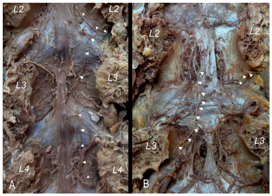
Figure 2.
Vertebral canal posterior view showing a double sinuvertebral nerve in the right side. (pattern 2). Both branches are labelled with white arrow heads. Intervertebral discs are numbered. (pll) posterior longitudinal ligament, (sg) spinal ganglion.
Figure 2.
Vertebral canal posterior view showing a double sinuvertebral nerve in the right side. (pattern 2). Both branches are labelled with white arrow heads. Intervertebral discs are numbered. (pll) posterior longitudinal ligament, (sg) spinal ganglion.
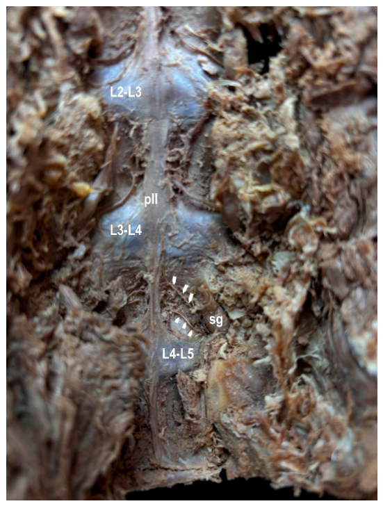
Figure 3.
(A) Cross-sectional image of SVN by immunohistochemical technique with anti-vasoactive intestinal peptide (VIP) antibody (40x). The presence of staining in most of the Schwann cell nuclei by immunohistochemical technique with anti-vasoactive intestinal peptide (VIP) antibody, even though the staining is slight, favors the demonstration of the autonomous component of the nerve. (B) Positive control of VIP made in pancreas (20x).
Figure 3.
(A) Cross-sectional image of SVN by immunohistochemical technique with anti-vasoactive intestinal peptide (VIP) antibody (40x). The presence of staining in most of the Schwann cell nuclei by immunohistochemical technique with anti-vasoactive intestinal peptide (VIP) antibody, even though the staining is slight, favors the demonstration of the autonomous component of the nerve. (B) Positive control of VIP made in pancreas (20x).
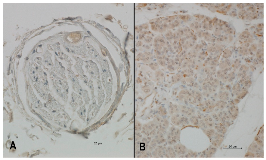
Figure 4.
(A) Cross-sectional image of SVN by immunohistochemical technique with Calcitonin gene-related peptide (CGRP) antibody (40x). The presence of intense staining in most of the Schwann cell nuclei by immunohistochemical technique with Calcitonin gene-related peptide (CGRP) antibody, favors the demonstration of the nociceptive component of the nerve. (B) Positive control of CGRP made in colon (20x).
Figure 4.
(A) Cross-sectional image of SVN by immunohistochemical technique with Calcitonin gene-related peptide (CGRP) antibody (40x). The presence of intense staining in most of the Schwann cell nuclei by immunohistochemical technique with Calcitonin gene-related peptide (CGRP) antibody, favors the demonstration of the nociceptive component of the nerve. (B) Positive control of CGRP made in colon (20x).
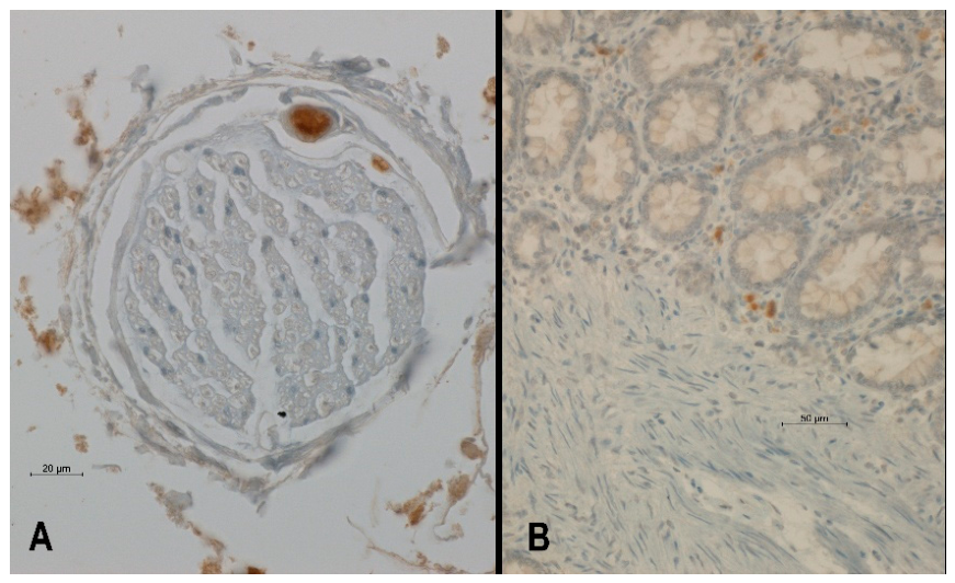

Table 1.
Frequency of each of the patterns and types at each of the vertebral levels studied.
| Type | L1 | L2 | L3 | L4 | L5 |
|---|---|---|---|---|---|
| Pattern 1/ Type 1a | 12 | 14 | 12 | 12 | 12 |
| Pattern 1/ Type 1b | 5 | 3 | 5 | 2 | 2 |
| Pattern 1/ Type 1c | 1 | 3 | 1 | 0 | 1 |
| Pattern 1/ Type 1d | 0 | 0 | 0 | 0 | 1 |
| Pattern 2 | 2 | 2 | 1 | 4 | 4 |
Disclaimer/Publisher’s Note: The statements, opinions and data contained in all publications are solely those of the individual author(s) and contributor(s) and not of MDPI and/or the editor(s). MDPI and/or the editor(s) disclaim responsibility for any injury to people or property resulting from any ideas, methods, instructions or products referred to in the content. |
© 2026 by the authors. Licensee MDPI, Basel, Switzerland. This article is an open access article distributed under the terms and conditions of the Creative Commons Attribution (CC BY) license (http://creativecommons.org/licenses/by/4.0/).
Copyright: This open access article is published under a Creative Commons CC BY 4.0 license, which permit the free download, distribution, and reuse, provided that the author and preprint are cited in any reuse.


