

Submitted:
04 June 2026
Posted:
09 June 2026
You are already at the latest version
Abstract
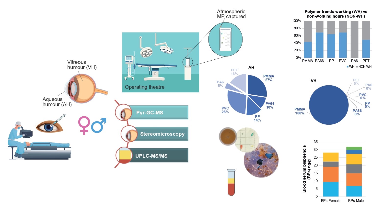
The widespread presence of microplastics (MPs) and associated plastic-derived com-pounds, bisphenols (BPs), in the environment – originating from plastic production or adsorption of environmental pollutants – results in unavoidable human exposure. There-fore, assessing their occurrence in human biological samples is essential for understand-ing potential health risks. In this study, pyrolysis gas chromatography-mass spectrometry was used to analyse human intraocular fluids – aqueous humour (AH) and vitreous hu-mour (VH)—to identify 11 microplastic polymer clusters. A method based on dansyl chloride derivatization combined with ultra-performance liquid chromatography-tandem mass spectrometry was employed to screen for five bisphenols (BPs) in blood serum col-lected from the same patients. Potential MPs isolated from ocular samples were qualita-tively assessed by stereomicroscopy. The number of particles ranged from 14 to 69 per gram in AH and from 21 to 34 per gram in VH. Among AH samples, the most frequently detected polymer clusters were C-PMMA, C-PVC, C-PET, and C-PP, with average levels ranging from 0.41 to 0.82 µg/g. The three VH samples contained only C-PMMA, with an average concentration of 0.55±0.12 µg/g. Polymers such as C-PA66 (0.30±0.21 µg/g) and C-PA6 (0.15±0.44 µg/g) were occasionally observed in AH. Analysis of 13 human serum samples demonstrated exposure to bisphenol A (BPA; 3.90–5.63 ng/mL) and bisphenol AF (BPAF; 2.13–4.54 ng/mL), but no clear correlation was observed between serum BP levels and intraocular MPs within the limitations of the present dataset. These findings suggest the presence of MPs and BPs in human biological samples; however, the results should be interpreted with caution owing to potential background contamination and the pilot na-ture of the study.
Keywords:
microplastics
; bisphenols
; aqueous and vitreous humour
; pyrolysis gas chromatography
; ultra-performance liquid chromatography-tandem mass spectrometry
1. Introduction
Plastic pollution is a pervasive environmental issue, with microplastics (MPs), defined as particles smaller than 5 mm, emerging as contaminants of global concern due to their persistence and potential biological effects. Human exposure to MPs occurs through multiple pathways, including ingestion, inhalation, and dermal contact, raising increasing concerns regarding their potential impacts on health [1,2]. Human exposure to MPs has increased markedly and its toxicity depends on material type, size, dose and chemical additives [3,4]. Zhao and You [5] reported a six-fold rise in MP ingestion and inhalation since 1990, especially in hotspots such as the USA, China, parts of the Middle East, North Africa and Scandinavia. Although assessing health impacts remains challenging, plastic particles can absorb environmental toxins, bind heavy metals, and release chemical additives capable of interacting with endocrine pathways [6,7,8]. MPs enter the body through ingestion, inhalation, dermal contact, and medical procedures [9,10,11].
However, current knowledge of human exposure and toxicity remains limited [12], yet micro- and nanoplastics can translocate to several organs, including the liver, spleen, lungs, kidneys, reproductive organs, and even the brain. The World Health Organization [13] states that no conclusive evidence of harm exists to date, but stresses the need for further research. Potential health effects include respiratory and gastrointestinal disturbances, oxidative stress and carcinogenesis [14].
Microplastics have been detected in a wide range of human tissues and body fluids, including the bone, brain, colon, heart, liver, kidney, the vascular system, lung, placenta, alveolar lavage fluid, breast milk, saliva and hands of young children, sputum, semen, testes, faeces and urine indicating their ability to translocate within the human body and raising concerns about potential systemic effects [15,16,17,18,19,20,21,22,23,24,25,26,27,28,29,30,31,32].
In contrast, studies examining MPs in the human eye remain sparse.
Despite the increasing number of studies on MPs in human tissues, the eye remains an underexplored organ, particularly with respect to internal compartments such as aqueous and vitreous humour [33]. Although the eye is highly sensitive, it has been relatively overlooked. MPs may enter ocular tissues through airborne exposure [34] or contact with contaminated detergents and cosmetics [35,36]. Growing evidence indicates that MPs can induce ocular surface inflammation, tissue damage, and cell death [37], although long-term effects and mechanisms remain unclear. To explore possible ocular exposure, researchers have begun analysing intraocular fluids, including aqueous (AH) and vitreous humour (VH), which are metabolically active and derived from blood ultrafiltration. The AH is critical for nutrient supply and regulation of intraocular pressure. Two recent studies have confirmed the presence of MPs in the AH and VH: 1745 particles <50 μm (mainly polyamide 6 cluster - C-PA 66, polyvinyl chloride cluster - C-PVC, and polystyrene cluster - C-PS) in the vitreous body, and five polymer types (polyethylene cluster - C-PE, C-PVC, polypropylene cluster - C-PP, polyamide 66 cluster - C-PA 66 and C-PS) in AH, with C-PE and C-PVC most abundant [38,39,40]. Flieger et al. [41] additionally detected bisphenol A (BPA) as MPs associated plastic-derived compound in ocular fluid during cataract surgery.
Bisphenols (BPs) have also gained scientific attention due to their endocrine-disrupting properties [42,43]. These plasticisers are widely used in polycarbonate plastics and epoxy resins [44]. Despite their environmental abundance, data on BPA analogues in human samples remain limited. Exposure occurs primarily through food, but also via inhalation and dermal contact [45]. Serum is considered a valuable biomatrix for assessing short-term exposure because of its contact with all body tissues [46]. For this purpose a sensitive ultra-performance liquid chromatography-tandem mass spectrometry (UPLC-MS/MS) method using dansyl chloride (Dns-Cl) derivatisation has been successfully applied to detect BPA, bisphenol AF (BPAF), bisphenol B (BPB), bisphenol F (BPF) and bisphenol S (BPS) in human serum.
This pilot study aims to explore the presence of MPs in AH and VH collected during routine ophthalmic procedures, and to provide preliminary insights into potential human exposure under controlled sampling conditions. The evaluation of both the morphology and physicochemical properties of MPs to which humans are exposed is of paramount importance, as these characteristics can significantly influence material toxicity and, consequently, human health. The subsequent steps in the analysis of MPs in human samples include sample collection, preparation anddigestion to isolate the MPs by filtration, and identification and quantification by combining the complementary approach of stereomicroscopy with destructive pyrolysis gas chromatography-mass spectrometry (Py-GC/MS) analysis, resulting in precise characterization of the mass fractions. Py-GC/MS, a proven method for identifying and quantifying microplastic polymers at trace levels was evaluated for its suitability in analyzing intraocular fluids [16,38,39,45,47]. MPs were classified by shape, colour and size using stereomicroscopy. In parallel, five BPs were quantified using UPLC–MS/MS in serum of the same patients to provide direct evidence of exposure to these plastic additives.
2. Materials and Methods
2.1. Study Population
This pilot study was conducted during a single surgical week, over three consecutive working days. AH, VH and serum samples were collected during routine ophthalmic surgeries at the University Hospital Sestre Milosrdnice (UHSM). These human samples were collected using different methods, depending primarily on the sample type. Collection conditions varied between clinical and non-clinical facilities, and multiple precautions were implemented to minimize MPs contamination during sampling.
Study involving 20 adult patients aged 18–85 years. Occupational background was classified according to potential environmental exposure: 15 retirees, 4 workers, and 1 pupil. The study population consisted of: 7 females (35.0%) and 13 males (65.0%) undergoing surgery for aphakia-related conditions. Age groups of adults were classified: 4 patients (≥18–<65 years), 8 elders patients (≥65–<75 years), and 8 very old elders patients (≥75 years). Participants were randomly recruited and predominantly consisted of older adults (>67 years). A total of 11 procedures were performed on the right eye (oculus dexter, OD) and 9 on the left eye (oculus sinister, OS) in patients. Nine patients had no history of previous ophthalmic surgery. Three patients had existing ocular implants (silicone oil, intraocular lens, or artificial capsule) in the operated eye, which were sampled intraoperatively. An additional eight participants had an intraocular lens (IOL) implanted in the contralateral, non-operated eye. In most cases, AH was collected at the start of surgery, prior to IOL implantation. Surgical procedures included cataract surgery (n = 16; 8 OD, 8 OS), combined cataract surgery with macular epiretinal membrane removal (n = 1, OD), pars plana vitrectomy for penetrating eye injury followed by cataract surgery (n = 2, OS and OD respectively), and anterior vitrectomy for secondary glaucoma (n = 1, OD).
2.2. Sample Collection
A total of 19 AH and 3 VH samples were collected from in total 20 patients undergoing ophthalmic surgery. The collected AH/VH volumes varied according to ocular anatomy (anterior chamber size, globe dimensions) and disease status. AH was obtained at the start of cataract surgery (age-related or secondary glaucoma) using a 27-gauge paracentesis syringe prior to viscoelastic injection, avoiding contact with the corneal endothelium, iris, and lens. VH samples were collected during combined cataract and epiretinal membrane surgery or pars plana vitrectomy. After a 25-gauge scleral puncture, filtered air was injected to flush residual saline, and 50-100 µL of VH was aspirated with a 2.5-mL syringe. All samples were transferred into pre-cleaned 2-mL glass vials, sealed, and stored at –20 °C. Of the 20 blood samples collected, 7 were excluded due to insufficient quality caused by haemolysis. As the affected patients did not consent to repeat sampling, a total of 13 serum samples were available for analysis. Venous blood samples were collected between 7:00 and 9:00 a.m. at the medical diagnostic laboratory of UHSM. All procedures were performed with precautions to minimize the risk of contamination with BPA analogues. Blood was drawn from the antecubital vein using a vacuum blood collection system and collected directly into 10 mL glass tubes without additives. Within 30–90 minutes of collection, samples were centrifuged at 2500 rpm for 15 minutes. Following centrifugation, serum was transferred into 5 mL glass vials using a disposable glass (Pasteur) pipette and sealed with polypropylene (PP) screw caps fitted with polytetrafluoroethylene (PTFE) septa. The samples were then stored at −20 °C until further analysis. Daily surgical blank controls were collected using physiological saline/ultrapure water under the same operating theatre conditions. All samples were transported to the Croatian Veterinary Institute, Regional Veterinary Institute Split, for analysis.
2.3. Surgical Environment Controls
Given the absence of standardized procedures for human sample collection for MPs analysis, strict contamination-prevention measures were implemented (glass equipment, metal tools, laboratory blanks). To characterize airborne MPs in the surgical theatre, validated methodology from Field et al. [48] was applied. The presence of MPs in the surgical environment further supports the possibility that intraoperative exposure may contribute to the detected MPs in ocular samples. Pre-combusted 400-mL glass beakers were deployed at eye level (1-1.8 m) for alternating 12-h intervals (working vs. non-working hours) over three days (n = 12). After collection, beakers were rinsed with pre-filtered distilled water and filtered through glass fibre filters (25 mm diameter, 1.2 μm pore size, Whatman GF/C™). Filters were dried and prepared for subsequent MPs examination.
2.4. Analytical Methods Development Using Animal Intraocular Fluids
To overcome the challenges posed by the limited sample volumes of AH (50-100 µL) and VH (250-300 µL), the method was developed using available long-lived animals, out of which five female cows over 2.5 years old, all of Simmental breed, in good breeding condition, and two pregnant (approximately 1 month and 4 months), whose whole eyes were supplied by abattoirs and frozen until dissection. The AH and VH were removed from the animals’ eyes after thawing, following a dissection protocol designed to avoid contamination and ensure sample integrity. The AH and VH samples were immediately transferred into 2 mL glass vials, sealed, and stored at −20 °C until analysis. For analyses, frozen bovine AH and VH samples were thawed at room temperature (25 ± 1 °C). The samples were homogenised using a vortex mixer (Vortex 3, IKA-Werke GmbH & Co. KG, Staufen, Germany). Subsequently, they were transferred using a glass Pasteur pipette into 2 mL Safe-Lock tubes (Eppendorf AG, Hamburg, Germany), which had been pre-cleaned with deionised water, methanol, and dichloromethane. The samples were then centrifuged at 14,800 rpm for 10 minutes to address mild compartmentalisation caused by autolysis. A direct detection method for BPs, based on derivatisation with Dns-Cl in animal AH/VH samples, was successfully applied following the approaches described by Flieger et al. [41] and Liu et al. [49]. Owing to the limited number of human samples and the prioritisation of microplastic polymer detection, BPs were determined only in supporting human serum samples. The vortexed and centrifuged samples were weighed in triplicate (approximate sample mass was 100 mg) into 1.5 mL glass vials (flat bottom, Macherey-Nagel GmbH) using a semi-microbalance (AND model BM-5, 5.2 g × 0.001 mg, σ = 0.0012 mg, A&D Company, Limited, Tokyo, Japan) and sealed with polypropylene (PP) screw caps fitted with red rubber septa (Macherey-Nagel GmbH). Wet digestion of the organic AH/VH matrix was performed via oxidation using 30% hydrogen peroxide (H₂O₂) at an elevated temperature of 65 °C to accelerate digestion, following approaches described in recent reviews on human tissue analysis [12,50]. Briefly, 1 mL of 30% H₂O₂ (Gramm-mol d.o.o., Zagreb, Croatia) was added to each sample, followed by overnight incubation (12 h) at 65 °C to ensure effective degradation of organic matter. After complete digestion and visual confirmation of solution clarity, vacuum filtration was carried out using glass fibre filters (25 mm diameter, 1.2 μm pore size, Whatman GF/C™, USA), which were pretreated at 500 °C for 8 h in a muffle furnace (Nabertherm model LT 40/11, Nabertherm GmbH, Lilienthal, Germany). The digested solution was transferred to the filtration unit using a glass Pasteur pipette. Residual material was quantitatively transferred by rinsing the digestion vial three times with 1 mL of 96% ethanol and subsequently rinsing the Pasteur pipette with an additional 2 mL of ethanol. A custom glass filtration setup was used to concentrate the retained particles in the central 9 mm diameter area of the filter. Particles larger than the 1.2 μm pore size were retained on the filter surface. The central 9 mm circle of the filter containing the analyte residue was excised using a custom-made ring-shaped rubber blade and it was appropriately sized to fit into a pyrolysis cup (approximately 80 μL volume). The filter was placed in a pre-cleaned glass Petri dish (4 cm × 1.2 cm, Karl Hecht, Sondheim, Germany), covered, and dried in an incubator (INCU-Line, VWR, Landsmeer, the Netherlands) at 45 °C for 4 h. The samples were then subjected to stereomicroscopy (SteREO Discovery V8, Carl Zeiss Jena GmbH, Germany, equipped with an Axiocam 105 R2 colour camera) and Py-GC/MS analysis (pyrolyzer EGA/PY-3030D with AS-1020E autosampler, Frontier Lab Ltd., Fukushima, Japan, coupled to GC-8890 and MSD 5977C, Agilent, Santa Clara, CA, USA) for the characterisation of MPs polymers. Stereomicroscopy was performed to estimate the total number of MP particles and to characterise them based on size, shape, and colour. Following stereomicroscopic analysis, the dried filters were further prepared for Py-GC/MS analysis of MPs polymer clusters. Blank samples were processed using the same procedure to ensure consistency and accuracy. Each analytical batch included a laboratory procedural blank to account for potential contamination introduced during sample handling and processing.
2.5. Human Intraocular Fluid Sample Treatment
Preparation of human AH/VH samples involved thawing at 25 °C, followed by vortexing and weighing into glass-stoppered test tubes (Šurlan, Pula, Croatia) using an analytical balance (RADWAG AS 82/220R2 PLUS, Radom, Poland). Wet oxidative digestion, previously validated for animal AH/VH matrices, was applied using 2 mL of 30% H₂O₂ at 65 °C for 12 hours. This step ensured efficient degradation of the organic matrix. Following digestion, samples were subjected to vacuum filtration using glass fibre filters (25 mm diameter, 1.2 μm pore size, Whatman GF/C™). The filters were subsequently rinsed with 96% ethanol, and the central 9 mm filter was excised, then placed in a glass Petri dish and incubated overnight at room temperature. The prepared filters were thus ready for stereomicroscopic analysis and Py-GC/MS characterisation of microplastic particles.
2.6. Stereomicroscopy Characterisation of Microparticles in Intraocular Fluids
This pilot study primarily focused on the chemical identification and quantification of polymer mass fractions. However, to obtain a qualitative representation of potential microplastic particles following sample pretreatment and prior to Py-GC/MS analysis, samples were subjected to stereomicroscopic inspection. For each particle, length (defined as the longest dimension), shape (according to Markley et al. 2024) [51], and colour were recorded. All glass fibre filters (1.2 µm GF/C, Whatman GF/C™) were examined under a stereomicroscope at ×8 magnification (SteREO Discovery V8, Carl Zeiss Jena GmbH, Germany). Images were captured using a digital camera (Axiocam 105 R2 colour, Carl Zeiss Jena GmbH, Germany) with exposure times ranging from 30 µs to 1 s and a frame rate of up to 30 fps (full frame). Each gridded filter was systematically analyzed from top to bottom and left to right, alternating the direction for each subsequent row. Particles visually identified as potential MPs were recorded according to number, colour, shape, and size. Shape categories were defined as: fragment (rounded, subrounded, subangular, angular), filament (long, thin fibre), film (irregular membrane), and sphere (spheroid). Colour categories included white, clear, blue, black, red, green, and yellow. Particle size was measured as the maximum dimension (length) following Abidli et al. (2019) [52]. Particles were assigned to one of nine size classes: 10–20 µm, 20–30 µm, 30–50 µm, 50–100 µm, 100–150 µm, 150–499 µm, 500–1499 µm, 1500–1999 µm, and 2000–3000 µm. Potential environmental contaminants, originating from the surgical environment or laboratory handling, were evaluated using blank samples collected during the surgical sampling phase and through procedural blanks processed alongside the microplastic polymer cluster method (sample preparation, isolation, detection, and quantification).
2.7. Py-GC/MS Identification and Quantification of Target Polymers
The chemical identity of the extracted microparticles was confirmed using Py-GC/MS. Identification and quantification of target polymers were performed using a multishot micro-oven pyrolyzer (EGA/PY-3030D) equipped with an autoshot sampler (AS-1020E, Frontier Lab Ltd., Fukushima, Japan) coupled to a GC/MS system (GC-8890 with MSD 5977C, Agilent, Santa Clara, CA, USA). Separation was achieved on an Ultra Alloy-5 column (30 m × 0.25 mm × 0.50 μm, Frontier Laboratories, Saikon, Japan). Ionization was carried out at 70 eV, and mass spectra were recorded at 1.56 scans/s over an m/z range of 40–400. The interface temperature was maintained at 320 °C to prevent cooling of the furnace. Sample identification was performed using MassHunter Qualitative/Quantitative Analysis Workstation (version 10). Detailed detection procedures are provided in Supplementary Tables (Tables S1-S3) and Figure S1. All measurements included an online derivatizing agent to enhance detection sensitivity for polyethylene terephthalate cluster C-PET, polycarbonate cluster C-PC, and C-PA6. This thermochemolysis was performed by adding 20 μL of tetramethylammonium hydroxide (TMAH, 10% in methanol, Sigma-Aldrich, Germany) to each sample, followed by vaporization at 65 °C. Polymer mass quantification was conducted using internal calibration curves. An inert solid matrix of 11 mixed polymer standards was weighed on a semi-microbalance (AND model BM-5, A&D Company, Limited, Tokyo, Japan) for calibration. For recovery experiments and verification of the MP isolation procedure, calibration curves were prepared using low-concentration microplastic standards in SiO₂ containing 11 common polymers: PE, PP, PS, acrylonitrile butadiene - ABS, styrene-butadiene rubber - SBR, PMMA, PC, PVC, PET, PA6, and PA66 (Frontier Laboratories Ltd., Fukushima, Japan) (Table S4). Seven different masses of calibration powder were weighed into pyrolysis cups and mixed with pre-cut 9 mm GF filters that had been pre-burned at 500 °C for 8 h (Nabertherm muffle furnace, LT 40/11m, Nabertherm GmbH, Lilienthal, Germany). Samples were spiked with 0.2 μg of an internal standard mixture comprising deuterated polystyrene (d5-PS) and poly(4-fluorostyrene) (Mr = 3000 g/mol, Polymer Source, Montreal, Canada). A 50 μL injection of the internal standard mixture was added directly to each pyrolysis cup and evaporated at 65 °C. The method was validated in accordance with EC Regulation No. 333/2007 [53]. Performance criteria included limit of detection (LOD), limit of quantification (LOQ), repeatability, reproducibility, specificity, linearity, and recovery. Precision was assessed at three concentration levels under repeatable conditions (RSDr). Animal AH and VH samples were spiked with the inert polymer matrix at three concentrations within the calibration range. Matrix effects were evaluated using calibration curves with and without the matrix, and a fixed amount of internal standard (0.2 µg d5-PS) and for each analyte was calculated as:
Matrix effect (%) = ((Average peak area with matrix) / (Average peak area without matrix) - 1) × 100.
If multiple indicator compounds were applicable, quantification of the target polymers was based on the analyte with the least matrix effect. Internal quality control was performed in each analytical batch by spiking available human samples to concentrations within the validated calibration range (Tables S5a, S5b). Results for linearity, limit of detection (LOD), limit of quantification (LOQ), recovery, and RSDr are presented in the Supplementary Materials (Table S6).
2.8. Sample Preparation and Bisphenol Analysis in Human Blood Serum
Frozen blood serum was thawed at room temperature (25 ± 1 °C) and vortexed (Genius 3, IKA Works GmbH & Co. KG, Staufen, Germany) to ensure homogeneity. Bisphenol extraction was performed using a rapid and straightforward liquid–liquid extraction (LLE) method, employing low sample and solvent volumes, as described by Owczarek et al. (2018) [54]. To 490 μL of serum, 10 μL of internal standard (IS) containing 500 ng/mL of 24BPS13C12 (Toronto Research Chemicals, Toronto, Canada, 96% purity) was added to all blood serum samples, followed by 1.5 mL of acetonitrile (ACN, UN1648, Biosolve, Chimie SARL, Dieuze, France). Samples were shaken for 30 s and left at room temperature for 10 min to allow protein precipitation. Subsequently, 250 mg of anhydrous Na₂SO₄ (Merck KGaA, Darmstadt, Germany) was added, and the mixture was vortexed to remove residual water. Samples were centrifuged (2 min, 6000 rpm, 3864 g), and the supernatant was transferred to clean glass tubes and evaporated to ~100 μL in a rotational vacuum concentrator (RCV2-18HCL, Christ, Osterode am Harz, Germany) at 40 °C and 1,300 rpm. For quantification, ultra-performance liquid chromatography–tandem mass spectrometry (UPLC-MS/MS) was used after derivatization with dansyl chloride (Dns-Cl, Merck KGaA, Darmstadt, Germany) to enhance sensitivity. Recoveries were evaluated using spiked human serum samples following Liu et al. (2023) [49]. The derivatization step was carried out according to the procedure reported by Liu et al. (2023) [53]. 100 μL of serum extract was mixed with 100 μL of water and 100 μL of sodium carbonate/sodium bicarbonate buffer (0.5 mol/L, pH 9). After vortexing, 100 μL of freshly prepared Dns-Cl solution (18 mg/mL in ACN) was added, and samples were incubated at 40 °C for 45 min. Excess Dns-Cl was quenched by adding 20 μL of 250 mM sodium hydroxide, followed by a 10-min incubation. Then, 100 μL of 20% formic acid was added to neutralize the sodium hydroxide. Samples were evaporated to dryness and reconstituted in 100 μL of ACN. The LOD was defined as the lowest concentration detectable with a signal-to-noise ratio (S/N) > 3, while the LOQ was defined as the lowest concentration giving S/N > 10. LODs for all BPs ranged from 0.1 to 1.0 ng/mL, and LOQs ranged from 1 to 5 ng/mL [55]. Chromatographic separation of five derivatized BPs (BPA, BPAF, BPB, BPF, BPS) was performed using a 1290 Infinity UPLC system (Agilent Technologies, Singapore) coupled to a G6460 ESI Triple Quad Mass Spectrometer (Agilent Technologies, Waldbronn, Germany). Separation was achieved on a Poroshell 120 EC-C18 column (100 × 2.1 mm, 2.7 μm) with a Poroshell 120 EC-C18 guard precolumn (2.1 mm i.d., 2.7 μm). Column temperature was maintained at 35 °C. Mobile phase A was 0.2% formic acid in water, and mobile phase B was 0.2% formic acid in acetonitrile. Gradient elution was applied at 0.3 mL/min, starting at 20% B, ramping to 80% B at 5 min, then to 90% B at 10 min, held until 11 min, and returned to 20% B at 13 min, held until 16 min. Injection volume was 15 μL. The ESI source operated in positive mode at 340 °C with 5 L/min gas flow and 4500 V capillary voltage. Data acquisition was performed using MRM transitions (Supplementary Table S7). Quantification was done using internal matrix-matched calibration curves (0.10–20 ng/mL), covering five concentration levels (0.75–20 ng/mL). The bisphenol method was validated according to EC Regulation No. 333/2007 [53]. Validation criteria included applicability, LOD, LOQ, precision (RSDr), specificity, linearity, and recovery. Precision was assessed at three concentration levels (1, 5, 10 ng/mL) in triplicate. Matrix effects were evaluated by comparing calibration curves prepared in matrix and in solvent using a fixed IS (24BPS13C12, 10 ng/mL). Internal quality control was performed for each analytical batch using negative control serum spiked to 5 or 10 ng/mL. Spiked samples were analyzed in duplicate to verify recovery. The method fulfilled all regulatory requirements and was deemed suitable for the determination of the five BPs in human blood serum. Results for linearity, LOD, LOQ, recovery, and RSDr are presented in Supplementary Materials (Table S8).
2.9. Data Analysis
Chromatographic and mass spectral data were processed using MassHunter WorkStation Quantitative Analysis for GC–MS (v12.0). Statistical analyses were performed in XLSTAT (Addinsoft, New York, USA), including environmental MP data (µg/m²), AH/VH MP concentrations (µg/g), and serum bisphenol levels (ng/mL). Data normality was assessed using the Shapiro–Wilk test. Differences related to surgical conditions and MP polymer concentrations (working space and time) were evaluated using the Fisher F-test. Non-normally distributed data were reported as median (IQR), and intergroup comparisons were performed using the Kruskal–Wallis test. Statistical significance was set at p < 0.05. Associations between MPs (particles/g and polymer mass fractions), bisphenol levels, and patient age were examined using Spearman correlation analysis. Graphical outputs were generated using Microsoft Excel, XLSTAT, and R (v4.3.1) with packages including ggplot2, tidyverse, ggstatsplot, and GGally.
3. Results
3.1. Results of Stereomicroscopy in Human AH and VH
All analysed human intraocular fluid samples contained potential MP particles, with an overall average of 10.5 ± 9.10 particles/g. Notably, the abundance of potential MPs detected in intraocular fluid samples was comparable to that observed in procedural and surgical blanks, indicating a possible contribution of background contamination despite the strict quality control measures applied. A more detailed analysis of the two intraocular fluid types and corresponding quality assurance/quality control (QA/QC) samples revealed average MPs concentrations of 11.2 ± 9.52 particles/g in AH and 6.92 ± 4.97 particles/g in VH. In comparison, blanks simulating sampling and analytical procedures, namely, the surgical condition blanks for AH and VH, as well as the procedural blank, showed average values of 9.25 ± 9.27 particles/g, 5.33 ± 4.03 particles/g, and 10.3 ± 7.76 particles/g, respectively. The lowest number of potential MPs observed in an ocular fluid sample was 14 particles/g (AH-5), while the highest was 69 particles/g (AH-7). The distribution of microplastic abundance in ocular fluid samples and QA/QC environmental controls is presented in Figure 1A and 1B.
Of the total 719 potential MP particles/g identified, 442 were classified as fragments, 184 as filaments, 84 as fibres, and 9 as spheres. The proportional distribution of MPs shapes is presented in Figure 2A and 2B.
The size of potential MPs ranged from 7.0 µm to 2981.7 µm in length, with mean ± standard deviation values of 199.6 ± 382.3 µm and 113.2 ± 234.1 µm for aqueous AH and VH, respectively. Morphometric distribution across the nine size classes showed that particles were predominantly fragments, followed by filaments, fibres, and spheres. The frequency distribution of potential MPs across size ranges is presented in Figure 3.
In both investigated intraocular fluids, a substantial proportion (often exceeding 50%) of detected potential MP particles fell within the smaller size ranges (e.g., <100 µm or <50 µm). The size of potential MPs in blanks collected under surgical conditions and during analytical procedures ranged from 11.8 µm to 737.2 µm in length. The mean ± standard deviation values were 42.3 ± 31.3 µm for BL-SC-AH, 40.39 ± 37.7 µm for BL-SC-VH, and 74.2 ± 140.8 µm for procedural blanks. Stereomicroscopic observations may indicate that fragments were the most prevalent potential MPs shape across both intraocular fluids, laboratory blanks, and surgical condition blanks (Figure 2.), supporting the applied quality control measures.
Filaments and films were also detected in the majority of samples (with films absent in some AH samples), whereas spheres were observed in all VH samples and in one AH sample. The 719 potential microplastic (MP) particles were classified into 10 colour categories, with the most prevalent in ocular samples being black (44.1%), grey (30.0%), brown (9.6%), orange (6.4%), transparent (4.17%), and blue/sky blue (3.76%) (Figure 4A). Black and grey were the predominant colours in both aqueous humour (AH) and vitreous humour (VH) samples. Blank samples exhibited a similar colour distribution; of the 152 potential microplastic (MP) particles detected across all sampling and procedural blanks, most were black (52.6%), grey (18.4%), brown (13.2%), orange (6.6%), transparent (3.29%), and blue/sky blue (3.76%) (Figure 4B).
3.2. Contamination Control and Methodological Limitations
Quality control measures implemented throughout the entire sampling-to-detection workflow, including Py-GC/MS analysis and stereomicroscopy, comprised laboratory procedural blanks and blanks collected under surgical conditions. MP particles were detected in these control samples Figure 5 A-B); however, comparison with sample data confirmed that the MPs identified in the study originated exclusively from the vitreous humour samples, reflecting the enclosed internal environment of the human eye.
The presence of MPs across all analysed samples may reflect continuous exposure pathways; however, given the comparable levels observed in blank controls, contributions from external contamination cannot be excluded. In addition to contamination-related challenges, several methodological limitations should be considered when interpreting the results. The application of Py-GC/MS provides robust identification and quantification of polymer mass fractions; however, it does not allow direct determination of particle number, size distribution, or morphology. Therefore, complementary stereomicroscopic analysis was employed, although this approach is inherently limited by visual identification and may be subject to misclassification of non-plastic particles. Furthermore, the relatively small sample volumes of intraocular fluids and the limited number of analysed samples, particularly for vitreous humour, constrain the statistical power of the study. Future research should integrate complementary analytical techniques, such as μFTIR or Raman spectroscopy, to improve particle-level characterisation and enhance methodological reliability.
3.3. Py-GC/MS Analyses of MPs in Human AH, VH and Surgical Environment Samples
Strict quality control measures were applied to ensure precise quantification and accurate characterisation of potential background contamination. Given the ubiquitous presence of MPs in the environment, both procedural controls and surgical environmental samples were analysed, and the results were reported as part of routine QA/QC assessments of plastic contamination [47]. These findings provide new insights into the quantities and types of polymer clusters present in surgical environments, where the use of plastic materials is considered beneficial for clinical practice. The results may inform future cell toxicity studies investigating the effects of human exposure to MPs and suggest a potential new exposure pathway associated with surgical procedures. Overall, the findings of this study should be considered preliminary and hypothesis-generating, warranting confirmation in larger and more comprehensive investigations.
Importantly, quality control measures and procedural blanks showed no detectable MPs, whereas sampling blanks exhibited a total MPs contamination of 2.21 µg/g, composed of C-PMMA, C-PA66, and C-PVC in descending order (Table 1).
MPs detected by Py-GC/MS in surgical blanks were not consistently present across all sampling days, and the concentrations observed were negligible. This finding highlights the minimal contribution of contamination relative to the total MPs detected by stereomicroscopy in both types of blanks. Furthermore, although eleven polymer types were targeted in the analysis, additional MPs may be associated with other polymer clusters not included in the current method. Overall, these results support the hypothesis that the detected MPs (PA66, PS, and PVC) originate from the AH and VH, representing the closed internal environment of the human eye.
In total, 6 out of the 11 investigated polymers were detected in ocular samples (Table 1). MPs were present in all 22 human samples analysed. The most prominent polymer clusters in AH samples were C-PMMA, C-PVC, C-PET, and C-PP, with average concentrations of 0.82 ± 0.32 µg/g, 0.41 ± 0.82 µg/g, 0.48 ± 0.70 µg/g, and 0.41 ± 0.82 µg/g, respectively. All three VH samples contained only C-PMMA, with an average concentration of 0.55 ± 0.12 µg/g. Additionally, C-PA66 (0.30 ± 0.21 µg/g) and C-PA6 (0.15 ± 0.44 µg/g) were frequently detected in AH samples. Other polymer clusters, including C-ABS, C-PC, C-PE, C-PS, and C-SBR, were not detected in any of the analysed ocular samples.
MPs were detected in all monitored surgical environmental samples, including both the operating theatre (OT) and anaesthetic room (AR), during both working hours (WH) and non-working hours (NWH). Across all sampled environments and time periods, total MP concentrations ranged from 12.3 to 969 μg m⁻² day⁻¹, with a mean atmospheric abundance of 74.8 ± 143.6 μg m⁻² day⁻¹. In the OT environment, the total MPs mass fraction varied considerably during the sampling period, with a mean value of 91.6 ± 190.6 μg m⁻² day⁻¹ and a range of 15 to 969 μg m⁻² day⁻¹ (Figure 5 A-B). Similarly, variation was observed in the AR, where the mean MP concentration was 57.9 ± 70.2 μg m⁻² day⁻¹, ranging from 12.3 to 292.9 μg m⁻² day⁻¹.
When comparing temporal differences, the mean MPs concentration in the AR during working hours was 54.6 ± 59.4 μg m⁻² day⁻¹, compared to 61.2 ± 81.2 μg m⁻² day⁻¹ during non-working hours. This difference was not statistically significant (p = 0.242). In the OT, the mean MP concentration during working hours was 112.3 ± 226.9 μg m⁻² day⁻¹, while during non-working hours it was lower, at 71.0 ± 149.7 μg m⁻² day⁻¹. Although MPs concentrations were higher during working hours in the OT—consistent with findings from previous studies—this difference was also not statistically significant.
The most dominant polymer clusters identified were C-PP (38% of total MPs), C-PET (30%), C-PVC (17%), and C-PMMA (10%). In contrast, C-PP, C-PA66 (accounting for 4% of the total MPs mass), and C-PA6 (1%) were not consistently detected throughout the monitoring period. Notably, C-PET was absent only in the OT during non-working hours on the first sampling day (Figure 6).
When comparing working and non-working hours across both environments (OT and AR combined), the average MPs abundance during working hours was 92 ± 166 μg m⁻² day⁻¹, whereas during non-working hours it was 68 ± 119 μg m⁻² day⁻¹. This difference was not statistically significant (p = 0.247, Fisher’s F-test).
3.4. Relationship Between MPs Mass Fraction Levels and Individual Participant Characteristics
Table 2 presents the presence of MPs in the AH of study participants, categorised according to individual characteristics outlined in (Table S9).
The analysis was further stratified by the six detected polymer clusters: C-PMMA, C-PVC, C-PET, C-PP, C-PA66, and C-PA6. Overall, total MPs levels were higher in female participants compared to males; however, this difference was not statistically significant (P > 0.05), with the exception of C-PET, which exhibited a significantly higher concentration in females. This pattern was generally consistent across most polymer types, except for C-PA66, where levels were lower in females, and C-PA6, which was not detected in the female subgroup. No statistically significant differences in MPs presence were observed based on occupation or type of ocular disease, nor in relation to the laterality of the eye, regardless of MPs polymer type (Table 2). When examining age groups, the pilot study population was divided into three classes. Notably, the very old subgroup (≥75 years) exhibited significantly higher concentrations of C-PA66. Additionally, C-PA6 was detected exclusively in the elder population, with statistically significant differences among the very old participants, whereas it was absent in younger age groups.
3.5. UPLC-MS/MS Analysis of BPs in Blood Serum
Due to the limited availability of AH and VH samples, this study focused on BP detection in blood serum. Using a sensitive detection A method based on dansyl chloride derivatisation (Table S7), comprehensive data on patient bisphenol levels were collected (Table 3). BPAF was detected in five samples (1.50–5.72 ng/ml), and in three of these, BPA was co-detected (3.90–5.63 ng/ml).
3.6. Correlation of BPs in Serum and MPs in Intraocular Fluids
Analysis of interrelationships among MPs polymer types, BPs, and potential MP particles per gram in the human samples revealed a strong positive correlation between PA66, PMMA, and PVC based on MP concentrations (Figure 7).
Notably, PA6 was positively correlated with PP and negatively correlated with BPA and the sum of bisphenols (ƩBPs), suggesting an association between certain polymers and BP levels, although these correlations do not imply causation.
When evaluating MP accumulation patterns in ocular fluids in relation to individuals’ lifelong exposure, no significant correlation was observed between total MPs levels and age across the pilot study population (Figure 8).
4. Discussion
4.1. Microplastics in Ocular Fluids and Surgery Environment
Available studies have shown that MP abundances in different human organ systems and biological samples range from 1.1 particles/g (lymphatic system, spleen) to 28.1 particles/g (colorectal cancer tissue) [17,19]. However, specific ocular fluid data are missing, and interpretation of the present findings therefore relies on careful consideration of potential sources and pathways of exposure. The ocular surface exposure to microplastics might be derived from airborne particles, contaminated fluids, medications and instrumentation [35]. The largest observed particle size in VH study of Zhong et al. [38] was 450 μm among the identified MPs and the majority of the identified MPs in the amalgamated vitreous humor sample exhibited diameters below 50 μm, with 63.8% falling in the range of 20–30 μm and 25.2% in the 30–50 μm range in these above MPs. SEM images further emphasised the spherical morphology of investigated VH samples. The smallest size particles have also been confirmed in surgery environment and samples suggesting that these particles might derive from the exogenous surgical environment, instruments and equipment packaging. Despite the similar fields of interest within the existing studies on the possible impacts posed by microplastics introduced into the body in surgery, their methods, results, and limitations do differ in some relevant markers. For example, mean or mode microplastic particle size have been reported as 92 ± 136 µm in surgery room (data obtained by µFTIR microscopy, air samples have not been chemically digested; Field et al. [48]., 35.29 ± 22.68 µm in blood after percutaneous coronary intervention (PCI) showing that after the intervention on patients who needed a PCI the level of MPs rose by more than a factor of 20 in the blood (data obtained by LDIR and SEM, blood samples were chemically digested in HNO3; Liu et al. [47] 0.78 ± 0.08 µm in synovial fluid after total knee arthroplasty (SEM, NaOH digestion) [56]. Fibres emerge as the most prevalent MPs shape, constituting 44.5% of MPs detected in human samples. Findings from multiple studies indicate that microfibres may accumulate at high levels in various human organs [17,19,23]. They can be as small as a few micrometres in diameter and are also lightweight, enabling them to be easily inhaled and ingested by humans. Microplastics occur in various shapes depending on their sources and the processes by which they are broken down in the environment. The occurrence of MPs in the surgical setting and their potential effects on surgical outcomes could influence the MPs morphometry in ocular fluids. Most of the particles were classified as fragments (78%) with fibres (20%) and spheres (2%) constituting the rest in the operating room as well as its adjacent anaesthetic room [48]. In general, microplastics found in human tissues are typically transparent or translucent rather than brightly coloured. Smaller particles are more likely to be transparent or translucent [17], whereas larger particles tend to be opaque and coloured [57]. However, some studies have reported microplastics of various colours in human tissues, including yellow, blue, green, and [21]. These colours may result from additives or pigments used during the production of plastic products, or from environmental factors such as exposure to UV radiation, which can cause plastics to degrade and discolour [58]. Furthermore 85% of identified MPs were clear in colour with black, blue, and brown composing the remainder (7%, 5% and 3% respectively) in the surgical environment. While the dominant colours of microplastics found in humans may vary, there is currently no evidence to suggest that the colours of microplastics have any direct effects on human health.
The MPs findings including both the operating theatre and anaesthetic room provide new insights into the quantities and types of polymer clusters present in surgical environments, where the use of plastic materials is considered beneficial for clinical practice. The results may inform future cell toxicity studies investigating the effects of human exposure to MPs and suggest a potential new exposure pathway associated with surgical procedures. Comparison with existing literature remains challenging due to the limited number of studies investigating MPs pollution in indoor environments and the wide variability in analytical methodologies employed. To date, no studies have reported mass-based quantification of MPs in indoor air, further complicating direct comparisons. Nevertheless, the polymer composition observed in the surgical environment in this study is consistent with the findings of Field et al. [48], who characterised MPs in a cardiothoracic operating theatre. Overall, the findings of this study should be considered preliminary and hypothesis-generating, warranting confirmation in larger and more comprehensive investigations. Furthermore the detected polymer profiles may reflect common environmental and clinical plastic sources; however, the relative contribution of each source cannot be clearly distinguished within the scope of this study.
According to published reports, the predominant polymers identified in human samples include alkyd resin, nylon, EVA, CPE, rayon/viscose, resin, PA, PBS, PC, PE, PET, PMMA, PP, PS, PU, PVA, PVAc, and PVC, typically occurring as irregular fragments and fibres [12]. In the review by Vdovchenko et al. [59], eight of the most used plastic types were evaluated: polyester (PES), PP, PA, PVC, PE, polyacrylate (PAC), PS, and polyurethane (PU). Their results indicate that the five most frequently detected MPs in humans are PES (38.8%), PA (17.1%), PU (15.3%), PP (9.4%), and PAC (8.0%), reflecting the prevalence of these widely used polymers.
Available studies investigating intraocular fluids AH and VH have reported the predominant presence of MPs such as PA, PE, PMMA, PP, PS, and PVC. Using LD-IR, MPs in VH were primarily detected in the size range of 20–30 μm [38]. In AH samples, PE, PVC, PP, and PA66 were identified using Py-GC/MS as the major constituents [39]. To date, in the only available human VH study, Zhong et al. [38] further performed Py-GC/MS analysis following LD-IR and confirmed the presence of MPs, predominantly PS, polyamide 66 (PA66), and PVC, in all vitreous samples. However,, PE, PMMA, and PP, which were detected in each VH sample by LD-IR, were not identified by Py-GC/MS, highlighting methodological differences in polymer detection. The representative profiles of MPs polymer clusters observed in this study are consistent with Py-GC/MS data from earlier mentioned studies, particularly for PVC, PP, and PA. To the best of our knowledge, these studies collectively cover comparable polymer profiles, with a broader range of polymers reported in AH analyses, including polylactic acid (PLA) and polybutylene adipate terephthalate (PBAT) [39], as well as ABS and SBR identified in the present study.
The relationship between MPs mass fraction levels and gender and age reveals statistically significant differences. The abundance of MPs in the aqueous humour of the female group may reflect differences in lifestyle factors; however, given the limited sample size, such interpretations should be considered exploratory. Cosmetic products, such as eyeshadows and mascaras containing microbeads or glitter, represent another significant route of ocular microplastic exposure [60]. Ineffective removal can lead to microplastic accumulation on ocular surfaces, resulting in prolonged irritation and increased susceptibility to ocular surface diseases. Notably, research in other organs confirms that females exhibit significantly higher gastrointestinal tract microplastic accumulation than males; however, evidence regarding sex-related differences in gastrointestinal accumulation is limited and, until confirmed across multiple studies, should be interpreted with caution [61]. This pilot study population was characterised by the presence of C-PA66 in the very old subgroup (≥75 years). Zhang et al. [39] found PA66 primarily in the aqueous humour of adults. This may be related to adults spending extended periods in working environments. Studies also indicate that PA66 has a relatively high migratory capacity ([62].
The individuals’ lifelong exposure in this study showed no significant correlation with total MPs abundance. According to Zhang et al., 2025, variations in microplastic abundance in aqueous humour may be linked to differing daily exposures to microplastics across age groups. Individuals employed in plastic manufacturing, recycling, or textile industries are exposed to high levels of airborne microplastics. One study found thousands of microplastic fibres on workers’ skin, hair, and even in saliva after shifts in plastic factories [63]. These workers are likely to experience continuous deposition of microplastics on the lungs and eyes.
4.2. Bisphenols in Blood Serum
Serum analyses are widely used to determine BPs analogue concentrations in blood, reflecting systemic distribution and potential effects on organs and tissues. In some studies, chemical analyses extend to specific tissues or organs, providing insight into localization and possible health impacts [64].
Previous studies have reported BPs in various biological matrices, highlighting multiple exposure routes. In urine, mean concentrations were 0.05 ng/ml for BPB and BPAF, 13.3 ng/ml for BPS, and 2.8 ng/ml for BPA, while BPF, BPS, BPAP, BPAF, BPP, and BPZ were detected in 2–10% of samples [65,66]. In blood, BPA ranged from 0.79–7.12 ng/ml and BPB from 0.88–11.94 ng/ml; other analogues including BPC, BPE, BPF, BPG, BPM, BPP, BPS, BPZ, BPFL, and BPBP were detected at 0.05–4.84 ng/ml [54,67]. BPA and BPS have also been detected in breast milk (0.002–1.16 ng/g and 0.23 ng/g, respectively), saliva (0.07–7.28 ng/ml), and plasma (0.17–12.51 ng/ml) [68]. These findings confirm that human exposure to bisphenols is diverse and widespread.
BPA is associated with serious health effects, including breast cancer, reduced fertility, genotoxicity, and respiratory issues in children [69]. It disrupts metabolic homeostasis, impairing energy, glucose, protein, and lipid metabolism [70]. BPAF, commonly used in plastic fibres, waveguides, epoxy resins, and polycarbonate plastics, also exerts endocrine effects by modulating the estrogen receptor pathway [71].
Regulatory actions limiting BPA use have led to the emergence of numerous structural analogues, all sharing the two-hydroxyphenyl structure. To date, sixteen BP analogues, including BPS, BPAF, BPAP, BPB, BPC, BPE, BPF, and BPZ, have been employed as industrial BPA substitutes [72]. Many of these analogues were introduced without comprehensive toxicity testing, raising concerns about their safety. Limited studies suggest that BPs can induce erythrocyte suicide (eryptosis) and biochemical or morphological changes in peripheral blood mononuclear cells [42,73,74].
Taken together, the results provide initial insights into the occurrence of MPs in intraocular fluids, while also highlighting substantial analytical and interpretative challenges that must be addressed in future studies.
5. Conclusions
Microplastics were detected in all analyzed ocular samples, while selected bisphenols, specifically BPA and BPAF, were identified in human serum, indicating potential exposure pathways. Detection of bisphenols in human serum suggests ongoing human exposure, although no established correlation has been identified between bisphenol levels in serum and microplastics in intraocular fluids, indicating that these represent distinct, though potentially related, exposure pathways. Six polymer types were detected – C-PMMA, C-PVC, C-PET, C-PP, C-PA66, and C-PA6—distributed across different ages and genders. The relationship between microplastics mass fraction levels and gender and age reveals statistically significant differences. Female groups were characterized by higher microplastics abundance and prevalence in the aqueous humour. The results indicate contamination of the surgical environment. To determine whether internal ocular exposure to microplastics is occurring, analytical data must enable identification and sufficiently exclude all non-plastic chemical entities. While the presented findings remain preliminary due to sample quantity and analytical limitations, the observed microplastics trends provide supportive knowledge relevant for further systemic assessments of the detected polymers in ocular fluids and tissues. Furthermore, in-depth mechanistic or clinical studies are required, including clarification of fundamental biological mechanisms of microplastics-induced ocular tissue damage to enable targeted interventions, while establishing high-risk cohorts to correlate exposure levels with clinical endpoints.
Supplementary Materials
The following supporting information can be downloaded at the website of this paper posted on Preprints.org. Table S1. Py-GC/MS method. Table S2. Main pyrolysis products of the 11 polymer cluters. Table S3. Specific marker and ions used for quantification of the 11 polymer clusters; characteristic Py-GC/MS decomposition products used for identification and quantification of detected polymer clusters indicated by “C-“ as pure polymers and potentially included polymer-related derivatives. Table S4. Polymer-Specific Mass Equivalents of MCS for Calibration Range in Py-GC/MS Analysis (MSC in SiO2, low level, Lot: 23031601) example. Table S5a. Recovery of microplastic polymer mass fractions spiked AH sample (AH 6). Table S5b. Recovery of microplastic polymer mass fractions spiked VH sample (VH 3). Table S6. Selected Py-GC/MS method performance indicators in animal ocular internal fluids (aqueous and vitreous humour, AH, VH): linearity range, limit of detection (LOD), limit of quantification (LOQ), recovery and precision (RSDr) for ocular tissues. Table S7. Optimised parameters for the mass spectrometry analysis of derivatized bisphenols. Table S8. Method performance criteria of UPLC-MS/MS method developed in human blood serum. Table S9. Clinical characteristics of patients. Figure S1. The chromatograms (on the left side) and mass spectrograms (on the right side) through pyrolysis-gas chromatography/mass spectrometry (Py-GC/MS) analysis of spiked AH sample, including poly methyl methacrylate cluster (C-PMMA) with 0.075 μg, polyamide-6 cluster (C-PA-6) with 0.135 μg, polypropylene cluster (C-PP) at 1.06 μg, polyvinyl chloride cluster (C-PVC) with 0.88 μg, polyamide-66 (PA-66) with 0.213 μg, styrene-butadiene rubber (SBR) with 0.400 μg, polyethylene terephthalate cluster (C-PET) with 0.251 μg, polycarbonate cluster (C-PC) with 0.08 μg, poly(4-fluorostyrene) ISTD 4PFS at 0.2 μg, Polystyrene cluster (C-PS) with 0.163 μg, polyethylene cluster (C-PE) at 3.29 μg (average sum of α-Alkenes (e.g. C14-C22)), and deuterated polystyrene (d5-PS) at 0.2 μg used in internal standard calibration with thermochemolysis (10% TMAH in MeOH).
Author Contributions
For research articles with several authors, a short paragraph specifying their individual contributions must be provided. The following statements should be used “Conceptualization, T.B., J.P. and A.G.; methodology, Z.V., G.M., V.M., S.Š., T.B., Z.J., F.DG., C.P., S.P., E.L. and I.L; validation, S.Š., G.M., Z.V., V.M., T.B., and Z.J.; formal analysis, A.G., V.M., C.P., S.P., E.L. and I.L.; investigation, T.B. Z.J., G.M., Z.V., S.Š., T.B. and F.DG.; resources, Z.V., G.M., S.Š., J.P., S.P. and E.L.; data curation, G.M., S.Š., T.B. Z.J. and C.P.; writing—original draft preparation, T.B. and J.P.; writing—review and editing, J.P., G.M., S.Š., A.G., Z.V., V.M., F.DG. S.P. and C.P.; visualization, T.B., J.P., V.M., F.DG. and C.P; supervision, T.B. E.L.; project administration, T.B. and J.P.; funding acquisition, T.B. and S.P. All authors have read and agreed to the published version of the manuscript.
Funding
This research was funded by the European Union NextGenerationEU and supported by the Ministry of Science and Education of the Republic of Croatia through the project No. NPOO 6 of Croatian Veterinary Institute titled „Occurrence of Microplastics and Bisphenols in Shellfish Along the Croatian Coast of the Adriatic Sea” (PLASTICshell).
Institutional Review Board Statement
In this section, please add the Institutional Review Board Statement and approval number for studies involving humans or animals. You might choose to exclude this statement if the study did not require ethical approval. Please note that the Editorial Office might ask you for further information. Please add “The study was conducted in accordance with the Declaration of Helsinki, and approved by the Medical Ethics Committee of University Hospital Sestre Milosrdnice (Protocol code: 003-06/26-03/001/ Urbroj: 251-29-11/3-26-1 and date of approval: 12.01.2026.
Informed Consent Statement
Informed consent was obtained from all subjects involved in the study.
Data Availability Statement
The original contributions presented in this study are included in the article/supplementary material. Further inquiries can be directed to the corresponding authors.
Conflicts of Interest
The authors declare no conflicts of interest.
Abbreviations
The following abbreviations are used in this manuscript:
| ACN | acetonitrile |
| AH | aqueous humour |
| anhydrous Na₂SO₄ | anhydrous natrium sulphate |
| AR | anaesthetic room |
| BPA | bisphenol A |
| BPAF | bisphenol AF |
| BPB | bisphenol B |
| BPBP | bisphenol BP |
| BPC | bisphenol C |
| BPE | bisphenol E |
| BPF | bisphenol F |
| BPFL | bisphenol FL |
| BPG | bisphenol G |
| BPM | bisphenol M |
| BPP | bisphenol P |
| BPS | bisphenol S |
| BPs | bisphenols |
| BPZ | bisphenol Z |
| C-ABS | acrylonitrile butadiene cluster |
| C-PA6 | polyamide 6 cluster |
| C-PA66 | polyamide 66 cluster |
| C-PC | polycarbonate cluster |
| C-PE | polyethylene cluster |
| C-PET | polyethylene terephthalate cluster |
| C-PP | polypropylene cluster |
| C-P | polystyrene cluster |
| C-PVC | polyvinyl chloride cluster |
| C-SBR | styrene-butadiene rubber cluster |
| Dns-Cl | dansyl chloride |
| ESI | Electrospray ionisation |
| GF/C | glass fibre filters |
| H₂O₂ | hydrogen peroxide |
| IOL | intraocular lens |
| IQR | interquartile range |
| LD-IR | Laser Direct Infrared |
| LLE | liquid–liquid extraction |
| LOD | limit of detection |
| LOQ | limit of quantification |
| MPs | microplastics |
| MRM | transitions |
| NWH | non-working hours |
| OD | oculus dexter |
| OS | oculus sinister |
| OT | operating theatre |
| PCI | percutaneous coronary intervention |
| PP | polypropylene |
| PTFE | polytetrafluoroethylene |
| Py-GC/MS | pyrolysis gas chromatography-mass spectrometry |
| RSDr | relative standard deviation |
| SEM | scanning electron microscopy |
| TMAH | tetramethylammonium hydroxide |
| UHSM | University Hospital Sestre Milosrdnice |
| UPLC- MS/MS | ultra-performance liquid chromatography-tandem mass spectrometry |
| VH | vitreous humour |
| WH | working hours |
| μFTIR | Fourier transform infrared spectroscopy |
References
- Dey, T.; Trasande, L.; Altman, R.; Wang, Z.; Krieger, A.; Bergmann, M.; Allen, D.; Allen, S.; Walker, T. R.; Wagner, M.; Syberg, K.; Brander, S. M.; Almroth, B. C. Global plastic treaty should address chemicals. Science 2022, 378, 841–842. [CrossRef]
- Bogdanović, T.; Pleadin, J. Microplastics as unexplored emerging contaminant: A challenge for food safety. In Advances in Food and Nutrition Research, 1st ed.; Fidel Toldrá, Ed. Academic Press Elsevier: 125 London Wall, London, EC2Y 5AS, United Kingdom, 2026, Volume 18, pp. 1-352. [CrossRef]
- Schirinzi, G. F.; Pérez-Pomeda, I.; Sanchís, J.; Rossini, C.; Farré, M.; Barceló, D. Cytotoxic effects of commonly used nanomaterials and microplastics on cerebral and epithelial human cells. Environ. Research. 2017, 159, 579–587. [CrossRef]
- Thompson, R. C.; Courtene-Jones, W.; Boucher, J.; Pahl, S.; Raubenheimer, K.; Koelmans, A. A. Twenty years of microplastic pollution research-what have we learned? Science 2024, 386, eadl2746. [CrossRef]
- Zhao, X.; You, F. Microplastic Human Dietary Uptake from 1990 to 2018 Grew across 109 Major Developing and Industrialized Countries but Can Be Halved by Plastic Debris Removal. Environ. Sci. Technol. 2024, 58, 8709–8723. [CrossRef]
- Rafa, N.; Ahmed, B.; Zohora, F.; Bakya, J.; Ahmed, S.; Ahmed, S. F.; Mofijur, M.; Chowdhury, A. A., Almomani, F. Microplastics as carriers of toxic pollutants: Source, transport, and toxicological effects. Environ. Pollut. 2024, 343, 123190. [CrossRef]
- Liu, S.; Huang, J.; Zhang, W.; Shi, L.; Yi, K.; Yu, H.; Zhang, C.; Li, S.; Li, J. Microplastics as a vehicle of heavy metals in aquatic environments: A review of adsorption factors, mechanisms, and biological effects. J. Environ. Manage 2022, 302, 113995. [CrossRef]
- Ullah, S.; Ahmad, S.; Guo, X.; Ullah, S.; Ullah, S.; Nabi, G.; Wanghe, K. A review of the endocrine disrupting effects of micro and nano plastic and their associated chemicals in mammals. Front. Endocrinol. 2023, 13, 1084236. [CrossRef]
- Field, D. T.; Green, J. L.; Bennett, R.; Jenner, L. C.; Sadofsky, L. R.; Chapman, E.; Loubani, M.; Rotchell, J. M. Microplastics in the surgical environment. Environ. Int. 2022, 170, 107630. [CrossRef]
- Leslie, H. A.; van Velzen, M. J. M.; Brandsma, S. H.; Vethaak, A. D.; Garcia-Vallejo, J. J.; Lamoree, M. H. Discovery and quantification of plastic particle pollution in human blood. Environ. Int. 2022, 163, 107199. [CrossRef]
- Li, K.; Li, W.; Sun, Y.; Ma, T.; Yuan, L.; Rong, Y.; Liu, X.; Fu, Y.; Yu, X.; Xu, X. Medical Microplastics: Research Progress on Exposure Pathways, Toxic Effects, and Detection Methods. Microplastics, 2026, 5, 61. [CrossRef]
- Barceló, D.; Picó, Y.; Alfarhan, A. H. Microplastics: Detection in human samples, cell line studies, and health impacts. Environ. Toxicol. Phar. 2023, 101, 104204. [CrossRef]
- World Health Organization (WHO). Dietary and inhalation exposure to nano- and microplastic particles and potential implications for human health. Geneva: WHO. 2022. https://www.who.int/publications/i/item/9789240059757.
- Snehamayee, N.; Somya, S.; Kumar, S. C.; Niranjan, M.; Ranjan, S. B.; Kumar, M. N. Microplastics and Human Health: A Comprehensive Review on Exposure Pathways, Toxicity, and Emerging Risks. Microplastics 2026, 5, 8. [CrossRef]
- Lu, S.; Wei, Y.; Xu, R.; Li, M.; Qv, Y.; Zha, Y.; Gong, M.; Wang, N.; Lu, X.; Jiang, X., Li, Z. New insights: Discovery of microplastics in human bone and skeletal muscle. Innov. Med. 2024, 2, 100100. http://creativecommons.org/licenses/by-nc-nd/4.0/.
- Nihart, A. J.; Garcia, M. A.; El Hayek, E.; Liu, R.; Olewine, M.; Kingston, J. D.; Castillo, E. F.; Gullapalli, R. R.; Howard, T.; Bleske, B.; Scott, J.; Gonzalez-Estrella, J.; Gross, J. M.; Spilde, M.; Adolphi, N. L.; Gallego, D. F.; Jarrell, H. S.; Dvorscak, G.; Zuluaga-Ruiz, M. E.; West, A. B.; Campen, M. J. Bioaccumulation of microplastics in decedent human brains. Nat. Med. 2025, 31, 1114–1119. [CrossRef]
- Ibrahim, Y. S.; Tuan Anuar, S.; Azmi, A. A.; Wan Mohd Khalik, W. M. A.; Lehata, S.; Hamzah, S. R.; Ismail, D.; Ma, Z. F.; Dzulkarnaen, A.; Zakaria, Z.; Mustaffa, N.; Tuan Sharif, S. E.; Lee, Y. Y. Detection of microplastics in human colectomy specimens. JGH Open, 2020, 5, 116–121. [CrossRef]
- Yang, Y.; Xie, E.; Du, Z.; Peng, Z.; Han, Z.; Li, L.; Zhao, R.; Qin, Y.; Xue, M.; Li, F.; Hua, K.; Yang, X. Detection of Various Microplastics in Patients Undergoing Cardiac Surgery. Environ. Sci. Technol. 2023, 57, 10911–10918. [CrossRef]
- Horvatits, T.; Tamminga, M.; Liu, B.; Sebode, M.; Carambia, A.; Fischer, L.; Püschel, K.; Huber, S.; Fischer, E. K. Microplastics detected in cirrhotic liver tissue. EBioMedicine 2022, 82, 104147. [CrossRef]
- Jiang, N.; Zheng, X,; Zhang, N.; Cao, Y. The detrimental effects of microplastic exposure on kidney function. Front. Med. 2025, 12, 1620733. [CrossRef]
- Amato-Lourenço, L. F.; Carvalho-Oliveira, R.; Júnior, G. R.; Dos Santos Galvão, L.; Ando, R. A.; Mauad, T.; Presence of airborne microplastics in human lung tissue. J. Hazard. Mater. 2021, 416, 126124. [CrossRef]
- Zhu, L.; Kang, Y.; Ma, M.; Wu, Z.; Zhang, L.; Hu, R.; Xu, Q.; Zhu, J.; Gu, X.; An, L. Tissue accumulation of microplastics and potential health risks in human. Sci. Tot. Environ. 2024, 915, 170004. [CrossRef]
- Ragusa, A.; Svelato, A.; Santacroce, C.; Catalano, P.; Notarstefano, V.; Carnevali, O.; Papa, F.; Rongioletti, M. C. A.; Baiocco, F.; Draghi, S.; D’Amore, E.; Rinaldo, D.; Matta, M.; Giorgini, E. Plasticenta: First evidence of microplastics in human placenta. Environ. Int. 2021, 146, 106274. [CrossRef]
- Baeza-Martínez, C.; Olmos, S.; González-Pleiter, M.; López-Castellanos, J.; García-Pachón, E.; Masiá-Canuto, M.; Hernández-Blasco, L.; Bayo, J. First evidence of microplastics isolated in European citizens’ lower airway. J. Hazard. Mater. 2022, 438, 129439. [CrossRef]
- Amiri, H.; Moradalizadeh, S.; Jahani, Y.; Nasiri, A. Biomonitoring of microplastics in saliva and hands of young children in kindergartens: identification, quantification, and exposure assessment. Environ. Monit. Assess. 2025, 197, 859. [CrossRef]
- Zhu, L.; Zhu, J.; Zuo, R.; Xu, Q.; Qian, Y.; An, L. Identification of microplastics in human placenta using laser direct infrared spectroscopy. Sci. Tot. Environ. 2023, 856, 159060. [CrossRef]
- Liu, S.; Guo, J.; Liu, X.; Yang, R.; Wang, H.; Sun, Y.; Chen, B.; Dong, R. Detection of various microplastics in placentas, meconium, infant feces, breastmilk and infant formula: A pilot prospective study. Sci. Total Environ. 2023, 854, 158699. [CrossRef]
- Quinzi, V.; Orilisi, G.; Vitiello, F.; Notarstefano, V.; Marzo, G.; Orsini, G.; A spectroscopic study on orthodontic aligners: First evidence of secondary microplastic detachment after seven days of artificial saliva exposure. Sci. Total. Environ. 2023, 866, 161356. Epub 2023 Jan 2. PMID: 36603638. [CrossRef]
- Huang, S.; Huang, X.; Bi, R.; Guo, Q.; Yu, X.; Zeng, Q.; Huang, Z.; Liu, T.; Wu, H.; Chen, Y.; Xu, J.; Wu, Y.; Guo, P. Detection and Analysis of Microplastics in Human Sputum. Environ. Sci. Technol. 2022, 56, 2476–2486. [CrossRef]
- Schwabl, P.; Köppel, S.; Königshofer, P.; Bucsics, T.; Trauner, M.; Reiberger, T.; Liebmann, B. Detection of Various Microplastics in Human Stool: A Prospective Case Series. Ann. Intern. Med., 2019, 171, 453–457. [CrossRef]
- Yan, Z.; Liu, Y.; Zhang, T.; Zhang, F.; Ren, H.; Zhang, Y. Analysis of Microplastics in Human Feces Reveals a Correlation between Fecal Microplastics and Inflammatory Bowel Disease Status. Environ. Sci. Technol. 2022, 56, 414–421. [CrossRef]
- Pironti, C.; Notarstefano, V.; Ricciardi, M.; Motta, O.; Giorgini, E.; Montano, L. First Evidence of Microplastics in Human Urine, a Preliminary Study of Intake in the Human Body. Toxics, 2022, 11, 40. [CrossRef]
- Verbraeken, H., Verstraete, A., Van de Velde, E., & Verschraegen, G. Penetration of gentamicin and ofloxacin in human vitreous after systemic administration. Graefes Arch. Clin. Exp. Ophthalmol. 1996, 234, 59–65. [CrossRef]
- Qi, Y.; Liu, X.; Chen, Y.; Wu, Y.; Sun, Y.; Liu, X.; Bao, Q.; Zhang, J.; Yuan, G.; Wang, T.; Sun, X.; Liu, S.; Gao, H. Environ. Sci. Technol. 2024, 58, 13636-13647. [CrossRef]
- Wu, D.; Lim, B.X.H.; Seah, I.; Xie, S.; Jaeger, J.E.; Symons, R.K.; Heffernan, A.L.; Curren, E.E.M.; Leong, S.C.Y.; Riau, A.K.; et al. Impact of Microplastics on the Ocular Surface. Int. J. Mol. Sci. 2023, 24, 3928. [CrossRef]
- Zhou, X.; Wang, G.; An, X.; Wu, J.; Fan, K.; Xu, L.; Li, C.; Xue, Y. Polystyrene microplastic particles: In vivo and in vitro ocular surface toxicity assessment. Environ. Poll. 2022, 303, 119126. [CrossRef]
- Upaphong, P.; Thonusin, C.; Wanichthanaolan, O.; Chattipakorn, N.; Chattipakorn, S. C. Consequences of exposure to particulate matter on the ocular surface: Mechanistic insights from cellular mechanisms to epidemiological findings. Environ. Poll. 2024, 345, 123488. [CrossRef]
- Zhong, Y.; Yang, Y.; Zhang, L.; Ma, D.; Wen, K.; Cai, J.; Cai, Z.; Wang, C.; Chai, X.; Zhong, J.; Liang, B.; Huang, Y.; Xian, H.; Li, Z.; Yang, X.; Chen, D.; Zhang, G.; Huang, Z. Revealing new insights: Two-centre evidence of microplastics in human vitreous humor and their implications for ocular health. Sci. Tot. Environ. 2024, 921, 171109. [CrossRef]
- Zhang, K.; Yu, L.; Qu, L.; Hui, N.; Chen, L.; Wang, J.; Yan, H. Identifying and analysing the microplastics in human aqueous humor by pyrolysis-gas chromatography/mass spectrometry. iScience, 2025, 28, 112078. [CrossRef]
- He, L.; Zheng, J.; Han, X.K.; Tao, T.Y.; Zeng, J.; Luo, W.; Chen, X.; Wang, J.M.; Sha, X.Y. Micro/nanoplastics and eye health: a review. Int J Ophthalmol. 2026, 18, 405-413. PMID: 41573008; PMCID: PMC1282065. [CrossRef]
- Flieger, J.; Śniegocki, T.; Dolar-Szczasny, J.; Załuska, W.; Rejdak, R. The First Evidence on the Occurrence of Bisphenol Analogues in the Aqueous Humor of Patients Undergoing Cataract Surgery. J. Clin. Med., 2022, 11, 6402. [CrossRef]
- Konieczna, A.; Rutkowska, A.; Rachoń, D. Health risk of exposure to Bisphenol A (BPA). Rocz Panstw Zakl Hig., 2015, 66, 5–11.
- Abbas, G.; Ahmed, U.; Ahmad, M. A. Impact of Microplastics on Human Health: Risks, Diseases, and Affected Body Systems. Microplastics 2025, 4, 23. [CrossRef]
- Pelch, K.; Wignall, J. A.; Goldstone, A. E.; Ross, P. K.; Blain, R. B.; Shapiro, A. J.; Holmgren, S. D.; Hsieh, J. H.; Svoboda, D.; Auerbach, S. S.; Parham, F. M.; Masten, S. A.; Walker, V.; Rooney, A.; Thayer, K. A. A scoping review of the health and toxicological activity of bisphenol A (BPA) structural analogues and functional alternatives. Toxicology 2019, 424, 152235. [CrossRef]
- Bogdanović, T.; Listeš, I.; Gjerde, J.; Petričević, S.; Jažo, Z.; Listeš, E.; Pleadin, J.; Sokolić, D.; Jadrešin, I.; & di Giacinto, F. Microplastic Polymer Mass Fractions in Marine Bivalves: From Isolation to Hazard Risk. Journal of Xenobiotics 2025, 15, 186. [CrossRef]
- Wang, J.; Hong, X.; Liu, W.; Zhang, L.; Yan, S.; Li, Z.; Zha, J. Comprehensive assessment of the safety of bisphenol A and its analogs based on multi-toxicity tests in vitro. J. Hazard. Mater. 2025, 486, 136983. [CrossRef]
- Liu, S.; Wang, C.; Yang, Y.; Du, Z.; Li, L.; Zhang, M.; Ni, S.; Yue, Z.; Yang, K.; Wang, Y.; Li, X.; Yang, Y., Qin, Y.; Li, J.; Yang, Y.; Zhang, M. Microplastics in three types of human arteries detected by pyrolysis-gas chromatography/mass spectrometry (Py-GC/MS). J. Hazard. Mater. 2024, 469, 133855. [CrossRef]
- Field, D.T.; Green, J.L.; Bennett, R.; Jenner, L.C.; Sadofsky, L.R.; Chapman, E.; Loubani, M.; Rotchell, J.M. Microplastics in the surgical environment. Environ. Int. 2022, 170, 107630.
- Liu, X.; Lv, Q.; Song, X.; Chen, Y.; Zhao, L.; Yan, M.; Hu, B.; Chen, D. Screening for Bisphenol Chemicals: A Strategy Based on Dansyl Chloride Derivatizatio n Coupled with In-Source Fragmentation by High-Resolution Mass Spectrometry. Anal. Chem. 2023, 95, 6227-6234. [CrossRef]
- Dzierżyński, E.; Gawlik, P. J.; Puźniak, D.; Flieger, W.; Jóźwik, K.; Teresiński, G.; Forma, A.; Wdowiak, P.; Baj, J.; Flieger, J. Microplastics in the Human Body: Exposure, Detection, and Risk of Carcinogenesis: A State-of-the-Art Review. Cancers 2024, 16, 3703. [CrossRef]
- Markley, L. A. T.; Driscoll, C. T.; Hartnett, B.; Mark, N.; Mateos Cárdenas, A.; Hapich, H. Guide for the Visual Identification & Classification of Plastic Particles. 2024. [CrossRef]
- Abidli, S.; Lahbib, Y.; Trigui, El.; Menif, N. Microplastics in commercial molluscs from the lagoon of Bizerte (Northern Tunisia). Mar Pollut Bull. 2019, 142, 243-252. [CrossRef]
- COMMISSION REGULATION (EC) No 333/2007 of 28 March 2007 laying down the methods of sampling and analysis for the official control of the levels of lead, cadmium, mercury, inorganic tin, 3-MCPD and benzo(a)pyrene in foodstuffs.
- Owczarek, K.; Kubica, P.; Kudłak, B.; Rutkowska, A.; Konieczna, A.; Rachoń, D.; Namieśnik, J.; Wasik, A. Determination of trace levels of eleven bisphenol A analogues in human blood serum by high performance liquid chromatography-tandem mass spectrometry. Sci. Tot. Environ. 2018, 628-629, 1362–1368. [CrossRef]
- Schiano, M.E.; Sodano, F.; Cassiano, C.; Fiorino, F.; Seccia, S.; Rimoli, M.G.; Albrizio, S. Quantitative Determination of Bisphenol A and Its Congeners in Plant-Based Beverages by Liquid Chromatography Coupled to Tandem Mass Spectrometry. Foods 2022, 11, 3853. [CrossRef]
- Minoda, Y,; Kobayashi, A.; Iwaki, H.; Miyaguchi, M.; Kadoya, Y.; Ohashi, H.; Yamano, Y.; Takaoka, K. Polyethylene Wear Particles in Synovial Fluid After Total Knee Arthroplasty. Clin. Orthop. 2003, 410,165–72.
- Abbasi, S.; Turner, A. Human exposure to microplastics: A study in Iran. J. Hazard. Mater. 2021, 403, 123799. [CrossRef]
- Lamichhane, G.; Acharya, A.; Marahatha, R.; Modi, B.; Paudel, R.; Adhikari, A. Microplastics in environment: global concern, challenges, and controlling measures. Int. J. Environ. Sci. Technol. 2023, 20, 4673–94. [CrossRef]
- Vdovchenko, A.; Resmini, M. Mapping Microplastics in Humans: Analysis of Polymer Types, and Shapes in Food and Drinking Water—A Systematic Review. Int. J. Mol. Sci. 2024, 25, 7074. [CrossRef]
- Not, C.; Chan, K.; So, M.W.K.; Lau, W.; Tang, L.T.; Cheung, C.K.H. State of microbeads in facial scrubs: persistence and the need for broader regulation. Environ. Sci. Pollut. Res. Int. 2025, 32, 11063–11071. [CrossRef]
- Zhu, S.; Gong, L.; Li, Y.; Xu, H.; Gu, Z.; Zhao, Y. Safety assessment of nanomaterials to eyes: an important but neglected issue. Adv. Sci. 2019, 6, 1802289. [CrossRef]
- Canellas, E.; Vera, P.; Song, X.C.; Nerin, C.; Goshawk, J.; Dreolin, N. The use of ion mobility time-of-flight mass spectrometry to assess the migration of polyamide 6 and polyamide 66 oligomers from kitchenware utensils to food. Food. Chem. 2021, 350, 129260. [CrossRef]
- Shahsavaripour, M.; Abbasi, S.; Mirzaee, M.; Amiri, H. Human occupational exposure to microplastics: a cross-sectional study in a plastic products manufacturing plant. Sci. Total Environ. 2023, 882, 163576. [CrossRef]
- Siracusa, J.S.; Yin, L,.; Measel, E.; Liang S.; Yu, X. Effects of bisphenol A and its analogs on reproductive health: A mini review. Reprod. Toxicol. 2018, 79, 96-123. [CrossRef]
- Asimakopoulos, A. G.; Xue, J.; De Carvalho, B. P.; Iyer, A.; Abualnaja, K. O.; Yaghmoor, S. S.; Kumosani, T. A.; Kannan, K. Urinary biomarkers of exposure to 57 xenobiotics and its association with oxidative stress in a population in Jeddah, Saudi Arabia. Environ. Res. 2016, 150, 573–581. [CrossRef]
- Rocha, B. A.; da Costa, B. R.; de Albuquerque, N. C., de Oliveira, A. R.; Souza, J. M.; Al-Tameemi, M.; Campiglia, A. D.; Barbosa, F. Jr. A fast method for bisphenol A and six analogues (S, F, Z, P, AF, AP) determination in urine samples based on dispersive liquid-liquid microextraction and liquid chromatography-tandem mass spectrometry. 2016, Talanta, 154, 511–519. [CrossRef]
- Cobellis, L.; Colacurci, N.; Trabucco, E.; Carpentiero, C.; Grumetto, L. Measurement of bisphenol A and bisphenol B levels in human blood sera from healthy and endometriotic women. Biomed. Chromatogr. 2009, 23, 1186–1190.
- Deceuninck, Y.; Bichon, E.; Marchand, P.; Boquien, C.-Y., Legrand, A.; Boscher, C.; Antignac, J.P.; Le Bizec, B. Determination of bisphenol A and related substitutes/analogues in human breast milk using gas chromatography-tandem mass spectrometry. Anal. Bioanal. Chem. 2015, 407, 2485–2497. [CrossRef]
- Rochester, J. R.; Bisphenol A and human health: a review of the literature, Reprod. Toxicol., 2013, 42, 132–155.. [CrossRef]
- Haq, M. E. U.; Akash, M. S. H.; Rehman, K.; Mahmood, M. H. Chronic exposure of bisphenol A impairs carbohydrate and lipid metabolism by altering corresponding enzymatic and metabolic pathways. Environ. Toxicol. Phar. 2020, 78, 103387. [CrossRef]
- Wang, H.; Liu, Z. H.; Zhang, J.; Huang, R. P.; Yin, H.; Dang, Z. Human exposure of bisphenol A and its analogues: understandings from human urinary excretion data and wastewater-based epidemiology. Environ. Sci. Pollut. R., 2020, 27, 3247–3256. [CrossRef]
- Wang, Y.; Wu, H.; Li, K.; Huang, R.; Liu, J.; Lu, Z.; Wang, Y.; Wang, J.; Du, Y.; Jin, X.; Xu, Y.; Li, B. Environmental triggers of autoimmunity: The association between bisphenol analogues and systemic lupus erythematosus. Ecotox. Environ. Safe. 2024, 278, 116452. [CrossRef]
- Maćczak, A.; Cyrkler, M.; Bukowska, B.; Michałowicz, J. Bisphenol A, bisphenol S, bisphenol F and bisphenol AF induce different oxidative stress and damage in human red blood cells (in vitro study). Toxicol. in Vitro. 2017, 41, 143-149.
- Michałowicz, J. Bisphenol A--sources, toxicity and biotransformation. Environ. Toxicol. Phar. 2014, 37, 738–758. [CrossRef]
Figure 1.
Box plots of potential microplastic (MPs) particles per gram in ocular fluids and blank samples. (A): T, potential MPs recovered from both ocular fluids combined; AH, aqueous humour; VH, vitreous humour; (B): T, potential MPs recovered from all blanks combined; BL-SC-AH, blank of surgical conditions obtained during AH sampling; BL-SC-VH, blank of surgical conditions obtained during VH sampling; PB, procedural blank. Boxes represent the interquartile range (IQR), with the median indicated by the horizontal line; whiskers denote the minimum and maximum values.
Figure 1.
Box plots of potential microplastic (MPs) particles per gram in ocular fluids and blank samples. (A): T, potential MPs recovered from both ocular fluids combined; AH, aqueous humour; VH, vitreous humour; (B): T, potential MPs recovered from all blanks combined; BL-SC-AH, blank of surgical conditions obtained during AH sampling; BL-SC-VH, blank of surgical conditions obtained during VH sampling; PB, procedural blank. Boxes represent the interquartile range (IQR), with the median indicated by the horizontal line; whiskers denote the minimum and maximum values.
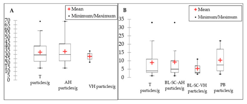
Figure 2.
Percentage of potential microplastic (MPs) particles in ocular fluids and control blank samples by shape category (A): AH aqueous humour (outer doughnut chart); VH, vitreous humour (inner doughnut chart); (B): BL-SC-AH, blank of surgical conditions obtained during AH sampling; BL-SC-VH, blank of surgical conditions obtained during VH sampling (middle doughnut chart); PB, procedural blank (outer doughnut chart).
Figure 2.
Percentage of potential microplastic (MPs) particles in ocular fluids and control blank samples by shape category (A): AH aqueous humour (outer doughnut chart); VH, vitreous humour (inner doughnut chart); (B): BL-SC-AH, blank of surgical conditions obtained during AH sampling; BL-SC-VH, blank of surgical conditions obtained during VH sampling (middle doughnut chart); PB, procedural blank (outer doughnut chart).
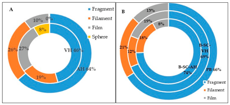
Figure 3.
Frequency of potential MPs shapes across the size ranges: blue bars represent total shape sample frequency, while grey bars denote aqueous humour (AH) or vitreous humour (VH). In both investigated intraocular fluids, a substantial proportion (often exceeding 50%) of detected potential MP particles fell within the smaller size ranges (e.g., <100 µm or <50 µm).
Figure 3.
Frequency of potential MPs shapes across the size ranges: blue bars represent total shape sample frequency, while grey bars denote aqueous humour (AH) or vitreous humour (VH). In both investigated intraocular fluids, a substantial proportion (often exceeding 50%) of detected potential MP particles fell within the smaller size ranges (e.g., <100 µm or <50 µm).
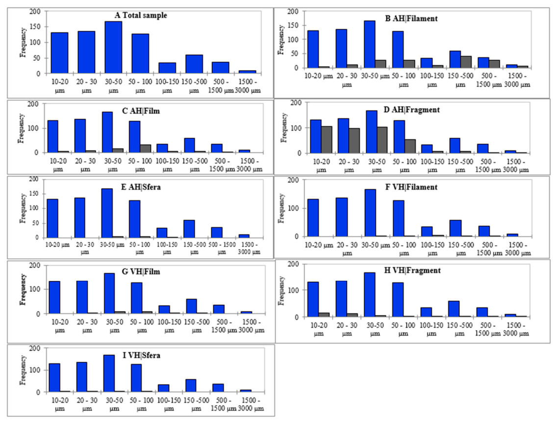
Figure 4.
Percentage of potential microplastic (MP) particles in ocular fluids and control blank samples by colour (A): Abundance of potential microplastic particles in aqueous humour (AH) (outer doughnut chart) and vitreous humour (inner doughnut chart) based on colour; (B): Abundance of potential microplastic particles in method blanks: PB, procedural blank (outer doughnut chart); sampling blanks: BL-SC-AH (inner doughnut chart), blank of surgical conditions obtained at AH sampling; BL-SC-VH (middle doughnut chart), blank of surgical conditions obtained at VH sampling, all based on colour.
Figure 4.
Percentage of potential microplastic (MP) particles in ocular fluids and control blank samples by colour (A): Abundance of potential microplastic particles in aqueous humour (AH) (outer doughnut chart) and vitreous humour (inner doughnut chart) based on colour; (B): Abundance of potential microplastic particles in method blanks: PB, procedural blank (outer doughnut chart); sampling blanks: BL-SC-AH (inner doughnut chart), blank of surgical conditions obtained at AH sampling; BL-SC-VH (middle doughnut chart), blank of surgical conditions obtained at VH sampling, all based on colour.
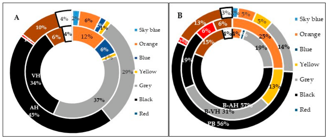
Figure 5.
Bar chart displaying mean microplastic abundance per sampling period for both (A) operating theatre - OT and (B) the anaesthetic room - AR. Abbreviations: WH, working hours; NWH, non-working hours.
Figure 5.
Bar chart displaying mean microplastic abundance per sampling period for both (A) operating theatre - OT and (B) the anaesthetic room - AR. Abbreviations: WH, working hours; NWH, non-working hours.
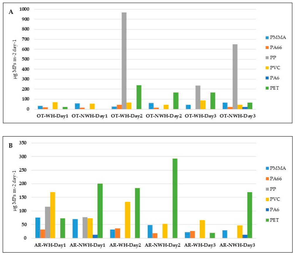
Figure 6.
Total MPs composition. Data is for the operating theatre (OT) and anaesthetic room (AR) combined for the entire 3-day sampling period, and for working and nonworking hours.
Figure 6.
Total MPs composition. Data is for the operating theatre (OT) and anaesthetic room (AR) combined for the entire 3-day sampling period, and for working and nonworking hours.
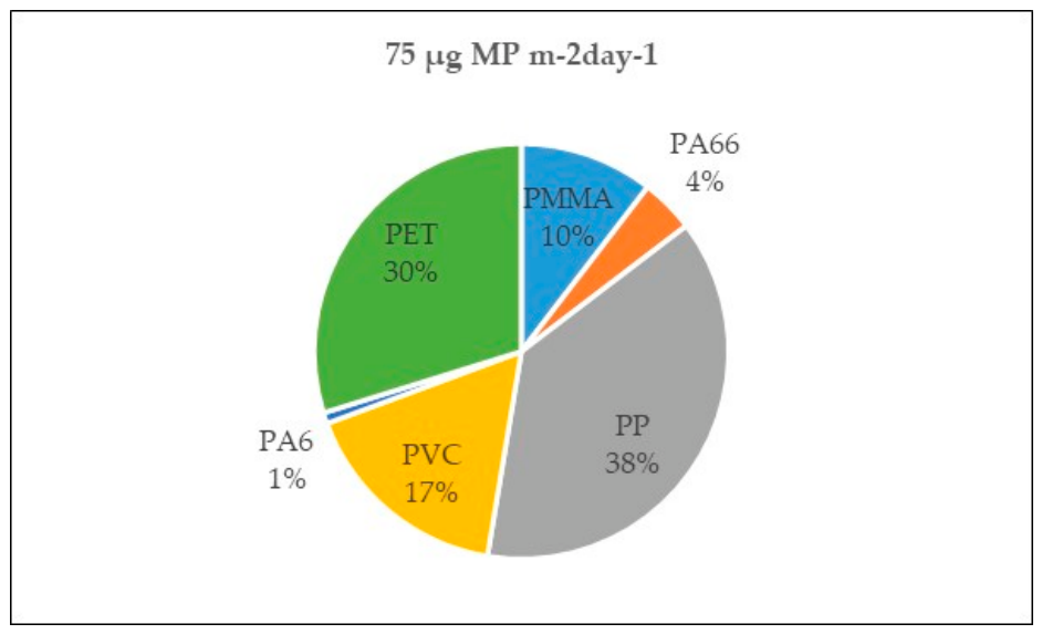
Figure 7.
Spearman correlogram between investigated MPs polymer mass fractions, bisphenols and potential microplastic particles per gram in all investigated human samples. BPA, Bisphenol A, BPAF, Bisphenol AF, ƩBPs the sum of BPA and its 4 analogues (BPAF, BPB, BPF, BPS); MPs, microplastics; C-PMMA: polymethyl methacrylate cluster, C-PA6: polyamide 6 cluster, C-PA66: polyamide 66 cluster, C-PP: polypropylene cluster, C-PVC: polyvinyl chloride cluster, C-PET: polyethylene terephthalate cluster, ƩMPs polymers, the sum of MPs polymers clusters (C-PMMA, C-PA6, C-PA66, C-PP, C-PVC, C-PET).
Figure 7.
Spearman correlogram between investigated MPs polymer mass fractions, bisphenols and potential microplastic particles per gram in all investigated human samples. BPA, Bisphenol A, BPAF, Bisphenol AF, ƩBPs the sum of BPA and its 4 analogues (BPAF, BPB, BPF, BPS); MPs, microplastics; C-PMMA: polymethyl methacrylate cluster, C-PA6: polyamide 6 cluster, C-PA66: polyamide 66 cluster, C-PP: polypropylene cluster, C-PVC: polyvinyl chloride cluster, C-PET: polyethylene terephthalate cluster, ƩMPs polymers, the sum of MPs polymers clusters (C-PMMA, C-PA6, C-PA66, C-PP, C-PVC, C-PET).
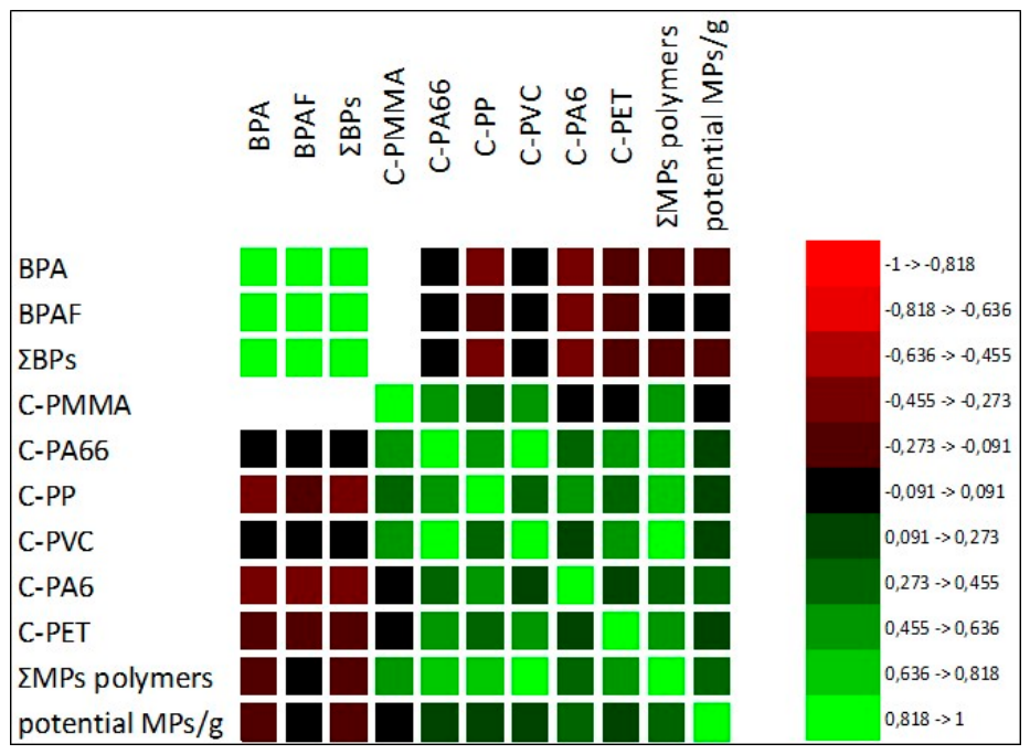
Figure 8.
Correlation analysis: Relationships between MP concentrations and patients age. Diagonal plots illustrate the distribution of data in logarithmic ratios. Lower plots display the scatter plots of data, while the upper plots indicate the correlation coefficients (r). Significant correlations are denoted by red markers. *P < 0.05; **P < 0.01; ***P < 0.001. Notes: MPs, microplastics; PA66, nylon 66; PS, polystyrene; PVC, polyvinyl chloride.
Figure 8.
Correlation analysis: Relationships between MP concentrations and patients age. Diagonal plots illustrate the distribution of data in logarithmic ratios. Lower plots display the scatter plots of data, while the upper plots indicate the correlation coefficients (r). Significant correlations are denoted by red markers. *P < 0.05; **P < 0.01; ***P < 0.001. Notes: MPs, microplastics; PA66, nylon 66; PS, polystyrene; PVC, polyvinyl chloride.
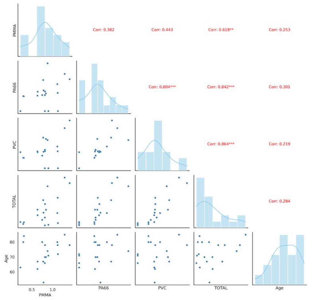

Table 1.
Microplastic polymer cluster concentrations in aqueous humor and vitreous humor samples. Results are expressed as mean value of 2 determinations calculate on 2 separate calibration curves.
Table 1.
Microplastic polymer cluster concentrations in aqueous humor and vitreous humor samples. Results are expressed as mean value of 2 determinations calculate on 2 separate calibration curves.
| Microplastic polymer cluster concentration (µg/g) | |||||||
| SAMPLE | C-PMMA5 | C-PA666 | C-PP7 | C-PVC8 | C-PA69 | C-PET10 | ƩMPs polymers11 |
| AH1-1 | 0.88 | 0.77 | 1.69 | 1.28 | <0.013 | 1.66 | 6.28 |
| AH-2 | 0.31 | 0.24 | <0.079 | 0.47 | <0.013 | 0.23 | 1.25 |
| AH-3 | 0.82 | 0.32 | 1.60 | 0.78 | <0.013 | 0.28 | 3.79 |
| AH-4 | 0.64 | 0.31 | <0.079 | 0.73 | <0.013 | 0.55 | 2.23 |
| AH-5 | 1.12 | 0.29 | 2.15 | 0.77 | <0.013 | 0.33 | 4.66 |
| AH-6 | 0.77 | 0.50 | <0.079 | 1.39 | 1.29 | 0.39 | 4.34 |
| AH-7 | 0.63 | 0.25 | <0.079 | 0.76 | <0.013 | 1.37 | 3.02 |
| AH-8 | 0.29 | 0.24 | <0.079 | 0.48 | <0.013 | <0.023 | 1.01 |
| AH-9 | 0.75 | 0.26 | <0.079 | 0.58 | <0.013 | <0.023 | 1.58 |
| AH-10 | 1.11 | 0.43 | <0.079 | 0.99 | <0.013 | <0.023 | 2.54 |
| AH-11 | 0.78 | 0.33 | <0.079 | 0.75 | <0.013 | 0.72 | 2.59 |
| AH-12 | 0.81 | 0.26 | <0.079 | 0.84 | <0.013 | <0.023 | 1.91 |
| AH-13 | 1.41 | 0.51 | <0.079 | 2.18 | <0.013 | 2.62 | 6.72 |
| AH-14 | 1.29 | 0.60 | 2.28 | 1.83 | 1.51 | <0.023 | 7.50 |
| AH-15 | 0.84 | 0.28 | 0.00 | 1.34 | <0.013 | 0.54 | 3.00 |
| AH-16 | 1.13 | <0.044 | <0.079 | <0.056 | <0.013 | <0.023 | 1.13 |
| AH-17 | 0.81 | <0.044 | <0.079 | <0.056 | <0.013 | <0.023 | 0.81 |
| AH-18 | 0.90 | <0.044 | <0.079 | <0.056 | <0.013 | <0.023 | 0.90 |
| AH-19 | 0.22 | <0.044 | <0.079 | 0.73 | <0.013 | 0.32 | 1.28 |
| average | 0.82 | 0.30 | 0.41 | 0.84 | 0.15 | 0.48 | 3.0 |
| range | 0.22-1.41 | <0.044-0.77 | <0.079-2.28 | <0.056-2.18 | <0.013-1.51 | <0.023-2.62 | 0.81 -7.55 |
| VH2-1 | 0.44 | <0.044 | <0.079 | <0.056 | <0.013 | <0.023 | 0.44 |
| VH-2 | 0.55 | <0.044 | <0.079 | <0.056 | <0.013 | <0.023 | 0.55 |
| VH-3 | 0.69 | <0.044 | <0.079 | <0.056 | <0.013 | <0.023 | 0.69 |
| average | 0.56 | 0.56 | |||||
| range | 0.44-0.69 | <0.044 | <0.079 | <0.056 | <0.013 | <0.023 | 0.44-0.69 |
| Blank SC3 1 | 0.49 | 0.49 | 0.00 | 0.95 | <0.013 | <0.023 | 1.92 |
| PB4 1 | <0.031 | <0.044 | <0.079 | <0.056 | <0.013 | <0.023 | 0.00 |
| Blank SC 2 | <0.031 | 0.00 | <0.079 | <0.056 | <0.013 | <0.023 | 0.00 |
| PB 2 | <0.031 | <0.044 | <0.079 | <0.056 | <0.013 | <0.023 | 0.00 |
| Blank SC 3 | 0.28 | <0.044 | <0.079 | <0.056 | <0.013 | <0.023 | 0.28 |
| PB 3 | <0.031 | <0.044 | <0.079 | <0.056 | <0.013 | <0.023 | 0.00 |
| average | 0.128 | 0.082 | - | 0.158 | - | - | 0.368 |
| range | <0.031-0.49 | <0.044-0.49 | <0.079 | <0.056 - 0.95 | <0.013 | <0.023 | 0.28 – 1.92 |
1AH aqueous humour; 2VH vitreous humour;, 3SC surgical conditions; 4PB, procedural blank; 5C-PMMA Poly methyl methacrylate cluster; 6C-PA6 Polyamide-6 cluster, 7C-PA66 Polyamide-6.6 cluster, 8C-PP Polypropylene cluster; 9C-PVC polyvinyl chloride cluster; 10C-PET polyethylene terephthalate cluster; values expressed as less then ‘<‘ represent polymer cluster limit of detection; 11ƩMPs polymers, total sum of MPs polymers.
Table 2.
Comparison of the presence of microplastics in eye vitreous humour according to the individual characteristics of the participants.
Table 2.
Comparison of the presence of microplastics in eye vitreous humour according to the individual characteristics of the participants.
| Variable | Characteristics | Category | Median | 1IQR | 2P-value |
| Total MPs | Age | Very old elders, ≥75 years |
3.188 | 3.566 | 0.662 |
| Elders, ≥65-.>75 years | 1.278 | 2.272 | |||
| Adults, ≥18->65 years | 2.454 | 1.463 | |||
| Gender | Female | 3.018 | 4.343 | 0.147 | |
| Male | 2.069 | 1.957 | 0.058 | ||
| Occupation | Retiree | 2.586 | 3.237 | 0.469 | |
| Worker | 1.402 | 1.402 | |||
| Type of disease in relation to the side of the eye | Oculus dexter | 3.018 | 4.343 | 0.401 | |
| Oculus sinister | 2.069 | 1.957 | |||
| C-PMMA | Age | Very old elders, ≥75 years |
0.799 | 0.301 | 0.472 |
| Elders, ≥65-.>75 years | 0.812 | 0.308 | 0.810 | ||
| Adults, ≥18->65 years | 0.723 | 0.234 | 0.155 | ||
| Gender | Female | 0.812 | 0.531 | 0.109 | |
| Male | 0.797 | 0.132 | 0.510 | ||
| Occupation | Retiree | 0.812 | 0.307 | 0.665 | |
| Worker | 0.723 | 0.248 | |||
| Type of disease in relation to the side of the eye | Oculus dexter | 0.812 | 0.531 | 0.395 | |
| Oculus sinister | 0.797 | 0.132 | |||
| C-PP | Age | Very old elders, ≥75 years |
0.8 | 1.806 | 0,013 |
| Elders, ≥65-.>75 years | 0.0 | 0 | - | ||
| Adults, ≥18->65 years | 0 | 0 | - | ||
| Gender | Female | 0 | 0.846 | 0.682 | |
| Male | 0 | ||||
| Occupation | Retiree | 0 | 0.800 | 0.100 | |
| Worker | 0 | 0 | |||
| Type of disease in relation to the side of the eye | Oculus dexter | 0 | 0.846 | 0.100 | |
| Oculus sinister | 0 | 0 | |||
| C-PA6 | Age | Very old elders, ≥75 years |
0 | 0 | 1.000 |
| Elders, ≥65-.>75 years | 0 | 0 | |||
| Adults, ≥18->65 years | 0 | 0 | |||
| Gender | Female | 0 | 0 | ||
| Male | 0 | 0 | 0.371 | ||
| Occupation | Retiree | 0 | 0 | 0.371 | |
| Worker | 0 | 0 | |||
| Type of disease in relation to the side of the eye | Oculus dexter | 0 | 0 | 0.919 | |
| Oculus sinister | 0 | 0 | |||
| Not apply | |||||
| C-PVC | Age | Very old elders, ≥75 years |
0.761 | 0.354 | 0.738 |
| Elders, ≥65-.>75 years | 0.728 | 1.063 | |||
| Adults, ≥18->65 years | 0.800 | 0.389 | |||
| Gender | Female | 0.764 | 0.552 | 0.984 | |
| Male | 0.741 | 0.529 | |||
| Occupation | Retiree | 0.749 | 0.782 | 0.345 | |
| Worker | 0.382 | 0.782 | |||
| Type of disease in relation to the side of the eye | Oculus dexter | 0.764 | 0.552 | 0.641 | |
| Oculus sinister | 0.741 | 0.529 | |||
| C-PA66 | Age | Very old elders, ≥75 years |
0.305 | 0.145 | 0.014 |
| Elders, ≥65-.>75 years | 0 | 0.406 | |||
| Adults, ≥18->65 years | 0.253 | 0.077 | |||
| Gender | Female | 0.249 | 0.163 | 0.901 | |
| Male | 0.272 | 0.321 | |||
| Occupation | Retiree | 0,293 | 0.177 | 0.097 | |
| Worker | 0.124 | 0.251 | |||
| Type of disease in relation to the side of the eye | Oculus dexter | 0.249 | 0.163 | 0.100 | |
| Oculus sinister | 0.272 | 0.321 | |||
| C-PET | Age | Very old elders, ≥75 years |
0.254 | 0.426 | 0.991 |
| Elders, ≥65-.>75 years | 0.324 | 0.470 | |||
| Adults, ≥18->65 years | 0.271 | 0.750 | |||
| Gender | Female | 0.327 | 1.401 | 0.014 | |
| Male | 0.138 | 0.426 | |||
| Occupation | Retiree | 0.324 | 0.548 | 0.360 | |
| Worker | 0 | 0.343 |
1IQR: interquartile range. 2Statistically significant differences between groups were tested using the Levene’s test (Mean) / Two-tailed test, Wilcoxon signed-rank test and Kruskal–Wallis test (P < 0.05). C-PMMA: poly methyl methacrylate cluster, C-PA6: polyamide-6 cluster, C-PA66: polyamide-6.6 cluster, C-PP: polypropylene cluster, C-PVC: polyvinyl chloride cluster, C-PET: polyethylene terephthalate cluster.
Table 3.
Concentration of bisphenols in human blood serum samples.
| Concentration of bisphenol compounds (ng/ml) | ||||||
| Sample | BPA1 | BPAF2 | BPB3 | BPF4 | BPS5 | ƩBPs6 |
| BS7-1 | 5.18 ± 0.64 | 4.36 ± 0.79 | nd8 | nd | nd | 9.54 |
| BS-2 | nd | 4.54 ± 1.67 | nd | nd | nd | 4.54 |
| BS-3 | 4.78 ± 1.14 | 2.72 ± 0.24 | nd | nd | nd | 7.50 |
| BS-4 | nd | nd | nd | nd | nd | - |
| BS-5 | nd | nd | nd | nd | nd | - |
| BS-6 | nd | nd | nd | nd | nd | - |
| BS-7 | nd | nd | nd | nd | nd | - |
| BS-8 | 4.05 ± 0.20 | 2.13 ± 0.89 | nd | nd | nd | 6.18 |
| BS-9 | nd | nd | nd | nd | nd | - |
| BS-10 | nd | nd | nd | nd | nd | - |
| BS-11 | nd | nd | nd | nd | nd | - |
| BS-12 | nd | 2.27 ± 0.39 | nd | nd | nd | 2.27 |
| BS-13 | nd | nd | nd | nd | nd | - |
1 BPA, Bisphenol A, 2 BPAF, Bisphenol AF, 3 BPB, Bisphenol B, 4 BPF, Bisphenol F, 5 BPS, Bisphenol S, 6 ƩBPs the sum of BPA and its 4 analogues (BPAF, BPB, BPF, BPS), 7BS, blood serum, 8nd-not detected.
Disclaimer/Publisher’s Note: The statements, opinions and data contained in all publications are solely those of the individual author(s) and contributor(s) and not of MDPI and/or the editor(s). MDPI and/or the editor(s) disclaim responsibility for any injury to people or property resulting from any ideas, methods, instructions or products referred to in the content. |
© 2026 by the authors. Licensee MDPI, Basel, Switzerland. This article is an open access article distributed under the terms and conditions of the Creative Commons Attribution (CC BY) license (http://creativecommons.org/licenses/by/4.0/).
Copyright: This open access article is published under a Creative Commons CC BY 4.0 license, which permit the free download, distribution, and reuse, provided that the author and preprint are cited in any reuse.


