

Submitted:
05 June 2026
Posted:
09 June 2026
You are already at the latest version
Abstract
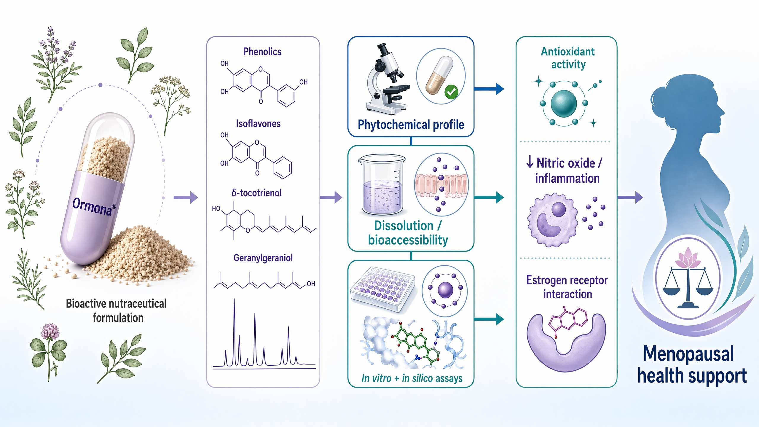
Menopause is associated with a progressive decline in estrogen levels, which contributes to increased oxidative stress, inflammatory processes, and metabolic and bone alterations that may favor the development of chronic conditions such as osteoporosis and cardiovascular diseases. Due to the potential risks associated with long-term hormone replacement therapy, the use of nutraceutical formulations containing phytoestrogens and antioxidant compounds has emerged as a potential alternative for the management of menopausal symptoms and associated metabolic alterations. In this context, the present study aimed to conduct the pharmacotechnical and phytochemical characterization of the nutraceutical formulation Ormona®, as well as to evaluate its antioxidant activity, cytotoxicity, anti-inflammatory potential, and molecular interactions with biological targets involved in estrogen signaling using integrated in silico and in vitro approaches. Pharmacotechnical analyses included weight variation, hardness, friability, and disintegration tests in order to evaluate the quality, uniformity, and stability of the formulation. Phytochemical screening and antioxidant activity were evaluated using the DPPH and ABTS radical-scavenging assays. In silico molecular docking studies were performed to investigate the interactions between the bioactive compounds present in the formulation and estrogen receptors and proteins involved in inflammatory pathways. In vitro assays were conducted to evaluate cytotoxicity in MRC-5 fibroblasts and anti-inflammatory activity by inhibiting nitric oxide production in macrophages. The formulation demonstrated satisfactory pharmacotechnical properties, relevant antioxidant activity, low cytotoxicity in fibroblast cells, potential anti-inflammatory activity, and favorable molecular interactions with estrogen receptors and inflammatory targets. These findings suggest that Ormona® may represent a promising nutraceutical formulation for menopausal health, acting through antioxidant, anti-inflammatory, and estrogen receptor-related mechanisms. However, further experimental and clinical studies are necessary to confirm its biological effects and therapeutic potential.
Keywords:
menopause
; nutraceutical
; phytoestrogens
; antioxidant activity
; molecular docking
; in vitro
1. Introduction
Menopause is a natural physiological transition characterized by the progressive decline in ovarian function and estrogen production, resulting in multiple systemic alterations affecting metabolic, cardiovascular, skeletal, and neuroendocrine systems. Estrogen deficiency is associated with increased oxidative stress, chronic low-grade inflammation, and dysregulation of bone remodeling, contributing to the development of several conditions, including osteoporosis, cardiovascular disease, and metabolic disorders [1,2]. Due to the multifactorial nature of menopausal syndrome, therapeutic strategies that target multiple biological pathways simultaneously have gained increasing scientific interest.
Hormone replacement therapy (HRT) is considered the most effective treatment for menopausal symptoms and prevention of bone loss. However, long-term use of HRT has been associated with important adverse effects, including increased risk of breast cancer, thromboembolic events, and cardiovascular complications [2,3]. These limitations have spurred the search for safer, more sustainable therapeutic alternatives, particularly those based on natural products and nutraceutical formulations with estrogen-like, antioxidant, and anti-inflammatory properties.
Plant-derived bioactive compounds, especially phytoestrogens such as isoflavones, have been widely investigated due to their structural similarity to estradiol and their ability to interact with estrogen receptors, particularly estrogen receptor beta (ERβ) [4,5]. In addition to their estrogenic activity, several phytochemicals, including tocotrienols and terpenoids, exhibit significant antioxidants and anti-inflammatory effects by reducing reactive oxygen species and inflammatory mediators such as nitric oxide and pro-inflammatory cytokines [6,7]. These combined mechanisms suggest that multi-component nutraceutical formulations may represent a promising strategy for the management of menopausal symptoms and prevention of associated complications, particularly those related to oxidative stress and bone metabolism.
In recent years, integrated preclinical approaches that combine pharmacotechnical characterization, phytochemical analysis, in silico molecular docking, and in vitro biological assays have become important tools for investigating nutraceutical formulations and bioactive compounds. Molecular docking studies predict interactions between phytochemicals and molecular targets involved in estrogen signaling, inflammation, and bone metabolism, while in vitro assays evaluate cytotoxicity, antioxidant activity, and anti-inflammatory effects at the cellular level, providing mechanistic and pharmacological evidence for the biological activity of complex formulations.
Therefore, the present study aimed to perform a comprehensive pharmacotechnical and phytochemical characterization of Ormona®, as well as to investigate its antioxidant activity, cytotoxicity profile, and molecular interactions with targets related to estrogen signaling and inflammatory pathways using integrated in silico and in vitro approaches, in order to provide preclinical evidence supporting its potential application for menopausal health management.
2. Materials and Methods
2.1. Nutraceutical formulation
Ormona® samples were provided by Ages Bioactive Compounds Co. (São Paulo, SP, Brazil). The analyzed batch (ORM220702; expiration date: July 21, 2025) consisted of purified oil of Bixa orellana L., and dry extracts of Myrciaria dubia McVaugh, Trifolium pratense L., and Euterpe oleracea Mart. The quantification methods and physicochemical characteristics are described in the patent application BR102022008408-4.
2.2. Morphological Characterization and Flow Properties of the Ormona®
The morphological characteristics of Ormona® granules were evaluated by scanning electron microscopy (SEM) using a T3030 Plus Tabletop microscope (Hitachi, Japan), operated at 15 kV. Flow properties were assessed through bulk density, tapped density, compressibility index (CI), Hausner factor (HF), and angle of repose. For bulk and tapped density measurements, 10 g of sample was transferred into a 50 mL graduated cylinder without compaction to obtain the initial volume (vb). The cylinder was then subjected to a series of manual taps (50–250) until a constant volume (vc) was reached. Bulk and tapped densities were calculated from the mass-to-initial-volume and mass-to-final-volume ratios, respectively. All measurements were performed in triplicate. The CI and HF were calculated from bulk and tapped densities to evaluate flowability and particle cohesiveness. The angle of repose was determined by allowing approximately 5 g of sample to flow through a standardized funnel positioned 20 cm above a flat surface, forming a conical pile. The height and radius of the cone were measured, and the angle (θ) was calculated accordingly.
2.3. Phytochemical Characterization
The phytochemical profile of Ormona® was evaluated through spectrophotometric and chromatographic analyses. Total phenolic content was determined by UV–Vis spectrophotometry using the Folin–Ciocalteu method, with gallic acid as the standard. Briefly, appropriately diluted samples were reacted with Folin–Ciocalteu reagent (3%, v/v) and sodium carbonate (10%, w/v), and after incubation for 30 min in the dark, absorbance was measured at 765 nm. Results were expressed based on a gallic acid calibration curve [8].
Isoflavone content was quantified by high-performance liquid chromatography (HPLC-UV/Vis) after acid hydrolysis, which converted glycosylated forms into their respective aglycones. Samples were solubilized in a mixture of acidified water (1% acetic acid) and acetonitrile (4:6, v/v), then magnetically stirred, filtered, and injected into a Shimadzu HPLC system equipped with a C18 column. The analysis was carried out under isocratic conditions at 35 °C, with a flow rate of 0.1 mL/min and an injection volume of 15 µL. Quantification was performed using external calibration curves of analytical standards (daidzein, glycitein, genistein, and genistin), with all analyses conducted in triplicate [9].
The levels of δ-tocotrienol and geranylgeraniol were determined by gas chromatography-mass spectrometry (GC-MS). Samples were prepared in hexane (5 mg/mL) and derivatized with BSTFA prior to analysis to enhance volatility. Chromatographic separation was achieved using a DB-5MS capillary column under a programmed temperature gradient, with helium as the carrier gas and electron impact ionization at 70 eV. Quantification was performed using calibration curves constructed from derivatized standard solutions over a defined concentration range. All analyses were performed in triplicate [10].
2.4. Free Radical Scavenging Assays
The antioxidant activity of Ormona® was evaluated using the DPPH and ABTS radical-scavenging assays. For the DPPH assay, a DPPH solution (≈1 mg in 12 mL of absolute ethanol) was prepared and adjusted to an absorbance of 1.00 ± 0.1 at 517 nm. Briefly, 30 µL of the sample and 270 µL of DPPH solution were added to a 96-well microplate and incubated in the dark for 30 min. Absorbance was measured at 517 nm using a microplate reader. Gallic acid was used as a reference standard and DMSO as a negative control [11,12].
For the ABTS assay, the ABTS radical was generated by reacting 0.7 mM ABTS with 2.4 mM potassium persulfate, then incubating in the dark at room temperature for 16 h. For the analysis, 30 µL of the sample and 270 µL of ABTS solution were mixed in a microplate and incubated in the dark for 30 min. Absorbance was measured at 630 nm, using gallic acid as a standard [13,14]. Antioxidant activity was expressed as the percentage of radical scavenging, calculated from the reduction in absorbance relative to the control.
2.5. In Vitro Bioaccessibility Assay
Ormona® capsules (size 00) were prepared to evaluate bioaccessibility and release properties. Empty capsules were weighed to determine the mean shell weight, and the filling mass (~650 mg) was estimated from the tapped density and capsule volume. Capsules were manually filled, individually weighed, and the net content was calculated by difference. Content uniformity was assessed in accordance with the Brazilian Pharmacopeia [15]. Disintegration tests were carried out using a pharmacopeia apparatus in a thermostatically controlled bath at 37 ± 1 °C, with purified water as the medium. Capsules were subjected to vertical movement (~30 cycles/min), and disintegration time was recorded for up to 30 min, with complete disintegration defined as the absence of any rigid residue, in accordance with pharmacopeia criteria [15].
Dissolution studies were performed using USP Apparatus I (basket method) to simulate gastrointestinal conditions and evaluate the release of gallic acid-derived phenolics. The assay was conducted in a dissolution tester (ETHIK, Brazil) with five vessels (n = 5), each containing 900 mL of dissolution medium (HCl buffer, pH 1.2, or phosphate buffer, pH 6.8), maintained at 37 ± 0.5 °C and rotated at 75 rpm. Aliquots (10 mL) were collected at predetermined intervals (5–120 min) and immediately replaced with fresh medium [15]. Samples were filtered and analyzed for total phenolic content using the Folin–Ciocalteu method, with absorbance measured at 765 nm and results expressed as mg of gallic acid equivalents (mg GAE/mL). Data were expressed as mean ± standard deviation (n = 5), and release profiles were plotted over time. The area under the curve (AUC₀–₁₂₀ min) was calculated using the trapezoidal method to estimate total phenolic release [16].
2.6. In Silico Analysis: Molecular Docking
Molecular docking studies were performed using AutoDock Vina [17] integrated into the PyRx 0.9.8 platform. Target proteins were obtained from the RCSB Protein Data Bank, including VEGFR2/KDR (PDB IDs: 3WZE and 2OH4), cyclooxygenase-2 (COX-2, PDB ID: 5IKR), estrogen receptor alpha (PDB IDs: 1A52 and 1L2I), and human placental aromatase (PDB ID: 3EQM). Protein structures were prepared by removing co-crystallized ligands, water molecules, and non-essential heteroatoms, followed by the addition of polar hydrogens and Gasteiger charges using AutoDock Tools (v1.5.7).
Docking protocol validation was conducted by redocking native ligands into their respective binding sites using predefined grid box parameters. The root-mean-square deviation (RMSD) values ranged from 0.49 to 1.05 Å, confirming the reliability of the protocol (RMSD < 2.0 Å) [17]. Bioactive compounds (biochanin A, formononetin, and genistein) were selected as ligands. Their structures were obtained from PubChem, optimized using BIOVIA Discovery Studio Visualizer (Dassault Systèmes, USA), and converted to .pdbqt format. Docking simulations were performed using default parameters. The best binding conformations were selected based on the lowest binding energy (kcal/mol), and molecular interactions were analyzed focusing on hydrogen bonds, hydrophobic interactions, π–π stacking, and van der Waals forces to elucidate ligand–protein stabilization mechanisms [18,19,20,21,22,23].
2.7. In Vitro Assays
Cell viability was evaluated in MRC-5 fibroblasts using the Alamar Blue (resazurin) assay, based on the reduction of resazurin to a fluorescent product in metabolically active cells [24]. Cells were seeded in 96-well plates (0.5 × 10⁴ cells/well) and allowed to adhere for 24 h. Subsequently, cells were treated with Ormona® at concentrations ranging from 1.56 to 100 µg/mL for 24, 48, and 72 h. After treatment, Alamar Blue solution was added, and fluorescence was measured after 3 h of incubation to determine cell viability.
Cytotoxicity and antioxidant activity were further assessed in J774.A1 macrophages using the MTT assay, based on mitochondrial reduction of MTT to formazan crystals [25]. Cells were seeded in 96-well plates (1 × 10⁶ cells/well) and incubated for 24 h at 37 °C with 5% CO₂. Cells were then treated with Ormona® (10, 25, and 50 µg/mL) for 24 h. After treatment, cells were washed with PBS and incubated with MTT solution for 3 h. Formazan crystals were solubilized with dimethyl sulfoxide (DMSO), and absorbance was measured at 560 nm. Results were expressed relative to untreated control cells.
Nitric oxide (NO) production was quantified by measuring nitrite levels using the Griess reaction [25]. Briefly, 50 µL of cell supernatant were mixed with an equal volume of Griess reagent (sulfanilamide and naphthyl-ethylenediamine) and incubated for 15 min. Absorbance was measured at 560 nm, and nitrite concentrations were determined using a sodium nitrate standard curve (15–1000 µM).
2.8. Statistical Analysis
Data were analyzed using GraphPad Prism software (version 7.0). Statistical differences among groups were assessed using one-way ANOVA followed by Tukey’s post hoc test for parametric data, or the Kruskal–Wallis test followed by Dunn’s post hoc test for non-parametric data, depending on the data distribution.
3. Results
3.1. Pharmacotechnical Properties And Morphology
Ormona® exhibited adequate pharmacotechnical properties for oral administration. The powder presented a Carr’s Index of 20.3% and a Hausner ratio of 1.25, indicating reasonable flowability and suitability for encapsulation processes. Scanning electron microscopy revealed irregularly shaped granules with heterogeneous surface morphology, consistent with plant-derived particulate systems (Figure 1 and Table 1).
3.2. Phytochemical Characterization and Free Radical Scavenging Activity of Ormona®
Total phenolic content in Ormona® was determined using the Folin–Ciocalteu method, with gallic acid as the reference standard. The calibration curve showed excellent linearity (R² = 0.994), indicating high reliability of the method. The sample had a total phenolic content of 25.23 mg gallic acid equivalents per gram (mg GAE/g), corresponding to approximately 2.52% of the formulation.
HPLC analysis revealed the presence of key isoflavones, including daidzein, glycitein, and genistein, with retention times of 33.6, 39.7, and 41.6 min, respectively. Quantification indicated concentrations of 27.2 µg/mL (5.4%) for daidzein, 11.6 µg/mL (2.3%) for glycitein, and 18.3 µg/mL (3.6%) for genistein. The chromatographic profile also suggested the presence of additional isoflavones, consistent with previous reports on Trifolium pratense, which may contribute to the biological activity due to their known estrogenic potential (Figure 2).
GC-MS analysis demonstrated excellent linearity for both δ-tocotrienol (R² = 0.998) and geranylgeraniol (R² = 0.999), confirming the robustness of the analytical method. Chromatographic and mass spectral analyses enabled clear identification of both compounds, with retention times consistent with analytical standards. Quantification revealed concentrations of 60.0 mg/g (6.0%) for δ-tocotrienol and 247.37 mg/g (24.74%) for geranylgeraniol. These values are consistent with previously reported data for Bixa orellana extracts, supporting the method’s reliability and confirming the high levels of these bioactive markers in Ormona® (Figure 3 and Figure 4).
Ormona® exhibited radical scavenging activity in both DPPH and ABTS assays, with inhibition values of 26.46 ± 2.03% and 75.83 ± 1.05%, respectively (Figure 5). The ABTS assay demonstrated significantly higher antioxidant activity than the DPPH assay, indicating a differential interaction of the formulation with distinct radical species (Figure 5).
3.3. Dissolution Profile and Bioaccessibility
The uniformity of mass of Ormona® capsules (n = 20) was highly consistent, with a mean weight of 669.35 ± 12.77 mg and a coefficient of variation of 1.91%, in compliance with Brazilian Pharmacopeia specifications. Disintegration tests showed that all capsules disintegrated within the pharmacopeia limit of 30 min in all tested media. The shortest disintegration time was observed in an acidic medium (pH 1.2; 5 min 12 s ± 49 s), followed by a phosphate buffer (pH 6.8; 6 min 35 s ± 1 min 28 s), and water (pH ~7.4; 15 min 22 s ± 3 min 04 s).
In vitro dissolution studies revealed a pH-dependent release profile of total phenolics. At 120 min, phenolic concentration reached 0.1344 mg GAE/mL in pH 6.8, compared to 0.0655 mg GAE/mL in pH 1.2. The area under the curve (AUC₀–₁₂₀) was 11.08 mg·min/mL for pH 6.8 and 6.545 mg·min/mL for pH 1.2 (Figure 6).
3.4. Molecular Docking Analysis
Molecular docking validation demonstrated the protocol’s high reliability, with RMSD values below 2.0 Å for all protein–ligand systems, ranging from 0.49 to 1.05 Å. The binding energy profiles of biochanin A, formononetin, and genistein against selected molecular targets associated with menopausal syndrome are presented in Figure 6. Docking simulations revealed that isoflavones exhibited binding affinities ranging from –6.5 to –9.2 kcal/mol across all targets. Among the evaluated proteins, VEGFR2 (3WZE) showed strong interactions with all ligands, particularly formononetin (–8.9 kcal/mol; Ki = 0.296 µM), followed by genistein and biochanin A. In contrast, weaker interactions were observed for the alternative VEGFR2 conformation (2OH4), with binding energies ranging from –6.5 to –6.7 kcal/mol.
For COX-2 (5IKR), genistein presented the highest affinity (–8.0 kcal/mol), whereas formononetin and biochanin A showed lower binding energies. Notably, genistein exhibited the strongest interaction among all systems with estrogen receptor α (1A52) (–9.2 kcal/mol; Ki = 0.178 µM), followed by interactions with 1L2I and 3EQM (Table 2). Overall, genistein consistently showed the highest binding affinity across multiple targets. Structural analysis revealed that ligand–protein stabilization involved hydrogen bonds, π–π stacking, π–sulfur interactions, and hydrophobic contacts. Genistein (Figure 7) formed the most extensive interaction networks across different targets, while formononetin (Figure 8) showed strong interactions with VEGFR2, and biochanin A exhibited more moderate and consistent binding profiles (Figure 9).
3.5. In Vitro Cytotoxicity and Antioxidant Activity
Ormona® cytotoxicity was evaluated in MRC-5 human lung fibroblasts using the Alamar Blue assay. The compound exhibited concentration-dependent cytotoxicity, with an IC₅₀ value of 17.14 µg/mL (95% CI: 12.26–23.96 µg/mL) after 72 h of treatment. Higher concentrations (50–100 µg/mL) reduced cell viability by more than 50%, whereas lower concentrations (≤12.5 µg/mL) showed limited cytotoxic effects (Figure 10).
In J774A.1 macrophages, cell viability was assessed under inflammatory conditions induced by lipopolysaccharide (LPS). LPS significantly reduced cell viability compared to the negative control (p < 0.05). Treatment with Ormona® at 10 and 25 µg/mL did not affect cell viability (p > 0.05), while 50 µg/mL significantly decreased viability (p < 0.001), indicating cytotoxicity at higher concentrations. The anti-inflammatory activity of Ormona® was evaluated by measuring nitric oxide (NO) production. Treatment with 25 and 50 µg/mL significantly reduced nitrite levels compared to the LPS group (p < 0.05 and p < 0.001, respectively), whereas 10 µg/mL showed no significant effect. These results indicate a dose-dependent inhibition of NO production (Figure 11).
4. Discussion
The present study demonstrated that the Ormona® formulation presents satisfactory pharmacotechnical characteristics, including adequate capsule weight uniformity and appropriate disintegration behavior. These parameters are essential for ensuring dose consistency and reliable release of bioactive compounds in solid oral dosage forms. The faster disintegration observed under acidic conditions is consistent with the behavior of gelatin capsules, which tend to hydrate and dissolve more rapidly in low pH environments, whereas slower disintegration under neutral conditions may be associated with reduced capsule shell hydration and ionic strength effects. In addition, the agglomeration of granules observed after capsule disintegration may reduce the surface area available for exposure, potentially influencing the dissolution rate and compound release.
The dissolution and bioaccessibility results revealed a pH-dependent release profile, with greater compound release under intestinal conditions compared to acidic conditions. This behavior is consistent with the physicochemical properties of phenolic compounds, which are weak acids and exhibit increased solubility at higher pH due to ionization [26,27]. Under acidic conditions, these compounds remain predominantly non-ionized, resulting in reduced solubility and dissolution. From a physiological perspective, this release pattern may be advantageous, as reduced gastric release may protect phenolic compounds from degradation, while enhanced intestinal release favors absorption at the primary site of uptake.
Furthermore, the antioxidant activity associated with the bioaccessible fraction suggests that the released compounds retain their functional properties after dissolution. Phenolic compounds and isoflavones are well known for their antioxidant capacity, acting as free radical scavengers and reducing oxidative stress through hydrogen donation and electron transfer mechanisms [28,29]. Given that oxidative stress is a key factor in aging and menopause-related physiological alterations, the observed antioxidant activity may contribute to the formulation’s potential biological effects [7,30].
The in vitro cytotoxicity assays demonstrated a concentration-dependent response, with lower concentrations presenting higher cell viability and therefore a more favorable safety profile. This behavior is consistent with the biological activity of phenolic compounds, which may exert cytoprotective effects at lower concentrations and cytotoxic effects at higher concentrations depending on cellular redox balance and metabolic conditions [29]. These findings suggest that the formulation has a safe profile within a specific concentration range, particularly relevant for nutraceutical applications.
In macrophage cells, the formulation significantly reduced nitric oxide production without inducing cytotoxicity at lower concentrations, indicating a true anti-inflammatory effect. Nitric oxide is a key inflammatory mediator produced by activated macrophages via inducible nitric oxide synthase (iNOS), and excessive production is associated with inflammatory responses and oxidative stress [31]. Therefore, the observed reduction in nitric oxide levels suggests that the formulation may modulate inflammatory pathways, potentially by inhibiting iNOS expression or activity.
Isoflavones such as genistein have been widely reported to exert anti-inflammatory effects by modulating signaling pathways, including NF-κB, and reducing the production of pro-inflammatory mediators, including nitric oxide and cytokines [32,33]. In addition, polyphenolic compounds have been shown to regulate macrophage activation and attenuate inflammatory responses, reinforcing their role as bioactive agents with therapeutic potential [34]. These findings support the hypothesis that the anti-inflammatory effects observed in this study are associated with the presence of phenolic compounds and isoflavones in the formulation.
The molecular docking analysis demonstrated that the bioactive compounds in the formulation, particularly isoflavones such as genistein, exhibit favorable binding affinities for molecular targets associated with menopausal symptoms, including estrogen receptors and proteins involved in inflammatory and metabolic pathways. The reliability of the docking results is supported by appropriate RMSD values, indicating consistency in ligand–receptor interactions.
Isoflavones are recognized as phytoestrogens due to their structural similarity to estradiol, which allows them to bind to estrogen receptors and modulate estrogen signaling pathways [35,36]. This interaction is particularly relevant in the context of menopause, where decreased estrogen levels are associated with various physiological alterations, including increased oxidative stress and inflammation [5]. Therefore, the interaction between isoflavones and estrogen receptors observed in the docking analysis supports the formulation’s potential estrogen-modulating activity.
In addition to Ormona® pathways, the ability of these compounds to interact with targets involved in inflammation and oxidative stress suggests a multitarget mechanism of action. Natural compounds, particularly polyphenols, are known to act on multiple molecular targets simultaneously, thereby enhancing their therapeutic potential in complex conditions [37,38]. This multitarget profile is consistent with the results observed in the in vitro assays and reinforces the biological plausibility of the formulation’s effects.
Taken together, the pharmacotechnical, bioaccessibility, antioxidant, and in silico findings suggest that the formulation may exert its biological effects through a synergistic mechanism involving antioxidant activity, modulation of inflammatory mediators, and interaction with estrogen receptors. This integrated mechanism may be particularly relevant to conditions associated with oxidative stress, inflammation, and hormonal changes, such as those associated with menopause.
However, some limitations should be considered. The study used in vitro and in silico approaches, which do not fully replicate the complexity of human physiology. Additionally, bioavailability, pharmacokinetics, and long-term safety were not evaluated. Therefore, future studies should focus on investigating these aspects and conducting clinical studies to confirm the formulation’s efficacy and safety and to further elucidate its mechanisms of action.
The higher scavenging activity observed in the ABTS assay can be attributed to the chemical properties of both the radical and the bioactive compounds present in Ormona®. The ABTS•⁺ radical is more hydrophilic and more reactive, allowing interaction with a broader range of antioxidant compounds, including both hydrophilic and lipophilic molecules [39,40]. This characteristic favors its interaction with phenolic compounds, which are efficient electron donors due to their hydroxyl groups, which stabilize free radicals [41].
In contrast, the DPPH radical presents lower hydrophilicity and greater steric hindrance, which may limit its interaction with certain antioxidant molecules. The antioxidant activity in the DPPH assay is primarily mediated by electron-transfer mechanisms and may be influenced by the molecular structure and accessibility of the bioactive compounds [40,41]. As a result, differences in reactivity between ABTS and DPPH assays are commonly observed in complex plant-based formulations.
Overall, the differential responses between DPPH and ABTS assays highlight the importance of employing multiple analytical methods to comprehensively evaluate antioxidant capacity. The pronounced activity observed in the ABTS assay reinforces the potential of Ormona® as a source of phenolic antioxidants with relevance to oxidative stress modulation [7,29].
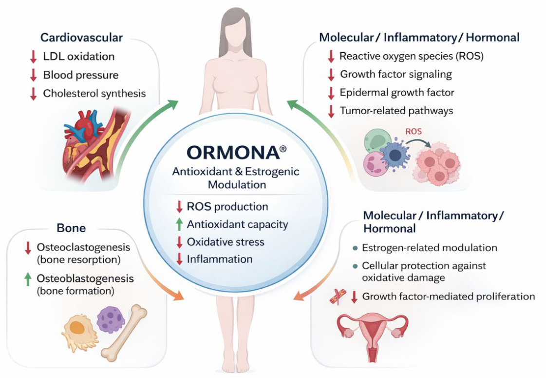
Taken together, the results suggest that Ormona® exerts its effects through a synergistic mechanism involving antioxidant activity, modulation of inflammatory pathways, and interaction with estrogen-related signaling. This multitarget approach aligns with current evidence supporting the use of nutraceuticals as complementary strategies for managing menopausal symptoms [42,43]. Additionally, bioactive compounds such as isoflavones and other polyphenols have been associated with improved vascular, metabolic, and inflammatory outcomes in postmenopausal populations [44,45].
Despite these promising findings, some limitations should be considered. This study used in vitro and in silico approaches; therefore, the results may not be directly extrapolated to human physiological conditions. Although the multi-model approach strengthens the mechanistic interpretation, clinical studies are necessary to confirm the efficacy and safety of Ormona® in human populations.
Future studies should focus on clinical validation, dose optimization, long-term safety evaluation, and further investigation of the molecular pathways underlying its pharmacological effects.
Figure 12.
Proposed systemic mechanisms of action of Ormona® through antioxidant and estrogen-related pathways.
Figure 12.
Proposed systemic mechanisms of action of Ormona® through antioxidant and estrogen-related pathways.
5. Conclusions
Ormona® demonstrated significant antioxidant and estrogenic effects through a multi-model preclinical approach. The formulation showed low cytotoxicity, reduced oxidative stress in vitro, and exhibited estrogen-like activity in vivo, improving bone-related parameters in ovariectomized rats. These findings suggest that Ormona® acts through a synergistic mechanism involving modulation of oxidative stress, inflammatory pathways, and estrogen receptor signaling.
Collectively, this study provides robust preclinical evidence supporting the potential of Ormona® as a safe and effective nutraceutical for the management of menopausal syndrome and prevention of associated complications, particularly bone loss. Further clinical studies are warranted to confirm their efficacy and safety in human populations.
6. Patents
This manuscript is associated with the patent application BR102022008408-4, filed with the Brazilian National Institute of Industrial Property (INPI). The patent application covers intellectual property related to the formulation, composition, and/or use of the nutraceutical products investigated in the present study.
Author Contributions
Conceptualization, J.C.T.C. and A.L.d.N.; methodology, A.L.d.N. and B.P.D.; software, E.C.A.; validation, A.L.d.N. and H.R.d.S.; formal analysis, E.C.A.; investigation, B.J.P.d.S. and H.R.d.S.; resources, I.A.F.d.S. and E.S.L.; data curation, E.C.A.; writing—original draft preparation, I.C.S.; writing—review and editing, J.C.T.C.; visualization, J.C.T.C.; supervision, J.C.T.C.; project administration, J.C.T.C.; funding acquisition, J.C.T.C. All authors have read and agreed to the published version of the manuscript.
Funding
This project received funding from the Ages Bioactive Compounds Company via the Priority Bioeconomy Program (PPBio - Priority Project: No. 22/2024), a public policy of the Superintendence of the Manaus Free Trade Zone (Suframa) coordinated by the Institute for Conservation and Sustainable Development of the Amazon (Idesam).
Institutional Review Board Statement
Not applicable.
Acknowledgments
The authors gratefully acknowledge the Brazilian National Council for Scientific and Technological Development (CNPq) and the Coordination for the Improvement of Higher Education Personnel (CAPES) for their support through the MAI-DAI project, which provided undergraduate research, master’s, and doctoral scholarships related to this study. The authors also acknowledge the sandwich doctoral scholarship awarded to Aline Lopes do Nascimento during her doctoral training.
Conflicts of Interest
The authors declare no conflicts of interest.
Abbreviations
The following abbreviations are used in this manuscript:
| ABTS | 2,2′-Azino-bis(3-ethylbenzothiazoline-6-sulfonic acid) |
| ADS | Sodium alendronate |
| AST | Aspartate aminotransferase |
| CG-MS / GC-MS | Gas chromatography–mass spectrometry |
| DA | Bulk density |
| DC | Tapped density |
| DMSO | Dimethyl sulfoxide |
| DPPH | 2,2-Diphenyl-1-picrylhydrazyl |
| DSHEA | Dietary Supplement Health and Education Act |
| ERα | Estrogen receptor alpha |
| ERβ | Estrogen receptor beta |
| EST | Estrogen |
| FDA | Food and Drug Administration |
| FH | Hausner ratio |
| GMP | Good Manufacturing Practices |
| HDL | High-density lipoprotein |
| HGMR | HMG-CoA reductase |
| HPLC | High-performance liquid chromatography |
| IMS | International Menopause Society |
| J774A.1 | Murine macrophage cell line |
| LDL | Low-density lipoprotein |
| MRC-5 | Human lung fibroblast cell line |
| NAMS | North American Menopause Society |
| OPG | Osteoprotegerin |
| ORM | Ormona® |
| OVW | Distilled water control |
| OVX | Bilateral ovariectomy used to induce menopause in rats |
| PDGF | Platelet-derived growth factor |
| RANKL | Receptor activator of nuclear factor kappa-B ligand |
| SEM | Scanning electron microscopy |
| SVM/VMS | Vasomotor symptoms |
| VEGF | Vascular endothelial growth factor |
| WHI | Women’s Health Initiative |
References
- Santoro, N.; Randolph, J.F., Jr. Reproductive Hormones and the Menopause Transition. Obstet. Gynecol. Clin. N. Am. 2011, 38, 455–466. [Google Scholar] [CrossRef]
- Davis, S.R.; Baber, R.; Panay, N.; Bitzer, J.; Pérez, S.C.; Islam, R.M.; Kaunitz, A.M. Global consensus position statement on menopausal hormone therapy. Climacteric 2022, 25(4), 335–343. [Google Scholar]
- Lobo, R.A. Hormone-replacement therapy: current thinking. Nat. Rev. Endocrinol. 2016, 13, 220–231. [Google Scholar] [CrossRef]
- Setchell, K.D.R.; Clerici, C. Equol: History, Chemistry, and Formation. J. Nutr. 2010, 140, 1355S–1362S. [Google Scholar] [CrossRef]
- Vitale, D.C.; Piazza, C.; Melilli, B.; Drago, F.; Salomone, S. Isoflavones: Estrogenic activity and biological effects. Curr. Med. Chem. 2013, 20(28), 3506–3516. [Google Scholar]
- Wolters, M.; Dejanovic, G.M.; Asllanaj, E.; Günther, K.; Pohlabeln, H.; Bramer, W.M.; et al. Effects of phytoestrogen supplementation on cardiovascular risk factors. Menopause 2020, 27(9), 1081–1092. [Google Scholar] [CrossRef]
- Zhang, H.; Liu, Z.D. Anti-aging and antioxidant effects of phytoestrogens: Mechanisms and clinical implications. Phytother Res. 2024, 38(2), 435–447. [Google Scholar]
- Nikolaeva, T.N.; Lapshin, P.V.; Zagoskina, N.V. Method for Determining the Total Content of Phenolic Compounds in Plant Extracts with Folin–Denis Reagent and Folin–Ciocalteu Reagent: Modification and Comparison. Russ. J. Bioorganic Chem. 2022, 48, 1519–1525. [Google Scholar] [CrossRef]
- Rodrigues, A.P.S.; et al. Ormona® SI and Ormona® RC—New nutraceuticals with geranylgeraniol, tocotrienols, anthocyanins, and isoflavones decrease high-fat diet-induced dyslipidemia in Wistar rats. Nutraceuticals 2022, 2(4), 311–322. [Google Scholar] [CrossRef]
- Rocha, C.F.; et al. Acute and reproductive toxicity evaluation of Ormona® SI and Ormona® RC in adult zebrafish. Pharmaceuticals 2022, 15(11). [Google Scholar] [CrossRef]
- Gulcin, I.; Alwasel, S.H. DPPH Radical Scavenging Assay. Processes 2023, 11, 2248. [Google Scholar] [CrossRef]
- Wołosiak, R.; Drużyńska, B.; Derewiaka, D.; Piecyk, M.; Majewska, E.; Ciecierska, M.; Worobiej, E.; Pakosz, P. Verification of the Conditions for Determination of Antioxidant Activity by ABTS and DPPH Assays—A Practical Approach. Molecules 2021, 27, 50. [Google Scholar] [CrossRef] [PubMed]
- Re, R.; et al. Antioxidant activity applying an improved ABTS radical cation decolorization assay. Free Radic. Biol. Med. 1999, 26(9–10), 1231–1237. [Google Scholar] [CrossRef]
- Brasil. Farmacopeia Brasileira, 7th ed.; ANVISA: Brasília, Brazil, 2024. [Google Scholar]
- Trott, O.; Olson, A.J. AutoDock Vina: Improving the speed and accuracy of docking with a new scoring function, efficient optimization, and multithreading. J. Comput. Chem. 2010, 31, 455–461. [Google Scholar] [CrossRef] [PubMed]
- Bitencourt-Ferreira, G.; Veit-Acosta, M.; De Azevedo, W.F., Jr. Hydrogen Bonds in Protein-Ligand Complexes. Methods Mol. Biol. 2019. [Google Scholar]
- Loeffler, J.R.; et al. Solvation theory of aromatic complexes as key for estimating drug binding. J. Chem. Inf. Model. 2020, 60(4), 2304–2313. [Google Scholar] [CrossRef]
- Zhong, D.; Douhal, A.; Zewail, A.H. Femtosecond studies of protein–ligand hydrophobic binding and dynamics: Human serum albumin. Proc. Natl. Acad. Sci. 2000, 97, 14056–14061. [Google Scholar] [CrossRef]
- Zhuang, W.-R.; Wang, Y.; Cui, P.-F.; Xing, L.; Lee, J.; Kim, D.; Jiang, H.-L.; Oh, Y.-K. Applications of π-π stacking interactions in the design of drug-delivery systems. J. Control. Release 2019, 294, 311–326. [Google Scholar] [CrossRef]
- Zhuang, S.; Zhang, J.; Zhang, C.; et al. Insight into the binding modes of ligands in protein targets by molecular docking and molecular dynamics simulations. J. Biomol. Struct. Dyn. 2019. [Google Scholar]
- Ahmed, S.A.; Gogal, R.M.; Walsh, J.E. A new rapid and simple non-radioactive assay to monitor and determine the proliferation of lymphocytes: an alternative to [3H]thymidine incorporation assay. J. Immunol. Methods 1994, 170, 211–224. [Google Scholar] [CrossRef]
- Green, L.C.; Wagner, D.A.; Glogowski, J.; Skipper, P.L.; Wishnok, J.S.; Tannenbaum, S.R. Analysis of nitrate, nitrite, and [15N]nitrate in biological fluids. Anal. Biochem. 1982, 126, 131–138. [Google Scholar] [CrossRef]
- D’archivio, M.; Filesi, C.; Varì, R.; Scazzocchio, B.; Masella, R. Bioavailability of the Polyphenols: Status and Controversies. Int. J. Mol. Sci. 2010, 11, 1321–1342. [Google Scholar] [CrossRef]
- Manach, C.; Williamson, G.; Morand, C.; Scalbert, A.; Rémésy, C. Bioavailability and bioefficacy of polyphenols in humans. I. Review of 97 bioavailability studies. Am. J. Clin. Nutr. 2005, 81, 230S–242S. [Google Scholar] [CrossRef] [PubMed]
- Shah, D.S.; Moravkar, K.K.; Jha, D.K.; Lonkar, V.; Amin, P.D.; Chalikwar, S.S. A concise summary of powder processing methodologies for flow enhancement. Heliyon 2023, 9, e16498. [Google Scholar] [CrossRef]
- Ingallina, C.; Capitani, D.; Mannina, L.; Carradori, S.; Locatelli, M.; Di Sotto, A.; Di Giacomo, S.; Toniolo, C.; Pasqua, G.; Valletta, A.; et al. Phytochemical and biological characterization of Italian “sedano bianco di Sperlonga” Protected Geographical Indication celery ecotype: A multimethodological approach. Food Chem. 2020, 309, 125649. [Google Scholar] [CrossRef]
- Zhang, Q.; Xu, X.; Li, M.; et al. Antiosteoporotic effect of hesperidin against ovariectomy-induced osteoporosis in rats via reduction of oxidative stress and inflammation. J. Biochem Mol. Toxicol. 2021, 35(8), e22832. [Google Scholar]
- Xia, W.; Khalil, R.A. Hormone Replacement Therapy and Cardiovascular Health in Postmenopausal Women. Int. J. Mol. Sci. 2025, 26, 5078. [Google Scholar] [CrossRef]
- Kim, Y.; Lee, S.R.; Cho, M.K.; et al. The 2025 menopausal hormone therapy guidelines: Korean Society of Menopause. J. Menopausal Med. 2025, 31(2), 53–78. [Google Scholar] [CrossRef]
- Salehi, B.; Sharifi-Rad, J.; Cappellini, F.; Reiner, Ž.; Zorzan, D.; Imran, M.; Sener, B.; Kilic, M.; El-Shazly, M.; Fahmy, N.M.; et al. The Therapeutic Potential of Anthocyanins: Current Approaches Based on Their Molecular Mechanism of Action. Front. Pharmacol. 2020, 11. [Google Scholar] [CrossRef]
- Jantan, I.; Haque, A.; Arshad, L.; Harikrishnan, H.; Septama, A.W.; Mohamed-Hussein, Z.-A. Dietary polyphenols suppress chronic inflammation by modulation of multiple inflammation-associated cell signaling pathways. J. Nutr. Biochem. 2021, 93, 108634. [Google Scholar] [CrossRef]
- Rietjens, I.M.C.M.; Louisse, J.; Beekmann, K. The potential health effects of dietary phytoestrogens. Br. J. Pharmacol. 2016, 174, 1263–1280. [Google Scholar] [CrossRef]
- Ko, K.P.; Kim, S.J.; Ahn, Y.; Park, S.J.; Kim, M.K.; Kim, S.S. Dietary soy isoflavones and estrogen receptor-mediated pathways: Implications for women’s health. Nutrients 2020. [Google Scholar]
- Vauzour, D.; Rodriguez-Mateos, A.; Corona, G.; Oruna-Concha, M.J.; Spencer, J.P.E. Polyphenols and Human Health: Prevention of Disease and Mechanisms of Action. Nutrients 2010, 2, 1106–1131. [Google Scholar] [CrossRef] [PubMed]
- Tsao, R. Chemistry and Biochemistry of Dietary Polyphenols. Nutrients 2010, 2, 1231–1246. [Google Scholar] [CrossRef]
- Setchell, K.D.R.; Clerici, C. Equol: History, Chemistry, and Formation. J. Nutr. 2010, 140, 1355S–1362S. [Google Scholar] [CrossRef]
- Re, R.; Pellegrini, N.; Proteggente, A.; Pannala, A.; Yang, M.; Rice-Evans, C. Antioxidant activity applying an improved ABTS radical cation decolorization assay. Free Radic. Biol. Med. 1999, 26, 1231–1237. [Google Scholar] [CrossRef] [PubMed]
- Munteanu, I.G.; Apetrei, C. Analytical Methods Used in Determining Antioxidant Activity: A Review. Int. J. Mol. Sci. 2021, 22, 3380–3410. [Google Scholar] [CrossRef]
- Shaffie, N.M.; Sharaf, H.; Morsy, F.; Badawi, M.; Abbas, N.F. Role of some phytoestrogens in recovering bone loss: histological results from experimental ovariectomized rat models. J. Arab. Soc. Med. Res. 2015, 10, 65. [Google Scholar] [CrossRef]
- Molyneux, P. The use of the stable free radical diphenylpicrylhydrazyl (DPPH) for estimating antioxidant activity. Songklanakarin J. Sci. Technol. 2004, 26(2), 211–219. [Google Scholar]
- Ionita, P. The Chemistry of DPPH· Free Radical and Congeners. Int. J. Mol. Sci. 2021, 22, 1545. [Google Scholar] [CrossRef]
- A Lobo, R.; Gompel, A. Management of menopause: a view towards prevention. Lancet Diabetes Endocrinol. 2022, 10, 457–470. [Google Scholar] [CrossRef]
- Rattanatantikul, T.; Maiprasert, M.; Sugkraroek, P.; Bumrungpert, A. Efficacy and Safety of Nutraceutical on Menopausal Symptoms in Post-Menopausal Women: A Randomized, Double-Blind, Placebo-Controlled Clinical Trial. J. Diet. Suppl. 2020, 19, 168–183. [Google Scholar] [CrossRef]
- Tan, S.; Meng, Q.; Jiang, Y.; Zhuang, Q.; Xi, Q.; Xu, J.; Zhao, J.; Sui, X.; Wu, G. Impact of oral nutritional supplements in post-discharge patients at nutritional risk following colorectal cancer surgery: A randomised clinical trial. Clin. Nutr. 2021, 40, 47–53. [Google Scholar] [CrossRef]
- Vitale, D.C.; Piazza, C.; Melilli, B.; Drago, F.; Salomone, S. Isoflavones: Estrogenic activity, biological effect and bioavailability. Eur. J. Drug Metab. Pharmacokinet. 2013, 38, 15–25. [Google Scholar] [CrossRef]
Figure 1.
Morphological characterization of Ormona® by scanning electron microscopy (SEM). Scanning electron microscopy images of Ormona® granules showing irregular morphology and heterogeneous surface characteristics, consistent with plant-derived particulate systems.
Figure 1.
Morphological characterization of Ormona® by scanning electron microscopy (SEM). Scanning electron microscopy images of Ormona® granules showing irregular morphology and heterogeneous surface characteristics, consistent with plant-derived particulate systems.
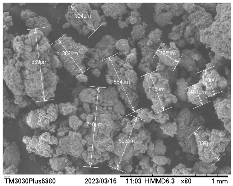
Figure 2.
Cromatograma resultante da análise do Ormona® por HPLC-UV (254-365 nm). Os picos identificados indicam a presença de (1) daidzeína (tr = 33,6 min), (2) gliciteína (tr = 39,7 min) e (3) genisteína (tr = 41,6 min).
Figure 2.
Cromatograma resultante da análise do Ormona® por HPLC-UV (254-365 nm). Os picos identificados indicam a presença de (1) daidzeína (tr = 33,6 min), (2) gliciteína (tr = 39,7 min) e (3) genisteína (tr = 41,6 min).
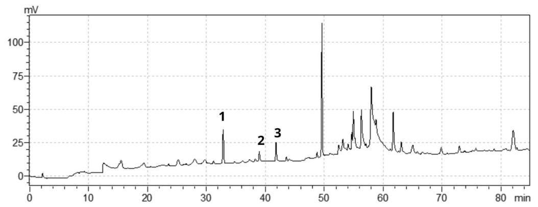
Figure 3.
Representative chromatogram of the Ormona® sample highlighting the peaks corresponding to geranylgeraniol (22.5 min) and δ-tocotrienol (33.0 min). In the chromatogram, the analytical standards are shown in black, and the Ormona® sample in red.
Figure 3.
Representative chromatogram of the Ormona® sample highlighting the peaks corresponding to geranylgeraniol (22.5 min) and δ-tocotrienol (33.0 min). In the chromatogram, the analytical standards are shown in black, and the Ormona® sample in red.
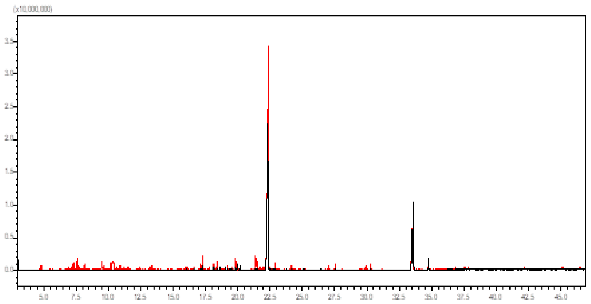
Figure 4.
(A) Mass spectrum of δ-tocotrienol, showing characteristic fragments consistent with its chemical structure. (B) Mass spectrum of geranylgeraniol, showing diagnostic ions typical of isoprenoid compounds.
Figure 4.
(A) Mass spectrum of δ-tocotrienol, showing characteristic fragments consistent with its chemical structure. (B) Mass spectrum of geranylgeraniol, showing diagnostic ions typical of isoprenoid compounds.
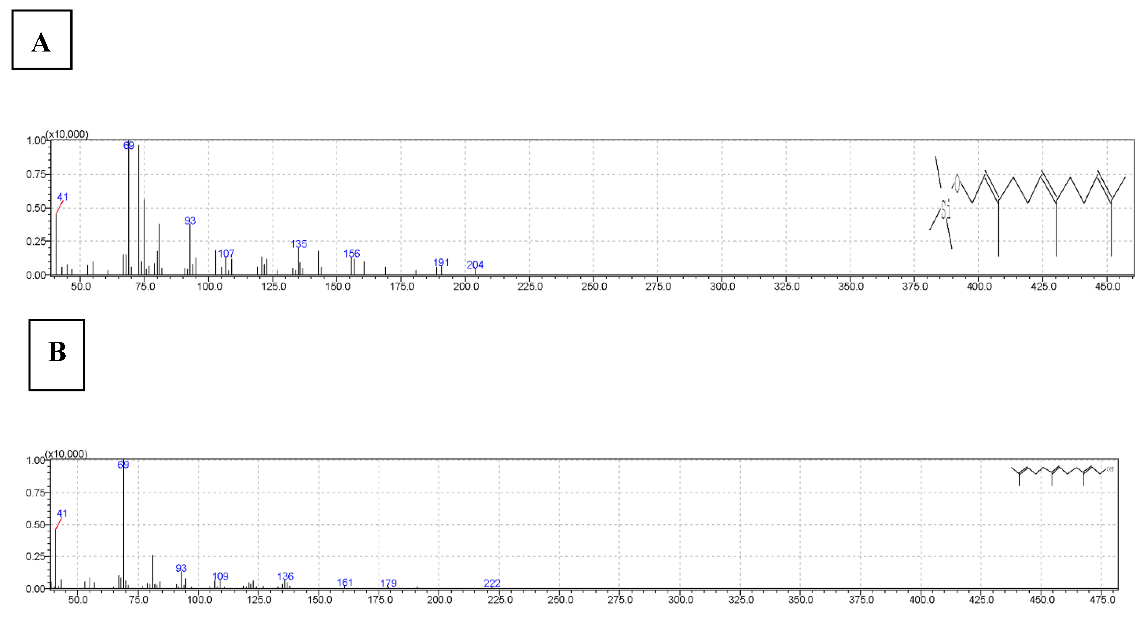
Figure 5.
Antioxidant activity of Ormona® (ORM) evaluated by ABTS and DPPH radical scavenging assays. Results are expressed as a percentage of inhibition, with gallic acid (GA) used as the reference standard.
Figure 5.
Antioxidant activity of Ormona® (ORM) evaluated by ABTS and DPPH radical scavenging assays. Results are expressed as a percentage of inhibition, with gallic acid (GA) used as the reference standard.
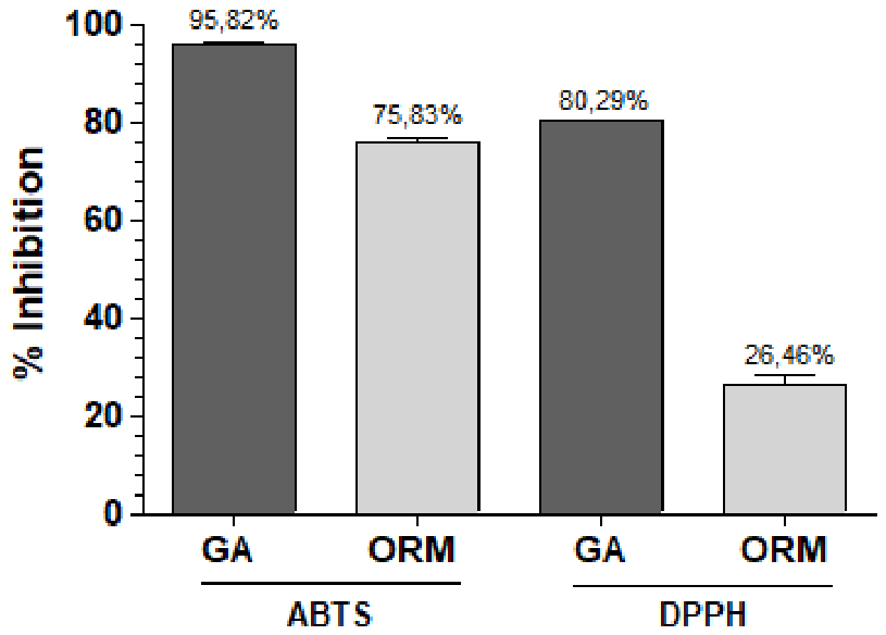
Figure 6.
In vitro dissolution profile of Ormona® under simulated gastrointestinal conditions. Dissolution profile of Ormona® capsules in simulated gastric (pH 1.2) and intestinal (pH 6.8) conditions. Data are expressed as a percentage of phenolic compound release over time.
Figure 6.
In vitro dissolution profile of Ormona® under simulated gastrointestinal conditions. Dissolution profile of Ormona® capsules in simulated gastric (pH 1.2) and intestinal (pH 6.8) conditions. Data are expressed as a percentage of phenolic compound release over time.
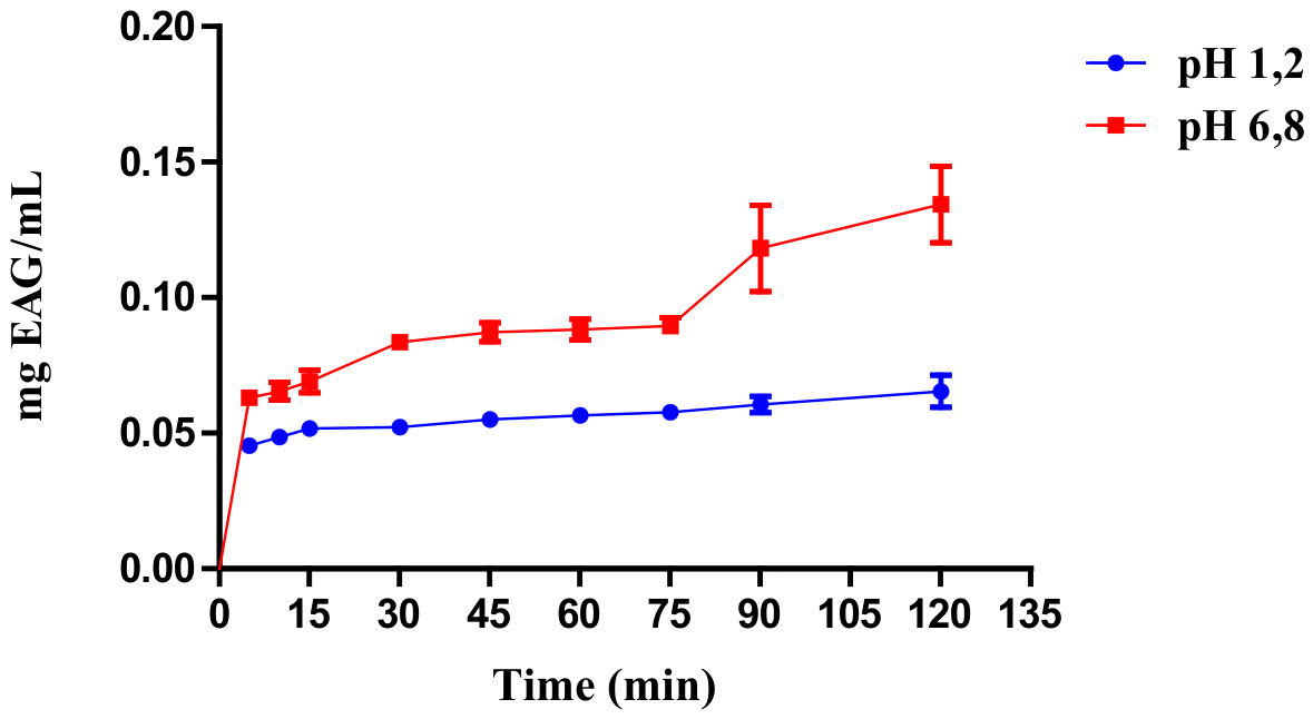
Figure 7.
Binding energy (kcal/mol) of isoflavones (biochanin A, formononetin, and genistein) against molecular targets associated with menopausal syndrome. Lower values indicate stronger binding affinity. Genistein exhibited the highest affinity across most targets.
Figure 7.
Binding energy (kcal/mol) of isoflavones (biochanin A, formononetin, and genistein) against molecular targets associated with menopausal syndrome. Lower values indicate stronger binding affinity. Genistein exhibited the highest affinity across most targets.
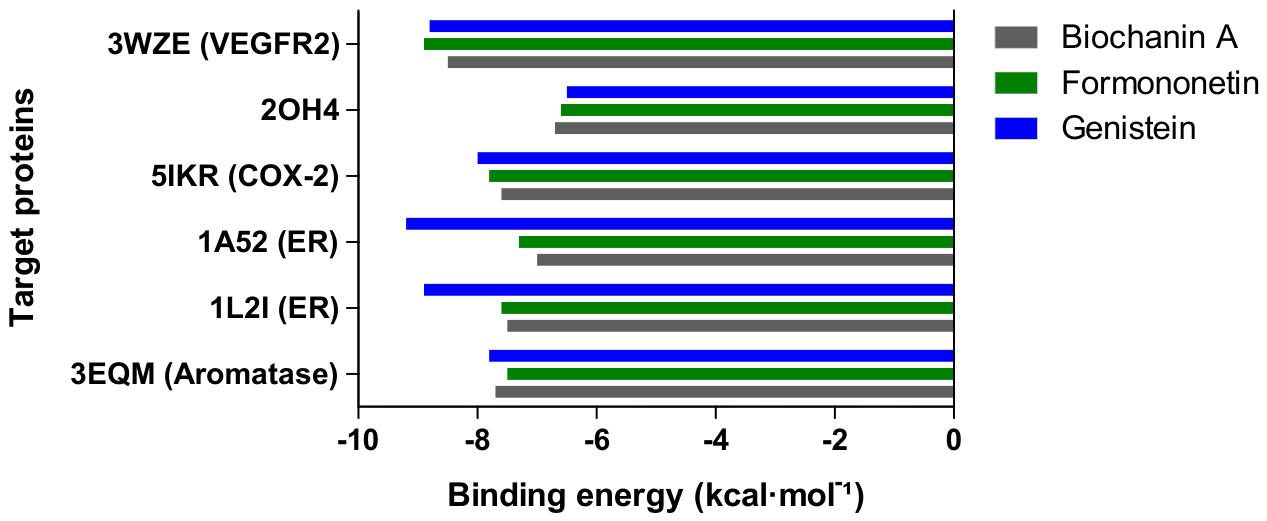
Figure 8.
3D (A) and 2D (B) representations of the Genistein –1A52 complex (Estrogen Receptor) illustrating the ligand binding orientation within the protein’s active site.
Figure 8.
3D (A) and 2D (B) representations of the Genistein –1A52 complex (Estrogen Receptor) illustrating the ligand binding orientation within the protein’s active site.
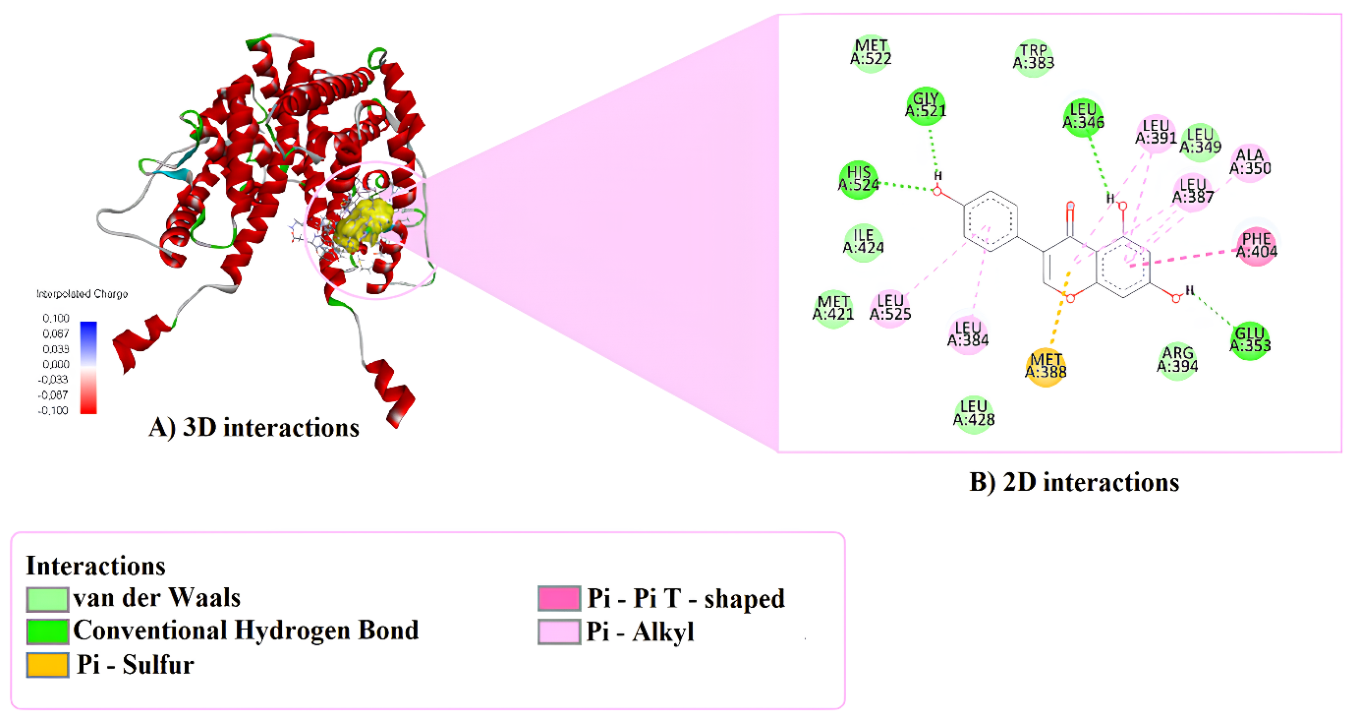
Figure 9.
3D (A) and 2D (B) representations of the Formononetin–3WZE complex (VEGFR2) illustrating the ligand binding orientation within the protein’s active site.
Figure 9.
3D (A) and 2D (B) representations of the Formononetin–3WZE complex (VEGFR2) illustrating the ligand binding orientation within the protein’s active site.
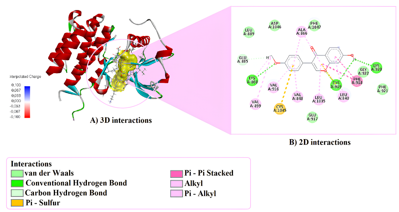
Figure 10.
Effect of Ormona® on MRC-5 cell viability after 72 h. (A) Cell viability (%) at increasing concentrations (1.5625–100 µg/mL), determined by the Alamar Blue assay. (B) Dose–response curve and IC₅₀ value (17.14 µg/mL) obtained by nonlinear regression. Data are expressed as mean ± SD (n = 3). *p < 0.05 vs. control.
Figure 10.
Effect of Ormona® on MRC-5 cell viability after 72 h. (A) Cell viability (%) at increasing concentrations (1.5625–100 µg/mL), determined by the Alamar Blue assay. (B) Dose–response curve and IC₅₀ value (17.14 µg/mL) obtained by nonlinear regression. Data are expressed as mean ± SD (n = 3). *p < 0.05 vs. control.
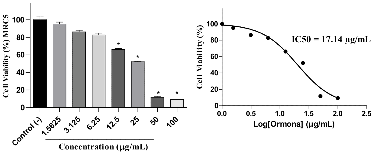
Figure 11.
Effect of Ormona® on J774A.1 macrophages under inflammatory conditions. (A) Cell viability (%) after treatment with Ormona® (10, 25, and 50 µg/mL) in the presence of LPS (1 µg/mL). (B) Nitric oxide (NO) production measured as nitrite levels (µM). Data are expressed as mean ± SEM (n = 5). Statistical analysis was performed using the Kruskal–Wallis test followed by Dunn’s post hoc test. *p < 0.05, ***p < 0.001.
Figure 11.
Effect of Ormona® on J774A.1 macrophages under inflammatory conditions. (A) Cell viability (%) after treatment with Ormona® (10, 25, and 50 µg/mL) in the presence of LPS (1 µg/mL). (B) Nitric oxide (NO) production measured as nitrite levels (µM). Data are expressed as mean ± SEM (n = 5). Statistical analysis was performed using the Kruskal–Wallis test followed by Dunn’s post hoc test. *p < 0.05, ***p < 0.001.
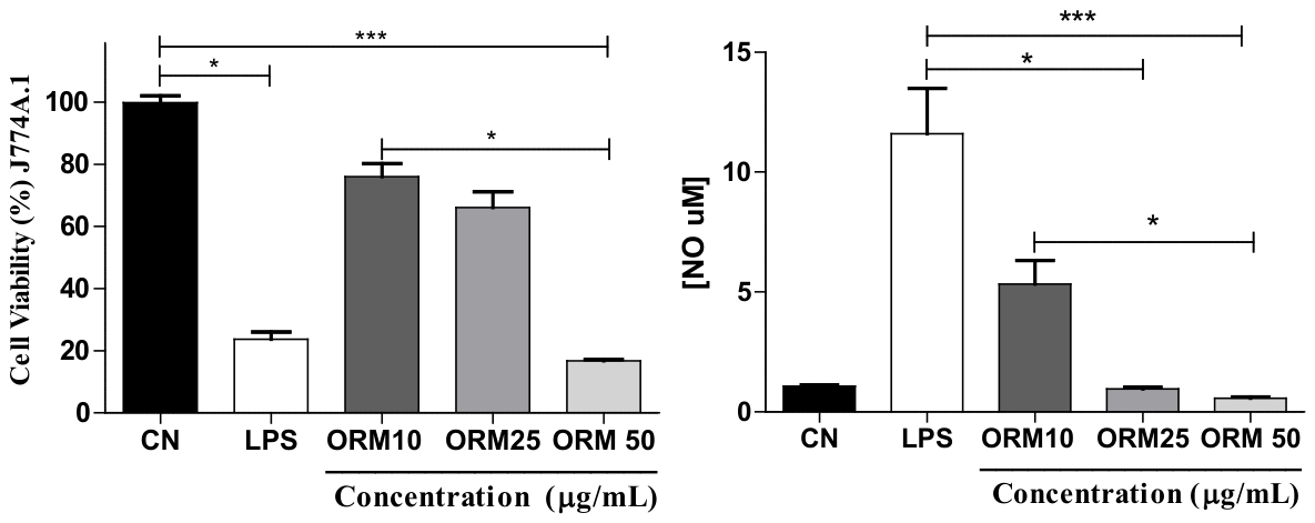

Table 1.
Pharmacotechnical properties of Ormona® formulation.
| Parameter | Obtain value | Flow Property |
|---|---|---|
| Bulk Density (g/mL) | 0.558 ± 0.03 | - |
| Tapped Density (g/mL) | 0.700 ± 0.02 | - |
| Carr’s Index or Compressibility Index | 20.3 ± 2.4 | Fair |
| Hausner Ratio | 1.25 ± 0.03 | Fair |
| Angle of Repose (º) | 26.82 ± 1.65 | Excellent |
Values are expressed as mean ± standard deviation (n = 3).
Table 2.
Binding Affinity of Isoflavones Present in Ormona®.
| Compounds | Proteins | Binding energy (kcal/mol) |
LE | FQ | BEI | Ki (μM) | |
|---|---|---|---|---|---|---|---|
| Biochanin A | 3WZE | -8.5 | 0.405 | 0.767 | 0.030 | 0.581 | |
| 2OH4 | -6.7 | 0.319 | 0.604 | 0.024 | 12.157 | ||
| 5IKR | -7.6 | 0.362 | 0.685 | 0.027 | 2.658 | ||
| 1A52 | -7.0 | 0.333 | 0.631 | 0.025 | 7.324 | ||
| 1L2I | -7.5 | 0.357 | 0.676 | 0.026 | 3.147 | ||
| 3EQM | -7.7 | 0.367 | 0.694 | 0.027 | 2.245 | ||
| Formononetin | 3WZE | -8.9 | 0.445 | 0.797 | 0.033 | 0.296 | |
| 2OH4 | -6.6 | 0.330 | 0.591 | 0.025 | 14.395 | ||
| 5IKR | -7.8 | 0.390 | 0.699 | 0.029 | 1.896 | ||
| 1A52 | -7.3 | 0.365 | 0.654 | 0.027 | 4.412 | ||
| 1L2I | -7.6 | 0.380 | 0.681 | 0.028 | 2.658 | ||
| 3EQM | -7.5 | 0.375 | 0.672 | 0.028 | 3.147 | ||
| Genistein | 3WZE | -8.8 | 0.440 | 0.789 | 0.033 | 0.350 | |
| 2OH4 | -6.5 | 0.325 | 0.582 | 0.024 | 17.044 | ||
| 5IKR | -8.0 | 0.400 | 0.717 | 0.030 | 1.353 | ||
| 1A52 | -9.2 | 0.460 | 0.824 | 0.034 | 0.178 | ||
| 1L2I | -8.9 | 0.445 | 0.797 | 0.033 | 0.296 | ||
| 3EQM | -7.8 | 0.390 | 0.699 | 0.029 | 1.896 | ||
BEI: Binding Efficiency Index, FQ: Fit Quality, Ki: Estimated Inhibition Constant, LE: Ligand Efficiency.
Disclaimer/Publisher’s Note: The statements, opinions and data contained in all publications are solely those of the individual author(s) and contributor(s) and not of MDPI and/or the editor(s). MDPI and/or the editor(s) disclaim responsibility for any injury to people or property resulting from any ideas, methods, instructions or products referred to in the content. |
© 2026 by the authors. Licensee MDPI, Basel, Switzerland. This article is an open access article distributed under the terms and conditions of the Creative Commons Attribution (CC BY) license (http://creativecommons.org/licenses/by/4.0/).
Copyright: This open access article is published under a Creative Commons CC BY 4.0 license, which permit the free download, distribution, and reuse, provided that the author and preprint are cited in any reuse.


