

Submitted:
03 June 2026
Posted:
05 June 2026
You are already at the latest version
Abstract
Introduction: Uterine leiomyoma (uterine fibroids, UF) are benign myometrial tumors affecting up to 70% of women and may cause significant symptoms. Their pathogenesis involves cytokines, steroid hormones, growth factors, and increased extracellular matrix deposition and remodeling, though it is not fully understood. Recent evidence associates low vitamin D levels with higher prevalence and severity of myomas. This clinical trial evaluates the effect of nutritional
supplement on UF.
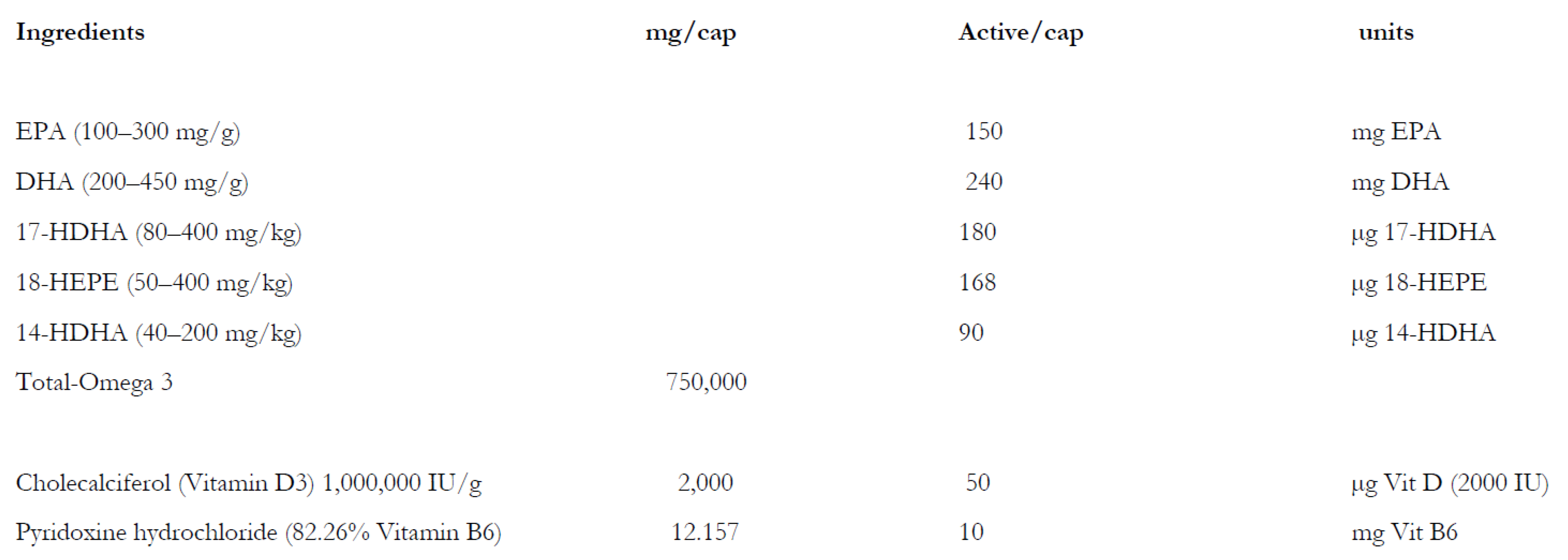
Material and Patients: Sixty (60) patients with uterine myoma received in a double-blind clinical trial for 16 weeks a nutritional formulation containing the omega 3 fatty acids EPA (eicosapentaenoic acid) and DHA (docosahexaenoic acid) 150 and 240 mg respectively, the selective pro resolving mediators (SPMs) 17-HDHA 180 µg, 18-HEPE 168 µg, 14-HDHA 90 µg, and vitamin D3 50 µg and vitamin B6 10 mg compared to placebo. The main outcomes were the development of bleeding, dysmenorrhea, volume reduction and quality of life assessment including female sexual desire.
Results: The group receiving the nutritional showed a significant improvement in the parameters bleeding reduction, dysmenorrhea’s and female sexual function compared to placebo. No changes could be observed regarding the volume reduction and estradiol values between the groups.
Conclusions: The nutritional containing Omega 3 fatty acid, SPMs, vitamin D and B6 represent a valid option for the symptomatic management of patients with uterine fibroids without comprising the hypothalamic pituitary axis of the women. Further research in grater groups of patients is needed to validate these first results.
Keywords:
uterine myoma
; inflammation
; omega e fatty acids
; vitamin D
; vitamin B6
; specialized pro-resolving mediators (SPMs)
Introduction
Uterine leiomyoma (myoma or uterine fibroids [UF]) are the most common nonmalignant pelvic tumors during the reproductive years [1], affecting up to 70% of women. Most myomas do not cause symptoms and grow slowly [2]. However, about 25% of patients experience clinically relevant symptoms [2,3]. UF incidence is especially high among African American women [4,5], who also experience more severe symptoms than Caucasian women [6]. Typical symptoms include heavy uterine bleeding, dysmenorrhea, dyspareunia, and constipation. Uterine leiomyoma can also cause infertility or abortion [2,7].
Myomas were described as monoclonal tumors of the smooth musculature. However, recent research on the Mediator Complex Subunit 12 gene, MED12(+) and MED12(-) fibroid variants, presents them as rather heterogeneous cell types by single-cell RNA sequencing [8]. The tumors, although benign, exhibit abnormal vascularization. Initiation and progression are driven by a variety of factors, including local hypoxia, gonadal steroids, and locally expressed angiogenic growth factors, such as EGF (epidermal growth factor) and VEGF (vascular endothelial growth factor). Cytokines and chemokines that are also involved in pathogenesis not only promote the proliferation of cells but also the deposition of ECM (extracellular matrix) [9], which is the main component of UF and an important factor in its development [1,9]. There also seems to be a link between myoma formation and genetic aberrations, as cytogenetic tests showed abnormalities in 40% of the samples tested: 65% overexpressed the HMGA2 (high mobility group AT-hook 2) gene. Furthermore, 70% of leiomyoma samples carried mutations in the subunit of a transcription regulator [10,11].
Estrogens and progesterone (P4) play a role in the initiation and propagation of UF. Progesterone receptors A and B (PR-A and PR-B) are induced by estrogen signaling at the estrogen receptor (ER), which increases progesterone’s effectiveness. As a result, progesterone-induced mitotic activity in UF cells rises [12,13]. Additional effects on growth factor regulation, microRNA expression, and ECM accumulation appear to be mediated by P4 [1,13,14,15]. Progesterone induces cell proliferation in UF tissue while reducing apoptosis. Its activity is mediated by growth factors such as TGFβ (transforming growth factor β), which act only in the presence of P4 [16]. Proliferation and growth of myoma may also be promoted by local and systemic inflammatory processes. Although this is debated [10,17,18], chronic inflammation is assumed to promote the pathogenesis of leiomyoma in this review.
Tumor types of various etiologies, including uterine fibroids, are associated with an inflammatory microenvironment induced by pro-inflammatory cytokines from immune and undifferentiated cells [17]. Pro-inflammatory cytokines such as interleukin 6 and 1 (IL-6, IL-1) and tumor necrosis factor alpha increase estrogen synthesis by modulating aromatase, estradiol 17-β-dehydrogenase, and estrone sulfatase activity. Inhibition of prostaglandin E2 synthesis also affects this balance [19,20]. As mentioned, ECM remodeling and accumulation are characteristic of inflammatory processes and are key in UF development. This is linked to excessive collagen accumulation and fewer micro vessels, which promote degenerative changes in the myometrium, including cellular senescence and involution. Collagenized areas show a hyaline and hypocellular morphology [21]. ECM generation is driven by cytokine and growth factor signaling and, in turn, provides a steady source of these molecules by stabilizing them.
Immune cells, especially monocytes and macrophages, respond to inflammatory signals and stimulate fibrotic changes in tumor tissue. TGF-β is a major player in these processes [22,23], along with TNF-α, granulocyte-macrophage colony-stimulating factor, and interleukins (IL) 1, IL6, IL11, IL13, and IL15, all involved in UF pathogenesis. Drugs used to treat UF may interfere with these inflammatory processes, so understanding these interactions is impor [22,23]tant [7].
Lima et al. recently showed [24] reduced Vitamin D Receptor (VDR) expression in leiomyoma tissue compared with myometrial tissue, which may be associated with the pathogenesis and development of human uterine leiomyomata. The cause of the reduced expression of these receptors remains unknown, and it is also unclear whether this event occurs concurrently with the onset or progression of uterine leiomyomas. Furthermore, Halder et al. [25] analyzed the effect of vitamin D on fibrosis-related protein expression in TGF-β-induced uterine leiomyoma cells in vitro. Myoma cells were treated with TGFß-3 with or without vitamin D. They found that TGF-3 induced the expression of fibronectin and collagen type 1, which was suppressed by vitamin D, suggesting that vitamin D is an antifibrotic factor in the treatment of benign uterine myomas. Halder et al. in a subsequent study [26] investigated the risk of benign uterine tumors in relation to VDR protein and determined the biological function of 1,25(OH)2D3 in regulating proteins involved in extracellular matrix formation, which is essential for leiomyoma formation. They identified reduced VDR levels in more than 60% of the uterine tumors analyzed compared to the adjacent myometrium. In fact, the levels of VDR in the myoma uteri samples were significantly lower than the levels in the adjacent myometrial samples. Al-Hendy et al. [27], who investigated the role of 1.25 (OH) 2D3 in the expression of sex steroid receptors in leiomyoma cells, realized that the deregulation of steroid hormones and their receptors could be a starting point for myoma growth since 1.25 (OH) 2D3 VDR expression acts as an antiestrogenic agent in these cells. They also showed a significant decrease in estrogen receptor levels in leiomyoma cells treated with 1,25(OH)2D3 and analyzed for receptor expression and localization. In contrast, 1.25 (OH) 2D3 induced its own VDR expression, suggesting that 1.25 (OH) 2D3 acts as an antagonist of hormone receptors, with antiestrogenic and antiprogestogen effects. Paffoni et al. [28] analyzed serum vitamin D levels in women with myoma and found that vitamin D concentrations were significantly lower in women with myomas than in women in the control group (11.1 vs. 18.0 ng/mL; p < 0.010; OR = 2.2). Similar results were obtained by Baird et al. [29], who assessed vitamin D and the risk of uterine myomas and found that women with the sufficient vitamin D had an estimated 32% reduction in the incidence of myomas com- pared with those with insufficient vitamin D. Sabry et al. [30] studied whether the low serum levels of vitamin D were associated with the increased risk and occurrence of uterine myomas and found that reduced serum levels of 1.25-(OH) 2D3 vitamin D were significantly associated with the occurrence of myomas. A statistically significant inverse correlation was also observed between serum 25-(OH) vitamin D levels and total leiomyoma volume in the case cohort. The above-described studies indicate that the loss of vitamin D function due to reduced vitamin D3 levels and/or reduced VDR expression may be associated with the growth and development of different types of neoplastic lesions. It reinforces the hypothesis that low VDR expression may be associated with the growth and development of myomas, making it an important biomarker in this pathology. Vitamin D is believed to regulate cell proliferation and differentiation, reduce angiogenesis, and stimulate apoptosis.
These changes may also be because of the genetic aspects of uterine fibroids. Approximately 40% of UL have non-random and tumor-specific chromosome abnormalities. This has allowed classification of some UF into well-defined subgroups, which include deletion of portions of 7q, trisomy 12, or rearrangements of 12q15, 6p21, or 10q22. Additional abnormalities, which appear consistently but less frequently, include rearrangements of chromosomes X, 1, 3, and 13. The variety of chromosomal rearrangements, including but not limited to translocation, deletion, and trisomy, predicts different molecular genetic mechanisms for UF formation and growth (31)
Vitamin D deficiency is recognized as a significant risk factor for fibroid development. Studies indicate that vitamin D can inhibit tumor cell division and reduce tumor size, though its precise role and that of its receptor in myoma pathophysiology remain unclear. Evidence suggests vitamin D and its analogs are promising, effective, and cost-efficient options for managing myomas and their symptoms [32]. Shahbazi et al.’s research supports this idea that the VDR rs2228570 variant is related to uterine fibroid —specifically, a correlation between the VDR TT genotype and an elevated risk of uterine fibroid occurrence [33]. Further, Yilmaz et al. demonstrated that the rs2228570 CC genotype may act as a risk-reducing factor, whereas the T allele may contribute to the risk of UL, in agreement with Shahbazi’s findings [34].
However, one of the findings indicated that there is no detected correlation between the VDR variants rs731236, rs1544410, and rs2228570, and the incidence of uterine leiomyoma in Caucasian women (Ciebiera et al., 2019) supporting the concept that SNPs linked to vitamin D metabolism and skin color are connected to the presence of uterine fibroids in women of Afro American origine [35].
Notably, among the scrutinized SNPs, rs12800438 near the DHCR7 gene and rs6058017 in the ASIP gene are implicated in vitamin D synthesis in the skin in these African American women [36].
This clinical trial evaluates the efficacy of a nutritional supplement containing vitamin D, vitamin B6, omega-3 fatty acids, and specialized pro-resolving lipid mediators for managing pain associated with uterine fibroids.
Material and Methods
Study Design
A randomized, double-blind, parallel, two-arm, placebo-controlled study will evaluate the efficacy and safety of a dietary supplement containing omega-3 fatty acids, vitamin D, and vitamin B6 in patients with uterine fibroids. Outcomes will include fibroid size, symptoms, and effects on the hypothalamic-pituitary-gonadal axis.
Eligible patients will be randomized to one of two treatment groups:
Group 1: Nutritional supplement
“Uterine Fibroids” capsules. One capsule will be administered every 24 hours for 4 months.
Group 2: Placebo
“Placebo” capsules. One capsule will be administered every 24 hours for 4 months.
The treatment period will last four months, with patients taking one capsule daily.
Study population
The study will enroll adult women aged 18 or older, up to menopause, with symptomatic uterine fibroids measuring at least 2 cm3.
Study timeline
Recruitment started in July 2024, and the last follow-up ended in December 2024.
Description of the treatment
The investigational product and placebo will be manufactured and packaged by Laboratorios Liconsa S.L. The supplement label will comply with local regulations, and all information will be provided in Spanish. During the study, products must be stored at room temperature, below 30 °C.
Table 1 shows the qualitative and quantitative composition of the nutritional supplement.
All products (nutritional supplement and placebo) will be provided by the sponsor in capsule form, with appropriate masking to maintain blinding.
Patients will be randomly assigned in a 1:1 ratio to receive either one capsule per day of the nutritional supplement or placebo, using the R software randomization function.
All patients in this random, masked study will take one capsule daily for four consecutive months.
Statistical analysis.
Participants with missing outcome data were excluded from the primary analysis (complete-case analysis). Microbial data were expressed as log10 CFU/mL. Statistical analyses and plotting were performed using R software (×64) version 4.0.3. Data normality was assessed using the Shapiro–Wilk test. Variables were reported as means ± standard deviation (SD) or 95% confidence intervals (CI), or as medians ± interquartile ranges (IQR), depending on distribution. Group comparisons used Kruskal-Wallis and Wilcoxon-Mann-Whitney tests for non-parametric data, with Bonferroni correction for multiple comparisons when indicated; Student’s t-test and ANOVA for parametric data, with Bonferroni adjustment; Wilcoxon’s rank-sum tests and PERMANOVA for microbial differences. Statistical significance was set at p < 0.05. As a proof-of-concept study, no formal sample size calculation was performed. The primary objective was to assess feasibility and signal of efficacy, acknowledging that the study was not powered to detect smaller effect sizes. Based on the sample, the minimal detectable effect size (Cohen’s d) for the primary outcome was approximately 0.8 with 80% power at alpha 0.05. More modest differences may have been missed, but the design allowed for a preliminary evaluation of trends and informed future, adequately powered studies.
Ethics
This randomized, double-blind, placebo-controlled trial was approved by the Research Ethics Committee of the HM Hospitales Madrid, Spain, Código CEIm HM Hospitales: 24.04.1985E5-GHM (approval date: 18/04/2024) and conducted in accordance with the Declaration of Helsinki. Written informed consent was obtained from all participants.
Clinical trial registration number:
ISRCTN69006251 https://doi.org/10.1186/ISRCTN69006251. Registration date: 18/02/2026.
Results
The primary objective was to assess the effect of a dietary supplement containing omega-3 fatty acids, vitamin D, and vitamin B6 on symptoms associated with uterine fibroids, including bleeding, pain, tolerability, and quality of life. Twenty-nine patients were evaluated in each group. Table 2 shows the demographics of the patients.

Table 2.
Demographics of patients.
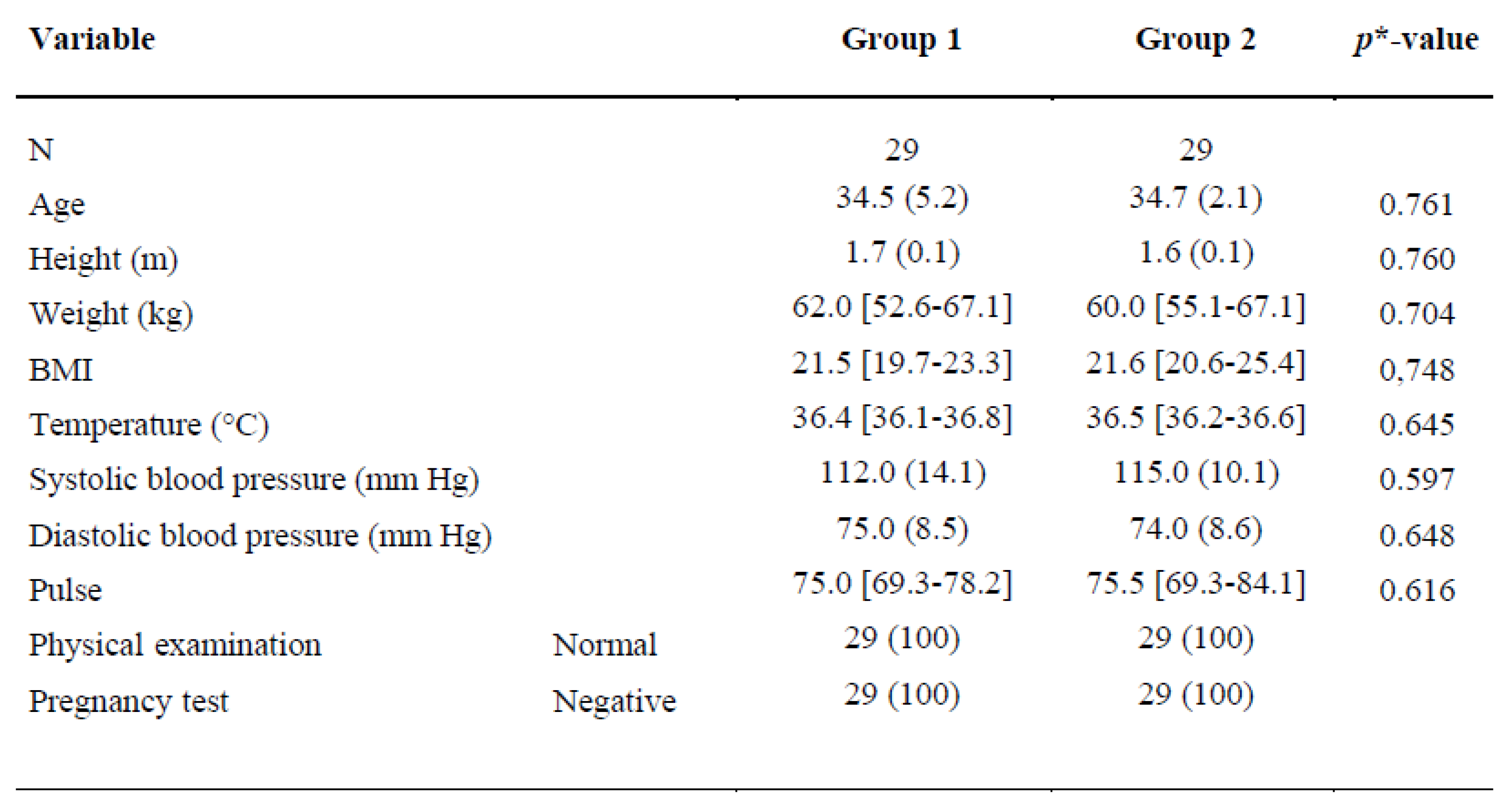 |
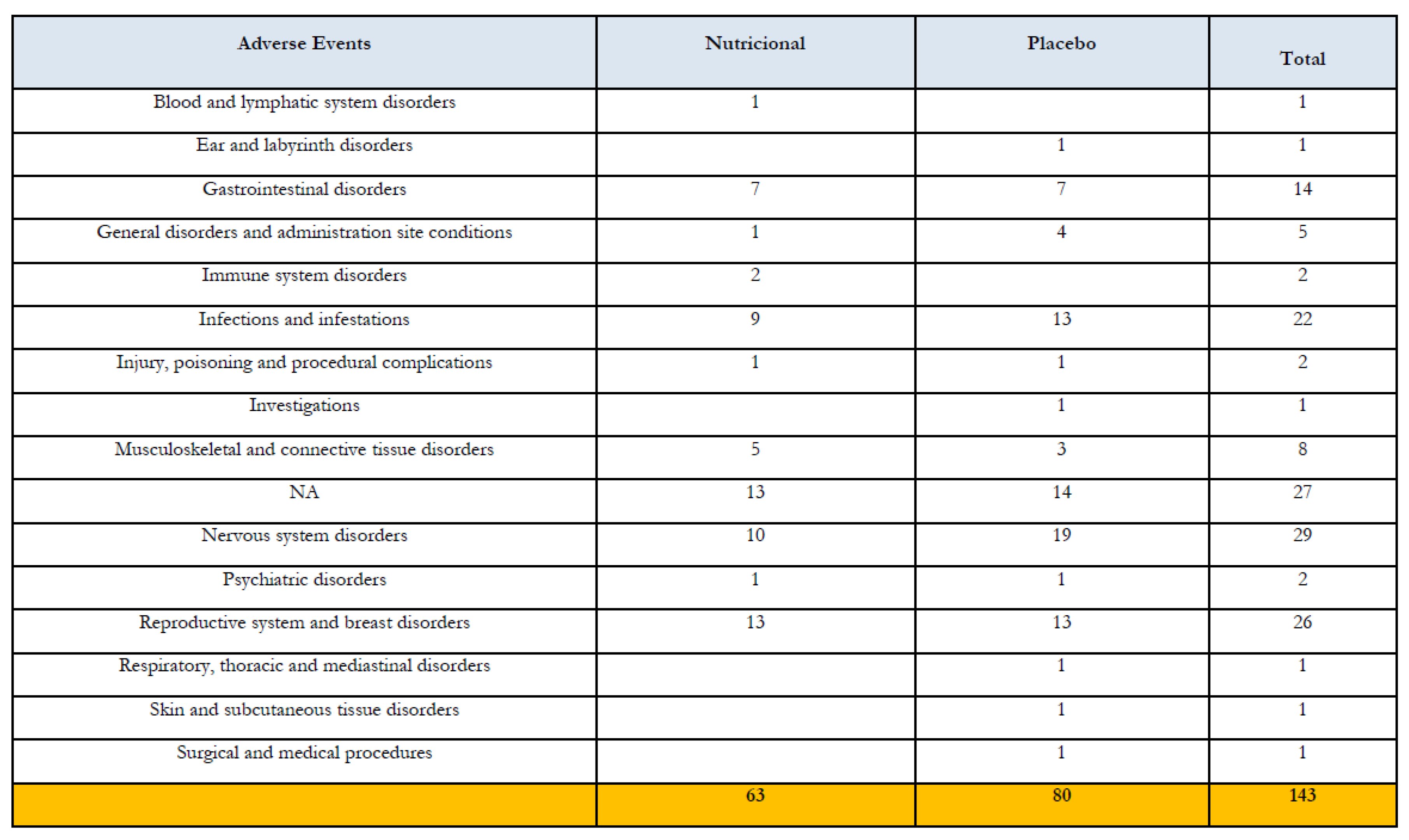
Table 3.
Adverse events classified by diagnostic and treatment group.
 |
Primary endpoints
Uterine bleeding intensity
The change in uterine bleeding intensity from the start to the end of treatment (week 16) was measured using a visual analogue scale (VAS) from 0 to 100, with higher scores indicating more intense bleeding. In the treated group, 14 out of 22 participants (64%) experienced a significant reduction in bleeding between visits 1 and 3 (p < 0.001). In contrast, in the placebo group, 8 out of 23 participants (35%) experienced a reduction, which was not statistically significant (p = 0.717).
These findings suggest the dietary supplement substantially reduces bleeding in patients with uterine fibroids. Figure 1 depicts the data.
Dysmenorrhea
The change in dysmenorrhea from the start to the end of treatment (week 16) was assessed using a visual analogue scale (VAS), with higher scores (0-100) indicating more severe pain.
In the treated group, the median VAS pain scores decreased from 33.0 (69.5) to 22.0 (51.4) after 16 weeks. The reduced interquartile range suggests more consistent pain relief among treated patients. Although not statistically significant, this trend indicates a potential treatment effect and supports further research with larger sample sizes and extended follow-up. assess changes in participants’ quality of life, the SF-36 Health Questionnaire was used, with surveys administered at 8 and 16 weeks. This questionnaire consists of 36 questions from which 8 indicators are extracted: physical function, physical role, bodily pain, vitality, social function, emotional role, mental health, and general health. The values for the 8 SF-36 indicators were calculated, and the total health status score was derived from them. Both the dimensions and total score were normalized for comparison. The results for vitality, emotional role, physical role, and mental health are shown below.
SF-36 results showed a significant improvement in the social dimension over time in the treated group (p = 0.049) and between groups (p = 0.035). This suggests the treatment may positively impact social interaction and well-being, although no significant changes were observed in other quality-of-life domains. Figure 2 depicts the data.
Assessment of sexual function
To assess this variable, the Female Sexual Function Index (FSFI) was used, consisting of 19 questions with a total score ranging from 0 to 36. Based on the scores obtained for each question, six dimensions were calculated to assess desire, arousal, lubrication, orgasm, satisfaction level, and pain during sexual intercourse (FSFI pain). The sum of these dimensions provides the total FSFI value. To compare between groups and individuals, the values of the six dimensions and the total FSFI were normalized and expressed as percentages, using the maximum and minimum values for each data set.
FSFI scores increased significantly (p < 0.001) in 42% of participants (10/24) in the treatment group by the end of the study, compared to 25% (6/24) in the placebo group. These results suggest the supplement improves female sexual function, with the greater effect in the treatment group possibly due to reduced bleeding. Figure 3 depicts the data.
Secondary Objectives
Uterine volume
When comparing both groups no statistically significant difference could be observed. Nevertheless, it is noteworthy that the uterine volume did not increase during the 14 weeks of treatment.
Interesting was that in the nutrient group of women on the individual level 10 patients (50%) showed a decrease in the volume size. Even if this decrease was not significant (p= 0.08) a positive trend could be observed. Only 3 patients experienced an increase in the uterine volume in the nutraceutical group compared to 5 out of 23 (22%) of the patients in the non-treated group. Figure 4 depicts the data.
Hormonal data
Another secondary objective was the effect of the food supplement on the hypothalamic-pituitary-gonadal axis was evaluated by measuring luteinizing hormone (LH), follicle-stimulating hormone (FSH), progesterone (P4), and estradiol.
After analyzing the data, no significant differences were found between the groups (treated vs. placebo) or overtime within each group (p > 0.05). The mean baseline levels were 4.9 pg/mL FSH, 5.6 pg/mL LH, 3.73 pg/mL P4, and 103.7 pg/mL in the case of estradiol. At the end of treatment, these values were 5.0 pg/mL for FSH, 4.85 pg/mL for LH, 2.29 pg/mL for P4, and 111.9 pg/mL for estradiol in the treated group. The values for the control group were: 5.1 pg/mL FSH, 4.65 pg/mL LH, 3.19 pg/mL P4, and 92.9 pg/mL estradiol.
No significant changes in hormone levels were observed at the group or individual level (p > 0.05). These findings indicate the dietary supplement did not significantly affect LH, FSH, P4, or estradiol levels, nor did it alter hormonal regulation of the hypothalamic-pituitary-gonadal axis.
Discussion
Findings and interpretation
This clinical trial is an initial investigation into the effects of vitamin D, EPA, DHA, and their monohydroxylated metabolites (SPMs) in women with uterine fibroids.
The primary outcome was a reduction in bleeding volume and dysmenorrhea, rather than tumor size. Improvements in general health and sexual function were also observed.
This phenomenon may result from the general mode of action of Omega-3 compounds, their active metabolites, and Vitamin D3. The pathogenesis of uterine fibroids remains unclear, but cytokine, growth factor, and steroid hormone signaling, along with inflammatory processes, may be involved. Vitamin D acts on human reproduction not only through calcium homeostasis but also as a direct regulator of aromatase gene expression. Evidence suggests that VDR-bound 1,25(OH)2D3 acts as a transcription factor to regulate the CYP19 gene, which encodes the aromatase enzyme in fat and ovarian tissues. This mechanism regulates estrogen [64]. Vitamin D can also stimulate the production of progesterone, various estrogens, and insulin-like growth factor-binding protein 1 (IGFBP-1), especially in cultured human ovarian cells. Together, these modes of vitamin D influence hormone-dependent benign tissues, such as uterine fibroids, by modulating growth and differentiation [65].
Other estrogen-dependent diseases, like endometriosis, seem to be modulated by Vitamin D. Activated CD4 and CD8 cells express vitamin D receptors and the enzymes 1α-hydroxylase and 24-hydroxylase. This induces local vitamin D production and negatively affects the potential for regression of endometriosis implants. In a large prospective cohort study, high plasma 25(OH)D was associated with a lower risk of endometriosis [66]. Vitamin D also inhibited ER and PR expression in a dose-dependent manner in leiomyoma cells [67]. In recent years, active resolution of inflammation, mainly driven by specialized pro-resolving lipid mediator molecules (SPMs), has been shown to be key to ending acute and chronic inflammation, as seen in diseases such as periodontitis, atherosclerosis, and diabetes.
Results in the context of what is known and clinical implications
The roles of SPMs and their influence on uterine myoma growth still need to be clarified. However, in a subset of women with erythrocyte FA measurements, lower odds of fibroids were seen among those with higher n-3 PUFA erythrocyte levels and higher odds among those with higher trans FA levels, suggesting n-3 PUFAs and trans FAs may be associated with fibroids [62]. In a preliminary study (unpublished data), a reduction in bleeding-related pain was also observed [68]. While these findings are promising, it is important to acknowledge that alternative explanations, such as placebo effects or regression to the mean, may have contributed to the observed improvements. Further research with larger sample sizes and additional controls is warranted to confirm these results.
Fibrosis, a characteristic feature of uterine leiomyoma, may result from inadequate resolution of inflammation. In pulmonary fibrosis, experimental LX4 Analogues (AT-LX4) reduced fibrosis in animal models [62]. LXA4 and benzo-LXA4, an analogue, also reduced fibrotic changes in the kidney in a rat model of early renal fibrosis [63], and resolvin RvE1 showed antifibrotic effects in a mouse model of obstructed kidney [64]. Although the role of SPMs in UF development is not yet clear, there are similarities with other chronic inflammatory diseases where inadequate resolution has been shown.
Research Implications
Further research may lead to new treatment options. In summary, inflammation always begins with its active resolution, as alpha signals omega in the signaling cascade [53].
No significant changes in estradiol levels were observed in either group, indicating that the effect is likely local and that the hypothalamic axis remains unaffected by the supplement. This suggests a favorable safety profile, as the product does not cause the hypoestrogenic side effects associated with some approved medications for uterine fibroids.
Strengths and Limitations
The main strength of this formulation it is non hormonal approach meaning that also women with contraindications to use of hormones may benefit by this product.
A notable limitation is the lack of clinical formulations containing vitamin D and omega-3 fatty acid derivatives for managing uterine fibroids. Further research on inflammation and SPMs may provide valuable insights into this condition.
Conclusions
The nutritional containing Omega 3 fatty acid, SPMs, vitamin D and B6 represent a valid option for the symptomatic management of patients with uterine fibroids without comprising the hypothalamic pituitary axis of the women. Further research in grater groups pf patients is needed to validate these first results.
Declaration Section
Ethics:
This randomized, double-blind, placebo-controlled trial was approved by the Research Ethics Committee of the HM Hospitales Madrid, Spain, Código CEIm HM Hospitales: 24.04.1985E5-GHM (approval date: 18/04/2024) and conducted in accordance with the Declaration of Helsinki.
Written informed consent to participate in the trial was obtained from all participants.
Author Contributions
Pedro-Antonio Regidor: Responsible for the concept of resolution of inflammation in uterine myoma. Responsible for writing the manuscript; Belen Orgaz: Responsible for statistical evaluation and graphic design; Santiago Palacios Gil-Acuna: Study investigator ; Manuel Marcos Fernández: Study inverstigator; Rodrigo Orozco Fernández. Study invetigator; Isabel Blanco Herráez. Study investigator; Luciana Bergamaschi Santa Cruz: Study investigator; Josué Jara Perez: Responsible for data curation; Beatriz Lazcoz Calvo: Responsible for scientific background; Rocio Gutierrez: Responsible for literature research and writing; Jose Miguel Rizo: Responsible for funding and data acquisition; Miguel Ángel Rodríguez Zambrano. Principal invetigator.
Funding
The trial was funded by Laboratorios Liconsa S.A., a subsidiary of Insud Pharma. The sponsor had no role in data collection, analysis, interpretation, or the decision to submit this work for publication.
Institutional Review Board Statement
Clinical trial registration number: ISRCTN69006251 https://doi.org/10.1186/ISRCTN69006251. Registration date: 18/02/2026; Informed Consent Statement: All patients were informed of the study characteristics, approved to participate, and signed an informed consent statement.
Informed Consent Statement
Not applicable.
Data Availability Statement
On request to the corresponding author.
Acknowledgments
Not applicable.
Conflicts of Interest
Legends.
References
- Islam, S.; Ciavattini, A.; Petraglia, F.; Castellucci, M.; Ciarmela, P. Extracellular matrix in uterine leiomyoma pathogenesis: A potential target of future therapeutics. Hum. Reprod. Update 2018, 24, 59–85. [Google Scholar] [CrossRef] [PubMed]
- Buttram, V.C., Jr.; Reiter, R.C. Uterine leiomyomata: Etiology, symptomatology, and management. Fertil. Steril. 1981, 36, 433–45. [Google Scholar] [CrossRef]
- Cramer, S.F.; Patel, A. The frequency of uterine leiomyomas. Am. J. Clin. Pathol. 1990, 94, 435–8. [Google Scholar] [CrossRef] [PubMed]
- Marshall, L.M.; Spiegelman, D.; Barbieri, R.L.; Goldman, M.B.; Manson, J.E.; Colditz, G.A.; Willett, W.C.; Hunter, D.J. Variation in the incidence of uterine leiomyoma among premenopausal women by age and race. Obstet. Gynecol. 1997, 90, 967–73. [Google Scholar] [CrossRef]
- Baird, D.D.; Dunson, D.B.; Hill, M.C.; Cousins, D.; Schectman, J.M. High cumulative incidence of uterine leiomyoma in black and white women: Ultrasound evidence. Am. J. Obstet. Gynecol. 2003, 188, 100–7. [Google Scholar] [CrossRef]
- Kjerulff, K.H.; Langenberg, P.; Seidman, J.D.; Stolley, P.D.; Guzinski, G.M. Uterine leiomyomas. Racial differences in severity, symptoms and age at diagnosis. J. Reprod. Med. 1996, 41, 483–90. [Google Scholar]
- Szydłowska, I.; Grabowska, M.; Nawrocka-Rutkowska, J.; Kram, A.; Piasecka, M.; Starczewski, A. Markers of Inflammation and Vascular Parameters in Selective Progesterone Receptor Modulator (Ulipristal Acetate)-Treated Uterine Fibroids. J. Clin. Med. 2021, 10. [Google Scholar] [CrossRef]
- Goad, J.; Rudolph, J.; Wei, J.-J.; Bulun, S.E.; Chakravarti, D.; Rajkovic, A. Single Cell atlas of uterine myometrium and leiomyomas reveals diverse and novel cell types of non-monoclonal origin. bioRxiv 2020, 2020, P. [Google Scholar] [CrossRef]
- Tal, R.; Segars, J.H. The role of angiogenic factors in fibroid pathogenesis: Potential implications for future therapy. Hum. Reprod. Update 2014, 20, 194–216. [Google Scholar] [CrossRef]
- Ciavattini, A.; Di Giuseppe, J.; Stortoni, P.; Montik, N.; Giannubilo, S.R.; Litta, P.; Islam, S.; Tranquilli, A.L.; Reis, F.M.; Ciarmela, P. Uterine fibroids: Pathogenesis and interactions with endometrium and endomyometrial junction. Obstet. Gynecol. Int. 2013, 2013, 173184. [Google Scholar] [CrossRef] [PubMed]
- Galindo, L.J.; Hernández-Beeftink, T.; Salas, A.; Jung, Y.; Reyes, R.; Montes de Oca, F.; Hernández, M.; Almeida, T.A. HMGA2 and MED12 alterations frequently co-occur in uterine leiomyomas. Gynecol. Oncol. 2018, 150, 562–568. [Google Scholar] [CrossRef] [PubMed]
- Grings, A.O.; Lora, V.; Ferreira, G.D.; Brum, I.S.; von Eye Corleta, H.; Capp, E. Protein expression of estrogen receptors alpha and beta and aromatase in myometrium and uterine leiomyoma. Gynecol. Obstet. Invest 2012, 73, 113–7. [Google Scholar] [CrossRef]
- Ishikawa, H.; Ishi, K.; Serna, V.A.; Kakazu, R.; Bulun, S.E.; Kurita, T. Progesterone is essential for maintenance and growth of uterine leiomyoma. Endocrinology 2010, 151, 2433–42. [Google Scholar] [CrossRef]
- Kawaguchi, K.; Fujii, S.; Konishi, I.; Nanbu, Y.; Nonogaki, H.; Mori, T. Mitotic activity in uterine leiomyomas during the menstrual cycle. Am. J. Obstet. Gynecol. 1989, 160, P. 637–41. [Google Scholar] [CrossRef]
- Islam, M.S.; Protic, O.; Stortoni, P.; Grechi, G.; Lamanna, P.; Petraglia, F.; Castellucci, M.; Ciarmela, P. Complex networks of multiple factors in the pathogenesis of uterine leiomyoma. Fertil. Steril. 2013, 100, P. 178–93. [Google Scholar] [CrossRef]
- Modugno, F.; Ness, R.B.; Chen, C.; Weiss, N.S. Inflammation and endometrial cancer: A hypothesis. Cancer Epidemiol. Biomark. Prev. 2005, 14, 2840–7. [Google Scholar] [CrossRef]
- Orciani, M.; Caffarini, M.; Biagini, A.; Lucarini, G.; Carpini, G.D.; Berretta, A.; Primio, R.D.; Ciavattini, A. Chronic Inflammation May Enhance Leiomyoma Development by the Involvement of Progenitor Cells. Stem Cells Int. 2018, 2018, P. 1716246. [Google Scholar] [CrossRef] [PubMed]
- Protic, O.; Toti, P.; Islam, M.S.; Occhini, R.; Giannubilo, S.R.; Catherino, W.H.; Cinti, S.; Petraglia, F.; Ciavattini, A.; Castellucci, M.; et al. Possible involvement of inflammatory/reparative processes in the development of uterine fibroids. Cell Tissue Res. 2016, 364, 415–27. [Google Scholar] [CrossRef] [PubMed]
- Macdiarmid, F.; Wang, D.; Duncan, L.J.; Purohit, A.; Ghilchick, M.W.; Reed, M.J. Stimulation of aromatase activity in breast fibroblasts by tumor necrosis factor alpha. Mol. Cell Endocrinol. 1994, 106, P. 17–21. [Google Scholar] [CrossRef]
- Purohit, A.; Newman, S.P.; Reed, M.J. The role of cytokines in regulating estrogen synthesis: Implications for the etiology of breast cancer. Breast Cancer Res. 2002, 4, P. 65–9. [Google Scholar] [CrossRef]
- Leppert, P.C.; Al-Hendy, A.; Baird, D.D.; Bulun, S.; Catherino, W.; Dixon, D.; Ducharme, M.; Harmon, Q.E.; Jayes, F.L.; Paul, E.; et al. Summary of the Proceedings of the Basic Science of Uterine Fibroids Meeting: New Developments February 28, 2020. F. S Sci. 2021, 2, P. 88–100. [Google Scholar] [CrossRef]
- Ciebiera, M.; Ali, M.; Prince, L.; Jackson-Bey, T.; Atabiekov, I.; Zgliczyński, S.; Al-Hendy, A. The Evolving Role of Natural Compounds in the Medical Treatment of Uterine Fibroids. J. Clin. Med. 2020, 9. [Google Scholar] [CrossRef]
- Zannotti, A.; Greco, S.; Pellegrino, P.; Giantomassi, F.; Carpini, G.D.; Goteri, G.; Ciavattini, A.; Ciarmela, P. Macrophages and Immune Responses in Uterine Fibroids. Cells 2021, 10. [Google Scholar] [CrossRef]
- Lima, M.S.O.; Borges da Silva, B.; Lima de Medeiros, M.; Ribeiro Dos Santos, A.; Davi do Nascimento Brazil, E.; Eulálio Filho, W.M.N.; Ibiapina, J.O.; Albuquerque Brito, A.G.; Lopes Costa, P.V. Evaluation of vitamin D receptor expression in uterine leiomyoma and nonneoplastic myometrial tissue: A cross-sectional controlled study. Reprod. Biol. Endocrinol. 2021, 19, P. 67. [Google Scholar] [CrossRef]
- Halder, S.K.; Goodwin, J.S.; Al-Hendy, A. 1,25-Dihydroxyvitamin D3 reduces TGF-beta3-induced fibrosis-related gene expression in human uterine leiomyoma cells. J. Clin. Endocrinol. Metab. 2011, 96, P. E754–62. [Google Scholar] [CrossRef] [PubMed]
- Halder, S.K.; Osteen, K.G.; Al-Hendy, A. 1,25-dihydroxyvitamin d3 reduces extracellular matrix-associated protein expression in human uterine fibroid cells. Biol. Reprod. 2013, 89, P. 150. [Google Scholar] [CrossRef] [PubMed]
- Al-Hendy, A.; Diamond, M.P.; El-Sohemy, A.; Halder, S.K. 1,25-dihydroxyvitamin D3 regulates expression of sex steroid receptors in human uterine fibroid cells. J. Clin. Endocrinol. Metab. 2015, 100, P. E572–82. [Google Scholar] [CrossRef]
- Paffoni, A.; Somigliana, E.; Vigano, P.; Benaglia, L.; Cardellicchio, L.; Pagliardini, L.; Papaleo, E.; Candiani, M.; Fedele, L. Vitamin D status in women with uterine leiomyomas. J. Clin. Endocrinol. Metab. 2013, 98, P. E1374–8. [Google Scholar] [CrossRef] [PubMed]
- Baird, D.D.; Hill, M.C.; Schectman, J.M.; Hollis, B.W. Vitamin d and the risk of uterine fibroids. Epidemiology 2013, 24, P. 447–53. [Google Scholar] [CrossRef]
- Sabry, M.; Halder, S.K.; Allah, A.S.; Roshdy, E.; Rajaratnam, V.; Al-Hendy, A. Serum vitamin D3 level inversely correlates with uterine fibroid volume in different ethnic groups: A cross-sectional observational study. Int. J. Womens Health 2013, 5, P. 93–100. [Google Scholar] [CrossRef]
- Hodge, J.C.; Morton, C.C. Genetic heterogeneity among uterine leiomyomata: Insights into malignant progression. Hum. Mol. Genet. 2007, 16 Spec No 1, R7–13. [Google Scholar] [CrossRef] [PubMed]
- Wise, L.A.; Ruiz-Narváez, E.A.; Haddad, S.A.; Rosenberg, L.; Palmer, J. R. Polymorphisms in vitamin D-related genes and risk of uterine leiomyomata. Fertil. Steril. 2014, 102, P, 503–510. [Google Scholar] [CrossRef]
- Shahbazi, S. Exploring the link between VDR rs2228570 and uterine leiomyoma in Iranian women. Egypt J. Med. Hum. Genet 2016, 17, P. 115–118. [Google Scholar] [CrossRef]
- Yılmaz, G.S.; Gül, T.; Attar, R.; Yıldırım, G.; İşbir, T. Association between fok1 polymorphism of vitamin D receptor gene with uterine leiomyoma in Turkish populations. J. Turk. Ger. Gynecol. Assoc. 2018, 19, P, 128–131. [Google Scholar] [CrossRef]
- Ciebiera, M.; Wrzosek, M.; Wojtyła, C.; Zaręba, K.; Nowicka, G.; Jakiel, G.; Włodarczyk, M. Vitamin D receptor gene polymorphisms and uterine fibroid incidence in Caucasian women. Arch. Med. Sci. 2019, 17 (6), p, 1643–1650. [Google Scholar] [CrossRef]
- Upadhyay, S.; Dubey, P.K. Gene variants polymorphisms and uterine leiomyoma: An updated review. Front Genet 2024, 15, 1330807. [Google Scholar] [CrossRef]
- Qu, X.; Zhang, X.; Yao, J.; Song, J.; Nikolic-Paterson, D.J.; Li, J. Resolvins E1 and D1 inhibit interstitial fibrosis in the obstructed kidney via inhibition of local fibroblast proliferation. J. Pathol. 2012, 228, P. 506–19. [Google Scholar] [CrossRef]
- Lagana, A.S.; Vitale, S.G.; Ban Frangez, H.; Vrtacnik-Bokal, E.; D’Anna, R. Vitamin D in human reproduction: The more, the better? An evidence-based critical appraisal. Eur. Rev. Med. Pharmacol. Sci. 2017, 21, 4243–4251. [Google Scholar]
- Colonese, F.; Laganà, A.S.; Colonese, E.; Sofo, V.; Salmeri, F.M.; Granese, R.; Triolo, O. Thepleiotropic Effects of Vitamin D in Gynaecological and Obstetric Diseases: An Overview on a Hot Topic. BioMed Res. Int. 2015, Article ID 986281, 11 pages. [Google Scholar] [CrossRef]
- Vergara, D.; Catherino, W.H.; Trojano, G.; Tinelli, A. Vitamin D: Mechanism of Action and Biological Effects in Uterine Fibroids. Nutrients 2021, 13, 597. [Google Scholar] [CrossRef] [PubMed]
- Martins, V.; Valença, S.S.; Farias-Filho, F.A.; Molinaro, R.; Simões, R.L.; Ferreira, T.P.T.; Silva, P.M.R.; Hogaboam, C.M.; Kunkel, S.L.; Fierro, I.M.; et al. ATLa, an aspirin-triggered lipoxin A4 synthetic analog, prevents the inflammatory and fibrotic effects of bleomycin-induced pulmonary fibrosis. J. Immunol. 2009, 182, 5374–81. [Google Scholar] [CrossRef] [PubMed]
- Harris, H.R.; Eliassen, A.H.; Doody, D.R.; Terry, K.L.; Missmer, S.A. Dietary fat intake, erythrocyte fatty acids, and risk of uterine fibroids. Fertil. Steril. 2020, 114, 837–847. [Google Scholar] [CrossRef]
- Börgeson, E.; Docherty, N.G.; Murphy, M.; Rodgers, K.; Ryan, A.; O’Sullivan, T.P.; Guiry, P.J.; Goldschmeding, R.; Higgins, D.F.; Godson, C. Lipoxin A (4) and benzo-lipoxin A (4) attenuate experimental renal fibrosis. FASEB J. 2011, 25, 2967–79. [Google Scholar] [CrossRef] [PubMed]
- Serhan, C.N.; Savill, J. Resolution of inflammation: The beginning programs the end. Nat. Immunol. 2005, 6, P. 1191–7. [Google Scholar] [CrossRef]
Figure 1.
Individual effect of dietary supplement intake on the intensity of uterine bleeding assessed using a visual analogue scale (0-100 points). The data corresponds to the values from the three visits for each of the participants in the treatment group (A) (n = 22) and placebo group (P) (n = 23). Only participants with complete data were included in the statistical analysis. The p-value indicated in the graph shows statistically significant differences (p < 0.05) between the mean values of the same variable over time (ANOVA test for repeated measures).
Figure 1.
Individual effect of dietary supplement intake on the intensity of uterine bleeding assessed using a visual analogue scale (0-100 points). The data corresponds to the values from the three visits for each of the participants in the treatment group (A) (n = 22) and placebo group (P) (n = 23). Only participants with complete data were included in the statistical analysis. The p-value indicated in the graph shows statistically significant differences (p < 0.05) between the mean values of the same variable over time (ANOVA test for repeated measures).
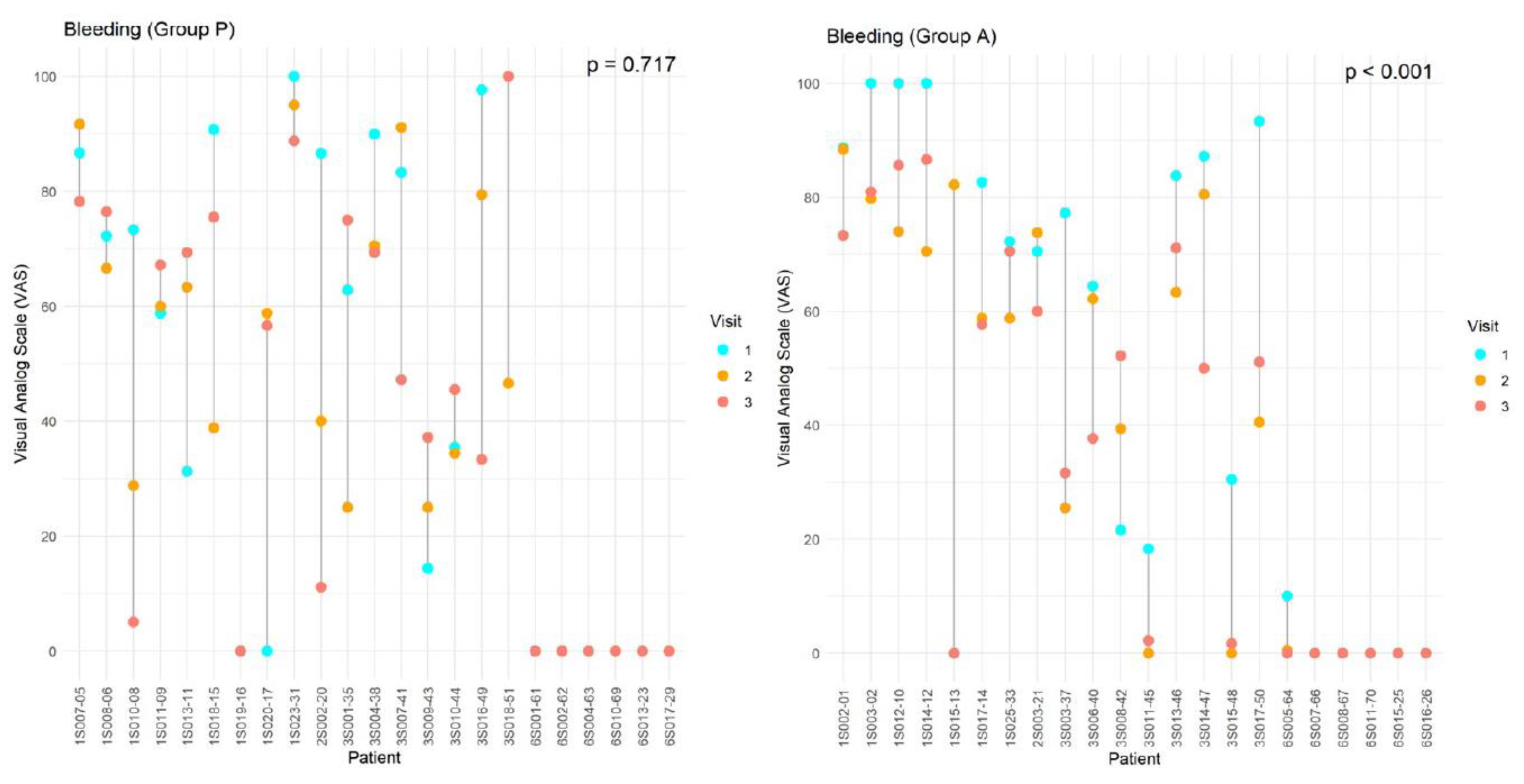
Figure 2.
Individual effect of dietary supplement intake on pain intensity of dysmenorrhea using a visual analogue scale (0-100 points). The data corresponds to the values from the three visits for each of the participants in the treatment group (A) (n = 22) and placebo group (P) (n = 23). Participants with complete data were included in the statistical analysis. The p-value indicated in the graph shows statistically significant differences (p < 0.05) between the mean values of the same variable over time (ANOVA test for repeated measures).
Figure 2.
Individual effect of dietary supplement intake on pain intensity of dysmenorrhea using a visual analogue scale (0-100 points). The data corresponds to the values from the three visits for each of the participants in the treatment group (A) (n = 22) and placebo group (P) (n = 23). Participants with complete data were included in the statistical analysis. The p-value indicated in the graph shows statistically significant differences (p < 0.05) between the mean values of the same variable over time (ANOVA test for repeated measures).
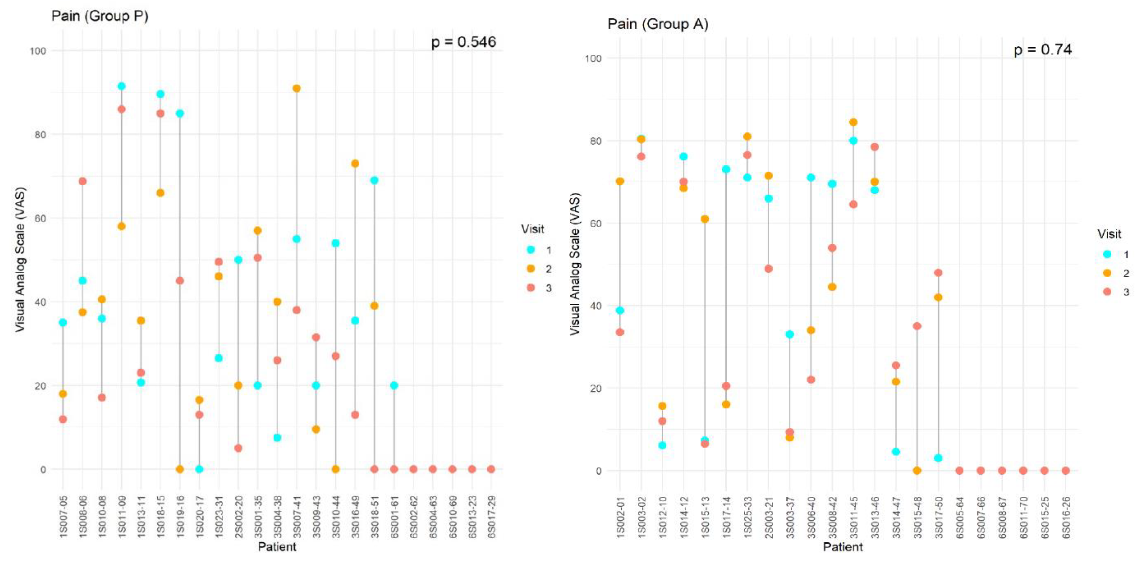
Figure 3.
Effect of dietary supplement intake on total sexual function as assessed by the Female Sexual Function Index (FSFI). The data corresponds to the values of the three visits for each of the participants in the treated group (A) (n = 24) and placebo group (P) (n = 24). Participants with complete data were included in the statistical analysis. The p-value indicated in the graph shows statistically significant differences (p < 0.05) between the mean values of the same variable over time (ANOVA test for repeated measures).
Figure 3.
Effect of dietary supplement intake on total sexual function as assessed by the Female Sexual Function Index (FSFI). The data corresponds to the values of the three visits for each of the participants in the treated group (A) (n = 24) and placebo group (P) (n = 24). Participants with complete data were included in the statistical analysis. The p-value indicated in the graph shows statistically significant differences (p < 0.05) between the mean values of the same variable over time (ANOVA test for repeated measures).
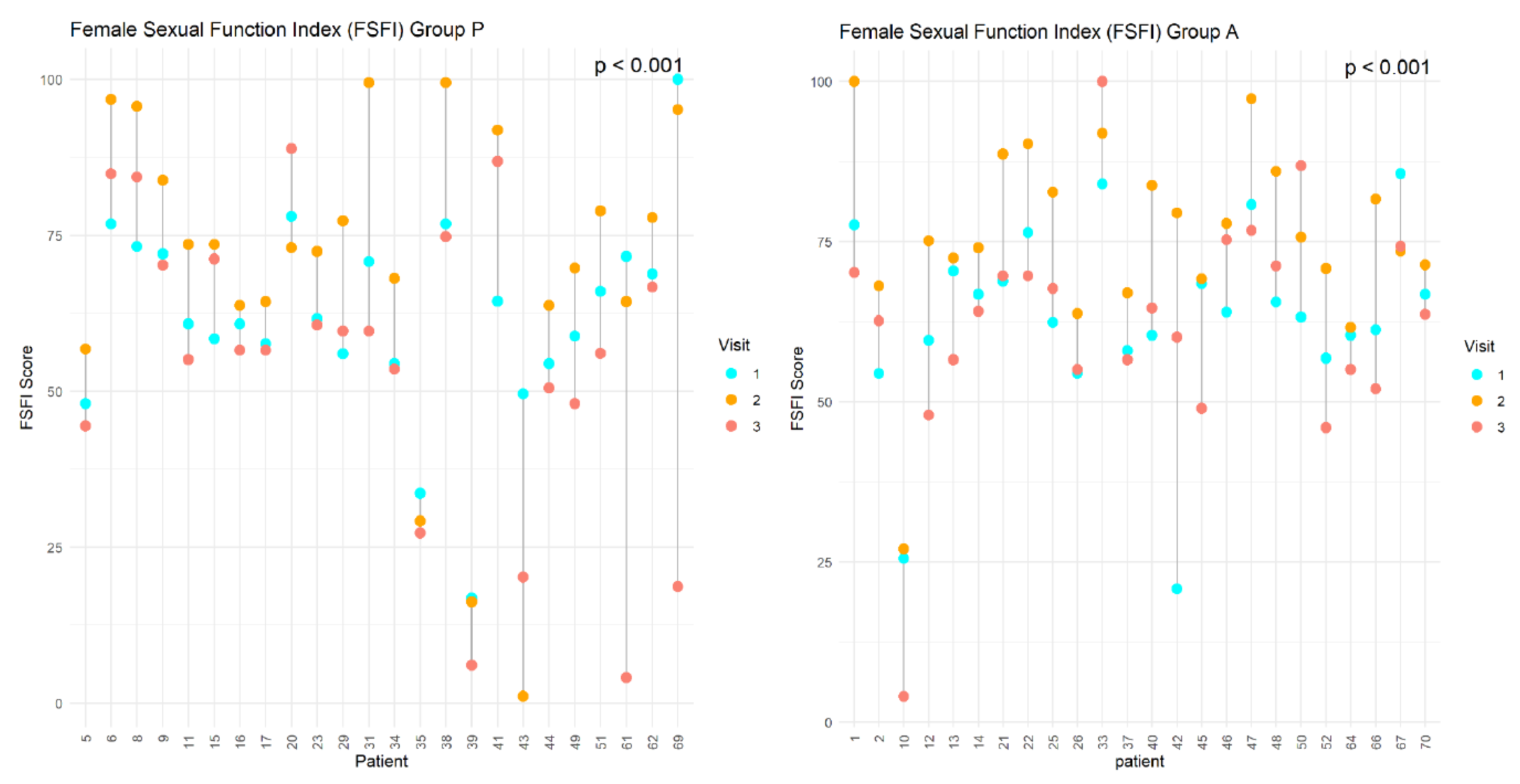
Figure 4.
Effect of dietary supplement intake on fibroid volume (cm3). The data corresponds to the values from the three visits for each of the participants in the treatment group (A) (n = 22) and placebo group (P) (n = 23). Only participants with complete data were included in the statistical analysis. The p-value indicated in the graph shows statistically significant differences (p < 0.05) between the mean values of the same variable over time (ANOVA test for repeated measures).
Figure 4.
Effect of dietary supplement intake on fibroid volume (cm3). The data corresponds to the values from the three visits for each of the participants in the treatment group (A) (n = 22) and placebo group (P) (n = 23). Only participants with complete data were included in the statistical analysis. The p-value indicated in the graph shows statistically significant differences (p < 0.05) between the mean values of the same variable over time (ANOVA test for repeated measures).
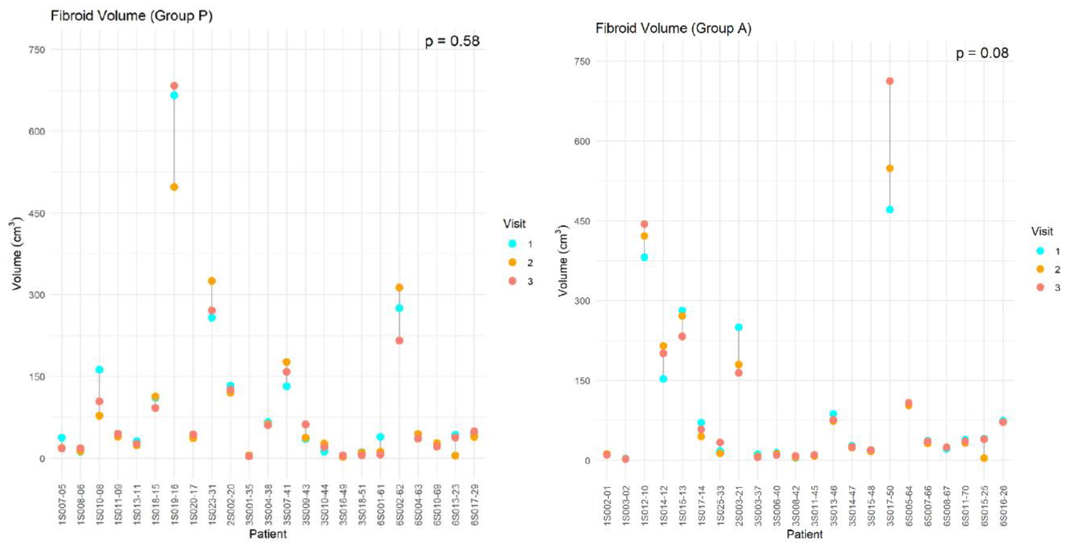
Table 1.
Content of the nutritional formulation.
 |
Disclaimer/Publisher’s Note: The statements, opinions and data contained in all publications are solely those of the individual author(s) and contributor(s) and not of MDPI and/or the editor(s). MDPI and/or the editor(s) disclaim responsibility for any injury to people or property resulting from any ideas, methods, instructions or products referred to in the content. |
© 2026 by the authors. Licensee MDPI, Basel, Switzerland. This article is an open access article distributed under the terms and conditions of the Creative Commons Attribution (CC BY) license (http://creativecommons.org/licenses/by/4.0/).
Copyright: This open access article is published under a Creative Commons CC BY 4.0 license, which permit the free download, distribution, and reuse, provided that the author and preprint are cited in any reuse.


