

Submitted:
01 June 2026
Posted:
03 June 2026
You are already at the latest version
Abstract
Mechanisms for maintenance of chronic viruses are poorly understood, particularly for archaeal viruses. Here, we identify the product of Sulfolobus spindle-shaped virus 1 (SSV1) ORF a291 as a virally-encoded toxin required for growth inhibition but dispensable for viral replication and virion production. Viruses lacking ORF a291 replicated their genomes and formed morphologically normal spindle-shaped particles yet failed to inhibit growth of uninfected Saccharolobus solfataricus. Substitution of residues at a predicted N-terminal signal peptide cleavage site abolished growth suppression without affecting replication, suggesting that secretion is essential for toxin function. Despite primary sequence divergence among fusellovirus toxin candidates, analysis of protein structure predictions revealed a conserved hydrolase-like fold across SSV1, SSV9 and SSV10 toxins. These findings demonstrate functional separation of viral replication and host growth suppression and support a model in which chronic archaeal viruses modulate host competition through antagonistic factors. This work expands the known diversity of viral toxins and suggests that fuselloviruses employ conserved strategies to promote persistence in extreme environments. Impact Statement: This work identifies a virally-encoded toxin, the product of ORF a291, in Sulfolobus spindle-shaped virus 1. Unlike most toxins, this toxin appears to be secreted and only affects uninfected cells. Our findings expand the known diversity of viral toxins and suggest that Fuselloviruses broadly employ these mechanisms to promote their persistence in extreme environments.
Keywords:
viral toxin
; archaea
; archaeal viruses
; extremophiles
; toxin-antitoxin systems
1. Introduction
Viruses are extremely important drivers of microbial evolution and can shape ecological and biogeochemical processes through influencing host fitness, population dynamics, and community structures (reviewed in [1,2,3,4]). In many well-studied systems, viruses exert these effects primarily through host cell lysis, thereby reducing host density and releasing cellular contents into the environment, to be recycled within the system (reviewed in [3,4]). This lytic paradigm has strongly influenced how viral activity in microbial populations are conceptualized [5]. However, ecological models describing virus-host interactions, particularly for lysogenic and chronic viruses, now extend beyond classical lytic Kill-the-Winner dynamics, where the most abundant hosts are killed, to include Piggyback-the-Winner models, in which viruses preferentially persist in abundant hosts; Kill-the-Competitor strategies, where viral activity suppresses competing host populations; and arms-race models where host defense mechanisms compete with viral anti-defense mechanisms, reflecting the diverse strategies by which viruses’ structure microbial populations (reviewed in [6]). However, not all virus infections conform to these models [7,8].
In contrast to lytic viruses, many archaeal viruses establish chronic infections that do not result in immediate host cell death [7,9]. These viruses often exit their hosts through budding or extrusion, allowing infected cells to survive and continue dividing while maintaining viral genomes as integrated and/or episomal DNA [9,10]. These infection strategies raise a fundamental ecological question: Can chronic viruses influence host population structure and competition if they do not reduce host density through lysis? In some ecological frameworks, temperate or chronic viral strategies may function analogously to Piggyback-the-Winner or Kill-the-Competitor dynamics, in which viral activity reshapes host competition without necessarily causing widespread lysis [6]. Geothermal environments where archaeal populations and their chronic viruses often dominate microbial communities [8,9,10,11], are excellent systems to study these interactions. Recently, ecosystem structuring via virus-mediated cellular antagonism has been proposed for Sulfolobus islandicus and Fusellovirus SSV9 [8,12].
Sulfolobus spindle shaped virus-1 (SSV1) is an extremophilic virus [13,14,15] that infects Saccharolobus (previously Sulfolobus) solfataricus, a hyper-thermophilic acidophile that thrives in geothermal hot springs worldwide [16]. Optimal growth conditions for Saccharolobus range from 65 °C to 80 °C and a pH of 4-3.5 [16,17]. SSV1, like all fuselloviruses, has a spindle-shaped virion, a morphology characteristic of many archaeal viruses [9,10], and buds from the host cell without killing it [10]. SSV1 has a double-stranded DNA circular genome 15.6 kbp in size [18], and unlike almost all extremophilic viruses, is genetically tractable [14,19]. Upon infection with SSV1, a copy of the virus genome integrates into the host genome [15], and about five copies of the viral genome are present per cell as episomal DNA at a given time [20]. Fusellovirus infection is widespread in Sulfolobus/Saccharolobus. Recently the related fusellovirus SSV9 was proposed to provide a selective advantage to infected cells via expression of a virus-encoded toxin, the product of SSV9 ORF b310 [21].
Here we investigate the product of SSV1 ORF a291, encoded upstream of the SSV1 capsid tail protein VP4 (Figure 1) in the genetically tractable SSV1 model. We tested whether SSV1 viruses lacking a functional ORF a291 could replicate their genomes, form virus particles or inhibit host growth, as measured by the formation of zones of clearing (halo assays) on lawns of uninfected cells. Taking together our experimental observations and additional sequence and structural analysis of ORF a291, we hypothesize that SSV1 ORF a291 encodes a secreted hydrolase-like toxin that enables infected cells to suppress the growth of uninfected neighbors, promoting SSV1 persistence in Saccharolobus populations in extreme environments.
2. Materials and Methods
2.1. Strains, Media, and Culture Conditions
All experiments were performed using Saccharolobus solfataricus strain S441 [22]. Cultures were maintained under aerobic conditions at 80 °C in 1x Yeast Sucrose (YS) media (pH 3.5) [15,19]. Liquid cultures were incubated in air with shaking at 150 rpm unless otherwise indicated. Semi-solid plates were made by mixing YS at 2x concentration with molten 1.4% (w/v in water) Gelrite® (Sigma-Aldrich) and pouring immediately. Plates were allowed to solidify at room temperature overnight prior to use. For transformation of S441, cells were prepared and electroporated as previously described [15,19]. Cell- free supernatants were prepared from liquid cultures at an OD600 of 0.8 and were centrifuged at 7,000 x g for 10 minutes at ambient temperature to pellet cellular debris. Supernatants were subsequently filtered through sterile 0.2 µm polyethersulfone (PES) syringe filters (MilliporeSigma) to remove remaining cells and debris. Filtered supernatants were either used immediately for halo assays or stored at 4 °C for future use. For viral passaging experiments, 0.5 to 1 ml of mid-log-phase culture (uninfected or infected) S. solfataricus S441 was diluted 1:10 in fresh 1x YS media and incubated at 80 °C with shaking. After 48 hours, the same dilution was performed. Three sequential passages were performed to assess viral maintenance.
2.2. Virus Constructs Used and Mutagenesis
Previously described SSV1 Tn5 insertion mutants [19] were grown from laboratory stocks. Targeted deletions of SSV1 ORFs a291 and c124 were constructed in the stand-in wild type EAI283 shuttle vector (SWT), which contains a stable Tn5 insertion [19]. Deletions were generated by inverse PCR amplification (LIPCR [19]) of the entire plasmid backbone (10-50 ng of template), excluding the desired open reading frame. Primers were designed flanking SSV1 ORFs a291 or c124 to allow for blunt-end ligation (Table S1). PCR amplification was performed using RepliQa HiFi DNA polymerase (Quantabio) under manufacturer-recommended conditions. 1 µL of amplified products were treated with a Kinase-Ligase-DpnI mixture (KLD; unpublished data) for 1 hour at 37 °C with 1 µl of 1U DpnI (New England Biolabs) to digest methylated template DNA, phosphorylation with 1 µl 1U T4 DNA kinase, 1 µl of 1U DNA ligase and 1 µl of the accompanying DNA ligase buffer (10X) (New England Biolabs) and ddH2O to a final volume of 10 µl. Ligated products were transformed into chemically competent E. coli Pir+ cells (Epicentre) using a standard heat-shock transformation protocol. Briefly, 2-5 µl of KLD reaction was added to 50 µl competent cells and incubated on ice for 30 minutes, followed by heat shock at 42 °C for 30 seconds. Cells were recovered in SOC medium for 1 hour at 37 °C with shaking prior to plating on selective LB agar. Plasmids were purified using a GeneJET Plasmid Miniprep Kit (Thermo Scientific) and verified by Sanger sequencing (Eurofins Genomics) prior to transformation into S. solfataricus. Site-directed point substitutions at the predicted signal peptide cleavage site of SSV1 ORF a291 (A25L/L26A) were introduced by LIPCR [19]. Primers used are indicated in supplemental material (Table S1).
2.3. Halo Assays
To assess growth inhibition phenotypes (halo formation), 3 µl aliquots of cell-free supernatants from transformed S. solfataricus S441 cultures were spotted onto lawns of uninfected S. solfataricus S441 embedded in semi-solid YS medium. Lawns were prepared by mixing mid-log-phase (OD600=0.2-0.4) S. solfataricus S441 cultures with YS medium with 0.7% (w/v) molten Gelrite prior to spreading on pre-warmed YS plates. Plates were allowed to solidify and equilibrate at 80 °C for 30 minutes before spotting. Plates were incubated for 4-5 days at 80 °C. Visible zones of clearing (halos) were recorded (see Figure 3 and Figure S1). Biological replicates (n=3) were performed for each construct. Negative controls included supernatants from uninfected S. solfataricus S441 cultures. Positive controls included supernatants from SWT SSV1-transformed cultures.
2.4. PCR Cell-Free Supernatant Analysis
To assess viral genome presence PCR amplification of the SSV1 structural gene vp1 and the ORF a291 region was used (Table S1) to screen for presence of viral DNA in cell-free culture supernatants (see above). Amplification was performed in 25 µl reactions containing 1x Taq buffer, 0.2 mM dNTPs, 0.5 µM forward and reverse primers, 1 U Taq DNA polymerase (New England Biolabs), and 1-3 µl filtered supernatant as template. Cycling conditions consisted of an initial denaturation at 95 °C for 30 seconds, followed by 35 cycles of denaturation at 95 °C for 30 seconds, primer annealing at 56 °C for 60 seconds, and extension at 68 °C for one minute/kbp of target sequence. A final extension step was performed at 68 °C for 5 minutes prior to a 10 °C hold, following manufacturer recommendations for Taq DNA polymerase (New England Biolabs).
PCR products were separated on 1% agarose gels stained with ethidium bromide, at a final concentration of 1.0 µg/ml and visualized under UV illumination using a gel documentation system (Axygen).
2.5. Transmission Electron Microscopy (TEM)
Virions were isolated from cell-free supernatant (above) by centrifugation at 100,000 x g for 2 hours at 4 °C. Pelleted virions were resuspended in sterile deionized water and adsorbed onto carbon-and formvar coated 400 mesh copper grids (Ted Pella) for 2 minutes. Samples were negatively stained with 2% (w/v) uranyl acetate for 1 minute and examined using a Talos® or Tecnai®, transmission electron microscope (FEI). Spindle-shaped virions were identified based on morphology consistent with previously described SSV1 particles [10,14,15].
2.6. Sequence Alignments and Analysis
Since SSV1 ORF a291 had no clear homologs in the NCBI database, 38 published fusellovirus genomes (Supplemental Table S2) were manually inspected and ORF sequences of around 300 amino acids located upstream of the vp4 tail protein gene were identified as putative toxin-encoding sequences. Redundant sequences were removed using Geneious Prime (v. 2025.2; Biomatters) sequence filtering, excluding sequences with >97% amino acid identity prior to alignments. Multiple sequence alignments were generated with ClustalW [26] with default parameters. Signal peptides were predicted using SignalP6.0 [28] (Supplemental Table S3).
2.7. Structural Modeling
Predicted protein structures were generated using AlphaFold3 [29]. Structural homology searches were performed using Foldseek [31], comparing predicted SSV1 ORF a291 models against the Protein Data Bank (PDB) [32]. E-values, TM-scores, an RMSD values were used to quantify structural similarity between predicted toxin models and experimentally determined structures. Structural superimpositions were visualized using ChimeraX [34]. All software tools were used with default parameters.
3. Results
3.1. SSV1 ORF a291 Is a Putative Toxin-Encoding Gene
SSV1 ORF a291 is located in the SSV1 genome between universally conserved ORF b129 and the vp4 tail protein gene [18,19], (Figure 1) in the same genomic context as the predicted SSV9 toxin gene ORF b310 [12]. The sequence of SSV1 ORF a291 is not well-conserved among SSVs, including SSV9 [19], and see below, yet Tn5 insertions in this gene generated SSV1 mutants that appeared to be non-functional [19]. In previous work, loss of virus function was inferred from the inability of mutant SSV1 genomes to inhibit growth on lawns of uninfected cells, as assessed by the absence of zones of clearing when transformed cells were spotted on lawns of uninfected cells [19]. However, growth inhibition could be caused by a virus-encoded toxin and be independent of virus genome replication, virion formation and infectivity. Thus, lack of growth inhibition is not necessarily indicative of lack of virus function. Given the genomic context of SSV1 ORF a291, lack of conservation, and phenotypes of insertion mutants, we decided to investigate SSV1 ORF a291’s potential role as a toxin.
3.2. An Intact SSV1 ORF a291 is Necessary for Host Growth Inhibition
To test the role of SSV1 ORF a291 in infection, we analyzed SSV1 genomes carrying transposon insertions within ORF a291 [19], deletion mutants of ORF a291, and deletions of both ORFs a291 and c124 (the ORF directly downstream of ORF a291 and potential antitoxin, see Discussion). We confirmed previous results [19] that cell-free supernatants from Saccharolobus solfataricus strain S441 [22] cultures transformed with virus genomes with Tn5 insertions within ORF a291 failed to produce a characteristic halo of growth inhibition when spotted on lawns of S. solfataricus strain S441 (Figure 2, Table 1). Cell-free supernatants of cultures transformed with SSV1 genomes with an intact ORF a291, but containing the same Tn5 insertion in ORF e178 [19], used as a replicative vector and control (hereafter referred to as Stand-in wild-type, SWT), did cause halos (Figure 3, Table 1). We also made in-frame deletions of SSV1 ORF a291, and SSV1 ORFs a291 and c124 in the SWT background to ensure that the altered phenotypes were not due to the Tn5 insertion in these ORFs. Both deletion mutants also failed to inhibit growth of uninfected cells (Figure 2, Table 1, Supplemental Figure S1).
Figure 2.
Representative halo assays for growth inhibition of S. solfataricus by SSV1. Three microliters of cell-free supernatants from S. solfataricus cultures that were transformed with different SSV1 genomes were spotted onto lawns of uninfected S. solfataricus strain S441 and incubated at 80 °C for 72 hours. A “+” indicates the quadrant in which supernatant from a known SWT infected culture was spotted. A “–“ indicates where the supernatant from an uninfected culture was spotted. “1% SDS” indicates where 3 µL of 1% Sodium Dodecyl Sulfate was spotted. Bottom half left panel 3 µL of culture supernatant from S. solfataricus S441 transformed with SWT SSV1 (ORF e178::Tn5) [19]. Bottom half right panel 3 µL of culture supernatant from S441 transformed with SSV1 a291::Tn5. (For other halo assays see Supplemental Figure S1).
Figure 2.
Representative halo assays for growth inhibition of S. solfataricus by SSV1. Three microliters of cell-free supernatants from S. solfataricus cultures that were transformed with different SSV1 genomes were spotted onto lawns of uninfected S. solfataricus strain S441 and incubated at 80 °C for 72 hours. A “+” indicates the quadrant in which supernatant from a known SWT infected culture was spotted. A “–“ indicates where the supernatant from an uninfected culture was spotted. “1% SDS” indicates where 3 µL of 1% Sodium Dodecyl Sulfate was spotted. Bottom half left panel 3 µL of culture supernatant from S. solfataricus S441 transformed with SWT SSV1 (ORF e178::Tn5) [19]. Bottom half right panel 3 µL of culture supernatant from S441 transformed with SSV1 a291::Tn5. (For other halo assays see Supplemental Figure S1).
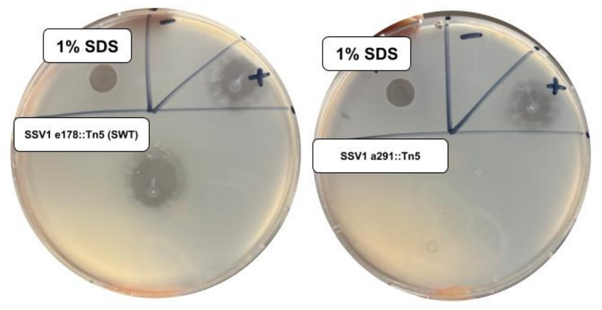

Table 1.
Genotype and phenotype summary of SSV1 ORF a291 mutants.
| Genotype / Mutation Description | Halo Formation† | Replication (PCR)‡ | Virions Observed§ |
|---|---|---|---|
| SSV1 e178::Tn5* (SWT) | + | + | + |
| SSV1 a291::Tn5 * | – | + | + |
| SSV1 e178::Tn5 Δa291 | – | + | + |
| SSV1 e178::Tn5 Δa291–c124 | – | + | + |
| SSV1 e178::Tn5 a291 (A25L/L26A) | – | + | + |
*For construction of mutants, see [19]; †Negative (-) halo formation indicates no halo formation from 3 transformations for which positive transformation controls generated halos (see Figure 2).; ‡Replication phenotype “+” indicates that SSV1 specific PCR amplicons were generated from cell-free supernatants of transformed cultures.; §Virions observed “+” indicates that typical fusellovirus virions were observed in cell-free supernatants by transmission electron microscopy (Figure 3).
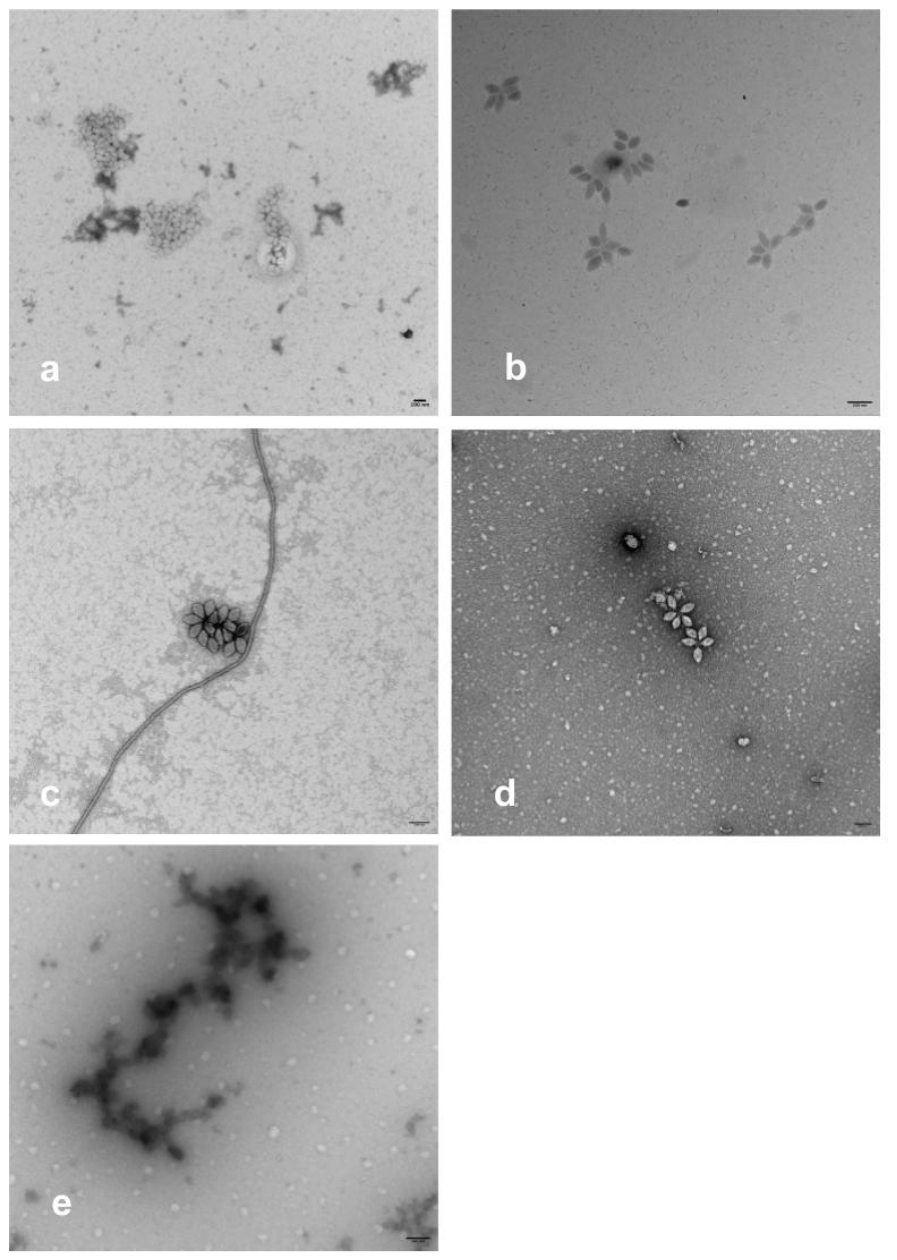
Figure 3.
Transmission electron micrographs of virions from S. solfataricus S441 cultures transformed with SSV1 viral genomes. Virions were stained with 1% Uranyl acetate and imaged with a FEI Tecnai transmission electron microscope or a FEI Talos transmission electron microscope. Scale bars represent 100 nm. a: Virions from SWT SSV1, b: Virions from SSV1 a291 Tn5 insertion mutant (EAI254 SSV1 a291::Tn5), c: and d: Virions from single and double deletions in SWT (ORFs a291 and a291-c124, respectively) and e: Virions produced by SSV1 a291 substitution mutant in SWT (ORF a291 A25L/L26A).
Figure 3.
Transmission electron micrographs of virions from S. solfataricus S441 cultures transformed with SSV1 viral genomes. Virions were stained with 1% Uranyl acetate and imaged with a FEI Tecnai transmission electron microscope or a FEI Talos transmission electron microscope. Scale bars represent 100 nm. a: Virions from SWT SSV1, b: Virions from SSV1 a291 Tn5 insertion mutant (EAI254 SSV1 a291::Tn5), c: and d: Virions from single and double deletions in SWT (ORFs a291 and a291-c124, respectively) and e: Virions produced by SSV1 a291 substitution mutant in SWT (ORF a291 A25L/L26A).
3.3. SSV1 Genomes with Disrupted or Deleted ORF a291 Still Replicate Their Genomes
To determine if SSV1 genomes lacking a functional ORF a291 were able to replicate and infect uninfected S. solfataricus, genome replication by mutant SSV1 genomes was checked. PCR with primers targeting the SSV1 major structural gene vp1 [23] and ORF a291 was performed on cell-free supernatants of S. solfataricus cultures transformed with different SSV1 genomes. SSV1-specific amplicons were generated from cell-free supernatants of S. solfataricus transformed with SWT SSV1, SSV1 genomes with Tn5 insertions in ORF a291, as well as genomes with deletions of ORF a291 and both ORFs a291 and c124 (Table 1). As controls, PCR from cell-free supernatants from cells mixed with DNA, but not electroporated, as well as cells without added DNA, did not generate SSV1-specific amplicons. Cultures from cells transformed with a Tn5 insertion at bp 8633 in SSV1 ORF a291 were passaged 3 times (see methods) without losing the ability to amplify SSV1-specific genes from cell-free supernatants, indicating that viruses generated by these mutants were stably maintained.
3.4. SSV1 Genomes with Disrupted or Deleted ORF a291 Can Form Virions
To determine if SSV1 ORF a291 is important for virion production, cell-free supernatants from cells transformed with SWT SSV1, and SSV1 virus genomes with Tn5 insertions in ORF a291 as well as SSV1 genomes with deletions of ORF a291 and both ORFs a291 and c124 were screened for typical spindle-shaped virions using transmission electron microscopy (TEM). Cell-free supernatants from S.solfataricus transformed with SWT SSV1, SSV1 with a Tn5 insertion in ORF a291, and deletions of SSV1 ORF a291 and deletion of SSV1 ORFs a291 and c124 all contained typical Fusellovirus-like particles (Table 1, Figure 3). This indicates that SSV1 ORF a291 is not necessary to generate virus particles.
3.5. Putative Toxin Gene Sequences in Fuselloviruses Are not Well Conserved
In previous analyses of SSV1 ORF a291, no clear homologs were found in other SSV genomes using BLASTp (e <0.001) [19]. However, there is syntenic conservation of the putative toxin gene locus among published SSV genomes and many SSV genomes have ORFs of approximately 300 amino acids at that locus (Figure 4) [21], Therefore, pairwise comparisons using SSV1 ORF a291 and other non-redundant putative SSV toxins (see methods) were performed. Only 1 other putative fusellovirus toxin sequence was more than 30% identical in amino acid sequence to SSV1 ORF a291, indicating that these proteins are highly divergent despite their conserved genomic context. Notably SSV9 ORF b310, which encodes the only other known fusellovirus-encoded toxin, was only 18.7% identical in amino acid sequence to SSV1 ORF a291 (Figure 4).
3.6. Alignment of Amino Acid Sequences of Putative Fusellovirus Toxins Indicates the Presence of a Conserved Signal Peptide and Multiple Related Toxins
In order to determine if there were conserved sequence motifs in putative SSV toxins, their amino acid sequences were aligned using ClustalW [26] with default parameters, and the resulting alignment was visualized in ESPript3.0 [27] (Figure 5). The only amino acids that are 100% conserved are at the N-terminus. Moreover, sequences in this region were generally much more conserved than in other parts of the sequence (Figure 5). Interestingly this N-terminal region corresponds to a predicted signal peptide in all of the putative toxin sequences, as determined by SignalP 6.0 [28] (Supplemental Table S3).
3.7. Changing the Putative Signal Peptide Cleavage Site of SSV1 ORF a291 Eliminates Toxin Activity
To determine if the predicted signal peptide was important for the function of SSV1 ORF a291, we switched the encoded amino acids at positions 25 and 26 in SWT SSV1 ORF a291, A25L/L26A using site-directed mutagenesis. This should eliminate cleavage by the Saccharolobus signal peptidase [28]. We then tested this mutant construct as above for causing growth inhibition, genome replication and virion formation. We found that this mutant did not cause growth inhibition but still replicated its genome and formed virions in a manner similar to complete deletion of SSV1 ORF a291 (Figure 2 and Figure 3, Supplemental Figure S2, Table 1), indicating that signal peptide cleavage secretion is essential for the function of the toxin.
3.8. The SSV1 ORF a291 Toxin and Other SSV Putative Toxin Proteins Share a Conserved Hydrolase-like Fold Despite Extreme Sequence Divergence
In order to predict the mechanism of action of the putative toxins, Alphafold3 models [29] were constructed for ORFs a291 (SSV1), b310 (SSV9) and 299 (SSV10) (Figure 6). SSV10 is a fusellovirus from Lassen Volcanic National Park, USA that is also genetically tractable [30]. SSV10 ORF 299 is only 22.9% identical to SSV1 ORF a291 (Figure 4 and Figure 5). All of these models contained a beta-sheet core flanked by short alpha-helicies and surface loops (Figure 6) with high confidence (data not shown). Despite low (>25%) amino acid sequence identity between these sequences, their predicted structures are extremely similar (Figure 6A–C). Foldseek [31] identified highly statistically significant structural matches in the PDB100 database [32] for all three predicted structures. The top match for SSV1 ORF a291, Fibrobacter succinogenes, 1,3-1,4-beta-D-glucanase PDB: 3AXD (E-Value 9.86 x 10⁻²⁴), has a very similar fold to the putative SSV toxin structures (Figure 6D). Superimposition of the X-ray crystallography derived structure 3AXD on the predicted structure of SSV1 ORF a291 (Figure 6E) demonstrates that SSV putative toxins have a canonical hydrolase-like structure.
4. Discussion
4.1. SSV1 ORF a291 Is Dispensable for Viral Replication but Required for Growth Suppression
Contrary to previous reports [19], PCR amplification of viral genes from cell-free supernatants, even after passaging, confirmed that ORF a291-deficient SSV1 viruses replicate (Table 1), and TEM analysis revealed virion morphology indistinguishable from wild-type SSV1 (Table 1, Figure 3). These findings demonstrate that SSV1 ORF a291 is not required for genome replication and virion assembly. However, an intact ORF a291 is required for SSV1 to inhibit growth of uninfected S. solfataricus. (Table 1, Figure 2). This functional separation indicates that SSV1 ORF a291 does not serve a structural or replicative role but instead may control host population dynamics. Accessory metabolic genes (AMGs) encoded in some viruses often mediate ecological interactions without being required for viral reproduction [35,36,37], but generally affect the metabolism of the infected host cell, not other cells [6].
4.2. Reinterpreting Halo Formation
Plaque formation on host cells has traditionally been interpreted as a proxy for viral infection and replication, particularly in lytic bacteriophage systems where zones of clearing result from cycles of infection and host cell lysis [3,4]. However, plaque formation is not exclusively associated with lytic infection. Filamentous bacteriophages such as M13 establish chronic, non-lytic infections yet still produce plaques due to reduced host growth and continuous virion extrusion rather than cell lysis [38]. Early studies reported plaque formation by SSV1 infecting Saccharolobus (previously Sulfolobus) solfataricus [15]. A rapid method of screening multiple viruses under identical conditions is by spotting a small amount of virus-containing liquid on a lawn of uninfected host cells and observing growth inhibition, also known as “halo” assays or spot testing [39]. This method is often used for fuselloviruses [13], since plaque assays for SSVs are notoriously challenging (see Supplemental Figure S2) [21]. These assays were based on the assumption that visible halos reflect direct inhibition of host growth due only to viral infection. However, our data demonstrate that this interpretation does not apply to SSV1.
Disruption or deletion of SSV1 ORF a291 abolished halo formation (Figure 2; Table 1), yet viruses with these mutations replicate their genomes (Table 1) and produce morphologically normal spindle-shaped virions (Figure 3). We infer that halo, and by extension plaque formation, by SSV1 reflects virus-encoded toxin-mediated growth suppression rather than merely viral spread and is not dependent on intact virions. Together, these results are consistent with the presence of a secreted protein factor that is sufficient to induce host growth suppression. Similar killing of uninfected Saccharolobus islandicus by SSV9, virus-free supernatants of infected cells, and otherwise uninfected cells overexpressing only SSV9 ORF b310 was interpreted as being due to a virus-encoded toxin [12].
4.3. SSV Toxin Evolutionary Divergence with Conserved Secretion-Dependent Function
Comparative sequence analysis revealed minimal amino acid identity among putative fusellovirus toxins (Figure 4), including only 18.7% identity between SSV1 ORF a291 and the previously examined SSV9 toxin b310 [12] and 22.92% identity in ORF 299 (toxin) of SSV10. Despite this divergence, putative toxin genes are encoded upstream of the tail protein gene vp4 across fusellovirus genomes, suggesting that functional constraints maintain genomic position. Moreover, the conservation of only putative signal peptides (Figure 5) between these putative toxin genes, despite rapid primary sequence evolution in the remainder of the protein indicates that secretion is critical for toxin function. Overexpression of a truncated SSV9 ORF b310 supports this interpretation [12]. Substitution of residues at the predicted signal peptide cleavage site abolished halo formation by SSV1 while preserving viral genome replication and virion production (Table 1, Figure 2 and Figure 3), similar to the complete deletion of ORF a291. Together, these findings indicate that fusellovirus toxins exhibit extensive sequence divergence while maintaining secretion-dependent function.
4.4. Structural Conservation of a Hydrolase-like Fold Across Divergent Fusellovirus Toxins
Despite extreme sequence divergence (Figure 4 and Figure 5), AlphaFold3 predictions indicate that fusellovirus toxins adopt a conserved beta-sheet-rich fold flanked by alpha-helices and surface loops (Figure 6A-C) [29]. As identified with FoldSeek [31], the predicted structures of these toxins is strikingly similar to a bacterial glucanase (Figure 6), an enzyme that hydrolyzes structural polysaccharides. Such enzymes typically cleave glycosidic bonds and modify extracellular substrates. The predicted SSV toxin structures share the canonical beta-sheet-rich fold of this enzyme class, suggesting potential catalytic or substrate-binding activity [33].
The preservation of structural architecture in the absence of detectable sequence similarity reflects functional constraints of the three-dimensional structure rather than primary sequence, a common feature of protein evolution [40,41,42]. Although the precise substrate remains unknown, this hydrolase-like architecture raises the possibility that these toxins act on extracellular targets, potentially altering cell surface components to suppress growth of neighboring cells.
4.5. Toxin-Antitoxin Systems and Viral Population Control
Toxin-antitoxin (TA) systems, also known as addiction modules, are well known in bacteria and archaea [43,44]. TA systems typically consist of a stable toxin and a labile antitoxin encoded as adjacent genes. Loss of the TA locus often leads to toxin-mediated growth arrest or cell death, thereby stabilizing maintenance of the genetic element43-44. Although TA systems are well characterized in bacterial chromosomes and plasmids, their roles in archaeal viruses remain largely unexplored [45]. Interestingly, SSV1 encodes a non-conserved ORF just downstream of ORF a291, ORF c124, and similar genome orientations of possible anti-toxins with putative toxin genes are found in other SSVs [12,19]. However, deletion of this ORF does not change the phenotype of SSV1 ORF a291 deletions (Table 1), nor does an insertion in this ORF in the presence of ORFa291 change halo formation [19].
Most bacterial TA toxins act intracellularly, targeting translation or DNA replication. By contrast, extracellular antagonistic toxins are commonly used in bacterial competition systems, but virally encoded secreted toxins are rare and poorly understood [12,46]. The presence of viral TA-like systems could promote a form of ‘virus addiction’, in which infected cells are protected while uninfected neighbors are selectively suppressed. Such a strategy would enhance viral persistence without requiring host lysis, aligning with ecological models in which viruses influence microbial communities beyond direct killing [5,6].
4.6. Ecological Implications: Growth Suppression as a Persistence Strategy
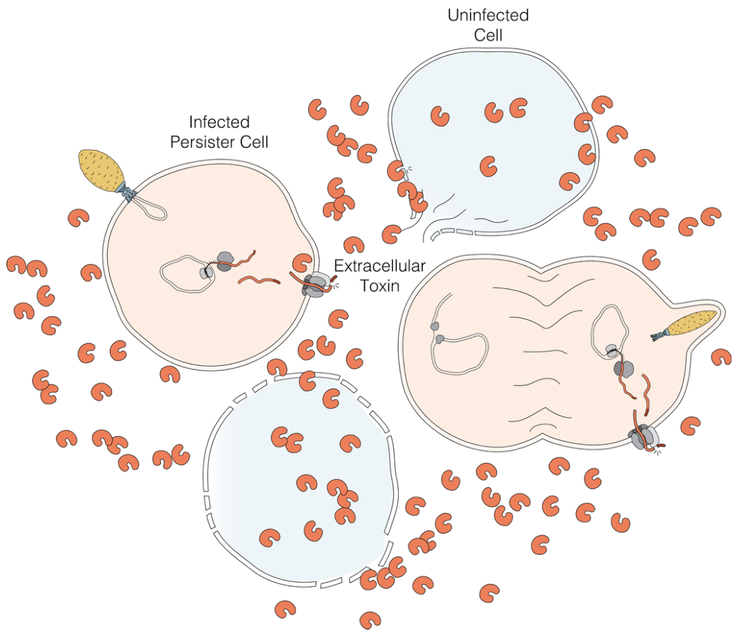
These findings indicate that SSV1 ORF a291 encodes a secreted toxin that suppresses the growth of neighboring uninfected cells while leaving viral replication and virion production intact. We propose a model (Figure 7) in which SSV1 establishes chronic infection in S.solfataricus that enables secretion of the SSV1 ORF a291 product into the extracellular environment, suppressing the growth of uninfected cells while infected cells persist. This framework is supported by prior work on SSV9-infected S. islandicus, in which SSV9 encodes the extracellular toxin b310 that kills uninfected hosts, particularly when overexpressed [12,21]. These toxin genes are conserved upstream of the vp4 structural gene across multiple fuselloviruses, suggesting functional conservation despite sequence divergence (Figure 4).
Unlike lytic viruses that reduce host density through destruction and nutrient release [3,4], SSV1 appears to modulate competitive dynamics without eliminating its host in a virus-host mutualism similar to SSV9 [12]. Our data support a mechanism in which growth suppression of uninfected cells enhances the relative fitness of infected hosts consistent with addiction module dynamics. However, the role, if any, of the associated SSV1 ORF c124 as a potential antitoxin remains to be determined.
In resource-limited geothermal environments, where microbial diversity is low and competition is intense [11], even modest suppression of competitors may promote long-term virus-host mutualism [12]. This strategy parallels addiction-module theory, in which maintenance of a genetic element confers a selective advantage through toxin-mediated enforcement [44,47]. However, fusellovirus toxins appear to extend this principle beyond intracellular killing to community level ecological control.
Supplementary Materials
The following supporting information can be downloaded at the website of this paper posted on Preprints.org. Table S1: Primers used in this study; Table S2: Fuselloviruses used for putative toxin identification; Table S3: Signal P results; Figure S1: Halo formation by SSV1 mutants; Figure S2: Plaque Assay.
Author Contributions
Conceptualization, J.C.A. and K.M.S.; Methodology, J.C.A. and K.M.S.; Validation, J.C.A., P. L. A., and G.T.; Formal Analysis, J.C.A.; Investigation, J.C.A., P. L. A., and G.T.; Resources, J.C.A., P. L. A., and G.T.; Data Curation, J.C.A., P. L. A., and G.T.; Writing – Original Draft Preparation, J.C.A. and K.M.S.; Writing – Review & Editing, J.C.A., P. L. A., G.T. and K.M.S.; Visuali-zation, J.C.A. and K.M.S.; Supervision, J.C.A. and K.M.S..; Project Administration, K.M.S.; Funding Acquisition, K.M.S.
Funding
This work was supported by Portland State University and grants 1R16GM154184-01 and 1S10GM154263-01 from the U.S. National Institutes of Health and MCB-1929273 from the U.S. National Science Foundation. Patrizia Lynnel Alpapara was supported by the U. S. National Science Foundation EES-1911026 (Louis Stokes Renewal STEM Pathways and Re-search Alliance: Pacific Northwest) and Guasåli Tomokane was supported by the U. S. Department of Education Asian American and Native American Pacific Islander-Serving Institutions Grant P031L220008.
Data Availability Statement
The original contributions presented in this study are included in the article/supplementary material. Further inquiries can be directed to the corresponding author.
Acknowledgments
We thank the members of the eXtreme Virus Lab at Portland State University for their support and contributions.
Conflicts of Interest
The authors declare no conflict of interest.
References
- Howard-Varona, C., Hargreaves, K. R., Abedon, S. T., & Sullivan, M. B. (2017). Lysogeny in nature: mechanisms, impact and ecology of temperate phages. The ISME Journal, 11(7), 1511-1520.
- Roux, S., Brum, J. R., Dutilh, B. E., Sunagawa, S., Duhaime, M. B., Loy, A., Poulos B., Solonenko N. & Sullivan, M. B. (2016). Ecogenomics and potential biogeochemical impacts of globally abundant ocean viruses. Nature, 537(7622), 689-693.
- Suttle, C. A. (2007). Marine viruses—major players in the global ecosystem. Nature Reviews Microbiology, 5(10), 801-812.
- Zimmerman, A. E., Howard-Varona, C., Needham, D. M., John, S. G., Worden, A. Z., Sullivan, M. B., Waldbauer J. R. & Coleman, M. L. (2020). Metabolic and biogeochemical consequences of viral infection in aquatic ecosystems. Nature Reviews Microbiology, 18(1), 21-34.
- Knowles, B., Silveira, C. B., Bailey, B. A., Barott, K., Cantu, V. A., Cobián-Güemes, A. G., & Rohwer, F. (2016). Lytic to temperate switching of viral communities. Nature, 531(7595), 466-470.
- Brown, T. L., Charity, O. J., & Adriaenssens, E. M. (2022). Ecological and functional roles of bacteriophages in contrasting environments: marine, terrestrial and human gut. Current Opinion in Microbiology, 70, 102229.
- Krupovic, M., Cvirkaite-Krupovic, V., Iranzo, J., Prangishvili, D., & Koonin, E. V. (2018). Viruses of Archaea: structural, functional, environmental and evolutionary genomics. Virus Research, 244, 181-193.
- Munson-McGee, J. H., Peng, S., Dewerff, S., Stepanauskas, R., Whitaker, R. J., Weitz, J. S., & Young, M. J. (2018). A virus or more in (nearly) every cell: ubiquitous networks of virus–host interactions in extreme environments. The ISME Journal, 12(7), 1706-1714.
- Prangishvili, D., Bamford, D. H., Forterre, P., Iranzo, J., Koonin, E. V., & Krupovic, M. (2017). The enigmatic archaeal virosphere. Nature Reviews Microbiology, 15(12), 724-739.
- Quemin, E. R., Chlanda, P., Sachse, M., Forterre, P., Prangishvili, D., & Krupovic, M. (2016). Eukaryotic-like virus budding in archaea. mBio, 7(5), 10-1128.
- Cadillo-Quiroz H, Didelot X, Held NL, Herrera A, Darling A, Reno ML, Krause DJ, Whitaker RJ. (2012) Patterns of gene flow define species of thermophilic Archaea. PLoS Biology. Feb;10(2):e1001265.
- DeWerff, S. J., Bautista, M. A., Pauly, M., Zhang, C., & Whitaker, R. J. (2021). Killer archaea: virus-mediated antagonism to CRISPR-immune populations results in emergent virus-host mutualism. mBio, 11(2), 10-1128.
- Iverson, E. A., & Stedman, K. M. (2012). A genetic system for studying archaeal viruses. Journal of Virology, 86, 12236–12242.
- Stedman, K. M., Schleper, C., Rumpf, E., & Zillig, W. (1999). Genetic requirements for the replication of archaeal virus SSV1. Journal of Virology, 77(5), 2850–2858.
- Schleper, C., Kubo, K., & Zillig, W. (1992). The virus-like element SSV1 of the extremely thermophilic archaeon Sulfolobus. Molecular & General Genetics, 232, 1–9.
- Huber, H., & Stetter, K. O. (2001). Order Sulfolobales. In W. B. Whitman (Ed.), Bergey’s Manual of Systematics of Archaea and Bacteria. John Wiley & Sons.
- Zillig, W., Stetter, K. O., Wunderl, S., Schulz, W., Priess, H., & Scholz, I. (1980). The Sulfolobus-“Caldariella” group: taxonomy on the basis of the structure of DNA-dependent RNA polymerases. Archives of Microbiology, 125(3), 259-269.
- Palm, P., Schleper, C., Grampp, B., Yeats, S., McWilliam, P., Reiter, W. D., & Zillig, W. (1991). Complete nucleotide sequence of the virus SSV1 of the archaebacterium Sulfolobus shibatae. Virology, 185(1), 242-250.
- Iverson, E. A., Goodman, D. A., Gorchels, M. E., & Stedman, K. M. (2017). Extreme mutation tolerance: nearly half of the archaeal fusellovirus sulfolobus spindle-shaped virus 1 genes are not required for virus function, including the minor capsid protein gene vp3. Journal of Virology, 91(10), 10-1128.
- Fusco, S., Liguori, R., Limauro, D., Bartolucci, S., She, Q., & Contursi, P. (2015). Transcriptome analysis of Sulfolobus solfataricus infected with two related fuselloviruses reveals novel insights into the regulation of CRISPR-Cas system. Biochimie, 118, 322-332.
- DeWerff, S. J., Zhang, C., Schneider, J., & Whitaker, R. J. (2022). Intraspecific antagonism through viral toxin encoded by chronic Sulfolobus spindle-shaped virus. Philosophical Transactions of the Royal Society B: Biological Sciences, 377(1842).
- Abshier, J., Marceau, C., & Stedman, K. M. (2025). Complete genome sequence of Saccharolobus solfataricus strain S441, from Lassen Volcanic National Park. Microbiology Resource Announcements, 14(7), e00288-25.
- Reiter, W. D., Palm, P., Henschen, A., Lottspeich, F., Zillig, W., & Grampp, B. (1987). Identification and characterization of the genes encoding three structural proteins of the Sulfolobus virus-like particle SSV1. Molecular and General Genetics MGG, 206(1), 144-153.
- Core Team, R. C. T. R. (2014). R: A language and environment for statistical computing. R Foundation for Statistical Computing, Vienna.
- Kolde, R. (2019). pheatmap: Pretty Heatmaps. R package version 1.0. 12.
- Thompson, J. D., Higgins, D. G., & Gibson, T. J. (1994). CLUSTAL W: improving the sensitivity of progressive multiple sequence alignment through sequence weighting, position-specific gap penalties and weight matrix choice. Nucleic Acids Research, 22(22), 4673-4680.
- Gouet, P., Robert, X., & Courcelle, E. (2003). ESPript/ENDscript: extracting and rendering sequence and 3D information from atomic structures of proteins. Nucleic Acids Research, 31(13), 3320-3323.
- Teufel, F., Almagro Armenteros, J. J., Johansen, A. R., Gíslason, M. H., Pihl, S. I., Tsirigos, K. D., Winther O., Brunak S., von Heijne G. & Nielsen, H. (2022). SignalP 6.0 predicts all five types of signal peptides using protein language models. Nature Biotechnology, 40(7), 1023-1025.
- Abramson, J., Adler, J., Dunger, J., Evans, R., Green, T., Pritzel, A., Ronneberger O. & Jumper, J. M. (2024). Accurate structure prediction of biomolecular interactions with AlphaFold 3. Nature, 630(8016), 493-500.
- Goodman, D. A., & Stedman, K. M. (2018). Comparative genetic and genomic analysis of the novel fusellovirus Sulfolobus spindle-shaped virus 10. Virus Evolution, 4(2), vey022.
- Van Kempen, M., Kim, S. S., Tumescheit, C., Mirdita, M., Lee, J., Gilchrist, C. L. & Steinegger, M. (2024). Fast and accurate protein structure search with Foldseek. Nature Biotechnology, 42(2), 243-246.
- Berman, H. M., Westbrook, J., Feng, Z., Gilliland, G., Bhat, T. N., Weissig, H., Shindyalov I. N. & Bourne, P. E. (2000) The protein data bank. Nucleic Acids Research, 28(1), 235-242.
- Huang, J. W., Cheng, Y. S., Ko, T. P., Lin, C. Y., Lai, H. L., Chen, C. C. & Guo, R. T. (2012). Rational design to improve thermostability and specific activity of the truncated Fibrobacter succinogenes 1, 3-1, 4-β-D-glucanase. Applied Microbiology and Biotechnology, 94(1), 111-121.
- Pettersen, E. F., Goddard, T. D., Huang, C. C., Meng, E. C., Couch, G. S., Croll, T. I., Morris, J. H. & Ferrin, T. E. (2021). UCSF ChimeraX: Structure visualization for researchers, educators, and developers. Protein Science, 30(1), 70-82.
- Hurwitz, B. L., & U’Ren, J. M. (2016). Viral metabolic reprogramming in marine ecosystems. Current Opinion in Microbiology, 31, 161-168.
- Thompson, L. R., Zeng, Q., Kelly, L., Huang, K. H., Singer, A. U., Stubbe, J., & Chisholm, S. W. (2011). Phage auxiliary metabolic genes and the redirection of cyanobacterial host carbon metabolism. Proceedings of the National Academy of Sciences, 108(39), E757-E764.
- Martin, C., Emerson, J. B., Roux, S., & Anantharaman, K. (2025). A call for caution in the biological interpretation of viral auxiliary metabolic genes. Nature Microbiology, 10(9), 2122-2129.
- Rakonjac, J., Bennett, N. J., Spagnuolo, J., Gagic, D., & Russel, M. (2011). Filamentous bacteriophage: biology, phage display and nanotechnology applications. Current Issues in Molecular Biology, 13(2), 51.
- Abedon, S. T. (2008). Phages, ecology, evolution. Bacteriophage ecology, 15, 1-28.
- Bamford, D. H. (2003). Do viruses form lineages across different domains of life?. Research in Microbiology, 154(4), 231-236.
- Illergård, K., Ardell, D. H., & Elofsson, A. (2009). Structure is three to ten times more conserved than sequence—a study of structural response in protein cores. Proteins: Structure, Function, and Bioinformatics, 77(3), 499-508.
- Koonin, E. V., Wolf, Y. I., & Karev, G. P. (2002). The structure of the protein universe and genome evolution. Nature, 420(6912), 218-223.
- Harms, A., Brodersen, D. E., Mitarai, N., & Gerdes, K. (2018). Toxins, targets, and triggers: an overview of toxin-antitoxin biology. Molecular cell, 70(5), 768-784.
- Hayes, F., & Van Melderen, L. (2011). Toxins-antitoxins: diversity, evolution and function. Critical Reviews in Biochemistry and Molecular Biology, 46(5), 386-408.
- Hall, J. P., Harrison, E., & Baltrus, D. A. (2022). Introduction: the secret lives of microbial mobile genetic elements. Philosophical Transactions of the Royal Society B: Biological Sciences, 377(1842).
- Makarova, K. S., Wolf, Y. I., Karamycheva, S., Zhang, D., Aravind, L., & Koonin, E. V. (2019). Antimicrobial peptides, polymorphic toxins, and self-nonself recognition systems in Archaea: an untapped armory for intermicrobial conflicts. mBio, 10(3), 10-1128.
- Villarreal, L. P. (2016). Persistent virus and addiction modules: an engine of symbiosis. Current Opinion in Microbiology, 31, 70-79.
Figure 1.
Genomic organization of SSV1. The circular SSV1 genome is shown with annotated open reading frames (ORFs) discussed herein. Universally conserved fusellovirus ORFs are indicated as thick black arrows, while non-conserved ORFs are shown in gray. Colored ORFs are less-conserved ORFs [19]. SSV1 ORF a291 (putative toxin gene) and adjacent SSV1 ORF c124 are located within a variable genomic region upstream of the conserved capsid/tail protein gene cluster (vp4-vp1-vp3). This genomic context is consistent with previously described fusellovirus toxin loci positioned near structural gene modules [21].
Figure 1.
Genomic organization of SSV1. The circular SSV1 genome is shown with annotated open reading frames (ORFs) discussed herein. Universally conserved fusellovirus ORFs are indicated as thick black arrows, while non-conserved ORFs are shown in gray. Colored ORFs are less-conserved ORFs [19]. SSV1 ORF a291 (putative toxin gene) and adjacent SSV1 ORF c124 are located within a variable genomic region upstream of the conserved capsid/tail protein gene cluster (vp4-vp1-vp3). This genomic context is consistent with previously described fusellovirus toxin loci positioned near structural gene modules [21].
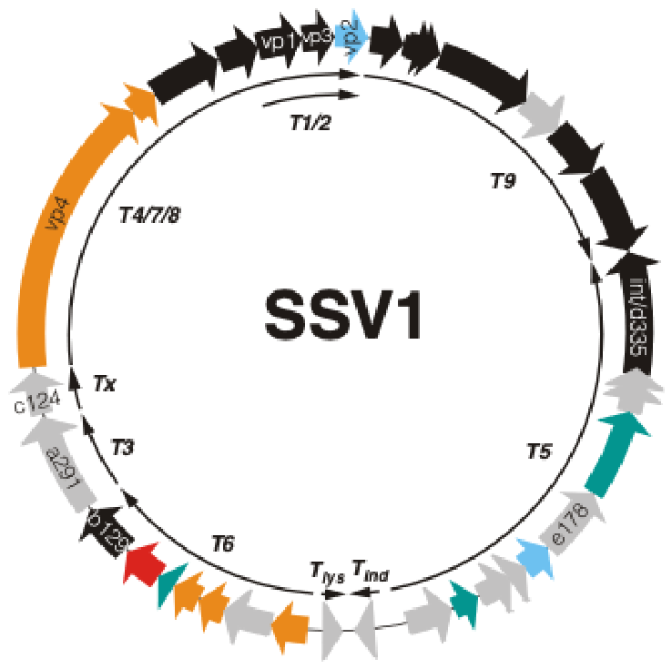
Figure 4.
Percent identity matrix of fusellovirus candidate toxin homologs. Pairwise amino-acid percent identities between fusellovirus toxin homologs were calculated using ClustalW and visualized using Rstudio v 4.5.2 [24] and pheatmap package v 1.0 [25]. Each cell shows the percent identity for the corresponding toxin pair, with warmer colors indicating higher sequence identities. Diagonal values represent self-comparisons. SSV1_ORF_a291 is highlighted in bold. Accession numbers for all sequences are listed next to fusellovirus ORF names.
Figure 4.
Percent identity matrix of fusellovirus candidate toxin homologs. Pairwise amino-acid percent identities between fusellovirus toxin homologs were calculated using ClustalW and visualized using Rstudio v 4.5.2 [24] and pheatmap package v 1.0 [25]. Each cell shows the percent identity for the corresponding toxin pair, with warmer colors indicating higher sequence identities. Diagonal values represent self-comparisons. SSV1_ORF_a291 is highlighted in bold. Accession numbers for all sequences are listed next to fusellovirus ORF names.
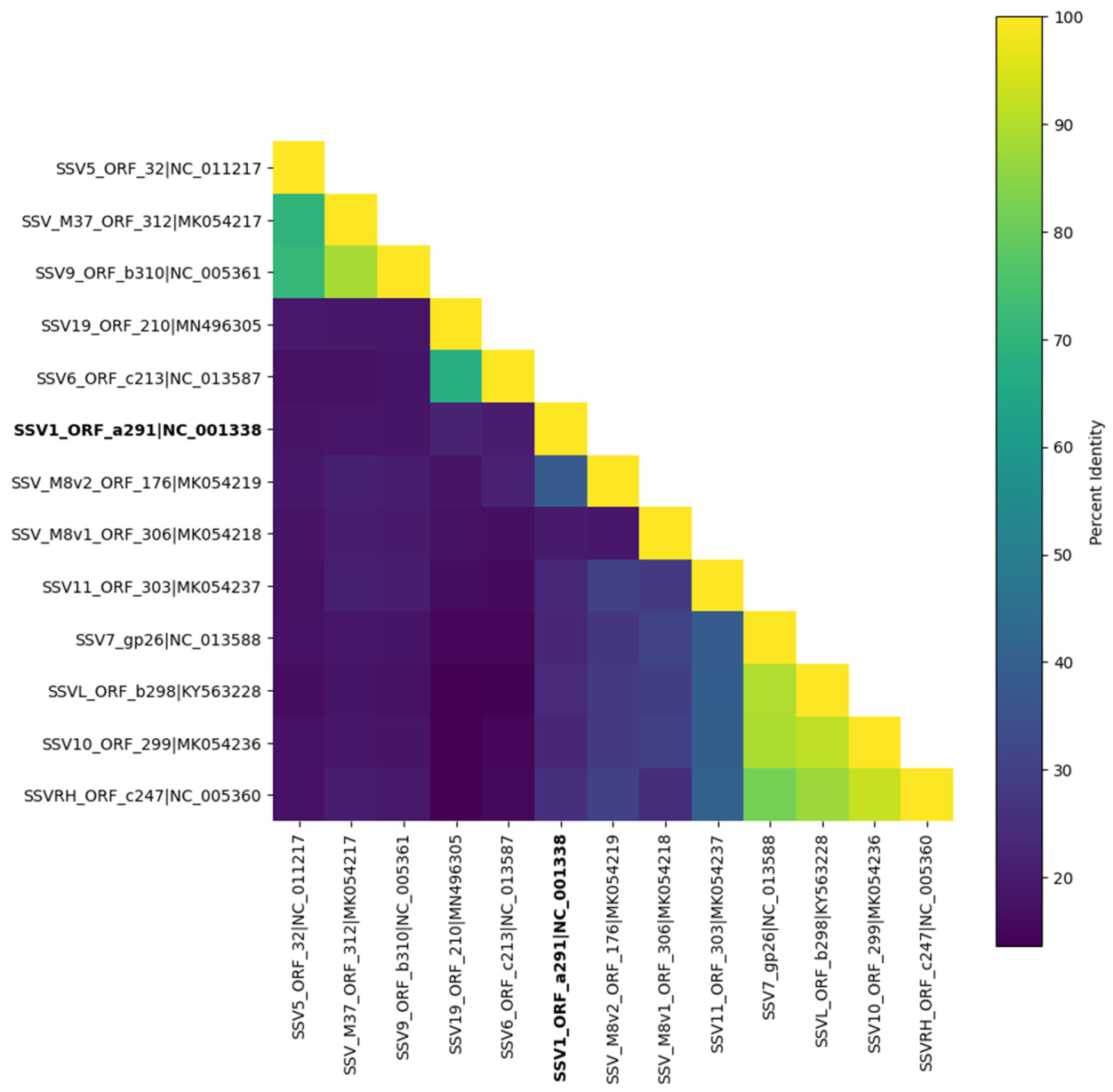
Figure 5.
Multiple sequence alignment of fusellovirus toxin candidates. A multiple sequence alignment of putative fusellovirus toxin proteins aligned with CLUSTALW and visualized using ESPript 3.0. Residues conserved in all sequences have a red background, and similar residues are indicated by red text and blue boxed highlighting. Sequence identifiers correspond to the viral toxin Genbank protein ID and associated open reading frame e.g., SSV1_ORF_a291|NC_001338.
Figure 5.
Multiple sequence alignment of fusellovirus toxin candidates. A multiple sequence alignment of putative fusellovirus toxin proteins aligned with CLUSTALW and visualized using ESPript 3.0. Residues conserved in all sequences have a red background, and similar residues are indicated by red text and blue boxed highlighting. Sequence identifiers correspond to the viral toxin Genbank protein ID and associated open reading frame e.g., SSV1_ORF_a291|NC_001338.
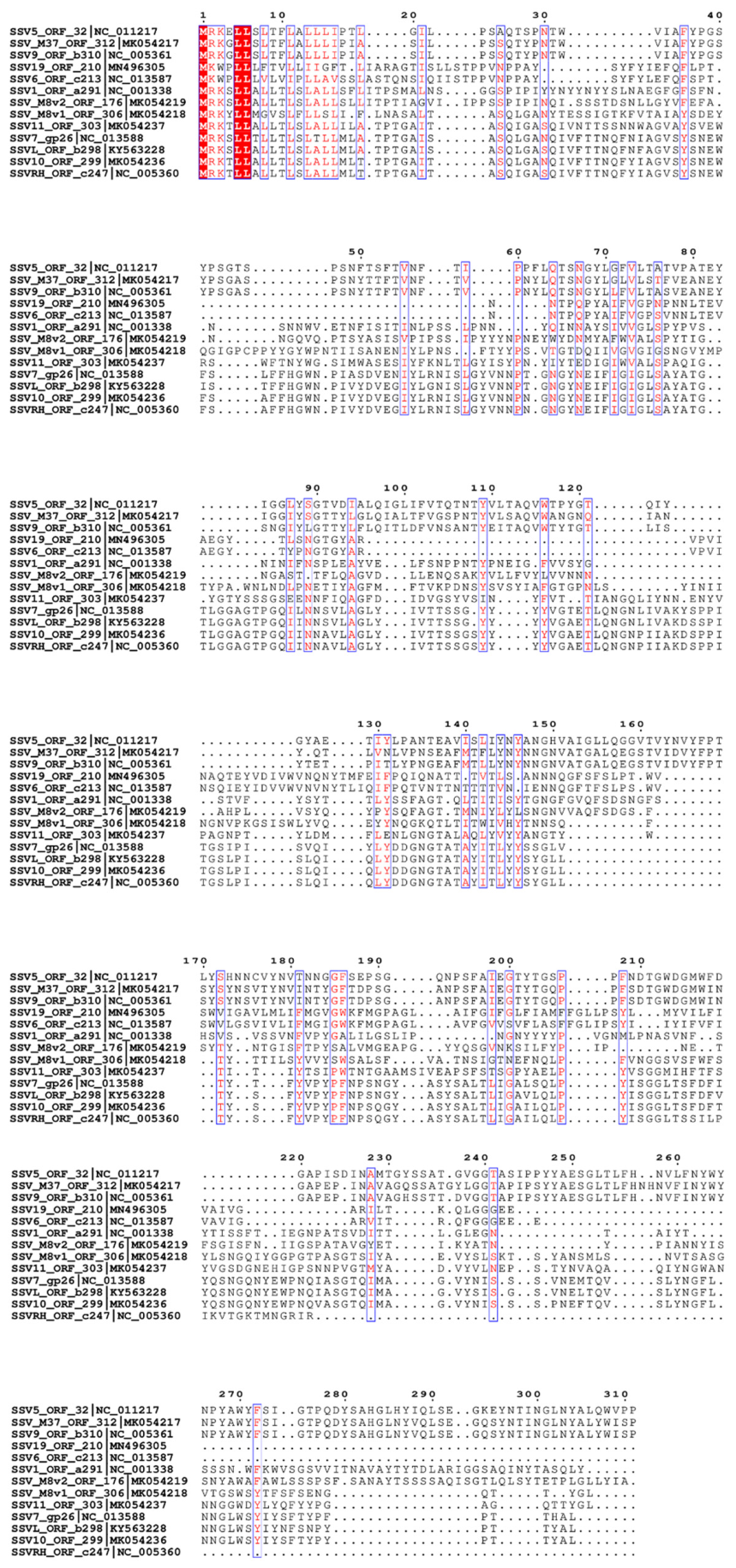
Figure 6.
(A–C) Alphafold3 structural predictions of SSV putative toxin proteins. Models of SSV1 a291 (A: blue), SSV9 b310 (B: maroon), and SSV10 orf299 (C: magenta). (D) Crystal structure of Fibrobacter succinogenes, 1,3-1,4-beta-D-glucanase (PDB: 3AXD), the best hit with Foldseek to the SSV1 ORF a291 predicted structure. The 3AXD structure (green) has a canonical hydrolase fold [33]. (E) Matchmaker (ChimeraX [34]) of SSV1 a291 (blue) with the hydrolase 3AXD (green), reveals a highly similar overall fold (RMSD 10.04 Å, TM-score = 0.453).
Figure 6.
(A–C) Alphafold3 structural predictions of SSV putative toxin proteins. Models of SSV1 a291 (A: blue), SSV9 b310 (B: maroon), and SSV10 orf299 (C: magenta). (D) Crystal structure of Fibrobacter succinogenes, 1,3-1,4-beta-D-glucanase (PDB: 3AXD), the best hit with Foldseek to the SSV1 ORF a291 predicted structure. The 3AXD structure (green) has a canonical hydrolase fold [33]. (E) Matchmaker (ChimeraX [34]) of SSV1 a291 (blue) with the hydrolase 3AXD (green), reveals a highly similar overall fold (RMSD 10.04 Å, TM-score = 0.453).
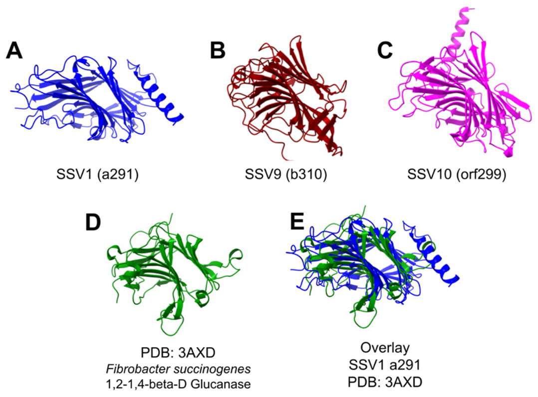
Figure 7.
Model of fusellovirus-mediated toxin production dynamics and population-level outcomes. Pink S.solfataricus cells are infected with yellow SSVs, replicate and produce virions and express and secrete virus-encoded toxins (red) which kill uninfected cells (blue). Pink infected cells are resistant to toxin and outcompete uninfected cells. © Rita Clare 2026.
Figure 7.
Model of fusellovirus-mediated toxin production dynamics and population-level outcomes. Pink S.solfataricus cells are infected with yellow SSVs, replicate and produce virions and express and secrete virus-encoded toxins (red) which kill uninfected cells (blue). Pink infected cells are resistant to toxin and outcompete uninfected cells. © Rita Clare 2026.
Disclaimer/Publisher’s Note: The statements, opinions and data contained in all publications are solely those of the individual author(s) and contributor(s) and not of MDPI and/or the editor(s). MDPI and/or the editor(s) disclaim responsibility for any injury to people or property resulting from any ideas, methods, instructions or products referred to in the content. |
© 2026 by the authors. Licensee MDPI, Basel, Switzerland. This article is an open access article distributed under the terms and conditions of the Creative Commons Attribution (CC BY) license (http://creativecommons.org/licenses/by/4.0/).
Copyright: This open access article is published under a Creative Commons CC BY 4.0 license, which permit the free download, distribution, and reuse, provided that the author and preprint are cited in any reuse.


