

Submitted:
30 May 2026
Posted:
02 June 2026
You are already at the latest version
Abstract
Vascular aging is a key factor in late-life health issues, including cardiovascular disease, stroke, and organ decline. It results from accumulated molecular, cellular, and structural damage like endothelial dysfunction, smooth muscle maladaptation, extracellular matrix failure, calcification, inflammation, and barrier breakdown. This accumulated damage interacts in ways that cause lasting changes to vascular mechanics and permeability. This chapter categorizes vascular damage into distinct physiologically recognizable groups, distinguishing root causes from signaling responses and emphasizing persistent structural and biochemical damage over transient dysregulation. This explains the limited durability of many signaling therapies and suggests that direct damage repair may provide more lasting and comprehensive benefits. We review evidence on endothelial cells, smooth muscle, extracellular matrix, mineralization, immune-vascular interactions, and hemodynamics, focusing on mechanisms that cause long-term damage. We discuss therapeutic strategies and risks of manipulating signaling pathways that vary across tissues. Finally, we explore the need for biomarkers reflecting specific vascular damage, advocating for a divide-and-conquer approach with targeted repairs and suitable endpoints to improve interventions that preserve vascular function.
Keywords:
plaque
; atherosclerosis
; fibrinolysis
; oxysterol
; vascular aging
; endothelial dysfunction
; vascular permeability
; barrier failure
; vascular leakage
; arterial remodeling
; vascular senescence
; oxidative stress
; reactive oxygen species
; eNOS uncoupling
; nitric oxide signaling
; lipid peroxidation
; advanced glycation end products
; cellular senescence
; senescence-associated secretory phenotype
; SASP
; endothelial senescence
; smooth muscle cell phenotypic switching
; elastin
; extracellular matrix remodeling
; collagen crosslinking
; lysyl oxidase
; vascular calcification
; arterial stiffness
; endothelial glycocalyx
; tight junctions
; plaque instability
; microvascular dysfunction
; diabetic vasculopathy
; chronic inflammation
; inflammaging
; endothelial barrier dysfunction
; geroscience
; biogerontology
1. Introduction
Aging can be viewed as the cumulative accumulation of molecular, cellular, and structural damage that progressively erodes tissue function [1,2]. This damage-accumulation framework describes aging as the progressive accumulation of diverse molecular and cellular lesions that outpace and evade endogenous repair mechanisms [3]. This process has been further described and formalized across multiple iterations of the Hallmarks of Aging [4], which together emphasize that age-related dysfunction arises from persistent lesions that outpace endogenous repair rather than from the gradual downregulation of beneficial processes [2]. Within this paradigm, effective intervention requires distinguishing between processes that merely modulate metabolism or signaling and those that directly repair, remove, or neutralize accumulated damage [5,6,7].
The vasculature exemplifies this framework with about 100,000 km of blood vessels in an adult, making it a large, widely distributed organ. It is subject to mechanical stress, circulating lipids, inflammatory mediators, and metabolic byproducts, becoming an early, visible site of age-related injury. Vascular aging is linked to hypertension, atherosclerosis, arterial stiffening, and higher cardiovascular risk, contributing to age-related disorders in the brain, kidneys, heart, and lungs [8]. Vascular senescence involves structural degeneration, impaired signaling, maladaptive remodeling, and increased disease vulnerability [2].
Despite its central role, vascular aging is often described vaguely, ignoring specific lesions that accumulate in vessel walls over time [1]. This chapter categorizes vascular aging by lesion type, including endothelial dysfunction, smooth muscle instability, extracellular matrix degradation, calcification, hemodynamic stress, immune dysregulation, mitochondrial failure, fibrosis, thrombosis, microclots, and vascular leakage (Figure 1). Though mechanistically overlapping, each represents a distinct failure mode with different repair implications.
This chapter also discusses the expanding concept of cellular senescence. Originally defined by Hayflick and Moorhead as a replicative limit in dividing cells [9], senescence is now viewed as a broader stress-induced phenotype that can occur independent of telomere shortening in post-mitotic and slowly dividing cells, such as vascular endothelial and smooth muscle cells. Senescent cells are identified by markers such as senescence-associated β-galactosidase activity and the p16 pathway activation [10], often adopting a senescence-associated secretory phenotype (SASP) that produces inflammatory and matrix-modifying signals [11]. While senescence is protective in some contexts, primarily as a defense against cancer, as well as other acute contexts such as wound repair, its accumulation in aging tissues disrupts tissue structure and function.
This chapter urges caution when extrapolating from mechanism to therapy. Many interventions that broadly alter gene expression or signaling pathways may improve vascular function but pose systemic risks, as some of these pathways are shared with stem cell differentiation and other homeostatic functions in other cells/tissues, many of which are yet to be fully characterized. Thus, therapeutic strategies are distinguished between damage repair, removal, and adjunctive modulation.
By organizing vascular aging around identifiable damage types and considering potential remediation strategies in this context, this chapter aims to clarify where current interventions fall short and where genuine repair-based approaches may ultimately offer durable benefits.
2. Endothelial Layer Failure
The vascular endothelium is a dynamic barrier that protects vessels, manages vascular permeability, controls inflammation, and responds to mechanical stress. Its progressive failure marks early vascular aging. This function depends on continuous endothelial nitric oxide (NO) production, intact tight junctions between cells, and a functional glycocalyx, all of which become progressively compromised with age and vascular disease [12,13]. Healthy arteries keep endothelial cells in a shear-protected, quiescent state that suppresses inflammation, limits permeability, and maintains NO signaling. When the glycocalyx thins, endothelial cells activate to recruit immune cells and allow plasma entry. NO signaling is closely tied to shear stress, as discussed in section 7.
Oxidative depletion of tetrahydrobiopterin (BH₄) causes endothelial NO synthase (eNOS) dysfunction. Oxidation to dihydrobiopterin (BH₂) lowers the BH₄/BH₂ ratio, enabling BH₂ to compete with BH₄ for eNOS binding, uncoupling eNOS, and shifting electron transfer from NO to superoxide production [12,13]. Superoxide reacts with NO to produce peroxynitrite, oxidizing BH4 into BH2, and causing eNOS uncoupling, creating a feed-forward loop that sustains oxidative stress and reduces NO.
The endothelial glycocalyx is a thin, sugar-rich layer on blood vessels that senses blood flow, blocks the passage of plasma proteins and cells, and regulates signaling. Glycocalyx damage impairs these functions, reducing shear sensing, KLF2/KLF4 activation, NO production, and anti-inflammatory responses [14]. Glycocalyx thinning increases permeability and exposes adhesion molecules, promoting leukocyte adhesion, inflammation, and a cycle of endothelial damage. This shifts the vessel from protected to activated, leading to lipid infiltration, oxidative stress, and dysfunction, creating a self-reinforcing damage cycle (Figure 2) [15].
Endothelial cell senescence involves chronic inflammation and barrier breakdown, mainly caused by oxidative and nitrosative stress, disturbed shear forces, mitochondrial dysfunction, oxidized lipids, and inflammation, leading to DNA damage and cell-cycle arrest [2,16]. Senescent endothelial cells adopt a SASP, characterized by persistent release of inflammatory cytokines and adhesion molecules that sustain local immune activation [17]. Concurrently, junctional integrity is compromised by disorganized adherens and tight junctions, increasing permeability and immune activation [15]. Together, SASP-driven signaling and junctional impairment establish a chronically inflamed, pro-adhesive endothelial environment that perpetuates vascular dysfunction [18].
Disruption of the asymmetric dimethylarginine (ADMA)/arginase axis results from oxidative stress and chronic inflammation, which inhibit dimethylarginine dimethylaminohydrolase (DDAH), leading to ADMA buildup. This increases arginase II via cytokines, ROS, and shear stress (Figure 3). The reduced L-arginine availability reinforces eNOS uncoupling and endothelial dysfunction. Arginase upregulation restricts substrate access, while ADMA accumulation competitively inhibits eNOS, overall constraining NO production [19]. This dual restriction of substrate and enzyme activity promotes endothelial dysfunction by maintaining low NO bioavailability and impaired vasodilation [20].
Therapeutic Strategies
Unfortunately, there is no direct clinical diagnostic test for measuring BH₄ deficiency in endothelia. Folate deficiency indicates a potential lack of BH₄ building blocks. Folates support BH₂-to-BH₄ recycling and protect against oxidative loss, thereby promoting sustained eNOS coupling [21]. Folic acid and/or 5-MTHF supplementation may help boost BH₄ levels. Sepiapterin (Sephience) can be an alternative precursor to increase intracellular BH₄ but depends on recycling pathways and may be limited by competitive interactions at eNOS [13]. Sephience is not FDA approved for this purpose. These approaches focus on restoring BH₄ redox balance rather than direct BH₄ supplementation, which alone does not correct eNOS uncoupling in patients with atherosclerosis [22].
Genetic activation of GTPCH1 boosts endothelial BH₄ levels and enhances NO production in experimental models [13,23], but no pharmacological interventions targeting GTPCH1 activation are available or in development. Systemic exogenous activation of GTPCH1 would likely cause side effects in other cell/organ systems.
When endothelial NO production is compromised, downstream signaling can be maintained through pharmacologic bypass of the NO pathway. Exogenous NO donors directly activate soluble guanylate cyclase (sGC) to increase cyclic GMP independently of endothelial function, while sGC stimulators enhance cGMP generation [13]. Phosphodiesterase type 5 (PDE5) inhibitors sustain signaling by preventing cGMP breakdown and prolonging protein kinase G activation. While they restore vasodilatory signaling, they do not address upstream endothelial defects that cause NO deficiency [17,24].
Senolytic and senomorphic strategies combat endothelial dysfunction by removing senescent cells or reducing their inflammation. Senolytics like dasatinib plus quercetin and navitoclax induce apoptosis in senescent vascular endothelial cells by disrupting survival pathways, killing senescent cells, and enhancing endothelial function in preclinical models [25]. Senomorphics aim to reverse senescence and inhibit the SASP without killing cells, thus reducing chronic local and systemic inflammation. Rapamycin and rapalogs suppress mTOR-driven SASP signaling and restore endothelial stability in aging vessels [26]. Suicide gene therapies are being developed targeting specific pathways such as p16. Emerging modalities like exosome delivery and in vivo reprogramming aim to target senescence pathways, highlighting the potential to modulate senescent endothelial states and disrupt inflammatory signaling in vascular aging. Gene- and mRNA-based rejuvenation restore endothelial function by correcting gene expression causing senescence. Endothelial-targeted gene therapy restoring factors like Sirt7 can reduce inflammation, repair microvascular defects, and reverse age-related dysfunction in progeroid models [8]. More selective and broad-spectrum senolytics are necessary to repair and maintain the aging vascular endothelium.
3. Atherosclerotic Plaque Development
Oxidized cholesterol links endothelial dysfunction to inflammation and plaque growth. Impaired barrier integrity and NO signaling cause cholesterol buildup in the subendothelial space, where it undergoes oxidation to form cytotoxic oxysterols, particularly 7-ketocholesterol (7KC) [1,27]. LDL oxidizes into oxLDL, which is rich in 7KC. Macrophage uptake of 7KC and oxLDL via scavenger receptors drives foam-cell formation, lysosomal stress, and impaired lipid clearance, sustaining inflammation. Macrophage dysfunction leads to excess free cholesterol, cholesterol crystallization, inflammasome activation, and cytokine release, amplifying vascular inflammation [28,29]. Together, impaired endothelial function and oxidized cholesterol accumulation create a cycle of inflammation, necrotic core expansion, and plaque destabilization in advanced atherosclerosis (Figure 4) [30,31].
Toxic oxysterols, particularly 7KC, are major contributors to vascular damage in atherosclerosis. When cholesterol reacts with oxygen free radicals, 7KC accumulates. 7-hydroxycholesterol (7HC) (7-alpha and 7-beta) is similarly toxic and atherogenic and thus much of what is said here about 7KC also applies to 7HC, but 7KC is more stable than 7-hydroxycholesterol so 7KC tends to accumulate while 7HC does not. Accumulation of 7KC within macrophages promotes foam-cell formation by driving lipid retention and suppressing cholesterol efflux [27]. Intracellular 7KC persistence causes further oxidative stress, lysosomal dysfunction, and impairs lipid clearance, promoting foam-cell formation. 7KC triggers apoptosis and necrosis in macrophages and vascular smooth muscle cells via membrane destabilization and calcium signaling, weakening plaques and enlarging the necrotic core [1,27].
Most cholesterol is synthesized in the liver and transported to other tissues by LDL (except the brain, which synthesizes its own). Oxidized LDL (oxLDL) carries oxidized cholesterol, particularly 7KC, linking lipid oxidation to vascular injury [27,32], though red blood cells have also been shown to be a major sink of 7KC in circulation, particularly in heart failure patients [32]. oxLDL is taken up by macrophages via scavenger receptors, driving lipid accumulation, lysosomal stress, and foam-cell formation [6]. OxLDL stimulates endothelial cells via LOX-1–dependent oxidative signaling, reducing NO and triggering pro-inflammatory adhesion molecule expression, which facilitates leukocyte recruitment [1,33]. OxLDL also stimulates innate immune activation, exacerbating vascular inflammation and plaque instability [27,28]. OxLDL blood tests are commercially available and affordable.
Cholesterol crystals drive inflammation and structural damage in atherosclerosis by damaging macrophages, disrupting membranes, and releasing lysosomal contents that activate the NLRP3 inflammasome and caspase-1–dependent maturation of IL-1β and IL-18, sustaining chronic vascular inflammation [28,29]. Beyond inflammasome signaling, the crystalline structure causes membrane rupture, leading to cell death and necrotic core expansion. Cholesterol crystals also act as nucleation sites for vascular calcification, promoting smooth muscle cell osteogenic transition and plaque mineralization [34,35].
Therapeutic Strategies
Preventative medicine in atherosclerosis focuses on limiting lipid accumulation in the vascular wall by reducing LDL, enhancing cholesterol efflux, and/or reducing oxidative stress. Lowering LDL remains the most common approach: statins lower circulating LDL by increasing hepatic cholesterol, while PCSK9 monoclonal antibodies and siRNA further reduce LDL-C, apoB, and oxLDL levels [36,37,38]. Statins have the deleterious side effect of raising blood sugar. Fibrates and HDL mimetics like apoA-I Milano and CSL112 that promote reverse cholesterol transport and mobilize plaque sterols, though clinical outcomes have been underwhelming [39,40]. Activation of liver X receptors (LXRs) promotes cholesterol efflux and reduces inflammation, but causes hepatic lipogenesis as an undesirable side effect [27,41]. Antioxidant therapies aim to reduce lipid oxidation and preserve NO, but large clinical trials show limited cardiovascular benefits [42,43].
True repair strategies aim to reverse atherosclerotic injury by directly removing or detoxifying cytotoxic oxysterols, particularly 7KC, rather than merely slowing their accumulation [6,27]. Engineered cyclodextrins are an investigational approach. Unlike conventional β-cyclodextrins, which lack selectivity and may deplete cholesterol from cell membranes, dimeric UDP-003 has high affinity for 7KC, enabling selective removal from plaque tissue [6,27,44,45]. Large clinical studies show that elevated plasma 7KC is a strong independent predictor of cardiovascular risk, with patients in the highest quartile exhibiting approximately double the risk of myocardial infarction, heart failure hospitalization, and all-cause mortality [46,47]. Lipidomic analyses show that 7KC accumulates in erythrocytes of heart failure patients at many times the level of healthy people [48]. In community-based cohorts initially free of cardiovascular disease, above-median plasma 7KC levels nearly double the five-year risk of cardiovascular events, highlighting its role as an early independent risk factor [46]. Removing 7KC restores macrophage function, reduces inflammation and oxidative stress, and promotes cholesterol efflux, with 7KC excreted in vivo [6].
Enhancing efferocytosis via CD47 antibodies improves clearance of apoptotic debris and stabilizes necrotic cores in animal models, representing a promising disease-modifying approach for advanced atherosclerosis currently in clinical development [49,50,51]. Antibodies against oxLDL reduce macrophage uptake of modified lipoproteins and attenuate inflammatory signaling, while LOX-1–targeting antibodies block endothelial oxLDL sensing, suppress oxidative signaling, and preserve NO bioavailability [16,33]. A randomized Phase 2 trial of the anti–LOX-1 antibody MEDI6570 showed dose-dependent reductions in soluble LOX-1 and IL-6 in patients with residual inflammation after myocardial infarction, although it did not reduce noncalcified plaque volume compared with placebo [52,53]. In early clinical research, the anti-oxLDL antibody orticumab decreased coronary inflammation, as measured by the perivascular fat attenuation index in patients with elevated baseline inflammation. This is a promising approach to reduce residual risk in cardiovascular disease [53,54].
4. Arterial Stiffening
Arterial stiffening is a key feature of vascular aging and chronic cardiovascular disease [27,55]. As large elastic arteries lose elasticity, hemodynamic stress increases ventricular load and cardiovascular risk. This loss of compliance/elasticity arises from the convergence of structural and cellular arterial wall changes, including extracellular matrix damage/remodeling, VSMC dysfunction, calcification, and fibrosis [56]. Elastin degradation and collagen accumulation increase matrix stiffness, while VSMC phenotypic switching to synthetic and osteogenic types decreases artery elasticity. Lipid-induced inflammation and calcification reinforce this, creating a cycle of fibrosis, calcification, and cell dysfunction that progressively stiffens arteries [57].
Elastin is the primary protein responsible for conferring elasticity to the extracellular matrix (ECM). Elastin fragmentation causes arterial stiffening by losing elastic recoil. Lifelong pulsatile strain damages elastic lamellae through mechanical fatigue, leading to fiber rupture, especially in large arteries [56,57]. This vulnerability is exacerbated by oxidative stress and inflammation, weakening elastin and increasing proteolysis risk. Elastases and matrix metalloproteinases degrade fibers, and limited elastin renewal makes this damage largely irreversible [56,58].
Pathological collagen crosslinking causes arterial stiffening by hardening the extracellular matrix and reducing vascular elasticity. Advanced Glycation End-products (AGEs) are diverse compounds formed through the non-enzymatic glycosylation of proteins, lipids, or nucleic acids, linking proteins like collagen and elastin to each other [7,59]. Oxidative and enzymatic crosslinking increases stiffness, with lysyl oxidase upregulation boosting collagen crosslinking, and oxidative stress promoting fibrosis and collagen overaccumulation [56,57]. These processes stiffen arteries and worsen smooth muscle dysfunction in aging [2].
Calcific microdeposits, mainly of hydroxyapatite, drive arterial stiffening and plaque vulnerability by decreasing elasticity and increasing local mechanical stress [60,61]. VSMCs undergo osteogenic switching under high phosphate, inflammatory, or oxidative conditions, marked by Runx2 and alkaline phosphatase induction, promoting mineral deposition [62]. Mineral nucleation is facilitated by extracellular vesicles and apoptotic bodies released from stressed VSMCs, with 7KC also contributing to apoptosis and calcium accumulation [27,63]. Small microcalcifications within fibrous caps create pronounced stiffness mismatch, concentrating stress and increasing rupture risk [64]. Persistent calcification stiffens arteries, worsening cardiovascular outcomes [61].
Alterations in arterial proteoglycans, especially increased versican and biglycan, promote LDL retention within the vessel wall and contribute to arterial stiffening. These proteoglycans bind LDL via negatively charged glycosaminoglycans, anchoring lipoproteins to the extracellular matrix and facilitating their oxidative modification [56,65]. Retained LDL oxidizes into oxLDL, driving local inflammation and phenotypic switching of vascular smooth muscle cells to a synthetic state. This increases proteoglycans and collagen hyperaccumulation, promotes lipid trapping, fibrosis, and ECM stiffening, resulting in arterial stiffening.
Basement membrane (BM) thickening causes vascular stiffening and microvascular dysfunction, particularly in metabolic disease. Chronic hyperglycemia increases deposition of collagen IV, laminin, and fibronectin while impairing ECM degradation, leading to progressive ECM accumulation [66,67]. Crosslinking stiffens the BM, altering the mechanical signals received by endothelial cells. This hardened, thick matrix interferes with endothelial signaling, increases permeability, and leads to cell dysfunction and apoptosis, exacerbating vascular [2].
Therapeutic Strategies
True repair strategies for arterial stiffening focuses on restoring vessel compliance by targeting matrix damage, such as collagen crosslinking, elastin degradation, and calcium deposits. Crosslink breakers like ALT-711 (multiple clinical trials) and C16 (preclinical only) chemically break bonds between glucose and structural proteins like collagen, making arteries less rigid [7,68]. This molecular bond breaking restores arterial flexibility (compliance) and improves endothelial function, aiding blood vessel relaxation. This could potentially improve cardiovascular function. Since elastin regenerates poorly in adults, elastin-based therapies might be expected to improve vascular elasticity and cardiovascular outcomes. Stabilizing existing fibers by limiting proteolytic degradation with therapeutic antibodies may preserve vascular elasticity [56,58], and are in preclinical development as a potential vascular therapy. Microinjecting tropoelastin (an elastin precursor) into stiffened cardiac tissue has been shown to restore elasticity to rodent hearts and partially restore heart function [69]. Modulating lysyl oxidase (LOX) ensures that elastin fibers are properly cross-linked and stabilized, strengthening the vessel wall and preventing weakening or tearing that could lead to aneurysm [57]. Removing calcium microdeposits could help reduce arterial rigidity caused by medial calcification. Calcium chelation has been shown to reduce hydroxyapatite burden in blood vessel walls, and targeted delivery of chelators to the vessel wall should avoid off-target effects [70,71]. These interventions target root causes of stiffness rather than downstream effects, are ambitious, and extraordinarily promising.
Modulating matrix metalloproteinase (MMP) activity limits elastin breakdown and fibrosis, preserving extracellular matrix during chronic inflammation [56,58]. Antiglycation therapies could reduce the formation of advanced glycation end products, reducing collagen crosslinking and downstream inflammatory signaling that further increase stiffness [7,72]. Lifestyle and hemodynamic optimization support vascular compliance by enhancing endothelial function and reducing mechanical stress. Interventions that preserve nitric oxide bioavailability, control blood pressure, lower atherogenic lipids, and reduce systemic inflammation, may also preserve vascular compliance. Combined with exercise and diet, these measures slow ECM injury and delay arterial stiffening [2,13].
5. Vascular Smooth Muscle Dysfunction
The vascular smooth muscle layer is made up of smooth muscle cells, elastic tissue, and collagen; and is a structural and adaptive tissue notable for its elasticity. Under chronic vascular stress, VSMCs shift from a contractile state to matrix-producing (“synthetic”) and osteogenic phenotypes. This impaired contractility promotes proliferation, migration, and extracellular matrix production, contributing to arterial wall thickening and loss of mechanical stability (Figure 5) [73]. Prolonged inflammation, oxidative stress, and metabolic signals lead VSMCs to osteogenic reprogramming, connecting VSM dysfunction to vascular calcification and arterial stiffening [62].
These phenotypic shifts are key to intimal thickening and plaque biology. Matrix-producing VSMCs migrate, clonally divide, deposit matrix, become foam-cell-like, and undergo apoptosis within the intima (the inside of the vascular wall) [74]. VSMC loss and osteogenic differentiation can compromise cap integrity and promote plaque growth. Additional discussion of the effects of mineralization of the VSM layer are discussed further in Section 6.
Phenotypic switching is driven by coordinated repression of the SRF (serum response factor)–Myocardin contractile program and activation of KLF4- and RUNX2-dependent networks. Pathological stimuli suppress SRF-Myocardin activity, reducing expression of contractile proteins and promoting a transition to a matrix-producing state [75]. KLF4 induction promotes pro-osteogenic gene expression and reduces contractile identity [76]. Sustained RUNX2 expression commits VSMCs to osteogenic transdifferentiation, repressing smooth muscle identity genes, and inducing mineralization-associated proteins, thereby linking VSMC reprogramming to blood vessel stiffening and plaque progression [61,62].
Maladaptive VSMC states arise from biochemical, mechanical, and epigenetic stressors that suppress contractile identity and promote inflammatory and osteogenic programs. Oxidized lipids like 7KC promote calcification via oxidative and lysosomal stress-driven release of calcium-rich apoptotic bodies [27]. Inflammatory cytokines such as TNF-α and IL-6 sustain osteogenic signaling and impair clearance of dying cells [77]. Increased matrix stiffness activates YAP/TAZ-dependent transcriptional switching toward proliferation and osteogenic programs [73]. Cytokine and stress signaling converge in the BMP and Wnt/β-catenin pathways, reinforcing RUNX2-driven reprogramming, while stiffening itself feeds back to amplify inflammatory cues in VSMCs [2,78]. Mechanistically, YAP/TAZ-dependent responses to stiff extracellular environments activate signaling like DVL3 redistribution, shifting VSMCs toward bone-forming transcriptional states [57].
Dysfunctional VSMCs become senescent in aged and atherosclerotic vessels and are associated with inflammatory signaling, matrix remodeling, and enter a pro-calcific state, and produce a SASP [2,79]. SASP factors like IL-6, chemokines, and MMPs reinforce inflammation, recruit immune cells, and degrade extracellular matrix, promoting cell dysfunction and osteogenic signaling [79,80]. Senescent VSMCs from human plaque tissue show telomere shortening and uncapping, which telomerase expression rescues in cultured primary VSMCs [81]. Senescent VSMCs also exhibit an increased osteogenic bias, boosting calcification exacerbated by mitochondrial dysfunction [72].
A key feature of VSM remodeling is that intimal VSMC buildup primarily results from clonal expansion of a small, highly adaptable VSMC subset, rather than even proliferation across the population [74,79]. These few cells proliferate extensively before migrating into the intima, forming non-diverse patches that dominate the dangerously expanding intimal tissue. Clonal expansion decreases diversity but allows phenotypic plasticity: progeny can form the fibrous cap or adopt macrophage-like lipid-scavenging states and foam-cell–like phenotypes within the necrotic core of the plaque [74]. This high-burden proliferation promotes replicative stress, cell senescence, and plaque instability [79,82].
Therapeutic Strategies
Mitochondrial failure is also implicated in VSMC pathology, characterized by mtDNA damage and reduced energy production that erodes the contractile phenotype [83,84]. Preclinical studies suggest that reversing genetic repression of mtDNA can restore bioenergetics and muscle function in animals [83]. Stress-induced mitophagy, including the PINK1–Parkin pathway, is a key quality-control mechanism preventing cell death [85]. Exploratory work on mitochondrial transfer (in the context of vascular endothelial cells) show a transient bioenergetic improvement, potentially caused by triggering endogenous recycling systems rather than providing durable organelle replacement [86].
VSMC identity is actively maintained by transcriptional circuits, therefore, epigenetic mechanisms provide additional points of leverage and help explain the stability of maladaptive states. DNA methylation regulates lineage-defining genes, where abnormal methylation suppresses contractile markers and promotes osteogenic pathways [87,88]. Noncoding RNAs provide key control points: miR-145 reinforces the contractile phenotype, whereas loss of miR-204 releases RUNX2-driven osteogenic programming during calcification [89,90,91,92,93]. Histone modifications further shape accessibility of these networks; HDAC-dependent regulation and chromatin disruption in senescent VSMCs contribute to dysfunction [14,25]. Epigenetic reprogramming is in its early stages of therapeutic development and difficult to target to specific tissues, but VSMC phenotypic switching is an attractive potential target for this nascent technology.
Several “identity-preserving” strategies aim to stabilize the contractile program and prevent osteogenic escape. Transcription factor modulation aims to preserve SRF-Myocardin activity, suppress KLF4/RUNX2 programs, and limit inflammatory signaling like NF-κB under calcifying stress [62,73,75,76]. In experimental systems, suppression of YAP can restore SRF–Myocardin activity and limit neointimal growth, including via locally delivered platforms such as drug-eluting stents [73]. Regenerative strategies remain exploratory but aim to rebuild healthier VSM architecture and improve VSMC survival; for example, inhibition of the long noncoding RNA NUDT6 improves vessel wall morphology in animal models [92].
Finally, vascular calcification results from VSMC osteogenic transdifferentiation and failure of mineralization inhibitors [60,62]. Interventions that preserve VSMC identity (upstream) and those that limit mineral nucleation or remove minerals should be viewed as complementary, with calcification-specific mechanisms and potential therapies discussed in Section 6 below.
6. Vascular Calcification
Vascular calcification is an active process in which contractile VSMCs adopt osteogenic programs and deposit hydroxyapatite within the vessel wall, mimicking aspects of skeletal mineralization [60,62]. Calcifying stimuli, such as high phosphate and oxidative stress suppress smooth muscle defining genes and promote osteogenic transcription, driven by RUNX2, alkaline phosphatase (ALP), and osteocalcin/osteopontin, with SASP and inflammation factors reinforcing this shift [62,76,94]. Mineralization occurs via VSMC-derived vesicles that nucleate hydroxyapatite in collagen and elastin-rich ECM, increasing arterial stiffness and reducing vascular elasticity (Figure 6) [60,62].
As calcification progresses, it becomes permanent: stable hydroxyapatite accumulates on long-lived ECM, which effectively replaces elastic tissue with rigid mineralized tissue [70]. Microcalcification amplifies local mechanical stress, and elastin fragmentation and VSMC loss accelerates mineral growth and compromises plaque stability. Calcification can be easily measured via low-dose X-ray imaging (calcium scoring), but measurable calcification is a sign that vascular degeneration is already in a relatively advanced state.
A key driver of mineral deposition is the formation of nucleation substrates within the vessel wall. Calcification occurs through mineral nucleation on matrix vesicles with calcium delivered by apoptotic bodies released from stressed vascular cells, which concentrate calcium and phosphate for localized hydroxyapatite formation. VSMC apoptosis can trigger calcification via apoptotic bodies that act as sites for calcium phosphate crystals [95]. Persistent VSMC apoptosis accelerates calcification by continuously producing mineral scaffolds, particularly when efferocytosis by macrophages is defective [63]. Oxidized cholesterol links plaque biology and calcification: 7KC and high phosphate synergize to induce lysosomal failure, oxidative stress, and calcium-dependent apoptosis in VSMCs, creating mineral-loaded apoptotic bodies that serve as calcification nuclei [27,96,97]. 7KC impairs autophagy–lysosomal function, a protective recycling pathway induced under phosphate load, promoting vesicle release and mineral nucleation [98].
The likelihood that nucleation substrates mature into durable mineral deposits also depends on the local mineral milieu, particularly on phosphate levels and the presence or absence of mineralization inhibitors. Klotho deficiency promotes calcification by disrupting phosphate and calcium homeostasis and increasing alkaline phosphatase activity, which reduces pyrophosphate, an inhibitor of hydroxyapatite formation [99]. This environment is further compromised by the loss of other mineralization prevention mechanisms, including inactive matrix Gla protein accumulation caused by vitamin K deficiency, fetuin-A depletion, and fewer mineralization inhibitors such as osteopontin [100,101,102]. Cholesterol crystals serve as danger signals and surfaces for calcium phosphate nucleation [28,29]. Under oxidative stress, 7KC activates PARP, causing the release of extracellular poly(ADP-ribose) (PAR). These PAR polymers bind calcium and nucleate calcium phosphate on the ECM, promoting arterial hardening [27,91,103,104].
Therapeutic Strategies
Therapeutic strategies for vascular calcification can be framed around two goals: preventing mineral deposition by addressing upstream drivers and reducing established mineral burden. “Cell-fate” strategies aim to lower osteogenic signals and improve debris clearance before it nucleates mineralization. Preclinical studies suggest that senolytics approaches (e.g., fisetin) may reduce osteogenic cues in the VSM layer, and PARP inhibition can decrease pro-calcification signals [91,105]. Enhancing efferocytosis is also mechanistically appealing: using biomimetic nano-degrader approaches to block CD47–SIRPα signaling has been shown to promote the clearance of apoptotic debris and reduce nucleation substrates, a potential disease-modifying strategy [50,63]. Because these approaches target common signaling and survival pathways, delivery constraints and plaque stage would be key to safety and efficacy.
Approaches targeting already deposited minerals are also being pursued. Chelation can dissolve hydroxyapatite, but systemic administration risks hypocalcemia and/or osteoporosis; thus, nanoparticle systems are being developed to target chelation to blood vessel walls [70,71,106,107,108,109,110]. Osteoporosis drugs, like bisphosphonates and denosumab, show mixed clinical evidence for delaying or reducing coronary calcification. Systematic reviews have found insufficient evidence of consistent benefits [64,111,112]. Finally, intravascular lithotripsy is a surgical procedure that mechanically fractures dense calcific deposits to address obstructions and compliance-limiting minerals [113]. Therapies targeting mineral removal are late-stage structural interventions, while upstream strategies aim to prevent mineralization by limiting extracellular calcium release and prevention of creation of nucleation substrates.
Because both “cell-fate” modulation and mineral removal carry systemic toxicity risk, effective anti-calcific therapy likely requires spatially restricted delivery to diseased arterial segments. Nanoparticles, drug-eluting stents, and drug-coated balloons can enable local chelation, preservation of VSMC identity, and enhancement of efferocytosis while minimizing off-target effects [50,71,73,114].
7. Hemodynamic Stress & Shear Forces
Hemodynamic shear stress plays a key role in vascular biology, influencing endothelial phenotypes and accounting for the distribution of atherosclerotic disease [115,116,117]. Stable, unidirectional laminar flow (laminar shear stress or LSS) promotes a quiescent, anti-inflammatory, and antithrombotic endothelium by aligning the cytoskeleton, stabilizing junctions, and activating a KLF2/KLF4-driven transcriptional program that sustains eNOS expression and NO bioavailability [116,118,119]. In contrast, disturbed flow, characterized by oscillatory shear (OSS), disrupts shear sensing by damaging epithelial tight junctions and the glycocalyx, reducing NO signaling, increasing epithelial permeability, leukocyte adhesion, and inflammatory gene expression [115,117,120]. These flow-dependent programs govern regional susceptibility to lipid infiltration, endothelial dysfunction, and early atherogenesis (Figure 3). The erosion of shear-dependent NO signaling marks the shift from vascular homeostasis to dysfunctional, senescent endothelial phenotypes described in previous sections [17,121]. Oscillatory shear stress only occurs in the high-pressure environment of arteries and not veins, which is the reason that atherosclerosis is limited to arteries.
Laminar and oscillatory shear stress exert opposing effects on endothelial function and vascular integrity. Sustained laminar flow promotes an atheroprotective, quiescent endothelial state by enhancing NO signaling, reinforcing tight junctions, and maintaining a hyaluronan-rich glycocalyx via KLF2/KLF4-related transcription [116,118,120]. Oscillatory shear at arterial branches suppresses these protective pathways, causing inflammation, cytoskeletal disorganization, increased permeability, and aiding leukocyte adhesion and lipoprotein entry [115,117]. These flow-dependent responses explain the focal localization of endothelial injury and early atherogenesis.
Disturbed flow magnifies vascular injury by synergizing with lipotoxic, inflammatory, and senescence-driven damage. Oscillatory shear also leads to endothelial barrier failure by increasing cell turnover and disrupting the structures that maintain vascular impermeability. Loss of glycocalyx and reduced cytoskeletal and intercellular tension weaken junctions, as studies show that decreased mechanical tension impairs cell junction assembly of cell-cell junctions, thereby reducing mechanotransduction and NO signaling and increasing vascular permeability [115,120,122,123]. Chronic disturbed flow increases endothelial apoptosis, senescence, and compensatory proliferation, creating transient gaps that facilitate macromolecule transport [2,15,117]. Shear stress driven barrier dysfunction promotes LDL absorption into the intima, leading to LDL oxidation, and worsening inflammation [16,65]. These signals promote VSMC phenotypic switching to synthetic and osteogenic states, contributing to matrix remodeling and calcification [60,76]. Together, these interacting lesions drive plaque complexity, stiffness, and instability.
Therapeutic Strategies
Exercise reduces hemodynamic injury by increasing laminar flow shear stress (LSS), reprogramming endothelial signaling toward an atheroprotective state. Clinical studies demonstrate that rhythmic muscle contraction increases blood flow, creating shear-mediated mechanotransduction that rapidly boosts NO production without affecting basal protein levels [121]. Sustained training increases eNOS expression, boosting NO bioavailability in coronary artery disease patients [124]. Repeated exercise also promotes vascular adaptation, with research demonstrating that eight weeks of training significantly reduces arterial wall thickness and normalizes wall-to-lumen ratios [125]. Another study confirms endurance exercise boosts circulating human endothelial progenitor cells, supporting vascular repair and turnover [126]. These systemic adaptations convert injury from disturbed flow into protective signals, improving vasodilation and reducing cardiovascular risk.
Local mechanical repair is an exploratory strategy aimed at correcting flow-induced vascular injury by restoring endothelial barrier function and stabilizing vascular cell phenotypes [73,90]. Pre-clinical studies indicate that surgical or endovascular remodeling could normalize disturbed shear stress, thereby limiting maladaptive smooth muscle proliferation and intimal hyperplasia [127].
Local drug delivery targets vascular injury caused by disturbed flow by focusing on flow-damaged arteries and reducing systemic exposure [74,114]. Pre-clinical studies show drug-eluting sorafenib stents reduce in-stent restenosis by inhibiting maladaptive phenotypic switching caused by hemodynamic stress [73].
Glycocalyx replacement is an exploratory concept aiming to repair endothelial injury from disturbed flow by restoring the luminal glycocalyx (GCX). Preclinical in vitro studies show that heparan sulfate (HS) loss weakens shear sensing, but replenishing HS could restore glycocalyx coverage and enhance barrier function [123,128]. Clinically, sulodexide boosts glycosaminoglycan availability and partly restores glycocalyx thickness and shear response in metabolic disease [129]. Pre-clinical studies suggest that pairing HS replacement with stabilizing signals like sphingosine-1-phosphate as a potential disease-modifying candidate to restore flow-aligned morphology and eNOS signaling under disturbed flow [128,130]. Other studies also show that heparinase inhibition can protect endogenous structure from flow-induced degradation [131]. Collectively, these replacement strategies aim to directly address flow-induced permeability defects and restore vasculoprotective shear signaling in high-risk arterial regions.
Vascular aging may reflect a fundamental mismatch between a single strong centralized pulsatile pump and a highly branched, long-lived elastic network, raising the speculative possibility that future bioengineered circulatory systems might favor distributed, low-amplitude flow generation to minimize shear injury.
8. Immune/Inflammatory Dysregulation
Aging is accompanied by chronic, low-grade systemic inflammation (“inflammaging”) that predisposes the vasculature to dysfunction, but is mechanistically distinct from focal vessel wall lesions that initiate atherosclerosis [132]. One major driver of inflammaging is senescent cells secreting SASP cytokines, driving oxidative stress and endothelial dysfunction [2,15]. In contrast, plaque initiation and progression depend on localized damage within the arterial wall, particularly oxidized lipids and cholesterol crystals, which activate macrophages and inflammasomes to promote foam-cell formation and plaque development [16,21]. Thus, systemic inflammaging creates a permissive inflammatory background, while vessel wall metabolic cues trigger localized plaque development and instability. Systemic factors like immune dysregulation, metabolic stress, mitochondrial dysfunction, and altered barrier function likely amplify vascular injury but are not primary drivers of vessel wall damage.
As discussed above, foam cell formation is a key immune metabolic lesion in atherosclerosis, caused by macrophage overload with oxidized lipids such as oxLDL and 7KC [6,27,28]. Foam cell driven cholesterol crystals activate the NLRP3 inflammasome, which boosts IL-1β and IL-18, maintaining local inflammation [28].
Persistent NLRP3 inflammasome activation links metabolic danger signals to chronic vascular inflammation in atherosclerosis [28,29]. Oxidized lipids prime macrophages via NF-κB–dependent NLRP3 and pro–IL-1β/IL-18 upregulation, while cholesterol crystals damage phagolysosomes and causing ionic fluxes [28,29]. The assembly of the NLRP3–ASC–caspase-1 complex drives maturation of IL-1β and IL-18 and triggers pyroptotic cell death, thereby enhancing local inflammation [29]. Genetic or pharmacologic inhibition of this pathway reduces lesion development, underscoring NLRP3 as a key driver of persistent plaque inflammation [28].
Chronic vessel-wall inflammation maintains atherosclerosis via dysfunctional endothelial cells, macrophages, and VSMCs [2,16]. Oxidized lipids activate endothelial NF-κB signaling, driving adhesion molecule expression and continuous leukocyte recruitment [30,133]. Within plaques, macrophage inflammasome activation sustains inflammation. Meanwhile, inflammatory signals prompt VSMC phenotypic switching and clonal expansion, supporting both fibrotic cap formation and core growth [74]. Senescence-associated cytokine release and matrix remodeling weaken plaque structure, fostering a cycle of inflammation, tissue damage, and lesion instability.
Therapeutic Perspective
True vascular repair requires removal of the primary triggers that sustain immune activation within diseased arterial segments. Selectively engineered cyclodextrin dimers, such as the candidate UDP-003, represent an exploratory but clinic-ready strategy that sequesters cytotoxic 7KC, reverses macrophage foam-cell phenotypes, and promotes in vivo clearance of oxysterols [5,6,44].
Pre-clinical studies show that the small molecule MCC950 reduces atherosclerotic lesions and macrophage infiltration by blocking NLRP3 inflammasome activation [35,134,135], but MCC950 is not safe for use in humans. A clinical study showed that the NLRP3 inhibitor GDC-2394 effectively suppresses pro-inflammatory biomarkers like IL-1β and IL-18, supporting its potential for human application [136]. A phase 1 trial recently reported a reduction in IL-1b in patients using NLRP3 inhibitor BG-102. Late-stage trials of CSL112 (human plasma-derived apolipoprotein A-I, a key component of HDL) are testing its ability to stabilize plaques and lower recurrent cardiovascular events through cholesterol efflux and anti-inflammatory effects [39]. In clinical trials, CSL112 did not significantly lower systemic levels of C-reactive protein (CRP), TNFα, IL-1β, or IL-6 across the general patient population, despite its strong anti-inflammatory effects in laboratory settings. Collectively, these strategies aim to stabilize the immune environment and promote vascular repair.
Clinical research shows that canakinumab, which neutralizes interleukin-1β, may lower cardiovascular events and reduce inflammation markers like C-reactive protein [137]. Pre-clinical studies show IL-1β antibody treatment can actually lead to detrimental outcomes, including increased plaque and senescence, if given during diet-induced regression [138]. Inhibiting NF-κB or p38 MAPK reduces SASP cytokines in senescent vascular cells, while macrophage-targeted nanotherapies suppress local inflammation [79,114].
9. Fibrosis
Vascular fibrosis is a maladaptive wound-healing process driven by the persistent activation of matrix-producing cells and excessive ECM deposition [90,139]. This stiffening involves the recruitment and transformation of various cell types, including adventitial Sca-1+ progenitors, infiltrating fibrocytes, and cells undergoing endothelial-to-mesenchymal transition [139]. Unlike regenerative remodeling, fibrotic ECM is disorganized, resistant to turnover, and has an increased collagen-to-elastin ratio, promoting arterial stiffening [56]. Mechanically, healthy tissue stiffness usually stays below 5 kPa, while fibrotic tissue can reach 20-100 kPa, making it up to 30 times stiffer than normal [140]. This condition is exacerbated by AGE crosslinking of collagen, locking the vasculature into a state of chronic dysfunction (Figure 7) [7,59].A small oligoclonal subset of vascular smooth muscle cells shifts to synthetic states, promoting collagen-rich fibrous cap formation and integrating inflammatory signals [74,79].
Excess extracellular matrix deposition, primarily excess collagen, is the primary characteristic of fibrosis and drives vascular stiffening and functional decline [57,141]. Chronic activation of TGF-β, PDGF, and angiotensin II reprograms vascular smooth muscle cells and fibroblasts toward persistent matrix-producing phenotypes, sustaining synthesis of collagen I/III and fibronectin [142,143]. These pathways are repeatedly reactivated by unresolved upstream damage signals including mechanical endothelial damage and oxidized lipid stress, maintaining low-grade cellular activation during plaque remodeling [57,144]. Over time, recurrent injury cycles establish a self-perpetuating fibrotic environment where elastic matrix is progressively replaced by rigid, disorganized collagen, resulting in neointimal thickening and reduced vascular compliance [2,56].
Accumulation of collagen and elastin crosslinks converts the extracellular matrix into a rigid scaffold that stabilizes fibrotic remodeling [57,141]. This process is driven by lysyl oxidase-mediated covalent crosslinking that stiffens fibrillar architecture in hypertension, restenosis, and diabetic microvasculature [57]. Chronic angiotensin II signaling further amplifies this response by inducing collagen I/III synthesis and suppressing the inhibitor RECK, sustaining a crosslink-permissive matrix [145]. Metabolic and oxidative stress promote non-enzymatic modifications, particularly, glycation and PAR-mediated mineral nucleation, further reinforcing matrix rigidity [78,104]. The resulting stiffened ECM sustains pathological mechanotransduction, inflammation, and calcification, creating durable molecular damage that drives arterial stiffness and fibrotic disease progression.
Fibrosis results from excess ECM synthesis and impaired matrix turnover, making tissue rigid and persistent [67]. Chronic metabolic and inflammatory stress disrupts the balance between MMPs and TIMPs, favoring inhibition of proteolysis and leading to progressive ECM accumulation.
All of this leads to adventitial fibrosis, caused by excess ECM accumulation within the vessel’s outer layer due to chronic inflammation, hypertension, and mechanical stress [139,146]. The adventitia is a progenitor-rich signaling compartment, where fibroblast progenitors, fibrocytes, and mesenchymal-like cells expand and adopt matrix-producing phenotypes [146]. Ang II–dependent signaling, oxidative stress, and mechanical stretch promote collagen synthesis and myofibroblast activation, while lysyl oxidase-mediated crosslinking stabilizes the newly deposited matrix [57,139]. Progressive adventitial stiffening drives outward remodeling, arterial rigidity, and structural vulnerability, contributing substantially to long-term vascular dysfunction.
Fibrosis causes significant pathophysiologic effects by converting a supple tissue environment into a rigid, inflammation-prone matrix that disrupts vascular structure and function [57,136]. Excess ECM deposition, lysyl oxidase-mediated cross-linking, and mineralization stiffen vessel walls, impair microvascular exchange, and predict cardiovascular events [67,104]. Matrix rigidity reprograms resident cells, promoting VSMC synthetic phenotypes and Runx2–NLRP3–driven inflammation [74,78]. These changes raise thrombotic risk, reduce compliance, and drive progressive organ dysfunction, making fibrosis a persistent and self-perpetuating pathology.
Fibrosis is sustained by a self-reinforcing loop between cellular senescence and TGF-β signaling. Senescent cells release SASP cytokines and proteases that activate TGF-β, promoting collagen synthesis, and ECM stiffening [2,72,143]. TGF-β in turn reinforces senescence and ECM accumulation, producing persistent vascular fibrosis and arterial stiffening.
Oxidative stress and AGEs reinforce fibrosis by inducing senescence, modifying ECM structure, and promoting calcification. ROS-driven DNA damage sustains SASP signaling and activates osteogenic programs in VSMCs, while AGE-mediated collagen cross-linking stiffens the matrix and accelerates mineral deposition [72,104,141].
Chronic inflammation sustains fibrosis by coupling immune activation with persistent ECM production and stiffening. Inflammasome signaling and NF-κB driven cytokines promote VSMC activation, matrix deposition, while macrophage dysfunction prevents proper clearance of the fibrotic tissue. This self-sustaining inflammatory fibrotic loop leads to persistent tissue rigidity and progressive vascular dysfunction [2,28,57].
Therapeutic Strategies
A clinical study on patients with isolated systolic hypertension found that the AGE crosslink breaker alagebrium (ALT-711) significantly improves endothelial flow-mediated dilation and reduces carotid pressure augmentation [7]. These findings are complemented by preclinical research in diabetic-hypertensive rats, which shows that alagebrium enhances antihypertensive drug effectiveness and restores vascular NO levels [147].
Preclinical research has shown that in vivo engineered ECM scaffolds with aligned microchannels create niches that facilitate host cell recruitment and functional integration for the regeneration of oriented tissues like arteries [148]. Another study showed that polymeric nanocapsules engineered for the sustained release of collagenase can maintain catalytic activity for up to ten days, significantly reducing fibrotic lesions in mice compared to free enzyme injections [141]. Overexpressing RECK, an endogenous membrane-anchored inhibitor, can effectively regulate MMP activity, reducing adverse remodeling and ECM deposition in models of hypertensive heart disease [145]. Although these localized strategies for targeted ECM remodeling strategies are still experimental, they hold promise for restoring vascular health by directly addressing structural damage.
Clinical experience with anti-fibrotic therapies provides important constraints on what is achievable through pharmacologic modulation of fibrosis. Although no therapies are approved specifically for vascular fibrosis, multiple anti-fibrotic agents have reached late-stage clinical development in pulmonary, hepatic, and systemic fibrotic diseases, establishing both the druggability of fibrotic pathways and the limits of signaling-based intervention.
Two agents are currently approved for idiopathic pulmonary fibrosis and related fibrosing interstitial lung diseases. Pirfenidone attenuates fibrosis through partial modulation of TGF-β signaling, antioxidant effects, and suppression of fibroblast proliferation [149,150,151]. Nintedanib is a multi-kinase inhibitor targeting PDGFR, FGFR, and VEGFR pathways and slows fibrotic progression across multiple organs, including lung and skin [152,153,154]. Importantly, both agents consistently slow functional decline but do not reverse established fibrosis, underscoring that existing scar tissue is largely resistant to remodeling once deposited.
Several late-stage programs have attempted to improve upon these outcomes. Pamrevlumab, a monoclonal antibody targeting connective tissue growth factor (CTGF), advanced to Phase 3 trials in idiopathic pulmonary fibrosis but failed to meet its primary endpoint, suggesting that targeting a single downstream mediator is insufficient to reverse entrenched fibrotic architecture [155,156,157]. In contrast, BI 1015550, a selective PDE4B inhibitor, has shown Phase 2 efficacy in preserving lung function without broad immunosuppression, and Phase 3 trials are ongoing. While promising, its effects remain consistent with slowing progression rather than removing fibrotic matrix [152,158]. Similarly, ISM001-055 is a small-molecule anti-fibrotic developed by Insilico Medicine that targets a novel, AI-identified fibrosis pathway distinct from canonical TGF-β inhibition. In early clinical studies for idiopathic pulmonary fibrosis, ISM001-055 demonstrated acceptable safety and signals of slowed functional decline, reinforcing the idea that fibrosis is pharmacologically tractable while still showing limited evidence for reversal of established scar [153,159].
Collectively, these clinical programs demonstrate that fibrosis is biologically modifiable but rarely reversible using systemic signaling inhibitors alone. They reinforce the concept that fibrotic extracellular matrix represents a durable structural lesion, requiring strategies that directly remodel, degrade, or replace pathological matrix rather than merely suppress its continued production.
Other preclinical research suggests that targeting key signaling pathways with inhibitors of TGF-β, connective tissue growth factor (CTGF), or lysyl oxidase (LOX) can disrupt the continuous synthesis and crosslinking of a rigid, collagen-rich matrix. Complementing these pathway inhibitors, early-stage clinical trials of the senolytic combination of dasatinib and quercetin suggest that they may be able to selectively eliminate senescent cells, removing SASP-driven stimuli that sustain fibroblast activation [25,105].
The developing therapies discussed above targeting calcification, senescence, and glucose crosslinking should help prevent and perhaps even partially ameliorate existing fibrosis. Durable reversal of vascular fibrosis likely requires local, targeted delivery of anti-fibrotics to diseased tissue to maximize effects while minimizing systemic toxicity caused by pharmacologic impacts on cell fate in other cells systems. Anti-fibrotic signaling interventions inhibit matrix production by suppressing TGF-β/Smad pathways or Ang II–driven ROS and NF-κB signaling, decreasing collagen synthesis.
10. Microclots & Impaired Fibrinolysis
Microclots and impaired fibrinolysis are central to microvascular obstruction, linking endothelial dysfunction to downstream ischemic injury. Endothelial activation under disturbed shear stress shifts the vascular surface toward a pro-coagulant state, marked by increased tissue factor and reduced antithrombotic signaling, promoting formation of fibrin- and platelet-rich microthrombi. Persistence of microclots is reinforced by suppression of fibrinolysis, driven largely by elevated plasminogen activator inhibitor-1 (PAI-1) [160]. Hypoxia further amplifies this imbalance by decreasing plasmin generation, leading to fibrin accumulation. This causes small vessel blockages, resulting in focal necrosis in the heart and brain, and creates a pro-ischemic microenvironment resistant to spontaneous resolution.
Endothelial injury is the initiating lesion that drives microclot formation and impaired fibrinolysis in microvascular obstruction. Loss of endothelial integrity shifts the normally antithrombotic, selectively permeable surface into a pro-coagulant and hyperpermeable interface that favors platelet adhesion, fibrin deposition, and vessel blockage [17,119]. Oscillatory shear stress, oxidative stress, and senescence combine to suppress eNOS-derived NO, increase tissue factor and PAI-1 expression, and disrupt tight junctions and glycocalyx structures [16,23]. These changes promote thrombosis, inhibit fibrinolysis, and link systemic risk factors to localized microvascular ischemic injury.
The aging endothelium and SASP drive a critical hemostatic imbalance promoting a prothrombotic state [2,17]. Senescent endothelial cells constitutively produce high levels of plasminogen activator inhibitor-1 (PAI-1), an increase that is associated with increased IL-1α [161]. This shift is reinforced by IL-6 trans-signaling, which establishes an inflammatory circuit that promotes PAI-1 production from the vascular wall [162]. Fibrinolysis is suppressed by sustained elevation of PAI-1, particularly under hypoxic and senescent conditions, limiting plasmin production and clot clearance [163,164].
Impaired fibrinolysis is a central mechanism sustaining microclots and driving microvascular obstruction. It is primarily mediated by pathological upregulation of plasminogen activator inhibitor-1 (PAI-1), which suppresses tPA- and uPA-dependent plasmin generation, particularly under hypoxic and inflammatory conditions, such as those created by the SASP [163,164]. In parallel, oxidative and glycation-dependent modifications of fibrinogen produce dense, lysis-resistant fibrin networks that further hinder clot clearance [165]. Together, elevated PAI-1 activity and structurally resistant fibrin stabilizes microthrombi, perpetuating ischemia and an anti-fibrinolytic vascular environment [160].
Fibrin(ogen) modification is a key determinant of microclot persistence and impaired fibrinolysis. Oxidative and metabolic stress induces post-translational modifications of fibrinogen, including oxidation, nitration, and glycation, creating dense fibrin networks resistant to plasmin-mediated degradation [59,165]. Modified fibrin stabilizes microthrombi, sustains microvascular obstruction, and aggravates ischemic injury [160]. By linking redox and metabolic stress to defective clot resolution, fibrin(ogen) modification represents a persistent molecular lesion responsible for chronic thrombosis.
Entrapment of blood elements represents the final structural mechanism driving microvascular obstruction, in which platelets, fibrin, leukocytes, and cellular debris accumulate within injured microvessels [160]. Endothelial injury and Tissue Factor (Factor III) exposure promote fibrin platelet aggregation, forming dense microthrombi that physically occlude small vessels [160]. Impaired fibrinolysis prevents clearance of these aggregates, locking the microcirculation in a pro-thrombotic, obstructive state that culminates in ischemia and tissue necrosis [163,164]. During ischemic events, large plasma proteins like fibrinogen extravasate and interact with perivascular tissue factor to stabilize significant extravascular fibrin deposits within the surrounding tissue [166]. This entrapment of blood elements eventually contributes to a complex series of thrombogenic events that sustain localized inflammatory signaling and prevent the resolution of the vascular lesion [119,144].
Microclots and impaired fibrinolysis culminate in microvascular obstruction (MVO), leading to ischemia, tissue necrosis, and maladaptive remodeling in highly perfusion-dependent organs such as the heart and brain. Occlusion of small vessels by fibrin platelet aggregates disrupts oxygen delivery and promotes hypoxia, which further suppresses fibrinolysis via PAI-1 upregulation, stabilizing microthrombi [164]. These lesions engage inflammatory pathways, including NLRP3-dependent IL-1β signaling, amplifying endothelial activation and smooth muscle proliferation [28,34]. Concurrent failure of efferocytosis expands necrotic cores and sustains inflammation, while endothelial barrier dysfunction increases permeability and injury, linking acute MVO to chronic vascular decline [50,167].
Therapeutic Strategies
Preclinical studies show that restoring NO signaling via eNOS overexpression or pharmacologic recoupling with folic acid limits platelet aggregation and reestablishes a non-thrombogenic endothelial surface, while clinical studies demonstrate that aerobic exercise similarly improves NO-mediated vascular function [21,121,124,168,169,170]. In parallel, glycocalyx repair is supported clinically by sulodexide, which restores glycocalyx dimensions and reduces permeability, and preclinically by heparan sulfate based strategies that stabilize barrier function [128,129,130,131,171]. Among available options, nattokinase represents the most practical systemic fibrinolytic currently available, while clinical thrombolytics are only indicated for breaking up acute blood clots, illustrating the core mechanistic principle.
Conventional plasminogen activators like tPA or streptokinase effectively dissolve fibrin but are limited by hemorrhagic risk when administered systemically [172]. Targeted delivery approaches concentrate fibrinolytic activity within the clot itself, markedly reducing the required dose while preserving efficacy. Experimental Fibrin-targeted nanoparticle systems delivering streptokinase achieve rapid clot dissolution at substantially lower concentrations than free enzyme, demonstrating the feasibility of localized thrombolysis [172]. Internally controlled studies of nattokinase show promising reductions in plaque volume in atherosclerosis patients, with angiography indicating regression [173,174]. These studies lacked placebo controls, however, limiting causal inference. Well-controlled studies are needed to persuade cardiologists to prescribe nattokinase to their patients and for health agencies to recommend the supplement. Clearing established microclots will require new approaches. Exploratory preclinical studies using fibrin-targeted nanoparticles on established microclots, localized micro-dose thrombolysis, and upregulating endogenous fibrinolysis are all promising methods to overcome lysis-resistant fibrin in chronic microvascular disease [56,59,165,172,175,176,177].
Preclinical research has shown that the PAI-1 inhibitor toddalolactone is an effective agent for reducing arterial thrombus burden in exploratory preclinical research. In vitro studies showed that targeted microdoses of alteplase can resolve microvascular obstructions at concentrations over 1,000 times lower than standard clinical doses [175,177]. Clinical studies and in vitro models further suggest that metabolic support with antioxidants such as ascorbic acid can improve endothelial function and provide concentration-dependent clot lysis, potentially preventing the biochemical crosslinking and glyoxidation that render aging fibrin resistant to standard enzymatic degradation [59,165,176].
Standard systemic administration of fibrinolytics requires significant caution because activating plasminogen throughout circulation creates a substantial risk of intracerebral hemorrhage [172]. Preclinical research shows that concentrating fibrinolytic activity via targeted nanoparticles can achieve effective lysis at drug concentrations orders of magnitude lower than standard systemic treatment.
11. Vascular Leakage & Barrier Failure
Vascular leakage reflects a fundamental loss of endothelial barrier integrity, allowing plasma proteins, lipids, and immune cells to escape into the vessel wall and surrounding tissues. Barrier stability depends on coordinated junctional and stabilizing signals, including VE-cadherin and angiopoietin-1, which maintain a selectively permeable vascular lining [178,179,180]. Disruption of permeability both results from and contributes to vascular aging, as endothelial senescence and matrix stiffening promote inflammation and smooth muscle cell loss [2,15,63]. Transient leaks may be reversible, but sustained inflammatory and mechanical stress converts functional instability into permanent structural failure, manifesting as microaneurysms, focal rupture, or hemorrhage [119,181].
Endothelial junctional disruption is a primary driver of vascular leakage, arising from coordinated failure of adherens and tight junctions [178,179]. Oxidative stress, inflammatory cytokines such as TNF-α and IL-1β, and loss of laminar flow weaken VE-cadherin, claudin-5, and occludin organization, promoting focal gap formation despite the stabilizing role of angiopoietin-1 [180]. VE-cadherin provides mechanical cohesion, but permeability signals in the VEGF axis destabilize VE-cadherin complexes via phosphorylation and cytoskeletal tension, generating paracellular gaps [178,182]. Tight junction integrity depends on claudin-5 and occludin to restrict size-selective permeability; their mislocalization or downregulation increases leakiness, particularly in metabolic disease and endothelial senescence. Because VE-cadherin signaling supports claudin-5 expression, adherens junction failure propagates tight junction breakdown, amplifying barrier dysfunction [15,67,183].
Basement membrane (BM) degradation or abnormal remodeling is a major contributor to vascular leakage by weakening structural support and endothelial signaling [67,178]. Proteolytic activation of MMP-2 and MMP-9 during hypoxia, oxidative stress, or inflammation degrades collagen IV and laminin, weakening barrier integrity and increasing permeability [67,178,184,185]. This degradation is caused by increased MMP-9 activity from endothelial cells and neutrophils, leading to a gradual loss of BM components [184]. Conversely, chronic metabolic disease causes BM thickening via excess matrix synthesis, cross-linking, and glycation, stiffening the matrix and impairing junction stability [57,67]. Because BM composition regulates VE-cadherin dependent junctions, both degradation and thickening destabilize the endothelial barrier and promote leakage [178].
Loss of endothelial cells through apoptosis or senescence disrupts monolayer continuity and directly compromises barrier integrity [178,182]. Apoptosis creates focal denudation, exposing thrombogenic matrix and increasing permeability under oxidative, inflammatory, or disturbed-flow conditions [117,186]. Senescence produces a chronic barrier defect, marked by disorganized adherens junctions, reduced VE-cadherin, and loss of tight junction proteins such as claudin-5 and occludin [15]. Senescent endothelial cells secrete a pro-inflammatory SASP that increases permeability, leukocyte adhesion, and vascular dysfunction, reinforcing progressive barrier failure [2,17]. While systemic stimuli such as CD95 engagement can cause rapid, widespread endothelial cell apoptosis, the resulting gaps often heal incompletely in aged vessels where re-endothelialization is severely impaired [187].
VSMCs provide structural support and tensile strength to the arterial wall; their loss via apoptosis or senescence leads to wall thinning and mechanical instability [58,63]. Apoptosis reduces medial cell density, weakens the fibrous cap, and promotes necrotic core expansion [63]. Senescent VSMCs lose contractile identity, adopt synthetic or osteogenic phenotypes, and secrete inflammatory and matrix-degrading factors that further destabilize the vessel wall [2,79]. Together, VSMC loss and phenotypic switching impair load-bearing capacity, increase stiffness, and heighten susceptibility to plaque rupture and aneurysmal degeneration.
Fragmentation and maladaptive ECM remodeling disrupt vascular compliance and barrier function by decoupling elasticity from structural support [55,57]. Elastin fragmentation, driven by protease activity and oxidative stress, reduces arterial resilience and generates bioactive fragments that promote inflammation and remodeling [56]. Compensatory collagen accumulation, reinforced by lysyl oxidase-mediated cross-linking and AGE formation, further stiffens the wall and causes compliance mismatch [57,66]. Superimposed calcification exacerbates rigidity, impairing hemodynamic adaptation, and increases susceptibility to barrier failure [104].
Barrier failure causes cascading mechanistic consequences that link local permeability defects to systemic vascular disease. Loss of junctional, glycocalyx, and BM integrity permits fluid and protein leakage, leading to edema and hemorrhage in vulnerable microvascular beds such as the retina [104,181]. Increased permeability speeds up atherogenesis by allowing LDL/oxLDL entry and monocyte recruitment, while oxidative stress reduces NO bioavailability and impairs vasorelaxation [13,15]. Concurrent endothelial and VSMC loss destabilizes plaque and promotes thrombosis risk [63,182]. Chronic barrier failure further promotes vascular calcification and stiffening, sustaining inflammation, maladaptive remodeling, and progressive cardiovascular dysfunction [2,188].
Vascular barrier failure reflects shared upstream stressors, oxidative stress, dyslipidemia, inflammation, and disturbed flow, that concurrently drive endothelial dysfunction, VSMC phenotypic switching, senescence, and maladaptive ECM remodeling. Oxidative and lipid-derived signals reduce NO bioavailability, activate NF-κB, and promote calcification and fibrotic programs, shifting the vessel toward a pro-inflammatory and mechanically fragile state [2]. Once initiated, barrier injury spreads through SASP signaling, extracellular vesicles, immune recruitment, and coagulation activation; leading to senescence, inflammation, and matrix degradation beyond the initial lesion [28,79]. Increased permeability permits lipid and leukocyte entry, amplifying inflammation and MMP activation. MMP-driven ECM breakdown further destabilizes the barrier, sustaining leakage, plaque vulnerability, and progressive vascular dysfunction [56,178].
Therapeutic Strategies
In vitro and preclinical in vivo research demonstrate restoration of BM integrity by binding endothelial cells to laminin-511 scaffolds via β1 and β3 integrins improves junctional tightness. Metabolic support with antioxidant cocktails significantly attenuates hypoxia-induced vascular leakage [47,189]. Other studies show that pharmacological inhibition of MMP-2 and MMP-9 effectively reduces microvascular permeability, and utilizing AGE breakers can reverse the biochemical crosslinking that stiffens vessel walls [68,190]. To address medial structural failure, preclinical investigations demonstrate that senolytic treatments with dasatinib and quercetin can eliminate dysfunctional SMCs and suppress their pro-inflammatory secretory phenotype to restore the mechanical tolerance of the vascular barrier [18,25].
Preclinical in vitro and in vivo research demonstrates that RhoA/ROCK inhibitors, such as fasudil and Y-27632, effectively reduce pathological cytoskeletal tension and stabilize adherens junctions to suppress inflammatory vascular leak [191,192]. Exploratory in vitro studies show that S1P analogs, including FTY720, activate receptors to drive Rac1-mediated cortical actin assembly, which provides a robust reinforcement of endothelial junctions and promotes cell quiescence [193,194]. These analogs additionally support anti-inflammatory signaling to stabilize the barrier against edemagenic stressors [193]. Other studies show PGE₂-EP2 receptor signaling induces significant hyperpermeability, suggesting that EP2 antagonists as potential therapeutic agents could reduce cAMP-mediated junction weakening and thereby tighten intercellular contacts [195].
Preclinical studies show antioxidants can blunt hypoxia-induced vascular leakage, while clinical studies demonstrate that high-dose ascorbic acid restores endothelial function by reducing oxidative stress [43,189]. Other studies show that stable angiopoietin-1 variants and synthetic Tie2 agonists, such as vasculotide, reinforce endothelial stability and suppress inflammatory cytokine signaling during systemic injury [196,197]. MMP activity has been identified as a key mediator of junctional disruption and BM degradation, with anti-MMP approaches limiting permeability under hypoxic and inflammatory conditions [185,190]. Collectively, these studies highlight the Tie2 axis and redox control as central regulators of vascular quiescence and barrier integrity.
Therapeutic strategies to repair the vascular barrier require caution, as excessive or poorly targeted interventions might disrupt adaptive repair, or cause off-target effects, particularly in chronic disease states. Excessive junction tightening, such as prolonged VEGF blockade, may impair physiological vascular intimal wall permeability and adaptive angiogenesis. Potential interventions should therefor aim to restore well-regulated barrier function while avoiding over-tightening endothelial contacts.
12. Conclusions
A divide-and-conquer approach that classifies vascular aging by discrete, physiologically recognizable forms of damage offers a more tractable path to durable vascular health than attempts to regenerate or replace the approximately 100,000 km of vasculature present in the adult human. By identifying specific molecular, cellular, and structural lesions, this approach enables targeted repair strategies aimed at restoring function rather than broadly modulating developmental or homeostatic programs.
However, many signaling pathways implicated in vascular degeneration are pleiotropic and context-dependent. Pathways that are beneficial in one tissue, such as bone formation or immune activation, may be maladaptive in the vascular wall, contributing to calcification, fibrosis, or instability. This creates inherent risk for systemically delivered therapies that broadly alter gene expression or signaling states, underscoring the need for spatially restricted and tissue-specific therapeutic technologies.
Finally, the translation of damage-repair strategies will depend critically on the availability of surrogate biomarkers that directly reflect the underlying lesions being targeted. Structural imaging modalities such as coronary CT angiography [198], arterial stiffness metrics such as pulse wave velocity, and calcium scoring provide quantifiable readouts that align well with several of the damage categories discussed in this chapter (Figure 8). Continued development and regulatory qualification of such lesion-linked endpoints will be essential for accelerating clinical evaluation and enabling iterative progress toward true vascular repair.
Institutional Review Board Statement
Not applicable.
Informed Consent Statement
Not applicable.
Data Availability Statement
No new data were created or analyzed in this study. Data sharing is not applicable to this article.
Acknowledgments
The author would like to thank Jainu Ajit for assistance with the manuscript. The author also thanks Aubrey de Grey for development of the `strategies for engineered negligible senescence’ and for feedback on this manuscript.
Conflicts of Interest
The author has an interest in Cyclarity Therapeutics which is developing UDP-003 for commercial purposes.
References
- Gargiulo, S., Gamba, P., Testa, G., Leonarduzzi, G. & Poli, G. The role of oxysterols in vascular ageing. J Physiol 594, 2095–2113 (2016). [CrossRef]
- Picos, A., Seoane, N., Campos-Toimil, M. & Viña, D. Vascular senescence and aging: mechanisms, clinical implications, and therapeutic prospects. Biogerontology 26, 118 (2025). [CrossRef]
- De Grey, A. D. N. J. et al. Time to Talk SENS: Critiquing the Immutability of Human Aging. Annals of the New York Academy of Sciences 959, 452–462 (2002).
- López-Otín, C., Blasco, M. A., Partridge, L., Serrano, M. & Kroemer, G. Hallmarks of aging: An expanding universe. Cell 186, 243–278 (2023). [CrossRef]
- Anderson, A. M. et al. Cyclodextrin dimers: A versatile approach to optimizing encapsulation and their application to therapeutic extraction of toxic oxysterols. International Journal of Pharmaceutics 606, 120522 (2021). [CrossRef]
- Bhargava, P. et al. Selective removal of 7-ketocholesterol by a novel atherosclerosis therapeutic candidate reverts foam cells to a macrophage-like phenotype. Atherosclerosis 119217 (2025). [CrossRef]
- Zieman, S. J. et al. Advanced glycation endproduct crosslink breaker (alagebrium) improves endothelial function in patients with isolated systolic hypertension. J Hypertens 25, 577–583 (2007). [CrossRef]
- Sun, S. et al. Vascular endothelium–targeted Sirt7 gene therapy rejuvenates blood vessels and extends life span in a Hutchinson-Gilford progeria model. Science Advances 6, eaay5556 (2020). [CrossRef]
- Hayflick, L. & Moorhead, P. S. The serial cultivation of human diploid cell strains. Experimental Cell Research 25, 585–621 (1961). [CrossRef]
- Serrano, M., Lin, A. W., McCurrach, M. E., Beach, D. & Lowe, S. W. Oncogenic ras provokes premature cell senescence associated with accumulation of p53 and p16INK4a. Cell 88, 593–602 (1997). [CrossRef]
- Dimri, G. P. et al. A biomarker that identifies senescent human cells in culture and in aging skin in vivo. Proc Natl Acad Sci U S A 92, 9363–9367 (1995). [CrossRef]
- Alkaitis, M. S. & Crabtree, M. J. Recoupling the Cardiac Nitric Oxide Synthases: Tetrahydrobiopterin Synthesis and Recycling. Curr Heart Fail Rep 9, 200–210 (2012). [CrossRef]
- Janaszak-Jasiecka, A., Płoska, A., Wierońska, J. M., Dobrucki, L. W. & Kalinowski, L. Endothelial dysfunction due to eNOS uncoupling: molecular mechanisms as potential therapeutic targets. Cell Mol Biol Lett 28, 21 (2023). [CrossRef]
- Wang, W. et al. Fluid shear stress stimulates phosphorylation-dependent nuclear export of HDAC5 and mediates expression of KLF2 and eNOS. Blood 115, 2971–2979 (2010). [CrossRef]
- Krouwer, V. J. D., Hekking, L. H. P., Langelaar-Makkinje, M., Regan-Klapisz, E. & Post, J. A. Endothelial cell senescence is associated with disrupted cell-cell junctions and increased monolayer permeability. Vascular Cell 4, 12 (2012). [CrossRef]
- Jiang, H. et al. Mechanisms of Oxidized LDL-Mediated Endothelial Dysfunction and Its Consequences for the Development of Atherosclerosis. Front Cardiovasc Med 9, 925923 (2022). [CrossRef]
- Ramírez, R. et al. Endothelial Senescence and the Chronic Vascular Diseases: Challenges and Therapeutic Opportunities in Atherosclerosis. J Pers Med 12, 215 (2022). [CrossRef]
- Ya, J. & Bayraktutan, U. Vascular Ageing: Mechanisms, Risk Factors, and Treatment Strategies. International Journal of Molecular Sciences 24, 11538 (2023). [CrossRef]
- Ryoo, S. et al. Endothelial arginase II: a novel target for the treatment of atherosclerosis. Circ Res 102, 923–932 (2008).
- Pernow, J. & Jung, C. Arginase as a potential target in the treatment of cardiovascular disease: reversal of arginine steal? Cardiovasc Res 98, 334–343 (2013). [CrossRef]
- Chalupsky, K., Kračun, D., Kanchev, I., Bertram, K. & Görlach, A. Folic Acid Promotes Recycling of Tetrahydrobiopterin and Protects Against Hypoxia-Induced Pulmonary Hypertension by Recoupling Endothelial Nitric Oxide Synthase. Antioxid Redox Signal 23, 1076–1091 (2015). [CrossRef]
- Cunnington, C. et al. Systemic and Vascular Oxidation Limits the Efficacy of Oral Tetrahydrobiopterin Treatment in Patients With Coronary Artery Disease. Circulation 125, 1356–1366 (2012). [CrossRef]
- Antoniades, C. et al. Induction of Vascular GTP-Cyclohydrolase I and Endogenous Tetrahydrobiopterin Synthesis Protect Against Inflammation-Induced Endothelial Dysfunction in Human Atherosclerosis. Circulation 124, 1860–1870 (2011). [CrossRef]
- Förstermann, U. & Sessa, W. C. Nitric oxide synthases: regulation and function. Eur Heart J 33, 829–837, 837a–837d (2012).
- Gadecka, A. et al. The senolytic cocktail, dasatinib and quercetin, impacts the chromatin structure of both young and senescent vascular smooth muscle cells. Geroscience 47, 3907–3925 (2025). [CrossRef]
- Lesniewski, L. A. et al. Dietary rapamycin supplementation reverses age-related vascular dysfunction and oxidative stress, while modulating nutrient-sensing, cell cycle, and senescence pathways. Aging Cell 16, 17–26 (2017). [CrossRef]
- Anderson, A. et al. 7-Ketocholesterol in disease and aging. Redox Biology 29, 101380 (2020).
- Duewell, P. et al. NLRP3 inflammasomes are required for atherogenesis and activated by cholesterol crystals. Nature 464, 1357–1361 (2010). [CrossRef]
- Rajamäki, K. et al. Cholesterol Crystals Activate the NLRP3 Inflammasome in Human Macrophages: A Novel Link between Cholesterol Metabolism and Inflammation. PLoS One 5, e11765 (2010). [CrossRef]
- Lizard, G. et al. Characterization and comparison of the mode of cell death, apoptosis versus necrosis, induced by 7beta-hydroxycholesterol and 7-ketocholesterol in the cells of the vascular wall. Arterioscler Thromb Vasc Biol 19, 1190–1200 (1999). [CrossRef]
- Vejux, A. et al. Phospholipidosis and down-regulation of the PI3-K/PDK-1/Akt signalling pathway are vitamin E inhibitable events associated with 7-ketocholesterol-induced apoptosis. J Nutr Biochem 20, 45–61 (2009). [CrossRef]
- Calara, F. et al. An Animal Model to Study Local Oxidation of LDL and Its Biological Effects in the Arterial Wall. ATVB 18, 884–893 (1998). [CrossRef]
- Xu, X., Gao, X., Potter, B. J., Cao, J.-M. & Zhang, C. Anti–LOX-1 Rescues Endothelial Function in Coronary Arterioles in Atherosclerotic ApoE Knockout Mice. Arteriosclerosis, Thrombosis, and Vascular Biology 27, 871–877 (2007). [CrossRef]
- Freigang, S. et al. Nrf2 is essential for cholesterol crystal-induced inflammasome activation and exacerbation of atherosclerosis. European Journal of Immunology 41, 2040–2051 (2011). [CrossRef]
- van der Heijden, T. et al. NLRP3 Inflammasome Inhibition by MCC950 Reduces Atherosclerotic Lesion Development in Apolipoprotein E-Deficient Mice-Brief Report. Arterioscler Thromb Vasc Biol 37, 1457–1461 (2017).
- Liu, C. et al. PCSK9 Inhibition: From Current Advances to Evolving Future. Cells 11, 2972 (2022). [CrossRef]
- Ray, K. K. et al. Effect of an siRNA Therapeutic Targeting PCSK9 on Atherogenic Lipoproteins. Circulation 138, 1304–1316 (2018). [CrossRef]
- Wolfrum, S., Jensen, K. S. & Liao, J. K. Endothelium-Dependent Effects of Statins. Arteriosclerosis, Thrombosis, and Vascular Biology 23, 729–736 (2003).
- Korjian, S. et al. Biological basis and proposed mechanism of action of CSL112 (apolipoprotein A-I [human]) for prevention of major adverse cardiovascular events in patients with myocardial infarction. Eur Heart J Cardiovasc Pharmacother 9, 387–398 (2023). [CrossRef]
- Nissen, S. E. et al. Effect of Recombinant ApoA-I Milano on Coronary Atherosclerosis in Patients With Acute Coronary Syndromes: A Randomized Controlled Trial. JAMA 290, 2292–2300 (2003).
- Hayashi, T. et al. Endothelial cellular senescence is inhibited by liver X receptor activation with an additional mechanism for its atheroprotection in diabetes. Proceedings of the National Academy of Sciences 111, 1168–1173 (2014). [CrossRef]
- Engler, M. M. et al. Antioxidant Vitamins C and E Improve Endothelial Function in Children With Hyperlipidemia. Circulation 108, 1059–1063 (2003).
- Heitzer, T., Schlinzig, T., Krohn, K., Meinertz, T. & Münzel, T. Endothelial Dysfunction, Oxidative Stress, and Risk of Cardiovascular Events in Patients With Coronary Artery Disease. Circulation 104, 2673–2678 (2001). [CrossRef]
- Anderson, A. M. et al. Addressing the complexities in measuring cyclodextrin-sterol binding constants: A multidimensional study. Carbohydrate Polymers 323, 121360 (2024). [CrossRef]
- Lizard, G. Reversal of foam cell formation by removal of 7-ketocholesterol with the cyclodextrin dimer UDP-003: a new potential drug candidate to promote atherosclerotic plaque regression. Atherosclerosis 409, 120419 (2025). [CrossRef]
- Wang, M. et al. Plasma 7-ketocholesterol levels and the risk of incident cardiovascular events. Heart 103, 1788–1794 (2017). [CrossRef]
- Song, J. et al. Endothelial Basement Membrane Laminin 511 Contributes to Endothelial Junctional Tightness and Thereby Inhibits Leukocyte Transmigration. Cell Reports 18, 1256–1269 (2017). [CrossRef]
- Tang, H.-Y. et al. Lipidomics reveals accumulation of the oxidized cholesterol in erythrocytes of heart failure patients. Redox Biol 14, 499–508 (2018). [CrossRef]
- Engelbertsen, D. et al. Increased lymphocyte activation and atherosclerosis in CD47-deficient mice. Sci Rep 9, 10608 (2019). [CrossRef]
- Gao, J. et al. Biomimetic Nano-Degrader Based CD47-SIRPα Immune Checkpoint Inhibition Promotes Macrophage Efferocytosis for Cardiac Repair. Advanced Science 11, 2306388 (2024). [CrossRef]
- Kojima, Y. et al. CD47 blocking antibodies restore phagocytosis and prevent atherosclerosis. Nature 536, 86–90 (2016). [CrossRef]
- O’Donoghue, M. L. et al. Antibody-mediated LOX-1 inhibition in patients with residual inflammation after myocardial infarction: a randomized phase 2 trial. Nat Med 31, 3553–3559 (2025). [CrossRef]
- Farina, C. J. et al. Inhibition of oxidized low-density lipoprotein with orticumab inhibits coronary inflammation and reduces residual inflammatory risk in psoriasis: a pilot randomized, double-blind placebo-controlled trial. Cardiovasc Res 120, 678–680 (2024). [CrossRef]
- Farina, C. J., Lu, W. & Nilsson, J. Orticumab: the potential to harness oxidized LDL to reduce coronary inflammation with plaque-targeted therapy. Curr Opin Lipidol 36, 170–178 (2025). [CrossRef]
- Mitchell, G. F. et al. Arterial stiffness and cardiovascular events: the Framingham Heart Study. Circulation 121, 505–511 (2010).
- Duca, L. et al. Matrix ageing and vascular impacts: focus on elastin fragmentation. Cardiovasc Res 110, 298–308 (2016). [CrossRef]
- Wang, Z.-J., Guan, Q.-W., Zhou, H.-H., Mao, X.-Y. & Chen, F.-H. Mechanistic insight into lysyl oxidase in vascular remodeling and angiogenesis. Genes Dis 10, 771–785 (2022). [CrossRef]
- Steed, M. M. et al. Functional consequences of the collagen/elastin switch in vascular remodeling in hyperhomocysteinemic wild-type, eNOS−/−, and iNOS−/− mice. Am J Physiol Lung Cell Mol Physiol 299, L301–L311 (2010). [CrossRef]
- Stirban, A., Gawlowski, T. & Roden, M. Vascular effects of advanced glycation endproducts: Clinical effects and molecular mechanisms. Mol Metab 3, 94–108 (2013). [CrossRef]
- Steitz, S. A. et al. Smooth muscle cell phenotypic transition associated with calcification: upregulation of Cbfa1 and downregulation of smooth muscle lineage markers. Circ Res 89, 1147–1154 (2001).
- Sun, Y. et al. Smooth Muscle Cell-Specific Runx2 Deficiency Inhibits Vascular Calcification. Circ Res 111, 543–552 (2012). [CrossRef]
- Byon, C. H. et al. Oxidative Stress Induces Vascular Calcification through Modulation of the Osteogenic Transcription Factor Runx2 by AKT Signaling *. Journal of Biological Chemistry 283, 15319–15327 (2008). [CrossRef]
- Clarke, M. C. H. et al. Chronic apoptosis of vascular smooth muscle cells accelerates atherosclerosis and promotes calcification and medial degeneration. Circ Res 102, 1529–1538 (2008). [CrossRef]
- Ruiz, J. L. et al. Nanoanalytical analysis of bisphosphonate-driven alterations of microcalcifications using a 3D hydrogel system and in vivo mouse model. Proceedings of the National Academy of Sciences 118, e1811725118 (2021). [CrossRef]
- Kang, H., Lu, J., Yang, J., Fan, Y. & Deng, X. Interaction of arterial proteoglycans with low density lipoproteins (LDLs): From theory to promising therapeutic approaches. Medicine in Novel Technology and Devices 3, 100016 (2019). [CrossRef]
- Hayden, M. R., Sowers, J. R. & Tyagi, S. C. The central role of vascular extracellular matrix and basement membrane remodeling in metabolic syndrome and type 2 diabetes: the matrix preloaded. Cardiovasc Diabetol 4, 9 (2005). [CrossRef]
- Roy, S. & Kim, D. Retinal Capillary Basement Membrane Thickening: Role in the Pathogenesis of Diabetic Retinopathy. Prog Retin Eye Res 82, 100903 (2021). [CrossRef]
- Cheng, G. et al. C16, a novel advanced glycation endproduct breaker, restores cardiovascular dysfunction in experimental diabetic rats. Acta Pharmacol Sin 26, 1460–1466 (2005). [CrossRef]
- Hume, R. D. et al. Tropoelastin Improves Post-Infarct Cardiac Function. Circ Res 132, 72–86 (2023). [CrossRef]
- Lei, Y., Sinha, A. & Vyavahare, N. Efficacy of reversal of aortic calcification by chelating agents. Calcif Tissue Int 93, 10.1007/s00223-013-9780–0 (2013). [CrossRef]
- Lei, Y., Nosoudi, N. & Vyavahare, N. Targeted chelation therapy with EDTA-loaded albumin nanoparticles regresses arterial calcification without causing systemic side effects. J Control Release 196, 79–86 (2014). [CrossRef]
- Whitehead, M. et al. ECM Modifications Driven by Age and Metabolic Stress Directly Promote Vascular Smooth Muscle Cell Osteogenic Processes. Arterioscler Thromb Vasc Biol 45, 424–442 (2025). [CrossRef]
- Huang, C., Zhang, W. & Zhu, Y. Drug-eluting stent specifically designed to target vascular smooth muscle cell phenotypic modulation attenuated restenosis through the YAP pathway. Am J Physiol Heart Circ Physiol 317, H541–H551 (2019). [CrossRef]
- Chappell, J. et al. Extensive Proliferation of a Subset of Differentiated, yet Plastic, Medial Vascular Smooth Muscle Cells Contributes to Neointimal Formation in Mouse Injury and Atherosclerosis Models. Circ Res 119, 1313–1323 (2016). [CrossRef]
- Tanaka, T. et al. Runx2 Represses Myocardin-Mediated Differentiation and Facilitates Osteogenic Conversion of Vascular Smooth Muscle Cells. Mol Cell Biol 28, 1147–1160 (2008). [CrossRef]
- Yoshida, T., Yamashita, M. & Hayashi, M. Kruppel-like factor 4 contributes to high phosphate-induced phenotypic switching of vascular smooth muscle cells into osteogenic cells. J Biol Chem 287, 25706–25714 (2012). [CrossRef]
- Bessueille, L. & Magne, D. Inflammation: a culprit for vascular calcification in atherosclerosis and diabetes. Cell. Mol. Life Sci. 72, 2475–2489 (2015). [CrossRef]
- Li, Z. et al. Runx2-NLRP3 axis orchestrates matrix stiffness-evoked vascular smooth muscle cell inflammation. Am J Physiol Cell Physiol 328, C467–C482 (2025). [CrossRef]
- Uryga, A. K. et al. Telomere damage promotes vascular smooth muscle cell senescence and immune cell recruitment after vessel injury. Commun Biol 4, 611 (2021). [CrossRef]
- Chou, L.-Y., Ho, C.-T. & Hung, S.-C. Paracrine Senescence of Mesenchymal Stromal Cells Involves Inflammatory Cytokines and the NF-κB Pathway. Cells 11, 3324 (2022). [CrossRef]
- Matthews, C. et al. Vascular Smooth Muscle Cells Undergo Telomere-Based Senescence in Human Atherosclerosis: Effects of Telomerase and Oxidative Stress. Circulation Research 99, 156–164 (2006).
- Kawai, K. et al. Clonal Proliferation Within Smooth Muscle Cells in Unstable Human Atherosclerotic Lesions. Arterioscler Thromb Vasc Biol 43, 2333–2347 (2023). [CrossRef]
- Liu, Y.-F. et al. Hypermethylation of mitochondrial DNA in vascular smooth muscle cells impairs cell contractility. Cell Death Dis 11, 35 (2020). [CrossRef]
- Yu, E. et al. Mitochondrial DNA damage can promote atherosclerosis independently of reactive oxygen species through effects on smooth muscle cells and monocytes and correlates with higher-risk plaques in humans. Circulation 128, 702–712 (2013). [CrossRef]
- Swiader, A. et al. Mitophagy acts as a safeguard mechanism against human vascular smooth muscle cell apoptosis induced by atherogenic lipids. Oncotarget 7, 28821–28835 (2016). [CrossRef]
- Lin, R.-Z. et al. Mitochondrial transfer mediates endothelial cell engraftment through mitophagy. Nature 629, 660–668 (2024). [CrossRef]
- Azechi, T., Sato, F., Sudo, R. & Wachi, H. 5-aza-2’-Deoxycytidine, a DNA methyltransferase inhibitor, facilitates the inorganic phosphorus-induced mineralization of vascular smooth muscle cells. J Atheroscler Thromb 21, 463–476 (2014). [CrossRef]
- Komal, S., Zhang, L.-R. & Han, S.-N. Potential regulatory role of epigenetic RNA methylation in cardiovascular diseases. Biomed Pharmacother 137, 111376 (2021). [CrossRef]
- Cheng, Y. et al. MicroRNA-145, a novel smooth muscle cell phenotypic marker and modulator, controls vascular neointimal lesion formation. Circ Res 105, 158–166 (2009). [CrossRef]
- Patel, N., Chin, D. D., Magee, G. A. & Chung, E. J. Therapeutic Response of miR-145 Micelles on Patient-Derived Vascular Smooth Muscle Cells. Front. Digit. Health 4, (2022). [CrossRef]
- Wang, C. et al. Poly(ADP-ribose) polymerase 1 accelerates vascular calcification by upregulating Runx2. Nat Commun 10, 1203 (2019). [CrossRef]
- Winter, H. et al. Targeting long non-coding RNA NUDT6 enhances smooth muscle cell survival and limits vascular disease progression. Molecular Therapy 31, 1775–1790 (2023). [CrossRef]
- Cui, R.-R. et al. MicroRNA-204 regulates vascular smooth muscle cell calcification in vitro and in vivo. Cardiovascular Research 96, 320–329 (2012). [CrossRef]
- Nakano-Kurimoto, R. et al. Replicative senescence of vascular smooth muscle cells enhances the calcification through initiating the osteoblastic transition. Am J Physiol Heart Circ Physiol 297, H1673-1684 (2009). [CrossRef]
- Proudfoot, D. et al. Apoptosis regulates human vascular calcification in vitro: evidence for initiation of vascular calcification by apoptotic bodies. Circ Res 87, 1055–1062 (2000).
- Saito, E., Wachi, H., Sato, F. & Seyama, Y. 7-ketocholesterol, a major oxysterol, promotes pi-induced vascular calcification in cultured smooth muscle cells. J Atheroscler Thromb 15, 130–137 (2008). [CrossRef]
- Sasaki, H., Watanabe, F., Murano, T., Miyashita, Y. & Shirai, K. Vascular smooth muscle cell apoptosis induced by 7-ketocholesterol was mediated via Ca2+ and inhibited by the calcium channel blocker nifedipine. Metabolism 56, 357–362 (2007). [CrossRef]
- Sudo, R., Sato, F., Azechi, T. & Wachi, H. 7-Ketocholesterol-induced lysosomal dysfunction exacerbates vascular smooth muscle cell calcification via oxidative stress. Genes Cells 20, 982–991 (2015). [CrossRef]
- Hu, M. C. et al. Klotho deficiency causes vascular calcification in chronic kidney disease. J Am Soc Nephrol 22, 124–136 (2011). [CrossRef]
- Roumeliotis, S., Dounousi, E., Salmas, M., Eleftheriadis, T. & Liakopoulos, V. Vascular Calcification in Chronic Kidney Disease: The Role of Vitamin K- Dependent Matrix Gla Protein. Front Med (Lausanne) 7, 154 (2020). [CrossRef]
- Speer, M. Y. et al. Smooth muscle cells deficient in osteopontin have enhanced susceptibility to calcification in vitro. Cardiovasc Res 66, 324–333 (2005). [CrossRef]
- Westenfeld, R. et al. Fetuin-A Protects against Atherosclerotic Calcification in CKD. J Am Soc Nephrol 20, 1264–1274 (2009). [CrossRef]
- Kiss, L. et al. Effects of 7-ketocholesterol on the activity of endothelial poly(ADP-ribose) polymerase and on endothelium-dependent relaxant function. International Journal of Molecular Medicine 18, 1113–1117 (2006). [CrossRef]
- Müller, K. H. et al. Poly(ADP-Ribose) Links the DNA Damage Response and Biomineralization. Cell Rep 27, 3124-3138.e13 (2019). [CrossRef]
- Razazian, M. et al. Fisetin ameliorates vascular smooth muscle cell calcification via DUSP1-dependent p38 MAPK inhibition. Aging 17, 885–907 (2025). [CrossRef]
- Arora, S., Halsey, G., Zohora, F.-T., Swiss, A. & Vyavahare, N. Targeted chelation therapy decreases NLRP3 expression by vascular cells and acts as senomorphic in Chronic Kidney Disorder induced Vascular Calcification. [CrossRef]
- Davies, B. M., Rutsch, F., Vyavahare, N. & Jones, A. Future treatments for the arteriopathy of ectopic calcification disorders. Front. Drug Discov. 3, 1249966 (2023). [CrossRef]
- Karamched, S. R., Nosoudi, N., Moreland, H. E., Chowdhury, A. & Vyavahare, N. R. Site-specific chelation therapy with EDTA-loaded albumin nanoparticles reverses arterial calcification in a rat model of chronic kidney disease. Sci Rep 9, 2629 (2019). [CrossRef]
- Nosoudi, N. et al. Reversal of Vascular Calcification and Aneurysms in a Rat Model Using Dual Targeted Therapy with EDTA- and PGG-Loaded Nanoparticles. Theranostics 6, 1975–1987 (2016). [CrossRef]
- Zohora, F. T., Arora, S., Swiss, A. & Vyavahare, N. Reversal of heavy arterial calcification in a rat model of chronic kidney disease using targeted ethylene diamine tetraacetic acid-loaded albumin nanoparticles. Cardiovasc Diagn Ther 14, 489–508 (2024). [CrossRef]
- Asadipooya, K. & Weinstock, A. Cardiovascular Outcomes of Romosozumab and Protective Role of Alendronate. Arterioscler Thromb Vasc Biol 39, 1343–1350 (2019). [CrossRef]
- Saunders, S. L. et al. Do bisphosphonates and RANKL inhibitors alter the progression of coronary artery calcification? A systematic review. BMJ Open 14, e084516 (2024). [CrossRef]
- Wang, Q. et al. Effectiveness and Safety of a Novel Intravascular Lithotripsy System for Severe Coronary Calcification: The CALCI-CRACK Trial. Can J Cardiol 40, 1657–1667 (2024). [CrossRef]
- Xu, H., Li, S. & Liu, Y.-S. Nanoparticles in the diagnosis and treatment of vascular aging and related diseases. Signal Transduct Target Ther 7, 231 (2022). [CrossRef]
- Chiu, J. J., Wang, D. L., Chien, S., Skalak, R. & Usami, S. Effects of disturbed flow on endothelial cells. J Biomech Eng 120, 2–8 (1998). [CrossRef]
- Dekker, R. J. et al. Endothelial KLF2 Links Local Arterial Shear Stress Levels to the Expression of Vascular Tone-Regulating Genes. The American Journal of Pathology 167, 609–618 (2005). [CrossRef]
- Jenkins, N. T. et al. Disturbed blood flow acutely induces activation and apoptosis of the human vascular endothelium. Hypertension 61, 615–621 (2013). [CrossRef]
- Nayak, L., Lin, Z. & Jain, M. K. “Go With the Flow”: How Krüppel-Like Factor 2 Regulates the Vasoprotective Effects of Shear Stress. Antioxid Redox Signal 15, 1449–1461 (2011).
- Sangwung, P. et al. KLF2 and KLF4 control endothelial identity and vascular integrity. JCI Insight 2, (2017). [CrossRef]
- Wang, G. et al. Shear Stress Regulation of Endothelial Glycocalyx Structure Is Determined by Glucobiosynthesis. Arterioscler Thromb Vasc Biol 40, 350–364 (2020). [CrossRef]
- Casey, D. P., Ueda, K., Wegman-Points, L. & Pierce, G. L. Muscle contraction induced arterial shear stress increases endothelial nitric oxide synthase phosphorylation in humans. Am J Physiol Heart Circ Physiol 313, H854–H859 (2017). [CrossRef]
- Ting, L. H. et al. Flow mechanotransduction regulates traction forces, intercellular forces, and adherens junctions. Am J Physiol Heart Circ Physiol 302, H2220-2229 (2012). [CrossRef]
- Lopez-Quintero, S. V. et al. High Glucose Attenuates Shear-Induced Changes in Endothelial Hydraulic Conductivity by Degrading the Glycocalyx. PLOS ONE 8, e78954 (2013). [CrossRef]
- Hambrecht, R. et al. Regular physical activity improves endothelial function in patients with coronary artery disease by increasing phosphorylation of endothelial nitric oxide synthase. Circulation 107, 3152–3158 (2003). [CrossRef]
- Thijssen, D. H. J. et al. Exercise-mediated changes in conduit artery wall thickness in humans: role of shear stress. Am J Physiol Heart Circ Physiol 301, H241-246 (2011). [CrossRef]
- Steiner, S. et al. Endurance training increases the number of endothelial progenitor cells in patients with cardiovascular risk and coronary artery disease. Atherosclerosis 181, 305–310 (2005). [CrossRef]
- Choi, J.-S., Hong, S. C., Kwon, H. M., Suh, S.-H. & Lee, J. S. Influences of Geometric Configurations of Bypass Grafts on Hemodynamics in End-to-Side Anastomosis. Korean J Thorac Cardiovasc Surg 44, 89–98 (2011). [CrossRef]
- Mensah, S. A. et al. Regeneration of glycocalyx by heparan sulfate and sphingosine 1-phosphate restores inter-endothelial communication. PLOS ONE 12, e0186116 (2017). [CrossRef]
- Broekhuizen, L. N. et al. Effect of sulodexide on endothelial glycocalyx and vascular permeability in patients with type 2 diabetes mellitus. Diabetologia 53, 2646–2655 (2010). [CrossRef]
- Mitra, R. et al. Co-Therapy with S1P and Heparan Sulfate Derivatives to Restore Endothelial Glycocalyx and Combat Pro-Atherosclerotic Endothelial Dysfunction. bioRxiv 2024.11.06.622347 (2024). [CrossRef]
- Lipowsky, H. H. & Lescanic, A. Inhibition of Inflammation Induced Shedding of the Endothelial Glycocalyx with Low Molecular Weight Heparin. Microvasc Res 112, 72–78 (2017). [CrossRef]
- Bai, H., Gao, Y., Hoyle, D. L., Cheng, T. & Wang, Z. Z. Suppression of Transforming Growth Factor-β Signaling Delays Cellular Senescence and Preserves the Function of Endothelial Cells Derived from Human Pluripotent Stem Cells. Stem Cells Transl Med 6, 589–600 (2017). [CrossRef]
- Tani, M., Kamata, Y., Deushi, M., Osaka, M. & Yoshida, M. 7-Ketocholesterol enhances leukocyte adhesion to endothelial cells via p38MAPK pathway. PLoS One 13, e0200499 (2018). [CrossRef]
- Zeng, W. et al. The selective NLRP3 inhibitor MCC950 hinders atherosclerosis development by attenuating inflammation and pyroptosis in macrophages. Sci Rep 11, 19305 (2021). [CrossRef]
- Geyer, M. Inhibition of NLRP3 by a CNS-penetrating indazole scaffold.
- Tang, F. et al. First-in-human phase 1 trial evaluating safety, pharmacokinetics, and pharmacodynamics of NLRP3 inflammasome inhibitor, GDC-2394, in healthy volunteers. Clin Transl Sci 16, 1653–1666 (2023). [CrossRef]
- Ridker, P. M. et al. Antiinflammatory Therapy with Canakinumab for Atherosclerotic Disease. New England Journal of Medicine 377, 1119–1131 (2017). [CrossRef]
- Karnewar, S. et al. IL-1β Inhibition Partially Negates the Beneficial Effects of Diet-Induced Atherosclerosis Regression in Mice. Arterioscler Thromb Vasc Biol 44, 1379–1392 (2024). [CrossRef]
- Wu, J. et al. The Origin of Matrix-Producing Cells that Contribute to Aortic Fibrosis in Hypertension. Hypertension 67, 461–468 (2016). [CrossRef]
- Blokland, K. E. C. et al. Substrate stiffness engineered to replicate disease conditions influence senescence and fibrotic responses in primary lung fibroblasts. Front. Pharmacol. 13, (2022). [CrossRef]
- Villegas, M. R. et al. Collagenase nanocapsules: An approach to fibrosis treatment. Acta Biomaterialia 74, 430–438 (2018). [CrossRef]
- Chen, K., Chen, J., Li, D., Zhang, X. & Mehta, J. L. Angiotensin II Regulation of Collagen Type I Expression in Cardiac Fibroblasts. Hypertension 44, 655–661 (2004). [CrossRef]
- Kubota, K. TGF- Stimulates Collagen (I) in Vascular Smooth Muscle Cells via a Short Element in the Proximal Collagen Promoter. Journal of Surgical Research 109, 43–50 (2003). [CrossRef]
- Khachigian, L. M., Lindner, V., Williams, A. J. & Collins, T. Egr-1-Induced Endothelial Gene Expression: A Common Theme in Vascular Injury. Science 271, 1427–1431 (1996). [CrossRef]
- Siddesha, J. M. et al. Angiotensin II stimulates cardiac fibroblast migration via the differential regulation of matrixins and RECK. J Mol Cell Cardiol 65, 9–18 (2013). [CrossRef]
- Majesky, M. W., Dong, X. R., Hoglund, V., Mahoney, W. M. & Daum, G. The Adventitia: A Dynamic Interface Containing Resident Progenitor Cells. Arterioscler Thromb Vasc Biol 31, 1530–1539 (2011).
- Zhang, B. et al. Alagebrium (ALT-711) improves the anti-hypertensive efficacy of nifedipine in diabetic-hypertensive rats. Hypertens Res 37, 901–907 (2014). [CrossRef]
- Zhu, M. et al. In vivo engineered extracellular matrix scaffolds with instructive niches for oriented tissue regeneration. Nat Commun 10, 4620 (2019). [CrossRef]
- King, T. E. et al. A Phase 3 Trial of Pirfenidone in Patients with Idiopathic Pulmonary Fibrosis. New England Journal of Medicine 370, 2083–2092 (2014). [CrossRef]
- Noble, P. W. et al. Pirfenidone in patients with idiopathic pulmonary fibrosis (CAPACITY): two randomised trials. The Lancet 377, 1760–1769 (2011). [CrossRef]
- Schaefer, C. J., Ruhrmund, D. W., Pan, L., Seiwert, S. D. & Kossen, K. Antifibrotic activities of pirfenidone in animal models. Eur Respir Rev 20, 85–97 (2011). [CrossRef]
- Richeldi, L. et al. Efficacy and safety of nintedanib in idiopathic pulmonary fibrosis. N Engl J Med 370, 2071–2082 (2014). [CrossRef]
- Distler, O. et al. Nintedanib for Systemic Sclerosis–Associated Interstitial Lung Disease. New England Journal of Medicine 380, 2518–2528 (2019). [CrossRef]
- Wollin, L. et al. Mode of action of nintedanib in the treatment of idiopathic pulmonary fibrosis. Eur Respir J 45, 1434–1445 (2015). [CrossRef]
- Raghu, G. et al. Pamrevlumab for Idiopathic Pulmonary Fibrosis: The ZEPHYRUS-1 Randomized Clinical Trial. JAMA 332, 380–389 (2024).
- Lipson, K. E., Wong, C., Teng, Y. & Spong, S. CTGF is a central mediator of tissue remodeling and fibrosis and its inhibition can reverse the process of fibrosis. Fibrogenesis Tissue Repair 5, S24 (2012). [CrossRef]
- Research update: Results from ZEPHYRUS-1 - A Phase 3 Clinical Trial – Action for Pulmonary Fibrosis. https://www.actionpf.org/news/research-update-results-from-zephyrus-a-phase-3-clinical-trial.
- Sisson, T. H. et al. Phosphodiesterase 4 inhibition reduces lung fibrosis following targeted type II alveolar epithelial cell injury. Physiol Rep 6, e13753 (2018). [CrossRef]
- Insilico Medicine announces positive topline results of ISM001-055 for the treatment of idiopathic pulmonary fibrosis (IPF) developed using generative AI. https://insilico.com/news/tnik-ipf-phase2a.
- Schwartz, R. S. et al. Microemboli and microvascular obstruction in acute coronary thrombosis and sudden coronary death: relation to epicardial plaque histopathology. J Am Coll Cardiol 54, 2167–2173 (2009).
- Comi, P., Chiaramonte, R. & Maier, J. A. Senescence-dependent regulation of type 1 plasminogen activator inhibitor in human vascular endothelial cells. Exp Cell Res 219, 304–308 (1995). [CrossRef]
- Kang, S. et al. IL-6 trans-signaling induces plasminogen activator inhibitor-1 from vascular endothelial cells in cytokine release syndrome. Proc Natl Acad Sci U S A 117, 22351–22356 (2020). [CrossRef]
- Kawasaki, T., Dewerchin, M., Lijnen, H. R., Vermylen, J. & Hoylaerts, M. F. Vascular release of plasminogen activator inhibitor-1 impairs fibrinolysis during acute arterial thrombosis in mice. Blood 96, 153–160 (2000).
- Pinsky, D. J. et al. Coordinated induction of plasminogen activator inhibitor-1 (PAI-1) and inhibition of plasminogen activator gene expression by hypoxia promotes pulmonary vascular fibrin deposition. J Clin Invest 102, 919–928 (1998). [CrossRef]
- Nencini, F. et al. Post-translational modifications of fibrinogen: implications for clotting, fibrin structure and degradation. Mol Biomed 5, 45 (2024). [CrossRef]
- Okada, Y., Copeland, B. R., Fitridge, R., Koziol, J. A. & del Zoppo, G. J. Fibrin contributes to microvascular obstructions and parenchymal changes during early focal cerebral ischemia and reperfusion. Stroke 25, 1847–1853; discussion 1853-1854 (1994). [CrossRef]
- Thorp, E., Cui, D., Schrijvers, D. M., Kuriakose, G. & Tabas, I. Mertk Receptor Mutation Reduces Efferocytosis Efficiency and Promotes Apoptotic Cell Accumulation and Plaque Necrosis in Atherosclerotic Lesions of Apoe−/− Mice. Arteriosclerosis, Thrombosis, and Vascular Biology 28, 1421–1428 (2008). [CrossRef]
- Huang, K., Wu, Y., Zhang, Y., Youn, J. Y. & Cai, H. Combination of folic acid with nifedipine is completely effective in attenuating aortic aneurysm formation as a novel oral medication. Redox Biol 58, 102521 (2022). [CrossRef]
- Kader, K. N. et al. eNOS-overexpressing endothelial cells inhibit platelet aggregation and smooth muscle cell proliferation in vitro. Tissue Eng 6, 241–251 (2000). [CrossRef]
- Xue, G.-H., Zhang, J.-W., Zhang, L., Zhang, H. & Zhang, B.-G. [Endothelial nitric oxide synthase gene transfected endothelial cells inhibit smooth muscle cell proliferation and platelet aggregation early in vitro]. Zhonghua Wai Ke Za Zhi 42, 1349–1352 (2004).
- Ying, J. et al. Sulodexide improves vascular permeability via glycocalyx remodelling in endothelial cells during sepsis. Front. Immunol. 14, (2023). [CrossRef]
- Marsh, J. N. et al. Fibrin-targeted perfluorocarbon nanoparticles for targeted thrombolysis. Nanomedicine (Lond) 2, 533–543 (2007). [CrossRef]
- Ren, N. N., Chen, H. J., Li, Y., Mcgowan, G. W. & Lin, Y. G. [A clinical study on the effect of nattokinase on carotid artery atherosclerosis and hyperlipidaemia]. Zhonghua Yi Xue Za Zhi 97, 2038–2042 (2017).
- Chen, H. et al. Effective management of atherosclerosis progress and hyperlipidemia with nattokinase: A clinical study with 1,062 participants. Front Cardiovasc Med 9, 964977 (2022). [CrossRef]
- Milusev, A. et al. Dissolving porcine and human microthrombi by short exposure to microdoses of alteplase in an in vitro model of microvascular obstruction. Sci Rep 15, 18114 (2025). [CrossRef]
- Yadav, S. N. et al. Assessment of clot-lysing and membrane-stabilizing capacity of ascorbic acid: In vitro approach with molecular docking. Toxicol Rep 13, 101831 (2024). [CrossRef]
- Yu, B. et al. Inhibition of PAI-1 Activity by Toddalolactone as a Mechanism for Promoting Blood Circulation and Removing Stasis by Chinese Herb Zanthoxylum nitidum var. tomentosum. Front Pharmacol 8, 489 (2017). [CrossRef]
- Claesson-Welsh, L., Dejana, E. & McDonald, D. M. Permeability of the Endothelial Barrier: Identifying and Reconciling Controversies. Trends in Molecular Medicine 27, 314–331 (2021). [CrossRef]
- Corada, M. et al. Vascular endothelial–cadherin is an important determinant of microvascular integrity in vivo. Proc Natl Acad Sci U S A 96, 9815–9820 (1999).
- Thurston, G. et al. Angiopoietin-1 protects the adult vasculature against plasma leakage. Nat Med 6, 460–463 (2000). [CrossRef]
- Kondo, S. et al. Apoptosis of Medial Smooth Muscle Cells in the Development of Saccular Cerebral Aneurysms in Rats. Stroke 29, 181–189 (1998). [CrossRef]
- Wang, Y. et al. Dissecting VEGF-induced acute versus chronic vascular hyperpermeability: Essential roles of dimethylarginine dimethylaminohydrolase-1. iScience 24, (2021). [CrossRef]
- Taddei, A. et al. Endothelial adherens junctions control tight junctions by VE-cadherin-mediated upregulation of claudin-5. Nat Cell Biol 10, 923–934 (2008). [CrossRef]
- Hanumegowda, U. M. et al. Basement membrane and matrix metalloproteinases in monocrotaline-induced liver injury. Toxicol Sci 76, 237–246 (2003). [CrossRef]
- Bauer, A. T., Bürgers, H. F., Rabie, T. & Marti, H. H. Matrix metalloproteinase-9 mediates hypoxia-induced vascular leakage in the brain via tight junction rearrangement. J Cereb Blood Flow Metab 30, 837–848 (2010). [CrossRef]
- Honda, K. et al. Lipid-Lowering Therapy With Ezetimibe Decreases Spontaneous Atherothrombotic Occlusions in a Rabbit Model of Plaque Erosion: A Role of Serum Oxysterols. Arterioscler Thromb Vasc Biol 38, 757–771 (2018).
- Janin, A. et al. CD95 engagement induces disseminated endothelial cell apoptosis in vivo: immunopathologic implications. Blood 99, 2940–2947 (2002). [CrossRef]
- Jaminon, A. M. G. et al. Matrix Gla protein is an independent predictor of both intimal and medial vascular calcification in chronic kidney disease. Sci Rep 10, 6586 (2020). [CrossRef]
- Irwin, D. C. et al. A potential role for reactive oxygen species and the HIF-1α-VEGF pathway in hypoxia-induced pulmonary vascular leak. Free Radic Biol Med 47, 55–61 (2009). [CrossRef]
- Soccal, P. M. et al. Matrix metalloproteinase inhibition decreases ischemia-reperfusion injury after lung transplantation. Am J Transplant 4, 41–50 (2004). [CrossRef]
- Nwariaku, F. E. et al. Rho inhibition decreases TNF-induced endothelial MAPK activation and monolayer permeability. Journal of Applied Physiology 95, 1889–1895 (2003). [CrossRef]
- Suzuki, K. et al. Fasudil, a Rho-kinase inhibitor, attenuates lipopolysaccharide-induced vascular hyperpermeability and colonic muscle relaxation in guinea pigs. J Surg Res 178, 352–357 (2012). [CrossRef]
- Garcia, J. G. N. et al. Sphingosine 1-phosphate promotes endothelial cell barrier integrity by Edg-dependent cytoskeletal rearrangement. J Clin Invest 108, 689–701 (2001).
- Wang, L., Chiang, E. T., Simmons, J. T., Garcia, J. G. N. & Dudek, S. M. FTY720-Induced Human Pulmonary Endothelial Barrier Enhancement is Mediated by c-Abl. Eur Respir J 38, 78–88 (2011). [CrossRef]
- Omori, K., Kida, T., Hori, M., Ozaki, H. & Murata, T. Multiple roles of the PGE2-EP receptor signal in vascular permeability. Br J Pharmacol 171, 4879–4889 (2014). [CrossRef]
- Alfieri, A. et al. Angiopoietin-1 variant reduces LPS-induced microvascular dysfunction in a murine model of sepsis. Crit Care 16, R182 (2012). [CrossRef]
- Kümpers, P. et al. The synthetic Tie2 agonist peptide vasculotide protects against vascular leakage and reduces mortality in murine abdominal sepsis. Crit Care 15, R261 (2011). [CrossRef]
- Baskaran, L. et al. Accelerating Drug Translation. JACC: Cardiovascular Imaging 18, 912–927 (2025). [CrossRef]
Figure 1.
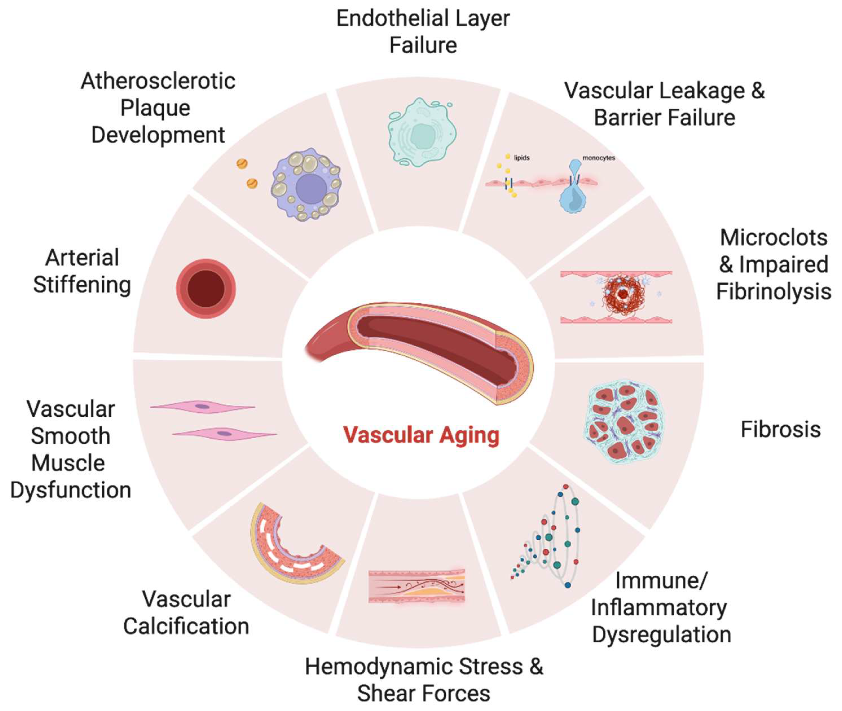
Figure 2.
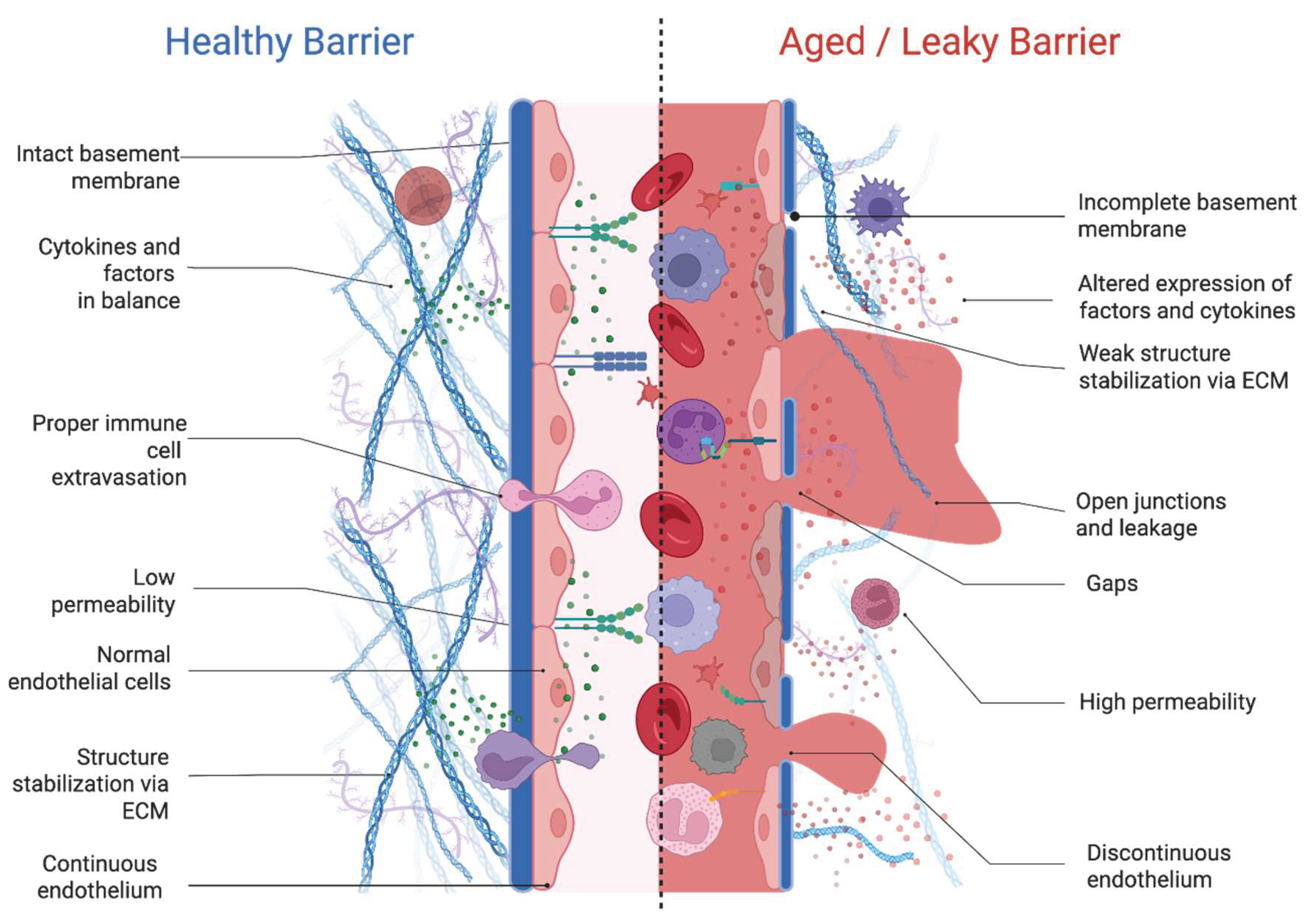
Figure 3.
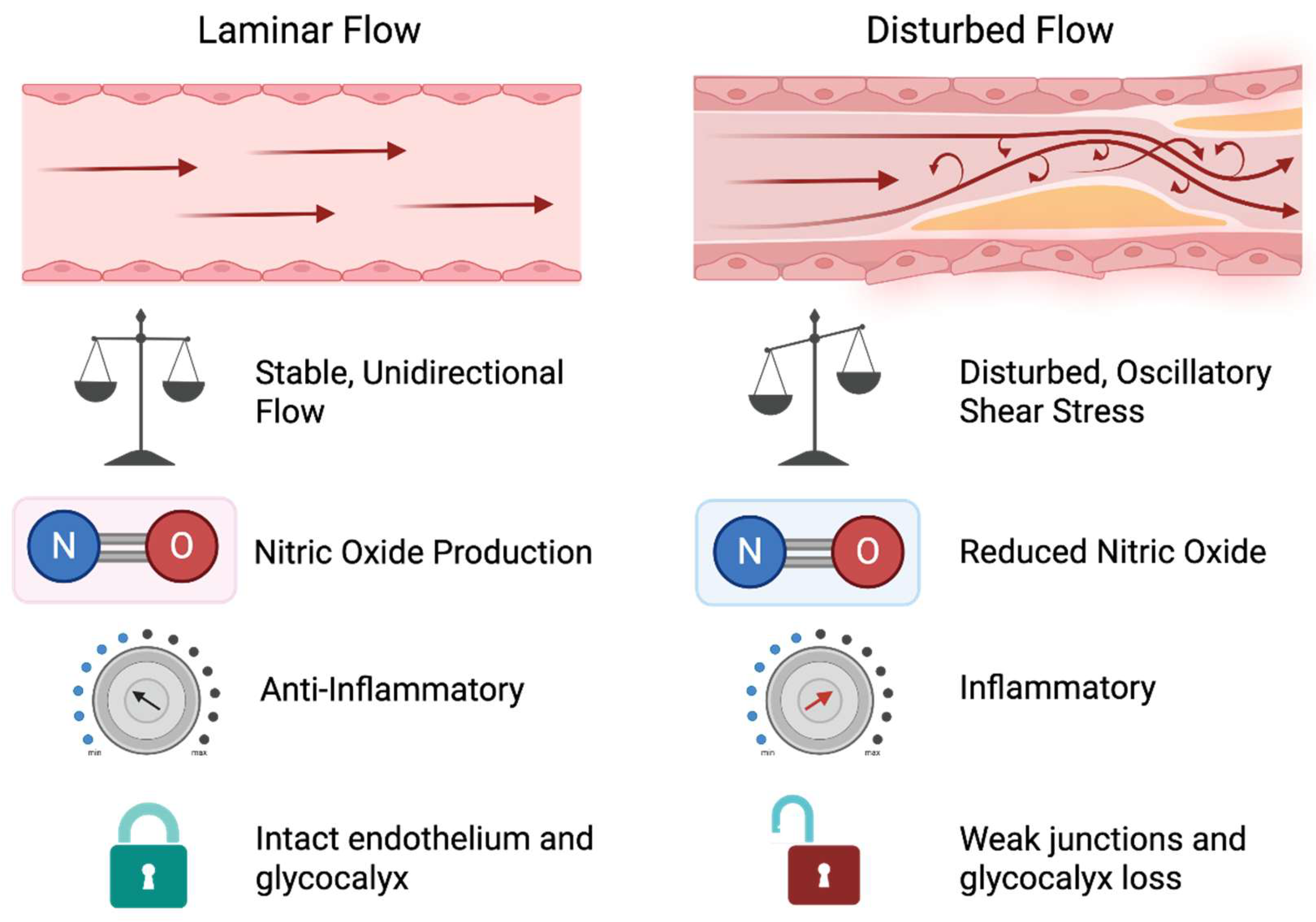
Figure 4.
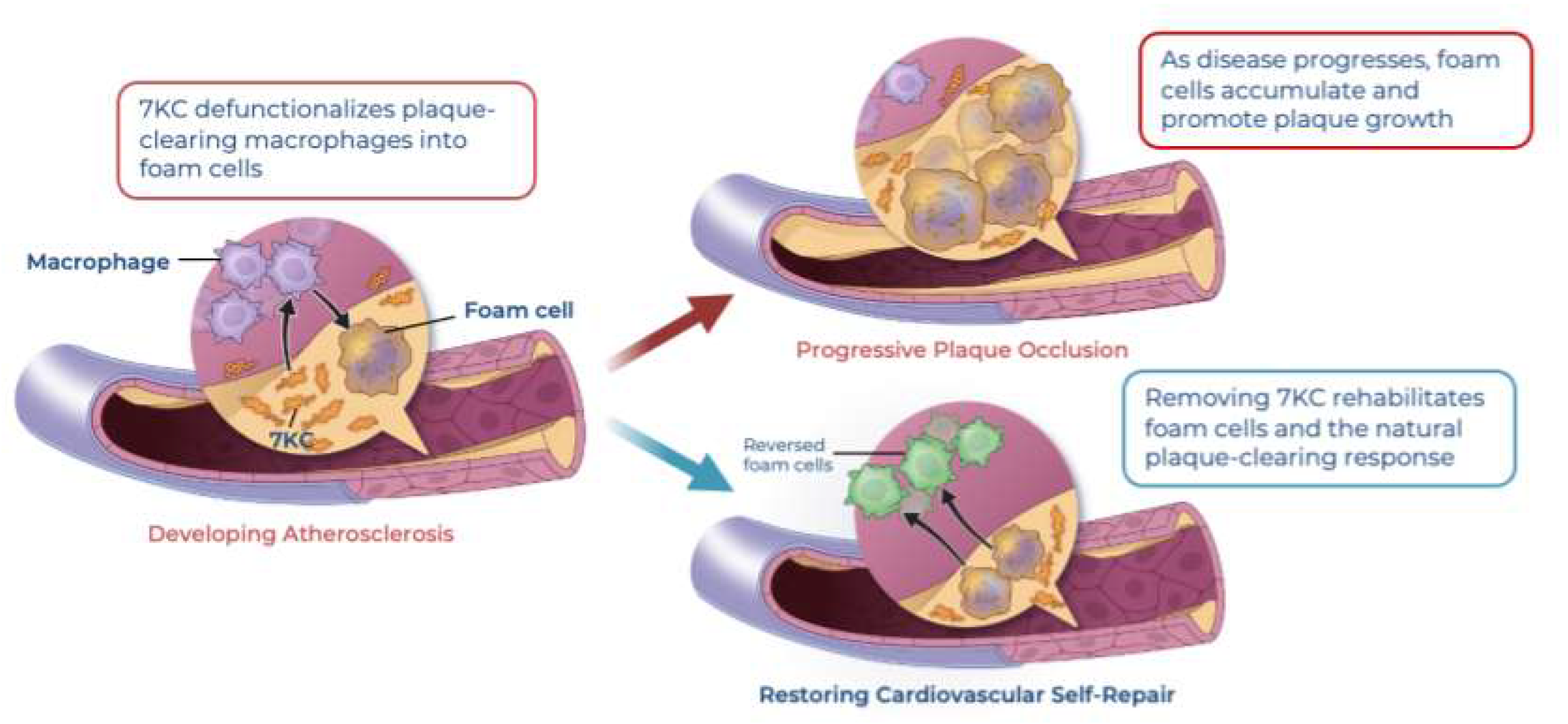
Figure 5.
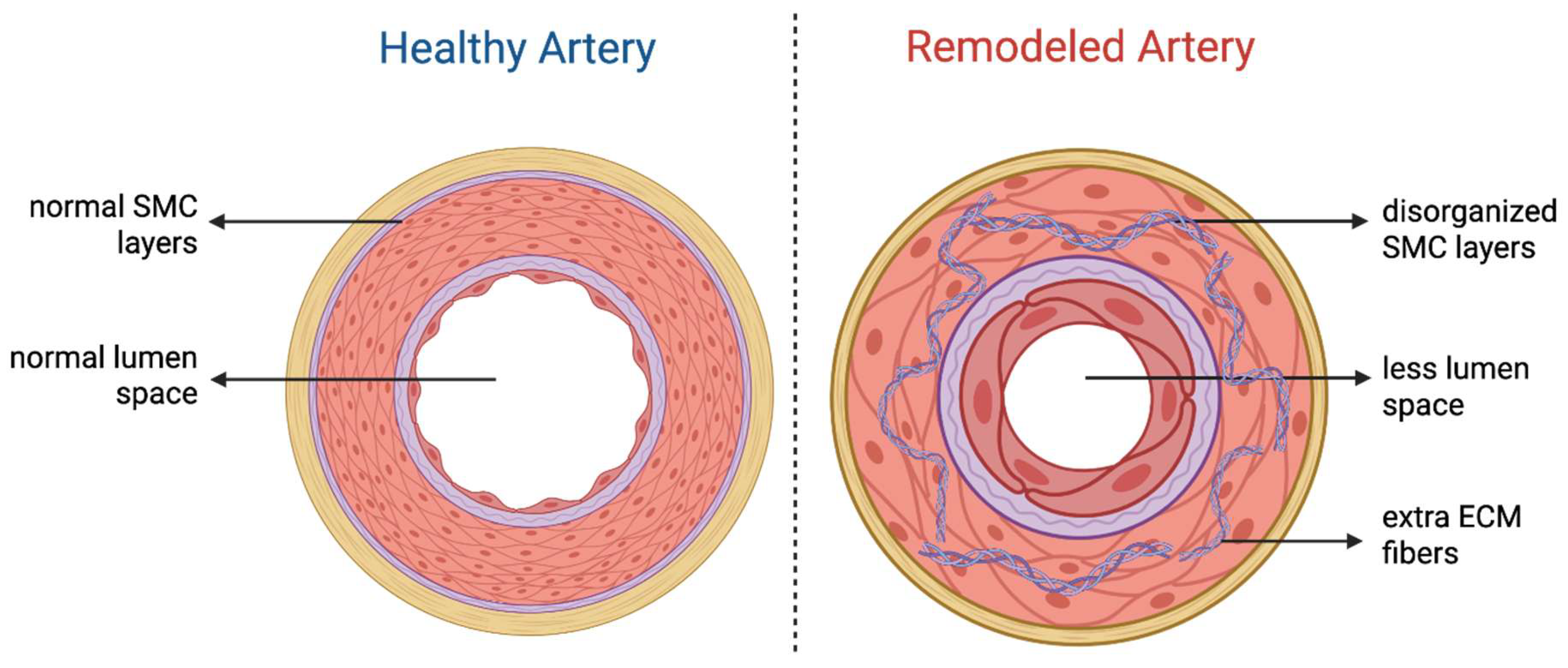
Figure 6.
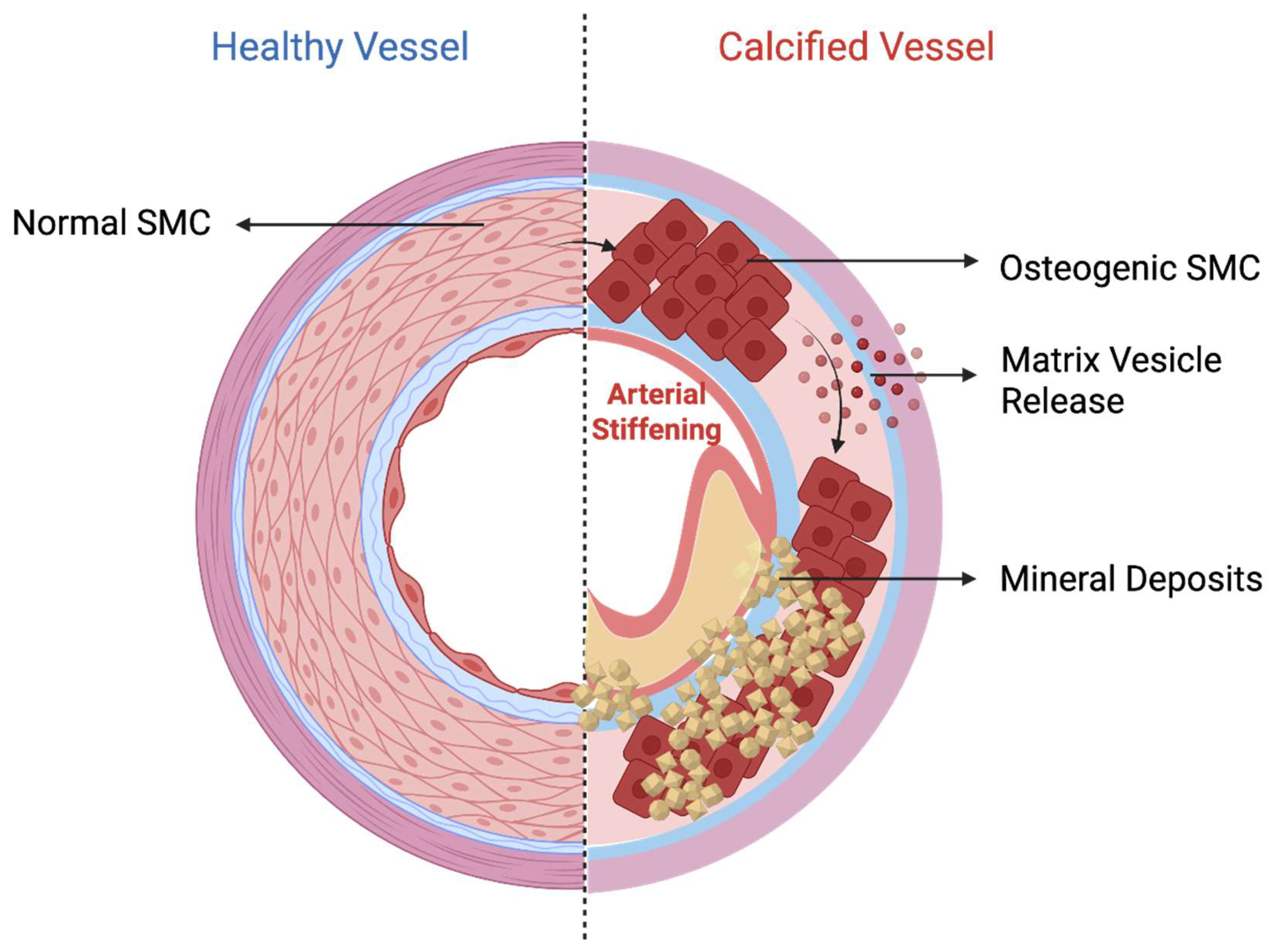
Figure 7.
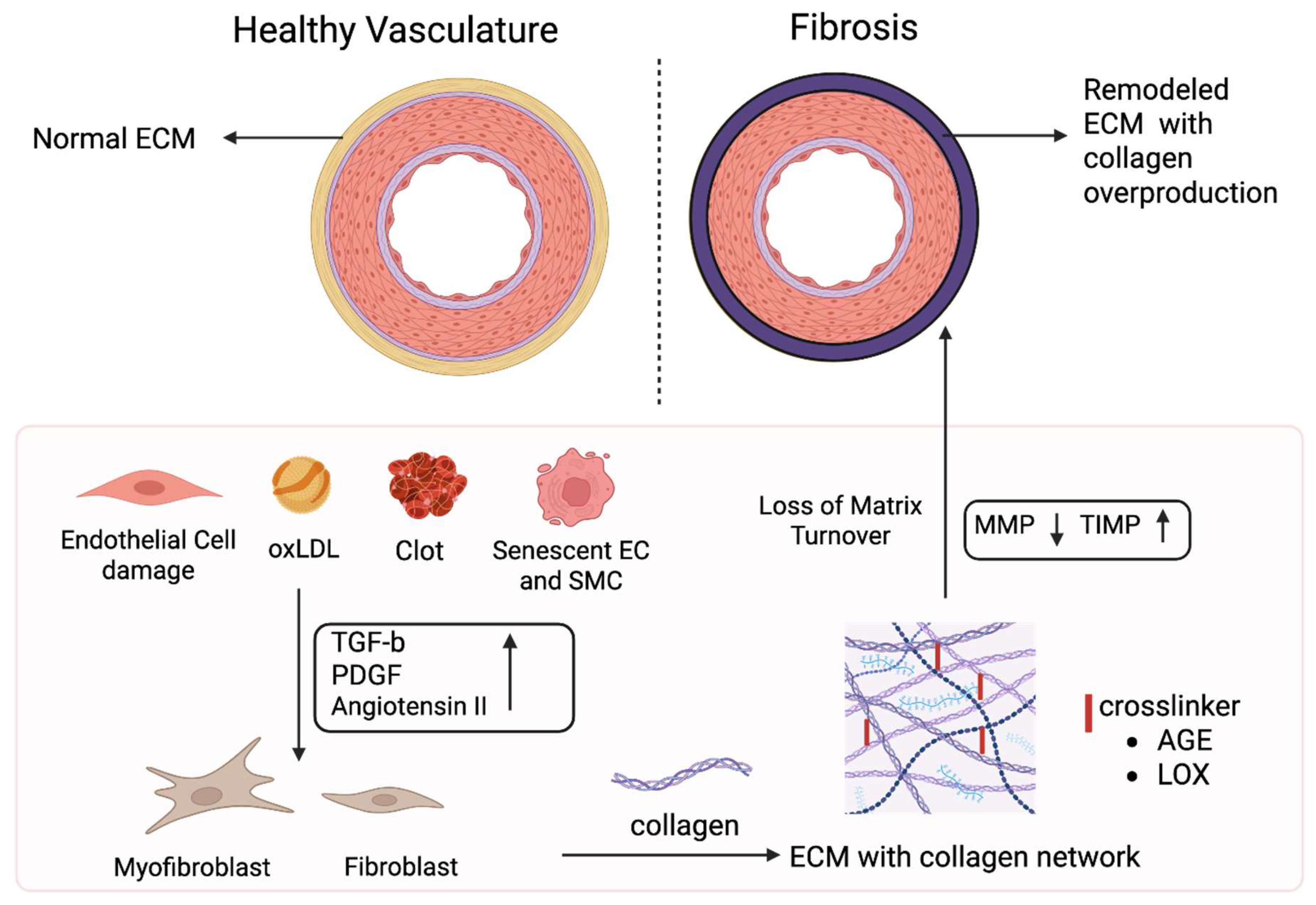
Figure 8.
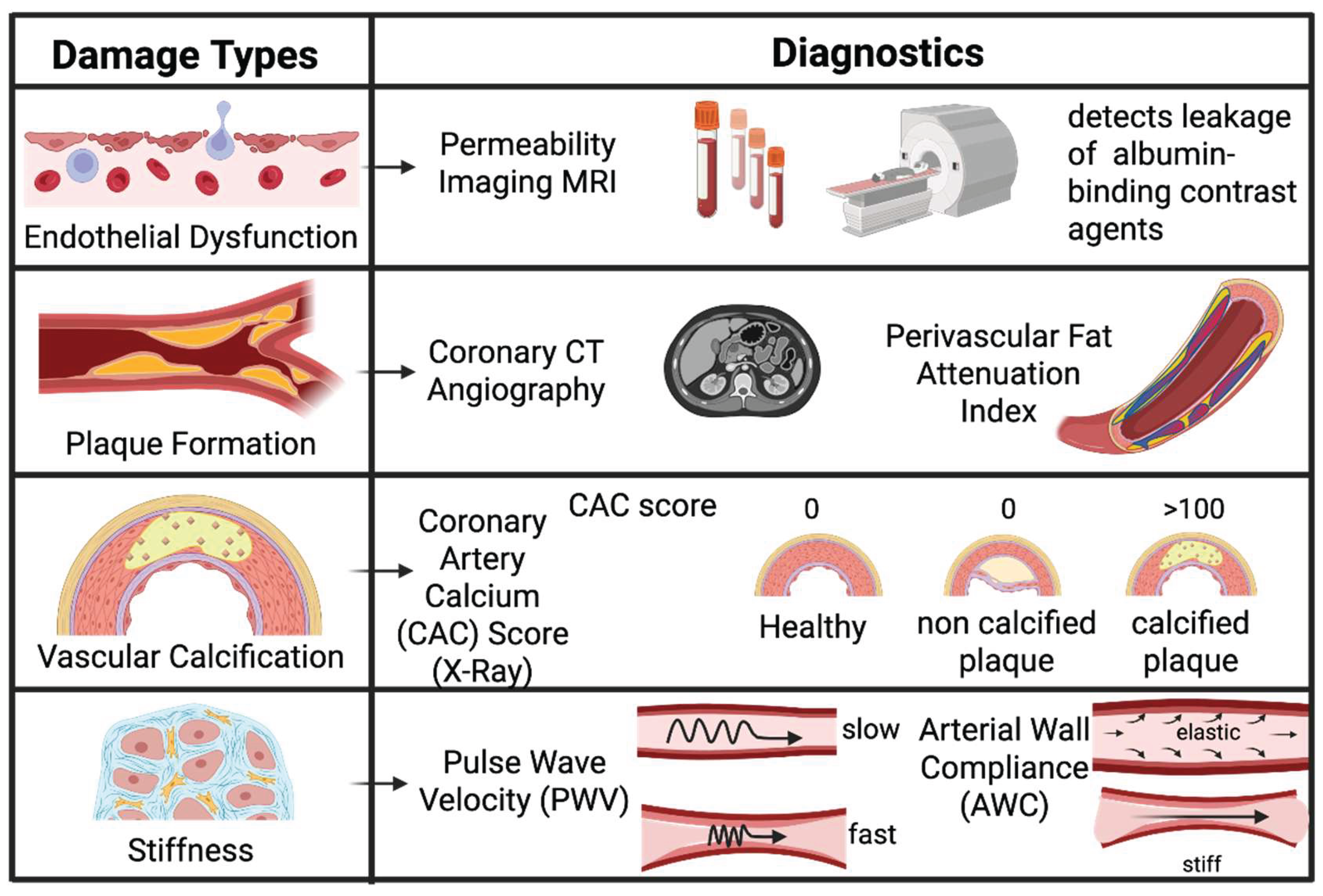
Disclaimer/Publisher’s Note: The statements, opinions and data contained in all publications are solely those of the individual author(s) and contributor(s) and not of MDPI and/or the editor(s). MDPI and/or the editor(s) disclaim responsibility for any injury to people or property resulting from any ideas, methods, instructions or products referred to in the content. |
© 2026 by the author. Licensee MDPI, Basel, Switzerland. This article is an open access article distributed under the terms and conditions of the Creative Commons Attribution (CC BY) license.
Copyright: This open access article is published under a Creative Commons CC BY 4.0 license, which permit the free download, distribution, and reuse, provided that the author and preprint are cited in any reuse.


