

Submitted:
28 September 2026
Posted:
01 October 2026
You are already at the latest version
Abstract
Alzheimer’s disease (AD) pathology is primarily driven by the self-assembly of amyloid-β (Aβ) peptides, a process significantly stabilized through π-π stacking interactions between the aromatic residues Phe19 and Phe20. To competitively disrupt this crucial interaction, we designed novel peptide inhibitors derived from the Aβ recognition motif KLVFFA (Aβ16−21), in which phenylalanine residues were replaced with L-3,4-dihydroxyphenylalanine (DOPA), a residue featuring an enhanced π-stacking and hydrogen-bonding catechol moiety. The DOPA-substituted peptides effectively inhibited Aβ1−42 (Aβ42) aggregation, as demonstrated by Thioflavin T fluorescence assays, circular dichroism spectroscopy, and transmission electron microscopy. These complementary analyses collectively exhibited substantial suppression of β-sheet formation and fibril elongation. Notably, the anti-aggregation potency was dependent on the substitution position, with the Phe19-substituted analogue (KDF) exhibiting stronger inhibitory activity than the Phe20-substituted analogue (KFD). The dual-substituted analogue (KDD) displayed the greatest overall anti-aggregation activity. Molecular simulations further supported the superior activity of KDF over KFD, revealing a more favorable binding free energy for KDF compared to the parent peptide. Furthermore, the DOPA-peptides potently mitigated Aβ42-induced neurotoxicity in SH−SY5Y cells, with the dual-substituted analogue (KDD) providing the highest level of cytoprotection. Collectively, these findings validate that targeted modulation of aromatic interactions through DOPA substitution represents a powerful molecular strategy for controlling Aβ42 self-assembly, presenting these peptides as promising therapeutic leads for AD.
Keywords:
Alzheimer’s disease
; amyloid-β
; aggregation
; KLVFFA
; neurotoxicity
1. Introduction
Alzheimer’s disease (AD) is the most prevalent neurodegenerative disorder, accounting for approximately 60–70% of all dementia cases worldwide. Clinically, AD manifests as progressive impairments in memory, orientation, and executive judgment, accompanied by personality and behavioral changes that ultimately compromise activities of daily living [1,2]. Neuropathologically, widespread synaptic loss and neuronal degeneration result from the aberrant accumulation of misfolded proteins, resulting in irreversible cognitive decline [3]. According to the amyloid cascade hypothesis, the pathological deposition of amyloid-β (Aβ) peptides is a central initiating factor in AD onset and progression [4]. Aβ is generated by sequential cleavage of the amyloid precursor protein by β-secretase and γ-secretase. Under physiological conditions, a dynamic equilibrium between Aβ production and clearance prevents pathological accumulation. However, disruption of this balance promotes self-assembly through a multistep aggregation pathway, progressing from soluble oligomers to mature fibrillar amyloid [5]. Emerging evidence implicates soluble oligomers as particularly neurotoxic species, as they disrupt membrane integrity, impair receptor-mediated signaling, perturb Ca2+ homeostasis, and increase the generation of reactive oxygen species (ROS). These effects, in turn, promote tau hyperphosphorylation and neurofibrillary tangle formation [6,7,8].
Given its central etiological role, inhibition of Aβ1−42 (Aβ42) aggregation remains a promising therapeutic strategy. Several classes of agents, including monoclonal antibodies, small molecules, and natural products, are currently under investigation. Anti-Aβ antibodies such as lecanemab and donanemab have demonstrated clinically meaningful efficacy by reducing brain amyloid burden [9,10]. Among small molecules, transthyretin stabilizers and compounds such as tolfenamic acid suppress oligomer formation by interfering with key aggregation interfaces [11,12]. Additionally, polyphenolic natural products including resveratrol and curcumin have been reported to attenuate β-sheet assembly through π–π stacking interactions with aromatic residues in the hydrophobic core of Aβ, thereby destabilizing amyloidogenic conformations [13,14,15].
Competitive inhibition of Aβ42 self-assembly using short peptide fragments derived from the native Aβ sequence has also emerged as an attractive molecular design principle. These short peptides can bind Aβ with high specificity and suppress the formation of toxic aggregates. We previously developed Aβ42 aggregation inhibitors based on the C-terminal region of Aβ42, the 42-residue isoform that is structurally and functionally distinct from the 40-residue Aβ40. The additional Ile41 and Ala42 residues confer markedly higher aggregation propensity and neurotoxicity [16]. One structural basis for this phenomenon is the stabilizing intermolecular hydrogen bond formed between the carboxylate side chain of Ala42 (–COO−) and the protonated ε-amino group of Lys28 (–NH3+), which reinforces parallel β-sheet packing during fibril maturation [17]. To competitively disrupt this interaction, we previously engineered peptide conjugates consisting of an Aβ42-derived address domain linked to an anchoring moiety such as caffeic acid or vitamin E. These hybrid constructs selectively bound Aβ42, interfered with key hydrogen-bonding motifs, inhibited β-sheet formation, and potently suppressed Aβ-induced neurotoxicity [18,19].
Numerous studies have sought to expand this pharmacophore concept by employing other partial sequences of Aβ, such as residues Aβ16–22 or Aβ26–36, both of which strongly interact with β-sheet interfaces and exhibit robust aggregation-inhibitory activity [20,21,22,23,24]. More recently, peptide analogues designed to target the two hydrophobic pharmacophores of Aβ, Aβ17–21 (LVFFA) and Aβ36–42 (VGGVVIA), have been shown to inhibit and even reverse Aβ fibrillization through a novel soft-aggregation mechanism, further demonstrating the therapeutic potential of pharmacophore-based peptide engineering [25]. To further enhance potency and metabolic stability, a variety of structural modification strategies have been applied to KLVFF- and LVFF-based peptides. These include backbone N-methylation to disrupt β-sheet propagation [26], cyclization to impose conformational constraints [27], and retro-inverso or D-amino acid substitution to improve resistance to proteolytic degradation [28]. In addition, terminal modifications and side-chain protection have been shown to modulate self-assembly behavior and strengthen interactions with amyloidogenic interfaces. Collectively, these approaches increase inhibitory efficacy by preorganizing key hydrophobic and aromatic residues, suppressing β-sheet formation, and stabilizing non-toxic off-pathway assemblies. Such chemically engineered peptide derivatives therefore represent a robust strategy for enhancing the anti-aggregation activity of Aβ-derived pharmacophores.
Building on these advances, we focused on rational modification of Phe19 and Phe20, two aromatic residues critically involved in π–π stacking within parallel β-sheet fibrils. During the early stages of aggregation, these phenylalanine residues align with counterparts in adjacent Aβ molecules, forming intermolecular aromatic stacking interactions that stabilize oligomer growth. To competitively disrupt this process while enhancing Aβ-binding affinity, we designed Aβ16–21 peptides in which Phe19 and Phe20 were substituted with L-3,4-dihydroxyphenylalanine (DOPA). The catechol moiety of DOPA confers superior π-stacking potential and hydrogen-bonding versatility. We hypothesized that DOPA-substituted peptides would preferentially associate with aggregation-prone regions of Aβ, diverting self-assembly toward nonproductive complexes and thereby attenuating fibril propagation.
To evaluate this hypothesis, we assessed the effects of DOPA-modified peptides on Aβ42 aggregation using thioflavin T (ThT) fluorescence assays, circular dichroism (CD) spectroscopy, and transmission electron microscopy (TEM). Furthermore, their cytoprotective effects against Aβ42-induced neurotoxicity were examined in human SH-SY5Y neuroblastoma cell lines. Collectively, our results demonstrate that rational modulation of intermolecular aromatic interactions represents a powerful molecular design strategy for controlling Aβ42 self-assembly. These findings highlight the potential of DOPA-substituted Aβ-derived peptides as promising therapeutic candidates for mitigating the pathological progression of AD.
2. Results and Discussion
2.1. Design Strategy for Aβ42 Aggregation Inhibitor Peptides
Aβ peptides readily assemble into intermolecular parallel β-sheet structures during aggregation, a process in which the phenyl rings of Phe19 and Phe20 align with equivalent residues on adjacent Aβ molecules to form stabilizing π–π stacking interactions [29,30]. These aromatic contacts significantly contribute to β-sheet stabilization and thereby facilitate fibril propagation. We hypothesized that the intrinsic aggregation-inhibitory activity of the Aβ partial sequence Aβ16–20 (KLVFF) originates from its ability to engage in Phe19- and Phe20-mediated aromatic interactions with high affinity, thereby competitively interfering with native intermolecular stacking motifs within full-length Aβ.
To enhance Aβ-binding affinity beyond that achieved by native aromatic residues, we focused on replacing the phenyl side chains of Phe19 and Phe20 with catechol-containing moieties capable of forming stronger and more versatile π–π interactions. Catechol and other phenolic rings are known to interact robustly with aromatic residues in Aβ. Moreover, several polyphenolic antioxidants, such as epigallocatechin and resveratrol, have been shown to suppress Aβ42 aggregation through competitive π–π stacking with the phenyl rings of Phe19 and Phe20 [31]. These findings support the concept that disruption of aromatic packing within the hydrophobic core effectively perturbs β-sheet formation by diverting aggregation interfaces toward alternative binding partners.
Based on this rationale, we designed a series of peptide analogues derived from the recognition sequence KLVFFA (KFF), in which Phe19 or Phe20 was substituted with DOPA (Dp), a residue possessing a catechol group (Figure 1). Previous studies have reported aggregation inhibition using peptides of various lengths centered around Aβ16–20 (KLVFF), and among these, the core Aβ16–20 motif (KLVFF) has been identified as crucial for aggregation [20,22,23,24]. In our approach, we selected this core region, specifically the most essential sequence, and extended it to Aβ16–21 (KLVFFA) to better capture the interaction dynamics with Aβ. To accurately facilitate interaction with the full-length Aβ, we then introduced DOPA substitutions at Phe19 or Phe20, creating the peptides KLVDpFA (KDF) and KLVFDpA (KFD). The double DOPA-substituted peptide, KLVDpDpA (KDD), was also designed to incorporate DOPA at both aromatic sites. These derivatives maintain high target specificity while providing enhanced potential for competitive aromatic interactions with full-length Aβ. We anticipated that incorporation of DOPA would strengthen noncanonical π–π stacking with phenylalanine residues of Aβ, thereby increasing binding affinity, promoting competitive occupation of aggregation-prone interfaces, and ultimately suppressing self-assembly more effectively than the unmodified KLVFFA sequence.
Collectively, this molecular design strategy exploits the aromatic packing dependencies of Aβ42 fibrillization and leverages catechol-mediated interactions to interfere with β-sheet stabilization. These peptide-based inhibitors are expected to bind competitively to aggregation hot spots, disrupt key intermolecular contacts, and robustly attenuate pathogenic Aβ42 aggregate formation.
2.2. Inhibition of Aβ42 Aggregation
To evaluate the effects of DOPA substitution on Aβ42 aggregation, we employed a ThT fluorescence assay. ThT selectively binds to β-sheet-rich amyloid fibrils, resulting in a marked increase in fluorescence intensity; thus, the progressive formation of β-sheet structures can be monitored in real time [32]. In the presence of aggregation inhibitors, reduced β-sheet formation leads to diminished ThT binding and consequently lowers fluorescence signals. Using this assay, we assessed the inhibitory activities of the designed peptide analogues (Figure 2).
When 20 μM Aβ42 was incubated without 20 μM peptides, fluorescence intensity steadily increased over time, consistent with the expected progression of fibril formation. After approximately four hours, the fluorescence signal reached a plateau, indicating that the majority of Aβ42 had assembled into mature fibrillar structures. This plateau intensity was defined as 100% aggregation for quantitative comparison with peptide inhibitors added at equimolar concentrations. Consistent with previous reports demonstrating that KLVFFA can interfere with Aβ self-assembly by competitively binding to the hydrophobic core of Aβ [20,21,22,24], the parent peptide KFF exhibited measurable inhibitory activity, reducing the endpoint fluorescence to approximately 80% of the control. In contrast, substitution of either Phe19 or Phe20 with DOPA significantly enhanced anti-aggregation potency. Incorporation of DOPA at position 19 (KDF) suppressed fluorescence to approximately 30%, demonstrating strong inhibition, while substitution at position 20 (KFD) also reduced fluorescence, albeit to a lesser extent than KDF (approximately 60%). These findings suggest positional dependence in the contribution of aromatic interactions to early aggregation events, with Phe19 likely playing a more prominent role in stabilizing β-sheet stacking interfaces.When both phenylalanine residues were simultaneously replaced with DOPA (KDD), aggregation was further suppressed, with fluorescence intensity dramatically reduced to approximately 20%. This pronounced inhibition suggests that the cooperative action of two catechol moieties enhances competitive binding to aromatic hot spots within Aβ, which may interfere with the π–π stacking interactions involved in fibril growth.
Collectively, these results demonstrate that DOPA substitution at Phe19 and Phe20 effectively augments the anti-aggregation capability of the KLVFFA scaffold in a position-dependent manner, and that dual substitution yields a synergistic enhancement of inhibitory activity.
2.3. CD Analysis of β-Sheet Formation
While the ThT fluorescence assay is widely used to monitor Aβ aggregation through the enhanced fluorescence of ThT bound to β-sheet-rich fibrils, it does not directly quantify the suppression of β-sheet secondary structure. To more directly evaluate the effects of peptide inhibitors on β-sheet formation, CD spectroscopy was conducted. Aggregated Aβ42 predominantly adopts a β-sheet structure characterized by a distinct negative band near 218 nm; thus, the intensity of this band serves as a quantitative indicator of β-sheet content during aggregation [33].
Mixtures containing 20 µM Aβ42 and 20 µM peptide were incubated at 37 °C for 4 hours prior to CD spectral measurements. Peptide-free controls were prepared under identical conditions. In peptide-free controls, aggregated Aβ42 exhibited a strong negative signal at approximately 218 nm, consistent with extensive β-sheet formation (Figure 3). Addition of the Aβ16–21 partial sequence (KFF), previously shown by ThT analysis to attenuate Aβ42 aggregation, reduced the 218 nm band intensity to around 75% of the control, indicating partial suppression of β-sheet assembly. Substitution of either Phe19 or Phe20 with DOPA further enhanced inhibition of secondary structure formation. In the presence of the mono-substituted analogues KFD and KDF, CD intensity decreased to approximately 50% and 40% of the control, respectively. Notably, simultaneous replacement of both phenylalanine residues with DOPA in KDD yielded the most pronounced suppression, reducing the CD signal to approximately 20% of the control, indicative of substantial inhibition of β-sheet formation.
Overall, the degree of CD intensity reduction correlated closely with the inhibition profiles observed in the ThT fluorescence assay, supporting the interpretation that the observed spectral attenuation reflects decreased formation of β-sheet-rich fibrillar structures. These findings suggest that the enhanced inhibitory potency of DOPA-substituted peptides arises from their ability to more effectively disrupt aromatic packing interactions critical for β-sheet assembly. The superior activity of KDF relative to KFD further implies positional sensitivity, with modification at Phe19 exerting a greater perturbative effect on fibril structure than Phe20 substitution.
2.4. TEM Analysis
Aggregation of Aβ42 proceeds through sequential structural transitions, in which soluble oligomeric intermediates evolve into extended fibrillar assemblies enriched in β-sheet secondary structure. These fibrillar aggregates are believed to contribute to amyloid plaque formation in vivo and are strongly associated with neurotoxicity. The ThT fluorescence and CD spectroscopy results demonstrated that the designed peptides suppress β-sheet formation and inhibit fibril elongation. To further elucidate the morphological consequences of peptide addition, TEM was employed.
Ten μM of Aβ42 was incubated in phosphate-buffered saline for 24 hours in the presence or absence of peptide inhibitors prior to TEM imaging (Figure 4). In the absence of peptide, abundant elongated fibrils were observed, forming dense, mesh-like networks characteristic of mature amyloid assemblies. This morphology is consistent with extensive β-sheet organization and reflects the strong fibrillogenic propensity of Aβ42 under physiological ionic conditions.
Upon addition of the parent peptide KFF, fibrillar structures were still detected. However, their abundance and network density were clearly reduced compared with the peptide-free control, consistent with biochemical analyses indicating that KFF partially interferes with fibril growth. More pronounced inhibitory effects were observed when the phenylalanine residues of KFF were substituted with DOPA. Both mono-substituted analogues, KDF and KFD, substantially suppressed fibril formation, with KDF exhibiting more pronounced effects, congruent with its superior inhibition of β-sheet assembly. Strikingly, the doubly substituted analogue KDD yielded only sparse, short, and poorly organized filaments, suggesting that formation of mature fibrillar aggregates was impeded.
The extent of fibril suppression observed by TEM correlated well with reductions in ThT fluorescence intensity and attenuation of the 218 nm β-sheet band in CD spectra, providing orthogonal validation of peptide-mediated aggregation inhibition. Collectively, these results indicate that substitution of phenylalanine with catechol-containing residues enhances the peptide scaffold’s ability to disrupt β-sheet stabilization, thereby preventing the hierarchical assembly of toxic fibrillar aggregates.
2.5. Inhibition of Aβ42-Induced Cytotoxicity
Aβ42 peptides exhibit potent neurotoxicity during self-assembly from soluble monomers into β-sheet-rich fibrillar aggregates. Soluble oligomeric intermediates formed at early aggregation stages are particularly deleterious, as they disrupt membrane integrity, dysregulate calcium ion influx, and enhance ROS generation, collectively provoking intracellular stress and ultimately cell death [7,34,35]. Consistent with these mechanistic insights, treatment of SH-SY5Y neuroblastoma cells with Aβ42 results in reduced cell viability and morphological deterioration, underscoring the pathological relevance of aggregation-driven cytotoxic.
To evaluate whether the DOPA-substituted peptides could mitigate Aβ42-induced neurotoxicity, SH-SY5Y cells were incubated with 15 μM Aβ for 48 hours in the absence or presence of peptide inhibitors. Importantly, in the absence of Aβ42, none of the DOPA-substituted peptides exhibited detectable cytotoxicity toward SH-SY5Y cells (data not shown), indicating that the introduced catechol-containing peptides themselves do not induce measurable cellular toxicity under the present experimental conditions. Treatment with Aβ42 alone decreased cell viability to approximately 40%, confirming robust cytotoxicity in this model system (Figure 5). Co-incubation with the parent KFF peptide modestly attenuated Aβ42 toxicity, increasing viability to approximately 60%, consistent with its partial inhibition of aggregation.
In contrast, substitution of phenylalanine residues with DOPA conferred markedly enhanced, concentration-dependent protective effects. Among the mono-substituted analogues, KDF exhibited superior neuroprotection compared with KFD. At 7.5 μM peptide, KFD increased viability to approximately 70%, whereas KDF restored viability to about 90%. Further increasing KDF concentration to 15 μM nearly nullified cytotoxicity, restoring viability to baseline levels. Notably, dual substitution of both phenylalanine residues (KDD) yielded the strongest protective effect, restoring cell viability to nearly 100% at 7.5 μM.
These results demonstrate that incorporation of catechol-containing residues substantially enhances the peptide scaffold’s ability to counteract Aβ42-induced neurotoxicity. The improved efficacy correlates with inhibition of β-sheet formation and fibrillization observed through ThT fluorescence, CD spectroscopy, and TEM, suggesting that suppression of oligomer–membrane interactions and downstream oxidative stress contribute to the observed cytoprotection.
2.6. Binding Free Energy Analysis
In this study, substitution of phenylalanine residues (Phe19 and Phe20) within the KFF sequence with DOPA was predicted to enhance π–π stacking interactions with Aβ, resulting in enhanced inhibition of β-sheet formation and reduced fibrillization compared with the parent peptide. These effects correlated with potent suppression of Aβ42-induced neurotoxicity in cultured neuronal cells. Notably, replacement of Phe19 with DOPA (KDF) produced greater inhibitory activity than substitution at Phe20 (KFD), suggesting positional dependence in the contribution of aromatic interactions to early aggregation processes.
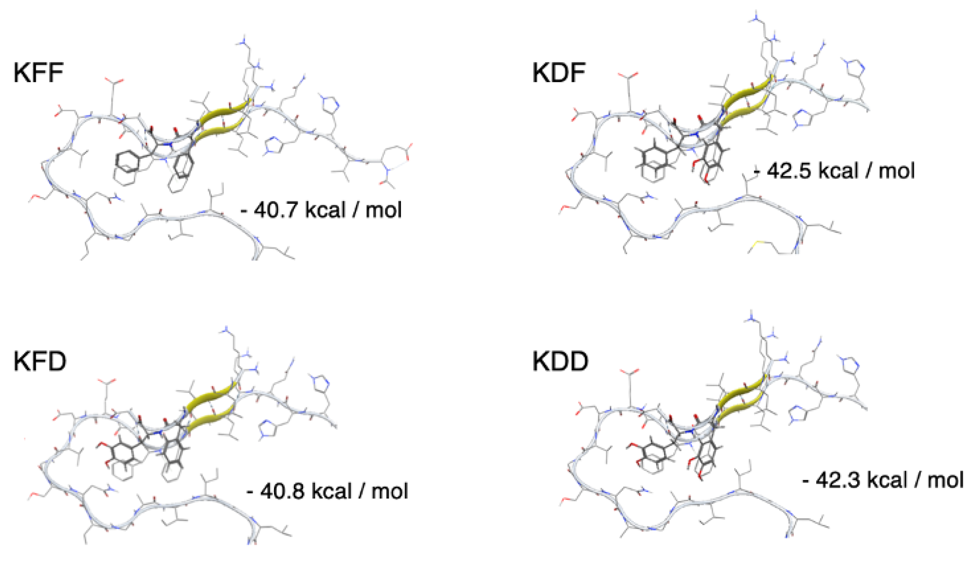
To elucidate the molecular basis of these differences, binding free energy calculations were performed using the Aβ11–42 1-fold structure (PDB ID: 2MXU) as a representative fibril-like conformation [17]. This atomic model of the Aβ42 amyloid fibril, determined by Xiao et al. from solid-state nuclear magnetic resonance (NMR) imaging, served as the structural template. Binding affinities of the peptide variants were evaluated at the Lys16–Ala21 region, a key self-association interface within the Aβ stacking motif. The binding free energy of the parent KFF peptide was calculated as −40.7 kcal·mol−1 (Figure 6). Substitution of Phe20 with DOPA in KFF yielded an essentially unchanged free energy of −40.8 kcal·mol−1, consistent with its modest enhancement in inhibitory activity compared to the parent sequence. In contrast, substitution at Phe19 to generate KDF lowered the binding free energy to −42.5 kcal·mol−1, indicating the formation of a more energetically favorable complex with Aβ. This improved affinity aligns with the experimentally observed increases in aggregation inhibition and neuroprotection. The doubly substituted analogue KDD also exhibited increased binding affinity, with a calculated free energy of −42.3 kcal·mol−1; however, the stabilization was comparable to that of KDF, suggesting that the first substitution at Phe19 has a disproportionately larger contribution to binding enhancement.
These results indicate that, while the computational binding free energy calculations suggest that KDF has a marginally more favorable affinity toward the Aβ fibril interface, the experimental inhibitory effects observed for KDD surpass those of KDF. This discrepancy highlights that factors beyond static binding energetics—such as cooperative hydrogen bonding, extended π–π stacking, and multiple interaction modes—significantly influence the overall inhibitory activity. The presence of two catechol moieties in KDD likely facilitates additional hydrogen bonds and aromatic interactions that are not fully captured in the simplified free energy models. Such synergistic effects underscore the importance of considering multiple physicochemical interaction pathways when evaluating and designing peptide inhibitors. These findings demonstrate that targeted catechol modifications can potentiate inhibition through cooperative interactions, making the disubstituted KDD a particularly promising candidate for further optimization.
Previous studies have shown that catechol structures can inhibit amyloid-β aggregation mainly through oxidation to quinones, which then react via Michael additions [36]. However, our energetic analysis suggests that catechol moieties may also contribute to inhibition through aromatic interactions, including possible π–π stacking with phenylalanine residues in Aβ. This provides a new mechanistic insight, indicating that catechol structures can serve not only as reactive oxidants but also as direct modulators of aromatic stacking—a critical stabilization factor for fibril formation. This observation highlights the multifactorial nature of peptide-mediated aggregation inhibition and suggests that further optimization of multivalent interaction surfaces could yield even more potent inhibitors. Interestingly, a recent study has suggested that the KLVFFA fragment itself may not play a dominant role in the formation of early Aβ oligomer complexes [37]. Our results do not necessarily contradict this observation because we propose that DOPA substitution fundamentally alters the interaction properties of the parent KLVFFA sequence. We hypothesize that the introduced catechol moieties enhance preferential recognition of aggregation-prone Aβ conformations, thereby competitively interfering with β-sheet formation. Further mechanistic studies are currently underway to test this hypothesis. Although direct experimental evidence for π–π stacking between the DOPA-substituted peptides and Aβ has not yet been obtained, the enhanced inhibitory activity together with the structural characteristics of the catechol moiety support the proposed mechanism. Further investigation using long-timescale molecular dynamics simulations is currently underway to clarify the molecular basis of these interactions.
3. Materials and Methods
3.1. Methods
The control peptide (KLVFFA) and DOPA-substituted analogues (KLVFDpA, KLVDpFA, and KLVDpDpA) were synthesized by standard Fmoc-based solid-phase peptide synthesis. Rink amide PEG resin, Fmoc-protected amino acids, and Fmoc-DOPA(3,4-diOxProp)-OH were purchased from Watanabe Chemical Industries, Ltd. (Hiroshima, Japan). Amino-acid couplings were performed in N,N-dimethylformamide (DMF) using HBTU/HOBt as coupling reagents in the presence of N,N-diisopropylethylamine, with each coupling step conducted for 1 h. Fmoc deprotection was carried out using 20% piperidine in DMF. After completion of chain assembly, the peptides were cleaved from the resin and globally deprotected with a trifluoroacetic acid/triisopropylsilane/H2O mixture (95:2.5:2.5, v/v/v) for 3 h. The reaction mixture was filtered, and the filtrate was concentrated under a nitrogen stream. The crude peptides were precipitated with cold diethyl ether, collected by centrifugation, washed with ether, and dried under reduced pressure. Purification was performed by preparative reverse-phase high-performance liquid chromatography (RP-HPLC) using a C18 column. The purity of the isolated peptides (>98%) was confirmed by analytical RP-HPLC. High-resolution mass spectrometry (HRMS) analysis confirmed the molecular masses of all peptides as follows:
HRMS (ESI) m/z [M+H]+ calcd for KLVFFA (C38H59N8O6), 723.4552; found, 723.4474; calcd for KLVFDpA (C38H59N8O8), 755.4450; found, 755.4372; calcd for KLVDpFA (C38H59N8O8), 755.4450; found, 755.4367; calcd for KLVDpDpA (C38H58N8O10), 787.4349; found, 787.4301.
3.2. Aβ42 Pretreatment
To minimize pre-aggregation, Aβ42 (0.25 mg) was dissolved in hexafluoro-2-propanol (HFIP, 0.20 mL) to generate a monomeric stock. HFIP was removed by vacuum centrifugal evaporation to yield a thin peptide film, which was stored until use. For each experiment (ThT fluorescence, CD spectroscopy, TEM, and cell viability assays), the HFIP film was freshly dissolved in the appropriate buffer immediately before assays. The concentration of Aβ42 for each assay was carefully optimized to clearly and sensitively evaluate the differences in the inhibitory effects among the peptides under the specific technical constraints of each experimental system. Accordingly, the experiments described in this study evaluate inhibition of Aβ42 aggregation from freshly prepared peptide solutions rather than disruption of pre-formed oligomers or mature fibrils.
3.3. ThT Fluorescence Assay
Aβ42 was dissolved in 0.3% ammonium hydroxide to 1 mM with brief sonication. Peptides were prepared in DMF at 3 mM; the final concentration of DMF in the assay mixture was 0.1% (v/v). Vehicle controls containing the same concentration of DMF without the peptide were included in all experiments to confirm the absence of solvent interference. For each condition, a master mix was assembled in the following order: 25 µL of 0.2 M phosphate buffer (pH 7.4), 64 µL of Milli-Q water, 5 µL of 2.0 M NaCl, 2 µL of 2 mM peptide stock (or DMF for controls), and 100 µL of 20 µM ThT in glycine–NaOH buffer (pH 9.0). A total of 196 µL of the master mix and 4 µL of the 1mM Aβ42 solution were added to each well of black, non-binding, 96-well plate (µClear; Greiner Bio-One) and incubated at 37 °C. Fluorescence was recorded at 0, 0.5, 1, 2, 3, 4, 6, 8, and 10 hours using a Varioskan LUX microplate reader (Thermo Fisher Scientific) with λ_ex 450 nm and λ_em 485 nm. Plates were shaken for 15 seconds immediately before each read. Data for each condition were obtained from three independent experiments. Note: In this assay system, even when compounds were added after 6 hours of incubation following fibril formation, no reduction in fluorescence was observed. This indicates that the decrease in fluorescence seen in earlier measurements reflects inhibition of fibril formation by the compounds, rather than a direct effect on ThT fluorescence itself. Control experiments confirmed that the DOPA-substituted peptides alone did not generate detectable ThT fluorescence under the assay conditions, indicating that the peptides themselves neither formed ThT-positive assemblies nor contributed to the observed fluorescence signals.
3.4. CD Spectroscopy
Aβ42 secondary structure was evaluated by CD spectroscopy. Aβ was dissolved in 0.3% NH4OH to 200 µM with brief sonication, and peptides were prepared in acetonitrile at 100 µM. For each measurement, samples were prepared in microcentrifuge tubes in the following order: 100 µL of 0.2 M phosphate buffer (pH 7.4), 230 µL of Milli-Q water, 20 µL of 2.0 M NaCl, 100 µL of 100 µM peptide solution (or acetonitrile for peptide-free controls), and 50 µL of 200 µM Aβ42 solution, and incubated at 37 °C for 4 hours. The final concentration of acetonitrile was kept at 20% (v/v) to avoid background absorbance in the far-UV region. After incubation, an aliquot was transferred to a 0.1 mm pathlength quartz cuvette, and spectra were recorded from 200–250 nm on a JASCO CD spectrometer. The spectrum of the corresponding vehicle (buffer/water/NaCl with matching solvent) was subtracted from all datasets. Furthermore, to fully correct for any peptide-specific contributions and potential signal changes during incubation, the spectrum of each peptide solution alone, incubated under identical conditions (37 °C for 4 hours), was subtracted from the respective mixture spectrum.
3.5. TEM
Aβ42 was dissolved in 0.3% ammonium hydroxide to 500 µM with brief sonication. Peptides were prepared in DMF at 1 mM; the final concentration of DMF in the assay mixture was 0.1% (v/v). For each condition, a master mix was prepared as follows: 50 µL of 0.2 M phosphate buffer (pH 7.4), 134 µL of Milli-Q water, 10 µL of 2.0 M NaCl, 2 µL of 1mM peptide stock (or DMF for controls). A total of 196 µL of the master mix and 4 µL of the 500 µM Aβ42 solution were combined in microcentrifuge tube and incubated at 37 °C for 24 hours. After incubation, samples were gently mixed, and when indicated, centrifuged at 4 °C for 10 minutes. Aliquots (5–10 µL) were applied to carbon-coated copper grids and allowed to adsorb for 2–3 minutes. Excess liquid was blotted, and grids were negative-stained with 2% (w/v) uranyl acetate for 6 minutes, followed by a 1-minute rinse with distilled water. Grids were air-dried overnight and imaged on a Hitachi H-7600 transmission electron microscope operating at 300 kV. Images were acquired under identical settings across conditions.
3.6. Cell Culture
The human neuroblastoma cell line SH-SY5Y was obtained from the American Type Culture Collection (ATCC, catalog number CRL-2266). The cells were maintained in a 1:1 (v/v) mixture of Dulbecco’s modified Eagle’s medium and Ham’s F-12 (DMEM/F-12) supplemented with 10% fetal bovine serum (FBS) and penicillin–streptomycin (50 U/mL and 50 µg/mL, respectively). Cells were cultured at 37 °C in a humidified incubator under 5% CO2.
3.7. Aβ42-Induced Cytotoxicity and Peptide Protection Assay
Peptides were dissolved in dimethyl sulfoxide (DMSO); the final concentration of DMSO in the culture medium was 0.1% (v/v), which showed no inherent cytotoxicity. Cells treated with the same concentration of DMSO served as the vehicle control. Vehicle controls containing an equivalent amount of DMSO were included and showed no effect on viability. SH-SY5Y cells were seeded at 1.0 × 10⁴ cells/100 µL/well in type I collagen–coated black 96-well plates (Nippi, Tokyo, Japan) and allowed to adhere for 72 hours. The culture medium was then replaced with phenol red–free DMEM/F-12 containing 2% FBS, 30 U/mL penicillin, and 30 µg/mL streptomycin. Test peptides (at the indicated concentrations) were added together with Aβ42 (stock 150 µM in 0.02% NH4OH, yielding a final Aβ42 concentration of 15 µM). After incubation for 48 hours at 37 °C under 5% CO2, cell viability was quantified using WST-8 assay (Dojindo, Kumamoto, Japan) according to the manufacturer’s protocol. Briefly, 10 µL of WST-8 reagent was added per well, and plates were incubated for 4 hours before measuring absorbance at 450 nm (reference 655 nm) using a GloMax® Multi microplate reader (Promega). Data were normalized to untreated controls, with appropriate vehicle controls included in all experiments.
3.8. Computational Modeling and Binding Free Energy Calculations
Binding affinities between Aβ42 fibrils and partial peptide variants were evaluated using a computational approach based on a previously reported structural model (PDB ID: 2MXU), which comprises 12 stacked Aβ units representing the 1-fold polymorph determined by solid-state NMR [17]. For accurate estimation of intermolecular interactions with the KLVFF motif, the central Aβ molecule (6th or 7th unit) was selected from model 1 to avoid edge effects during energy minimization. Peptide variants were constructed by substituting one or both phenylalanine residues in KLVFF with DOPA, resulting in the following sequences: KFF (unmodified), KDF (Phe19 → DOPA), KFD (Phe20 → DOPA), and KDD (both Phe residues → DOPA). Initial binding poses were generated by aligning the KLVFF motif with the Lys16–Ala21 region of the selected Aβ fibril. Geometry optimization and free energy calculations were conducted using MOE (Chemical Computing Group) using the Amber10:EHT force field. All atoms were allowed to move freely during optimization under the Born solvation, while the Aβ backbone was constrained to preserve the fibril structure. A force tolerance threshold was set to 0.1 RMS kcal·mol−1·Å−2. The dielectric constants were set to 1 and 80 for the interior and exterior of each complex, respectively. Binding free energies were calculated using the GBVI/WSA method implemented in MOE. A comparative analysis of the four peptide variants provided insight into the effects of DOPA substitution on interaction strength with the Aβ fibril, particularly within the central stacking interface.
4. Conclusions
In this study, we designed and synthesized a series of novel peptide derivatives in which the aromatic residues Phe19 and Phe20 within the Aβ partial sequence KLVFFA were substituted with a catechol-bearing DOPA moiety. Their inhibitory effects on Aβ42 aggregation and associated neurotoxicity were systemically evaluated. ThT fluorescence assays, CD spectroscopy, and TEM collectively demonstrated that DOPA substitution significantly suppresses β-sheet formation and fibrillar elongation, with the extent of inhibition dependent on both the number and positional arrangement of substituted residues. In particular, modification at Phe19 yielded pronounced enhancement of inhibitory activity, consistent with binding free energy calculations that revealed more thermodynamically favorable interactions with Aβ. Toxicity assays in SH-SY5Y cells further showed that DOPA-bearing peptides attenuate Aβ42-induced cell death in a dose-dependent manner, with the doubly substituted analogue (KDD) displaying the highest level of neuroprotection. These findings reinforce the central role of aromatic interactions in promoting Aβ42 self-assembly and suggest that the combined strategy of disrupting π–π stacking and expanding hydrogen-bonding networks via catechol incorporation is an effective molecular approach for aggregation inhibition.
Moreover, given that the catechol structure of DOPA possesses potent radical scavenging activity, it is also anticipated that these modified peptides could effectively eliminate reactive oxygen species generated during amyloid-β aggregation, further contributing to neuroprotection. This dual functionality—aggregation disruption and antioxidant activity—may synergistically mitigate the pathogenic processes involved in AD.
While these findings highlight the promising therapeutic potential of DOPA-modified peptides, further optimization is necessary. Since Aβ42 aggregation involves multifaceted interactions beyond the Lys16–Ala21 region, future endeavors should focus on structural modifications—such as peptide cyclization, incorporation of noncanonical amino acids, and reduction of molecular size—to improve pharmacokinetic properties, including blood-brain barrier permeability and proteolytic stability. Advancing these peptides through such optimization strategies will be crucial to translate their inhibitory capacity into effective therapeutics for AD.
Author Contributions
Conceptualization, K.F.; Methodology, K.F., K.M., Y.O., T.T., T.M., Y.D. and M.S.; Software, Y.O.; Validation, K.F. Y.O. and A.O; Investigation, K.F., Y.O., T.T. and K.T.; Data Curation, K.F.; Writing-Original Draft Preparation, K.F.; Writing-Review and Editing; K.F., Y.O. and A.O.; Supervision, K.F. and A.O.; Project Administration, K.F. and A.O.; Funding Acquisition, K.F.
Funding
This research was funded by Grant-in-Aid No. 22K06508 from the Ministry of Education, Culture, Sports, Science, and Technology, Japan.
Institutional Review Broad Statement
Not applicate.
Informed Consent Statement
Not applicable.
Data Availability Statement
The data presented in this study are available in the article.
Conflicts of Interest
The authors declare no conflicts of interest.
Abbreviations
| Aβ | amyloid-β |
| AD | Alzheimer’s disease |
| CD | circular dichroism |
| DMF | N,N-dimethylformamide |
| DMSO | dimethyl sulfoxide |
| DOPA | L-3,4-dihydroxyphenylalanine |
| HRMS | High-resolution mass spectrometry |
| KDD | KLVDpDpA |
| KDF | KLVDpFA |
| KFD | KLVFDpA |
| KFF | KLVFFA |
| NMR | nuclear magnetic resonance |
| RP-HPLC | reverse-phase high-performance liquid chromatography |
| TEM | transmission electron microscopy |
| ThT | thioflavin T |
References
- Prince, M.; Bryce, R.; Albanese, E.; Wimo, A.; Ribeiro, W.; Ferri, C. P. The global prevalence of dementia: a systematic review and metaanalysis. Alzheimers Dement 2013, 9(1), 63–75 e62. [Google Scholar] [CrossRef] [PubMed]
- DeTure, M. A.; Dickson, D. W. The neuropathological diagnosis of Alzheimer’s disease. Mol. Neurodegener. 2019, 14(1), 32. [Google Scholar] [CrossRef] [PubMed]
- Breijyeh, Z.; Karaman, R. Comprehensive Review on Alzheimer’s Disease: Causes and Treatment. Molecules 2020, 25(24). [Google Scholar] [CrossRef] [PubMed]
- Selkoe, D. J.; Hardy, J. The amyloid hypothesis of Alzheimer’s disease at 25 years. EMBO Mol. Med. 2016, 8(6), 595–608. [Google Scholar] [CrossRef] [PubMed]
- Chen, G. F.; Xu, T. H.; Yan, Y.; Zhou, Y. R.; Jiang, Y.; Melcher, K.; Xu, H. E. Amyloid beta: structure, biology and structure-based therapeutic development. Acta Pharmacol. Sin. 2017, 38(9), 1205–1235. [Google Scholar] [CrossRef] [PubMed]
- Huang, Y. R.; Liu, R. T. The Toxicity and Polymorphism of beta-Amyloid Oligomers. Int. J. Mol. Sci. 2020, 21(12). [Google Scholar] [CrossRef] [PubMed]
- Sengupta, U.; Nilson, A. N.; Kayed, R. The Role of Amyloid-beta Oligomers in Toxicity, Propagation, and Immunotherapy. EBioMedicine 2016, 6, 42–49. [Google Scholar] [CrossRef] [PubMed]
- Zhao, L. N.; Long, H. W.; Mu, Y.; Chew, L. Y. The toxicity of amyloid beta oligomers. Int. J. Mol. Sci. 2012, 13(6), 7303–7327. [Google Scholar] [CrossRef] [PubMed]
- Kim, B. H.; Kim, S.; Nam, Y.; Park, Y. H.; Shin, S. M.; Moon, M. Second-generation anti-amyloid monoclonal antibodies for Alzheimer’s disease: current landscape and future perspectives. Transl. Neurodegener. 2025, 14(1), 6. [Google Scholar] [CrossRef] [PubMed]
- Cummings, J.; Osse, A. M. L.; Cammann, D.; Powell, J.; Chen, J. Anti-Amyloid Monoclonal Antibodies for the Treatment of Alzheimer’s Disease. BioDrugs 2024, 38(1), 5–22. [Google Scholar] [CrossRef] [PubMed]
- Ghadami, S. A.; Chia, S.; Ruggeri, F. S.; Meisl, G.; Bemporad, F.; Habchi, J.; Cascella, R.; Dobson, C. M.; Vendruscolo, M.; Knowles, T. P. J.; et al. Transthyretin Inhibits Primary and Secondary Nucleations of Amyloid-beta Peptide Aggregation and Reduces the Toxicity of Its Oligomers. Biomacromolecules 2020, 21(3), 1112–1125. [Google Scholar] [CrossRef] [PubMed]
- Adwan, L.; Subaiea, G. M.; Zawia, N. H. Tolfenamic acid downregulates BACE1 and protects against lead-induced upregulation of Alzheimer’s disease related biomarkers. Neuropharmacology 2014, 79, 596–602. [Google Scholar] [CrossRef] [PubMed]
- Pagano, K.; Tomaselli, S.; Molinari, H.; Ragona, L. Natural Compounds as Inhibitors of Abeta Peptide Aggregation: Chemical Requirements and Molecular Mechanisms. Front Neurosci. 2020, 14, 619667. [Google Scholar] [CrossRef] [PubMed]
- Jakubowski, J. M.; Orr, A. A.; Le, D. A.; Tamamis, P. Interactions between Curcumin Derivatives and Amyloid-beta Fibrils: Insights from Molecular Dynamics Simulations. J. Chem. Inf. Model 2020, 60(1), 289–305. [Google Scholar] [CrossRef] [PubMed]
- Porat, Y.; Abramowitz, A.; Gazit, E. Inhibition of amyloid fibril formation by polyphenols: structural similarity and aromatic interactions as a common inhibition mechanism. Chem. Biol. Drug Des. 2006, 67(1), 27–37. [Google Scholar] [CrossRef] [PubMed]
- Benilova, I.; Karran, E.; De Strooper, B. The toxic Abeta oligomer and Alzheimer’s disease: an emperor in need of clothes. Nat. Neurosci. 2012, 15(3), 349–357. [Google Scholar] [CrossRef] [PubMed]
- Xiao, Y.; Ma, B.; McElheny, D.; Parthasarathy, S.; Long, F.; Hoshi, M.; Nussinov, R.; Ishii, Y. Abeta(1-42) fibril structure illuminates self-recognition and replication of amyloid in Alzheimer’s disease. Nat. Struct. Mol. Biol. 2015, 22(6), 499–505. [Google Scholar] [CrossRef] [PubMed]
- Arai, T.; Ohno, A.; Kazunori, M.; Kakizawa, T.; Kuwata, H.; Ozawa, T.; Shibanuma, M.; Hara, S.; Ishida, S.; Kurihara, M.; et al. Design, synthesis, and evaluation of Trolox-conjugated amyloid-beta C-terminal peptides for therapeutic intervention in an in vitro model of Alzheimer’s disease. Bioorg Med. Chem. 2016, 24(18), 4138–4143. [Google Scholar] [CrossRef] [PubMed]
- Arai, T.; Ohno, A.; Mori, K.; Kuwata, H.; Mizuno, M.; Imai, K.; Hara, S.; Shibanuma, M.; Kurihara, M.; Miyata, N.; et al. Inhibition of amyloid fibril formation and cytotoxicity by caffeic acid-conjugated amyloid-beta C-terminal peptides. Bioorg Med. Chem. Lett. 2016, 26(22), 5468–5471. [Google Scholar] [CrossRef] [PubMed]
- Abedin, F.; Kandel, N.; Tatulian, S. A. Effects of Abeta-derived peptide fragments on fibrillogenesis of Abeta. Sci. Rep. 2021, 11(1), 19262. [Google Scholar] [CrossRef] [PubMed]
- Itoh, S. G.; Okumura, H. Promotion and Inhibition of Amyloid-beta Peptide Aggregation: Molecular Dynamics Studies. Int. J. Mol. Sci. 2021, 22(4). [Google Scholar] [CrossRef] [PubMed]
- Lu, J.; Cao, Q.; Wang, C.; Zheng, J.; Luo, F.; Xie, J.; Li, Y.; Ma, X.; He, L.; Eisenberg, D.; et al. Structure-Based Peptide Inhibitor Design of Amyloid-beta Aggregation. Front Mol. Neurosci. 2019, 12, 54. [Google Scholar] [CrossRef] [PubMed]
- Tjernberg, L. O.; Lilliehook, C.; Callaway, D. J.; Naslund, J.; Hahne, S.; Thyberg, J.; Terenius, L.; Nordstedt, C. Controlling amyloid beta-peptide fibril formation with protease-stable ligands. J. Biol. Chem. 1997, 272(19), 12601–12605. [Google Scholar] [CrossRef] [PubMed]
- Tjernberg, L. O.; Naslund, J.; Lindqvist, F.; Johansson, J.; Karlstrom, A. R.; Thyberg, J.; Terenius, L.; Nordstedt, C. Arrest of beta-amyloid fibril formation by a pentapeptide ligand. J. Biol. Chem. 1996, 271(15), 8545–8548. [Google Scholar] [CrossRef] [PubMed]
- Liu, S.; Peng, F.; Liu, J.; Wu, W.; He, L.; Cao, H.; Liu, C.; Qiu, F.; Zhang, W. Peptides with Soft-Aggregation Behavior Inhibit and Reverse Amyloid-beta Fibrillization. Small 2025, 21(36), e05459. [Google Scholar] [CrossRef] [PubMed]
- Kokkoni, N.; Stott, K.; Amijee, H.; Mason, J. M.; Doig, A. J. N-Methylated peptide inhibitors of beta-amyloid aggregation and toxicity. Optimization of the inhibitor structure. Biochemistry 2006, 45(32), 9906–9918. [Google Scholar] [CrossRef] [PubMed]
- Arai, T.; Sasaki, D.; Araya, T.; Sato, T.; Sohma, Y.; Kanai, M. A cyclic KLVFF-derived peptide aggregation inhibitor induces the formation of less-toxic off-pathway amyloid-beta oligomers. Chembiochem 2014, 15(17), 2577–2583. [Google Scholar] [CrossRef] [PubMed]
- Taylor, M.; Moore, S.; Mayes, J.; Parkin, E.; Beeg, M.; Canovi, M.; Gobbi, M.; Mann, D. M.; Allsop, D. Development of a proteolytically stable retro-inverso peptide inhibitor of beta-amyloid oligomerization as a potential novel treatment for Alzheimer’s disease. Biochemistry 2010, 49(15), 3261–3272. [Google Scholar] [CrossRef] [PubMed]
- Stankovic, I. M.; Niu, S.; Hall, M. B.; Zaric, S. D. Role of aromatic amino acids in amyloid self-assembly. Int. J. Biol. Macromol. 2020, 156, 949–959. [Google Scholar] [CrossRef] [PubMed]
- Kumar, J.; Namsechi, R.; Sim, V. L. Structure-Based Peptide Design to Modulate Amyloid Beta Aggregation and Reduce Cytotoxicity. PLoS ONE 2015, 10(6), e0129087. [Google Scholar] [CrossRef] [PubMed]
- Li, F.; Zhan, C.; Dong, X.; Wei, G. Molecular mechanisms of resveratrol and EGCG in the inhibition of Abeta(42) aggregation and disruption of Abeta(42) protofibril: similarities and differences. Phys. Chem. Chem. Phys. 2021, 23(34), 18843–18854. [Google Scholar] [CrossRef] [PubMed]
- Biancalana, M.; Koide, S. Molecular mechanism of Thioflavin-T binding to amyloid fibrils. Biochim Biophys. Acta 2010, 1804(7), 1405–1412. [Google Scholar] [CrossRef] [PubMed]
- Greenfield, N. J. Using circular dichroism spectra to estimate protein secondary structure. Nat. Protoc. 2006, 1(6), 2876–2890. [Google Scholar] [CrossRef] [PubMed]
- Kawahara, M.; Ohtsuka, I.; Yokoyama, S.; Kato-Negishi, M.; Sadakane, Y. Membrane Incorporation, Channel Formation, and Disruption of Calcium Homeostasis by Alzheimer’s beta-Amyloid Protein. Int. J. Alzheimers Dis. 2011, 2011, 304583. [Google Scholar] [CrossRef] [PubMed]
- Demuro, A.; Mina, E.; Kayed, R.; Milton, S. C.; Parker, I.; Glabe, C. G. Calcium dysregulation and membrane disruption as a ubiquitous neurotoxic mechanism of soluble amyloid oligomers. J. Biol. Chem. 2005, 280(17), 17294–17300. [Google Scholar] [CrossRef] [PubMed]
- Sato, M.; Murakami, K.; Uno, M.; Nakagawa, Y.; Katayama, S.; Akagi, K.; Masuda, Y.; Takegoshi, K.; Irie, K. Site-specific inhibitory mechanism for amyloid beta42 aggregation by catechol-type flavonoids targeting the Lys residues. J. Biol. Chem. 2013, 288(32), 23212–23224. [Google Scholar] [CrossRef] [PubMed]
- Lee, H. T.; Chang, H. W.; Lin, Y. T.; Huang, S. J.; Wu, M. H.; Tsai, C. S.; Tu, L. H.; Lee, M. C.; Chen, Y. R.; Chan, J. C. C. Oligomeric Protein Complexes Formed by Beta Amyloid Peptides and Their Molecular Associates. Chemistry 2025, 31(38), e202501099. [Google Scholar] [CrossRef] [PubMed]
Figure 1.
KLVFFA (KFF) and DOPA-substituted peptides.
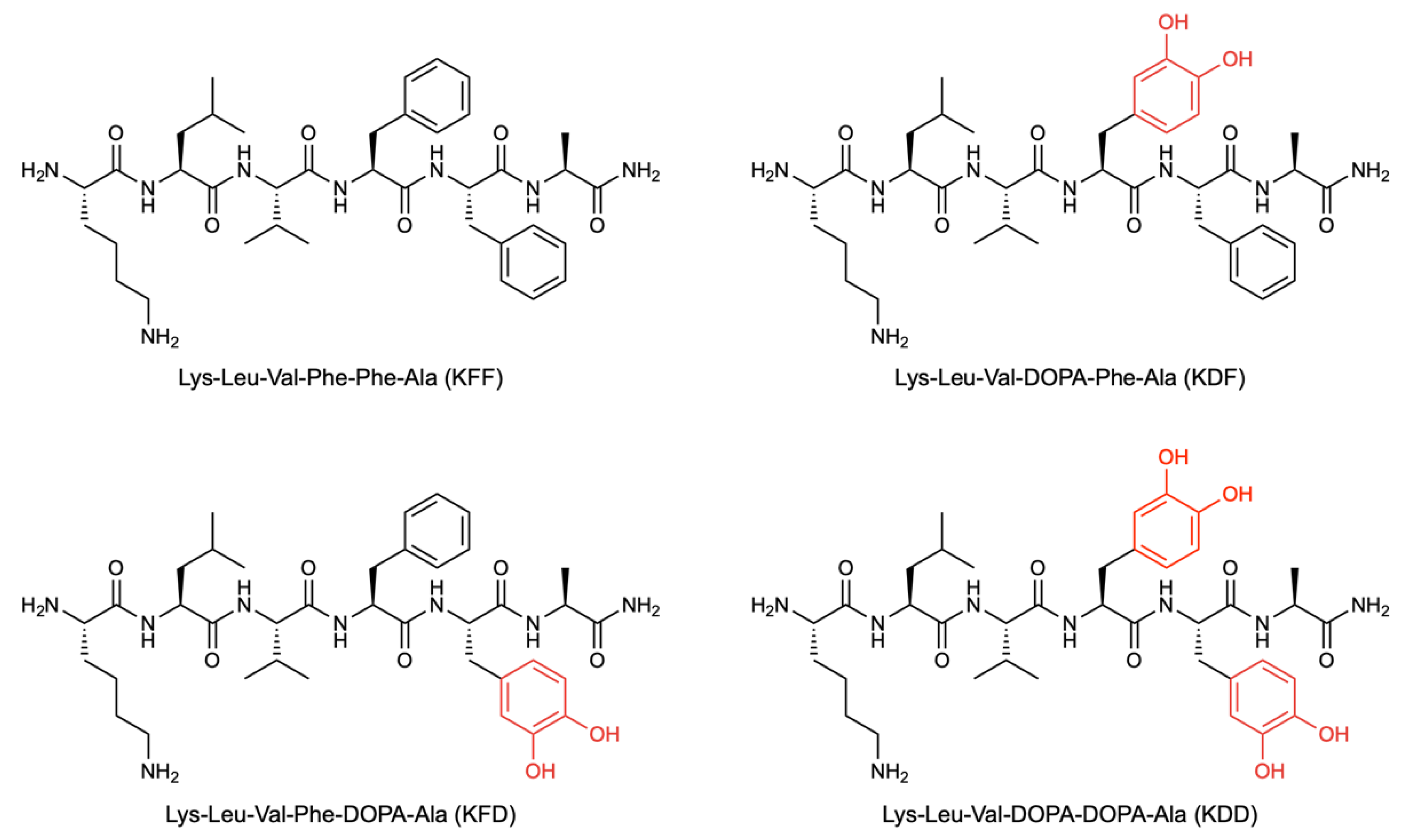
Figure 2.
Inhibitory effects of KFF and DOPA-substituted peptides on Aβ42 aggregation kinetics monitored by ThT fluorescence. Aβ42 (20 µM) was incubated with or without peptides (20 µM) at 37 °C, and Thioflavin T (ThT) fluorescence intensity was measured over time to monitor fibril formation. In the presence of KFF or DOPA-substituted peptides, Aβ42 aggregation proceeded but with reduced ThT fluorescence intensity compared to Aβ42 alone, indicating a lower degree of fibril formation under these conditions.
Figure 2.
Inhibitory effects of KFF and DOPA-substituted peptides on Aβ42 aggregation kinetics monitored by ThT fluorescence. Aβ42 (20 µM) was incubated with or without peptides (20 µM) at 37 °C, and Thioflavin T (ThT) fluorescence intensity was measured over time to monitor fibril formation. In the presence of KFF or DOPA-substituted peptides, Aβ42 aggregation proceeded but with reduced ThT fluorescence intensity compared to Aβ42 alone, indicating a lower degree of fibril formation under these conditions.
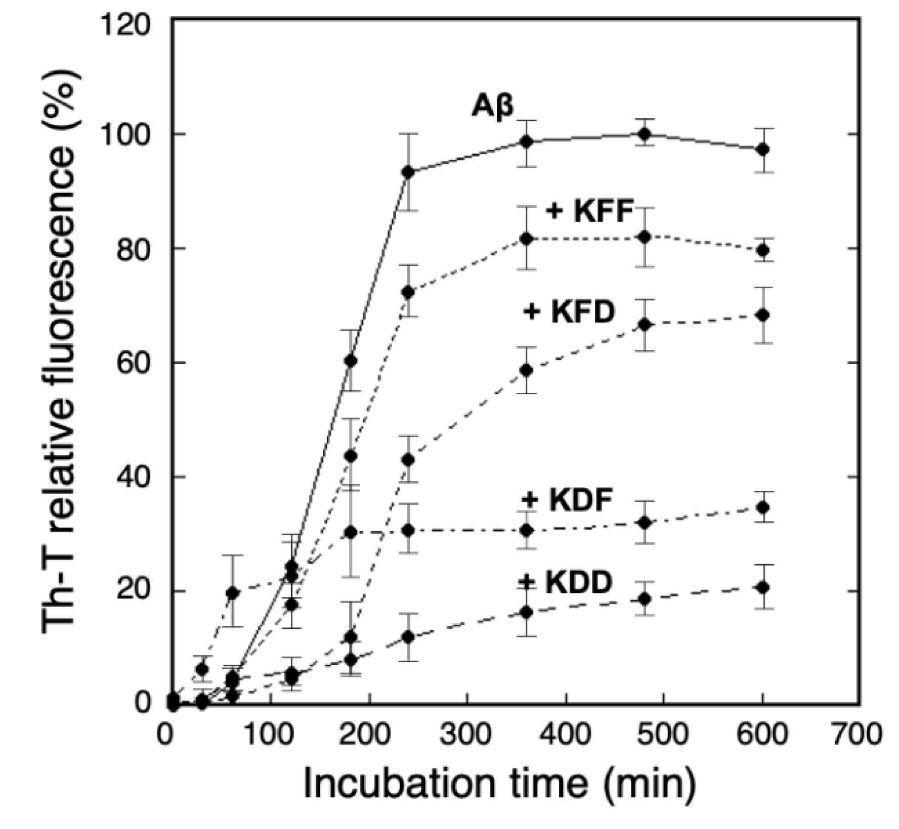
Figure 3.
Effects of synthetic peptides on Aβ42 β-sheet formation analyzed by CD measurements. CD spectra of Aβ42 (20 µM) were recorded after incubation at 37 °C for 4 hours in the absence or presence of peptides (20 µM). Aβ42 alone exhibited a characteristic negative band near 218 nm, indicative of β-sheet-rich structures. Upon addition of KFF or DOPA-substituted peptides, the intensity of this β-sheet-associated signal was reduced, indicating a lower extent of β-sheet formation in the presence of these peptides.
Figure 3.
Effects of synthetic peptides on Aβ42 β-sheet formation analyzed by CD measurements. CD spectra of Aβ42 (20 µM) were recorded after incubation at 37 °C for 4 hours in the absence or presence of peptides (20 µM). Aβ42 alone exhibited a characteristic negative band near 218 nm, indicative of β-sheet-rich structures. Upon addition of KFF or DOPA-substituted peptides, the intensity of this β-sheet-associated signal was reduced, indicating a lower extent of β-sheet formation in the presence of these peptides.
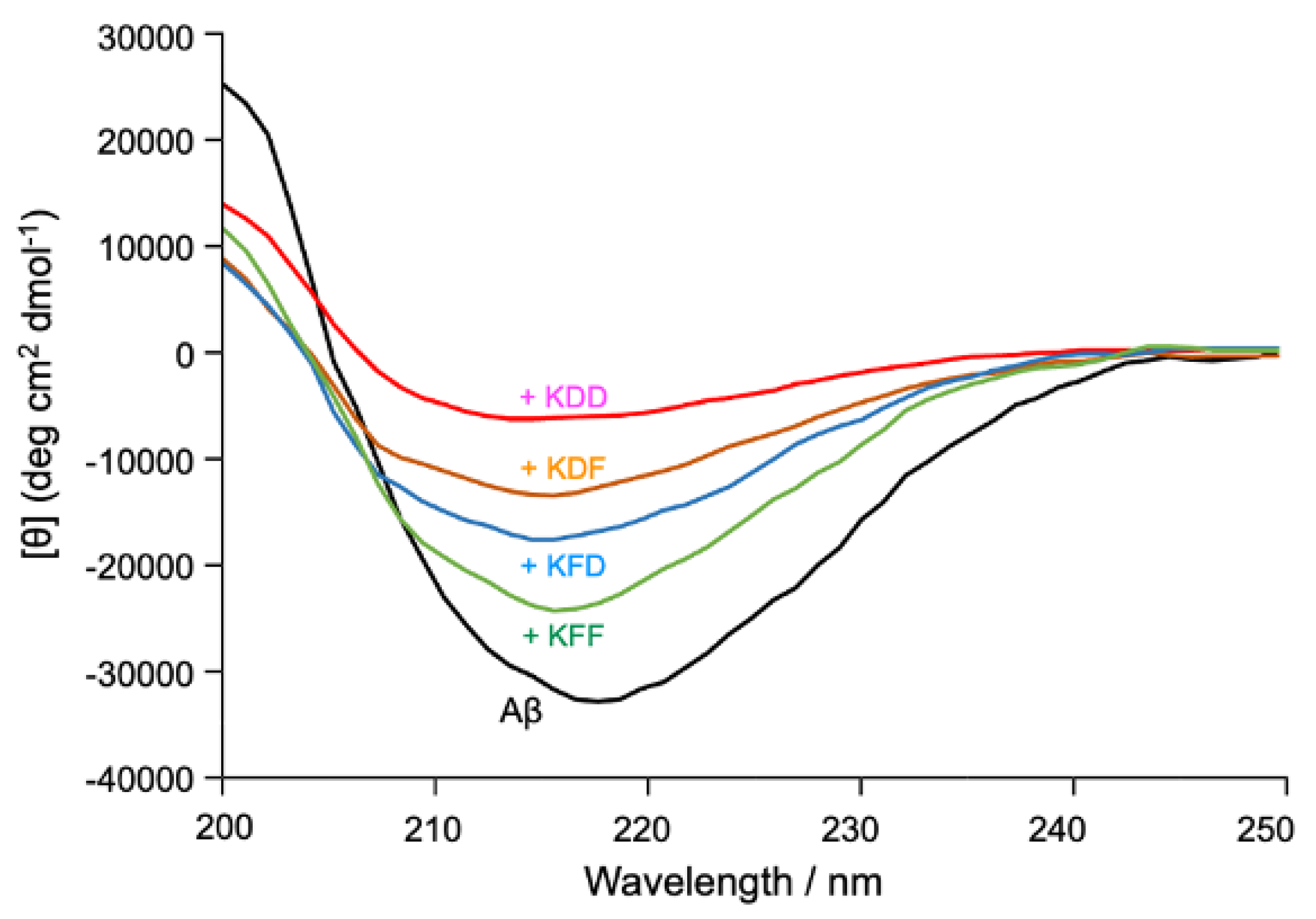
Figure 4.
TEM analysis of Aβ42 fibril morphology in the presence of synthetic peptides. Aβ42 (10 µM) was incubated alone or with synthetic peptides (10 µM) at 37 °C for 24 hours, followed by negative staining and transmission electron microscopy. In the absence of peptides, extensive fibrillar structures characteristic of amyloid aggregates were observed. In contrast, samples incubated with KFF or DOPA-substituted peptides showed a noticeable reduction in fibrillar morphology, with fewer and shorter fibrils observed under the same conditions.
Figure 4.
TEM analysis of Aβ42 fibril morphology in the presence of synthetic peptides. Aβ42 (10 µM) was incubated alone or with synthetic peptides (10 µM) at 37 °C for 24 hours, followed by negative staining and transmission electron microscopy. In the absence of peptides, extensive fibrillar structures characteristic of amyloid aggregates were observed. In contrast, samples incubated with KFF or DOPA-substituted peptides showed a noticeable reduction in fibrillar morphology, with fewer and shorter fibrils observed under the same conditions.
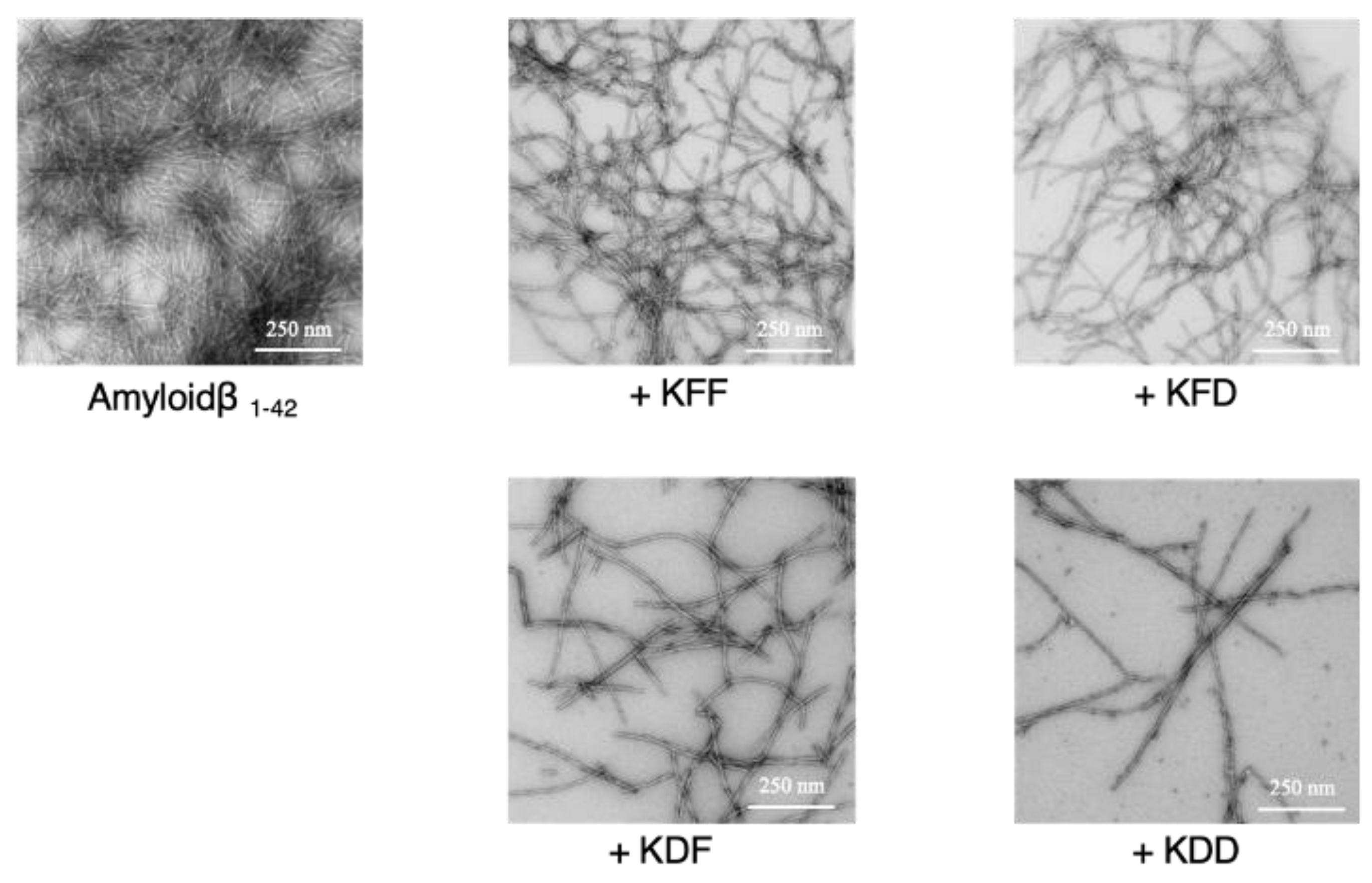
Figure 5.
Protective effects of synthetic peptides against Aβ42-induced cytotoxicity in SH-SY5Y cells. SH-SY5Y cells were cultured in the presence of Aβ42 (15 µM) with or without peptides (7.5 µM and 15 µM) at 37 °C for 48 hours. Cell viability was assessed using the WST-8 assay. Aβ42 treatment significantly reduced cell viability compared to control. Co-incubation with synthetic peptides attenuated Aβ42-induced cytotoxicity in a concentration-dependent manner. Among the peptides tested, KDD showed the most pronounced protective effect, followed by KDF, KFD, and KFF. Statistical significances were determined by Student’s t-tests with P < 0.05 (*), P < 0.01 (**), P < 0.001 (***) as the level of significance (vs Aβ42 addition).
Figure 5.
Protective effects of synthetic peptides against Aβ42-induced cytotoxicity in SH-SY5Y cells. SH-SY5Y cells were cultured in the presence of Aβ42 (15 µM) with or without peptides (7.5 µM and 15 µM) at 37 °C for 48 hours. Cell viability was assessed using the WST-8 assay. Aβ42 treatment significantly reduced cell viability compared to control. Co-incubation with synthetic peptides attenuated Aβ42-induced cytotoxicity in a concentration-dependent manner. Among the peptides tested, KDD showed the most pronounced protective effect, followed by KDF, KFD, and KFF. Statistical significances were determined by Student’s t-tests with P < 0.05 (*), P < 0.01 (**), P < 0.001 (***) as the level of significance (vs Aβ42 addition).
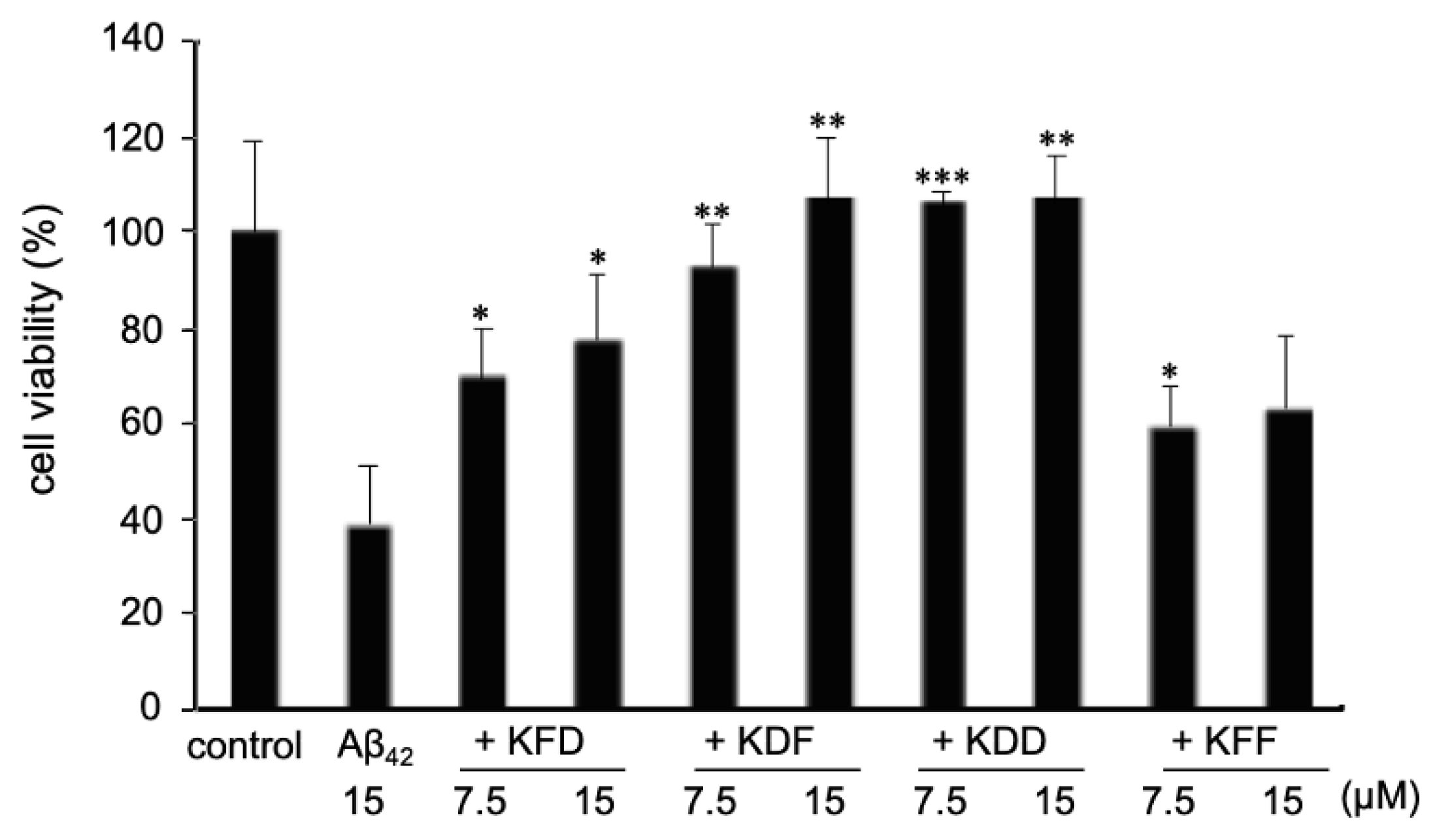
Figure 6.
Binding free energies of peptides docked to Aβ42 fibril core. Atomic models of hexapeptides docked into the Lys16–Ala21 region of Aβ42 fibril (based on PDB ID: 2MXU). Binding free energies were calculated using MOE with the Amber10:EHT force field after energy minimization. DOPA substitutions, especially at Phe19 (KDF and KDD), resulted in reduced binding energies, suggesting enhanced affinity to the Aβ42 fibril interface.
Figure 6.
Binding free energies of peptides docked to Aβ42 fibril core. Atomic models of hexapeptides docked into the Lys16–Ala21 region of Aβ42 fibril (based on PDB ID: 2MXU). Binding free energies were calculated using MOE with the Amber10:EHT force field after energy minimization. DOPA substitutions, especially at Phe19 (KDF and KDD), resulted in reduced binding energies, suggesting enhanced affinity to the Aβ42 fibril interface.
Disclaimer/Publisher’s Note: The statements, opinions and data contained in all publications are solely those of the individual author(s) and contributor(s) and not of MDPI and/or the editor(s). MDPI and/or the editor(s) disclaim responsibility for any injury to people or property resulting from any ideas, methods, instructions or products referred to in the content. |
© 2026 by the authors. Licensee MDPI, Basel, Switzerland. This article is an open access article distributed under the terms and conditions of the Creative Commons Attribution (CC BY) license (http://creativecommons.org/licenses/by/4.0/).
Copyright: This open access article is published under a Creative Commons CC BY 4.0 license, which permit the free download, distribution, and reuse, provided that the author and preprint are cited in any reuse.