

Submitted:
28 May 2026
Posted:
29 May 2026
You are already at the latest version
Abstract
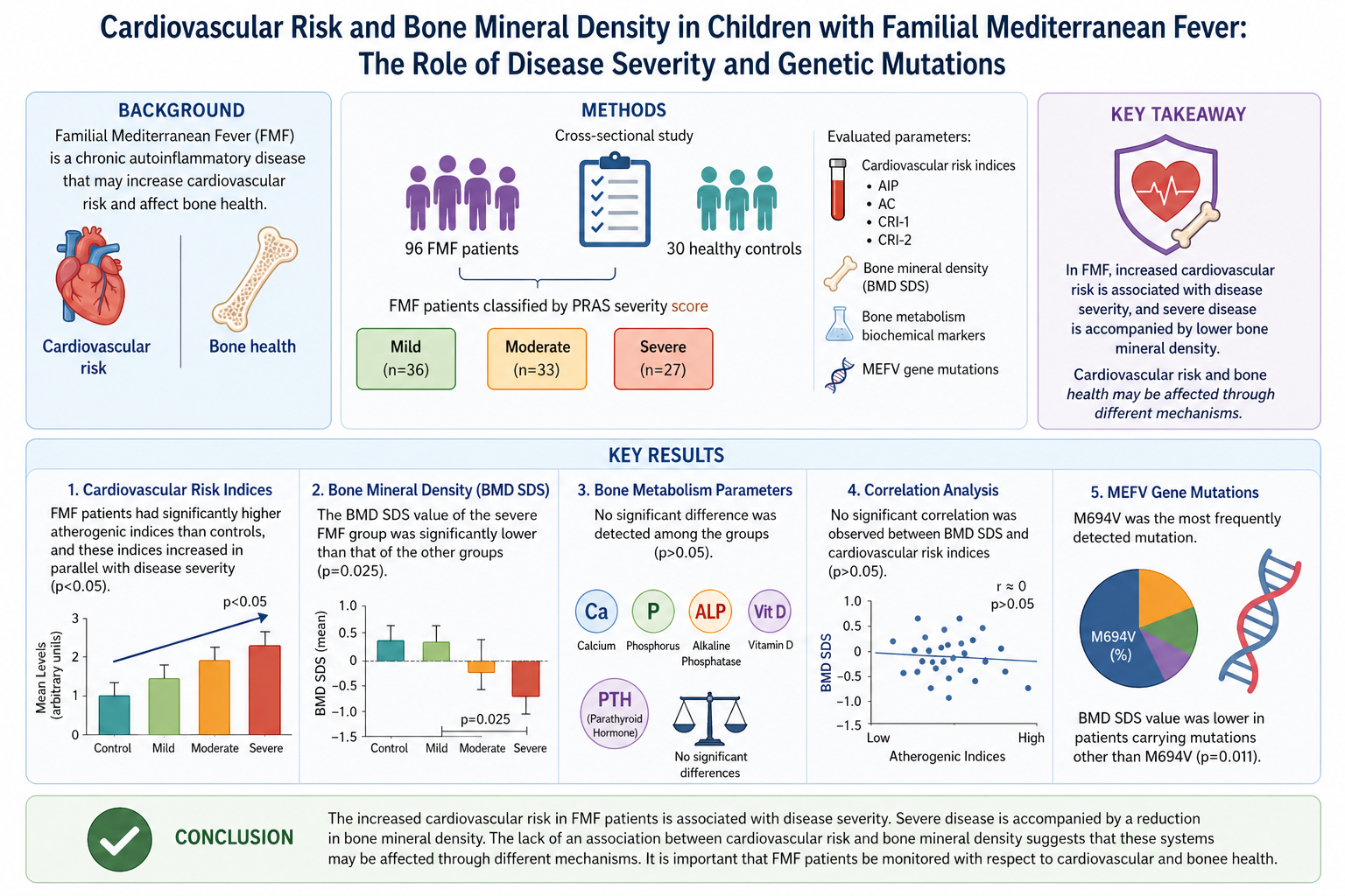
Objective: In the present study, cardiovascular risk indices (Atherogenic Index of Plasma [AIP], Atherogenic Coefficient [AC], Castelli Risk Index-1 [CRI-1], and Castelli Risk Index-2 [CRI-2]) and bone mineral density (BMD SDS) were evaluated in patients with Familial Mediterranean Fever (FMF), and the association of these parameters with disease severity and MEFV gene mutations was investigated.
Methods: A total of 126 participants (96 FMF patients and 30 healthy controls) were included in the study. FMF patients were classified as mild, moderate, or severe according to the PRAS severity score. Cardiovascular risk indices, biochemical parameters of bone metabolism, and BMD SDS values were compared among the groups. The relationship between BMD SDS and atherogenic indices was assessed by Spearman correlation analysis. The results were also analyzed according to MEFV gene mutations.
Results: No significant difference was detected among the groups in terms of sex or age (p>0.05). FMF patients were found to have substantially higher cardiovascular risk indices than the control group, and these indices increased in parallel with disease severity (p< 0.05). The BMD SDS value of the severe FMF group was significantly lower than that of the other groups (p=0.025). No difference was detected among the groups with respect to bone metabolism parameters (p>0.05). No significant correlation was observed between BMD SDS and cardiovascular risk indices (p>0.05). M694V was the most frequently detected mutation, and the BMD SDS value was lower in patients carrying mutations other than M694V (p=0.011).
Conclusion: The increased cardiovascular risk in FMF patients is associated with disease severity. Severe disease is accompanied by a reduction in bone mineral density. The lack of an association between cardiovascular risk and bone mineral density suggests that these systems may be affected through different mechanisms. It is important that FMF patients be monitored with respect to cardiovascular and bone health.
Keywords:
child
; cardiovascular risk
; familial Mediterranean fever
; bone mineral density
1. Introduction
Familial Mediterranean Fever (FMF) is one of the most common autoinflammatory diseases of childhood; it is inherited in an autosomal recessive manner and is characterized by serositis and recurrent febrile attacks. The principal pathogenesis of the disease is related to overactivation of the pyrin inflammasome secondary to MEFV gene mutations and to the resulting chronic inflammatory response [1,2].
Although asymptomatic periods are present between attacks, inflammatory activity is thought to remain incompletely suppressed, and this may contribute to organ damage in the long term. Attack frequency varies from patient to patient and can range from once every three to four months to weekly or more frequent occurrences. Attack periods can substantially impair daily life and stand in clear contrast to the complete sense of well-being observed during attack-free intervals [3].
FMF is frequently encountered, particularly in populations originating from the Mediterranean basin such as Turks, Armenians, Jews, and Arabs [4]. Turkey is one of the countries in which the disease is most frequently observed, with a prevalence of approximately 1:1000 [5]; however, in one study the prevalence was found to decrease to 6:10000 in the northwestern regions [6].
Although studies addressing cardiovascular risk markers and bone mineral density in children with FMF are available, studies in which these two systems are jointly evaluated in relation to disease severity and genetic mutations are quite limited. In addition, given the effects of MEFV gene mutations on disease severity and the inflammatory response, the association of these genetic variations with cardiovascular risk and bone metabolism also appears worth investigating.
In the present study, the aim was to investigate the association of cardiovascular risk markers and bone mineral density parameters with disease severity and Mediterranean FeVer (MEFV) gene mutations in children and adolescents diagnosed with FMF.
2. Materials and Methods
This study was planned as a cross-sectional and comparative study conducted at the Pediatrics Clinic of Kastamonu Training and Research Hospital between January 2026 and May 2026. A total of 126 children and adolescents, comprising 96 FMF patients and 30 healthy controls, were enrolled in the study. The diagnosis of FMF was established according to the Yalçınkaya criteria [7], and MEFV gene mutations were assessed in the genetic analyses of the patients. Patients diagnosed with FMF were divided into three groups as mild (n=32), moderate (n=32), and severe (n=32) using the Pras disease severity score [8] and taking into account its pediatric-adapted version [9]. The control group was composed of healthy children with no chronic disease and with similar characteristics in terms of age and sex (n=30).
Triglyceride (TG), parathyroid hormone (PTH), alkaline phosphatase (ALP), phosphorus (P), calcium (Ca), high-density lipoprotein cholesterol (HDL-C), low-density lipoprotein cholesterol (LDL-C), total cholesterol (TC), and vitamin D (25-OH vitamin D) levels were measured in the patients, and bone mineral density (BMD) was assessed. Clinical symptoms (abdominal pain, fever, arthritis, chest pain, and similar findings) and MEFV gene mutations of the patients diagnosed with FMF were recorded.
Cardiovascular risk indices were calculated using the following formulas [10]:
Atherogenic Index of Plasma (AIP) = log(TG/HDL-C),
Atherogenic Coefficient (AC) = (TC − HDL-C)/HDL-C,
Castelli Risk Index-1 (CRI-1) = TC/HDL-C,
Castelli Risk Index-2 (CRI-2) = LDL-C/HDL-C.
All blood analyses were performed on samples obtained after at least 10–12 hours of fasting, following written informed consent at the time of admission. Serum concentrations of TG, TC, HDL-C, and LDL-C were analyzed by spectrophotometry (Beckman Coulter AU 5800), and Ca, P, ALP, PTH, and vitamin D concentrations were analyzed by chemiluminescence and spectrophotometry (Ca, P, and ALP: Beckman Coulter AU5800 clinical chemistry analyzer Inc., CA, USA; PTH and vitamin D: Beckman Coulter UniCel DxI 800 Access immunoassay analyzer Inc., CA, USA). Bone mineral density (BMD) measurements were performed at the lumbar vertebrae (L1–L4) using the dual-energy X-ray absorptiometry (DEXA) method (Stratos DR, DMS Imaging, Le Montueux, FRANCE), after the patient had been informed about radiation exposure and written informed consent had been obtained. The results were obtained in g/cm2 and recorded as standard deviation scores adjusted for age and sex (BMD SDS) using the Chıld Metrıcs calculation program developed by Ceddcozum [11]. MEFV gene mutations were analyzed by real-time PCR (DNA-Technology, Moscow, RUSSIA).
The four groups were compared in terms of demographic data, AIP, AC, CRI-1, and CRI-2, as well as Ca, P, ALP, PTH, vitamin D, and BMD SDS values. Spearman correlation analysis was performed to investigate whether an association existed between BMD SDS values and cardiovascular risk indices in patients diagnosed with FMF. Patients with FMF were divided into two groups, those carrying the M694V mutation and those carrying mutations other than M694V, and they were compared with respect to AIP, AC, CRI-1, CRI-2, and BMD SDS values.
2.1. Exclusion Criteria
Individuals with the following criteria were excluded from the study: those with diabetes mellitus, chronic renal failure, liver disease, or endocrine disorders; those receiving medications that affect bone metabolism (corticosteroids, antiepileptics, and similar drugs); and those with obesity, malnutrition, or another chronic inflammatory disease.
2.2. Statistical Analysis
SPSS program, ver26.0 (IBM Corp., Armonk, NY, USA), was used to conduct statistical analyses. The Kolmogorov–Smirnov test was used to determine if continuous variables were normal. While non-normally distributed data were displayed as median (IQR), regularly distributed continuous variables were portrayed as mean±SD. ANOVA for regularly distributed variables and the Kruskal–Wallis test for non-normally distributed variables were used to compare four separate groups. When necessary, the Tukey test was used for post hoc pairwise comparisons. Spearman’s rank correlation coefficient was used to evaluate correlations between continuous variables. The independent samples t-test or the Mann–Whitney U test, if applicable, were used to compare FMF patients with and without mutations. Distribution patterns were illustrated via pie charts. A two-sided method with a 95% confidence level was used for all statistical analyses. A p-value of less than 0.05 was deemed statistically significant.
2.3. Ethical Approval
Approval for this study was granted by the Ethics Committee of Kastamonu University Faculty of Medicine (decision no: 2025-28; approval date: 15 January 2026). The study was conducted in accordance with the principles of the Declaration of Helsinki. Written informed consent was obtained from the parents of all participants.
3. Results
A total of 126 participants (51 [40.5%] boys and 75 [59.5%] girls) were included in the study, and the mean age was 9.22±4.69 years. No significant difference was observed among the four groups in terms of age (p=0.992) or sex (p=0.870).
The distribution of clinical symptoms in patients with FMF is shown in Figure 1, and the distribution of MEFV gene mutations is shown in Figure 2.
Post hoc analyses demonstrated significant pairwise differences among the groups with respect to the cardiovascular risk indices AIP, AC, CRI-1, and CRI-2 (p<0.05). The cardiovascular risk indices of FMF patients were found to be significantly higher than those of the control group, and these indices were observed to rise significantly as disease severity increased (p<0.001) (Table 1).
In post hoc comparisons, the BMD SDS value was found to be significantly lower in FMF patients in the severe group than in FMF patients with moderate and mild disease and in the control group (p=0.025). No significant difference in BMD SDS was detected between FMF patients with moderate or mild disease and the control group (p>0.05). The biochemical parameters related to bone metabolism, namely Ca, P, ALP, vitamin D, and PTH, were observed to be within normal ranges in all four groups, and no statistically significant difference was found among the groups for these parameters (p>0.05) (Table 2).
In the Spearman correlation analysis, no significant correlation was detected between BMD SDS and the atherogenic indices AIP, AC, CRI-1, and CRI-2 (Table 3).
In comparisons between FMF patients with the M694V mutation and FMF patients with mutations other than M694V, no significant differences were found in the AIP, AC, CRI-1, or CRI-2 measurements (p>0.05), whereas the BMD SDS value was found to be significantly lower in patients carrying mutations other than M694V than in those carrying the M694V mutation (p=0.011) (Table 4).
4. Discussion
In the present study, cardiovascular risk indices in children and adolescents diagnosed with FMF were shown to be significantly higher than those in healthy controls, and they were observed to rise markedly as disease severity increased. In addition, bone mineral density was found to be significantly reduced in the severe disease group. However, no significant correlation was found between cardiovascular risk indices and BMD. In the genotype-based analysis, BMD SDS was determined to be lower in patients carrying mutations other than M694V.
Familial Mediterranean Fever is a chronic inflammatory disease caused by mutations in the MEFV gene, which encodes the pyrin protein that is thought to play a role in inflammatory pathways. These mutations may result in loss of pyrin function, leading to uncontrolled inflammation, which in turn may give rise to endothelial dysfunction, prothrombotic states, and functional alterations in the microcirculation, ultimately predisposing to atherosclerosis [12]. In addition, FMF patients have been shown in previous studies to have abnormal lipid profiles [13], and epidemiological studies have demonstrated that dyslipidemia on its own is sufficient to drive the atherosclerotic process even in the absence of other risk factors.
In recent years, atherogenic indices have emerged as indicators of atherosclerosis and cardiovascular disease [10,14]. Therefore, the monitoring of these parameters is of considerable importance for early intervention regarding potential cardiovascular risks. In the study by Bhardwaj et al. [15], cardiovascular risk indices were found to be markedly elevated in individuals with angiographically confirmed coronary artery disease. Similarly, other studies have indicated that these indices may be used as prognostic indicators in atherosclerotic cases [15,16,17]. In addition, in the study by İçli et al. [18], a positive correlation was found between AIP values in patients with FMF and carotid intima–media thickness, which is used to define preclinical atherosclerosis.
Numerous studies in the literature have demonstrated that cardiovascular risk indices in children with FMF are higher than those in healthy controls [10,13,19]. In the study by Abo Hola et al. [12], cardiovascular risk indices in children with FMF were found to be significantly higher during attacks than during attack-free intervals, and the indices during attack-free intervals were also found to be significantly higher than those of healthy controls. In our study, in line with the literature, the cardiovascular risk indices were found to be significantly higher in FMF patients than in the healthy control group, and these indices were also observed to rise significantly as the severity of FMF increased. The observation that cardiovascular risk indices are higher in FMF patients than in healthy controls and become more pronounced with increasing disease severity suggests that these parameters could be used in clinical follow-up as early markers of subclinical atherosclerosis. Accordingly, it can be stated that, in children with FMF, it is important not only to focus on symptom control but also to evaluate long-term cardiovascular risk and to plan appropriate preventive strategies.
The most widely used method to assess bone mineral density (BMD) is dual-energy X-ray absorptiometry (DXA) [20]. Because peak bone mass is not attained until the second or third decade of life [21], comparing children’s BMD with that of young adults is not appropriate; for this reason, T-scores should not be used in the pediatric age group. Instead, calculation of the BMD Z-score, which represents the SD score relative to age-matched controls, is a more appropriate approach [22].
In FMF patients, inflammatory cytokines such as IL-1, IL-6, IL-17, and TNF-α stimulate osteoclasts, increase bone resorption, and inhibit osteogenesis, thereby reducing bone mineral density [23,24]. Previous studies have also reported that BMD and BMD Z-scores are significantly lower in children diagnosed with FMF than in control groups [25,26,27,28]. Similarly, adult studies have also reported that BMD Z-scores in FMF patients are significantly lower than those in controls [29,30]. On the other hand, in the studies conducted by Koşan et al. [31] and Atağ et al. [32] in children diagnosed with FMF, no significant association was found between disease severity score and BMD Z-score. In our study, in agreement with the majority of studies in the literature, a significant decline in BMD Z-score was detected as FMF disease severity increased. The fact that the reduction in bone mineral density was particularly pronounced in the severe disease group supports the adverse effect of chronic inflammation on bone metabolism.
It has been proposed that, in chronic inflammatory diseases, atherosclerosis and osteoporosis may develop through shared inflammatory pathways [33]. Accordingly, a significant association between cardiovascular risk and bone mineral density might be expected. However, no significant correlation was detected between BMD SDS and atherogenic indices in our study. Similarly, several pediatric studies in FMF have also failed to demonstrate a significant association between lipid parameters and BMD [34]. On this basis, it can be stated that, in FMF, the responses of the cardiovascular system and bone tissue to inflammation are regulated through different mechanisms.
MEFV gene mutations, which constitute the genetic basis of Familial Mediterranean Fever, can influence the disease phenotype and severity. In particular, the M694V mutation has been reported to be associated with a more severe clinical course through its predisposition to the development of amyloidosis [35,36]. Additional studies have also demonstrated that a more severe phenotype, with high fever, splenomegaly, and musculoskeletal manifestations, is generally associated with high-penetrance mutations such as M694V [37,38]. In another study, however, no difference was observed in disease severity scores between common mutations (M694V, V726A, and M680I) and rare mutations [39]. Likewise, in the study by Salah et al. [28], no significant association was detected between MEFV gene mutation and BMD Z-score in children with FMF. Siverekli et al. [24] also reported that no significant association was found between mutation type and BMD. In our study, bone mineral density values were found to be significantly lower in patients carrying mutations other than M694V than in patients carrying the M694V mutation. This unexpected finding suggests that mutation-specific inflammatory pathways, or additional genetic and environmental regulatory factors, may affect bone metabolism in different ways. Moreover, it can be stated that, in FMF, genetic variations may influence not only the clinical severity but also the risk of long-term complications.
5. Limitations
The present study has several limitations. First, the single-center design and the limited sample size may restrict the generalizability of the results. In addition, the changes in lipid parameters over time and their relationship with inflammatory markers were not assessed. Furthermore, factors that may influence bone mineral density, such as patients’ physical activity levels, dietary habits, and pubertal stages, were not evaluated in detail. Consideration of these variables in future studies will allow a more comprehensive interpretation of the findings.
6. Conclusion
In children and adolescents with FMF, cardiovascular risk and bone health should be evaluated through a holistic approach that takes disease severity and genetic features into account. The inclusion of cardiovascular risk indices in routine follow-up and the regular monitoring of bone mineral density in the severe disease group may contribute to the early recognition of possible long-term complications and to the early implementation of preventive approaches.
Author Contributions
Conceptualization, E.Ç., F.Ç.İ., S.G. E.E.; methodology, E.Ç., S.G., F.Ç.İ. E.E. H.O.K.; software, E.Ç, S.G. M.A. A.E.G, M.A.B., E.E.; validation, E.Ç., M.A.B, M.A. E.Y. H.O.K, A.E.G.; formal analysis, E.Ç. F.Ç.İ, E.A. E.Y. H.O.K; investigation, E.Ç. F.Y. E.A, E.E; resources, E.Ç. S.G., M.A.B, F.Ç.İ. F.Y. H.O.K, E.Y.; data curation, E.Ç., S.G., F.Ç.İ, M.A.B, F.Y., A.E.G., M.A. E.Y. E.A.; writing—original draft preparation, E.Ç., S.G., F.Y. F.Ç.İ.; writing—review and editing, E.Ç. F.Ç.İ., S.G. M.A.B., F.Y., M.A, A.E.G, E.A., E.E; visualization, E.Ç. M.A., M.A.B., A.E.G, H.O.K, E.Y. supervision, E.Ç., S.G. F.Ç.İ., H.O.K., E.E.; project administration, E.Ç. F.Ç.İ., E.A. E.E; funding acquisition, E.Ç. F.Ç.İ, S.G., F.Y, M.A.B, E.Y., M.A., A.E.G. E.A, H.O.K, E.E. All authors have read and agreed to the published version of the manuscript.
Funding
This research received no external funding.
Institutional Review Board Statement
The study was conducted in accordance with the Declaration of Helsinki, and approved by the Ethics Committee of Kastamonu University Faculty of Medicine (decision no: 2025-28; approval date: 15 January 2026).
Informed Consent Statement
Informed consent was obtained from all subjects involved in the study.
Data Availability Statement
The original contributions presented in this study are included in the article. Further inquiries can be directed to the corresponding author.
Conflicts of Interest
The authors declare no conflicts of interest.
References
- Özdoğan, H.; Uğurlu, S. Familial Mediterranean Fever. La Presse Médicale 2019, 48 1 Pt 2, e61–e76. [Google Scholar] [CrossRef]
- The International FMF Consortium. Ancient missense mutations in a new member of the RoRet gene family are likely to cause familial Mediterranean fever. Cell. 1997, 90(4), 797–807. [Google Scholar] [CrossRef]
- Livneh, A.; Langevitz, P.; Zemer, D.; Padeh, S.; Migdal, A.; Sohar, E.; Pras, M. The Changing Face of Familial Mediterranean Fever. Semin. Arthritis Rheum. 1996, 26, 612–627. [Google Scholar] [CrossRef] [PubMed]
- Alghamdi, M. Familial Mediterranean Fever, Review of the Literature. Clin. Rheumatol. 2017, 36, 1707–1713. [Google Scholar] [CrossRef] [PubMed]
- Lancieri, M.; Bustaffa, M.; Palmeri, S.; Prigione, I.; Penco, F.; Papa, R.; Volpi, S.; Caorsi, R.; Gattorno, M. An Update on Familial Mediterranean Fever. Int. J. Mol. Sci. 2023, 24, 9584. [Google Scholar] [CrossRef] [PubMed]
- Tunca, M.; Ozdogan, H.; Kasapcopur, O.; Yalcinkaya, F.; Tutar, E.; Topaloglu, R.; Yilmaz, E.; Arici, M.; Bakkaloglu, A.; Besbas, N.; et al. Familial Mediterranean Fever (FMF) in Turkey: Results of a Nationwide Multicenter Study. Medicine 2005, 84, 1–11. [Google Scholar]
- Yalçinkaya, F.; Ozen, S.; Ozçakar, Z.B.; Aktay, N.; Cakar, N.; Düzova, A.; Kasapçopur, O.; Elhan, A.H.; Doganay, B.; Ekim, M.; et al. ANewSetofCriteria for the Diagnosis of Familial Mediterranean Fever in Childhood. Rheumatology 2009, 48, 395–398. [Google Scholar] [CrossRef]
- Pras, E.; Livneh, A.; Balow, J.E.; Pras, E.; Kastner, D.L.; Pras, M.; et al. Clinical differences between North African and Iraqi Jews with familial Mediterranean fever. Am. J. Med. Genet. 1998, 75(2), 216–9. [Google Scholar] [CrossRef]
- Ozen, S.; Demirkaya, E.; Amaryan, G.; Koné-Paut, I.; Polat, A.; Woo, P.; et al. Paediatric Rheumatology International Trials Organisation; Eurofever Project. Results from a multicentre international registry of familial Mediterranean fever: impact of environment on the expression of a monogenic disease in children. Ann. Rheum. Dis. 2014, 73(4), 662–7. [Google Scholar] [CrossRef]
- El Zayat, R.S.; Hassan, F.M.; Aboelkhair, N.T.; Abdelhakeem, W.F.; Abo Hola, A.S. Serum endocan, asymmetric dimethylarginine and lipid profile in children with familial Mediterranean fever. Pediatr Res 2024, 23. [Google Scholar] [CrossRef]
- Demir, K.; Konakçı, E.; Özkaya, G.; Kasap Demir, B.; Özen, S.; Aydın, M. New Features for Child Metrics: Further Growth References and Blood Pressure Calculations. J. Clin. Res. Pediatr. Endocrinol. 2020, 12(2), 125–129. [Google Scholar] [CrossRef]
- Abo Hola, A.; El Zayat, R.S.; Shehata, W.A.; Elashmawy, M.I.; Khalaf, N.E.; El Zefzaf, H., MS. Metabolic consequences and nailfold capillary changes in children with familial Mediterranean fever. Ital. J. Pediatr. Res. 2025, 51, 35. [Google Scholar]
- Çakırca, G.; Çelik, M.M. Lipid profile and atherogenic indices and their associa tion with platelet indices in familial Mediterranean fever. Turk. Kardiyol. Dern. Ars. 2018, 46(3), 184–90. [Google Scholar]
- Kim, S.H.; Cho, Y.K.; Kim, Y.J.; et al. Association of the atherogenic index of plasma with cardiovascular risk beyond the traditional risk factors: a nationwide population-based cohort study. Cardiovasc Diabetol. 2022, 21, 81. [Google Scholar]
- Bhardwaj, S.; Bhattacharjee, J.; Bhatnagar, M.K.; Tyag, S. Atherogenic index of plasma, castelli risk index and atherogenic coefficient- new parameters in assessing cardiovascular risk. Int. J. Pharm. Bio Sci. 2013, 3, 354–64. [Google Scholar]
- Sujatha, R.; Kavitha, S. Atherogenic indices in stroke patients: A retrospective study. Iran. J. Neurol. 2017, 16(2), 78. [Google Scholar] [PubMed]
- Acar, O.; Sarac, G.A.; Rota, D.D.; Aksoy, H. Evaluation of pro atherogenic lipid profile and high atherohenic indexes in patients with Behçet’s disease: A case–control study. J. Cosmet. Dermatol. 2023, 22(6), 1887–1892. [Google Scholar] [PubMed]
- Icli, A.; Cure, E.; Uslu, A.U.; et al. The relationship between atherogenic index and carotid artery atherosclerosis in familial mediterranean fever: A pilot study. Angiology 2017, 68(4), 315–321. [Google Scholar]
- Ugurlu, S.; Seyahi, E.; Cetinkaya, F.; Ozbakir, F.; Balci, H.; Ozdogan, H. Intima-media thickening in patients with familial Mediterranean fever. Rheumatology 2009, 48(8), 911–5. [Google Scholar] [CrossRef]
- Williams, K.M. Update on Bone Health in Pediatric Chronic Disease. Endocrinol. Metab. Clin. North Am. 2016, 45(2), 433–41. [Google Scholar] [CrossRef]
- Henry, Y.M.; Fatayerji, D.; Eastell, R. Attainment of peak bone mass at the lumbar spine, femoral neck and radius in men and women; relative contributions of bone size and volumetric bone mineral density. Osteoporos. Int. 2004, 15, 263–273. [Google Scholar] [CrossRef]
- Gordon, Catherine M. Çocuklarda kemik yoğunluğunun değerlendirilmesi. Curr. Opin. Endocrinol. Diabetes Turk. Ed. 2006, 1. [Google Scholar]
- Duzova, A.; Bakkaloglu, A.; Besbas, N.; Topaloglu, R.; Ozen, S.; Ozaltin, F.; Bassoy, Y.; Yilmaz, E. Role of serum amyloid A (SAA) in monitoring subclinical inflammation and in colchicine dosage in familial Mediterranean fever. Clin. Exp. Rheumatol. 2003, 21, 509–14. [Google Scholar]
- Siverekli, N.B.; Sahin, O.; Senel, S.; Hayta, E.; Kaptanoglu, E.; Elden, H. Bone mineral density in familial Mediterranean fever. Rheumatol. Int. 2012, 32, 2453–7. [Google Scholar] [CrossRef]
- Duzova, A.; Ozaltın, F.; Ozon, A.; Besbas, N.; Topaloglu, R.; Ozen, S.; et al. Bone mineral density in children with familial Mediterranean fever. Clin. Rheumatol. 2004, 23(3), 230–4. [Google Scholar] [CrossRef]
- Gök, Emine. Ailevi Akdeniz Ateşi tanısı ile izlenen prepubertal ço cuklarda kemik mineral içeriğinin değerlendirilmesi. Göztepe Eği tim ve Araştırma Hastanesi Çocuk Sağlığı ve Hastalıkları Servisi, Uzmanlık tezi. İstanbul. 2006. [Google Scholar]
- Yildirim, K.; Karatay, S.; Cetinkaya, R.; Uzkeser, H.; Erdal, A.; Capoglu, I.; et al. Bone mineral density in patients with familial Mediterranean fever. Rheumatol. Int. 2010, 30(3), 305–8. [Google Scholar] [CrossRef] [PubMed]
- Salah, S.; Masry, S.A.; Sheba, H.F.; Banna, R.A.; Saad, W. Bone Mineral Density in Egyptian Children with Familial Mediterranean Fever. J. Med. Sci. 2016, 41(1), 2–8. [Google Scholar]
- Yuksel, S.; Samli, H.; Colbay, M.; Dundar, U.; Acarturk, G.; Demir, S.; et al. Increased serum osteoprotegerin levels associated with decreased bone mineral density in familial Mediterranean fever. Tohoku J. Exp. Med. 2009, 217(4), 321–7. [Google Scholar] [CrossRef]
- Suyani, E.; Ozturk, M.A.; Deger, S.M.; Demirag, M.D.; Goker, B.; Haznedaroglu, S. Decreased bone mineral density in adult familial Mediterranean fever patients: a pilot study. Clin. Rheumatol. 2008, 27(9), 1171–5. [Google Scholar] [CrossRef]
- Koşan, C.; Sepetçigil, O.; Çayır, A.; Kaya, A.; Özkan, B. Evaluation Bone Metabolism in Children with Familial Mediterranean Fever. Turk. J. Pediatr. Dis. 2012, 6(3), 139–145. [Google Scholar]
- Atag, E.; Ocal, S.; Erguven, M. Evaluation of bone mineral density in prepubertal children with Familial Mediterranean Fever. ÇOcuk Dergisi—Journal Child 2021, 21(2), 89–93. [Google Scholar] [CrossRef]
- Demer, Linda L.; Tintut, Yin. Vascular calcification: Pathobiology of a multifaceted disease. Circulation 2008, 117(22), 2938–48. [Google Scholar] [CrossRef]
- Atabek, M.E.; Pirgon, Ö.; Alp, H.; Alp, E. Lipids and Bone Density in Children. J. Clin. Res. Pediatr. Endocrinol. 2010, 2(1), 49–51. [Google Scholar]
- Shohat, M.; Halpern, G.J. Familial Mediterranean fever—A review. Genet. Med. 2011, 13(6), 487–98. [Google Scholar] [CrossRef]
- Kasifoglu, T.; Bilge, S.Y.; Sari, I.; Solmaz, D.; Senel, S.; Emmungil, H.; et al. Amyloidosis and its related factors in Turkish patients with familial Mediter ranean fever: A multicentre study. Rheumatology 2014, 53, 741–5. [Google Scholar] [CrossRef]
- Brik, R.; Shinawi, M.; Kasinetz, L.; Gershoni-Baruch, R. The Musculoskeletal Manifestations of Familial Mediterranean Fever in Children Genetically Diagnosed with the Disease. Arthritis Rheum. 2001, 44, 1416–1419. [Google Scholar] [CrossRef]
- Cazeneuve, C.; Sarkisian, T.; Pêcheux, C.; Dervichian, M.; Nédelec, B.; Reinert, P.; et al. MEFV-Gene Analysis in Armenian Patients with Familial Mediterranean Fever: Diagnostic Value and Unfavorable Renal Prognosis of the M694V Homozygous Genotype-Genetic and Therapeutic Implications. Am. J. Hum. Genet. 1999, 65, 88–97. [Google Scholar] [CrossRef]
- Soylemezoglu, O.; Kandur, Y.; Duzova, A.; et al. Familial Mediterranean fever with a single MEFV mutation: comparison of rare and common mutations in a Turkish paediatric cohort. Clin. Exp. Rheumatol. 2015, 33, 152–155. [Google Scholar]
Figure 1.
Distribution of clinical symptoms in patients with FMF.
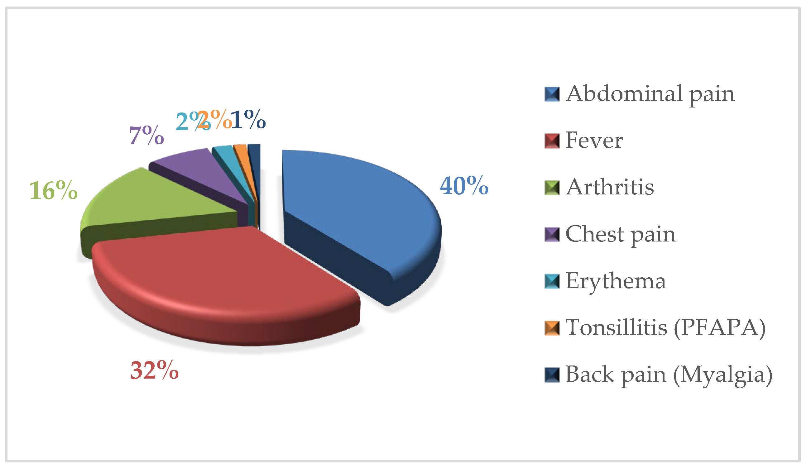
Figure 2.
Distribution of MEFV gene mutations among FMF patients.
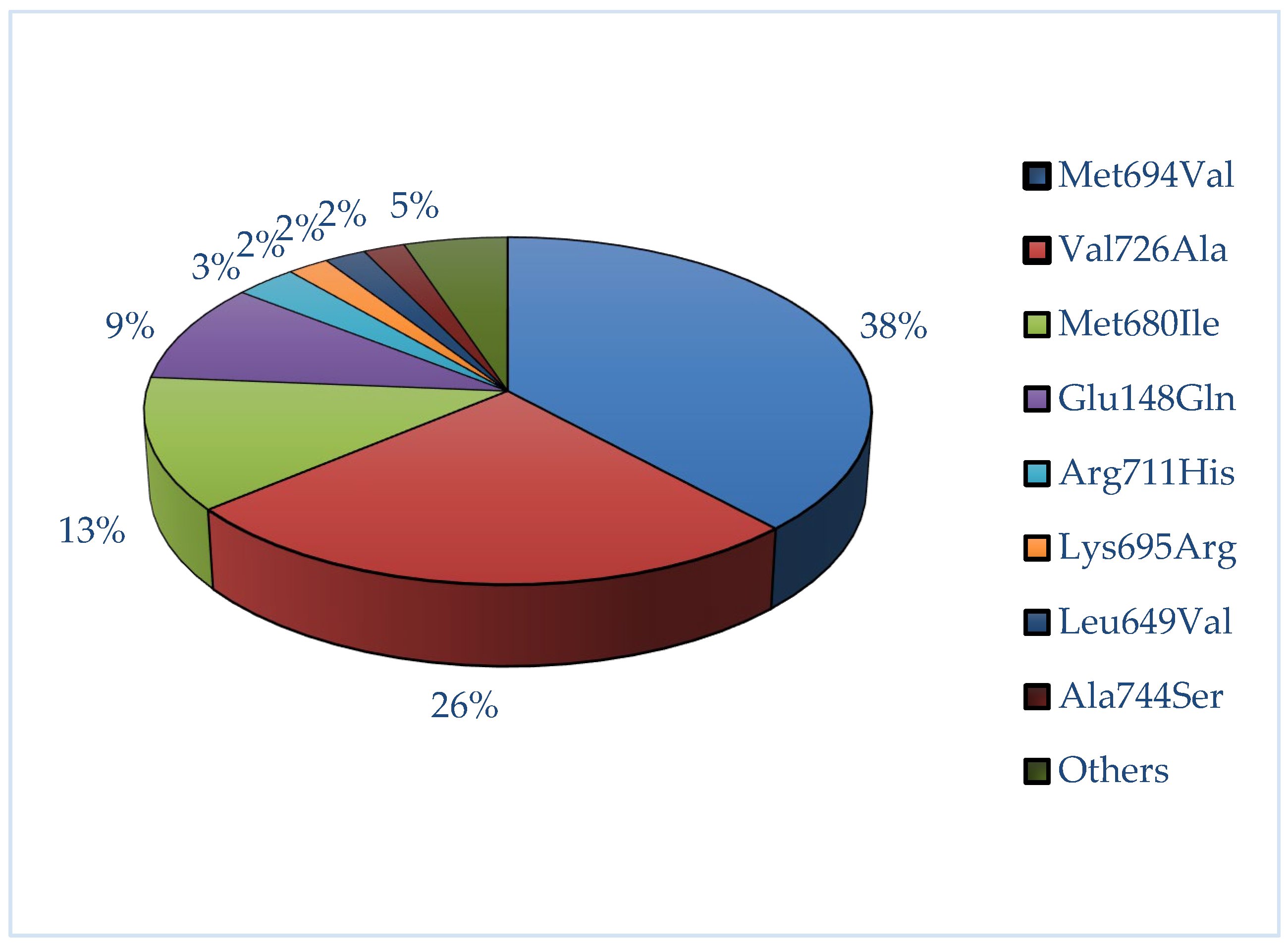

Table 1.
Comparison of cardiovascular risk indices among FMF severity groups and controls.
| Parameters | Severe group (n:32) | Moderate group (n:32) | Mild group (n:32) | Control (n:30) | test value | p | Post Hoc Comparison |
|---|---|---|---|---|---|---|---|
| AIP | 0.36±0.34 | 0.22±0.17 | -0.06±0.15 | -0.07±0.21 | 27.262 | <0.001 | 1-3, 1-4, 2-3, 2-4 |
| AC | 2.95±1.15 | 2.13±0.77 | 1.70±0.42 | 1.62±0.39 | 20.406 | <0.001 | 1-2, 1-3, 1-4, 2-4 |
| CRI-1 | 3.95±1.15 | 3.13±0.77 | 2.70±0.42 | 2.62±0.39 | 20.406 | <0.001 | 1-2, 1-3, 1-4, 2-4 |
| CRI-2 | 2.59±1.03 | 1.99±0.53 | 1.52±0.32 | 1.46±0.31 | 22.025 | <0.001 | 1-2, 1-3, 1-4, 2-3, 2-4 |
AIP: Atherogenic Index of Plasma, AC: Atherogenic Coefficient, CRI-1: Castelli Risk Index-1, CRI-2: Castelli Risk Index-2.
Table 2.
Comparison of BMD SDS values and bone metabolism parameters in FMF patients and the control group.
Table 2.
Comparison of BMD SDS values and bone metabolism parameters in FMF patients and the control group.
| Parameters | Severe group (n:32) | Moderate group (n:32) | Mild group (n:32) | Control (n:30) | test value | p | Post Hoc Comparison |
|---|---|---|---|---|---|---|---|
| Ca | 9.40 (9.22-9.77) | 9.30 (9.2-9.5) | 9.35 (9.2-9.67) | 9.40 (9.20-9.72) | 2.112 | 0.550 | |
| P | 5.09±0.41 | 5.06±0.43 | 5.04±0.59 | 5.07±0.38 | 0.074 | 0.974 | |
| ALP | 167.66±27.82 | 166.59±15.54 | 165.34±8.81 | 168.30±14.21 | 0.161 | 0.922 | |
| Vitamin D | 23.0 (22.0-25.75) | 24.0 (23.0-26.75) | 24.0 (23.0-26.0) | 24.0 (23.0-27.0) | 2.476 | 0.480 | |
| PTH | 47.15 (47.0-48.0) | 47.0 (46.25-48.0) | 47.0 (46.22-48.0) | 47.0 (45.0-48.0) | 1.515 | 0.679 | |
| BMD SDS | 0.29 (-1.22-1.77) | 0.90 (0.40-1.37) | 0.72 (0.13-2.65) | 1.43 (0.57-2.27) | 9.369 | 0.025 | 1-4 |
Ca: Calcium, P: Phosphorus, ALP: Alkaline phosphatase, PTH: Parathyroid hormone, BMD SDS: Bone mineral density standard deviation score.
Table 3.
Spearman correlation analysis of BMD SDS and cardiovascular risk indices.
| AIP | AC | CRI-1 | CRI-2 | |
|---|---|---|---|---|
| BMD SDS | rho=-0.041 p=0.647 |
rho=-0.117 p=0.191 |
rho=-0.117 p=0.191 |
rho=-0.046 p=0.612 |
| AIP | rho=0.499 p<0.001 |
rho=0.499 p<0.001 |
rho=0.570 p<0.001 |
|
| AC | rho=1.000 | rho=0.864 p<0.001 |
||
| CRI-1 | rho=0.864 p<0.001 |
|||
| CRI-2 |
AIP: Atherogenic Index of Plasma, AC: Atherogenic Coefficient, CRI-1: Castelli Risk Index-1, CRI-2: Castelli Risk Index-2, BMD SDS: Bone mineral density standard deviation score.
Table 4.
Metabolic and bone-related parameters according to mutation status in FMF patients.
| Mutation other than M694V (n:60) | Mutation M694V (n:36) | test value | p | |
|---|---|---|---|---|
| AIP | 0.19±0.28 | 0.17±0.30 | 0.295 | 0.769 |
| AC | 2.30±0.94 | 2.23±0.99 | 0.312 | 0.756 |
| CRI-1 | 3.30±0.94 | 3.23±0.99 | 0.312 | 0.756 |
| CRI-2 | 1.98±0.84 | 2.06±0.79 | -0.499 | 0.619 |
| BMD SDS | 0.38 (-1.09-1.13) | 0.95 (0.15-2.67) | 782.5 | 0.011 |
AIP: Atherogenic Index of Plasma, AC: Atherogenic Coefficient, CRI-1: Castelli Risk Index-1, CRI-2: Castelli Risk Index-2, BMD SDS: Bone mineral density standard deviation score.
Disclaimer/Publisher’s Note: The statements, opinions and data contained in all publications are solely those of the individual author(s) and contributor(s) and not of MDPI and/or the editor(s). MDPI and/or the editor(s) disclaim responsibility for any injury to people or property resulting from any ideas, methods, instructions or products referred to in the content. |
© 2026 by the authors. Licensee MDPI, Basel, Switzerland. This article is an open access article distributed under the terms and conditions of the Creative Commons Attribution (CC BY) license (http://creativecommons.org/licenses/by/4.0/).
Copyright: This open access article is published under a Creative Commons CC BY 4.0 license, which permit the free download, distribution, and reuse, provided that the author and preprint are cited in any reuse.