

Submitted:
27 May 2026
Posted:
28 May 2026
You are already at the latest version
Abstract
Background: Staphylococcus aureus — particularly methicillin-resistant S. aureus (MRSA) — remains one of the leading causes of skin and soft tissue infections (SSTIs) worldwide. Patients with renal edema, including those with nephrotic syndrome and chronic kidney disease (CKD), represent a population of increased susceptibility due to combined barrier dysfunction, immune impairment, and altered pharmacokinetics. Despite its substantial clinical relevance, the mechanistic relationship between renal edema and cutaneous staphylococcal infection has received limited integrative attention. Objectives: This narrative review aims to: (1) examine the pathophysiological mechanisms through which renal edema predisposes to cutaneous S. aureus infection; (2) characterize the virulence mechanisms that facilitate bacterial persistence and invasion in the renal host; (3) describe the clinical spectrum of SSTIs in this population; (4) evaluate current and emerging therapeutic strategies adapted to renal dysfunction; and (5) discuss the management challenges of severe S. aureus infection in critically ill renal patients. Methods: Search terms included combinations of Staphylococcus aureus, MRSA, skin and soft tissue infections, renal edema, nephrotic syndrome, chronic kidney disease, ICU, vancomycin pharmacokinetics, augmented renal clearance, and anti-virulence therapy. Priority was given to clinical guidelines, systematic reviews, translational studies, and landmark investigations relevant to the pathophysiology and management of cutaneous staphylococcal infection in renal disease. Results: Three converging pathophysiological axes appear to underlie the increased susceptibility of patients with renal edema to cutaneous S. aureus infection: (1) mechanical barrier disruption caused by edema-associated skin tension, blistering, fissuring, and proteinaceous exudate; (2) nephrotic and uremic immunodeficiency, including urinary immunoglobulin loss, neutrophil dysfunction mediated by protein-bound uremic retention solutes (PBURS), complement depletion, and T-cell dysregulation; and (3) lymphatic dysfunction impairing regional immune surveillance and antigen clearance. S. aureus exploits these abnormalities through adhesins, toxin-mediated tissue injury, biofilm formation, and coordinated immune evasion pathways regulated in part by the ArlRS/MgrA cascade. In critically ill patients, management is further complicated by major pharmacokinetic variability, including augmented renal clearance, expanded volume of distribution, and extracorporeal renal support, necessitating individualized dosing strategies and AUC/MIC-guided vancomycin monitoring. Emerging anti-virulence approaches targeting alpha-toxin, quorum-sensing systems, and immune evasion pathways represent promising adjunctive therapeutic strategies. Conclusions: Renal edema creates a biologically permissive environment for cutaneous S. aureus infection through the interaction of barrier dysfunction, immune impairment, and altered antimicrobial pharmacokinetics. Optimal management requires early recognition, renal-adapted antimicrobial therapy, therapeutic drug monitoring, source control, and multidisciplinary care. Continued development of anti-virulence and immunomodulatory therapies may further improve outcomes in this vulnerable patient population.
Keywords:
Staphylococcus aureus
; MRSA
; skin and soft tissue infections
; renal edema
; nephrotic syndrome
; chronic kidney disease
; cutaneous infection
; virulence factors
; biofilm
1. Introduction
Staphylococcus aureus is one of the most clinically significant bacterial pathogens known to medicine, responsible for a broad spectrum of disease, from superficial skin pustules to fatal septicemia. Its dual capacity to colonize healthy individuals asymptomatically and to cause invasive, life-threatening illness in vulnerable hosts has sustained its prominence in infectious disease over multiple decades. Global meta-analysis data published in 2024 report a worldwide prevalence of S. aureus colonization of approximately 24.8% (95% CI, 20.4–29.3%), with methicillin-resistant S. aureus (MRSA) accounting for approximately 5.8% of all isolates globally [1].
S. aureus is the predominant pathogen in skin and soft tissue infections (SSTIs) in the United States, with clinical presentations ranging from superficial impetigo and folliculitis to monomicrobial necrotizing fasciitis. In 2023, SSTIs attributable to S. aureus were associated with an age-standardized all-cause mortality rate of 0.5 per 100,000 globally [2]. Moreover, in 2025, S. aureus was associated with more than 1 million deaths worldwide (range 816,000–1,470,000), cementing its position as the leading cause of bacterial infection-related mortality [3].
Patients with chronic kidney disease (CKD) or acute glomerular pathology represent a group of particular vulnerability. CKD is characterized by progressive impairment of renal filtration, culminating in the accumulation of uremic toxins and a state of composite immune dysfunction. S. aureus exploits this diminished host defense on multiple levels — impaired neutrophil chemotaxis, defective phagocytosis, disrupted T-cell signaling, and reduced immunoglobulin production all converge to create a permissive environment for bacterial invasion and propagation [4].
The cutaneous barrier is further compromised by the mechanical and biochemical consequences of renal edema, a defining feature of nephrotic syndrome and advanced CKD. Skin tension, blistering, and exudate create portals of entry for S. aureus, while impaired lymphatic drainage disrupts the local immunological surveillance that would ordinarily contain superficial colonization [5,6].
Rather than a simple manifestation of fluid overload, renal edema creates a complex pathological microenvironment characterized by compromised structural barriers, dysregulated innate and adaptive immunity, impaired lymphatic clearance, and altered antimicrobial pharmacokinetics. These interconnected abnormalities may collectively facilitate the transition of S. aureus from asymptomatic colonizer to invasive pathogen.
This review synthesises the current evidence on these interlocking pathophysiological mechanisms, delineates the clinical spectrum of cutaneous staphylococcal infections in the renal patient, appraises therapeutic strategies adapted to the unique constraints of renal function impairment, and addresses the specific challenges of managing S. aureus infections in the intensive care unit (ICU) — where critical illness, dialysis-dependent pharmacokinetics, and staphylococcal virulence converge to create the most demanding management scenario in this patient population.
2. Renal Edema as a Gateway to Cutaneous Infection: From Hemodynamics to Immune Failure
This section examines the mechanistic basis of enhanced cutaneous susceptibility to S. aureus in patients with renal disease. The following subsections address, in sequence: the haemodynamic and structural mechanisms underlying oedema formation in nephrotic syndrome and CKD (2.1); the specific patterns of immune dysfunction — nephrotic immunodeficiency — that reduce host capacity to resist staphylococcal invasion (2.2); the contribution of uremic toxin accumulation to innate and adaptive immune failure (2.3); the role of lymphatic dysfunction in abolishing regional immunological surveillance (2.4); and the broader spectrum of cutaneous manifestations through which CKD creates the integumentary conditions permissive for infection (2.5).
2.1. Mechanisms of Edema Formation in Renal Disease
Edema is one of the cardinal features of nephrotic syndrome, occurring alongside hypoalbuminemia, proteinuria, and dyslipidemia. Its pathogenesis has historically been framed by two competing hypotheses. The classical "underfill" model postulates that urinary albumin loss reduces plasma oncotic pressure, causing fluid to shift into the interstitium. The resulting intravascular hypovolaemia triggers neurohormonal compensatory mechanisms, activation of the renin-angiotensin-aldosterone system (RAAS), release of antidiuretic hormone (ADH), and sympathetic nervous system stimulation, which increase renal sodium and water retention, perpetuating edema [7]. Conversely, the "overfill" model, supported by direct measurements of circulating volume in many adult nephrotic patients, proposes that primary intrarenal sodium retention, independent of oncotic changes, is the driving mechanism, leading to plasma volume expansion and peripheral edema. Contemporary evidence suggests that both mechanisms operate, with their relative contributions varying by underlying etiology, glomerular pathology, and disease stage [7].
The glomerular filtration barrier (GFB) normally restricts protein passage through the coordinated action of the glomerular endothelium, the glomerular basement membrane (GBM), and podocyte foot processes with their slit diaphragm architecture. Loss of podocyte function — through effacement of foot processes or disruption of the slit diaphragm — is the shared structural endpoint of most nephrotic conditions, including minimal change disease (MCD), focal segmental glomerulosclerosis (FSGS), and membranous nephropathy [8,9].
A clinically important but underappreciated consequence of sustained edema is its direct effect on the integumentary system. Edema increases skin tension, mechanical blistering, skin breakage, and extrusion of proteinaceous exudate — all of which compromise the structural integrity of the cutaneous barrier and create a culture medium for bacterial colonization and infection [5]. In severe or longstanding edema, skin weeping and maceration further reduce local concentrations of antimicrobial peptides and disrupt the acid mantle, thereby compounding susceptibility to S. aureus.
2.2. Nephrotic Immunodeficiency: Mechanisms and Consequences
Nephrotic syndrome is associated with a specific pattern of immune dysfunction — sometimes termed "nephrotic immunodeficiency" — that substantially increases susceptibility to encapsulated bacteria and, notably, to S. aureus [5]. The immune dysfunction associated with nephrotic syndrome is multifactorial and extends beyond simple urinary protein loss. Both innate and adaptive immune pathways are disrupted through interrelated mechanisms involving immunoglobulin depletion, complement loss, T-cell dysregulation, and treatment-related immunosuppression. The principal mechanisms contributing to nephrotic immunodeficiency, along with their clinical implications, are summarized in Table 1.
Among these mechanisms, urinary loss of immunoglobulins — particularly IgG — is perhaps the most clinically significant. IgG is required for opsonization of S. aureus, and its depletion reduces the efficiency of neutrophil-mediated bacterial killing. Concurrently, T-cell transformation dysfunction — characterized by an imbalance between Th2 and Th1 subsets and reduced regulatory T cell (Treg) function — impairs the adaptive immune response to staphylococcal surface antigens, facilitating persistent colonization and recurrent infection [9].
2.3. Uremic Toxin-Mediated Immune Dysfunction in CKD
Beyond the specific mechanisms of nephrotic syndrome, the broader context of CKD imposes a distinct and compounding layer of immune dysfunction, driven primarily by the accumulation of protein-bound uremic retention solutes (PBURS) — including indoxyl sulfate, p-cresyl sulfate, and trimethylamine N-oxide (TMAO). These molecules are derived from the gut microbiota's metabolic activity and progressively accumulate as renal clearance diminishes [4].
PBURS impair neutrophil function at multiple levels. Uremic toxins inhibit neutrophil chemotaxis — the directed migration toward sites of infection — and reduce phagocytic capacity, such that neutrophils from CKD patients are significantly less effective at killing S. aureus than those from healthy controls. Critically, when neutrophils from CKD patients are cultured in a non-uremic medium, their phagocytic capacity is restored, confirming the direct causative role of circulating toxins rather than intrinsic neutrophil defects [4].
PBURS also impair the adaptive immune system: CKD patients exhibit premature T-cell aging (immunosenescence), defective dendritic cell function, and reduced cellular and humoral immune responses. This creates a state of paradoxical immune activation (chronic low-grade inflammation, endothelial damage) combined with immune incompetence (impaired antibacterial capacity), a combination uniquely permissive for S. aureus skin colonization and invasive infection [4,10].
2.4. Lymphatic Dysfunction and Regional Immunological Surveillance
The lymphatic system serves as the principal conduit for immune cell trafficking from peripheral tissues to regional lymph nodes, fulfilling a critical role in immunological surveillance. In renal edema, the lymphatic system is subjected to sustained overload because interstitial fluid volume exceeds its normal clearance capacity. This functional lymphatic insufficiency mirrors the pathophysiology of secondary lymphoedema and is associated with comparable immunological consequences [11]. In states of lymphatic dysfunction, defective lymphatic conduits prevent the homing of leukocytes and dendritic cells from peripheral tissues to draining lymph nodes. The resulting impairment of immune surveillance converts the edematous integument into an "immunologically vulnerable area" — a concept directly supported by the increased frequency of infections, including cellulitis and erysipelas, in edematous limbs [11]. Soft-tissue infections in this context carry a risk of progression to sepsis and may necessitate lifelong prophylactic antibiotic therapy in the most severe cases.
A 2023 experimental study by Cąkała-Jakimowicz et al. formally demonstrated that interrupting lymphatic flow markedly worsens local cutaneous inflammation in the presence of commensal staphylococci. The combination of edema and bacterial inoculum induces severe local inflammation and delays the antibacterial protection processes in neighboring lymph nodes — establishing a mechanistic basis for the empirically observed susceptibility of edematous skin to staphylococcal superinfection [6].
2.5. Cutaneous Manifestations in Chronic Kidney Disease: The Dermatological Interface
CKD produces a spectrum of cutaneous manifestations that reflect the systemic consequences of uremia, immune dysfunction, and altered dermal biology. A 2024 review by Arriaga Escamilla et al. comprehensively cataloged these manifestations, identifying infectious complications as a major source of morbidity. Infections are predominantly caused by Gram-positive cocci — primarily S. aureus and coagulase-negative staphylococci (CONS) — with Gram-negative organisms, fungi, and polymicrobial infections occurring in immunocompromised subsets [12]. A defining feature of CKD-associated staphylococcal infections is the propensity of S. aureus and CONS to elaborate extracellular mucopolysaccharides, forming biofilms on compromised skin surfaces and vascular access sites. Biofilms confer marked resistance to both immune clearance and antibiotic penetration — contributing to the chronicity and recurrence that characterize infections in this population [12]. Key host-related risk factors for access-site and cutaneous infections in CKD patients include immunosuppression, prior bacteremia, poor hygiene, adjacent skin infection, high BMI, iron overload, and, perhaps most importantly, hypoalbuminemia, which reduces both antimicrobial peptide transport and the nutritional substrate for wound healing [12].
The susceptibility of patients with renal edema to cutaneous S. aureus infection arises from the convergence of multiple interdependent pathophysiological processes rather than from a single isolated defect. Mechanical disruption of the edematous skin barrier facilitates bacterial entry, nephrotic and uremic immune dysfunction impairs both innate and adaptive antibacterial responses, and lymphatic insufficiency compromises regional immune surveillance and antigen clearance. Together, these mechanisms create a biologically permissive environment for persistent colonization, recurrent SSTIs, and invasive staphylococcal disease. The major mechanistic pathways linking renal edema to increased susceptibility to cutaneous S. aureus infection are summarized in Figure 1.
3. Virulence Mechanisms of S. aureus in the Cutaneous Context
The pathogenic potency of S. aureus in the skin derives from a precisely orchestrated, multi-layered virulence program. This section deconstructs that programme across three levels of analysis: the molecular mechanisms governing initial colonisation and host surface adhesion (3.1); the panel of secreted toxins — alpha-toxin, Panton-Valentine Leukocidin, and exfoliative toxins — that directly damage cutaneous tissues and neutralise immune effectors (3.2); and the transcriptional regulatory cascade (ArlRS/MgrA) that coordinates immune evasion in real time during skin infection and constitutes a compelling therapeutic target (3.3). Throughout, the specific relevance of each mechanism to renal edema is highlighted.
3.1. Colonization Dynamics and Surface Adhesins
The pathogenic success of S. aureus in the skin begins with colonization — the establishment of a stable bacterial reservoir on the integumentary surface or within follicular structures, from which invasive infection can subsequently arise. Approximately 30% of the general population carries S. aureus asymptomatically in the anterior nares, with a transient proportion carrying S. aureus on the skin. Nasal, cutaneous, and oropharyngeal colonization serve as the primary reservoirs for both self-infection and transmission to susceptible contacts [13].
Surface adhesins mediate the initial attachment of S. aureus to host epithelial cells and extracellular matrix proteins. The clumping factors (ClfA and ClfB), fibronectin-binding proteins (FnBPA, FnBPB), and surface proteins such as SasG and IsdA facilitate adherence to corneocytes, fibronectin, fibrinogen, and loricrin — structural components of the stratum corneum. ClfB, in particular, has been identified as a key virulence factor in skin infection models and is under investigation as a vaccine target [13,14].
3.2. Secreted Toxins: Alpha-Toxin, PVL, and Exfoliative Toxins
S. aureus deploys an arsenal of secreted toxins that directly damage cutaneous tissues and neutralize host immune effectors. The virulence toolkit operates synergistically — toxins with complementary mechanisms reinforce each other's activity, creating functional redundancy that complicates therapeutic targeting [15].
Alpha-toxin (Hla) is a pore-forming cytotoxin that disrupts the integrity of epithelial and endothelial cell membranes, causing cell lysis and the release of pro-inflammatory danger signals. In a human skin explant model, alpha-toxin produced high tissue toxicity and complete loss of epithelial integrity, confirming its central role in the pathogenesis of superficial staphylococcal SSTIs [16]. Antibodies raised against alpha-toxin mitigated tissue damage in a concentration-dependent manner in the same model, supporting anti-toxin strategies as a potential adjunctive therapeutic approach [16].
Panton-Valentine Leukocidin (PVL) is a two-component pore-forming toxin (LukS-PV and LukF-PV) produced by fewer than 5% of S. aureus strains globally, but with a dramatically higher prevalence in community-associated MRSA (CA-MRSA) lineages. PVL forms cytolytic pores in the membranes of polymorphonuclear neutrophils (PMNs), monocytes, and macrophages, inducing apoptosis and lysing the primary cellular effectors of the early cutaneous innate immune response. PVL genes are detected in 93% of strains associated with furunculosis and in 55% of cellulitis isolates, compared with near-absent detection in hospital-acquired and device-related infections [17,18,19].
Exfoliative toxins (ETs) — including ETA, ETB, and ETD — are serine proteases that cleave desmoglein-1, the desmosomal cadherin responsible for intercellular adhesion in the superficial epidermis. Cleavage of desmoglein-1 produces the characteristic superficial skin cleavage of staphylococcal scalded skin syndrome (SSSS) and bullous impetigo. The clinical relevance of ETs extends to patients with renal disease: ET clearance depends on adequate renal function, and patients with CKD or AKI may accumulate ETs to locally or systemically toxic concentrations, even in the absence of bacteremia [15,20].
The pathogenic success of S. aureus in edematous and immunocompromised tissues depends on a coordinated repertoire of adhesins, toxins, immune evasion systems, and regulatory pathways. Several of these virulence determinants assume particular clinical relevance in patients with CKD and renal edema due to impaired immune clearance and altered toxin handling. The major virulence factors implicated in cutaneous infection and their relevance in renal disease are summarized in Table 2.
3.3. Regulatory Mechanisms of Cutaneous Immune Evasion
The pathogenic program of S. aureus during skin infection is not constitutive but dynamically regulated in response to host signals. A landmark 2021 study by Kwiecinski et al. identified the ArlRS two-component regulatory system and its downstream effector MgrA as the principal controllers of cutaneous immune evasion [21,22,23].
S. aureus strains lacking ArlRS or MgrA show dramatically reduced virulence in murine skin infection models: they fail to form the structured abscess architecture that normally sequesters bacteria from immune effectors, express lower levels of leukocidins, CHIPS (chemotaxis inhibitory protein), and SCIN (staphylococcal complement inhibitor), and are unable to kill neutrophils, block chemotaxis, degrade neutrophil extracellular traps (NETs), or survive direct neutrophil attack [21]. ArlRS and MgrA thus represent compelling therapeutic targets — pharmacological disruption of this regulatory cascade could simultaneously disable multiple immune evasion mechanisms, restoring susceptibility to innate immune clearance [22,23].
4. Clinical Implications
Translating the pathophysiological and virulence mechanisms described in sections 2 and 3 into clinical practice requires an understanding of how they manifest at the bedside. This section covers three interconnected clinical dimensions: the spectrum of SSTI presentations specifically observed in patients with renal oedema, and how the edematous tissue environment modifies the natural history of each (4.1); the bidirectional relationship between staphylococcal bacteremia and acute kidney injury, and the vicious cycle this creates (4.2); and the diagnostic challenges unique to edematous renal patients, where classical inflammatory signs may be masked and atypical presentations are common (4.3).
4.1. Clinical Spectrum of S. aureus SSTIs in Renal Patients
The clinical presentations of S. aureus SSTIs in patients with renal edema span the full spectrum from superficial to life-threatening disease, but several features are particularly characteristic of this population. The edematous tissue environment lowers the inoculum required for infection, accelerates local spread by disrupting fascial barriers waterlogged with interstitial fluid, and favors the development of chronic, poorly healing wounds. Additionally, immunosuppressive agents commonly used in the management of nephrotic syndrome — corticosteroids, calcineurin inhibitors, and rituximab — further impair bactericidal defenses.
The pathogenic bacterium S. aureus, the most common pathogen isolated in SSTIs in the United States, presents with clinical features ranging from superficial infections with local symptoms to monomicrobial necrotizing fasciitis with systemic manifestations and potentially fatal outcomes [2]. In CKD patients, this spectrum is skewed toward more severe presentations, with a higher rate of bacteremic dissemination and multifocal disease.
The spectrum of cutaneous S. aureus infection in renal patients ranges from superficial localized disease to rapidly progressive necrotizing infection and sepsis. Edema-associated barrier disruption, lymphatic dysfunction, vascular access devices, and immunosuppressive therapy all contribute to distinct clinical risk profiles in this population. The major SSTI presentations and their associated renal-specific risk factors are outlined in Table 3.
4.2. The Bidirectional Relationship: Staphylococcal Infection and Acute Kidney Injury
The relationship between S. aureus and renal function is bidirectional: renal impairment predisposes to staphylococcal infection, while staphylococcal bacteremia (SAB) can precipitate or worsen acute kidney injury (AKI). AKI is a frequent and clinically significant complication of SAB, impacting management decisions and prognosis. A 2022 multicentre retrospective cohort study by Westgeest et al. quantified the incidence of AKI within 14 days of SAB onset and evaluated its association with 30-day mortality. The study demonstrated that AKI occurred at a clinically meaningful frequency and was independently associated with adverse outcomes, establishing a mechanistic basis for the "staphylo-renal" vicious cycle: bacteremia triggers tubular injury and glomerulonephritis, worsening edema and immune dysfunction, which in turn promote further infection [24].
Mechanisms by which SAB induces AKI include: (1) haematogenous seeding of the renal parenchyma, with cortical abscesses or diffuse nephritis; (2) immune complex-mediated glomerulonephritis, driven by circulating staphylococcal superantigens and immune complexes depositing in glomerular capillaries; (3) haemodynamic insult from sepsis-associated vasodilation; and (4) direct nephrotoxicity from antibiotic therapy — particularly aminoglycosides and high-trough vancomycin — compounding the infective injury.
4.3. Diagnostic Considerations in the Renal Patient
Diagnosis of staphylococcal SSTIs in patients with renal edema presents specific clinical challenges. The classical inflammatory signs — erythema, warmth, and tenderness — may be attenuated or partially masked by edema, delaying recognition. Necrotizing infections in particular may be overlooked in the early stages, as the overlying skin can appear relatively normal while deep fascial necrosis progresses. A high index of suspicion, supported by imaging (MRI or CT for deep infections) and early surgical exploration where indicated, is essential [12].
Microbiological sampling should include wound swabs or pus cultures from all accessible lesions, with blood cultures mandatory in the presence of systemic inflammatory signs. Susceptibility testing must include MRSA screening, given the high MRSA prevalence in healthcare-associated settings and dialysis populations. Nasal swabbing for MRSA colonization status should be performed in all hospitalized CKD patients, as colonization status directly guides empirical antibiotic selection and decolonization protocols [25,26,27].
5. Therapeutic Perspectives
Management of staphylococcal SSTIs in patients with renal edema requires adapting standard antibiotic protocols to the pharmacokinetic and immunological constraints of renal disease. This section addresses five therapeutic domains in sequence: the evidence-based principles and guideline recommendations governing SSTI treatment stratified by severity and MRSA risk (5.1); the comparative efficacy and renal dose adjustments of novel anti-MRSA agents approved in the past decade (5.2); network meta-analysis evidence comparing these agents in MRSA infections (5.3); the pharmacokinetic consequences of oedema on antibiotic distribution and clearance, and the implications for dosing (5.4); and decolonisation strategies specifically validated in the nephrological setting (5.5).
5.1. General Principles of SSTI Management
Antibiotic selection for S. aureus SSTIs must be guided by (1) the severity of infection, (2) the local and patient-specific MRSA risk, (3) the patient's renal function, and (4) susceptibility data from cultures when available. The IDSA and UK guidelines both adopt a severity-stratified approach, distinguishing between non-purulent cellulitis (predominantly streptococcal), purulent infections (primarily staphylococcal), and severe/complicated SSTIs.
For mild purulent SSTI (abscesses, furunculosis), incision and drainage (I&D) remains the cornerstone of management, with systemic antibiotics reserved for systemic signs, marked cellulitis, or immunocompromised hosts — including CKD patients on immunosuppression. For moderate-to-severe SSTI with systemic features, empirical antibiotic therapy targeting both MSSA and streptococci is appropriate, with escalation to anti-MRSA agents when risk factors are present [28,29,30,31].
For confirmed or strongly suspected MRSA SSTIs, UK guidelines recommend intravenous glycopeptides (vancomycin or teicoplanin) for severe cellulitis or soft-tissue infections; linezolid (oral or intravenous) or daptomycin (intravenous) as validated alternatives [28]. The IDSA guidelines (2011) recommend a first-generation cephalosporin or antistaphylococcal penicillin for MSSA, or vancomycin, linezolid, daptomycin, telavancin, or ceftaroline where MRSA risk factors are present [29].
5.2. Novel Anti-MRSA Agents: Evidence and Renal Considerations
Over the past decade, several new antibiotics with activity against MRSA have received regulatory approval for SSTIs, expanding the therapeutic armamentarium beyond vancomycin and linezolid. A 2023 in vitro study evaluated ceftobiprole, dalbavancin, tedizolid, and comparators against 124 clinical MRSA isolates from SSTIs (2020–2022), demonstrating that vancomycin remains preferred for complicated SSTIs, with linezolid and daptomycin as established alternatives, while newer agents offer additional options in specific clinical scenarios [32].
Selection of anti-MRSA therapy in patients with renal dysfunction requires balancing antimicrobial efficacy against the risk of nephrotoxicity, altered pharmacokinetics, dialysis-related clearance, and the feasibility of therapeutic drug monitoring. Several newer agents have expanded the available therapeutic options for complicated SSTIs, although renal-specific pharmacokinetic data remain limited for some compounds. The principal anti-MRSA agents, renal dose adjustments, and major clinical considerations are summarized in Table 4.
5.3. Comparative Efficacy of Anti-MRSA Agents
A network meta-analysis published in 2024 evaluated the efficacy and safety of multiple antibiotics for MRSA infections. The analysis compared clinical cure and microbiological eradication rates, as well as adverse event profiles, across subgroups, including complex SSTIs (cSSSIs and cSSTIs) and pneumonia [33]. While vancomycin remains the benchmark comparator, the analysis identified several agents — including linezolid, tedizolid, and daptomycin — with non-inferior or superior clinical cure rates in specific subgroup analyses, providing evidence-based guidance for individual patient selection.
For persistent or refractory MRSA bacteremia — a scenario encountered in dialysis patients with vascular access infections — the combination of daptomycin plus ceftaroline has been studied as salvage therapy. A 2023 retrospective study evaluated outcomes of daptomycin + ceftaroline versus alternative therapy in patients with persistent MRSA bacteremia, stratified by baseline renal function (creatinine clearance and renal replacement therapy status). The combination demonstrated clinical benefit in treatment-refractory cases, though its use in patients with edematous CKD requires careful pharmacokinetic monitoring [34].
5.4. Pharmacokinetic Considerations in Renal Edema
Edematous states substantially alter antibiotic pharmacokinetics, increasing the risk of therapeutic failure due to underdosing or toxicity from drug accumulation. Increased volume of distribution (Vd) — driven by expanded interstitial and total body water — lowers peak serum concentrations of hydrophilic antibiotics (vancomycin, beta-lactams, aminoglycosides), requiring higher loading doses to achieve therapeutic levels. Conversely, impaired renal clearance prolongs the half-life of renally excreted antibiotics, necessitating dose reduction or interval extension to avoid accumulation.
Multidisciplinary algorithms for targeted antimicrobial therapy of severe S. aureus infections, published in 2023, organize antibiotic selection by site of infection and use PK/PD parameters to inform therapeutic decision-making [35]. For patients on renal replacement therapy (RRT), including intermittent hemodialysis (IHD) and continuous renal replacement therapy (CRRT), the pharmacokinetic profiles of virtually all anti-MRSA agents are further altered, with drug clearance influenced by dialysis modality, membrane type, and ultrafiltration rate. Specialized dosing guidelines for RRT patients are referenced in institutional antimicrobial stewardship resources.
5.5. Decolonization Strategies in the Nephrological Setting
Decolonization — the systematic eradication of S. aureus nasal and skin carriage — is a well-established strategy for reducing the risk of subsequent invasive infection in high-risk populations. In nephrology, decolonization is particularly relevant for patients undergoing peritoneal dialysis (PD) and hemodialysis (HD) with arteriovenous fistulae or central venous catheters, where staphylococcal access-site infections carry substantial morbidity.
A systematic review and meta-analysis (Grothe et al.) of 9 studies comprising 839 PD patients colonized with S. aureus found that both topical mupirocin and systemic antibiotic decolonization significantly reduced the incidence of access-site infections compared with placebo or no treatment. Mupirocin 2% nasal ointment, applied to the anterior nares twice daily for 5 days (with or without skin decolonization using chlorhexidine), remains the standard first-line approach [25]. An important caveat is the emergence of mupirocin resistance, which has reached clinically significant levels in some settings. For patients colonized with mupirocin-resistant MRSA (MupR-MRSA), alternative decolonization regimens — including retapamulin, povidone-iodine, or systemic rifampicin — may be considered, though evidence remains limited and should be guided by local susceptibility patterns.
6. Management of S. aureus Infections in the Intensive Care Unit (ICU)
The intensive care unit represents the most challenging arena for managing S. aureus infections in the renal patient, where the compounding of critical illness, organ failure, and invasive monitoring creates a uniquely hostile pharmacological and immunological environment. This section addresses four interrelated aspects of ICU management: the epidemiology and clinical presentations of staphylococcal infections in the critically ill renal patient, with their associated mortality burden (6.1); the profound pharmacokinetic alterations — augmented renal clearance, expanded volume of distribution, dialysis-dependent clearance — that render standard dosing unreliable in this population (6.2); the evidence base for AUC/MIC-guided vancomycin dosing as the current pharmacodynamic standard of care in the ICU (6.3); and the integrated principles of empirical antibiotic strategy, mandatory source control, and antimicrobial stewardship specific to the ICU context (6.4).
6.1. Epidemiology and Clinical Significance in the ICU
S. aureus — and in particular MRSA — is one of the leading causative agents of severe infections in the intensive care unit, accounting for a disproportionate share of ICU-acquired bacteremia, ventilator-associated pneumonia (VAP), catheter-related bloodstream infections (CRBSI), and complicated SSTIs. The critically ill patient with renal edema represents a convergence of the most adverse prognostic factors: immune dysfunction, vascular access requirements, disrupted skin barriers, and a pharmacokinetic environment radically different from that of the non-ICU patient.
Evidence-based algorithms for targeted antibiotic therapy of severe S. aureus infections in critically ill adult patients have been developed through multidisciplinary expert consensus, incorporating a review and organizing therapeutic choices by site of infection and PK/PD parameters — distinguishing between MSSA and MRSA, covering endocarditis, primary bacteremia, intravascular device infections, community-acquired pneumonia, CNS infections, and necrotizing skin and soft tissue infections [35].
The critically ill renal patient faces a specific and compounding threat: sepsis-associated kidney injury is common in critically ill patients and significantly increases morbidity and mortality, with several complex pathophysiological factors contributing — including macrocirculatory and microcirculatory changes, mitochondrial dysfunction, and metabolic reprogramming [36]. When S. aureus is the precipitating pathogen, the renal injury both compromises drug clearance and amplifies the immunological vulnerability that drove the infection in the first place, creating a vicious cycle of escalating severity.
A striking illustration is provided by a 2025 case report of a steroid-treated patient with end-stage renal disease who developed fulminant PVL-positive MSSA pneumonia requiring venovenous extracorporeal membrane oxygenation (VA-ECMO) and continuous haemodiafiltration. Severe hyperkalemia, metabolic acidosis, and lactic acidosis complicated the course, with persistent hemodynamic compromise despite maximal ICU support — illustrating the lethal convergence of staphylococcal virulence and end-stage renal disease in the critical care setting [37].
In critically ill renal patients, S. aureus infection may present through multiple overlapping syndromes, including catheter-related bloodstream infection, ventilator-associated pneumonia, necrotizing SSTI, and septic shock with secondary organ dysfunction. Early recognition of the primary infectious focus and prompt diagnostic evaluation are essential because mortality rises substantially once bacteremia and multiorgan failure develop. The principal ICU presentations, diagnostic priorities, and associated mortality impact are summarized in Table 5.
6.2. Pharmacokinetic Alterations in the ICU Renal Patient
The pharmacokinetics of antibiotics — and of vancomycin in particular — are profoundly altered in the critically ill patient, making standard dosing regimens unreliable. Two opposing physiological extremes must be recognized and actively managed.
Augmented renal clearance (ARC) occurs paradoxically in a subset of ICU patients — particularly younger, hyperdynamic septic patients — where glomerular filtration is supranormal (eGFR >130 mL/min/1.73 m2). ARC is not rare in the ICU — estimates suggest it affects 30–65% of young critically ill patients — and represents a pharmacokinetic emergency requiring active recognition [38].
Conversely, established CKD and renal edema produce the opposite problem: drug accumulation through impaired clearance, risking nephrotoxicity from the very agents needed to treat the infection. Critically ill patients admitted to the ICU may have markedly altered pharmacokinetic parameters compared to non-critically ill patients; individualized dose adjustment and therapeutic drug monitoring (TDM) of vancomycin are therefore essential. A 24-hour AUC target of 700 µg·h/mL has been proposed as a more reliable pharmacokinetic parameter to achieve sufficient clinical efficacy while preventing vancomycin-induced nephrotoxicity in this population [39].
Critically ill patients on extracorporeal organ support, continuous renal replacement therapy (CRRT), intermittent hemodialysis (IHD), or ECMO, pose an additional pharmacokinetic challenge: drug clearance depends on membrane type, surface area, ultrafiltration rate, and dialysate flow, rendering standard dosing nomograms unreliable [40]. Liaison with clinical pharmacology and infectious disease is mandatory for these patients.
6.3. AUC-Guided Vancomycin Dosing in the ICU
The 2020 revised consensus guidelines from ASHP/IDSA/SIDP marked a paradigm shift in vancomycin monitoring: from trough-based to AUC/MIC-based dosing. The guidelines recommend a loading dose of 15–20 mg/kg followed by maintenance dosing targeting an AUC/MIC of 400–600 mg·h/L (based on an assumed MRSA MIC of 1 mg/L), with AUC/MIC estimated by multiplying the vancomycin steady-state concentration by 24 to approximate AUC₀₋₂₄ [41].
For MRSA bacteremia, multivariate analyses have confirmed that lower initial AUC/MIC is a significant independent risk factor for treatment failure. Current IDSA recommendations use AUC as the primary pharmacodynamic monitoring indicator before formal MIC data become available [42]. A multicentre retrospective Japanese ICU study (2020–2022) quantified nephrotoxicity risk: the data indicate that initial dosing should target an AUC on day 2 not exceeding 500 µg·h/mL in patients at high risk of AKI — a critical threshold for patients at risk of renal edema [43].
Monte Carlo pharmacokinetic simulations comparing vancomycin infusion modes in critically ill MRSA patients demonstrated that the probability of target attainment (PTA) at AUC₀₋₂₄/MIC 400–600 varies significantly by renal function stratum, with continuous infusion and optimized two-step infusion offering PK/PD advantages over standard intermittent infusion in patients with altered renal pharmacokinetics [44].
Antibiotic management of severe S. aureus infection in critically ill renal patients requires continuous integration of infection severity, renal function, extracorporeal support modalities, and pharmacokinetic/pharmacodynamic targets. Expanded volume of distribution, augmented renal clearance, and dialysis-dependent drug removal frequently necessitate individualized dosing strategies and therapeutic drug monitoring. The major ICU anti-staphylococcal agents and their pharmacokinetic considerations in renal impairment are summarized in Table 6.
6.4. Empirical Strategy, Source Control, and Antimicrobial Stewardship
In the ICU patient with suspected S. aureus sepsis and renal edema, empirical antibiotic selection must simultaneously address four imperatives: (1) adequate MRSA coverage initiated within one hour of sepsis recognition; (2) appropriate loading doses to account for expanded volume of distribution; (3) avoidance of further nephrotoxic insult; and (4) daily reassessment with de-escalation as culture data become available. Dual coverage for MRSA should be considered for high-risk ICU patients; an initial loading-dose strategy is essential to rapidly attain the target, with subsequent dosing guided by renal function and infectious disease consultation [45].
Antibiotic therapy alone is insufficient in ICU S. aureus infections associated with identifiable foci. Source control — surgical debridement of necrotizing SSTIs, removal of infected vascular catheters, and drainage of deep abscesses — is a prerequisite for treatment success. Management of S. aureus bloodstream infections in critically ill patients requires a structured multidisciplinary approach integrating infectious disease consultation, echocardiography to exclude infective endocarditis, and repeated blood cultures to document bacteremia clearance — with persistent bacteremia beyond 72 hours triggering investigation for metastatic foci including renal cortical abscesses, osteomyelitis, and septic arthritis [46].
Antimicrobial stewardship in the ICU context demands: daily antibiotic reassessment ("antibiotic time-out"); de-escalation from broad-spectrum empirical therapy to targeted agents as soon as susceptibility data permit; avoidance of combination therapy beyond the initial empirical phase unless specifically indicated (e.g., daptomycin + ceftaroline for persistent MRSA bacteraemia); and rigorous monitoring for antibiotic-associated nephrotoxicity — particularly in patients already burdened by renal oedema and CKD.
7. Discussion
7.1. Principal Findings and Their Synthesis
The central thesis of this review — that renal edema constitutes a specific and mechanistically coherent risk factor for cutaneous S. aureus infection — is supported by converging evidence from nephrology, microbiology, immunology, and clinical pharmacology. Three pathophysiological axes emerge with particular consistency across the literature: barrier disruption by edema-induced skin tension and exudate, nephrotic and uremic immunodeficiency, and lymphatic dysfunction impairing regional surveillance. The mechanistic plausibility of each is supported by experimental data, from murine skin infection models demonstrating the role of ArlRS/MgrA in abscess formation [21], demonstrating that interrupted lymph flow directly worsens staphylococcal skin inflammation [6], and is clinically supported by the epidemiological observations of elevated SSTI and bacteremia rates in CKD and dialysis populations [12].
What distinguishes the present synthesis from prior reviews is the explicit integration of ICU pharmacokinetic considerations — augmented renal clearance, volume-of-distribution expansion, and extracorporeal drug removal — into a single framework alongside pathophysiology and virulence. This integration is clinically essential: a clinician managing an MRSA-bacteremic patient on CRRT who is also experiencing renal edema faces simultaneously a pathogen selected for immune evasion, a host whose defenses have been systematically dismantled, and an antibiotic whose pharmacokinetics are profoundly and unpredictably altered. The 2021 ARC case, in which maximum-dose vancomycin failed due to supranormal renal clearance, encapsulates this complexity [47].
7.2. Controversies and Unresolved Questions
Several areas of genuine scientific controversy merit explicit discussion. First, the relative contribution of the "underfill" versus "overfill" model of nephrotic edema remains context-dependent and not fully resolved: while both mechanisms operate, their relative weight varies by etiology, glomerular lesion type, and disease stage, and this variation may have implications for the type of edema-associated immune dysfunction observed [7]. A patient with predominantly intravascular underfill may have a different pattern of immunoglobulin loss than one with sodium retention-driven overfill, with potentially different susceptibility profiles.
Second, the causal role of PVL in SSTI severity remains contested. While epidemiological data associate PVL-positive strains with recurrent furunculosis and necrotizing infections [17,18], experimental studies using isogenic PVL-knockout strains have yielded conflicting results depending on the animal model and route of infection. The clinical significance of PVL in non-necrotizing SSTIs in immunocompromised renal patients — where the neutrophil target of PVL is already functionally impaired by PBURS — has not been prospectively evaluated. This is a material gap, as it may justify routine PVL genotyping of isolates from CKD patients.
Third, the evidence base for decolonization in CKD is primarily derived from peritoneal dialysis cohorts, and its generalisability to pre-dialysis CKD, hemodialysis via arteriovenous fistula, and post-transplant patients is uncertain. The emergence of mupirocin resistance — now reported at clinically relevant frequencies in some dialysis units — further complicates the picture and has not been systematically addressed in prospective interventional trials in renal populations [25].
7.3. Antimicrobial Resistance: The Overarching Threat
The clinical challenges described throughout this review must be contextualized within the global AMR crisis. A 2024 systematic analysis with forecasts to 2050 estimated that MRSA represented the pathogen-drug combination responsible for both the largest increase in attributable AMR burden from 1990 to 2021 and the largest absolute attributable mortality in 2021, across five WHO super-regions — despite being classified as "high" rather than "critical" priority due to its treatability profile [48]. Forecasts project 1.91 million deaths attributable to AMR annually by 2050, with MRSA remaining a significant contributor [48].
For renal patients specifically, the AMR burden is compounded by the structural conditions of nephrological care: high antibiotic exposure over years of chronic disease; repeated hospitalizations; dialysis access, which provides a persistent portal for healthcare-associated MRSA acquisition; and immunosuppressive therapies that reduce the selective pressure required to contain resistant clones. The mortality rate from MRSA infection in hemodialysis patients is reported to be five times higher than in hemodialysis patients without MRSA infection — a stark illustration of the intersection between renal vulnerability and antimicrobial resistance [49].
7.4. Emerging Therapeutic Strategies: Anti-Virulence and Immunomodulatory Approaches
Given the limitations of conventional antibiotic therapy — nephrotoxicity, resistance selection, and inadequate penetration into biofilms — there is growing interest in anti-virulence strategies that target pathogenic mechanisms without exerting bactericidal pressure. Several such approaches show translational promise, particularly for patients with renal edema.
Anti-alpha-toxin monoclonal antibodies represent the most clinically advanced anti-virulence approach. The human anti-Hla monoclonal antibody YG1, identified by phage display against the Hla epitope centered on residues N209 and F210, blocked Hla-mediated erythrocyte lysis and provided protection against peritoneal infection, bacteremia, and pneumonia in murine models in a concentration-dependent manner [50]. A complementary bispecific strategy combining anti-alpha-toxin (MEDI4893*) with anti-ClfA (11H10) demonstrated enhanced strain coverage and improved survival compared to either antibody alone in bacteremia and dermonecrosis models, suggesting that multi-target neutralization may overcome the virulence redundancy that limits single-agent approaches [51].
Quorum-sensing inhibitors targeting the Agr system — which coordinates toxin production and biofilm dispersion — represent a further avenue. However, as noted in a 2025 review, QS inhibition faces a paradox: it may reinforce biofilm formation in chronic infections, where Agr activity is already attenuated [52]. For the CKD patient, in whom biofilm-associated access-site infections predominate, this limitation is clinically relevant.
The ArlRS/MgrA regulatory cascade, identified as the master controller of S. aureus cutaneous immune evasion (section 3.3), represents a particularly attractive target: disrupting it simultaneously disables the expression of leukocidin, CHIPS, SCIN, and nuclease, restoring neutrophil access and phagocytic killing [21]. Small-molecule inhibitors of ArlRS and MgrA have been identified in silico and in vitro, but no clinical candidates have yet entered human trials. Given that the neutrophil pool in patients with renal edema is already impaired by PBURS, any intervention that partially restores neutrophil-mediated killing may yield disproportionate clinical benefit in this population.
By combining antimicrobial therapies with approaches that neutralize toxins and modulate the immune response, it may be possible to improve outcomes in patients with S. aureus infections — a principle articulated in the 2025 comprehensive toxin review and directly applicable to renal edema, where conventional antibiotics alone are inadequate [53].
7.5. Limitations of the Present Review
Several methodological limitations of this narrative review must be acknowledged. First, the intersection of renal edema and cutaneous staphylococcal infection has not been the subject of dedicated prospective clinical trials; the pathophysiological links described are synthesized from mechanistic studies, observational cohorts, and clinical series that were not designed to address this specific intersection. Causal inference from such heterogeneous data is inherently limited.
Second, the populations studied in the cited literature are heterogeneous: nephrotic syndrome, CKD stages 3–5, ESRD on hemodialysis, and peritoneal dialysis patients differ substantially in their immune phenotype, pharmacokinetics, and risk of MRSA exposure. Generalization across these subgroups requires caution.
Third, while the review covers literature through March 2023, rapidly evolving areas — particularly anti-virulence therapeutics, Bayesian pharmacokinetic software for vancomycin dosing, and molecular diagnostics — may have developed significantly since the cited studies were published. Clinicians should consult current guideline updates and institutional antimicrobial stewardship resources for the most up-to-date recommendations.
7.6. Research Agenda
The following research priorities emerge from the synthesis and critical appraisal above:
(1) Prospective cohort studies specifically designed to characterize the incidence, clinical spectrum, and outcomes of S. aureus SSTIs in patients with nephrotic syndrome and CKD, stratified by edema severity, immunosuppressive burden, and MRSA carriage status.
(2) Pharmacokinetic studies of novel anti-MRSA agents (tedizolid, dalbavancin, oritavancin) in oedematous CKD and ICU patients, specifically addressing the impact of expanded volume of distribution and dialysis modality on target attainment.
(3) Randomized controlled trials of decolonization regimens for mupirocin-resistant MRSA in dialysis populations, evaluating alternatives including retapamulin, povidone-iodine, and oral rifampicin combinations.
(4) Translational studies evaluating ArlRS/MgrA inhibitors in murine models of edema-associated staphylococcal skin infection, specifically in the context of uremic immune dysfunction.
(5) Biomarker discovery studies to identify readily measurable serum or urine biomarkers (e.g., uremic toxin profiles, complement factor levels, neutrophil function assays) that predict SSTI risk and severity in CKD, enabling risk-stratified decolonization and prophylactic strategies.
8. Conclusions
Renal edema represents far more than a passive manifestation of fluid overload. The combined effects of barrier disruption, nephrotic and uremic immune dysfunction, impaired lymphatic surveillance, and altered antimicrobial pharmacokinetics create a biologically permissive microenvironment for cutaneous Staphylococcus aureus infection. In this setting, S. aureus can exploit structural skin fragility, diminished innate and adaptive immune responses, and the therapeutic limitations imposed by renal dysfunction to establish persistent colonization, recurrent SSTIs, and invasive disease. The interaction between host vulnerability and bacterial virulence is particularly important in patients with CKD, nephrotic syndrome, dialysis dependence, and critical illness. Edematous tissues favor bacterial adherence and biofilm persistence, while toxin-mediated injury, immune evasion pathways, and regulatory systems such as the ArlRS/MgrA cascade amplify disease severity. At the same time, an expanded volume of distribution, augmented renal clearance, extracorporeal renal support, and fluctuating renal function substantially complicate antibiotic dosing and the attainment of therapeutic targets, especially in ICU patients. Consequently, optimal management of staphylococcal SSTIs in renal patients requires a multidisciplinary, mechanistically informed approach that integrates early diagnosis, source control, renal-adapted antimicrobial therapy, therapeutic drug monitoring, infection prevention, and antimicrobial stewardship. Decolonization strategies, careful vascular access management, and prompt recognition of invasive infection are essential components of care for nephrological populations at high risk of recurrence and bacteremia. Beyond conventional antibiotic therapy, emerging anti-virulence and immunomodulatory approaches may offer important future therapeutic opportunities. Strategies targeting alpha-toxin, quorum-sensing systems, biofilm regulation, and immune evasion pathways have the potential to complement existing antimicrobial therapies while limiting selective pressure for resistance. Given the growing global burden of MRSA and the increasing prevalence of advanced renal disease, further translational and clinical research focused specifically on edema-associated staphylococcal infection is urgently needed.
Author Contributions
M.-E.C and D.C.C.; methodology, L.B. and M.B.N.; software, M.V.B., V.P., and M.-Z.A.; validation, M.V.B. and M.-Z.A.; resources, D.F., and V.P.; data curation, D.-M.P., D.F., and V.P.; writing—original draft preparation, M.V.B. and M.-Z.A.; writing—review and editing, M.-E.C and D.C.C.; supervision, L.B. and M.B.N.; project administration, C.C.V. All authors have read and agreed to the published version of the manuscript.
Funding
The Article Processing Charges were funded by the University of Medicine and Pharmacy of Craiova, Romania.
Institutional Review Board Statement
Not applicable.
Informed Consent Statement
Not applicable.
Data Availability Statement
No new data were created or analyzed in this study. Data sharing is not applicable to this article.
Acknowledgments
Mariana-Emilia Caragea and Daniel Cosmin Caragea share equal contributions and status as main/first authors.
Conflicts of Interest
The authors declare no conflicts of interest.
References
- Adeiza, Shuaibu Suleiman; Islam, Md. Aminul; Shittu, Aminu. Global, regional, and national burdens: An overlapping meta-analysis on Staphylococcus aureus and its drug-resistant strains. One Health Bull. 2024, 4(4), 164–180. [Google Scholar] [CrossRef] [PubMed]
- Linz, M.S.; Mattappallil, A.; Finkel, D.; Parker, D. Clinical Impact of Staphylococcus aureus Skin and Soft Tissue Infections. Antibiotics. 2023, 12(3), 557. [Google Scholar] [CrossRef]
- Navidifar, T.; Zare Banadkouki, A.; Parvizi, E.; Mofid, M.; Golab, N.; Beig, M.; Sholeh, M. Global prevalence of macrolide-resistant Staphylococcus spp.: a comprehensive systematic review and meta-analysis. Front Microbiol. 2025, 16, 1524452. [Google Scholar] [CrossRef]
- Espi, M.; Koppe, L.; Fouque, D.; Thaunat, O. CKD-Associated Immune Dysfunctions: Impact of Protein-Bound Uremic Retention Solutes on Immune Cells. Toxins 2020, 12(5), 300. [Google Scholar] [CrossRef]
- Gupta, S.; Pepper, R. J.; Ashman, N.; Walsh, S. B. Nephrotic Syndrome: Oedema Formation and Its Treatment With Diuretics. Front Physiol. 2019, 9, 1868. [Google Scholar] [CrossRef]
- Cąkała-Jakimowicz, M.; Domaszewska-Szostek, A.; Puzianowska-Kuznicka, M. Interruption of Lymph Flow Worsens the Skin Inflammation Caused by Saprophytic Staphylococcus epidermidis. Biomedicines 2023, 11, 3234. [Google Scholar] [CrossRef] [PubMed]
- Frățilă, V.-G.; Lupușoru, G.; Sorohan, B.M.; Obrișcă, B.; Mocanu, V.; Lupușoru, M.; Ismail, G. Nephrotic Syndrome: From Pathophysiology to Novel Therapeutic Approaches. Biomedicines 2024, 12, 569. [Google Scholar] [CrossRef] [PubMed]
- Verma, P.R.; Patil, P. Nephrotic Syndrome: A Review. Cureus 2024, 16(2), e53923. [Google Scholar] [CrossRef]
- Seitz-Polski, B.; Audard, V.; Ghiggeri, G. M.; Tomas, N. M. Editorial: Immune dysfunction in nephrotic syndrome - recent advances and new roads ahead. Front Immunol. 2022, 13, 985925. [Google Scholar] [CrossRef]
- Cohen, G. Immune Dysfunction in Uremia 2020. Toxins 2020, 12, 439. [Google Scholar] [CrossRef]
- Yuan, Y.; Arcucci, V.; Levy, S. M.; Achen, M. G. Modulation of Immunity by Lymphatic Dysfunction in Lymphedema. Front Immunol. 2019, 10, 76. [Google Scholar] [CrossRef] [PubMed]
- Arriaga Escamilla, D.; Lakhani, A.; Antony, S.; Salazar Villegas, K. N.; Gupta, M.; Ramnath, P.; Murillo Pineda, M. I.; Bedor, A.; Banegas, D.; Calderon Martinez, E. Dermatological Manifestations in Patients With Chronic Kidney Disease: A Review. Cureus 2024, 16(1), e52253. [Google Scholar] [CrossRef]
- Touaitia, R.; Mairi, A.; Ibrahim, N.A.; Basher, N.S.; Idres, T.; Touati, A. Staphylococcus aureus: A Review of the Pathogenesis and Virulence Mechanisms. Antibiotics 2025, 14, 470. [Google Scholar] [CrossRef]
- Lacey, K. A.; Mulcahy, M. E.; Towell, A. M.; Geoghegan, J. A.; McLoughlin, R. M. Clumping factor B is an important virulence factor during Staphylococcus aureus skin infection and a promising vaccine target. PLoS Pathog. 2019, 15(4), e1007713. [Google Scholar] [CrossRef] [PubMed]
- Di Bella, S.; Marini, B.; Stroffolini, G.; Geremia, N.; Giacobbe, D. R.; Campanile, F.; Bartoletti, M.; Alloisio, G.; Di Risio, L.; Viglietti, G.; Principe, L.; Costantino, V.; Busetti, M.; Zerbato, V.; Mearelli, F.; Biolo, G.; Nunnari, A.; Cafiero, C. M.; di Masi, A. The virulence toolkit of Staphylococcus aureus: a comprehensive review of toxin diversity, molecular mechanisms, and clinical implications. Eur. J. Clin. Microbiol. Infect. Dis. 2025, 44(8), 1797–1816. [Google Scholar] [CrossRef]
- Olaniyi, R. O.; Pancotto, L.; Grimaldi, L.; Bagnoli, F. Deciphering the Pathological Role of Staphylococcal α-Toxin and Panton-Valentine Leukocidin Using a Novel Ex Vivo Human Skin Model. Front. Immunol. 2018, 9, 951. [Google Scholar] [CrossRef]
- Saeed, K.; Gould, I.; Esposito, S.; Ahmad-Saeed, N.; Ahmed, S. S.; Alp, E.; Bal, A. M.; Bassetti, M.; Bonnet, E.; Chan, M.; et al. Panton-Valentine leukocidin-positive Staphylococcus aureus: a position statement from the International Society of Chemotherapy. Int. J. Antimicrob. Agents Correction in Int J Antimicrob Agents. 2018, 52, 125. doi: 10.1016/j.ijantimicag.2018.03.005.. 2018, 51, 16–25. [Google Scholar] [CrossRef]
- Rentinck, M. N.; Krüger, R.; Hoppe, P. A.; Humme, D.; Niebank, M.; Pokrywka, A.; Stegemann, M.; Kola, A.; Hanitsch, L. G.; Leistner, R. Skin infections due to Panton-Valentine leukocidin (PVL)-producing S. aureus-Cost effectiveness of outpatient treatment. PLoS ONE 2021, 16(6), e0253633. [Google Scholar] [CrossRef]
- Aoki, A.; Hatamiya, Y.; Fukamatsu, H.; Sugiyama, S.; Yamamoto, T.; Aoyama, Y. Panton-Valentine Leukocidin-Producing Community-Acquired Methicillin-Resistant Staphylococcus aureus in Skin and Soft Tissue Infections: Clinical and Epidemiological Insights From Japan. J. Dermatol. 2025, 52(11), 1682–1690. [Google Scholar] [CrossRef]
- Geeta Sai, G.; Devi, S.; Jameela Wahab, A. Unusual Presentation of Staphylococcal Scalded Skin Syndrome in an Elderly Patient With Acute Kidney Injury: A Case Report. Cureus 2024, 16(3), e56853. [Google Scholar] [CrossRef] [PubMed]
- Kwiecinski, J. M.; Kratofil, R. M.; Parlet, C. P.; Surewaard, B. G. J.; Kubes, P.; Horswill, A. R. Staphylococcus aureus uses the ArlRS and MgrA cascade to regulate immune evasion during skin infection. Cell Rep. 2021, 36(4), 109462. [Google Scholar] [CrossRef]
- Crosby, H. A.; Schlievert, P. M.; Merriman, J. A.; King, J. M.; Salgado-Pabón, W.; Horswill, A. R. The Staphylococcus aureus Global Regulator MgrA Modulates Clumping and Virulence by Controlling Surface Protein Expression. PLoS Pathog. 2016, 12(5), e1005604. [Google Scholar] [CrossRef] [PubMed]
- Crosby, H. A.; Keim, K.; Kwiecinski, J. M.; Langouët-Astrié, C. J.; Oshima, K.; LaRivière, W. B.; Schmidt, E. P.; Horswill, A. R. Host-derived protease promotes aggregation of Staphylococcus aureus by cleaving the surface protein SasG. mBio 2024, 15(4), e0348323. [Google Scholar] [CrossRef]
- Westgeest, A. C.; Schippers, E. F.; Delfos, N. M.; Visser, L. G.; de Fijter, J. W.; de Boer, M. G. J.; Lambregts, M. M. C. Acute kidney injury in Staphylococcus aureus bacteremia. Eur. J. Clin. Microbiol. Infect. Dis. 2022, 41(3), 431–437. [Google Scholar] [CrossRef]
- Grothe, C.; Taminato, M.; Belasco, A.; Sesso, R.; Barbosa, D. Prophylactic treatment of chronic renal disease in patients undergoing peritoneal dialysis and colonized by Staphylococcus aureus: a systematic review and meta-analysis. BMC Nephrol. 2016, 17(1), 115. [Google Scholar] [CrossRef]
- Eryilmaz Eren, E.; Karagöz, N.; Saatçi, E.; Çelik, İ.; Alp Meşe, E. A Six-Year Surveillance of Nasal Methicillin-Resistant Staphylococcus aureus Colonization on Intensive Care Unit Admission: Do We Need Screening? Infect. Dis. Rep. 2025, 17, 136. [Google Scholar] [CrossRef] [PubMed]
- Moin, Safia; Salman, Bina; Ahmad, Aasim. Staphylococcus aureus Bacteraemia in Patients with Chronic Kidney Disease: Single-Centre Data from Pakistan. EMJ. 2024, 9((1)), 68–76. [Google Scholar] [CrossRef]
- Brown, N. M.; Goodman, A. L.; Horner, C.; Jenkins, A.; Brown, E. M. Treatment of methicillin-resistant Staphylococcus aureus (MRSA): updated guidelines from the UK. JAC Antimicrob. Resist 2021, 3(1), dlaa114. [Google Scholar] [CrossRef]
- Liu, C.; Bayer, A.; Cosgrove, S. E.; Daum, R. S.; Fridkin, S. K.; Gorwitz, R. J.; Kaplan, S. L.; Karchmer, A. W.; Levine, D. P.; Murray, B. E.; J Rybak, M.; Talan, D. A.; Chambers, H. F.; Infectious Diseases Society of America. Clinical practice guidelines by the infectious diseases society of america for the treatment of methicillin-resistant Staphylococcus aureus infections in adults and children. Clin. Infect. Dis. 2011, 52(3), e18–e55. [Google Scholar] [CrossRef] [PubMed]
- Stout, L.; Stephens, M.; Hashmi, F. Purulent Skin and Soft Tissue Infections, Challenging the Practice of Incision and Drainage: A Scoping Review. Nurs. Res. Pract. 2023, 2023, 5849141. [Google Scholar] [CrossRef]
- Sartelli, M.; Coccolini, F.; Kluger, Y.; Agastra, E.; Abu-Zidan, F. M.; Abbas, A. E. S.; Ansaloni, L.; Adesunkanmi, A. K.; Augustin, G.; Bala, M.; Baraket, O.; Biffl, W. L.; Ceresoli, M.; Cerutti, E.; Chiara, O.; Cicuttin, E.; Chiarugi, M.; Coimbra, R.; Corsi, D.; Cortese, F.; Catena, F. WSES/GAIS/WSIS/SIS-E/AAST global clinical pathways for patients with skin and soft tissue infections. World J. Emerg. Surg. 2022, 17(1), 3. [Google Scholar] [CrossRef]
- Maraki, S.; Mavromanolaki, V.E.; Stafylaki, D.; Iliaki-Giannakoudaki, E.; Hamilos, G. In Vitro Activities of Ceftobiprole, Dalbavancin, Tedizolid and Comparators against Clinical Isolates of Methicillin-Resistant Staphylococcus aureus Associated with Skin and Soft Tissue Infections. Antibiotics 2023, 12, 900. [Google Scholar] [CrossRef]
- Liu, Q.; He, D.; Wang, L.; Wu, Y.; Liu, X.; Yang, Y.; Chen, Z.; Dong, Z.; Luo, Y.; Song, Y. Efficacy and Safety of Antibiotics in the Treatment of Methicillin-Resistant Staphylococcus aureus (MRSA) Infections: A Systematic Review and Network Meta-Analysis. Antibiotics 2024, 13, 866. [Google Scholar] [CrossRef]
- Patel, D.; Brown, M. L.; Edwards, S.; Oster, R. A.; Stripling, J. Outcomes of Daptomycin Plus Ceftaroline Versus Alternative Therapy for Persistent Methicillin-resistant Staphylococcus aureus (MRSA) Bacteraemia. Int. J. Antimicrob. Agents 2023, 61(3), 106735. [Google Scholar] [CrossRef]
- Gatti, M.; Viaggi, B.; Rossolini, G.M.; Pea, F.; Viale, P. Targeted Therapy of Severe Infections Caused by Staphylococcus aureus in Critically Ill Adult Patients: A Multidisciplinary Proposal of Therapeutic Algorithms Based on Real-World Evidence. Microorganisms 2023, 11, 394. [Google Scholar] [CrossRef]
- Górczyńska-Kosiorz, S.; Lejawa, M.; Goławski, M.; Tomaszewska, A.; Fronczek, M.; Maksym, B.; Banach, M.; Osadnik, T. The Impact of Haplotypes of the FTO Gene, Lifestyle, and Dietary Patterns on BMI and Metabolic Syndrome in Polish Young Adult Men. Nutrients 2024, 16, 1615. [Google Scholar] [CrossRef]
- Yoshizaki, S.; Shimizu, K. Fulminant Methicillin-Sensitive Staphylococcus aureus Pneumonia in a Steroid-Treated Patient With End-Stage Renal Disease: A Rapidly Fatal Case. Cureus 2025, 17(5), e84155. [Google Scholar] [CrossRef] [PubMed]
- Davis, J. S.; Petersiel, N.; Tong, S. Y. C. How I manage a patient with MRSA bacteraemia. Clin. Microbiol. Infect. 2022, 28(2), 190–194. [Google Scholar] [CrossRef]
- Ghasemiyeh, P.; Vazin, A.; Zand, F.; Haem, E.; Karimzadeh, I.; Azadi, A.; Masjedi, M.; Sabetian, G.; Nikandish, R.; Mohammadi-Samani, S. Pharmacokinetic assessment of vancomycin in critically ill patients and nephrotoxicity prediction using individualized pharmacokinetic parameters. Front Pharmacol. 2022, 13, 912202. [Google Scholar] [CrossRef] [PubMed]
- Ruiz-Ramos, J.; Gras-Martín, L.; Ramírez, P. Antimicrobial Pharmacokinetics and Pharmacodynamics in Critical Care: Adjusting the Dose in Extracorporeal Circulation and to Prevent the Genesis of Multiresistant Bacteria. Antibiotics 2023, 12, 475. [Google Scholar] [CrossRef] [PubMed]
- Rybak, M.J.; Le, J.; Lodise, T.P.; Levine, D.P.; Bradley, J.S.; Liu, C.; Mueller, B.A.; Pai, M.P.; Wong-Beringer, A.; Rotschafer, J.C.; Rodvold, K.A.; Maples, H.D.; Lomaestro, B.M. Therapeutic monitoring of vancomycin for serious methicillin-resistant Staphylococcus aureus infections: A revised consensus guideline and review by the American Society of Health-System Pharmacists, the Infectious Diseases Society of America, the Pediatric Infectious Diseases Society, and the Society of Infectious Diseases Pharmacists. Am. J. Health Syst. Pharm. 2020, 77(11), 835–864. [Google Scholar] [CrossRef] [PubMed]
- Xiao, Q.; Zhang, H.; Wu, X.; Qu, J.; Qin, L.; Wang, C. Augmented Renal Clearance in Severe Infections-An Important Consideration in Vancomycin Dosing: A Narrative Review. Front Pharmacol. 2022, 13, 835557. [Google Scholar] [CrossRef]
- Ishigo, T.; Matsumoto, K.; Yoshida, H.; Tanaka, H.; Ibe, Y.; Fujii, S.; Fukudo, M.; Fujihara, H.; Yamaguchi, F.; Ebihara, F.; Maruyama, T.; Hamada, Y.; Samura, M.; Nagumoi, F.; Komatsu, T.; Tomizawa, A.; Takuma, A.; Chiba, H.; Nishi, Y.; Enoki, Y.; Suzuki, A. Relationship between nephrotoxicity and area under the concentration-time curve of vancomycin in critically ill patients: a multicenter retrospective study. Microbiol. Spectr. 2024, 12(7), e0373923. [Google Scholar] [CrossRef]
- Song, X.; Han, M. Pharmacokinetic/Pharmacodynamic Target Attainment of Vancomycin, at Three Reported Infusion Modes, for Methicillin-Resistant Staphylococcus aureus (MRSA) Bloodstream Infections in Critically Ill Patients: Focus on Novel Infusion Mode. Front Cell Infect. Microbiol. 2022, 12, 874401. [Google Scholar] [CrossRef]
- Guarino, M.; Perna, B.; Cesaro, A.E.; Maritati, M.; Spampinato, M.D.; Contini, C.; De Giorgio, R. 2023 Update on Sepsis and Septic Shock in Adult Patients: Management in the Emergency Department. J. Clin. Med. 2023, 12, 3188. [Google Scholar] [CrossRef]
- Kimmig, A.; Hagel, S.; Weis, S.; Bahrs, C.; Löffler, B.; Pletz, M. W. Management of Staphylococcus aureus Bloodstream Infections. Front Med. 2021, 7, 616524. [Google Scholar] [CrossRef]
- Pata, R. K.; Bastola, C.; Nway, N.; Patel, M. J.; Adhikari, S. Augmented Renal Clearance in a Case of Sepsis Leading to Vancomycin Failure Despite Increasing Dose As per the Estimated Glomerular Filtration Rate. Cureus 2021, 13(3), e14183. [Google Scholar] [CrossRef] [PubMed]
- GBD 2021 Antimicrobial Resistance Collaborators. Global burden of bacterial antimicrobial resistance 1990-2021: a systematic analysis with forecasts to 2050. Lancet 2024, 404(10459), 1199–1226. [Google Scholar] [CrossRef]
- SINGH, K.; RAJU, V.; NIKALJI, R.; JAWALE, S.; PATEL, H.; AHDAL, J.; JAIN, R. MRSA Infections in Patients With Renal Disorders: A Review. World J. Nephrol. Urol. 2019, 8(1), 8–13. [Google Scholar] [CrossRef]
- Liu, F.; Guan, Z.; Liu, Y.; Li, J.; Liu, C.; Gao, Y.; Ma, Y.; Feng, J.; Shen, B.; Yang, G. Identification of a Human Anti-Alpha-Toxin Monoclonal Antibody Against S. aureus Infection. Front Microbiol. 2021, 12, 692279. [Google Scholar] [CrossRef] [PubMed]
- Tkaczyk, C.; Hamilton, M. M.; Sadowska, A.; Shi, Y.; Chang, C. S.; Chowdhury, P.; Buonapane, R.; Xiao, X.; Warrener, P.; Mediavilla, J.; Kreiswirth, B.; Suzich, J.; Stover, C. K.; Sellman, B. R. Targeting Alpha Toxin and ClfA with a Multimechanistic Monoclonal-Antibody-Based Approach for Prophylaxis of Serious S. aureus Disease. mBio 2016, 7(3), e00975. [Google Scholar] [CrossRef]
- Touati, A.; Ibrahim, N. A.; Idres, T. Disarming S. aureus: Review of Strategies Combating This Resilient Pathogen by Targeting Its Virulence. Pathogens 2025, 14(4), 386. [Google Scholar] [CrossRef]
- Di Bella, S.; Marini, B.; Stroffolini, G.; Geremia, N.; Giacobbe, D. R.; Campanile, F.; Bartoletti, M.; Alloisio, G.; Di Risio, L.; Viglietti, G.; Principe, L.; Costantino, V.; Busetti, M.; Zerbato, V.; Mearelli, F.; Biolo, G.; Nunnari, A.; Cafiero, C. M.; di Masi, A. The virulence toolkit of S. aureus: comprehensive review of toxin diversity, molecular mechanisms, and clinical implications. Eur. J. Clin. Microbiol. Infect. Dis. 2025, 44(8), 1797–1816. [Google Scholar] [CrossRef]
Figure 1.
Mechanistic pathways linking renal edema to increased susceptibility to cutaneous Staphylococcus aureus infection. Three converging pathophysiological axes contribute to the enhanced susceptibility of patients with renal edema to cutaneous S. aureus infection. Axis 1: Mechanical barrier disruption. Progressive interstitial fluid accumulation increases skin tension and mechanical stress, promoting epidermal stretching, blistering, fissuring, and microtrauma. Protein-rich exudate on the skin surface supports bacterial adherence and growth, while dilution of lipid barrier components and antimicrobial peptides further compromises cutaneous defense. These changes facilitate bacterial penetration, persistent colonization, and biofilm formation. Axis 2: Nephrotic and uremic immunodeficiency. Urinary loss of immunoglobulins and complement components in nephrotic syndrome impairs opsonization and bacterial clearance. Concurrent accumulation of protein-bound uremic retention solutes (PBURS) contributes to neutrophil dysfunction, including impaired chemotaxis, phagocytosis, regulation of the oxidative burst, and neutrophil extracellular trap (NET) formation. T-cell dysregulation, dendritic cell dysfunction, and macrophage impairment collectively weaken adaptive immune priming and antibacterial host defense, resulting in increased susceptibility to S. aureus infection. Axis 3: Lymphatic dysfunction. Chronic interstitial overload exceeds lymphatic drainage capacity, leading to lymphatic congestion, structural vessel injury, and impaired immune cell trafficking. Reduced antigen transport and defective regional immune surveillance limit effective inflammatory resolution and facilitate persistent edema, chronic inflammation, recurrent infection, and bacterial persistence within edematous tissues. Together, these three interconnected mechanisms create a permissive microenvironment that facilitates S. aureus adhesion, immune evasion, tissue invasion, and recurrent cutaneous infection in patients with renal edema.
Figure 1.
Mechanistic pathways linking renal edema to increased susceptibility to cutaneous Staphylococcus aureus infection. Three converging pathophysiological axes contribute to the enhanced susceptibility of patients with renal edema to cutaneous S. aureus infection. Axis 1: Mechanical barrier disruption. Progressive interstitial fluid accumulation increases skin tension and mechanical stress, promoting epidermal stretching, blistering, fissuring, and microtrauma. Protein-rich exudate on the skin surface supports bacterial adherence and growth, while dilution of lipid barrier components and antimicrobial peptides further compromises cutaneous defense. These changes facilitate bacterial penetration, persistent colonization, and biofilm formation. Axis 2: Nephrotic and uremic immunodeficiency. Urinary loss of immunoglobulins and complement components in nephrotic syndrome impairs opsonization and bacterial clearance. Concurrent accumulation of protein-bound uremic retention solutes (PBURS) contributes to neutrophil dysfunction, including impaired chemotaxis, phagocytosis, regulation of the oxidative burst, and neutrophil extracellular trap (NET) formation. T-cell dysregulation, dendritic cell dysfunction, and macrophage impairment collectively weaken adaptive immune priming and antibacterial host defense, resulting in increased susceptibility to S. aureus infection. Axis 3: Lymphatic dysfunction. Chronic interstitial overload exceeds lymphatic drainage capacity, leading to lymphatic congestion, structural vessel injury, and impaired immune cell trafficking. Reduced antigen transport and defective regional immune surveillance limit effective inflammatory resolution and facilitate persistent edema, chronic inflammation, recurrent infection, and bacterial persistence within edematous tissues. Together, these three interconnected mechanisms create a permissive microenvironment that facilitates S. aureus adhesion, immune evasion, tissue invasion, and recurrent cutaneous infection in patients with renal edema.
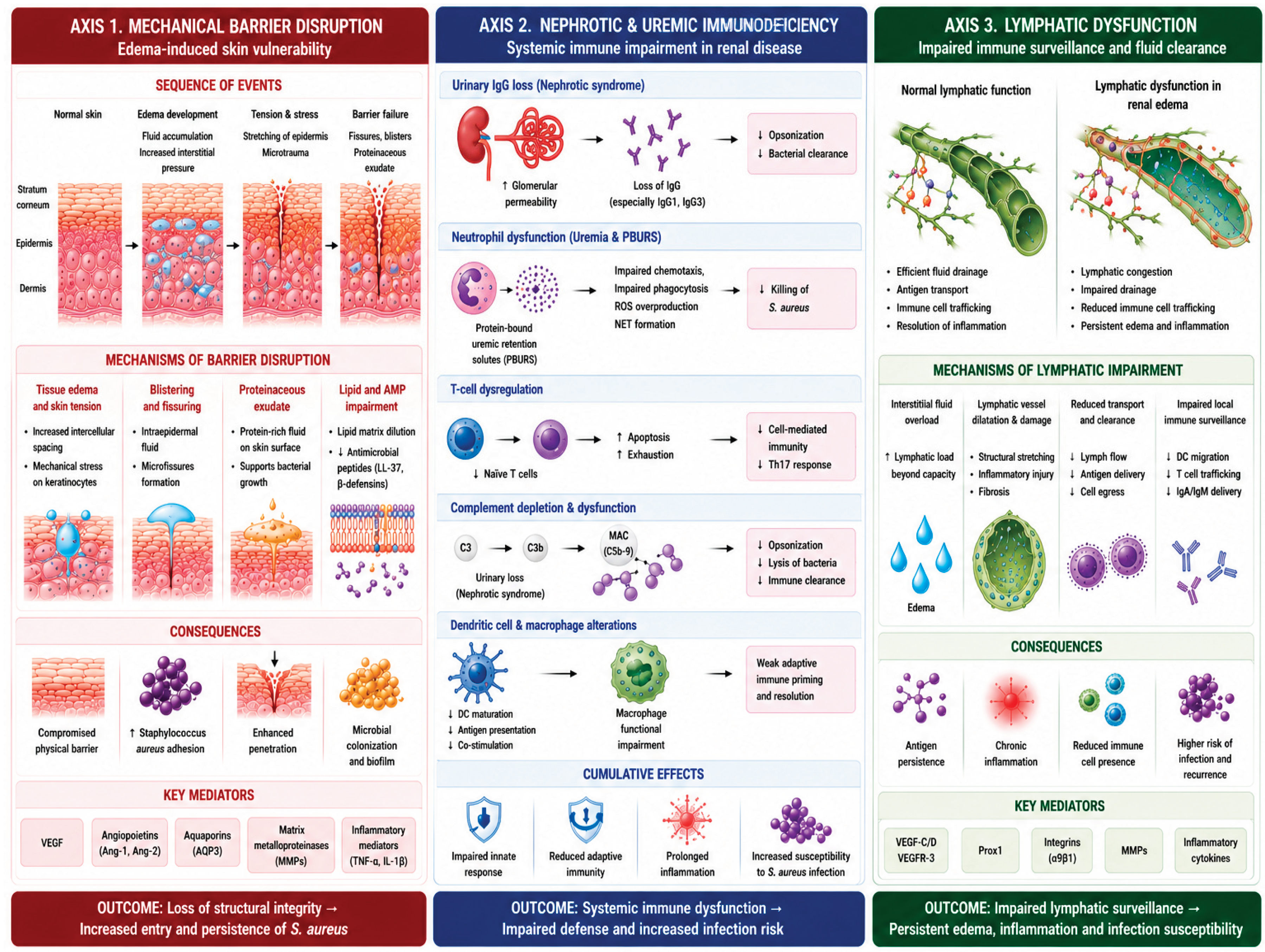

Table 1.
Mechanisms of nephrotic immunodeficiency and their clinical consequences.
| Mechanism | Pathophysiological Basis | Clinical Consequence |
| Urinary loss of immunoglobulins |
Massive proteinuria includes IgG loss; Reduced opsonization capacity |
Increased susceptibility to encapsulated and extracellular bacteria, including S. aureus |
| T-cell dysfunction | Th2/Th1 imbalance; reduced Treg function; increased Th17 activity |
Impaired cell-mediated immunity; Recurrent and persistent infections |
| B-cell dysregulation | Increased memory B-cell activity; altered CD23 release; anti-CD40 autoantibodies |
Dysregulated antibody production; Impaired humoral immune response |
| Complement loss | Urinary loss of complement factors (Factor B, Factor D, Properdin) | Defective opsonophagocytosis; Increased bacterial survival |
| Corticosteroid therapy |
Immunosuppressive treatment used in NS management | Iatrogenic immunosuppression compounding nephrotic immunodeficiency |
| Hypoalbuminaemia | Reduced serum albumin as acute-phase protein carrier |
Reduced drug and antimicrobial peptide transport; Nutritional depletion |
Table 2.
Key S. aureus virulence factors, mechanisms, and relevance in patients with renal disease.
| Toxin/Factor | Mechanism |
Predominant Clinical Manifestation |
Relevance in Renal Patients |
| Alpha-toxin (Hla) |
Pore formation in epithelial/endothelial membranes |
Furuncles, wound infections, cellulitis |
Enhanced effect in hypoalbuminemia; reduced opsonization |
| PVL (LukS/F-PV) |
Pore formation in neutrophils, monocytes, and macrophages |
Necrotizing furunculosis, recurrent abscesses, CA-MRSA SSTI |
PMN dysfunction in CKD amplifies PVL-mediated immune evasion |
| Exfoliative toxins (ETA, ETB) | Desmoglein-1 cleavage; intraepidermal cleavage |
Bullous impetigo, SSSS |
Renal clearance-dependent; toxic accumulation in CKD/AKI |
| TSST-1 (Superantigen) |
Non-specific T-cell activation; cytokine storm |
Toxic shock syndrome, multiorgan failure | Exaggerated systemic response in immunocompromised CKD patients |
| IsdA/SpA (Protein A) |
Fc binding; complement evasion; iron sequestration | Persistent bacteremia, biofilm formation |
Biofilm formation on skin and vascular access sites in dialysis patients |
| ArlRS/MgrA (Regulatory cascade) |
Controls the expression of immune evasion factors (leukocidins, SCIN, CHIPS) |
Abscess formation; neutrophil evasion |
Therapeutic target; dysregulated in high-inoculum CKD infections |
Table 3.
Clinical spectrum of S. aureus SSTIs and associated risk factors in patients with renal edema.
Table 3.
Clinical spectrum of S. aureus SSTIs and associated risk factors in patients with renal edema.
|
Clinical Presentation |
Key Features |
Typical Organism |
Risk Factors in CKD/Edema |
| Impetigo/ Ecthyma |
Superficial erosions; honey-coloured crusting; ecthyma penetrates the dermis |
S. aureus (MSSA/MRSA), GAS | Skin maceration from edema; poor hygiene; hypoalbuminemia |
| Folliculitis/ Furunculosis |
Perifollicular pustules; nodular abscesses; frequent recurrence with PVL strains |
S. aureus (PVL-positive) |
Increased nasal carriage; immunosuppression; skin moisture |
| Cellulitis | Non-purulent spreading erythema, warmth, edema; lymphangitis possible |
S. aureus, GAS |
Pre-existing lymphatic dysfunction; skin barrier disruption |
| Erysipelas | Raised, sharply demarcated erythema; predominantly superficial lymphatics |
GAS >> S. aureus | Lymphoedema; renal edema; Repeated episodes worsen lymphatics |
| Wound/ Access-site Infection |
Purulent discharge; dehiscence; biofilm on catheter/fistula |
S. aureus, MRSA, CONS | Dialysis access; peritoneal catheter; Reduced skin immunity |
| Necrotizing Fasciitis (Type II) |
Rapid spread; systemic toxicity; dishwater exudate; pain disproportionate to appearance |
S. aureus (monomicrobial) |
High mortality; delayed diagnosis in edematous limbs |
Table 4.
Anti-MRSA antibiotics for SSTIs: mechanisms, renal dose adjustments, and clinical considerations.
Table 4.
Anti-MRSA antibiotics for SSTIs: mechanisms, renal dose adjustments, and clinical considerations.
| Antibiotic | Class |
MRSA Activity |
Dose Adjustment in CKD |
Key Notes |
| Vancomycin | Glycopeptide | MSSA/ MRSA first-line |
Required (AUC/MIC monitoring); CrCl <50 mL/min requires dose reduction |
Nephrotoxic; avoid high troughs in CKD; AUC-guided dosing preferred |
| Teicoplanin | Glycopeptide | MRSA first-line |
Required; loading doses followed by dose/interval adjustment | Less nephrotoxic than vancomycin; once-daily dosing possible |
| Linezolid | Oxazolidinone | MRSA/ MSSA |
No renal dose adjustment required | Oral bioavailability ~100%; risk of serotonin syndrome; myelosuppression with prolonged use |
| Daptomycin | Cyclic lipopeptide |
MRSA/ MSSA |
Required (CrCl <30 mL/min: q48h dosing) | Inactivated by pulmonary surfactant; CPK monitoring required; NOT for pneumonia |
| Ceftaroline | 5th-gen cephalosporin |
MRSA/ MSSA |
Required (CrCl <50 mL/min: dose reduction) | Only beta-lactam approved for MRSA; useful in vancomycin-intolerant patients |
| Tedizolid | Oxazolidinone (2nd-gen) | MRSA/ MSSA |
No renal dose adjustment required |
Once-daily; shorter course (6 days) vs. linezolid; less myelosuppression |
| Dalbavancin | Lipoglycopeptide | MRSA/ MSSA |
Required (single 1500 mg dose; adjust if CrCl <30) | Once-weekly or single-dose IV; useful for outpatient SSTI completion therapy |
| Oritavancin | Lipoglycopeptide | MRSA/ MSSA |
Limited data in severe CKD | Single-dose IV; long half-life (245h); limited renal CKD data |
Table 5.
Clinical presentations of S. aureus infection in the ICU renal patient, with diagnostic priorities and mortality impact.
Table 5.
Clinical presentations of S. aureus infection in the ICU renal patient, with diagnostic priorities and mortality impact.
|
ICU Presentation |
Typical Source |
MRSA Risk |
Key Diagnostic Step |
Mortality Impact |
| Catheter-related BSI (CRBSI) | CVC/dialysis catheter |
High (≥40% in dialysis) |
Blood cultures ×2 + catheter tip culture |
30-day mortality 20–30% |
| Ventilator-associated pneumonia (VAP) | Endotracheal tube biofilm | High in post-influenza, immunosuppressed | BAL/protected brush + quantitative culture |
Attributable mortality 10–15% |
| Necrotising SSTI → sepsis | Oedematous skin fissure/wound |
Variable; PVL strains are more common in CA | CT/MRI + surgical exploration |
Mortality 25–35% in renal patients |
| Haematogenous seeding | Primary bacteremia (CRBSI, SSTI) | MRSA bacteremia → metastatic foci |
Echocardiography (IE exclusion); repeat BCs |
Infective endocarditis: 30% mortality |
| Septic shock with AKI |
Bacteremia + toxin-mediated vasodilation |
MRSA/MSSA both; TSST-1 strains |
SOFA score, lactate, organ function panel |
>50% mortality in MRSA septic shock + CKD |
Table 6.
Antibiotic management of S. aureus/MRSA infections in critically ill ICU patients with renal impairment.
Table 6.
Antibiotic management of S. aureus/MRSA infections in critically ill ICU patients with renal impairment.
| Antibiotic | ICU Indication | Dosing in CKD/Edema | Key PK/PD Consideration | Avoid/Caution |
| Vancomycin IV |
MRSA BSI, VAP, cSSTI — first line | AUC/MIC 400–600; loading 25–30 mg/kg in edema; TDM mandatory |
Expanded Vd in edema → higher loading dose; ARC → rapid clearance |
Nephrotoxic; AUC >500 on day 2 → ↑ AKI risk |
| Daptomycin IV |
MRSA BSI, right-sided endocarditis, SSTI |
6–10 mg/kg q24h; q48h if CrCl <30; CPK monitoring |
Inactivated by pulmonary surfactant — do NOT use for pneumonia/VAP | Monitor CPK weekly; rhabdomyolysis risk |
| Ceftaroline IV |
MRSA BSI salvage, SSTI, pneumonia | 600 mg q8h in normal renal function; adjust by CrCl |
Only beta-lactam approved for MRSA; useful combination with daptomycin in persistent MRSA bacteremia |
Limited data in ESRD; pharmacist-guided dosing required |
| Linezolid IV/PO | MRSA VAP, SSTI, step-down therapy | 600 mg q12h — no renal dose adjustment required |
Tissue penetration superior to vancomycin for lung/SSTI; 100% oral bioavailability | Serotonin syndrome; myelosuppression >14 days; avoid with SSRIs |
| Teicoplanin IV |
MRSA BSI, SSTI — alternative to vancomycin | Loading 6 mg/kg q12h ×3, then q24–48h by CrCl | Less nephrotoxic than vancomycin; once-daily maintenance; TDM target trough >15–20 mg/L |
Slower bactericidal activity vs. vancomycin |
| Daptomycin + Ceftaroline | Persistent MRSA bacteremia (salvage) | Dose adjust both by renal function; pharmacist-guided | Synergistic: ceftaroline restores daptomycin susceptibility in daptomycin-tolerant strains | High cost; reserve for refractory cases |
Disclaimer/Publisher’s Note: The statements, opinions and data contained in all publications are solely those of the individual author(s) and contributor(s) and not of MDPI and/or the editor(s). MDPI and/or the editor(s) disclaim responsibility for any injury to people or property resulting from any ideas, methods, instructions or products referred to in the content. |
© 2026 by the authors. Licensee MDPI, Basel, Switzerland. This article is an open access article distributed under the terms and conditions of the Creative Commons Attribution (CC BY) license (http://creativecommons.org/licenses/by/4.0/).
Copyright: This open access article is published under a Creative Commons CC BY 4.0 license, which permit the free download, distribution, and reuse, provided that the author and preprint are cited in any reuse.


