

Submitted:
26 May 2026
Posted:
28 May 2026
You are already at the latest version
Abstract
The amide macrocyclic complexes of zinc (ZnMc1, ZnMc2, and ZnMc3) were synthesized through template condensation of oxalic acid with different diamines (p-phenylenediamine, ethylenediamine, and propylenediamine). These zinc complexes were characterized using FT-IR, UV-Vis, conductance measurements, and DFT analysis, then tested for in vitro antibacterial and anticancer activities against Bacillus subtilis and HCT-15 cell lines, respectively. The results suggest that the complexes have promising antimicrobial and anticancer properties. The molecular structures of these zinc (II) complexes were also optimized theoretically, and their electronic and thermodynamic properties were determined with density functional theory (DFT). Finally, molecular docking studies using AutoDock evaluated the biological relevance of the synthesized complexes, identifying the most effective binding modes between the complexes and the receptor protein's active site. Two protein receptors, 6COX and 7AHL, were used to dock the macrocyclic complexes. The binding energies provide insights into their pharmaceutical potential. SwissADME and pkCSM were used to predict pharmacokinetic properties and toxicity. The ADMET profile of these macrocyclic architectures revealed their oral bioavailability and drug-like properties. Zinc complexes are known for their medicinal applications. So, these complexes were synthesized and studied as promising drug candidates for the future, though further research is needed to develop potent inhibitors.
Keywords:
macrocyclic complexes
; template condensation
; antibacterial activity
; in-silico docking
; DFT
1. Introduction
Nitrogen- containing organic compounds have demonstrated promising bioactivities [1,2,3,4,5]. Among these nitrogen compounds, macrocyclic compounds and their metal complexes have long intrigued chemists [6]. Nature’s macrocycles are well- characterized and serve remarkably diverse roles in biological systems. Synthetic macrocyclic metal complexes have been used for various biological applications, and the synthesis of these frameworks has increased significantly over the past two decades. Examples of natural metal- coordinated macrocyclic complexes include chlorophyll, myoglobin, hemoglobin, and vitamin B 12 [7,8]. In these macrocyclic structures, the metal plays a key role in stabilizing the complexes. These metal- coordinated complexes resist degradation and show enhanced thermal stability [9]. Amide- based macrocyclic complexes are highly interesting due to their unique coordination chemistry, which creates a geometrically constrained environment around a metal ion [10,11,12,13,14]. These large structures also have notable applications as antibacterial and antioxidant agents [15,16]. Zinc is the most essential mineral and is biocompatible with the human body. It is vital for various metabolic and biological processes, playing a crucial role in multiple physiological functions. Zinc also exhibits antimicrobial properties [17]. Zinc ions can effectively penetrate microbial cells and inhibit their growth by disrupting cellular processes [18]. Zinc also acts as an antioxidant by generating reactive oxygen species (ROS), which can damage bacterial cells. Zinc complexes have diverse applications, especially in medicinal chemistry [19]. Therefore, the search for new zinc complexes with strong biological activity is growing, as they could help overcome resistance challenges posed by existing antibiotics. Additionally, chemists are exploring additional applications, which have strengthened our interest and motivated us to synthesize macrocyclic complexes. Building on our previous work, the current aim is to synthesize and characterize novel N4-amide macrocyclic zinc complexes using various diamines and oxalic acid, followed by computational and biological evaluation.
2. Materials and Methods
2.1. Chemicals
All the required chemicals and solvents purchased were of analytical grade. They were used as received without purification. FT-IR spectrophotometer was used to record IR spectra in the range 4000-200 cm-1 using Nujol Mull. The literature method was used to determine the metal contents in the complexes [20]. A digital conductivity meter (HPG System, G-3001) was used for measuring conductivity. Melting points were determined using an electrical melting point apparatus.
2.2. Synthesis
Zinc metal was used as a template for synthesizing the macrocyclic complexes. An ethanolic solution (~20 ml) of zinc chloride (5 mmol) was refluxed with an ethanolic solution (~50 ml) of p-phenylenediamine/ethylenediamine/propylenediamine (10 mmol), respectively, for 30 minutes. Subsequently, to the above mixture solution, 10 mmol oxalic acid in 25 ml of ethanol was added, and refluxed with 1-2 drops of Conc.HCl for 6-8 hours. The condensation reaction yielded the ZnMc1, ZnMc2, and ZnMc3 complexes (Scheme 1). The solution was kept undisturbed at room temperature for 24 hours in desiccators. The colored precipitates of complexes were formed in good yield (~55-60%). They were filtered and washed with solvents such as methanol, ethanol, acetone, and diethyl ether and dried. The solubility test reveals their polar nature as they were found to be soluble in DMF and DMSO but difficult to dissolve in common organic solvents. They were found to decompose above 270 °C.
2.3. Biological Assays
2.3.1. In Vitro Antibacterial Activity
The in vitro antibacterial activity of synthesized zinc macrocyclic complexes was evaluated using the agar well diffusion method [21] with Muller–Hinton agar (MHA) medium. The study focused on Gram-positive bacteria, Bacillus subtilis (MTCC 441). All microbial cultures were adjusted to a 0.5 McFarland standard, corresponding to approximately 1.5 × 10⁸ cfu/mL [22,23]. Each petri dish was poured with 20 ml of MHA medium, and the agar plates were swabbed with 100 μl of each test bacterium’s inoculum, then incubated for 15 min to allow for adsorption. Wells with an 8 mm diameter were punched into the seeded agar plates using a sterile cork borer, and these wells were loaded with 100 μl of each metal complex at concentrations of 50, 75, and 100 mg/ml in 50% DMSO. All plates were incubated at 37 ºC for 24 hours. The antibacterial activity of each synthesized complex was determined by measuring the zone of growth inhibition against the test microorganisms using a zone reader (Hi Antibiotic zone scale). Dimethyl sulfoxide (DMSO) served as a negative control, while Gentamycin was used as a positive control.
2.3.2. Anticancer Activity
The human cancer cell lines used in this study were HCT-15, cultured in Roswell Park Memorial Institute (RPMI, Sigma-Aldrich, United States) supplemented with 10% fetal bovine serum (Sigma-Aldrich, United States) in tissue culture flasks at a 5% CO2 atmosphere and 37 °C [24]. The cells were seeded at 3 × 10⁴ cells per well in 100 µL of Dulbecco’s Modified Eagle’s Medium (DMEM) [for HCT-15 cells, from Roswell Park Memorial Institute (RPMI)] containing 10% fetal bovine serum (FBS) in a 96-well tissue culture plate and incubated for 72 hours at 37 °C in a CO2 incubator maintaining 5% CO2 and 90% relative humidity. Afterwards, the cells were exposed to different concentrations of zinc complexes prepared in DMEM/RPMI, specifically 100, 50, 25, and 12.5 µM, for 24 hours. Subsequently, the cells were incubated with 100 µL of MTT dye (0.5 mg/mL) in a CO2 incubator for an additional 4 hours. After incubation, the purple formazan produced by the cells appeared as dark crystals at the bottom of the wells. The culture medium was carefully removed from each well to avoid disrupting the monolayer. Then, 100 µL of dimethyl sulfoxide (DMSO) was added to each well. The plates were thoroughly mixed to dissolve the formazan crystals, resulting in a purple solution. The absorbance of the 96-well plates was measured at 570 nm using a Labsystems Multiskan EX ELISA reader against a reagent blank. The results were calculated as the micromolar concentration of 50% cell growth inhibition (IC50) for each complex. The MTT assay was performed in three independent experiments, each conducted in triplicate.
2.4. Computational Studies
2.4.1. DFT Studies
An electronic structure of atoms, molecules, and solids can be computed with an effective DFT method [25]. To interpret the reactivity and chemical structure, electronic structures are calculated. Ground state of synthesized complexes was optimized. To find the optimized geometry first-principles calculations based on density functional theory within a generalized gradient approximation (GGA) were employed. For greater reliability in our results, we use the Perdew–Burke–Ernzerhof [26] form of the GGA with the Quantum Espresso package [27] based on ultrasoft pseudopotentials [28] and a plane wave basis. Local minima of the energy landscape are obtained through structural relaxation starting with different initial structures using Hellman–Feynman forces and stresses [29]. The kinetic energy cutoff for wave functions was 50 Ry, and a gamma-only k-mesh was used in sampling Brillouin zones. Around 20 Å vacuum space was chosen to avoid any interaction between the periodic images.
2.4.2. Molecular Docking Studies
Molecular docking is a computational approach used to predict the orientation and binding poses of a ligand when it interacts with a target receptor, generally a protein or nucleic acid [30]. Established docking techniques assume rigid or semi-flexible receptor and ligand representations, employing standardized search algorithms such as genetic algorithms, Monte Carlo simulations, and progressive construction methods, integrated with experimental, force-field-based, or knowledge-based scoring functions to evaluate binding affinity [31]. It is widely known that these traditional methods are computationally capable and extensively used for virtual screening; they usually strive to perfectly capture protein flexibility, solvent effects, and induced-fit conformational transformations [32]. Current advancements in molecular docking have addressed these drawbacks by combining receptor ensemble docking, flexible docking, and hybrid docking-molecular dynamics approaches that more accurately account for conformational variations [33]. Moreover, the integration of Python-related scripts has remarkably exceeded scoring functions, pose predictions, and ranking efficacy by integrating complex non-linear interaction patterns from mass structural datasets [34]. Cloud computing, GPU acceleration, and AI-derived docking platforms have also made large-scale, high-throughput docking operations possible with improved speed and accuracy, making modern docking a high-powered tool in structure-based drug development and drug repurposing [35]. In the current study, the two receptor protein targets with PDB ID (6COX, 7AHL) were used, respectively, structures obtained from the PDB database for the docking process. The protein preparation was done using multiple techniques, such as the binding site evaluation, which was done using the built-in Python script, and then these x, y, z values were applied in AutoDock tools to set the center and size of the grid box. For receptor protein 6COX- the size of grid was – 68, 84, 74 and center was- 27.732, 29.435, 40.186, and for receptor protein 7AHL- the size of the grid was- 93, 73, 87, and center values were- 54.711, 7.741, 35.624. From homomer chains of the target protein structure, the A chains were selected for docking. Two molecules ZnMc2 and ZnMc3 were selected and prepared as ligands [36]. The docking operation was run through AutoDock Vina in Anaconda Prompt.
3. Results and Discussion
3.1. Analytical Studies
The analytical data for macrocyclic complexes indicate their formula as: [Zn(CxHyN4O4)Cl2], where x=16,10 or 8; y= 12 or 16. The zinc complexes were soluble in DMF and DMSO but insoluble in water. They are thermally stable to 250 °C and decompose without melting. The chloride ion test was found to be positive after decomposing the complexes. Attempts to crystallize the complexes using mixtures of solvents, slow diffusion, or low-temperature crystallization were unsuccessful. However, analytical, spectroscopic, and magnetic data enable us to predict the structures of the complexes. All the complexes give satisfactory elemental analyses, as given in Table 1. A distorted octahedral geometry is confirmed for these complexes.
Conductivity in DMSO indicated them to be non-electrolytes (15–20 ohm-1 cm2 mol-1)[37]. The molar conductance value indicates the formation of a covalent bond between the chloride ion and Zn(II). These complexes are non-electrolytes in nature, indicating the presence of a chloride ion inside the coordination sphere. The characteristic asymmetric and symmetric stretching vibrations band due to the amino group (NH2) in the IR spectra of diamines was absent in all the amide complexes. The band due to the OH group of dicarboxylic acids was also absent in the synthesized complexes. The absence of these characteristic bands indicates the formation of a macrocyclic framework. The IR absorption frequencies of the amide group occur at 1670-1680, 1565-1585, 1240-1270, and 650-682 cm-1 for ZnMc1-ZnMc3 complex [13,15]. The lower values in the amide bands in the range 3240–3260 cm−1, confirm the coordination of N–H with zinc metal [38]. The Far IR spectra show the band at 450-485 cm−1 corresponding to (M–N) stretching vibrations, highlighting the coordination of amide nitrogen with zinc metal [39]. The stretching vibration band in the range 300–330 cm−1is attributed to M-Cl bond [40,41]. The absorption band in the range 1445-1480 cm−1, 1040-1075 cm−1 and 725-760 cm−1 may be assigned to phenyl ring vibrations in ZnMc1.
3.2. Pharmacological Studies
3.2.1. Antibacterial Activity
In this study, all the synthesized macrocyclic complexes were evaluated against Gram-positive bacteria, Bacillus subtilis. All three synthesized macrocyclic complexes were screened for their antibacterial activities. All the tested complexes show variable antibacterial activities against B. subtilis at 75 and 100 mg/mL concentrations. Standard antibiotic, specifically gentamycin, was used to compare the antibacterial activities exhibited by the synthesized complexes (Table 2). Zinc complexes (ZnMc1-ZnMc3) were tested for in vitro cytotoxicity against HCT-15 (human colon cancer) using MTT assay. The reduction in cell viability obtained by a specific increase in concentration of complexes against a fixed number of cancer cells is shown in Table 3. IC50 values (µM) of the complexes are presented in Table 3. ZnMc1 exhibits the highest activity in inhibiting cell proliferation.
3.3. Computational Studies
3.3.1. DFT Studies
The relaxed geometry of the molecules is shown in Figure 1. DFT calculations indicate the formation of one primary (P) and one secondary (S) bond between Zinc and Nitrogen atom. As the molecules contain more than one bond between the same elements, the maximum and minimum bond lengths are given in Table 4. Total energy per atom for each molecule is also presented in Table 4. Furthermore, the energies of HOMO and LUMO are found (Figure 2), and the band gap Eg is calculated as the difference between HOMO and LUMO. Electronegativity and electronic affinity can be calculated, respectively, from the two relationships (2) and (3) [42,43].
For synthesized molecules, the electronic states, i.e., EHOMO, ELUMO, and are given in eV in Table 5. The Electron affinity (E.A), Ionization potentials (I.P), and electronegativity (X) are also given for the studied molecules
Our results show that the HOMO energies follow the trend of increasing energy for compounds ZnMc1 >ZnMc3>ZnMc2
whereas the LUMO energies for compounds can be arranged as follows: ZnMc1 >ZnMc3>ZnMc2
The band gap for the complexes increases in order ZnMc1 >ZnMc3>ZnMc2, and electronegativity decreases as ZnMc1 >ZnMc3>ZnMc2
According to Koopman’s theory [44], the following equations (3) and (4) can express the ionization potential and electron affinity, as shown in the Table
I.P = - EHOMO
E.A = ELUMO
Our results show the highest I.P for ZnMc2, whereas the lowest value is shown by ZnMc1, from eq (3) it is clear that E.A will follow the same trend as shown by ELUMO.
3.3.2. Molecular Docking
The docking operation was run through AutoDock Vina in Anaconda Prompt. The binding affinity with both proteins was studied for two complexes. Macrocyclic complex, ZnMc2 and ZnMc3 show docking score of -6.5k/Cal and -6.4 k/Cal with PDB ID- 6COX and -5.30 k/Cal and -5.4 k/Cal with protein ID- 7AHL(Table 6). These hit candidates were further checked on BioVia for interaction analysis, and they also showed van der Waals and Pi-Pi interactions with the respective target proteins [45]. (Figure 3 and Figure 4) Eventually, this study gives a justification for these molecules to take them into consideration for further validation through a molecular dynamics approach.
3.4. ADMET Analysis
SwissADME [46] and pkCSM [47] were used for analyzing several physicochemical properties ( Table 7, Table 8, Table 9 and Table 10). The three macrocyclic complexes have molecular weights less than 500 and do not violate the criteria for hydrogen bond acceptors (HBA less than 10), hydrogen bond donors (HBD less than 5), and rotatable bonds (RB less than 5). Also, they do not exceed the maximum limit of LogP value of 5 [48]. Table 8 depicts the drug-likeness rules (Lipinski’s rule of 5, Ghose rule, Veber’s rule, Egan’s rule, and Muegge’s rule) and bioavailability. These rules ensure the drug-like properties of test macrocyclic complexes. Violation of more than one parameter reduces the drug’s ability (Figure 5). All three complexes meet all the parameters. Complex ZnMc2 and ZnMc3 did not follow the Ghose rule. The bioavailability of the test macrocyclic complexes was found to be 0.55. Since Veber‘s rule is obeyed by these complexes, this suggests good oral bioavailability of all the complexes.The parameters of ADMET, like log S (water solubility), TPSA (topological polar surface area), GI absorption, Blood Brain Barrier permeability, and P-glycoprotein substrates, were studied. ZnMc1 is found to inhibit the CYP1A2 inhibitor and the CYP3A4 inhibitor. so, can slow down the activity of Cytochrome P450 1A2 (CYP1A2) and Cytochrome P450 3A4 (CYP3A4) enzyme (Table 10) and act as a substrate for P glycoprotein.
Toxicity is an important parameter that dictates the efficiency of a drug as a medicine. Toxicity is an important parameter that dictates the efficiency of a drug as a medicine. The complexes were found to be non- AMES toxic (non-mutagenic), non-hepatotoxic, non-skin sensitizer, and non-hERG inhibitor (Table 10). Oral rat acute and chronic toxicity (LD50 and LOAEL) values predicted are given in the table and are in safe range. These parameters suggest that, in the future, these macrocyclic complexes might act as a good drug candidate
4. Conclusions
This study reports the synthesis of amide macrocyclic complexes of zinc using the template method with oxalic acid and different diamines (p-phenylenediamine, ethylenediamine, and propylenediamine). Spectroscopic studies predicted an octahedral geometry. These complexes demonstrate activity against bacterial strains. In these zinc complexes, nitrogen coordinates to the zinc ion and shares its charge, reducing polarity and increasing lipophilicity. This lipophilicity helps the macrocyclic complexes cross bacterial membranes easily and exhibit antibacterial activity. The structures of these zinc (II) complexes were also optimized theoretically, and their electronic and thermodynamic properties were analyzed using density functional theory (DFT). The energy gap between HOMO and LUMO, as well as electronegativity and electron affinity, were calculated with DFT, in silico molecular docking studies of the ZnMc2 macrocyclic complex with Cyclooxygenase-2 (Prostaglandin Synthase-2) and alpha-hemolysin from Staphylococcus revealed various interactions. ADMET parameters indicate that these macrocyclic complexes could serve as promising drug candidates in the future, though further research is necessary to develop potent inhibitors.
Acknowledgments
Thanks are also due to the authorities of Amity University, Uttar Pradesh, Lucknow, for providing the necessary research facilities.
Conflicts of Interest
The authors declare no conflict of interest.
Abbreviations
The following abbreviations are used in the manuscript
B.M.-Bohr Magneton, CFU-Colony Forming Unit, DMF-N- N-dimethylformamide, DMSO-Dimethylsulphoxide, IR-Infrared, MIC-Minimum Inhibitory Concentration, MTCC-Microbial Type Culture Collection, NCCLS-National Committee for Clinical Laboratory Standards, MHA-Mueller Hinton Agar, MHB-Mueller Hinton Broth, RMSD-Root Mean Square Deviation
References
- Singh, D.P.; Kumar, R.; Kamboj, M.; Jain, K. Synthesis, Spectral, and Antibacterial Studies of 16-Membered Tetraazamacrocyclic Complexes. J. Coord. Chem. 2009, 62, 2995–3002. [Google Scholar] [CrossRef]
- Kishore, R.; Kamboj, M.; Shukla, M.; Chaturvedi, D.; Srivastava, N. Novel strategy for synthesis of cyclic dithiocarbamates catalyzed by Triton B. Asian J. Chem. 2019, 31(5), 1091–1094. [Google Scholar] [CrossRef]
- Kishore, R.; Kamboj, M.; Shukla, M.; Chaturvedi, D. Triton-B mediated efficient synthesis of thioxoimidazolidine-4,5-diones. Asian J. Chem. 2020, 32(1), 183–186. [Google Scholar] [CrossRef]
- Kishore, R.; Kamboj, M.; Shukla, M. Triton-B Mediated One-Pot Multicomponent Synthesis of 3,5 Substituted Tetrahydro-2H-1,3,5-Thidiazine-2-Thiones. Orient. J. Chem. 2018, 34(6), 2878–2883. [Google Scholar] [CrossRef]
- Waziri, I.; Nthehang, T.S.; Oyedeji-Amusa, M.O.; Oyebamiji, A.K.; Adeyinka, A.S.; Muller, A.A. Chem. Sel. 2025, 10(35), e01578. [CrossRef]
- Chandra, S.; Gupta, L.K.; Agrawal, S. Modern spectroscopic and biological approach in the characterization of a novel 14-membered [N4] macrocyclic ligand and its transition metal complexes. Trans. Met. Chem. 2007, 32, 240–245. [Google Scholar] [CrossRef]
- Tusek-Bozic, L.; Marotta, E.; Traldi, P. Efficient solid-state microwave-promoted complexation of a mixed dioxa-diaza macrocycle with an alkali salt. Synthesis of a sodium ethyl 4-benzeneazophosphonate complex. Polyhedron 2007, 26, 1663–1668. [Google Scholar] [CrossRef]
- Chandra, S.; Gupta, R.; Gupta, N.; Bawa, S.S. Biologically Relevant Macrocyclic Complexes of Copper Spectral, Magnetic, Thermal and Antibacterial Approach. Trans. Met. Chem. 2006, 31, 147–151. [Google Scholar] [CrossRef]
- Curtis, N.F. Macrocyclic coordination compounds formed by condensation of metal-amine complexes with aliphatic carbonyl compounds. Coord. Chem. Rev. 1968, 3, 3–47. [Google Scholar] [CrossRef]
- Niasari, M.S.; Daver, F. In situ one-pot template synthesis (IOPTS) and characterization of copper (II) complexes of 14-membered hexaaza macrocyclic ligand 3,10-dialkyl-dibenzo-1,3,5,8,10,12-hexaazacyclotetradecane. Inorg. Chem. Commun. 2006, 9(2), 175–179. [Google Scholar] [CrossRef]
- Lozovan, V.; Kravtsov, V.; Bulhac, I.; Petuhov, O.; Vlase, T.; Bourosh, P. N. Tetraaza macrocyclic complexes of Cu(II), Co(III) and Ni(II) derived from template condensation of 2,6-pyridinedicarbohydrazide and 2,6-diacetylpyridine: Stability investigation and coloring properties on plastics. J. Mol. Str. 2025, 1321(3), 140021. [Google Scholar] [CrossRef]
- Hanaa, E.; Ohyla, E. Novel (N) Macrocyclic Metal Complexes: Synthesis, Characterization, Spectral Studies and Anticancer Activity. Open Chem. J. 2018, 5, 51–63. [Google Scholar] [CrossRef]
- Subhash; Chaudhary, A.; Jyoti; Kumar, M.; Mamta; Solanki, R. Synthesis, structural elucidation, DFT investigations, biological evaluation and molecular docking studies of tetraamide-based macrocyclic cobalt (II) complexes. J. Iran. Chem. Soc. 2023, 20, 2339–2362. [Google Scholar] [CrossRef]
- Chandra, S.; Pundir, M. Spectroscopic characterization of chromium(III), manganese(II) and nickel(II) complexes with a nitrogen donor tetradentate, 12-membered azamacrocyclic ligand. Spectrochim. Acta A. 2008, 69, 1–7. [Google Scholar] [CrossRef]
- Shubash; Kumar, M.; Devi, A.; Chaudhary, A. Novel amide-based macrocyclic Co(ii) complexes: correlating structural, computational, and biological properties through DFT and docking. RSC Adv. 2026, 16, 3169–3191. [Google Scholar] [CrossRef]
- Singh, D.P.; Kumar, R.; Kamboj, M.; Grover, V.; Jain, K. Template Synthesis and Characterization of N6 12-Membered Macrocyclic Complexes Derived from Isatin and Ethylenediamine. Russ. J. Coord. Chem. 2008, 34, 233–235. [Google Scholar] [CrossRef]
- Singh, D.P.; Kamboj, M.; Kumar, K.; Jain, K.; Sharma, C.; Aneja, K.R. Synthesis, Spectroscopic Studies and Biological Screening of 18-Membered Octaazamacrocyclic Complexes Derived from Acetonylacetone and Thiocarbohydrazide. Russ. J. Inorg. Chem. 2011, 56, 1396–1401. [Google Scholar] [CrossRef]
- Mamta; Chaudhary, A. Synthesis, spectroscopic characterization, in vitro antimicrobial activity, antioxidant study, and theoretical approaches towards DNA gyrase, DHFR enzyme, NADPH enzyme of N8-tetraoxomacrocyclic complexes of Zn(II). J. Mol. Struct. 2024, 1295, 136743. [Google Scholar] [CrossRef]
- Godoy-Gallardo, M.; Eckhard, U.; Delgado, L. M.; de Roo Puente, Y.J.D.; Hoyos-Nogués, M.; Gil, F.J.; Perez, R.A. Antibacterial approaches in tissue engineering using metal ions and nanoparticles: From mechanisms to applications. Bioact. Mater. 2021, 6, 4470–4490. [Google Scholar] [CrossRef] [PubMed]
- Abendrot, M.; Chęcińska, L.; Kusz, J.; Lisowska, K.; Zawadzka, K.; Felczak, A.; Kalinowska-Lis, U. Zinc(II) Complexes with Amino Acids for Potential Use in Dermatology: Synthesis, Crystal Structures, and Antibacterial Activity. Molecules 2020, 25, 951. [Google Scholar] [CrossRef] [PubMed]
- Vogel, A.I. A textbook of quantitative chemical analysis; Longman, London, 1989. [Google Scholar]
- Andrews, J.M. Determination of minimum inhibitory concentrations. J. Antimicrob. Chemother. 2001, 48, 5–16. [Google Scholar] [CrossRef] [PubMed]
- Kamboj, M.; Singh, D.P.; Singh, A.K.; Chaturvedi, D. Molecular Modeling, in-Silico Docking and Antibacterial Studies of Novel Template Wangled Macrocyclic Complexes Involving Isatin Moiety. J. Mol. Struct. 2020, 1207, https://doi.org/10.1016/j.molstruc.2019.127602; Kamboj, M.; Jain, K.; Singh, D.P. Template Engineered Tetraazamacrocyclic Complexes and Their Antibacterial Studies. In Asian Journal of Chemistry; 2018; Volume 30, pp. 1431–1434. [Google Scholar] [CrossRef]
- Ahmad, I.; Beg, A.J. Antimicrobial and phytochemical studies on 45 Indian medicinal plants against multi-drug-resistant human pathogens. J. Ethnopharmacol. 2001, 74, 113–123. [Google Scholar] [CrossRef]
- Srivastava, N.; Kishore, R. Cleaner, Greener Synthesis, Characterization, and Anticancer Evaluation of 2-Thioxo-1,3- Thiazolanes. Indian J. Heterocycl. Chem. 2021, 31, 265–271. Available online: https://connectjournals.com/01951.2021.31.265.
- Roy, A. K. A new density functional method for electronic structure calculation of atoms and molecules. physics.chem-ph. 2019. Available online: https://arxiv.org/abs/1904.08806.
- Perdew, J.P.; Burke, K.; Ernzerhof, M. Generalized Gradient Approximation Made Simple. In Phys. Rev. Lett.;Phys. Rev. Lett.; Perdew, J.P., Burke, K., Ernzerhof, M., Eds.; 1996; Volume 78, p. 1396. [Google Scholar] [CrossRef]
- Giannozzi, P.; Baroni, S.; Bonini, N.; et al. QUANTUM ESPRESSO: a modular and open-source software project for quantum simulations of materials. J. Phys. Condens. Matter. 2009, 28 21, 395502. [Google Scholar] [CrossRef]
- Laasonen, K.; Car, R.; Lee, C.; Vanderbilt, D. Implementation of ultrasoft pseudopotentials in ab initio molecular dynamics. Phys. Rev. B. 1991, 43, 6796. [Google Scholar] [CrossRef]
- Hellmann, H. Einführung in die Quantenchemie. Leipzig: Franz Deuticke. p. 285. Feynman, R. P. 1939. “Forces in Molecules”. Physical Review 1939, 56, 340–343. [Google Scholar]
- Rasul, H.O.; Ghafour, D.D.; Aziz, B.K.; Hassan, B.A.; Rashid, T.A.; Kivrak, A. Decoding Drug Discovery: Exploring A-to-Z In Silico Methods for Beginners. Appl. Biochem. Biotechnol. 2025, 197(3), 1453–1503. [Google Scholar] [CrossRef]
- Huang, C.; Wei, L.; Yuan, W.; Lu, Y.; Zhang, G.; Yan, Z. Molecular Mechanisms of Aspartame-Induced Kidney Renal Papillary Cell Carcinoma Revealed by Network Toxicology and Molecular Docking Techniques. Int. J. Mol. Sci. 2026, 27(1), 77. [Google Scholar] [CrossRef] [PubMed]
- Martins, F.G.; Santos, H.A.; Sousa, S.F. A Review of Current Computational Tools for Peptide–Protein Docking, J. Comput. Chem. 2026, 47(5), e70328. [Google Scholar] [CrossRef]
- de Angelo, R.M.; de Sousa, D. S.; da Silva, A.P.; Chiari, L.P.>A.; da Silva, A.B.F.; Honório, K.M. Molecular Docking: State-of-the-Art Scoring Functions and Search Algorithms. In book: Computer-Aided and Machine Learning-Driven Drug Design; Maltarollo, V.G., Ed.; Springer Nature Switzerland: Cham, Switzerland, 2024. [Google Scholar] [CrossRef]
- Kiouri, D. P.; Batsis, G. C.; Chasapis, C. T. Structure-Based Approaches for Protein–Protein Interaction Prediction Using Machine Learning and Deep Learning. Biomol. 2025, 15(1), 141. [Google Scholar] [CrossRef] [PubMed]
- Singh, S.; Kumar, S.; Quadir, S.S.; Bhandari, S.; Baniya, B.; Joshi, G.; Jain, C.P.; Choudhary, D. Artificial Intelligence: Preface, Applications and Future Perspective in Relation to Pharmaceutical Sector. J. Pharm. Innov. 2025, 20, 39. [Google Scholar] [CrossRef]
- Robin, X.; Škrinjar, P.; Waterhouse, A.M.; Studer, G.; Tauriello, G.; Durairaj, J.; Schwede, T. Beyond Single Chains: Benchmarking Macromolecular Complex Prediction Methods With the Continuous Automated Model EvaluatiOn ( CAMEO ). Proteins Struct. Funct. Genet. 2026, 94(1), 403–413. [Google Scholar] [CrossRef]
- Singh, D.P.; Kumar, R.; Kamboj, M.; Jain, K. Biologically Active Tetraazamacrocyclic Complexes of Co(II), Ni(II), Cu(II) and Zn(II). Acta Chim. Slov. 2009, 56, 780–785. [Google Scholar]
- Siddiqi, Z.A.; Khan, M.M.; Khalid, M.; Kumar, S. Spectral and electrochemical characterization of bimetallic complexes of a novel 32-membered unsymmetrical [N12] macrocycle. Transit. Met. Chem. 2007, 32, 927–935. [Google Scholar] [CrossRef]
- Tyagi, P.; Chandra, S.; Saraswat, B.S.; Sharma, D. Design, spectral characterization, DFT and biological studies of transition metal complexes of Schiff base derived from 2-aminobenzamide, pyrrole and furan aldehyde. Spectrochim. Acta A. 2015, 43, 1–11. [Google Scholar] [CrossRef]
- Kamboj, D.P.; Kumar, M.; Jain, K.; Sharma, K.C. Divalent Metal Macrocyclic Complexes Derived from Acetonylacetone and Carbohydrazide with Their Spectroscopic and Antibacterial Studies. J. Coord. Chem. 2011, 64, 502–510. [Google Scholar] [CrossRef]
- Kumar, K.; Kamboj, M.; Jain, K.; Singh, D.P. Spectroscopic and Antibacterial Studies of New Octaazamacrocyclic Complexes Derived from Carbohydrazide and Isatin. Spectrochim. Acta A Mol. Biomol. Spectrosc. 2014, 128, 243–247. [Google Scholar] [CrossRef] [PubMed]
- Arivazhagan, M.; Manivel, S.; Jeyavijayan, S.; Meenakshi, R. Vibrational spectroscopic (FTIR and FT-Raman), first-order hyperpolarizability, HOMO, LUMO, NBO, Mulliken charge analyses of 2-ethylimidazole based on Hartree–Fock and DFT calculations. Spectrochim. Acta A. 2015, 134, 493–501. [Google Scholar] [CrossRef] [PubMed]
- Al – Saadawy, N. Synthesis, Characterization, and Theoretical Study of Some New Organotellurium Compounds Derived from Camphor. Indones. J. Chem. 2022, 22, 437–448. [Google Scholar] [CrossRef]
- Scheinmann, F. An Introduction to Spectroscopic Methods for the Identification of Organic Compounds: Mass Spectrometry, Ultraviolet Spectroscopy, Electron Spin Resonance Spectroscopy, Nuclear Magnetic Resonance Spectroscopy (Recent Developments), Use of Various Spectral Methods Together, and Documentation of Molecular Spectra; Elsevier Science: Amsterdam, Netherlands, 2014. [Google Scholar]
- Raen, R.; Islam, M. M.; Islam, R.; Islam, M.R.; Jarin, T. Functional characterization and structural prediction of hypothetical proteins in monkeypox virus and identification of potential inhibitors. Mol. Divers. 2025, 29(2), 1589–1617. [Google Scholar] [CrossRef]
- Daina, A.; Michielin, O.; Zoete, V. SwissADME: a free web tool to evaluate pharmacokinetics, drug-likeness and medicinal chemistry friendliness of small molecules. Sci. Rep. 2017, 3(7), M42717. [Google Scholar] [CrossRef] [PubMed]
- Pires, D.E.; Blundell, T.L.; Ascher, D.B. pkCSM: Predicting Small-Molecule Pharmacokinetic and Toxicity Properties Using Graph-Based Signatures. J. Med. Chem. 2015, 58(9), 4066–72. [Google Scholar] [CrossRef] [PubMed]
- Daina, A.; Michielin, O.; Zoete, V. iLOGP: a simple, robust, and efficient description of n-octanol/water partition coefficient for drug design using the GB/SA approach. J. Chem. Inf. Model. 2014, 54(12), 3284–301. [Google Scholar] [CrossRef] [PubMed]
Scheme 1.
Template Synthesis of macrocyclic Zinc complexes a) ZnMc1 b) ZnMc2 c) ZnMc3 from oxalic acid and diamine (p-phenylenediamine/ethylenediamine/propylenediamine).
Scheme 1.
Template Synthesis of macrocyclic Zinc complexes a) ZnMc1 b) ZnMc2 c) ZnMc3 from oxalic acid and diamine (p-phenylenediamine/ethylenediamine/propylenediamine).
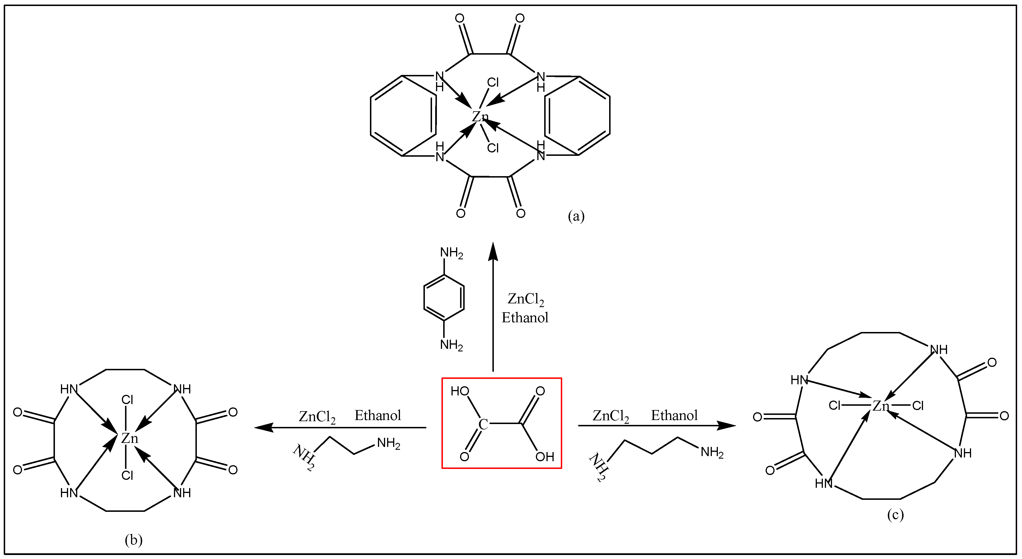
Figure 1.
The optimized structure of the molecules as found using the DFT calculations. Green, red, pink, steel grey, grey, and brown represent Cl, O, H, Zn, N, and C, respectively.
Figure 1.
The optimized structure of the molecules as found using the DFT calculations. Green, red, pink, steel grey, grey, and brown represent Cl, O, H, Zn, N, and C, respectively.
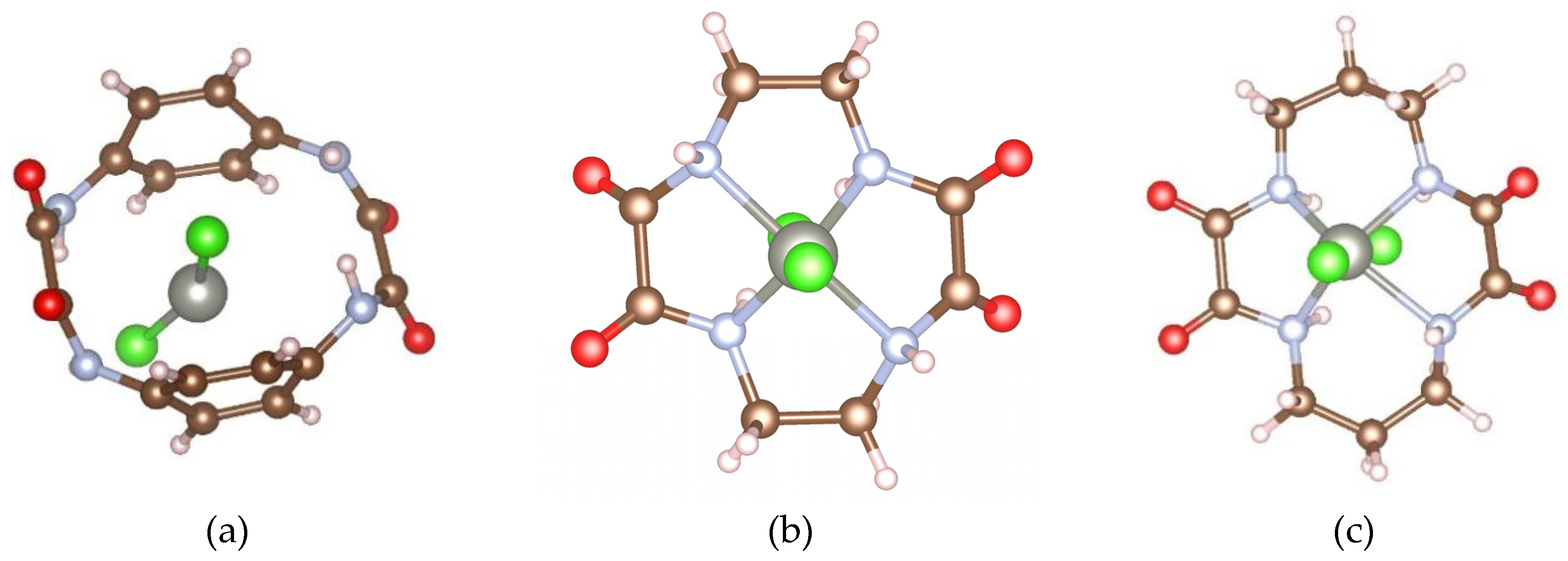
Figure 2.
(a–c) HOMO (upper panel) and (d–f) LUMO (lower panel) of synthesized molecules. Green, red, pink, steel grey, grey, and brown represent Cl, O, H, Zn, N, and C, respectively.
Figure 2.
(a–c) HOMO (upper panel) and (d–f) LUMO (lower panel) of synthesized molecules. Green, red, pink, steel grey, grey, and brown represent Cl, O, H, Zn, N, and C, respectively.
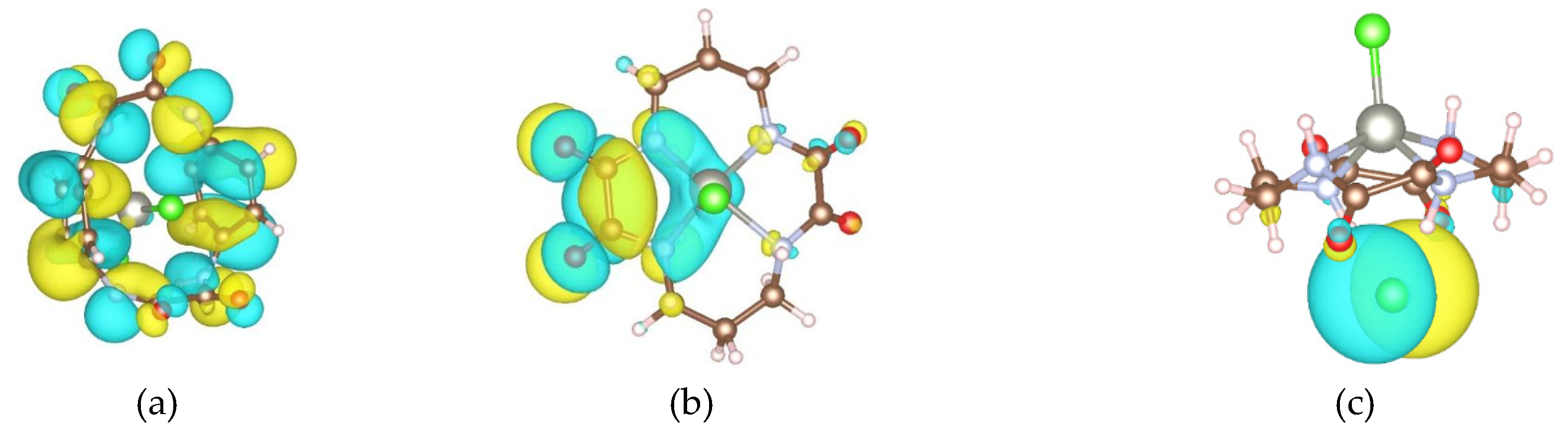
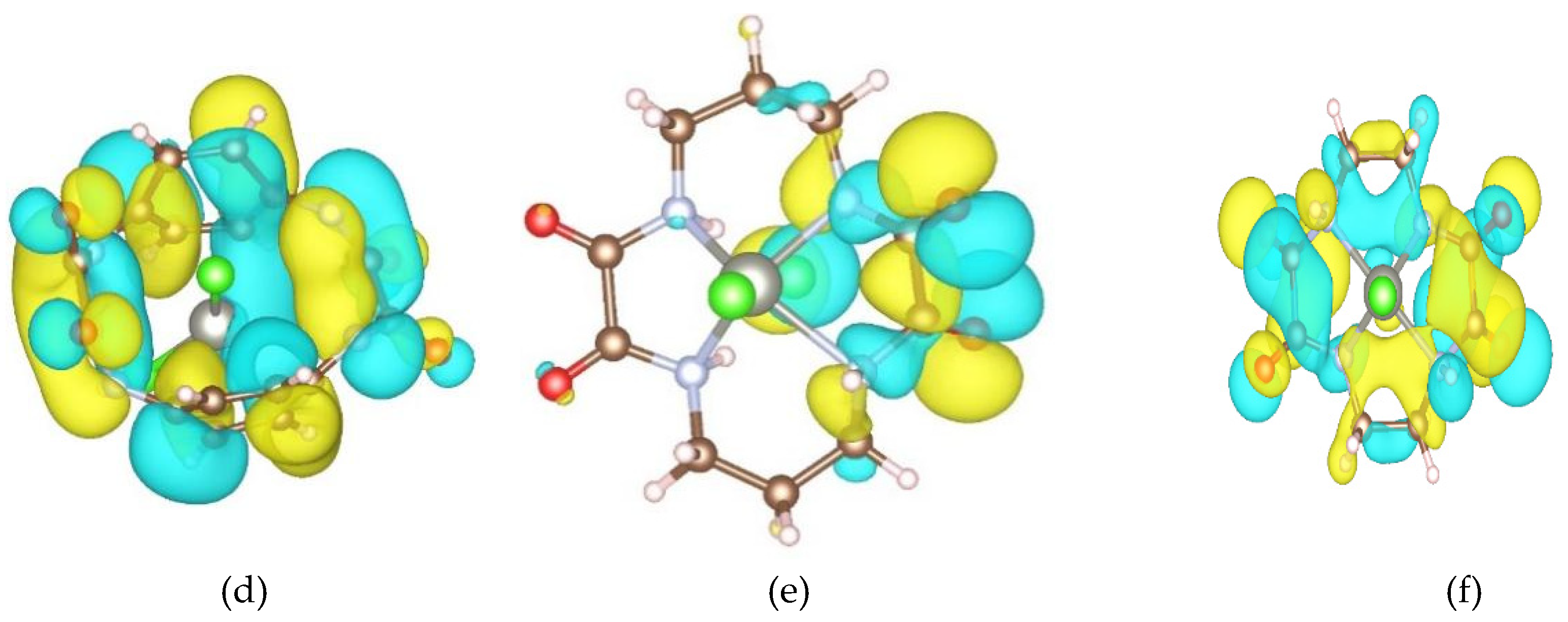
Figure 3.
2D and 3D docked structure of ZnMc2 with 7AHL.
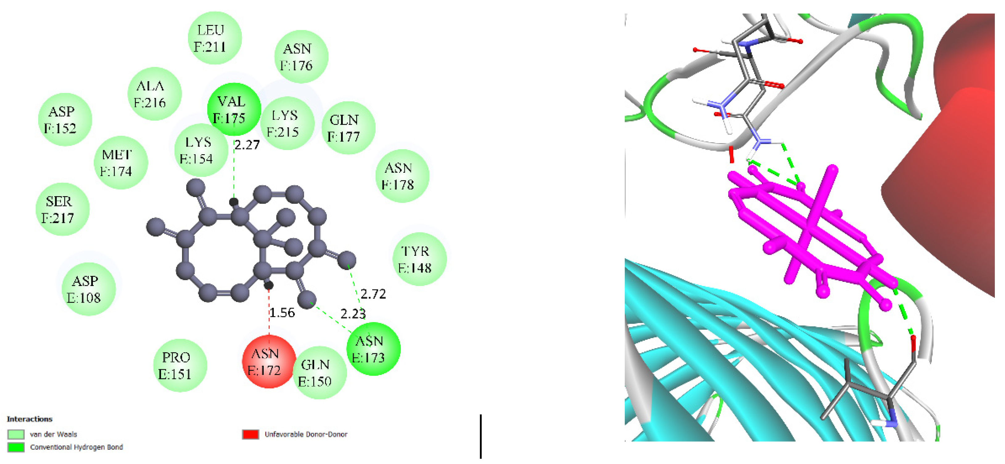
Figure 4.
2D and 3D docked structure of ZnMc3 with COX.
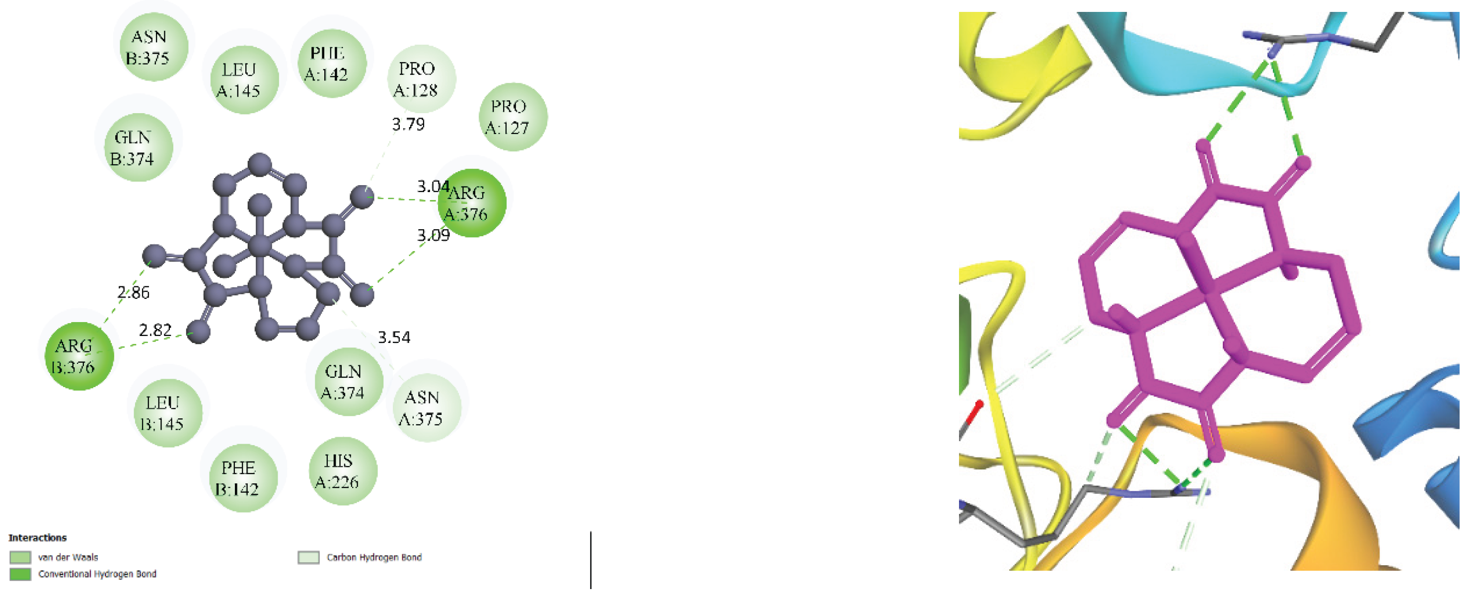
Figure 5.
Bioavailability Radar image obtained from SwissADME (Pink area in the figure represents optimal range of particular property) for ZnMc1, ZnMc2 and ZnMc3. (LIPO: lipophilicity; SIZE: size as molecular weight; POLAR: polarity asTPSA; INSOLU: insolubility in water; INSATU: insaturation as per fraction of carbons in the sp3 hybridization; FLEX: flexibility as per rotatable bonds).
Figure 5.
Bioavailability Radar image obtained from SwissADME (Pink area in the figure represents optimal range of particular property) for ZnMc1, ZnMc2 and ZnMc3. (LIPO: lipophilicity; SIZE: size as molecular weight; POLAR: polarity asTPSA; INSOLU: insolubility in water; INSATU: insaturation as per fraction of carbons in the sp3 hybridization; FLEX: flexibility as per rotatable bonds).
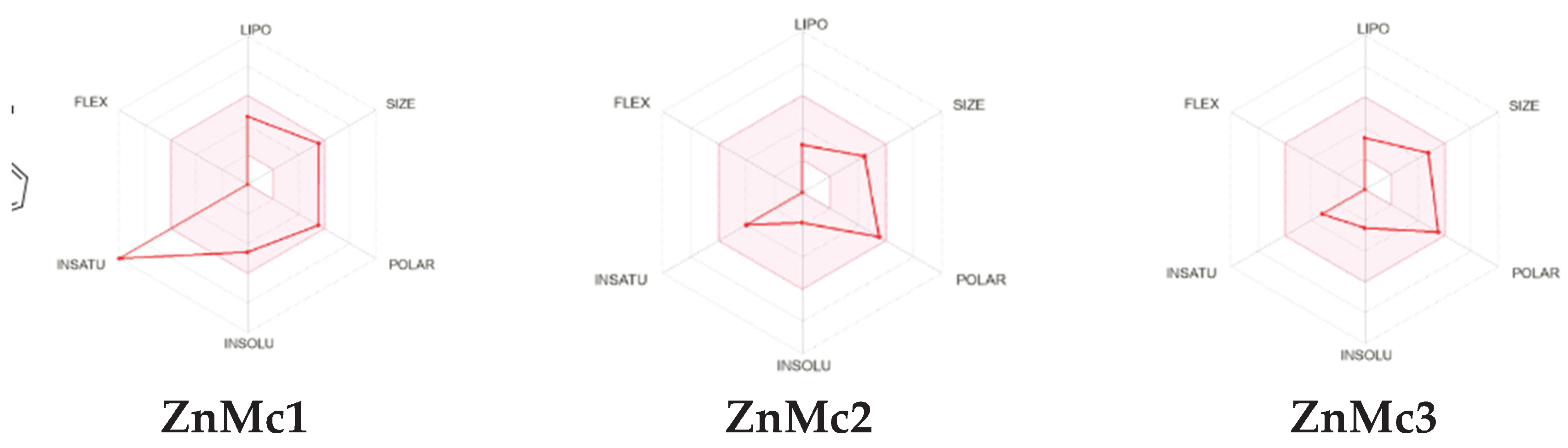

Table 1.
Analytical data of Zinc Macrocyclic complexes. Found (Calcd.) %.
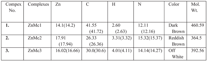 |
Table 2.
Antibacterial activities of synthesized complexes against Bacillus subtilis.
| S.No. | Bacterial Strain |
Diameter of zone of inhibition, mm | ||||
|---|---|---|---|---|---|---|
| Concentration (mg/mL) |
Gentamycin (Standard) (mm) |
Control (DMSO) | ||||
| 50 |
75 |
100 |
50 mg/mL |
50% | ||
| ZnMc1 |
Bacillus subtilis (Gram-positive) |
0 | 1 | 1.2 | 2.8 | 0 |
| ZnMc2 | 0 | 1.1 | 1.1 | 2.8 | 0 | |
| ZnMc3 | 0 | 1.3 | 1.5 | 2.8 | ||
Table 3.
Cytotoxicity of DMSO soluble compounds on HCT-15 cell line.
| S.No | Concentration(μg/mL) | Cytotoxicity % | ||
|---|---|---|---|---|
| A | C | D | ||
| 1. | 48 | 39.22 | 21.57 | 9.48 |
| 2. | 24 | 33.68 | 14.35 | 2.51 |
| 3. | 12 | 27.07 | 5.19 | 1.78 |
| 4. | 6 | 20.95 | 2.21 | 1.03 |
| 5. | 3 | 10.61 | 1.01 | 0.76 |
| 6. | 1.5 | 2.02 | 0.43 | 0.28 |
Table 4.
Bond length between various elements in the molecule. (As the molecules contain more than one bond between the same elements, the maximum and minimum bond lengths are given in Table 4).
Table 4.
Bond length between various elements in the molecule. (As the molecules contain more than one bond between the same elements, the maximum and minimum bond lengths are given in Table 4).
| Molecule | EHOMO (eV) | EFERMI (eV) | ELUMO (eV) | E.A (eV) | I.P (eV) | X = (EHOMO +ELUMO)/2 (eV) | Eg= (EHOMO -ELUMO) (eV) | Total Energy (eV) |
|---|---|---|---|---|---|---|---|---|
| ZnMc1 | -4.8266 | -4.0393 | -2.8770 | -2.8770 | 4.8266 | -3.8518 | 1.9496 | -990.73591 |
| ZnMc 2 | -6.5898 | -5.0874 | -3.7995 | -3.7995 | 6.5898 | -5.1947 | 2.7903 | -885.33233 |
| ZnMc3 | -6.4932 | -5.0798 | -3.5538 | -3.5538 | 6.4932 | -5.0235 | 2.9394 | -843.65217 |
Table 5.
EHOMO, ELUMO, Eg, Electron affinity (E.A), Ionization potentials (I.P) and electronegativity (X) are given in eV.
Table 5.
EHOMO, ELUMO, Eg, Electron affinity (E.A), Ionization potentials (I.P) and electronegativity (X) are given in eV.
| Molecule/Bond | Zn-Cl | C-N | C-H | C-O | N-H | C-C | N-Zn | Energy/atom (Ry) |
|---|---|---|---|---|---|---|---|---|
| ZnMc1 | 2.231(2.214) | 1.436 (1.397) | 1.090 | 1.217 (1.212) | 1.020 | 1.636 (1.403) | 3.612 (2.909) | -25.4034849 |
| ZnMc2 | 3.375 (2.168) | 1.484 (1.415) | 1.100 (1.099) | 1.215 (1.211) | 1.055 (1.029) | 1.569 (1.534) | 2.385 (2.155) | -23.9279009 |
| ZnMc3 | 3.727 (2.163) | 1.489 (1.404) | 1.100 (1.097) | 1.214 (1.205) | 1.099 (1.020) | 1.571 (1.550) | 2.219(2.140) | -27.2145863 |
| Molecule/Bond | Zn-Cl | C-N | C-H | C-O | N-H | C-C | N-Zn | Energy/atom (Ry) |
| ZnMc1 | 2.231(2.214) | 1.436 (1.397) | 1.090 | 1.217 (1.212) | 1.020 | 1.636 (1.403) | 3.612 (2.909) | -25.4034849 |
| ZnMc2 | 3.375 (2.168) | 1.484 (1.415) | 1.100 (1.099) | 1.215 (1.211) | 1.055 (1.029) | 1.569 (1.534) | 2.385 (2.155) | -23.9279009 |
| ZnMc3 | 3.727 (2.163) | 1.489 (1.404) | 1.100 (1.097) | 1.214 (1.205) | 1.099 (1.020) | 1.571 (1.550) | 2.219(2.140) | -27.2145863 |
Table 6.
Binding Affinity scores of complexes with the Receptor Protein.
| Sr.no. | Complex | PDB no | Receptor | Binding Affinity |
|---|---|---|---|---|
| 1. | ZnMc2 |
7AHL | ALPHA-HEMOLYSIN FROM STAPHYLOCOCCUS AUREUS | -5.3 |
| 6COX | CYCLOOXYGENASE-2 (PROSTAGLANDIN SYNTHASE-2) | -6.5 | ||
| 2. | ZnMc3 | 7AHL | ALPHA-HEMOLYSIN FROM STAPHYLOCOCCUS AUREUS | -5.4 |
| 6COX | CYCLOOXYGENASE-2 (PROSTAGLANDIN SYNTHASE-2) | -6.4 |
Table 7.
Molecular properties, descriptors, and lead-likeness of the amide macrocyclic complexes.
| Complex | Formula | Mw | HBD | HBA | Nrot | Logp | TPSA | Logs | Csp3 | Leadlikeness |
|---|---|---|---|---|---|---|---|---|---|---|
| Znmc1 | C16H12Cl2N4O4Zn | 460.58 | 4 | 4 | 0 | 1.8093 | 116.40 | -4.59 | 0 | No |
| Znmc2 | C8H12Cl2N4O4Zn | 364.49 | 4 | 4 | 0 | -2.1587 | 116.40 | -1.88 | 0.50 | No |
| Znmc3 | C10H16Cl2N4O4Zn | 392.54 | 4 | 4 | 0 | -1.3785 | 116.40 | -2.5 | 0.6 | No |
MW- molecular weight, HBD-hydrogen bond donors, HBA- hydrogen bond acceptors, nRB- number of rotatable bonds, LogP- partition co-efficient, Csp3.
Table 8.
Drug-likeness and bioavailability of the amide macrocyclic complexes.
| Compound | Lipinski | Ghose | Veber | Egan | Muegge | Bioavailability score | No of Violations |
|---|---|---|---|---|---|---|---|
| ZnMc1 | Yes | Yes | Yes | Yes | Yes | 0.55 | 0 |
| ZnMc2 | Yes | No | Yes | Yes | Yes | 0.55 | 1 |
| ZnMc3 | yes | no | yes | yes | yes | 0.55 | 1 |
Table 9.
ADMET physicochemical properties of amide macrocyclic complexes.
| Complex | GI absorption | BBB permeant | P-gp Substrate | CYP1A2 inhibitor | CYP2C19 inhibitor | CYP2C9 inhibitor | CYP2D6 inhibitor | CYP3A4 inhibitor |
|---|---|---|---|---|---|---|---|---|
| ZnMc1 | High | No | Yes | yes | No | No | no | yes |
| ZnMc2 | Low | No | No | No | No | No | No | No |
| ZnMc3 | Low | No | No | No | No | No | No | no |
Table 10.
Toxicity prediction of amide macrocyclic complexes.
| Complexes | AMES toxicity | Hepatoxicity | Max. tolerated dose (human) mg/kg/day | Oral rat Acute Toxicity (LD50) |
Oral Rat Chronic Toxicity (LOAEL) (mol/kg) |
T.Pyriformis toxicity (log ug/L) |
hERG I inhibitor | hERG II inhibitor | Skin Sensitisation |
|---|---|---|---|---|---|---|---|---|---|
| 1 | No | No | 0.181 | 2.218 | 1.246 | 0.3 | no | no | no |
| 2 | No | no | 1.087 | 2.929 | 1.83 | 0.144 | no | no | no |
| 3 | No | no | 0.835 | 3.188 | 1.568 | 0.148 | no | no | no |
Disclaimer/Publisher’s Note: The statements, opinions and data contained in all publications are solely those of the individual author(s) and contributor(s) and not of MDPI and/or the editor(s). MDPI and/or the editor(s) disclaim responsibility for any injury to people or property resulting from any ideas, methods, instructions or products referred to in the content. |
© 2026 by the authors. Licensee MDPI, Basel, Switzerland. This article is an open access article distributed under the terms and conditions of the Creative Commons Attribution (CC BY) license (http://creativecommons.org/licenses/by/4.0/).
Copyright: This open access article is published under a Creative Commons CC BY 4.0 license, which permit the free download, distribution, and reuse, provided that the author and preprint are cited in any reuse.