
Submitted:
24 May 2026
Posted:
25 May 2026
You are already at the latest version
Abstract
Leptin is the oldest studied adipokine, and its mechanism of action in the regulation of energy metabolism remains a hot topic of current research. In this paper, we systematically review the progress of clinical and basic research on leptin and energy metabolism from 1994 to 2025. It is shown that leptin can regulate the energy metabolism homeostasis through autocrine, paracrine and neurohumoral pathways (e.g., hypothalamic-leptin-melanocortin axis). In addition, the effects of mainstream weight loss strategies such as dietary control, pharmacological interventions and exercise on leptin levels and their underlying mechanisms were investigated in this paper, with the aim of providing a theoretical basis for the clinical application of leptin in metabolic diseases (e.g., obesity, diabetes mellitus). Future studies need to further clarify the molecular mechanisms of leptin resistance and explore precise intervention strategies based on the leptin signaling pathway.
Keywords:
leptin
; energy metabolism
; obesity
; endocrine regulation
1. Introduction
Energy homeostasis is the state in which an organism maintains a long-term dynamic equilibrium between energy intake and energy expenditure through a series of physiological regulatory mechanisms. It is a core physiological process for the maintenance of life and health. It is not a simple static equilibrium, but rather a dynamic, finely regulated process. Governed by the central nervous system (particularly the hypothalamus), it involves a complex network of communication between multiple organs and numerous signalling molecules. Its purpose is to coordinate energy intake, expenditure and storage in order to maintain long-term stability in body weight and metabolism.Understanding energy homeostasis is crucial for elucidating the pathogenesis of metabolic diseases such as obesity and diabetes, and for developing strategies for their prevention and treatment. Among these, leptin—the ‘satiety signal’ produced by adipose tissue—is a key regulatory molecule involved in maintaining energy homeostasis.
Leptin, the best-known adipokine, has been the subject of extensive research since its discovery in 1994. It has been found that leptin plays a role in energy metabolism by suppressing appetite, increasing energy expenditure and maintaining insulin sensitivity[1,2]. Research indicates that leptin acts as a homeostatic signal (a fat signal) that regulates body weight; for example, mice lacking the leptin gene (ob/ob mice) and humans with congenital leptin deficiency caused by rare genetic mutations in the leptin gene become severely obese[3,4,5]. However, administering leptin to ob/ob mice reduces food intake and increases energy expenditure, leading to an energy deficit and weight loss. Furthermore, leptin monotherapy in obese individuals with congenital leptin deficiency can also result in sustained weight loss.
Furthermore, leptin is a cytokine secreted primarily by white adipose tissue in proportion to fat mass; consequently, leptin levels reflect the body’s fat stores. In terms of energy metabolism, leptin binds to leptin receptors to activate downstream signalling molecules, thereby promoting the body’s energy metabolism. It acts primarily on the hypothalamus and, via the peripheral nervous system, regulates the energy-metabolising organs (adipose tissue, liver, skeletal muscle, etc.)[6]. Adipose tissue also contains a large number of leptin receptors; this finding demonstrates that leptin may also regulate the physiological activity of adipose tissue via an autocrine pathway[7]. However, in a state of obesity, abnormal proliferation of adipose tissue leads to excessive leptin secretion, resulting in reduced sensitivity to leptin and the development of leptin resistance. This causes disturbances in the body’s energy metabolism and insulin resistance, ultimately leading to a series of metabolic syndromes[8,9,10].
This review aims to analyse how leptin maintains energy homeostasis through different secretion patterns; it discusses the role of various therapeutic approaches for energy homeostasis imbalances in the context of leptin mechanisms; and examines gaps and limitations in the current evidence, as well as areas for future research and clinical translation.
2. Mechanisms Regulating Leptin Secretion and Energy Metabolism Homeostasis
2.1. Primary Mode of Secretion – Endocrine
2.1.1. Target Organ – the Hypothalamus
The effects of leptin on energy metabolism primarily involve central nervous system signalling pathways[3,4,5,11,12]. A comparison between hypothalamic leptin receptor knockout mice and db/db mice revealed that the two groups exhibit similar metabolic phenotypes. Although the residual leptin signalling in the hypothalamic leptin receptor knockout mice is still capable of regulating their physiological functions to some extent, this compensatory effect is insufficient to reverse their obese phenotype[13,14]. This demonstrates that the hypothalamus is the primary site for leptin-mediated regulation of the body’s energy balance. Hypothalamic extensor cells play a key role in this process; for the transcellular transport of leptin, leptin and epidermal growth factor (EGF) must sequentially activate the LepR(leptin receptor):EGFR complex, enabling leptin to traverse the brain and reach its target neurons[15].
The central melanocortin system plays a key role in the satiety effects of leptin and the activation of the sympathetic nervous system[16]. This system comprises a variety of cell types, including neurons expressing pro-opiomelanocortin (POMC), agouti-related protein (AgRP), melanocortin-3 receptor (MC3R) and melanocortin-4 receptor (MC4R).Research has shown that corticotropin-releasing hormone (CRH) promotes the degradation of POMC in the anterior pituitary; it was found that treatment with a CRH antagonist (αCRH) reduces the effect of leptin on the expression of c-fos-like immunoreactivity (cFLI) in the paraventricular nucleus (PVN) and ventromedial hypothalamus (VMH), thereby attenuating leptin’s appetite-suppressing and weight-reducing effects.The study also found a strong negative correlation between CRH and peripheral leptin levels, suggesting that leptin stimulates the hypothalamus to release CRH, which in turn inhibits leptin secretion by fat cells, thereby maintaining a dynamic equilibrium[17,18].Hypothalamic POMC and AgRP neurons are primarily located in the arcuate nucleus (ARC). The hypothalamic ARC is a key structure involved in suppressing food intake and stimulating thermogenesis in brown adipose tissue (BAT) and autonomic activity.Studies involving ARC-damaged rats have revealed that leptin’s regulation of food intake and body weight requires an intact ARC to be effective, suggesting that the integrity of the ARC plays a crucial role in the functioning of the leptin signalling pathway[19].In the ARC, leptin exerts its appetite-suppressing effect by upregulating the expression of POMC in neurons and downregulating the expression of AgRP in neurons[20]. Among these, signal transduction and transcription activator 3 (STAT3) is a transcription factor involved in a variety of biological functions[21]; it acts as a downstream signalling molecule of leptinand serves as an essential ‘messenger’ for transcriptional regulation in both POMC neurons and AGRP/NPY neurons[22,23]. Other genes in these neurons also play an important role in leptin-mediated energy metabolism. Specific knockout of Sir2-type 1 (Sirt1) in POMC neurons in mice severely impairs the normal binding of leptin to phosphoinositide 3-kinase (PI3K) signalling in POMC neurons, thereby disrupting the remodelling of periglandular white adipose tissue (WAT). At the same time, sympathetic activity and browning of the periglandular fat in mutant mice are also reduced, suggesting that Sirt1 in POMC neurons plays an important physiological role in regulating visceral fat browning[24].Similarly, through genetic knockout and overexpression in POMC neurons, it was found that the presence of the Sequestosome 1 (p62) and zinc-α2-glycoprotein (AZGP1) genes both contribute to increased leptin sensitivity; however, in terms of mechanism, p62 interacts with STAT3, promoting its phosphorylation to initiate POMC transcription and enhance leptin sensitivity. This indicates that p62 can directly regulate STAT3/POMC signalling and enhance leptin sensitivity[25]. AZGP1, on the other hand, enhances leptin-JAK2-STAT3 signalling and increases leptin sensitivity by interacting with acyl-GMP kinase (AGK) to block its ubiquitin-mediated degradation[26].
Cytokin-induced SH2 domain protein (CISH) is co-expressed with the leptin receptor on AgRP neurons in the ARC; the absence of CISH leads to a reduction in the basal expression of AgRP in the arcuate nucleus of the brain. Mice lacking CISH are more sensitive to leptin; although CISH expression itself is not regulated by leptin, CISH’s negative regulation of leptin’s anorexigenic effects makes it a key regulator of metabolic balance in vivo[27].As our understanding of AgRP neurons has deepened, it has been discovered that they secrete NPY, AgRP and γ-aminobutyric acid (GABA) simultaneously; they are therefore also referred to as NAG neurons. GABAergic RIP-Cre neurons in the arcuate nucleus can coordinately drive energy expenditure, enhance the thermogenic effects of leptin, and prevent diet-induced obesity[28].Neurons in the ARC that express both calcitonin gene-related peptide receptors (Calcr) and leptin receptors include NAG neurons and non-NAG neurons. Experiments have shown that leptin can directly activate LepRb and Calcr in these cells, producing related effects[29].Mice lacking forkhead box O1 (FOXO1) in POMC or AgRP neurons exhibit favourable metabolic characteristics, characterised by a lean body conformation and increased sensitivity to insulin and leptin.Specific knockout of the G protein-coupled receptor 17 (Gpr17) gene in AgRP neurons produces the same phenotype as knockout of the FOXO1 gene in the same cell type, providing preliminary evidence that the G protein-coupled receptor Gpr17 acts as an effector of the appetite-promoting signal from FOXO1 in AgRP neurons[30].
α-Melanocyte-stimulating hormone (α-MSH) is a melanocortin receptor agonist produced by POMC neurons, whereas AgRP is an endogenous melanocortin receptor antagonist[31]. Experiments have shown that α-MSH can enhance leptin sensitivity and preadipocyte proliferation by activating the Neurogenic locus notch homolog 1 (Notch1) signalling pathway, whilst simultaneously inhibiting endoplasmic reticulum stress in preadipocytes[32]. However, there is no direct correlation between α-MSH levels and leptin resistance, demonstrating that there is no direct proportional relationship between α-MSH and leptin concentrations[33].The G protein stimulatory α-subunit (Gsα) facilitates the coupling of various receptors, including MC4R, to intracellular cAMP. Following the generation of Gsα-deficient mice in the dorsomedial hypothalamus (DMH), impaired leptin signalling was observed, accompanied by increased expression of the leptin signalling inhibitor protein tyrosine phosphatase 1B (PTP1B) in the DMH. This may account for the mice’s excessive food intake, reduced energy expenditure, decreased locomotor activity and diminished cold-induced thermogenesis[34].The absence of MC3R and MC4R impairs leptin-induced nutrient distribution, appetite suppression and energy metabolism; however, their specific roles require further investigation[35,36].Tropomyosin receptor kinase B (TrkB) signalling originating in the hypothalamus can directly regulate appetite, metabolism and taste preferences downstream of leptin and MC4R[37]. Brain-derived neurotrophic factor (BDNF) is one of the endogenous ligands for the tyrosine kinase receptor TrkB, and theVentromedial Hypothalamus(VMN) is a key site where BDNF inhibits High-Fat Diet(HFD)-induced obesity[38]. VGF (VGF nerve growth factor inducible) is a propeptide particularly abundant in the hypothalamus, and its expression is upregulated by environmental enrichment (EE). Research findings indicate that hypothalamic VGF expression is regulated by leptin, melanocortin receptor agonists and food deprivation, and largely parallels BDNF expression[39].
POMC neurons are also found in the nucleus tractus solitarius (NTS) of the medulla oblongata in the brainstem; in recent years, the importance of brainstem nuclei in regulating energy balance has been recognised.The forebrain and hindbrain are connected by extensive synaptic networks, enabling them to act both independently and in concert in regulating the body’s energy metabolism. Research has shown that activating leptin receptors in the NTS of the hindbrain can influence the effects of leptin on the forebrain via neural networks[4,40,41]. For example, leptin in the posterior brain enhances the precision of energy balance control by lowering the threshold for leptin signalling in the anterior brain[11]. Under basal conditions, leptin receptors in the posterior brain may antagonise the activity of receptors in the anterior brain in order to protect lean tissue and adipose tissue[42]. Metabolic activity in the forebrain is transmitted via neural pathways to the hindbrain, thereby influencing peripheral tissues. For example, the thermogenic effect of leptin, mediated by RIP-Cre GABAergic neurons in the arcuate nucleus, may involve signals from Paraventricular Hypothalamic nucleus(PVH) neurons being transmitted via NTS GABAergic neurons to neurons in the raphe pallidus (RPa), which in turn activate 5-HT(1A) receptors on local pre-motor sympathetic neurons, ultimately regulating BAT thermogenesis through neuronal hyperpolarisation.Recent research evidence suggests that Lepr neurons in the dorsal dorsomedial hypothalamic nucleus (dDMH) and docosahexaenoic acid (DHA) integrate signals from upstream Lepr neurons in the preoptic area (POA) and the ARC, releasing equal amounts of the neurotransmitters GABA and glutamate, respectively, which are transmitted downstream to regulate thermoregulation and dynamically adapt to various environmental changes, including ambient temperature and energy status.For example, glutamate is released at the synapse and binds to neurons in the globus pallidus anterior, activating pre-sympathetic motor neurons in the RPa, which in turn stimulates thermogenesis in BAT[43,44,45,46,47]. Furthermore, lepr-positive glial cells in the dorsal vagal complex (DVC) at the posterior end of the brainstem may be involved in the transport of leptin into the brainstem, and the leptin-activated neural circuits in the hypothalamus may access white adipose tissue via the DVC[48,49].
In addition to the involvement of the melanocortin system, other hypothalamus-mediated leptin pathways exist within the central nervous system[50,51]. For example, following central leptin administration, obese mice lacking a functional melanocortin system (MC4R-KO mice) exhibit higher interscapular brown adipose tissue (iBAT) temperatures than their wild-type littermates, suggesting that high levels of leptin drive sympathetic activation of iBAT via a non-melanocortin pathway[52]. Research has shown that the VMH is involved in leptin’s role in suppressing food intake, suggesting that the VMH also contains LEPR[53,54]. Leptin can directly activate Steroidogenic Factor-1 (SF1)-positive neurons in the VMH, which is a key mechanism underlying leptin’s anti-obesity effects.The absence of SF-1 leads to dysregulation of insulin and leptin homeostasis, resulting in increased food intake and delayed-onset obesity under both normal and high-fat dietary conditions. Furthermore, ablation of SF-1 reduces energy expenditure and physical activity, an effect that is more pronounced in aged mice[55,56]. Carnitine palmitoyltransferase 1C (CPT1C) in the VMH acts as a downstream regulator of AMP-Activated Protein Kinase (AMPK), which governs BAT thermogenesis. Knocking out CPT1C in mice inhibits BAT thermogenesis and weight loss, demonstrating that CPT1C is essential for leptin-driven BAT thermogenesis[57].
Neurons in the PVH also express leptin receptors, which mediate the anti-obesity and thermogenic effects of leptin. Experiments have shown that microinjection of rAAV-lep into the PVH of diet-induced obese rats reduces energy intake and increases energy expenditure; it normalises body weight and blood levels of leptin, insulin, free fatty acids and glucose, whilst gastric ghrelin secretion increases over the extended observation period. This indicates that enhancing the response to leptin in the PVH can reverse diet-induced obesity and hyperinsulinaemia, and block the central stimulatory effects of elevated endogenous ghrelin on food intake and obesity[58].Secondly, leptin can target a specific subpopulation of Oxytocin (OXT) neurons within the PVH to reduce body weight[59].The Single-Hearted Origin 1 gene (Sim1) is highly expressed in the PVH and parts of the amygdala. Studies have shown that the specific loss of LepR in Sim1 neurons leads to a reduction in both peripheral and core body temperature in mice, decreased energy expenditure at room temperature, and impaired non-shivering thermogenesis in response to cold stimulation. This demonstrates the role of LepR signalling in Sim1 neurons in regulating body weight, core body temperature and non-shivering thermogenesis[60].
In the nervous system, in addition to neurons, there are also large numbers of glial cells, which perform auxiliary functions such as supporting, nourishing, protecting and insulating neurons; indeed, the central nervous system effects of leptin are also dependent on the support of glial cells. Experiments have shown that knocking out the leptin receptor in astrocytes can prevent morbid obesity induced by a high-fat diet and leptin receptor mutations in neurons, suggesting that the absence of leptin signalling in astrocytes helps to better preserve leptin signalling in neurons. This demonstrates that there is a competitive and negative regulatory relationship between leptin signalling in neurons and that in astrocytes. Astrocyte leptin signalling is a key factor contributing to obesity, hyperleptinaemia and impaired neuronal leptin signalling[61].(Figure 1)
2.1.2. Target Organ – the Liver
Current research indicates that leptin’s primary effect on the liver is to improve glucose metabolism, such as by enhancing glycogen storage and reducing gluconeogenesis; these may constitute important compensatory mechanisms for the suppression of insulin secretion. Leptin can also temporarily enhance insulin’s effect on glycogen synthesis, a phenomenon associated with the inhibition of phosphorylase A; this effect can be reversed by brief incubation with glucagon[62,63].In well-differentiated hepatocellular carcinoma cells, treatment with leptin alone had no effect on the insulin signalling pathway; however, pretreatment with leptin transiently enhanced insulin-induced tyrosine phosphorylation of Insulin Receptor Substrate 1 (IRS-1) and the binding of PI3K to IRS-1, whilst simultaneously inhibiting the tyrosine phosphorylation of Insulin Receptor Substrate 2 (IRS-2) and the binding of PI3K to IRS-2. Leptin can also induce serine phosphorylation of Protein Kinase B (PKB) and glycogen synthase kinase 3, but its potency is lower than that of insulin, and the effects of these hormones are not simply additive. These results indicate that there are complex interactions between leptin and insulin signalling pathways[64].
2.1.3. Target Organ – the Muscle
As the primary organ of energy metabolism, skeletal muscle is also directly influenced by leptin[65]. Leptin resistance induced by obesity is clearly manifested in skeletal muscle; the onset of obesity inhibits leptin-stimulated fatty acid oxidation in skeletal muscle[66]. Leptin-stimulated fatty acid oxidation in skeletal muscle can be mediated through pathways involving AMPK and acetyl-CoA carboxylase (ACC); however, this effect is confined to muscle fibres, and the function of this pathway is impaired in the obese state. This suggests that leptin resistance in obesity may be associated with impaired fatty acid oxidation via this pathway[67].In cellular experiments, treatment of C2C12 myotubes with leptin rapidly induced the expression of acyl-CoA oxidase (ACOX), demonstrating that, in the early stages of overnutrition and prior to the development of leptin resistance, peroxisomes may work in conjunction with mitochondria to clear excess lipids from non-adipose tissues[68]. However, an increase in leptin concentration alone does not affect skeletal muscle responsiveness to leptin; for instance, raising circulating leptin levels to concentrations comparable to those in obese individuals enhances the AMPK signalling pathway in human skeletal muscle, leading to increased fatty acid oxidation[69]. Furthermore, experiments have identified the N-terminal peptide (17-peptide) of leptin as the primary site responsible for its effects on glucose metabolism and energy homeostasis[70]. The mechanisms underlying whether obese patients exhibit impaired leptin-stimulated fatty acid oxidation in skeletal muscle, alongside increased fatty acid uptake and esterification, require further investigation.
2.2. Supplementary Mechanism—Autocrine Regulation
Leptin primarily regulates various signalling molecules and pathways by binding to leptin receptors; it is involved in carbohydrate, lipid and energy metabolism, reduces food intake and increases thermogenesis, thereby promoting weight loss[57,71]. Adipose tissue serves not only as a secretory organ for leptin but also as its primary target organ. Results from studies involving the knockout of leptin receptors in mouse adipose tissue, as well as in vitro studies of leptin’s effects on rat adipocytes, indicate that adipocytes can regulate energy metabolism in adipose tissue via an autocrine pathway without the involvement of the central nervous system[72,73].
Early in vitro studies have shown that leptin can directly promote fatty acid oxidation in fat cells, thereby increasing lipid consumption; it can also directly inhibit insulin-mediated fat synthesis in fat cells[74,75,76]. Adenovirus-induced leptin overexpression rapidly reduces body fat in rats, promotes the oxidation and breakdown of fatty acids in white adipocytes, leading to the atrophy of these cells, which become filled with mitochondria that are smaller than those found in brown adipocytes.This phenomenon primarily stimulates mitochondrial biogenesis by activating STAT3 and Peroxisome Proliferator-Activated Receptor Gamma Coactivator 1-α (PGC-1α) in fat cells. Studies on ASKO mice, which specifically lack the STAT3 gene in adipocytes, have shown that leptin-induced lipolysis in these mice is impaired, suggesting that leptin directly regulates lipid metabolism via the STAT3 molecule in adipocytes[77]. It was found that direct treatment of adipocytes with leptin significantly upregulated the mRNA expression of PGC-1α, cytochrome c (Cytc) , carnitine palmitoyltransferase 1 (CPT1), Uncoupling Protein 2( UCP2) and Uncoupling Protein 3 (UCP3), genes that play a crucial role in fatty acid oxidation and energy expenditure; simultaneously, fatty acid synthase (FAS) expression in adipocytes was reduced, whilst acetyl-CoA carboxylase (ACC) expression and the phosphorylation levels of AMPK were both significantly elevated.These results indicate that leptin stimulates mitochondrial biogenesis, increases fatty acid oxidation, thermogenesis and energy expenditure by activating PGC-1α[72,78,79]. However, the effect of leptin on PGC-1α is significantly attenuated in PPARα-knockout mice, demonstrating that PPARα in adipocytes plays a crucial role in the action of leptin[80]. Administration of exogenous leptin to ob/ob mice revealed that, following leptin treatment, the levels of hormone-sensitive lipase (HSL), UCP2, adrenergic receptor 3 (ADR3), mitofusion protein 2 (Mfn2), sirtuin 3 (Sirt3), sterol regulatory element-binding factor 1 (SREBF1), B-cell lymphoma 2 (Bcl-2), Bcl-2-associated X protein (Bax), Cysteine-aspartate protease 3 (Caspase 3), tumour necrosis factor α (TNF-α), adiponectin and angiopoietin 2 (Ang-2); whilst the expression of stearoyl-CoA desaturase 1 (SCD1), FAS and retinol-binding protein 4 (RBP4) was reduced.These results indicate that leptin treatment in ob/ob mice alters gene expression in adipose tissue, which not only promotes lipid mobilisation and energy expenditure but also contributes to apoptosis and angiogenesis[81]. Following exogenous leptin administration in rats, adipose tissue also exhibited similar apoptotic features, including inter-nucleosomal DNA breaks, elevated levels of DNA strand breaks, reduced total DNA content and decreased cell volume. However, these apoptotic features were not observed in rats maintained on a matched diet or in other tissues of the leptin-treated rats[82].
The cytokine signalling suppressor of growth hormone 3 (SOCS3) is considered a leptin-resistant factor; in obese rats fed a high-fat diet, the mRNA and protein levels of SOCS3 in epididymal fat were significantly increased. In vitro experiments revealed that after incubating SOCS3-knockdown adipocytes with 50 nM leptin for 6 hours, the mRNA expression of acetyl-CoA carboxylase (a marker of de novo fatty acid synthesis) was reduced, whilst the expression of acetyl-CoA oxidase mRNA (a marker of fat oxidation) increased, demonstrating that SOCS3 acts as a negative regulator of leptin-mediated fatty acid oxidation in adipocytes[83]. SOCS2 is another member of the SOCS family and is widely expressed in muscle, nerve, pancreatic and adipose tissues. Studies have shown that leptin increases SOCS2 mRNA levels in mouse inguinal adipose tissue; however, it has been found that genes associated with fatty acid oxidation that are up-regulated by leptin ,such as PGC-1α, (Nuclear Respiratory Factor 1) NRF-1, TFAM, CPT-1b, AOX1, Cytochrome c Oidase subunit 2 (COX2) and UCP2 are attenuated by SOCS2. Furthermore, SOCS2 reduces the levels of mitochondrial complexes I and III, the fatty acid oxidase MCAD, long-chain acyl-CoA dehydrogenase (LCAD), and cytochrome C, as well as the release of free fatty acids. This indicates that SOCS2 exerts a negative effect on mitochondrial fatty acid oxidation, and these effects are closely linked to the LepR/JAK2/AMPK pathway[84].
Neuromodulatory protein B (NMB) is a member of the iridin-like peptide family and has been shown to reduce food intake when administered systemically. Its expression in adipose tissue is regulated by leptin; following leptin administration to both obese and control mice, mRNA expression of NMB in adipose tissue is reduced in both groups. Given its anorexigenic effects, it may represent a novel physiological mechanism for regulating appetite via the ‘adipose-hypothalamic axis’[85]. Activation of fatty acid-binding protein 4 (FABP4) reduces the expression of leptin, CPT-1 and AOX1 in mouse adipocytes; conversely, leptin treatment downregulates FABP4. FABP4 can reverse leptin-induced mitochondrial fatty acid oxidation, and this effect is closely associated with the inhibition of the Akt/mTOR signalling pathway[86](Figure 2).
3. The Circadian Rhythm of Leptin Secretion
The metabolic effects of leptin in adipose tissue exhibit circadian rhythms[87,88]. It has been demonstrated that the circadian rhythm of circulating leptin levels is determined by the timing of food intake. Mice typically feed in the dark; however, whilst a short-term high-fat diet eliminates circadian fluctuations in plasma leptin levels[89], feeding mice during the day may also cause circadian-related fluctuations in their central leptin sensitivity, thereby inducing leptin resistance[90]. Leptin resistance is a hallmark of human obesity and plays a key role in obesity and metabolic syndrome induced by circadian dysfunction[91].
4. Therapeutic Transformation
4.1. Medicines
Some obese patients exhibit leptin resistance; consequently, leptin sensitizers, rather than leptin itself, are expected to serve as therapeutic agents for obesity. Leptin sensitizers are primarily intended to address conditions characterised by excessively high leptin concentrations or leptin resistance. Studies have shown that oral administration of 1,3-butanediol (BD) has a therapeutic effect on obese mice with leptin resistance; this effect is closely associated with increased ATP concentrations in the hypothalamus and elevated plasma levels of β-hydroxybutyrate[92]. Inhibitors of the cellular histone deacetylase 6 (HDAC6), such as tolvaptatin A, are also potent leptin sensitizers and anti-obesity agents. Mechanistically, they confer central leptin sensitivity by inhibiting peripheral HDAC6. Furthermore, the anti-obesity effects of tolvaptatin A are attenuated in animals accompanied by a reduction in the central leptin-melanocortin feedback loop, including in db/db and MC4R-KO mice[93]. JD5037, a peripherally restricted CB₁R inverse agonist, reverses hyperleukaemia by reducing the expression and secretion of leptin in adipocytes and increasing renal clearance of leptin, thereby enhancing the sensitivity of obese mice to endogenous leptin[94]. Resveratrol also reduces leptin secretion from fat cells in rats[95]. Treatment with 5-aminoimidazole-4-carboxamide-1-β-D-furanoside (AICAR) can also increase hypothalamic leptin sensitivity, thereby reducing fat content[96]. Rapamycin, an inhibitor of the mammalian target of rapamycin (mTOR), reduces food intake and fat mass in diet-induced obese mice, but is ineffective in animals with leptin deficiency. Bowen Tan recently discovered that increased mTOR activity in POMC neurons leads to leptin resistance, whilst rapamycin restores the action of leptin on POMC neurons. This suggests that rapamycin can suppress the enhanced activity of mTOR, thereby improving leptin resistance and alleviating associated symptoms of obesity[97].
Obesity patients often experience a ‘weight loss plateau’ whilst trying to lose weight, and the advent of leptin sensitisers may well break the vicious cycle of ‘weight loss followed by weight regain’. This is because when a person loses weight, the number of fat cells decreases and the secretion of leptin falls accordingly; upon receiving this signal, the brain activates a compensatory mechanism by increasing appetite and reducing metabolic rate.Leptin sensitizers not only restore the brain’s sensitivity to high concentrations of leptin during periods of obesity characterised by leptin resistance—thereby directly suppressing appetite, boosting metabolism and promoting weight loss—but also, once weight has been lost and leptin levels have returned to the normal range, they can help maintain weight and prevent weight regain by ensuring the brain remains sensitive to these normal leptin levels.However, most leptin sensitizers currently available are effective only in cases of high leptin concentrations and leptin resistance, and do not produce any significant response in individuals with normal leptin levels. Consequently, future research could explore approaches that go beyond the sole use of leptin sensitizers, such as integrating them into a comprehensive treatment regimen—for example, by combining them with other types of medication—or utilising them as a chronic disease management medication for long-term use to regulate energy balance and maintain normal physiological states.However, even though many similar drug candidates have shown promising results in laboratory studies, there is still a long way to go from animal trials to the successful development of drugs that are both effective and safe for human use. At present, there is no ‘leptin sensitiser’ in the strict sense of the term that has been widely approved for clinical use. Some existing drugs (such as the GLP-1 receptor agonists semaglutide and tirzopentide) have been found to potentially improve leptin sensitivity indirectly, in addition to their primary functions, but they are not dedicated leptin sensitizers.
In addition to leptin sensitizers, there are also drugs that exert leptin-related anti-obesity effects by activating downstream leptin pathways. Konjac glucomannan (KGM) significantly reduced leptin and fatty acid signalling in adipose tissue, activated thermogenesis in brown adipose tissue, suppressed the expression of POMC and activated the expression of AgRP, thereby reducing food intake and increasing energy expenditure[98].The black soy isoflavone-isoflavone-free peptide mixture (BSP) activates the leptin-downstream JAK2/STAT3 signalling pathway in cellular models and increases STAT3 phosphorylation levels in the hypothalamus of ob/ob mice. BSP also increases the phosphorylation of AMPK and acetyl-CoA carboxylase in C2C12 myoblasts in a dose-dependent manner. These results suggest that BSP reduces appetite and high-fat diet-induced weight gain through leptin-like STAT3 phosphorylation and AMPK activation, particularly when combined with exercise[99]. In vitro studies have shown that vanadates can also enhance leptin-induced phosphorylation of JAK2 and STAT3 in CHO cells. Furthermore, vanadates (PTP inhibitors) reverse the inhibitory effect of SOCS3 on leptin-induced STAT3 phosphorylation, thereby exerting a therapeutic effect against obesity[100].Tungstate also relies on the leptin system to exert its weight-reducing effects, specifically by increasing energy expenditure in animals, including the expression of key genes involved in BAT thermogenesis. Furthermore, it can influence the gene expression of the hypothalamic neuropeptides NPY, AgRP and GART, which are regulated by leptin[101]. A water extract of Gastrodia elata (GEB) containing phenolic compounds reduces fat accumulation in adipocytes by activating lipid oxidation and enhancing leptin signalling, thereby reducing insulin resistance in diet-induced obese rats[102].The improvement in energy homeostasis induced by the Intestinal Blood Stasis-Resolving Decoction (CCEET) and the Modified Intestinal Blood Stasis-Resolving Decoction (MCCEET) is associated with the enhancement of the hypothalamic leptin signalling pathway, including increased signal transduction and phosphorylation of transcription factors, as well as reduced phosphorylation of AMPK[103]. Studies have also shown that PNS (Panax notoginseng saponins) can participate in the remodelling of brown adipose tissue by regulating the leptin/AMPK/STAT3 signalling pathway induced by the gut microbiota. This leads to increased energy expenditure and a reduction in obesity[104].
4.2. Diet
Fasting significantly reduces the concentration of leptin in the blood. This is a normal physiological response by which the body adapts to a state of energy deprivation; its primary purpose is to conserve energy and stimulate appetite, but this effect diminishes over time. On average, leptin levels drop significantly by 66% in the first week, after which the rate of decline gradually slows. However, changes in circulating leptin levels associated with food restriction do not reflect changes in body fat mass.In obese patients, fluctuations in energy intake over a period of days or weeks are the primary determinants of plasma leptin concentrations; leptin levels correlate with changes in blood glucose and can override the regulatory effects of body fat mass. In large animals such as horses, short-term fasting reduces leptin levels without significantly altering body weight[105,106]. However, the decline in leptin levels resulting from acute fasting in obese patients was significantly lower than that in a control group with a matched body mass index who maintained a normal diet, and this pattern persisted for up to 12 weeks. Following the end of the fasting period, although serum leptin levels rose again, they remained significantly lower than those in the control group[107]. Compared with lean individuals, fasting overweight men exhibited altered postprandial acute energy substrate utilisation, with reduced carbohydrate oxidation and strongly inhibited lipid oxidation, which may be attributed to some degree of leptin resistance. These data also suggest that if these changes in acute energy substrate utilisation persist over a long period, they may explain long-term physical obesity[108]. Furthermore, the study found that leptin inhibits excitatory synaptic input to neurons in the lateral hypothalamic area (LHA) that project to the ventral tegmental area (VTA) and express orexin and melanin. Both energy expenditure induced by acute food deprivation and energy over-storage caused by a high-fat diet attenuate the inhibitory effect of leptin on synaptic transmission[109].
Research has also found that dietary patterns can influence circulating leptin concentrations and the action of leptin; for example, a ketogenic diet characterised by very low carbohydrate and high fat intake exacerbates insulin resistance in non-obese rats with type 2 diabetes and impairs energy and glucose homeostasis by attenuating hypothalamic leptin signalling[107]. Conversely, increasing sucrose intake in the diet leads to leptin resistance; experiments have revealed that the mechanism underlying this is associated with differences in specific sites of leptin signalling in the hypothalamus[110,111]. However, when a low-fat diet is adopted in place of a high-sugar, high-fat diet, leptin responsiveness rapidly returns to normal[112]. Cross-sectional studies have found that the Mediterranean diet—characterised by a low intake of saturated fatty acids (7–8%) and a higher intake of unsaturated fatty acids—is likely to influence body mass index and maximum oxygen uptake via the mediating action of leptin[113,114].Some studies also suggest that carbohydrate intake increases energy expenditure and leptin levels more effectively than fat intake, and that elevated circulating leptin levels contribute to the weight-loss benefits of a high-carbohydrate, low-fat diet[110,115,116]. Therefore, when following a high-fat, low-carbohydrate diet, the resulting decrease in circulating leptin is likely to contribute to weight gain.The post-transcriptional regulation of leptin, as well as the development and reversal of leptin resistance, also involve the availability of substrates; even without regulating the rate of lipolysis in fat cells, leptin is capable of directly modulating the balance of nutrient utilisation. This regulatory mechanism is sufficient to regulate fat mass without altering food intake or energy expenditure[117].However, other experimental data suggest that, rather than the structure, proportions and combinations of nutrients in the diet, leptin is actually more closely correlated with the absolute intake of the three macronutrients than with their percentages[118]. For example, leptin concentrations are positively correlated with the carbohydrate content of the diet[119], and carbohydrate intake can offset the exercise-induced reduction in circulating leptin[120]. Nevertheless, several experimental findings indicate that leptin levels’ response to energy restriction, as well as the relationship between leptin and insulin, are highly correlated with the composition of dietary fatty acids.The fatty acid composition of the diet is a key determinant of circulating leptin levels in induced obesity. In summary, the composition of dietary fatty acids determines circulating leptin levels. Studies have shown that, compared with the butter group, rats fed ad libitum on fish oil and safflower oil had plasma leptin concentrations 60% higher, indicating a hyperleptinaemic effect in animals consuming a diet rich in polyunsaturated fatty acids, which can be normalised to the levels observed with saturated fat consumption through mild energy restriction. This suggests that modifying the diet can improve the role of leptin in obesity[121].This may be related to the number of carbon atoms in fatty acids. Studies investigating the effects of short-chain fatty acids (SCFAs) and long-chain fatty acids (LCFAs) on leptin expression in bovine adipocytes found that removing foetal bovine serum and reducing glucose levels in the culture medium of differentiated adipocytes decreased leptin mRNA expression. Subsequent addition of acetate, butyrate or propionate restored and increased leptin expression in a dose-dependent manner, whereas the addition of LCFAs inhibited leptin expression. Mechanistically, leptin expression in bovine adipocytes is upregulated by SCFAs via stimulation of G protein-coupled receptors, whereas it is downregulated by LCFAs[122].
4.3. Exercise
Traditionally, it has been believed that weight loss through exercise is achieved primarily by burning calories. However, research suggests that exercise itself acts as a powerful biological signal that directly regulates the levels and sensitivity of the key hormone leptin, thereby improving obesity at both the hormonal and neural levels. It is not merely a matter of ‘burning calories’, but rather of ‘resetting’ the body’s energy regulation system.
For example, exercise can lower leptin levels. Following exercise, leptin concentrations in the blood decrease (both at the mRNA level and in plasma), but this effect is delayed; this suggests that exercise does not reduce the leptin gene itself, but rather regulates its ‘expression’ process[123,124,125]. Research has found that a decrease in blood leptin levels can be observed within a few hours of acute exercise (a single session), whilst there is no corresponding change in fat mass. This suggests that exercise directly triggers a physiological mechanism that instructs fat cells to ‘reduce the synthesis and secretion of leptin’, rather than relying on a reduction in fat cell size to decrease secretion[126].This rapid decline in leptin concentration, independent of body fat mass, is likely an adaptive response by the body; it temporarily reduces ‘satiety signals’, helping to ensure that individuals retain sufficient appetite after exercise to consume the energy and nutrients needed to repair the body. This suggests that every bout of exercise offers immediate metabolic benefits[124]. The mechanism is likely that a single bout of exercise immediately enhances the hypothalamus’s response to leptin signals (such as increased ERK1/2 phosphorylation), thereby boosting energy expenditure[127]. Following long-term exercise training, however, overall leptin levels decline; this is partly due to a reduction in total body fat, which indirectly affects leptin production.More importantly, the brain has become more sensitive to leptin; even though total leptin levels have fallen, the brain’s response to it has become stronger (this is why exercise has a more pronounced effect on people with normal metabolism, as their leptin system is inherently more sensitive)[128,129].Furthermore, the experiments revealed that when the GP130 protein was knocked out in mice, the weight-loss effects of exercise were significantly reduced, suggesting that exercise relies on IL-6 signals released by fat cells to enhance the hypothalamus’s sensitivity to leptin and insulin. This demonstrates that the effects of exercise are not mediated by a single hormone, but rather result from the synergistic interaction of multiple hormones and cytokines—including leptin, insulin and IL-6—at the network level[130].
Furthermore, obesity is often accompanied by chronic inflammation, which interferes with leptin signalling; exercise can suppress inflammation and thus help restore the function of the leptin signalling pathway. However, research has shown that it is moderate-intensity exercise that effectively improves leptin resistance and inflammation, whereas high-intensity exercise may trigger a strong inflammatory response, which could actually offset the benefits of exercise in improving leptin resistance[131]. Furthermore, studies have found that following exercise and dietary interventions, adipose tissue in different areas of the body responds differently (glutes vs. abdomen). This explains why changes occur at different rates in various parts of the body during fat loss[132].
It is well known that exercise and controlling food intake are key strategies for managing obesity. For over a decade, scientists have been investigating whether leptin plays a role in the effects of exercise on obesity. Research on exercise interventions indicates that dietary control can reduce body weight and leptin levels, but only when combined with exercise can it effectively improve leptin resistance and achieve long-term weight management[133]. Otherwise, at low leptin levels, the body is more prone to feeling hungry and entering an energy-saving mode (a plateau or weight regain). Furthermore, for obese individuals, starting with moderate-intensity aerobic exercise may be an effective and lower-risk method for improving leptin sensitivity and suppressing inflammation[134].
5. Summary and Outlook
Obesity has long been a major challenge in the field of healthcare, and finding solutions to this problem has become a key area of research within the scientific community. Energy metabolism encompasses both energy intake and energy expenditure, and ensuring that energy expenditure exceeds energy intake has long been regarded as the primary method for addressing obesity. As the first adipokine to be discovered, leptin has been extensively studied for its role in reducing food intake and increasing energy expenditure. Although some progress has been made in understanding the effects of leptin on metabolic function and its underlying molecular mechanisms, there remains a considerable distance before it can be applied as a therapeutic agent in clinical practice. Elucidating the network pathways through which leptin exerts its effects will facilitate the exploration of its immense potential in the treatment of metabolic diseases. In summary, understanding the role of leptin in energy metabolism helps to uncover new approaches for increasing energy expenditure and reducing energy intake, which is of great significance for the research, prevention and treatment of fat accumulation associated with metabolic disorders.
Author Contributions
Conceptualization,C. W. and X. R.; methodology, C. W. ; literature analysis and data curation, C. W. ; writing—original draft preparation, C. W. ; writing—review and editing, C. W. and X. R.; visualization, C. W. ; supervision,X. R.. All authors have read and agreed to the published version of the manuscript.
Funding
This research received no external funding. The APC was paid by the first author.
Institutional Review Board Statement
Not applicable.
Informed Consent Statement
Not applicable.
Data Availability Statement
No new data were created or analysed in this study. Data sharing is not applicable to this article.
Acknowledgments
We thank Dr. Siyuan Xu for his thorough review of the initial draft and his detailed, constructive feedback, which greatly improved the quality and clarity of this work. We are grateful for his time and expertise. During the preparation of this manuscript, the authors used DeepL (DeepL Translator, version DeepL Free) for the purposes of English language translation and grammatical polishing. The authors have reviewed and edited the output and take full responsibility for the content of this publication.
Conflicts of Interest
The authors declare no conflicts of interest.
References
- Wang, X.; Zhang, S.; Li, Z. Adipokines in glucose and lipid metabolism. Adipocyte 2023, 12(1), 2202976. [Google Scholar] [CrossRef]
- Keim, N. L.; Stern, J. S.; Havel, P. J. Relation between circulating leptin concentrations and appetite during a prolonged, moderate energy deficit in women. Am. J. Clin. Nutr. 1998, 68(4), 794–801. [Google Scholar] [CrossRef] [PubMed]
- Evans, M. C.; Lord, R. A.; Anderson, G. M. Multiple leptin signalling pathways in the control of metabolism and fertility: A means to different ends? Int. J. Mol. Sci. 2021, 22(17). [Google Scholar] [CrossRef] [PubMed]
- Hwa, J. J.; Ghibaudi, L.; Compton, D.; Fawzi, A. B.; Strader, C. D. Intracerebroventricular injection of leptin increases thermogenesis and mobilizes fat metabolism in ob/ob mice. Horm. Metab. Res. 1996, 28(12), 659–663. [Google Scholar] [CrossRef]
- Galgani, J. E.; Greenway, F. L.; Caglayan, S.; Wong, M.-L.; Licinio, J.; Ravussin, E. Leptin replacement prevents weight loss-induced metabolic adaptation in congenital leptin-deficient patients. J. Clin. Endocrinol. Metab. 2010, 95(2), 851–855. [Google Scholar] [CrossRef]
- Kietzmann, T.; Mäkelä, V. H. The hypoxia response and nutritional peptides. Peptides 2021, 138, 170507. [Google Scholar] [CrossRef]
- Liu, Y.; Li, Y.; Liang, J.; Sun, Z.; Wu, Q.; Liu, Y.; Sun, C. Leptin: An entry point for the treatment of peripheral tissue fibrosis and related diseases. Int. Immunopharmacol. 2022, 106, 108608. [Google Scholar] [CrossRef] [PubMed]
- Triantafyllou, G. A.; Paschou, S. A.; Mantzoros, C. S. Leptin and hormones: Energy homeostasis. Endocrinol. Metab. Clin. North Am. 2016, 45(3), 633–645. [Google Scholar] [CrossRef]
- Kolaczynski, J. W.; Ohannesian, J. P.; Considine, R. V.; Marco, C. C.; Caro, J. F. Response of leptin to short-term and prolonged overfeeding in humans. J. Clin. Endocrinol. Metab. 1996, 81(11), 4162–4165. [Google Scholar] [CrossRef]
- Cooling, J.; Barth, J.; Blundell, J. The high-fat phenotype: Is leptin involved in the adaptive response to a high fat (high energy) diet? Int. J. Obes. Relat. Metab. Disord. 1998, 22(11), 1132–1135. [Google Scholar] [CrossRef]
- Guo, K.; McMinn, J. E.; Ludwig, T.; Yu, Y.-H.; Yang, G.; Chen, L.; Loh, D.; Li, C.; Chua, S. J.; Zhang, Y. Disruption of peripheral leptin signaling in mice results in hyperleptinemia without associated metabolic abnormalities. Endocrinology 2007, 148(8), 3987–3997. [Google Scholar] [CrossRef]
- Ceddia, R. B.; William, W. N. J.; Lima, F. B.; Flandin, P.; Curi, R.; Giacobino, J. P. Leptin stimulates uncoupling protein-2 mRNA expression and Krebs cycle activity and inhibits lipid synthesis in isolated rat white adipocytes. Eur. J. Biochem 2000, 267(19), 5952–5958. [Google Scholar] [CrossRef]
- Ige, S.; Alaoui, K.; Al-Dibouni, A.; Dallas, M. L.; Cagampang, F. R.; Sellayah, D.; Chantler, P. D.; Boateng, S. Y. Leptin-dependent differential remodeling of visceral and pericardial adipose tissue following chronic exercise and psychosocial stress. FASEB J. 2024, 38(1), e23325. [Google Scholar] [CrossRef]
- Walder, K.; Filippis, A.; Clark, S.; Zimmet, P.; Collier, G. R. Leptin inhibits insulin binding in isolated rat adipocytes. J. Endocrinol. 1997, 155(3), R5–7. [Google Scholar] [CrossRef] [PubMed]
- William, W. N. J.; Ceddia, R. B.; Curi, R. Leptin controls the fate of fatty acids in isolated rat white adipocytes. J. Endocrinol. 2002, 175(3), 735–744. [Google Scholar] [CrossRef] [PubMed]
- Ramsay, T. G. Porcine leptin inhibits lipogenesis in porcine adipocytes. J.Anim.Sci 2003, 81(12), 3008–3017. [Google Scholar] [CrossRef] [PubMed]
- Cernkovich, E. R.; Deng, J.; Bond, M. C.; Combs, T. P.; Harp, J. B. Adipose-specific disruption of signal transducer and activator of transcription 3 increases body weight and adiposity. Endocrinology 2008, 149(4), 1581–1590. [Google Scholar] [CrossRef]
- Commins, S. P.; Watson, P. M.; Padgett, M. A.; Dudley, A.; Argyropoulos, G.; Gettys, T. W. Induction of uncoupling protein expression in brown and white adipose tissue by leptin. Endocrinology 1999, 140(1), 292–300. [Google Scholar] [CrossRef]
- Orci, L.; Cook, W. S.; Ravazzola, M.; Wang, M.-Y.; Park, B.-H.; Montesano, R.; Unger, R. H. Rapid transformation of white adipocytes into fat-oxidizing machines. Proc. Natl. Acad. Sci. U.S.A 2004, 101(7), 2058–2063. [Google Scholar] [CrossRef]
- Luo, G.-F.; Yu, T.-Y.; Wen, X.-H.; Li, Y.; Yang, G.-S. Alteration of mitochondrial oxidative capacity during porcine preadipocyte differentiation and in response to leptin. Mol. Cell. Biochem 2008, 307(1–2), 83–91. [Google Scholar] [CrossRef]
- Lee, Y.; Yu, X.; Gonzales, F.; Mangelsdorf, D. J.; Wang, M.-Y.; Richardson, C.; Witters, L. A.; Unger, R. H. PPAR alpha is necessary for the lipopenic action of hyperleptinemia on white adipose and liver tissue. Proc. Natl. Acad. Sci. U.S.A 2002, 99(18), 11848–11853. [Google Scholar] [CrossRef]
- Zhang, W.; Chai, B.; Li, J.-Y.; Wang, H.; Mulholland, M. W. Effect of des-acyl ghrelin on adiposity and glucose metabolism. Endocrinology 2008, 149(9), 4710–4716. [Google Scholar] [CrossRef]
- Qian, H.; Azain, M. J.; Compton, M. M.; Hartzell, D. L.; Hausman, G. J.; Baile, C. A. Brain administration of leptin causes deletion of adipocytes by apoptosis. Endocrinology 1998, 139(2), 791–794. [Google Scholar] [CrossRef]
- Gu, H.; Liu, L.; Ma, S.; Liu, Y.; Ren, Y.; Zhai, L.; Yu, F.; An, L.; Yang, J. Inhibition of SOCS-3 in adipocytes of rats with diet-induced obesity increases leptin-mediated fatty acid oxidation. Endocrine 2009, 36(3), 546–554. [Google Scholar] [CrossRef]
- Zhang, T.; Chen, Y.; Cai, J.; Pan, M.; Sun, Q.; Zhang, J.; Sun, C. SOCS2 inhibits mitochondrial fatty acid oxidation via suppressing LepR/JAK2/AMPK signaling pathway in mouse adipocytes. Oxid. Med. Cell. Longev. 2020, 2020, 3742542. [Google Scholar] [CrossRef]
- Hoggard, N.; Bashir, S.; Cruickshank, M.; Miller, J. D. B.; Speakman, J. R. Expression of neuromedin B in adipose tissue and its regulation by changes in energy balance. J. Mol. Endocrinol. 2007, 39(3), 199–210. [Google Scholar] [CrossRef] [PubMed]
- Fischer, A. W.; Hoefig, C. S.; Abreu-Vieira, G.; de Jong, J. M. A.; Petrovic, N.; Mittag, J.; Cannon, B.; Nedergaard, J. Leptin raises defended body temperature without activating thermogenesis. Cell Rep. 2016, 14(7), 1621–1631. [Google Scholar] [CrossRef]
- Kong, D.; Tong, Q.; Ye, C.; Koda, S.; Fuller, P.M.; Krashes, M.J.; Vong, L.; Ray, R.S.; Olson, D.P.; Lowell, B.B. GABAergic RIP-Cre neurons in the arcuate nucleus selectively regulate energy expenditure. Cell. 2012, 151(3), 645–57. [Google Scholar] [CrossRef] [PubMed]
- Martins, F. F.; Bargut, T. C. L.; Aguila, M. B.; Mandarim-de-Lacerda, C. A. Thermogenesis, fatty acid synthesis with oxidation, and inflammation in the brown adipose tissue of ob/ob (-/-) mice. Ann. Anat. 2017, 210, 44–51. [Google Scholar] [CrossRef]
- Muoio, D. M.; Dohm, G. L.; Fiedorek, F. T. J.; Tapscott, E. B.; Coleman, R. A. Leptin directly alters lipid partitioning in skeletal muscle. Diabetes 1997, 46(8), 1360–1363. [Google Scholar] [CrossRef] [PubMed]
- Steinberg, G. R.; Parolin, M. L.; Heigenhauser, G. J. F.; Dyck, D. J. Leptin increases FA oxidation in lean but not obese human skeletal muscle: Evidence of peripheral leptin resistance. Am. J. Physiol. Endocrinol. Metab. 2002, 283(1), E187-192. [Google Scholar] [CrossRef]
- Janovská, A.; Hatzinikolas, G.; Staikopoulos, V.; McInerney, J.; Mano, M.; Wittert, G. A. AMPK and ACC phosphorylation: Effect of leptin, muscle fibre type and obesity. Mol. Cell. Endocrinol. 2008, 284(1–2), 1–10. [Google Scholar] [CrossRef] [PubMed]
- Ceci, R.; Sabatini, S.; Duranti, G.; Savini, I.; Avigliano, L.; Rossi, A. Acute, but not chronic, leptin treatment induces acyl-CoA oxidase in C2C12 myotubes. Eur. J. Nutr. 2007, 46(6), 364–368. [Google Scholar] [CrossRef]
- Wolsk, E.; Mygind, H.; Grøndahl, T. S.; Pedersen, B. K.; van Hall, G. The role of leptin in human lipid and glucose metabolism: The effects of acute recombinant human leptin infusion in young healthy males. Am. J. Clin. Nutr. 2011, 94(6), 1533–1544. [Google Scholar] [CrossRef] [PubMed]
- Toyoshima, Y.; Gavrilova, O.; Yakar, S.; Jou, W.; Pack, S.; Asghar, Z.; Wheeler, M. B.; LeRoith, D. Leptin improves insulin resistance and hyperglycemia in a mouse model of type 2 diabetes. Endocrinology 2005, 146(9), 4024–4035. [Google Scholar] [CrossRef]
- Aiston, S.; Agius, L. Leptin enhances glycogen storage in hepatocytes by inhibition of phosphorylase and exerts an additive effect with insulin. Diabetes 1999, 48(1), 15–20. [Google Scholar] [CrossRef]
- Szanto, I.; Kahn, C. R. Selective interaction between leptin and insulin signaling pathways in a hepatic cell line. Proc. Natl. Acad. Sci. U.S.A 2000, 97(5), 2355–2360. [Google Scholar] [CrossRef]
- Levi, J.; Gray, S. L.; Speck, M.; Huynh, F. K.; Babich, S. L.; Gibson, W. T.; Kieffer, T. J. Acute disruption of leptin signaling in vivo leads to increased insulin levels and insulin resistance. Endocrinology 2011, 152(9), 3385–3395. [Google Scholar] [CrossRef] [PubMed]
- Rhee, S. D.; Sung, Y.-Y.; Jung, W. H.; Cheon, H. G. Leptin inhibits rosiglitazone-induced adipogenesis in murine primary adipocytes. Mol. Cell. Endocrinol. 2008, 294(1–2), 61–69. [Google Scholar] [CrossRef]
- Geiser, F.; Körtner, G.; Schmidt, I. Leptin increases energy expenditure of a marsupial by inhibition of daily torpor. Am. J. Physiol. 1998, 275(5), R1627–1632. [Google Scholar] [CrossRef]
- Combatsiaris, T. P.; Charron, M. J. Downregulation of uncoupling protein 2 mRNA in white adipose tissue and uncoupling protein 3 mRNA in skeletal muscle during the early stages of leptin treatment. Diabetes 1999, 48(1), 128–133. [Google Scholar] [CrossRef]
- Desai, B. N.; Harris, R. B. S. Integrated effects of leptin in the forebrain and hindbrain of male rats. Endocrinology 2013, 154(8), 2663–2675. [Google Scholar] [CrossRef]
- Ring, L. E.; Zeltser, L. M. Disruption of hypothalamic leptin signaling in mice leads to early-onset obesity, but physiological adaptations in mature animals stabilize adiposity levels. J. Clin. Invest 2010, 120(8), 2931–2941. [Google Scholar] [CrossRef]
- Duquenne, M.; Folgueira, C.; Bourouh, C.; Millet, M.; Silva, A.; Clasadonte, J.; Imbernon, M.; Fernandois, D.; Martinez-Corral, I.; Kusumakshi, S.; Caron, E.; Rasika, S.; Deliglia, E.; Jouy, N.; Oishi, A.; Mazzone, M.; Trinquet, E.; Tavernier, J.; Kim, Y.-B.; Prévot, V. Leptin brain entry via a tanycytic LepR-EGFR shuttle controls lipid metabolism and pancreas function. Nat. Metab. 2021, 3(8), 1071–1090. [Google Scholar] [CrossRef]
- Satoh, N.; Ogawa, Y.; Katsuura, G.; Numata, Y.; Masuzaki, H.; Yoshimasa, Y.; Nakao, K. Satiety effect and sympathetic activation of leptin are mediated by hypothalamic melanocortin system. Neurosci. Lett. 1998, 249(2–3), 107–110. [Google Scholar] [CrossRef]
- Masaki, T.; Yoshimichi, G.; Chiba, S.; Yasuda, T.; Noguchi, H.; Kakuma, T.; Sakata, T.; Yoshimatsu, H. Corticotropin-releasing hormone-mediated pathway of leptin to regulate feeding, adiposity, and uncoupling protein expression in mice. Endocrinology 2003, 144(8), 3547–3554. [Google Scholar] [CrossRef]
- Gioldasi, S.; Karvela, A.; Rojas-Gil, A. P.; Rodi, M.; de Lastic, A.-L.; Thomas, I.; Spiliotis, B. E.; Mouzaki, A. Metabolic association between leptin and the corticotropin releasing hormone. Endocr. Metab. Immune Disord. Drug Targets 2019, 19(4), 458–466. [Google Scholar] [CrossRef] [PubMed]
- Tang-Christensen, M.; Holst, J. J.; Hartmann, B.; Vrang, N. The arcuate nucleus is pivotal in mediating the anorectic effects of centrally administered leptin. Neuroreport 1999, 10(6), 1183–1187. [Google Scholar] [CrossRef]
- Bagnasco, M.; Dube, M. G.; Kalra, P. S.; Kalra, S. P. Evidence for the existence of distinct central appetite, energy expenditure, and ghrelin stimulation pathways as revealed by hypothalamic site-specific leptin gene therapy. Endocrinology 2002, 143(11), 4409–4421. [Google Scholar] [CrossRef] [PubMed]
- Abdennebi-Najar, L.; Desai, M.; Han, G.; Casillas, E.; Jean, D.; Arieh, G.; Ross, M. G. Basal, endogenous leptin is metabolically active in newborn rat pups. J. Matern. Fetal Neonatal Med. 2011, 24(12), 1486–1491. [Google Scholar] [CrossRef] [PubMed]
- Xu, A. W.; Ste-Marie, L.; Kaelin, C. B.; Barsh, G. S. Inactivation of signal transducer and activator of transcription 3 in proopiomelanocortin (Pomc) neurons causes decreased Pomc expression, mild obesity, and defects in compensatory refeeding. Endocrinology 2007, 148(1), 72–80. [Google Scholar] [CrossRef]
- Gong, L.; Yao, F.; Hockman, K.; Heng, H. H.; Morton, G. J.; Takeda, K.; Akira, S.; Low, M. J.; Rubinstein, M.; MacKenzie, R. G. Signal transducer and activator of transcription-3 is required in hypothalamic agouti-related protein/neuropeptide Y neurons for normal energy homeostasis. Endocrinology 2008, 149(7), 3346–3354. [Google Scholar] [CrossRef] [PubMed]
- Ramadori, G.; Fujikawa, T.; Fukuda, M.; Anderson, J.; Morgan, D. A.; Mostoslavsky, R.; Stuart, R. C.; Perello, M.; Vianna, C. R.; Nillni, E. A.; Rahmouni, K.; Coppari, R. SIRT1 deacetylase in POMC neurons is required for homeostatic defenses against diet-induced obesity. Cell Metab. 2010, 12(1), 78–87. [Google Scholar] [CrossRef]
- Elias, C. F.; Lee, C.; Kelly, J.; Aschkenasi, C.; Ahima, R. S.; Couceyro, P. R.; Kuhar, M. J.; Saper, C. B.; Elmquist, J. K. Leptin activates hypothalamic CART neurons projecting to the spinal cord. Neuron 1998, 21(6), 1375–1385. [Google Scholar] [CrossRef]
- Delezie, J.; Gill, J. F.; Santos, G.; Karrer-Cardel, B.; Handschin, C. PGC-1β-expressing POMC neurons mediate the effect of leptin on thermoregulation in the mouse. Sci. Rep. 2020, 10(1), 16888. [Google Scholar] [CrossRef]
- Naser, W.; Maymand, S.; Rivera, L. R.; Connor, T.; Liongue, C.; Smith, C. M.; Aston-Mourney, K.; McCulloch, D. R.; McGee, S. L.; Ward, A. C. Cytokine-inducible SH2 domain containing protein contributes to regulation of adiposity, food intake, and glucose metabolism. FASEB J. 2022, 36(5), e22320. [Google Scholar] [CrossRef]
- Kong, D.; Tong, Q.; Ye, C.; Koda, S.; Fuller, P. M.; Krashes, M. J.; Vong, L.; Ray, R. S.; Olson, D. P.; Lowell, B. B. GABAergic RIP-Cre neurons in the arcuate nucleus selectively regulate energy expenditure. Cell 2012, 151(3), 645–657. [Google Scholar] [CrossRef]
- Pan, W.; Adams, J. M.; Allison, M. B.; Patterson, C.; Flak, J. N.; Jones, J.; Strohbehn, G.; Trevaskis, J.; Rhodes, C. J.; Olson, D. P.; Myers, M. G. J. Essential role for hypothalamic calcitonin receptor‒expressing neurons in the control of food intake by leptin. Endocrinology 2018, 159(4), 1860–1872. [Google Scholar] [CrossRef] [PubMed]
- Ren, H.; Cook, J. R.; Kon, N.; Accili, D. Gpr17 in AgRP neurons regulates feeding and sensitivity to insulin and leptin. Diabetes 2015, 64(11), 3670–3679. [Google Scholar] [CrossRef] [PubMed]
- Song, Y.; Cone, R. D. Creation of a genetic model of obesity in a teleost. FASEB J. 2007, 21(9), 2042–2049. [Google Scholar] [CrossRef]
- Brito, N. A.; Brito, M. N.; Bartness, T. J. Differential sympathetic drive to adipose tissues after food deprivation, cold exposure or glucoprivation. Am. J. Physiol. Am. J. Physiol. Regul. Integr. Comp. Physiol 2008, 294(5), R1445–1452. [Google Scholar] [CrossRef]
- Soos, S.; Petervari, E.; Szekely, M.; Jech-Mihalffy, A.; Balasko, M. Complex catabolic effects of central alpha-MSH infusion in rats of altered nutritional states: Differences from leptin. J. Mol. Neurosci. 2011, 43(2), 209–216. [Google Scholar] [CrossRef]
- Chen, M.; Wilson, E. A.; Cui, Z.; Sun, H.; Shrestha, Y. B.; Podyma, B.; Le, C. H.; Naglieri, B.; Pacak, K.; Gavrilova, O.; Weinstein, L. S. G(s)α deficiency in the dorsomedial hypothalamus leads to obesity, hyperphagia, and reduced thermogenesis associated with impaired leptin signaling. Mol. Metab. 2019, 25, 142–153. [Google Scholar] [CrossRef]
- Zhang, Y.; Kilroy, G. E.; Henagan, T. M.; Prpic-Uhing, V.; Richards, W. G.; Bannon, A. W.; Mynatt, R. L.; Gettys, T. W. Targeted deletion of melanocortin receptor subtypes 3 and 4, but not CART, alters nutrient partitioning and compromises behavioral and metabolic responses to leptin. FASEB J. 2005, 19(11), 1482–1491. [Google Scholar] [CrossRef]
- Godar, R.; Dai, Y.; Bainter, H.; Billington, C.; Kotz, C. M.; Wang, C. F. Reduction of high-fat diet-induced obesity after chronic administration of brain-derived neurotrophic factor in the hypothalamic ventromedial nucleus. Neuroscience 2011, 194, 36–52. [Google Scholar] [CrossRef] [PubMed]
- Foglesong, G. D.; Huang, W.; Liu, X.; Slater, A. M.; Siu, J.; Yildiz, V.; Salton, S. R. J.; Cao, L. Role of hypothalamic VGF in energy balance and metabolic adaption to environmental enrichment in mice. Endocrinology 2016, 157(3), 983–996. [Google Scholar] [CrossRef] [PubMed]
- Vaill, M. I.; Desai, B. N.; Harris, R. B. S. Blockade of the cerebral aqueduct in rats provides evidence of antagonistic leptin responses in the forebrain and hindbrain. Am. J. Physiol. Endocrinol Metab 2014, 306(4), E414-423. [Google Scholar] [CrossRef] [PubMed]
- Harris, R. B. S. Low-dose leptin infusion in the fourth ventricle of rats enhances the response to third-ventricle leptin injection. Am. J. Physiol. Endocrinol Metab 2017, 313(2), E134–E147. [Google Scholar] [CrossRef]
- Harris, R. B. S. Low-dose infusions of leptin into the nucleus of the solitary tract increase sensitivity to third ventricle leptin. Am. J. Physiol. Endocrinol Metab 2019, 316(5), E719–E728. [Google Scholar] [CrossRef]
- Harris, R. B. S.; Desai, B. N. Fourth-ventricle leptin infusions dose-dependently activate hypothalamic signal transducer and activator of transcription 3. Am. J. Physiol. Endocrinol Metab 2016, 311(6), E939–E948. [Google Scholar] [CrossRef]
- Harris, R. B. S. Leptin-induced increase in body fat content of rats. Am. J. Physiol. Endocrinol Metab 2013, 304(3), E267-281. [Google Scholar] [CrossRef]
- Enriori, P. J.; Sinnayah, P.; Simonds, S. E.; Garcia Rudaz, C.; Cowley, M. A. Leptin action in the dorsomedial hypothalamus increases sympathetic tone to brown adipose tissue in spite of systemic leptin resistance. J. Neurosci. 2011, 31(34), 12189–12197. [Google Scholar] [CrossRef]
- Zhang, Y.; Kerman, I. A.; Laque, A.; Nguyen, P.; Faouzi, M.; Louis, G. W.; Jones, J. C.; Rhodes, C.; Münzberg, H. Leptin-receptor-expressing neurons in the dorsomedial hypothalamus and median preoptic area regulate sympathetic brown adipose tissue circuits. J. Neurosci. 2011, 31(5), 1873–1884. [Google Scholar] [CrossRef]
- Morrison, S. F. Activation of 5-HT1A receptors in raphe pallidus inhibits leptin-evoked increases in brown adipose tissue thermogenesis. Am. J. Physiol. Regul. Integr. Comp. Physiol. 2004, 286(5), R832–837. [Google Scholar] [CrossRef]
- Boghossian, S.; Lecklin, A.; Dube, M. G.; Kalra, P. S.; Kalra, S. P. Increased leptin expression in the dorsal vagal complex suppresses adiposity without affecting energy intake and metabolic hormones. Obesity 2006, 14(6), 1003–1009. [Google Scholar] [CrossRef]
- Dallaporta, M.; Pecchi, E.; Pio, J.; Jean, A.; Horner, K. C.; Troadec, J. D. Expression of leptin receptor by glial cells of the nucleus tractus solitarius: Possible involvement in energy homeostasis. J. Neuroendocrinol. 2009, 21(1), 57–67. [Google Scholar] [CrossRef]
- Bagnasco, M.; Dube, M. G.; Kalra, P. S.; Kalra, S. P. Evidence for the existence of distinct central appetite, energy expenditure, and ghrelin stimulation pathways as revealed by hypothalamic site-specific leptin gene therapy. Endocrinology 2002, 143(11), 4409–4421. [Google Scholar] [CrossRef]
- Satoh, N.; Ogawa, Y.; Katsuura, G.; Numata, Y.; Tsuji, T.; Hayase, M.; Ebihara, K.; Masuzaki, H.; Hosoda, K.; Yoshimasa, Y.; Nakao, K. Sympathetic activation of leptin via the ventromedial hypothalamus: Leptin-induced increase in catecholamine secretion. Diabetes 1999, 48(9), 1787–1793. [Google Scholar] [CrossRef]
- Harris, R. B. S. Phosphorylation of STAT3 in hypothalamic nuclei is stimulated by lower doses of leptin than are needed to inhibit food intake. Am. J. Physiol. Endocrinol Metab 2021, 321(1), E190–E201. [Google Scholar] [CrossRef]
- Harris, R. B. S. Low-dose peripheral leptin infusion produces selective activation of ventromedial hypothalamic and hindbrain STAT3. Am. J. Physiol. Endocrinol Metab 2023, 325(1), E72–E82. [Google Scholar] [CrossRef]
- Kinyua, A. W.; Yang, D. J.; Chang, I.; Kim, K. W. Steroidogenic factor 1 in the ventromedial nucleus of the hypothalamus regulates age-dependent obesity. PLoS ONE 2016, 11(9), e0162352. [Google Scholar] [CrossRef]
- Dhillon, H.; Zigman, J. M.; Ye, C.; Lee, C. E.; McGovern, R. A.; Tang, V.; Kenny, C. D.; Christiansen, L. M.; White, R. D.; Edelstein, E. A.; Williams, S. C.; Elmquist, J. K. Leptin directly activates SF1 neurons in the VMH, and this action by leptin is required for normal body-weight homeostasis. Neuron 2006, 49(2), 191–203. [Google Scholar] [CrossRef]
- Rodríguez-Rodríguez, R.; Miralpeix, C.; Fosch, A.; Pozo, M.; Calderón-Domínguez, M.; Perpinyà, X.; Vellvehí, M.; López, M.; Herrero, L.; Serra, D.; Casals, N. CPT1C in the ventromedial nucleus of the hypothalamus is necessary for brown fat thermogenesis activation in obesity. Mol. Metab. 2019, 19, 75–85. [Google Scholar] [CrossRef]
- Bagnasco, M.; Dube, M. G.; Katz, A.; Kalra, P. S.; Kalra, S. P. Leptin expression in hypothalamic PVN reverses dietary obesity and hyperinsulinemia but stimulates ghrelin. Obes. Res. 2003, 11(12), 1463–1470. [Google Scholar] [CrossRef]
- Perello, M.; Raingo, J. Leptin activates oxytocin neurons of the hypothalamic paraventricular nucleus in both control and diet-induced obese rodents. PLoS ONE 2013, 8(3), e59625. [Google Scholar] [CrossRef]
- Cakir, I.; Diaz-Martinez, M.; Lining Pan, P.; Welch, E. B.; Patel, S.; Ghamari-Langroudi, M. Leptin receptor signaling in Sim1-expressing neurons regulates body temperature and adaptive thermogenesis. Endocrinology 2019, 160(4), 863–879. [Google Scholar] [CrossRef]
- Segal-Lieberman, G.; Bradley, R. L.; Kokkotou, E.; Carlson, M.; Trombly, D. J.; Wang, X.; Bates, S.; Myers, M. G. J.; Flier, J. S.; Maratos-Flier, E. Melanin-concentrating hormone is a critical mediator of the leptin-deficient phenotype. Proc. Natl. Acad. Sci. U.S.A 2003, 100(17), 10085–10090. [Google Scholar] [CrossRef]
- Alon, T.; Friedman, J. M. Late-onset leanness in mice with targeted ablation of melanin concentrating hormone neurons. J. Neurosci. 2006, 26(2), 389–397. [Google Scholar] [CrossRef]
- Bjursell, M.; Gerdin, A.-K.; Ploj, K.; Svensson, D.; Svensson, L.; Oscarsson, J.; Snaith, M.; Törnell, J.; Bohlooly-Y, M. Melanin-concentrating hormone receptor 1 deficiency increases insulin sensitivity in obese leptin-deficient mice without affecting body weight. Diabetes 2006, 55(3), 725–733. [Google Scholar] [CrossRef]
- Kowalski, T. J.; Spar, B. D.; Weig, B.; Farley, C.; Cook, J.; Ghibaudi, L.; Fried, S.; O'Farrell, L.; Del Vecchio, R.; Fenyk-Melody, J. Effects of a selective melanin-concentrating hormone 1 receptor antagonist on food intake and energy homeostasis in diet-induced obese mice. Eur. J. Pharmacol. 2006, 535(1–3), 182–191. [Google Scholar] [CrossRef]
- Davis, J. F.; Choi, D. L.; Schurdak, J. D.; Fitzgerald, M. F.; Clegg, D. J.; Lipton, J. W.; Figlewicz, D. P.; Benoit, S. C. Leptin regulates energy balance and motivation through action at distinct neural circuits. Biol. Psychiatry 2011, 69(7), 668–674. [Google Scholar] [CrossRef]
- Goforth, P. B.; Leinninger, G. M.; Patterson, C. M.; Satin, L. S.; Myers, M. G. J. Leptin acts via lateral hypothalamic area neurotensin neurons to inhibit orexin neurons by multiple GABA-independent mechanisms. J. Neurosci. 2014, 34(34), 11405–11415. [Google Scholar] [CrossRef]
- Jayaram, B.; Pan, W.; Wang, Y.; Hsuchou, H.; Mace, A.; Cornelissen-Guillaume, G. G.; Mishra, P. K.; Koza, R. A.; Kastin, A. J. Astrocytic leptin-receptor knockout mice show partial rescue of leptin resistance in diet-induced obesity. J. Appl. Physiol. 2013, 114(6), 734–741. [Google Scholar] [CrossRef]
- Yura, S.; Ogawa, Y.; Sagawa, N.; Masuzaki, H.; Itoh, H.; Ebihara, K.; Aizawa-Abe, M.; Fujii, S.; Nakao, K. Accelerated puberty and late-onset hypothalamic hypogonadism in female transgenic skinny mice overexpressing leptin. J. Clin. Invest 2000, 105(6), 749–755. [Google Scholar] [CrossRef] [PubMed]
- Singh, U. P.; Krishna, A.; Bhatnagar, K. P. Changes in serum leptin, insulin, androstenedione and luteinizing hormone during ovarian cycle in the bat, Taphozous longimanus. Acta Biol. Hung. 2008, 59(1), 1–16. [Google Scholar] [CrossRef]
- Sharrock, K. C. B.; Kuzawa, C. W.; Leonard, W. R.; Tanner, S.; Reyes-García, V.; Vadez, V.; Huanca, T.; McDade, T. W. Developmental changes in the relationship between leptin and adiposity among Tsimané children and adolescents. Am. J. Hum. Biol. 2008, 20(4), 392–398. [Google Scholar] [CrossRef] [PubMed]
- Luukkaa, V.; Savontaus, E.; Rouru, J.; Virtanen, K. A.; Boss, O.; Huhtaniemi, I.; Koulu, M.; Pesonen, U.; Huupponen, R. Effects of estrous cycle and steroid replacement on the expression of leptin and uncoupling proteins in adipose tissue in the rat. Gynecol. Endocrinol. 2001, 15(2), 103–112. [Google Scholar] [CrossRef]
- Dos Santos, Z. A.; Da Silva, R. J.; Bacurau, R. F. P.; Tirapegui, J.; Ribeiro, S. M. L. Effect of food restriction and intense physical training on estrous cyclicity and plasma leptin concentrations in rats. J. Nutr. Sci. Vitaminol. 2011, 57(1), 1–8. [Google Scholar] [CrossRef] [PubMed]
- Carlton, E. D.; Cooper, C. L.; Demas, G. E. Metabolic stressors and signals differentially affect energy allocation between reproduction and immune function. Gen. Comp. Endocrinol. 2014, 208, 21–29. [Google Scholar] [CrossRef]
- French, S. S.; Greives, T. J.; Zysling, D. A.; Chester, E. M.; Demas, G. E. Leptin increases maternal investment. Proc. Biol. Sci. 2009, 276(1675), 4003–4011. [Google Scholar] [CrossRef]
- Schubring, C.; Englaro, P.; Siebler, T.; Blum, W. F.; Demirakca, T.; Kratzsch, J.; Kiess, W. Longitudinal analysis of maternal serum leptin levels during pregnancy, at birth and up to six weeks after birth: Relation to body mass index, skinfolds, sex steroids and umbilical cord blood leptin levels. Horm. Res. 1998, 50(5), 276–283. [Google Scholar] [CrossRef]
- Ehrhardt, R. A.; Slepetis, R. M.; Bell, A. W.; Boisclair, Y. R. Maternal leptin is elevated during pregnancy in sheep. Domest. Anim. Endocrinol. 2001, 21(2), 85–96. [Google Scholar] [CrossRef]
- Highman, T. J.; Friedman, J. E.; Huston, L. P.; Wong, W. W.; Catalano, P. M. Longitudinal changes in maternal serum leptin concentrations, body composition, and resting metabolic rate in pregnancy. Am. J. Obstet. Gynecol. 1998, 178(5), 1010–1015. [Google Scholar] [CrossRef]
- Zhang, X.-Y.; Wang, D.-H. Different physiological roles of serum leptin in the regulation of energy intake and thermogenesis between pregnancy and lactation in primiparous Brandt's voles (Lasiopodomys brandtii). Comp. Biochem Physiol. C Toxicol. Pharmacol. 2008, 148(4), 390–400. [Google Scholar] [CrossRef]
- Stocker, C. J.; Wargent, E.; O'Dowd, J.; Cornick, C.; Speakman, J. R.; Arch, J. R. S.; Cawthorne, M. A. Prevention of diet-induced obesity and impaired glucose tolerance in rats following administration of leptin to their mothers. Am. J. Physiol. Regul. Integr. Comp. Physiol. 2007, 292(5), R1810–1818. [Google Scholar] [CrossRef] [PubMed]
- McFadin, E. L.; Morrison, C. D.; Buff, P. R.; Whitley, N. C.; Keisler, D. H. Leptin concentrations in periparturient ewes and their subsequent offspring. J. Anim. Sci. 2002, 80(3), 738–743. [Google Scholar] [CrossRef] [PubMed]
- Zhang, X. Y.; Wang, D. H. Thermogenesis, food intake and serum leptin in cold-exposed lactating Brandt's voles Lasiopodomys brandtii. J. Exp. Biol. 2007, 210 Pt 3, 512–521. [Google Scholar] [CrossRef]
- Xiao, X. Q.; Grove, K. L.; Grayson, B. E.; Smith, M. S. Inhibition of uncoupling protein expression during lactation: Role of leptin. Endocrinology 2004, 145(2), 830–838. [Google Scholar] [CrossRef]
- Cui, J.-G.; Tang, G.-B.; Wang, D.-H.; Speakman, J. R. Effects of leptin infusion during peak lactation on food intake, body composition, litter growth, and maternal neuroendocrine status in female Brandt's voles (Lasiopodomys brandtii). Am. J. Physiol. Regul. Integr. Comp. Physiol. 2011, 300(2), R447–459. [Google Scholar] [CrossRef] [PubMed]
- Yura, S.; Sagawa, N.; Ogawa, Y.; Masuzaki, H.; Mise, H.; Matsumoto, T.; Ebihara, K.; Fujii, S.; Nakao, K. Augmentation of leptin synthesis and secretion through activation of protein kinases A and C in cultured human trophoblastic cells. J. Clin. Endocrinol. Metab. 1998, 83(10), 3609–3614. [Google Scholar] [CrossRef]
- Ng, P. C.; Lam, C. W.; Lee, C. H.; Wong, G. W.; Fok, T. F.; Chan, I. H.; Ma, K. C.; Wong, E. Leptin and metabolic hormones in preterm newborns. Arch. Dis. Child. Fetal Neonatal Ed. 2000, 83(3), F198–202. [Google Scholar] [CrossRef] [PubMed]
- Woodside, B.; Abizaid, A.; Walker, C. Changes in leptin levels during lactation: Implications for lactational hyperphagia and anovulation. Horm. Behav. 2000, 37(4), 353–365. [Google Scholar] [CrossRef]
- Saito, T. R.; Suzuki, M.; Aoki-Komori, S.; Tanaka, M. Food intake and leptin concentrations of lactating rats nursing various sized litters. Reprod. Med. Biol. 2005, 4(3), 203–206. [Google Scholar] [CrossRef]
- Zhang, X.-Y.; Li, Y.-L.; Wang, D.-H. Large litter size increases maternal energy intake but has no effect on UCP1 content and serum-leptin concentrations in lactating Brandt's voles (Lasiopodomys brandtii). J. Comp. Physiol. B 2008, 178(5), 637–645. [Google Scholar] [CrossRef]
- Lage, M.; Garcia-Mayor, R. V.; Tomé, M. A.; Cordido, F.; Valle-Inclan, F.; Considine, R. V.; Caro, J. F.; Dieguez, C.; Casanueva, F. F. Serum leptin levels in women throughout pregnancy and the postpartum period and in women suffering spontaneous abortion. Clin. Endocrinol. (Oxf) 1999, 50(2), 211–216. [Google Scholar] [CrossRef]
- Gavrilova, O.; Barr, V.; Marcus-Samuels, B.; Reitman, M. Hyperleptinemia of pregnancy associated with the appearance of a circulating form of the leptin receptor. J. Biol. Chem. 1997, 272(48), 30546–30551. [Google Scholar] [CrossRef] [PubMed]
- Golla, N.; Chopra, A.; Boya, S.; Kumar, T. V. C.; Onteru, S. K.; Singh, D. High serum free fatty acids and low leptin levels: Plausible metabolic indicators of negative energy balance in early lactating Murrah buffaloes. J. Cell. Physiol. 2019, 234(6), 7725–7733. [Google Scholar] [CrossRef]
- Zhang, X.-Y.; Jing, B.-B.; Wang, D.-H. Cold exposure does not decrease serum leptin concentration, but increases energy intake and thermogenic capacity in pregnant Brandt's voles (Lasiopodomys brandtii). Zoology 2009, 112(3), 206–216. [Google Scholar] [CrossRef]
- Lõhmus, M.; Björklund, M. Leptin affects life history decisions in a passerine bird: A field experiment. PLoS ONE 2009, 4(2), e4602. [Google Scholar] [CrossRef]
- Sadri, H.; Mielenz, M.; Morel, I.; Bruckmaier, R. M.; van Dorland, H. A. Plasma leptin and mRNA expression of lipogenesis and lipolysis-related factors in bovine adipose tissue around parturition. J. Anim. Physiol. Anim. Nutr. 2011, 95(6), 790–797. [Google Scholar] [CrossRef] [PubMed]
- Pico, C.; Jilkova, Z. M.; Kus, V.; Palou, A.; Kopecky, J. Perinatal programming of body weight control by leptin: Putative roles of AMP kinase and muscle thermogenesis. Am. J. Clin. Nutr. 2011, 94, 1830S–1837S. [Google Scholar] [CrossRef]
- Priego, T.; Sánchez, J.; Palou, A.; Picó, C. Leptin intake during the suckling period improves the metabolic response of adipose tissue to a high-fat diet. Int. J. Obes. 2010, 34(5), 809–819. [Google Scholar] [CrossRef]
- Inoue, D. S.; Panissa, V. L.; Antunes, B. M.; et al. Reduced leptin level is independent of fat mass changes and hunger scores from high-intensity intermittent plus strength training. J. Sports Med. Phys. Fit. 2018, 58(7–8), 1045–1051. [Google Scholar] [CrossRef]
- Caldeira, R. S.; Panissa, V. L. G.; Inoue, D. S.; et al. Impact to short-term high intensity intermittent training on different storages of body fat, leptin and soluble leptin receptor levels in physically active non-obese men: A pilot investigation. Clin. Nutr. ESPEN 2018, 28, 186–192. [Google Scholar] [CrossRef] [PubMed]
- Trim, W. V.; Walhin, J.-P.; Koumanov, F.; et al. The impact of long-term physical inactivity on adipose tissue immunometabolism. J. Clin. Endocrinol. Metab. 2022, 107(1), 177–191. [Google Scholar] [CrossRef]
- Olive, J. L.; Miller, G. D. Differential effects of maximal- and moderate-intensity runs on plasma leptin in healthy trained subjects. Nutrition 2001, 17(5), 365–369. [Google Scholar] [CrossRef]
- Gaspar, R. C.; Muñoz, V. R.; Kuga, G. K.; et al. Acute physical exercise increases leptin-induced hypothalamic extracellular signal-regulated kinase1/2 phosphorylation and thermogenesis of obese mice. J. Cell. Biochem 2019, 120(1), 697–704. [Google Scholar] [CrossRef] [PubMed]
- Pagano, C.; Marzolo, M.; Granzotto, M.; Ricquier, D.; Federspil, G.; Vettor, R. Acute effects of exercise on circulating leptin in lean and genetically obese fa/fa rats. Biochem Biophys. Res. Commun. 1999, 255(3), 698–702. [Google Scholar] [CrossRef] [PubMed]
- Laing, B. T.; Do, K.; Matsubara, T.; et al. Voluntary exercise improves hypothalamic and metabolic function in obese mice. J. Endocrinol. 2016, 229(2), 109–122. [Google Scholar] [CrossRef]
- Heiston, E. M.; Eichner, N. Z.; Gilbertson, N. M.; Malin, S. K. Exercise improves adiposopathy, insulin sensitivity and metabolic syndrome severity independent of intensity. Exp. Physiol. 2020, 105(4), 632–640. [Google Scholar] [CrossRef]
- Chang, B.; Song, C.; Gao, H.; et al. Leptin and inflammatory factors play a synergistic role in the regulation of reproduction in male mice through hypothalamic kisspeptin-mediated energy balance. Reprod. Biol. Endocrinol. 2021, 19(1), 12. [Google Scholar] [CrossRef] [PubMed]
- You, T.; Wang, X.; Murphy, K. M.; et al. Regional adipose tissue hormone/cytokine production before and after weight loss in abdominally obese women. Obesity 2014, 22(7), 1679–1684. [Google Scholar] [CrossRef] [PubMed]
- Kang, S.; Kim, K. B.; Shin, K. O. Exercise training improve leptin sensitivity in peripheral tissue of obese rats. Biochem Biophys. Res. Commun. 2013, 435(3), 454–459. [Google Scholar] [CrossRef] [PubMed]
- Bendinelli, B.; Masala, G.; Della Bella, C.; et al. Adipocytokine plasma level changes in a 24-month dietary and physical activity randomised intervention trial in postmenopausal women. Eur. J. Nutr. 2023, 62(3), 1185–1194. [Google Scholar] [CrossRef]
Figure 1.
Neuro-humoral regulation.CRH: Corticotropin-Releasing Hormone; Sim1:Single-minded homolog 1; OXT: Oxytocin; Gsα:G-protein subunit alpha (stimulatory); PTP1B: Protein Tyrosine Phosphatase 1B; trkB: Tropomyosin receptor kinase B; BDNF: Brain-Derived Neurotrophic Factor; AMPK: AMP-Activated Protein Kinase; SF-1: Steroidogenic Factor 1; VGF: VGF nerve growth factor ind ucible; CPT1C: Carnitine Palmitoyltransferase 1C; POMC: Pro-opiomelanocortin; AgRP: Agouti-related peptide; GABA: Gamma-Aminobutyric Acid; CISH: Cytokine-Ind ucible SH2-containing protein; Calcr: Calcitonin receptor; Gpr17: G protein-coupled receptor 17; FOXO1: Forkhead box protein O1; PI3K: Phosphoinositide 3-kinase; Sirt1: Sirtuin 1; JAK2: Janus kinase 2; STAT3: Signal Transducer and Activator of Transcription 3; P62: Sequestosome 1; AGK(ub): ubiquitinated Acylglycerol Kinase; AZGP1: Zinc-alpha-2-glycoprotein; Notch1: Neurogenic locus notch homolog protein 1; FOXO: Forkhead box O transcription factors;α-MSH: Alpha-melanocyte-stimulating hormone; PVH: Paraventricular Hypothalamic nucleus; DVC: Dorsal Vagal Complex; DMH: Dorsomedial Hypothalamus; VMH: Ventromedial Hypothalamus; ARC: Arcuate Nucleus; 3V: Third Ventricle; mPOA: medial Preoptic Area; NTS: Nucleus Tractus Solitarius; RPa: Raphe Pallid us nucleus.
Figure 1.
Neuro-humoral regulation.CRH: Corticotropin-Releasing Hormone; Sim1:Single-minded homolog 1; OXT: Oxytocin; Gsα:G-protein subunit alpha (stimulatory); PTP1B: Protein Tyrosine Phosphatase 1B; trkB: Tropomyosin receptor kinase B; BDNF: Brain-Derived Neurotrophic Factor; AMPK: AMP-Activated Protein Kinase; SF-1: Steroidogenic Factor 1; VGF: VGF nerve growth factor ind ucible; CPT1C: Carnitine Palmitoyltransferase 1C; POMC: Pro-opiomelanocortin; AgRP: Agouti-related peptide; GABA: Gamma-Aminobutyric Acid; CISH: Cytokine-Ind ucible SH2-containing protein; Calcr: Calcitonin receptor; Gpr17: G protein-coupled receptor 17; FOXO1: Forkhead box protein O1; PI3K: Phosphoinositide 3-kinase; Sirt1: Sirtuin 1; JAK2: Janus kinase 2; STAT3: Signal Transducer and Activator of Transcription 3; P62: Sequestosome 1; AGK(ub): ubiquitinated Acylglycerol Kinase; AZGP1: Zinc-alpha-2-glycoprotein; Notch1: Neurogenic locus notch homolog protein 1; FOXO: Forkhead box O transcription factors;α-MSH: Alpha-melanocyte-stimulating hormone; PVH: Paraventricular Hypothalamic nucleus; DVC: Dorsal Vagal Complex; DMH: Dorsomedial Hypothalamus; VMH: Ventromedial Hypothalamus; ARC: Arcuate Nucleus; 3V: Third Ventricle; mPOA: medial Preoptic Area; NTS: Nucleus Tractus Solitarius; RPa: Raphe Pallid us nucleus.
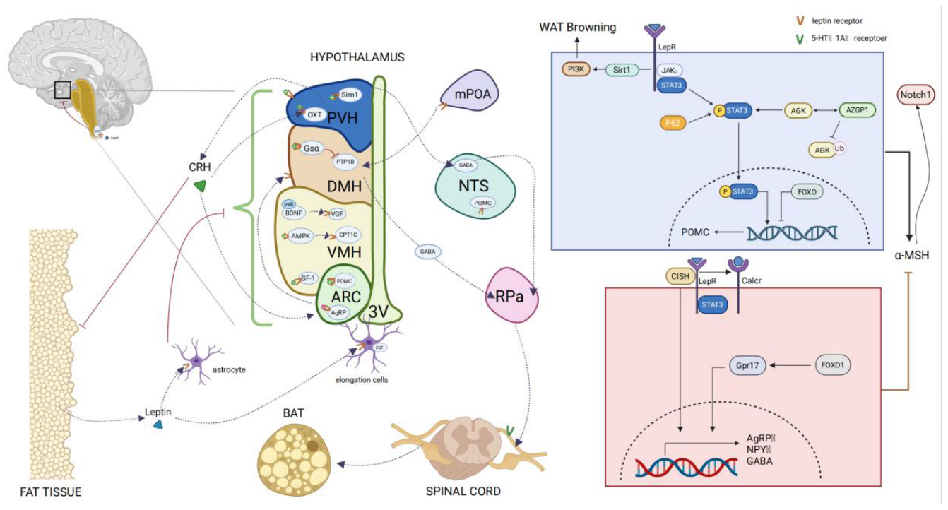
Figure 2.
Autocrine regulation. BCL-2: B-cell lymphoma 2; BAX: BCL2-associated X protein; Caspase3: Cysteinyl aspartate specific proteinase 3; TNF: Tumor Necrosis Factor; Ang2: Angiopoietin 2; ADRB3: Ad renoceptor Beta 3; RBP3: Retinol-Binding Protein 3; CPT1: Carnitine Palmitoyltransferase 1; CPT1b: Carnitine Palmitoyltransferase 1B; AOX1: Aldehyde Oxidase 1; ACOX1: Acyl-CoA Oxidase 1; FAS: Fatty Acid Synthase; ACC: Acetyl-CoA Carboxylase; SREBP-1: Sterol Regulatory Element-Binding Transcription Factor 1; SCD: Stearoyl-CoA Desaturase; UCP1: Uncoupling Protein 1; UCP2: Uncoupling Protein 2; UCP3: Uncoupling Protein 3; Cyt C: Cytochrome C; MFN2: Mitofusin 2; COX2: Cytochrome c Oxidase Subunit II; Sirt3: Sirtuin 3; TFAM: Transcription Factor A Mitochondrial; NRF: Nuclear Respiratory Factor; FABP4: Fatty Acid-Binding Protein 4; AKT-mTOR: AKT (Protein Kinase B) - mechanistic Target of Rapamycin; STAT3: Signal Transducer and Activator of Transcription 3; JAK2: Janus Kinase 2; SOCS2: Suppressor of Cytokine Signaling 2; SOCS3: Suppressor of Cytokine Signaling 3; LepR: Leptin Receptor; NMB: Neuromedin B; PPARα: Peroxisome Proliferator-Activated Receptor Alpha; PGC-1α: Peroxisome Proliferator-Activated Receptor Gamma Coactivator 1-alpha; pAMPK: phosphorylated AMP-Activated Protein Kinase; HSL: Hormone-Sensitive Lipase; WAT: White adipose tissue.
Figure 2.
Autocrine regulation. BCL-2: B-cell lymphoma 2; BAX: BCL2-associated X protein; Caspase3: Cysteinyl aspartate specific proteinase 3; TNF: Tumor Necrosis Factor; Ang2: Angiopoietin 2; ADRB3: Ad renoceptor Beta 3; RBP3: Retinol-Binding Protein 3; CPT1: Carnitine Palmitoyltransferase 1; CPT1b: Carnitine Palmitoyltransferase 1B; AOX1: Aldehyde Oxidase 1; ACOX1: Acyl-CoA Oxidase 1; FAS: Fatty Acid Synthase; ACC: Acetyl-CoA Carboxylase; SREBP-1: Sterol Regulatory Element-Binding Transcription Factor 1; SCD: Stearoyl-CoA Desaturase; UCP1: Uncoupling Protein 1; UCP2: Uncoupling Protein 2; UCP3: Uncoupling Protein 3; Cyt C: Cytochrome C; MFN2: Mitofusin 2; COX2: Cytochrome c Oxidase Subunit II; Sirt3: Sirtuin 3; TFAM: Transcription Factor A Mitochondrial; NRF: Nuclear Respiratory Factor; FABP4: Fatty Acid-Binding Protein 4; AKT-mTOR: AKT (Protein Kinase B) - mechanistic Target of Rapamycin; STAT3: Signal Transducer and Activator of Transcription 3; JAK2: Janus Kinase 2; SOCS2: Suppressor of Cytokine Signaling 2; SOCS3: Suppressor of Cytokine Signaling 3; LepR: Leptin Receptor; NMB: Neuromedin B; PPARα: Peroxisome Proliferator-Activated Receptor Alpha; PGC-1α: Peroxisome Proliferator-Activated Receptor Gamma Coactivator 1-alpha; pAMPK: phosphorylated AMP-Activated Protein Kinase; HSL: Hormone-Sensitive Lipase; WAT: White adipose tissue.
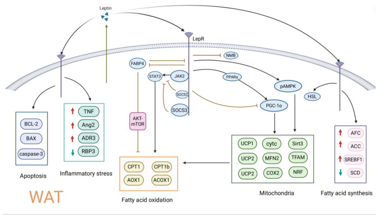
Disclaimer/Publisher’s Note: The statements, opinions and data contained in all publications are solely those of the individual author(s) and contributor(s) and not of MDPI and/or the editor(s). MDPI and/or the editor(s) disclaim responsibility for any injury to people or property resulting from any ideas, methods, instructions or products referred to in the content. |
© 2026 by the authors. Licensee MDPI, Basel, Switzerland. This article is an open access article distributed under the terms and conditions of the Creative Commons Attribution (CC BY) license (http://creativecommons.org/licenses/by/4.0/).
Copyright: This open access article is published under a Creative Commons CC BY 4.0 license, which permit the free download, distribution, and reuse, provided that the author and preprint are cited in any reuse.


