

Submitted:
19 May 2026
Posted:
21 May 2026
You are already at the latest version
Abstract
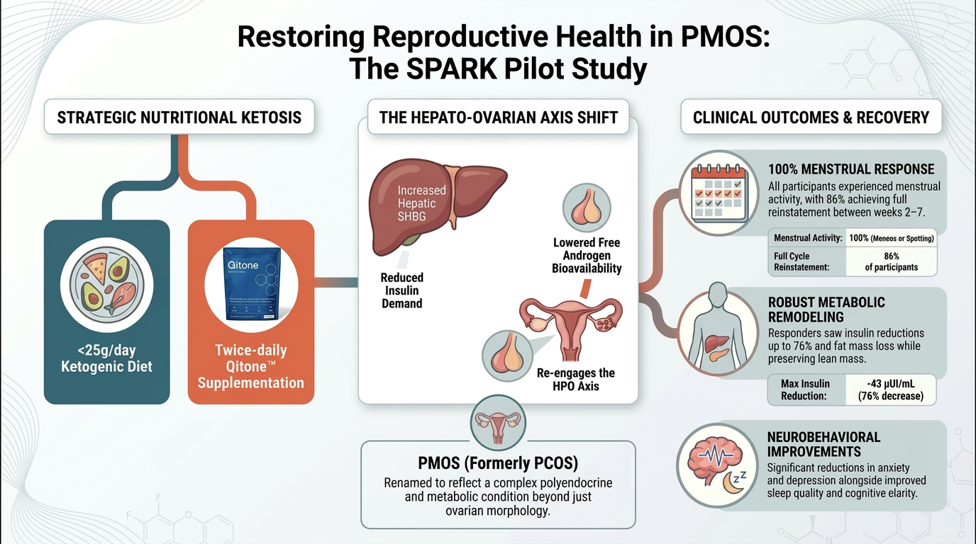
Background: The SPARK (Symptoms of PCOS ameliorated by responses to Ke-to-adaptation) pilot study investigates whether nutritional ketosis can improve repro-ductive and metabolic outcomes in women with Polyendocrine Metabolic Ovarian Syndrome (PMOS). PMOS is a common endocrinopathy characterized by anovulation, hyperandrogenism, and metabolic dysfunction. Objective: To evaluate if a ketogenic diet (KD) or exogenous ketone supplementation (KS) with Qitone™ (bis-octanoyl-(R)-1,3-butanediol) can restore ovulation and menstrual activity. Methods: This ongoing 12-week randomized trial assigns women with PMOS to either a KD or KS intervention. Assessments include body composition by DXA, metabolic and reproduc-tive biomarkers (e.g., insulin, testosterone, SHBG), and cycle tracking via basal body temperature. Results: Preliminary data from seven participants (KD: n=3; KS: n=4) show that 100% demonstrated spontaneous menstrual activity within 2–7 weeks. Full menstrual reinstatement occurred in 86% of participants (KD=3, KS=3), including those with long-standing amenorrhea. Ovulation was predicted in 43% of the cohort. Key metabolic improvements included significant insulin reductions, favorable body composition changes (fat mass loss with lean mass preservation), and enhanced neurobehavioral well-being, including reduced anxiety and improved cognitive clarity. Conclusion: Preliminary findings suggest that both sustained and intermittent ketogenic interven-tions can restore menstrual and ovulatory function through multi-node endo-crine-metabolic recovery, independent of significant weight loss. These results highlight the potential of ketogenic therapies for PMOS but require confirmation in larger trials.
Keywords:
ketogenic diet
; ketone supplementation
; PMOS
; anovulation
; women’s health
; reproductive metabolism
; Qitone
1. Introduction
Polyendocrine metabolic ovarian syndrome (PMOS; formerly polycystic ovary syndrome [PCOS]) affects 6–13% of women of reproductive age and is the leading cause of anovulatory infertility worldwide, yet approximately 70% of those affected remain undiagnosed [1]. In May 2026, following a 14-year global consensus process involving more than 22,000 stakeholders, the condition was formally renamed from PCOS to PMOS in a Lancet publication led by Teede et al., reflecting recognition that the disorder extends beyond ovarian morphology to encompass a complex polyendocrine and metabolic condition. The present manuscript adopts this updated nomenclature throughout [2]. The disorder is defined by the Rotterdam criteria as the presence of at least two of the following three features: oligo- or anovulation, clinical or biochemical hyperandrogenism, and polycystic ovarian morphology [3]. Importantly, PMOS is not solely a reproductive condition. A large proportion of affected women, including those with normal BMI, as represented in the present cohort, demonstrate insulin resistance and compensatory hyperinsulinemia, which drive downstream endocrine dysfunction through a coordinated hepato-ovarian axis: elevated insulin suppresses hepatic sex hormone-binding globulin (SHBG) production while simultaneously stimulating ovarian androgen biosynthesis, creating a self-reinforcing cycle of androgen excess and menstrual irregularity [4,5,6]. These interacting pathways explain why menstrual dysfunction in PMOS is not simply a consequence of elevated weight, and why interventions that reduce insulin exposure, rather than merely inducing caloric restriction, may be required to restore ovulatory cyclicity.
Standard dietary management of PMOS has focused primarily on caloric restriction and weight reduction. While weight loss can modestly improve insulin sensitivity and reproductive outcomes in some women, a purely weight-centric model fails to address the underlying endocrine-metabolic drivers of cycle dysfunction, particularly in women with normal or near-normal BMI [7,8]. Ketogenic dietary approaches represent a mechanistically distinct strategy: by markedly reducing carbohydrate intake, they lower insulin demand continuously, shift substrate oxidation toward fat, and elevate circulating β-hydroxybutyrate (BHB), a metabolite that functions not only as an alternative fuel but as a signaling molecule capable of modulating inflammatory pathways, histone deacetylase activity, and neuroendocrine tone [9,10,11]. Emerging trials have demonstrated improvements in reproductive and metabolic outcomes following ketogenic interventions in women with PMOS that were independent of large weight changes, suggesting that altered metabolic substrate utilization, rather than energy deficit alone, may drive reproductive benefit [12,13,14].
Our group previously demonstrated that despite equivalent weight loss, menstrual improvements occurred only in women achieving nutritional ketosis, not in those assigned to a low-fat diet, implicating circulating BHB, rather than caloric deficit, as a critical signal for reproductive recovery [15]. However, adherence to strict carbohydrate restriction remains a meaningful barrier for many women with PMOS, given the condition’s overlapping metabolic and psychosocial burden. Exogenous ketone supplementation (KS) offers an alternative approach: by delivering BHB directly without requiring carbohydrate restriction, it can transiently elevate circulating ketones within a mixed-diet context [16]. To date, only one trial has examined KS in PMOS, reporting acute reductions in androgen and glucose concentrations but not evaluating menstrual outcomes [17]. Whether sustained ketosis (via KD) and intermittent ketone exposure (via KS) produce comparable reproductive benefits, and whether they do so through convergent or distinct endocrine pathways, remains unknown.
Beyond reproductive endpoints, PMOS is an endocrine-metabolic-neurobehavioral condition. Women with PMOS report disproportionately high rates of anxiety, depression, sleep disturbance, and cognitive fatigue, yet few intervention trials have systematically evaluated how metabolic therapies influence these dimensions [18,19,20]. Given that improved sleep, mood, and cognitive function may themselves support circadian alignment, stress resilience, and dietary adherence, these outcomes are not merely secondary, they may be mechanistically linked to metabolic recovery [21,22,23]. The present case series therefore incorporates longitudinal PROMIS® assessments of mood, sleep, and cognitive function alongside reproductive and endocrine outcomes, capturing the multi-domain nature of improvement that participants experienced.
Here we report preliminary findings from the SPARK study, in which seven women with PMOS completed a 12-week randomized intervention of either a ketogenic diet (KD; n = 3) or twice-daily exogenous ketone supplementation with Qitone™ while maintaining a mixed diet (KS; n = 4). Participants spanned a wide BMI range and presented with diverse menstrual histories, including primary amenorrhea, long-standing secondary amenorrhea, and oligomenorrhea. We hypothesized that both interventions would disrupt the insulin–SHBG–androgen axis and in doing so support menstrual reinstatement and ovulatory recovery, with KD producing more sustained metabolic remodeling and KS producing intermittent ketone signaling. The heterogeneity of outcomes, including cases where menstrual activity returned despite persistently elevated total testosterone, illustrates the multi-node nature of PMOS endocrine recovery and motivates reporting these findings in advance of full trial completion.
2. Materials and Methods
Participants
Women with symptoms of PMOS were recruited from the greater Columbus, Ohio area to join the SPARK study. Inclusion criteria encompassed women with PMOS that followed the Rotterdam criteria, experiencing two of the three central symptoms of PMOS: oligo-amenorrhea, hyperandrogenism, and polycystic ovaries. Additionally, women in the study ranged from 18-40 years old and had a BMI of >18kg/m. Exclusion criteria included current adherence to a low-carbohydrate diet (<30% of total energy from carbohydrates), pregnancy or breastfeeding, postmenopausal status, clinically significant weight loss within the past six months (>10% of body weight), type 1 diabetes, and anovulation unrelated to PMOS.
Eligible participants provided written informed consent during an in-person screening visit. At baseline, participants completed questionnaires assessing dietary habits, medical and menstrual history, and physical activity. Seven women were enrolled and randomized to one of two intervention arms: a ketogenic diet (KD; n = 3) or a ketone supplementation arm (KS; n = 4). All participants reported irregular menses or amenorrhea before enrollment. One participant used hormonal contraception throughout the study. Baseline characteristics of completers are presented in Table 1.
Participant demographics and body composition values at baseline for women diagnosed with polyendocrine metabolic ovarian syndrome (PMOS) according to the Rotterdam criteria (n = 7). Lean and fat mass were assessed using dual-energy X-ray absorptiometry (DXA).
Experimental Design
This 12-week intervention followed a prospective, double-arm randomized design. All lab visits were in-person at the Physical Activity and Education Services (PAES) Building at The Ohio State University (Columbus, OH). Visits were scheduled between 06:00–08:00 AM with participants arriving fasted (>8 h since last meal), well rested (>7 h sleep/night), and adequately hydrated (urine specific gravity < 1.025). At each visit, body weight and height were measured using standardized procedures while wearing light clothing and no shoes.
At baseline, the research/study team assessed medications, confirmed last menstrual bleeding, collected demographic data. A sample of urine was collected to assess for specific gravity, testing diurnal pattern of urine hormones. A trained phlebotomist collected venous blood for metabolic and reproductive panels (e.g., insulin, testosterone, SHBG, LH, FSH, estradiol, progesterone, lipid profile); samples were processed within 2 hours (centrifuged, aliquoted) and stored at −80 °C. Diet counseling and accountability were provided at each visit for this in the KD arm, and study foods/supplements for the next interval were dispensed; subsequent appointments were scheduled every biweekly within a ±2-day window to maintain consistent morning sampling. For participants in the KS arm, at every biweekly visit, the assigned supplement was administered under observation with capillary sampling at baseline and 60 minutes to document the acute post-dose response and adherence. At every biweekly visit, same measurements were repeated for KS and KD participants, respectively. (Figure 1).
Body Composition
Height and weight were measured using a SECA 703 digital stadiometer and scale (Hamburg, Germany) at baseline and endpoint. BMI was calculated as weight in kilograms divided by height in meters squared. Lean mass and fat mass were quantified by dual X-ray absorptiometry (DXA) on a Lunar iDXA system (GE, Madison, WI, USA) at baseline and endpoint. Subjects were carefully aligned within detection limits on the DXA bed. The average duration of a whole-body scan lasted 7 min. A single, whole-body, DXA measurement was performed on each subject to estimate lean and fat mass (CoreScanTM enCORE software version 14.10) using two-dimensional projection data created by low energy, fan beam x-ray to create a model consisting of bone, adipose, and lean tissue compartments.
Surveys
Participants completed questionnaires collecting their menstrual and medical histories at baseline. At each biweekly testing visit, depressive symptoms, sleep disturbance (restfulness, sleep quality & satisfaction), anxiety symptoms, and cognitive function (perceived mental capabilities) were self-reported via PROMIS ® (Patient-Reported Outcomes Measurement Information System) surveys by the participants [24]. Each of the four surveys contained statements (e.g., My worries overwhelmed me) that were rated on a 5-point Likert scale (0-4) representing frequency or intensity in the past week. All surveys were administered on facility-provided devices in quiet, monitored conditions during testing visits. Survey scores were added, and data were tabulated biweekly to observe any significant changes in mood, sleep, or cognitive function over the study period.
Diet and Supplementation
Participants in the KD arm received individualized nutrition counseling and an educational handout at baseline. Those in the KS arm consumed an exogenous ketone supplement (Qitone™)(Component Health Limited, Dublin Ireland) providing the pro-ketone bis-octanoyl-(R)-1,3-butanediol twice daily throughout the study, with one of the two daily doses administered in-lab under supervision during their biweekly visits. During the first four weeks, all participants received structured meal support. For the diet arm of the study, pre-prepared ketogenic meals were provided by Factor Meals, a commercial meal delivery service specializing in low-carbohydrate, high-fat meals along with keto-friendly groceries for breakfast. Meals were selected from the weekly rotating ketogenic menu and individually portioned to provide approximately, less than 25g of carbohydrates per day, a total daily caloric intake of 65-75% of fat, and a total daily caloric intake of 20-25% of protein all totaling up to a daily caloric range of 1,400-1,800 kcal, adjusted per individual based on baseline energy and weight needs. Meal compositions were verified using nutritional labels provided by Factor. In the supplement arm of the study, each participant received two meals per day, seven days per week, along with groceries to make their own breakfast all based on their caloric needs and matched similar to their usual diet. All participants were instructed to maintain consistent dietary intake throughout the study, especially after the first 4-week period concluded. Specifically for the KD group, participants were asked to adhere to the provided ketogenic guidelines and optional recipes. Throughout the study, participants were monitored through the Healthie app for data collection, adherence, and counseling. Participants in the KS arm were provided with Qitone™ in powder form. The supplement was unflavored and designed for oral consumption after mixing with 250 ml of any beverage. Participants were instructed to consume two scoops (19 g/scoop) of Qitone™ daily (12.5 g active ingredient per scoop), divided between breakfast and dinner. To ensure compliance, participants were given a supply of the supplement at the baseline visit and received additional supplies as needed during scheduled biweekly visits. Weekly reminder messages via the Healthie app were sent to encourage adherence to the supplement and the diet. Participants were also instructed to maintain their exercise routines throughout the study and to avoid other ketone supplements or metabolic aids. Any adverse effects or gastrointestinal discomfort were to be reported immediately through the Healthie app and through their biweekly testing visits.
Capillary Metabolites
Fasting capillary BHB (mmol/L) and glucose (mg/dL) were measured daily using reagent strips with a handheld meter (KetoMojo GK+ Meter, Napa, California). Participants obtained samples on waking after an ≥8-hour fast following a self-administered standard finger-stick procedures; values were automatically uploaded via the KetoMojo application to the study portal. Data were reviewed for completeness and plausibility by a registered dietitian. At biweekly lab visits, participants randomized to the KS arm measured BHB and glucose levels at baseline and 60 minutes after ingestion to assess the acute post-dose BHB response; these readings also served as an adherence check for the KS arm.
Blood Collection and Processing
Fasting venous blood samples were collected biweekly between 06:00 and 08:00 AM. Samples were processed within two hours of collection, centrifuged, and the resulting serum and plasma were aliquoted and stored at −80 °C until analysis by the Clinical Research Center for Clinical Research Management of The Ohio State University Wexner Medical Center and The Ohio State University College of Medicine in Columbus, Ohio. Fasting serum insulin was measured using a solid-phase sandwich Enzyme-Linked Immunosorbent Assay (ELISA; Novus Biologicals, Cat# NBP2-60077). The assay was performed according to the manufacturer’s instructions, with concentrations determined in µIU/mL. This assay was utilized to monitor longitudinal shifts in hyperinsulinemia and calculate markers of insulin sensitivity. Total testosterone was quantified using a competitive ELISA (Novus Biologicals, Cat# NBP3-23553). This assay features a detection range of 0.63–40 ng/mL and a sensitivity of 0.24 ng/mL. In accordance with the manufacturer’s recommendation for human serum and plasma, all samples were assayed undiluted. This protocol was selected to ensure all participant values remained within the established standard curve (0.63–40 ng/mL), providing an accurate measure of the severe biochemical hyperandrogenism observed in this cohort. SHBG was measured using the Meso Scale Discovery (MSD) U-PLEX platform (Plates #1: 23P2TAZ879; #2: 23P2TAC880). The assay utilized a 4-parameter logistic (4-PL) algorithm with a weight of 1/y² to establish the standard curve. To ensure samples remained within the measurable detection range (2.51–26,300 pg/mL), all serum samples were subjected to a 4,000-fold dilution prior to analysis. Final concentrations were adjusted for this dilution factor and converted from pg/mL to the clinical standard of nmol/L.
Basal Body Temperature
Basal body temperature (BBT) was assessed nightly using one of two validated wearable devices provided to participants: a vaginally inserted sensor or a ring-style continuous temperature monitor. Both devices were designed to capture true core or near-core body temperature during sleep, corresponding to the post-ovulatory rise in progesterone. Participants inserted or wore their assigned device before sleep and removed it upon waking. Temperature data were automatically synced to a secure, study-approved mobile application for daily cycle tracking.
Research personnel accessed participant data through a secure, password-protected physician portal, allowing continuous monitoring of temperature trends without participant reporting or manual data downloads. BBT patterns were evaluated longitudinally to identify ovulatory shifts and trends over time.
3. Results
Individual case presentations are provided to highlight the distinct metabolic, hormonal, and psychosocial responses observed among a subset of women completing the SPARK intervention. Each case includes descriptive changes in body composition, menstrual outcomes, and relevant clinical indices, such as anxiety, depressive symptoms, sleep quality, and cognitive function, collected longitudinally across the 12-week period. These individualized reports illustrate the variability in adaptation patterns to both KD and KS arms, emphasizing differences in time to menstrual reinstatement, magnitude of metabolic improvement, and psychological response. Together, these cases capture the diverse yet promising effects of ketogenic-based interventions on menstrual regularity, metabolic health, and overall well-being in women with PMOS. Full summary of changes can be found in Supplementary Table 1.
3.1. Ketogenic Diet
Participants randomized to the KD arm all achieved nutritional ketosis (R-βHB ≥ 0.5 mmol/L) within 2–14 days of initiation, verified via the Ke-to-Mojo® GK+ device. Upon achieving nutritional ketosis, R-BHB values ranged between 0.5 mmol/L to 2.0 mmol/L, accompanied by steady declines in fasting glucose and improvements in energy stability and mood. PROMIS® surveys were completed biweekly to monitor depressive symptoms, anxiety, sleep disturbance, and cognitive function. DXA-derived body-composition data revealed preferential reductions in fat mass with preservation of lean mass (Figure 2). No significant adverse events occurred, and adherence remained high through weekly check-ins and digital tracking.
3.1.1. SPARK 002
A 33-year-old woman with PMOS and lifelong amenorrhea presented with a BMI of 45.8 kg/m² and 50.8% body fat. She had never menstruated spontaneously and was not on hormonal or insulin-sensitizing medications. Within 14 days of KD initiation, she reached nutritional ketosis (R-βHB = 0.7 mmol/L) and experienced her first spontaneous menses four days later, her first lifetime menstrual event, followed by predicted ovulation on Day 41 (Figure 3). Over 12 weeks, body mass decreased from 79.3 → 67.9 kg (−11.4 kg), with fat mass −10.0 kg and reduced lean mass (-1.9 kg) (Figure 2). PROMIS® scores reflected reduced anxiety and sleep disturbance, with improved restfulness, emotional stability, and cognitive clarity.
3.1.2. SPARK003
A 25-year-old competitive rugby player with PMOS and oligomenorrhea (cycles every 120–180 days) presented with a BMI of 45.1 kg/m² and 52.6% body fat. She achieved nutritional ketosis within 48 hours (R-βHB ≈ 0.6 mmol/L) and remained in ketosis intermittently throughout the study, adjusting protein intake for training (Figure 4). Spontaneous menses resumed by week 4, notably within a normalized interval relative to her prior 120 to 180 day cycles, with intermittent spotting between cycles, and the second menses recurred at week 11. Body mass declined 110.9 → 105.6 kg (−5.4 kg); fat mass −7.5 kg; gained lean mass +1.4 kg (Figure 2). PROMIS® data indicated reductions in anxiety and depressive symptoms, better sleep satisfaction, and reduced cognitive fatigue by week 8. She reported greater focus and steadier energy during competition.
3.1.3. SPARK008
A 31-year-old woman with PMOS, and oligomenorrhea (BMI 35.3 kg/m², 43.1% body fat) achieved ketosis within six days (βHB = 0.6 mmol/L). Spontaneous menses occurred at week 4 and predicted ovulation on Day 16 followed by spotting (Figure 5). Weight decreased by 5.4 kg with lower fasting glucose and improved insulin sensitivity (Figure 2). PROMIS® scores showed marked reductions in anxiety (7 → 0) and depressive symptoms (2 → 0), improved sleep (15 → 8), and better cognitive function (4 → 0).
KD Conclusion
Women in the KD group exhibited rapid induction of nutritional ketosis, significant reductions in adiposity, and spontaneous menstrual onset, accompanied by measurable improvements in PROMIS®-derived mood, sleep, and cognition.
3.2. Ketogenic Supplement
Participants randomized to the KS arm consumed a twice-daily ketone ester while maintaining a mixed diet (>30% carbohydrate). Mean BHB concentrations one hour after ingestion of the supplement ranged from 0.5 to 1.3 mmol/L, representing partial or intermittent ketosis. Even brief or modest elevations in circulating βHB may have engaged key metabolic signaling pathways—improving mitochondrial efficiency, modulating inflammation, and influencing central nervous system energetics. Participants frequently described steadier energy, reduced appetite variability, and clearer cognition, consistent with mild central ketone signaling despite ongoing carbohydrate intake.
3.2.1. SPARK004
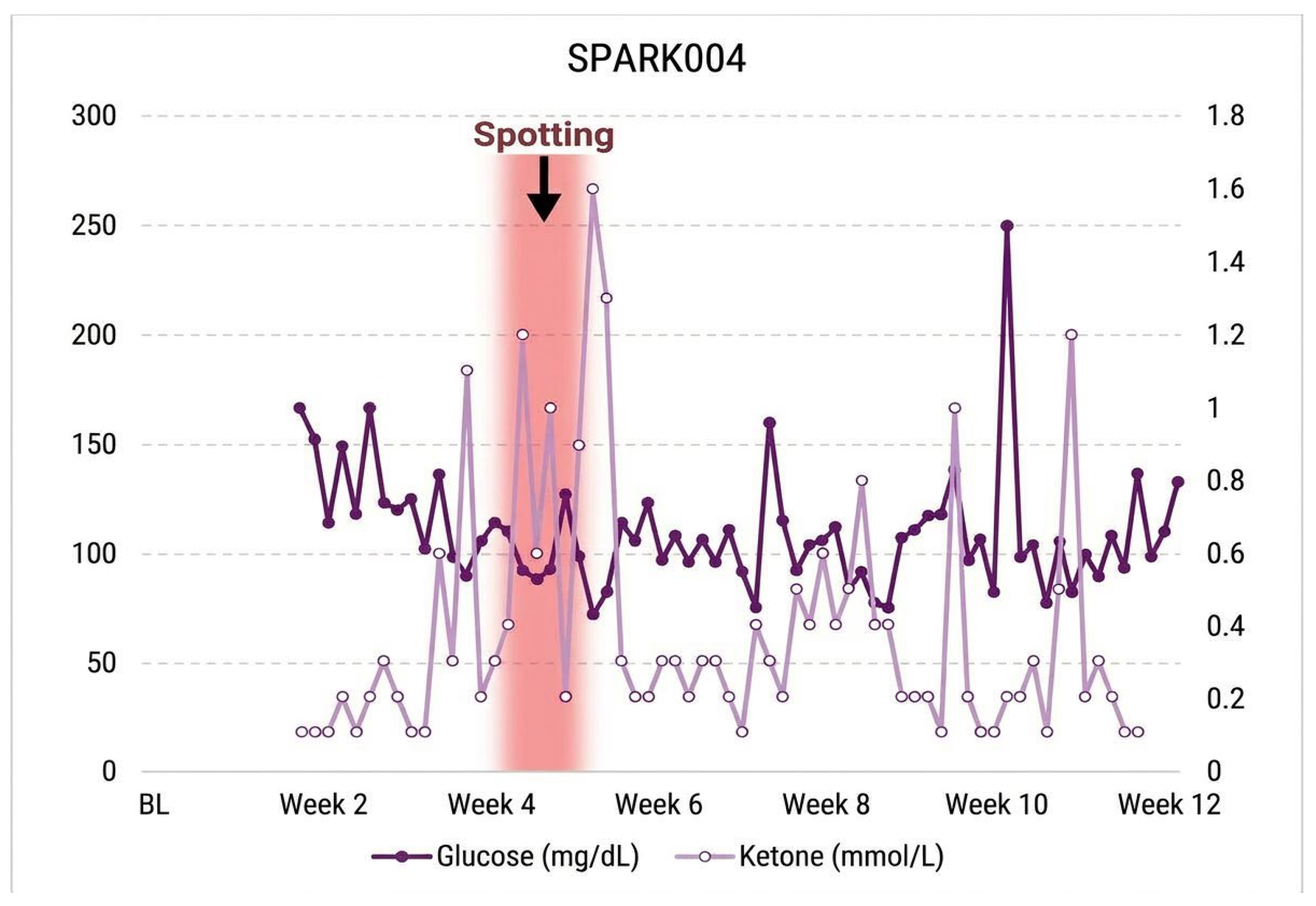
A 21-year-old woman with six-month secondary amenorrhea (BMI 20.6 kg/m²) achieved intermittent ketosis with BHB levels ranging from 0.5 to 1.3 mmol one hour after ingestion of the supplement. Spontaneous menstrual spotting appeared at week 5, though full menses did not resume, suggesting early endocrine responsiveness (Figure 7). DXA showed total-mass +2.1 kg, fat −1.3 kg, and lean +2.4 kg (Figure 6). PROMIS® cognitive-function scores improved (17 → 6). Participant reported noticeable improvement in concentration and motivation, describing feeling ‘less scattered and more alert’ after supplementation, particularly during morning hours, suggesting cognitive sensitivity to exogenous βHB peaks.
Figure 6.
| KS Body Composition Changes. Changes in total, fat, and lean mass measured by DXA among participants in the KS arm of the SPARK-pilot study. Reductions in fat mass with preservation of lean mass were observed over the 12-week intervention period. BL = Baseline; WK12 = Testing week 12.
Figure 6.
| KS Body Composition Changes. Changes in total, fat, and lean mass measured by DXA among participants in the KS arm of the SPARK-pilot study. Reductions in fat mass with preservation of lean mass were observed over the 12-week intervention period. BL = Baseline; WK12 = Testing week 12.
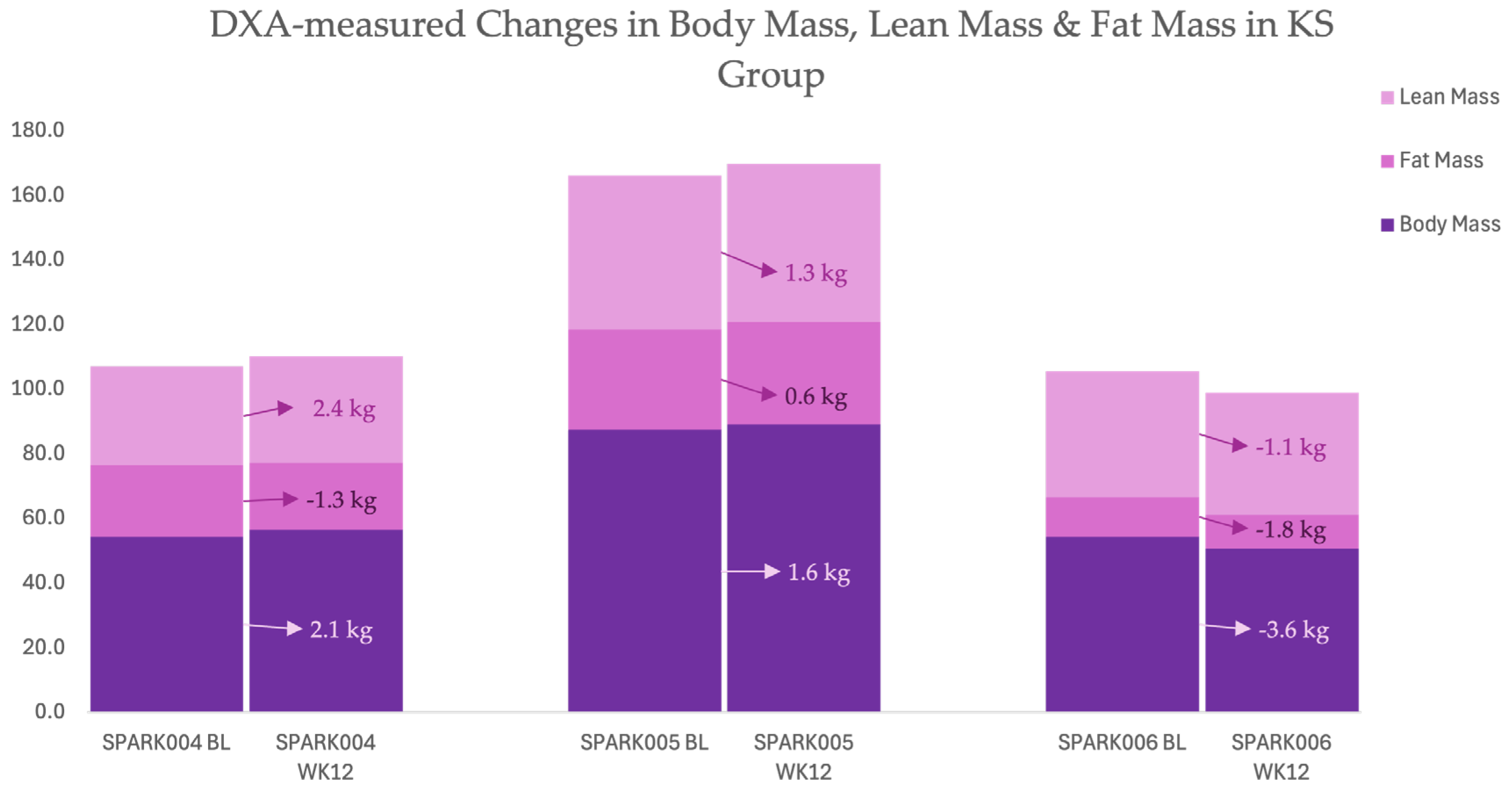
Figure 7.
| Daily BHB and glucose trends with timing of menstrual spotting in SPARK004. Daily BHB and glucose trends with timing of menstrual spotting during KS intervention. Shaded bar marks week 5 spotting; intermittent ketosis (0.5–1.3 mmol/L) occurred one-hour post-supplement ingestion.
Figure 7.
| Daily BHB and glucose trends with timing of menstrual spotting in SPARK004. Daily BHB and glucose trends with timing of menstrual spotting during KS intervention. Shaded bar marks week 5 spotting; intermittent ketosis (0.5–1.3 mmol/L) occurred one-hour post-supplement ingestion.
3.2.2. SPARK005
A 21-year-old woman with irregular 29–40-day cycles (BMI 30.5 kg/m²) achieved intermittent ketosis, levels ranging from 0.5 to 0.8 one hour after ingestion of the supplement. DXA showed total-mass +1.6 kg, fat +0.6 kg, and lean +1.3 kg (Figure 6). Spontaneous menses resumed by week 7 (Figure 8). PROMIS® surveys showed resolution of anxiety (6 → 0) and depressive symptoms (10 → 0) with better sleep and cognitive performance (21 → 6). In addition to mood improvement, the participant noted decreased late-evening hunger and more consistent daytime energy, suggesting modulation of appetite hormones or glycemic stability.
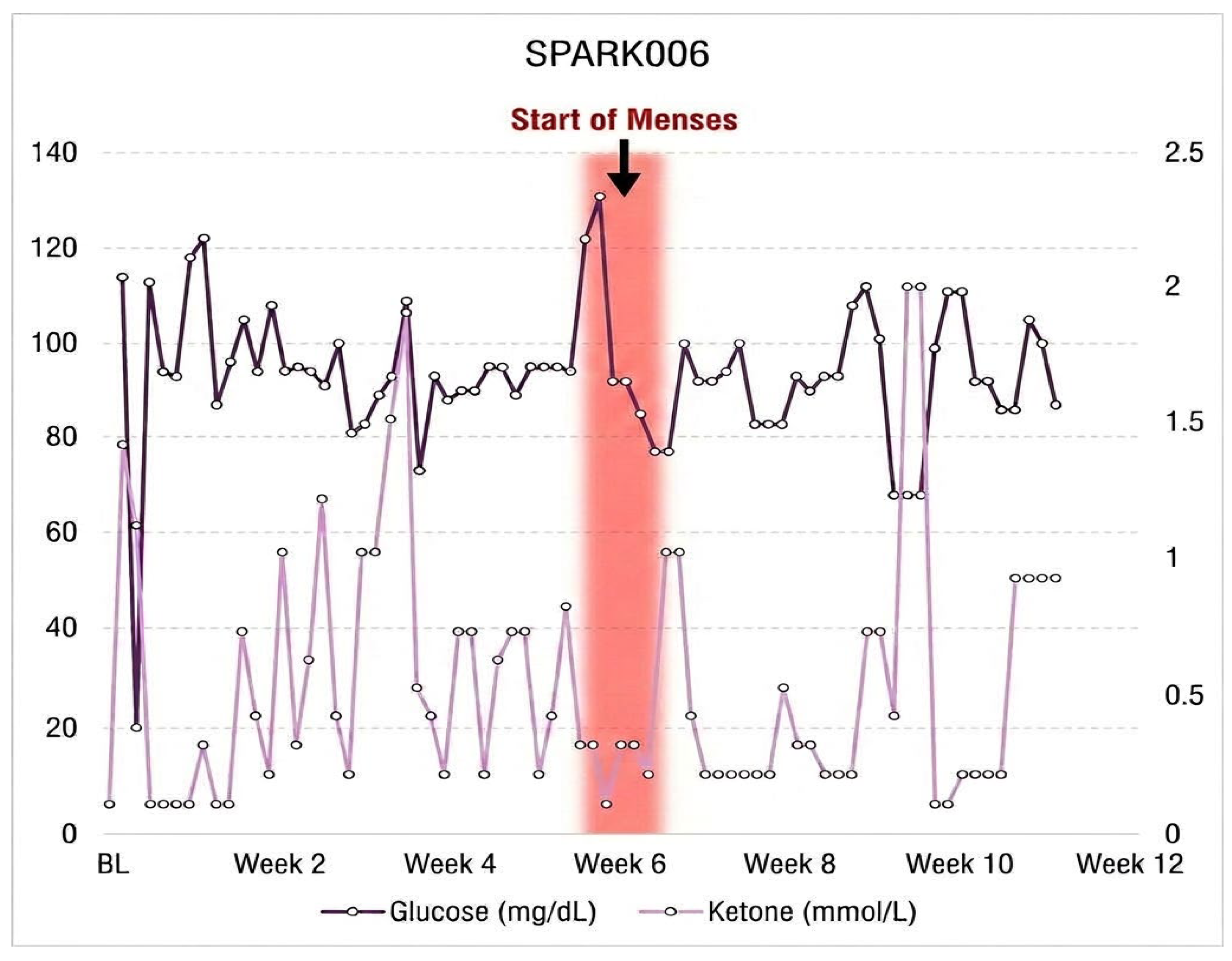
Figure 9.
| Daily BHB and glucose trends with timing of menses in SPARK006. Daily BHB and glucose trends with timing of menses reinstatement in a woman with five-year secondary amenorrhea. Shaded bar indicates week 6 menses following sustained βHB levels of 0.7–1.0 mmol/L.
Figure 9.
| Daily BHB and glucose trends with timing of menses in SPARK006. Daily BHB and glucose trends with timing of menses reinstatement in a woman with five-year secondary amenorrhea. Shaded bar indicates week 6 menses following sustained βHB levels of 0.7–1.0 mmol/L.
3.2.3. SPARK006
A 21-year-old woman with long-standing five-year secondary amenorrhea (BMI 21.7 kg/m²) achieved intermittent ketosis, with levels ranging from 0.7 to 1.0 mmol one hour after ingestion of the supplement. DXA showed total-mass -3.6 kg, fat −1.8 kg, and lean -1.1 kg (Figure 6). Spontaneous menses resumed by week 6. PROMIS® anxiety and cognitive scores improved (anxiety 5 → 2; cognitive 4 → 1). Following menses reinstatement, the participant reported improved exercise tolerance and post-meal satiety. Stable PROMIS® sleep scores coincided with fewer nocturnal awakenings, suggesting enhanced circadian alignment.
3.2.4. SPARK007
A 22-year-old woman with oligomenorrhea (29–55 day cycles) and BMI 33.0 kg/m² showed minimal body composition changes on DXA: total mass −0.5 kg, fat mass +0.7 kg, and lean mass −1.5 kg (Figure 6). She achieved intermittent ketosis with levels ranging from 0.6 to 1.0 one hour after the ingestion of the supplement. Menstrual cycles spontaneously normalized to a regular 4-week interval, with predicted ovulation preceding each cycle (Figure 10). PROMIS® scores improved substantially in depressive symptoms (23 → 10) and anxiety (16 → 7) with better sleep and cognition (24 → 19). The participant also described increased social engagement and productivity, paralleling declines in depression and anxiety, which may indicate improved neurocognitive efficiency and emotional regulation associated with ketone metabolism.
KS Conclusion
Participants in the KS group achieved intermittent ketosis yet demonstrated consistent improvements in menstrual regularity, mood, and cognitive function. PROMIS® data confirmed reductions in depressive symptoms, anxiety, and sleep disturbance, paralleling subjective increases in energy and focus.
3.3. Group Changes
Restoration of menstrual cyclicity was observed in all previously amenorrheic participants, typically within 2–6 weeks of intervention onset. Table 2 summarizes individual responses by intervention type. Both KD and KS groups demonstrated evidence of improved regularity across the 12-week period.
3.3.1. Hormonal and Metabolic Biomarker Responses
Insulin Dynamics. The delta changes in serum insulin highlight a stark contrast between metabolic responders and those exhibiting resistance. SPARK008 demonstrated the most profound clinical improvement, with a continuous downward trajectory reaching a delta of approximately -43 µIU/mL by Week 12. In contrast, SPARK004 exhibited “metabolic resistance,” with insulin levels remaining relatively stable before showing a late-study increase of nearly +20 µIU/mL at Week 10. The remaining participants (002, 003, 005, 006, and 007) maintained insulin levels closer to their baseline values throughout the intervention (Figure 11).
SHBG Volatility. SHBG levels acted as a sensitive “metabolic mirror,” reflecting individual hepatic responses to the intervention. Two distinct outliers emerged in the delta data:
Metabolic Recovery (SPARK003): This participant saw a robust increase in SHBG, reaching a delta of approximately +30 nmol/L by Week 12.
Metabolic Collapse (SPARK004): Conversely, SPARK004 experienced a precipitous decline in SHBG beginning at Week 4, culminating in a negative delta of nearly -65 nmol/L by the study’s conclusion. Other participants remained relatively stable or showed modest positive deltas in the 10–20 nmol/L range (Figure 11).
Testosterone. While total testosterone remained elevated in the “male range” for many subjects, the delta graphs show individual downward trends for those with the greatest metabolic improvements. SPARK008 and SPARK002 both recorded negative deltas, with reductions of approximately -4 ng/mL and -2 ng/mL respectively. Conversely, SPARK007 saw an upward trend, ending with a positive delta of approximately +3 ng/mL (Figure 11).
4. Discussion
Reframing dietary intervention in PMOS: from weight loss to endocrine-metabolic restoration
Conventional dietary management in PMOS has historically emphasized calorie restriction and weight loss [25]. While weight loss can improve insulin sensitivity and reproductive outcomes in some women with PMOS, a purely weight-centric model may underperform because it does not consistently target the endocrine-metabolic biology that drives cycle dysfunction: insulin resistance, compensatory hyperinsulinemia, altered hepatic SHBG production, androgen excess, and disrupted hypothalamic-pituitary-ovarian (HPO) signaling [26,27,28]. The SPARK intervention was designed to test a different premise. Rather than inducing an intentional energy deficit, calories were matched to estimated maintenance needs, and counseling prioritized whole food, nutrient-dense meals with adequate protein intake. Therefore, the observed changes are best interpreted as evidence of altered metabolic signaling and substrate partitioning rather than simple calorie restriction.
This distinction is important because PMOS-related menstrual dysfunction is not merely a consequence of body weight. Amenorrhea and oligomenorrhea reflect disrupted ovulatory physiology, and restoring menstrual activity likely requires changing the hormonal environment that permits follicular maturation, ovulation, and luteal progesterone exposure [29,30,31,32]. In this context, KD and KS both interventions generate a metabolic environment characterized by lower glucose reliance, altered insulin responses, and circulating BHB, although with different magnitude-duration profiles in women diagnosed with PMOS. KD represents a sustained metabolic shift, whereas KS provides intermittent ketone exposure superimposed on the participant’s usual diet. The observation that both approaches were associated with menstrual activity raises an important mechanistic question: is sustained nutritional ketosis necessary for reproductive benefit, or can intermittent ketone signaling contribute to endocrine responsiveness.
Insulin, SHBG, testosterone, and the hepato-ovarian axis
A central mechanistic framework for interpreting these preliminary findings is the insulin–SHBG–androgen axis, more broadly the hepato-ovarian axis, which can be viewed as a self-reinforcing “vicious cycle” of metabolic and reproductive dysfunction [28,33]. In this model, hyperinsulinemia has two coordinated effects. First, insulin directly stimulates ovarian theca-cell androgen production, particularly in ovaries that remain relatively insulin-sensitive despite systemic insulin resistance [34]. Second, elevated insulin, along with increased carbohydrate and lipogenic flux, suppresses hepatic SHBG production, reducing the circulating binding capacity for testosterone, and increasing the bioavailable androgen fraction. At the hepatic level, high portal insulin concentrations inhibit HNF-4α activity, the primary transcriptional driver of SHBG gene expression in hepatocytes, providing a mechanistic link between insulin exposure and reduced SHBG levels [35,36,37]. These processes may reinforce one another, greater insulin exposure drives androgen excess while simultaneously weakening SHBG-mediated buffering, positioning SHBG as both a regulator of androgen availability and a reflection of underlying hepatic insulin sensitivity.
The hormone subset demonstrates why this model is useful, but also why it should not be oversimplified. Some participants showed patterns that were highly consistent with improved metabolic function. SPARK008 in the KD arm had the highest baseline insulin concentration in the hormone subset (56.78 µIU/mL) and experienced a 76.0% reduction by week 12 (13.61 µIU/mL). This was accompanied by a reduction in total testosterone from 20.11 to 15.96 ng/mL and repeated, spontaneous menstrual activity during the study period. Similarly, SPARK005 in the KS arm began with high insulin (28.54 µIU/mL) and very low SHBG (6.60 nmol/L), but by week 12 insulin decreased to 22.85 µIU/mL and SHBG more than doubled to 13.62 nmol/L, with menstrual activity recorded at week 8. These patterns support the idea that lowering insulin exposure and/or improving hepatic SHBG output may help reduce androgen bioavailability and permit reproductive-axis responsiveness.
However, the data also show that menstrual activity did not require a uniform biomarker pattern across all participants. SPARK004 in the KS arm maintained persistently high insulin concentrations, peaking at 53.31 µIU/mL at week 10, while SHBG declined dramatically from 72.06 nmol/L at baseline to 7.10 nmol/L at week 12. Despite this unfavorable insulin-SHBG profile, spotting was recorded at week 6. SPARK006 also had relatively low insulin at baseline but showed increased SHBG over time and menstrual activity at week 6 after long-standing secondary amenorrhea. SPARK007, with a history of oligomenorrhea, had menstrual activity at baseline and again at weeks 4, 8, and 12 despite modest insulin changes and persistently high total testosterone. These cases suggest that ketotic exposure may initiate reproductive responsiveness through multiple pathways, and that menstrual activity may precede full normalization of fasting insulin, SHBG, or total testosterone.
The strongest interpretation is therefore not that every participant followed a linear “insulin down, SHBG up, testosterone down” pathway. Rather, the data suggest that ketogenic exposure may destabilize the chronic PMOS endocrine-metabolic state by acting on several nodes at once: insulin exposure, hepatic SHBG regulation, androgen bioavailability, substrate oxidation, inflammation, and possibly neuroendocrine feedback. This multi-node effect may explain why menstrual activity was observed even when single fasting biomarkers remained abnormal.
From metabolic signaling to menses: why menstrual activity may return
The return or increased regularity of menstrual activity can be mechanistically linked to changes in insulin and androgen biology. PMOS is commonly characterized by chronic oligo-ovulation or anovulation, faster GnRH/LH pulsatility, relative FSH insufficiency, impaired follicular maturation, and luteal progesterone inadequacy [38]. Hyperinsulinemia contributes to this state by increasing ovarian androgen production and lowering SHBG, thereby increasing free androgen exposure [28]. Excess androgen can impair follicular development and may reduce the normal sensitivity of the hypothalamic-pituitary-ovarian axis to progesterone feedback [39]. Without consistent ovulation, progesterone exposure remains inadequate, and the system fails to re-establish normal cyclic negative feedback. This creates a self-reinforcing loop in which insulin resistance and androgen excess may sustain menstrual irregularity.
Within this framework, the SPARK findings can be interpreted as evidence that ketogenic exposure may help shift participants out of this loop. Reduced carbohydrate flux in the KD arm likely lowered insulin demand more continuously, while KS likely produced intermittent BHB elevations that may have supported metabolic flexibility, appetite regulation, or inflammatory signaling. If insulin exposure decreases sufficiently, the liver may increase SHBG production, reducing free testosterone even when total testosterone remains high. Lower bioavailable androgen tone could improve follicular dynamics, estradiol feedback, LH surge reliability, and luteal progesterone exposure. Once ovulation resumes, progesterone can help re-establish negative feedback at the hypothalamic-pituitary level, supporting more regular cycle patterning.
This interpretation is strengthened by the fact that menstrual activity was observed in participants with long-standing menstrual dysfunction and in participants with irregular cycles. At study completion, this pilot will integrate LH testing, basal body temperature patterns, urine hormone metabolites, progesterone confirmation, and cycle-length data to distinguish withdrawal bleeding, spotting, anovulatory bleeding, and true ovulatory menses.
The testosterone paradox: total testosterone remained high despite menstrual activity
One of the most important findings is the persistence of very high total testosterone values. Across the hormone subset, total testosterone ranged from approximately 3.09 to 20.11 ng/mL, values that are far above conventional female reference ranges and, in several cases, overlap with adult male-range concentrations (31 ng/dL). This creates an apparent paradox: how could menstrual activity occur while total testosterone remained pathologically elevated?
Despite these pathologically high concentrations, the high rate of menstrual reinstatement may suggest that the absolute concentration of total testosterone may be less critical for ovulatory function than the bioavailability of the hormone [40]. The persistence of high total testosterone may be explained by the contribution of adrenal-derived 11-oxygenated androgens, which recent research identifies as a predominant circulating androgen pool in women with PMOS [41]. Unlike ovarian androgens, these adrenal metabolites (such as 11-ketotestosterone) are not controlled by the HPO axis but are influenced by adrenal insulin signaling. This could suggest that while nutritional ketosis targets the ovarian component via insulin suppression, the adrenal component may require longer durations or different metabolic thresholds to fully normalize.
Body composition and lean mass as metabolic buffers
Body composition provided a more informative picture of metabolic change than weight alone. Despite maintenance-calorie targets, several participants showed favorable changes in fat mass with preservation or modest increases in lean mass. These changes were most apparent in the KD arm and were also present in some KS participants. For example, SPARK003 in the KD arm increased lean mass from 49.55 to 50.89 kg while fat mass decreased from 57.50 to 50.04 kg. SPARK008 in the KD arm decreased fat mass from 36.34 to 34.06 kg, although lean mass also decreased from 52.28 to 49.11 kg. SPARK004 in the KS arm increased lean mass from 30.63 to 32.99 kg while fat mass decreased from 22.10 to 20.80 kg. SPARK006 in the KS arm decreased fat mass from 12.23 to 10.40 kg, although lean mass also declined modestly.
These patterns support the argument that body composition, rather than scale weight alone, is a more appropriate endpoint for PMOS interventions [42]. Changes in fat mass and lean mass may reflect improved nutrient partitioning, altered insulin signaling, and substrate utilization, even when body weight changes are modest. LBM may also act as a metabolic buffer because skeletal muscle is a major site of glucose disposal and insulin-mediated substrate handling [43]. Participants with higher lean mass, including SPARK003, 007, and 008, generally maintained moderate-to-higher SHBG concentrations across the study compared with participants with very low baseline SHBG or pronounced SHBG suppression. In contrast, SPARK004 had the lowest lean mass at baseline and showed the most severe decline in SHBG after week 4, although this pattern cannot establish causality in a small sample. Preserving or increasing lean mass may be an important component of improving endocrine outcomes in women with the metabolic PMOS phenotype and should be considered in the design of future interventions and analyses.
BHB as a signal, not only a fuel
The KS arm is especially useful for separating ketone signaling from sustained carbohydrate restriction. KD provides continuous reductions in carbohydrate flux and insulin demand, whereas KS produces acute BHB elevations that may occur without full dietary ketosis. The fact that menstrual activity and neurobehavioral improvements were observed in KS participants raises the possibility that BHB itself may contribute to PMOS-relevant physiology, although this cannot be proven from the current data.
BHB is not merely an alternative fuel. It can act as a signaling molecule through hydroxycarboxylic acid receptor 2 (HCAR2/GPR109A), histone deacetylase inhibition, AMPK-related signaling, and PPAR-alpha-linked oxidative programs [44,45]. These pathways may bias metabolism toward fat oxidation and may attenuate low-grade inflammation, a feature commonly observed in PMOS [46]. Such effects could plausibly improve ovarian, hepatic, adipose, or central nervous system signaling even when fasting insulin and SHBG responses are incomplete. This may explain why intermittent ketone exposure in the KS arm was associated with reproductive and quality-of-life improvements, but with more variable body-composition and endocrine responses than KD.
Neurobehavioral recovery and holistic well-being
Beyond endocrine and body-composition outcomes, the SPARK study captured psychosocial and neurocognitive dimensions of metabolic restoration. Women with PMOS frequently report anxiety, depression, fatigue, sleep disturbance, and cognitive symptoms often described as brain fog [47]. Longitudinal PROMIS data indicated improvements in cognitive clarity, mood, and sleep quality across both arms. Participants in the KS arm reported feeling less scattered and more alert shortly after supplementation, consistent with the possibility that circulating BHB may support central nervous system energetics even in the absence of sustained nutritional ketosis [48].
These outcomes are important because menstrual recovery in PMOS should not be interpreted narrowly as a reproductive event alone. PMOS is an endocrine-metabolic-neurobehavioral condition, and improvements in mood, sleep, cognition, and perceived energy may support adherence, stress resilience, circadian alignment, and behavioral consistency. The relationship is likely bidirectional: improved metabolic signaling may improve neurobehavioral function, while better sleep and lower perceived stress may improve insulin sensitivity and reproductive-axis regulation. Therefore, quality-of-life outcomes should remain central in future PMOS intervention studies rather than secondary afterthoughts.
The SPARK findings in the context of the PMOS renaming
In May 2026, following a 14-year international consensus process, the condition was formally renamed from polycystic ovary syndrome to polyendocrine metabolic ovarian syndrome (PMOS), with the change published in The Lancet and endorsed by 56 leading academic, clinical, and patient organizations [2]. The core argument for the rename was that the old name reduced a complex, multisystem condition to an inaccurate reference to ovarian cysts, obscuring its endocrine and metabolic effects and contributing to delayed diagnosis, fragmented care, and inadequate research framing. The SPARK data offer direct empirical support for this renaming at the level of individual patient biology.
The “polyendocrine” component of the new name is perhaps best validated by the hormone data in this cohort. No single endocrine axis tells a complete or consistent story. Insulin, SHBG, and total testosterone moved in different directions and by different magnitudes across participants, yet spontaneous menstrual reinstatement occurred across this heterogeneous landscape. SPARK008 recovered through dramatic insulin reduction; SPARK005 through SHBG normalization; SPARK006 through a pattern that fit neither cleanly. The data suggest that PMOS disrupts endocrine regulation at multiple nodes simultaneously, the hypothalamic-pituitary-ovarian axis, the hepato-ovarian insulin–SHBG axis, and adrenal androgen production, and that recovery may engage different nodes in different individuals. This multi-axis pathophysiology is precisely what the prefix “polyendocrine” is intended to capture, and it is exactly what a name centered on ovarian cysts would have obscured.
The “metabolic” component is validated by the nature of the intervention itself. Menstrual reinstatement in the SPARK cohort was not achieved through a reproductive intervention, no ovulation-inducing medications, no hormonal therapy, no surgical approach. It was achieved by changing the metabolic environment: reducing carbohydrate flux, lowering insulin demand, and elevating circulating BHB. That a metabolic manipulation, and in the KS arm, a relatively modest one, was sufficient to restore menstrual activity in women with long-standing amenorrhea makes the case that PMOS is fundamentally a metabolic disorder with reproductive consequences, rather than a reproductive disorder with metabolic associations. The prominence of body composition remodeling, insulin dynamics, and ketone signaling throughout these results reinforces that framing. Notably, several participants in this cohort had normal or near-normal BMI, a presentation that would historically have been underweighted under a clinical framework organized around ovarian morphology and weight management. The metabolic dysfunction in these women was real and responsive to intervention, further supporting the argument that the condition’s metabolic character is not contingent on obesity.
Finally, the PROMIS data extend the “polyendocrine” argument beyond the classic reproductive and metabolic axes. Improvements in anxiety, depressive symptoms, sleep quality, and cognitive function were documented across both arms and appeared in some participants before full menstrual reinstatement. These neurobehavioral dimensions are downstream of the same neuroendocrine disruption, altered GnRH pulsatility, dysregulated cortisol and insulin feedback, and systemic inflammation, that drives the reproductive and metabolic features of PMOS [49,50]. Their improvement in response to a metabolic intervention underscores that PMOS is a condition whose endocrine reach extends into the central nervous system. A name that foregrounds the polyendocrine and metabolic character of the disorder is therefore not merely more scientifically accurate; it is more clinically actionable, because it directs both patients and providers toward the upstream metabolic drivers that the SPARK results suggest can be meaningfully targeted.
5. Conclusions
These preliminary findings suggest that ketogenic interventions may address PMOS through mechanisms that extend beyond caloric restriction and weight loss. KD and KS appear to create metabolic environments capable of influencing insulin exposure, SHBG regulation, androgen bioavailability, body composition, ketone signaling, neurobehavioral function, and menstrual activity. The data do not support a single linear mechanism in which all participants improve through identical insulin-SHBG-testosterone changes. Instead, they suggest a multi-node recovery model in which different participants may respond through different dominant pathways.
The most clinically relevant observation is that menstrual activity occurred across both sustained and intermittent ketogenic exposure conditions, including in participants with substantial endocrine abnormalities. This supports the possibility that targeting metabolic flexibility and ketone biology may help re-engage cycle physiology in women with PMOS, even when total testosterone remains elevated or when fasting biomarkers have not fully normalized. Larger, controlled studies with ovulation confirmation, free androgen measures, adrenal androgen profiling, and cycle-phase-aware ketone analytics are needed to determine whether ketogenic interventions can reliably restore ovulatory menstrual cyclicity and to identify which PMOS phenotypes are most likely to benefit.
Supplementary Materials
The following supporting information can be downloaded at the website of this paper posted on Preprints.org. Table S1.
Author Contributions
Conceptualization, M.L.K. and A.C.; methodology, M.L.K. and A.C.; formal analysis, M.L.K. and A.C.; investigation, A.C., G.H., M.M., C.M.P.-L., A.R.M., E.W., C.C., K.R., A.D., Y.N., C.I., H.H, Q.S., and N.K.; resources, M.L.K. and J.V.; data curation, A.C.; writing—original draft preparation, A.C. and M.L.K.; writing—review and editing, G.H., M.M., C.M.P.-L., A.R.M., E.W., C.C., K.R., A.D., Y.N., C.I., H.H., T.B., M.B., E.B., J.V., and M.L.K.; visualization, M.L.K. and A.C.; supervision, M.L.K., T.B., M.B., J.V., and E.B.; project administration, M.L.K.; funding acquisition, M.L.K. and J.V. All authors have read and agreed to the published version of the manuscript.
Funding
This research received no external funding.
Institutional Review Board Statement
This study was approved by the Institutional Review Board of Ohio State University—Department of Human Sciences (IRB # 2023H0368), in accordance with the latest version of the Declaration of Helsinki (2013). Prior to enrollment, all women were fully informed of any risks and discomforts associated with the experiments before providing written consent. This study is registered at ClinicalTrials.gov (Identifier: NCT07227363).
Informed Consent Statement
Informed consent was obtained from all subjects prior to their involvement in each study.
Data Availability Statement
The data analyzed and presented in this manuscript can be provided upon reasonable request.
Acknowledgments
The authors would like to acknowledge the undergraduate and graduate members of the SHE IS Laboratory for their continued support of participant testing, study coordination, and laboratory operations, including Aydan Jordan, Raja Mohammed, Mehrukh Sofi, and Hafsah Rizvi. The authors also gratefully acknowledge the support of the Morgan–McCarthy Family Foundation for their commitment to advancing women’s health research. The authors further thank Qitone™ by Component Health (Ireland) for providing study products and logistical support related to the intervention materials used in this work. Most importantly, the authors sincerely thank all study participants for their time, effort, and dedication to this research. The authors dedicate this work in memory of Emmanuella Bostock, whose passion for advancing women’s health research and thoughtful contributions to this project will not be forgotten.
Conflicts of Interest
JV receives royalties for low-carbohydrate nutrition books; is founder, consultant, and stockholder of Virta Health, Inc. and is a member of the advisory boards for Simply Good Foods. The remaining authors declare that the research was conducted in the absence of any commercial or financial relationships that could be construed as a potential conflict of interest. Qitone was provided gratis by Component Health Ltd. (Ireland). Component Health Ltd. (Ireland) markets Qitone to consumers. Component Health Ltd. (Ireland) provided no funding for the study, and had no role in the design, management, data collection, analysis, interpretation of data, decision to submit publications, or writing of publications.
Abbreviations
The following abbreviations are used in this manuscript:
| PMOS | Polyendocrine Metabolic Ovarian Syndrome |
| PCOS | Polycystic Ovary Syndrome |
| KD | Ketogenic Diet |
| KS | Ketone Supplementation / Ketogenic Supplement |
| BHB | β-Hydroxybutyrate (also written as βHB) |
| R-βHB | R-enantiomer of β-Hydroxybutyrate |
| SHBG | Sex Hormone-Binding Globulin |
| LH | Luteinizing Hormone |
| FSH | Follicle-Stimulating Hormone |
| DEXA | Dual-Energy X-ray Absorptiometry (also written as DXA) |
| BMI | Body Mass Index |
| BBT | Basal Body Temperature |
| PROMIS® | Patient-Reported Outcomes Measurement Information System |
| HPO | Hypothalamic-Pituitary-Ovarian (axis) |
| GnRH | Gonadotropin-Releasing Hormone |
| HNF-4α | Hepatocyte Nuclear Factor 4-alpha |
| HCAR2/GPR109A | Hydroxycarboxylic Acid Receptor 2 |
| AMPK | AMP-Activated Protein Kinase |
| PPAR-alpha | Peroxisome Proliferator-Activated Receptor Alpha |
| ELISA | Enzyme-Linked Immunosorbent Assay |
| MSD | Meso Scale Discovery |
| 4-PL | 4-Parameter Logistic (algorithm) |
| LBM | Lean Body Mass |
| PAES | Physical Activity and Education Services (Building) |
| IRB | Institutional Review Board |
| NIH | National Institutes of Health |
| NCT | (ClinicalTrials.gov identifier prefix) |
| BL | Baseline |
| WK12 | Week 12 |
References
- Chai, O.; Feng, X.; Tang, N.; Cui, D.; Li, H.; Tian, W.; Fan, J.; Wang, Y.; Hou, Z. Global and Regional Burden of Polycystic Ovary Syndrome, 1990–2021: A Systematic Analysis of the Global Burden of Disease Study 2021. BMC Women’s Health 2025, 25, 437. [Google Scholar] [CrossRef]
- Teede, H.J.; Khomami, M.B.; Morman, R.; Laven, J.S.E.; Joham, A.E.; Costello, M.F.; Patil, M.; Rees, D.A.; Berry, L.; Cree, M.G.; et al. Polyendocrine Metabolic Ovarian Syndrome, the New Name for Polycystic Ovary Syndrome: A Multistep Global Consensus Process. The Lancet 2026, S0140673626007178. [Google Scholar] [CrossRef]
- The Rotterdam ESHRE/ASRM-sponsored PCOS consensus workshop group Revised 2003 Consensus on Diagnostic Criteria and Long-Term Health Risks Related to Polycystic Ovary Syndrome (PCOS). Hum. Reprod. 2004, 19, 41–47. [CrossRef]
- Rosenfield, R.L. Polycystic Ovary Syndrome and Insulin-Resistant Hyperinsulinemia. J. Am. Acad. Dermatol. 2001, 45, S95–S104. [Google Scholar] [CrossRef]
- Deswal, R.; Yadav, A.; Dang, A.S. Sex Hormone Binding Globulin—an Important Biomarker for Predicting PCOS Risk: A Systematic Review and Meta-Analysis. Syst. Biol. Reprod. Med. 2018, 64, 12–24. [Google Scholar] [CrossRef]
- Weiskirchen, R.; Lonardo, A. The Ovary–Liver Axis: Molecular Science and Epidemiology. IJMS 2025, 26, 6382. [Google Scholar] [CrossRef]
- Sengupta, P.; Dutta, S. Beyond Weight Loss: Toward Phenotype-Guided, Regionally Calibrated Interventions in Polycystic Ovary Syndrome (PCOS). Med. Rev. 2026, 6, 162–165. [Google Scholar] [CrossRef]
- Herbert, S.; Woolf, K. Moving beyond Weight: A Narrative Review of the Dietary and Lifestyle Management for Reducing Cardiometabolic Risk in Polycystic Ovary Syndrome (PCOS). Nutrients 2023, 15, 5069. [Google Scholar] [CrossRef]
- Cavaleri, F.; Bashar, E. Potential Synergies of β -Hydroxybutyrate and Butyrate on the Modulation of Metabolism, Inflammation, Cognition, and General Health. J. Nutr. Metab. 2018, 2018, 1–13. [Google Scholar] [CrossRef]
- Volek, J.S.; Phinney, S.D.; Krauss, R.M.; Johnson, R.J.; Saslow, L.R.; Gower, B.; Yancy, W.S.; King, J.C.; Hecht, F.M.; Teicholz, N.; et al. Alternative Dietary Patterns for Americans: Low-Carbohydrate Diets. Nutrients 2021, 13, 3299. [Google Scholar] [CrossRef]
- Wang, B.; Wu, J.; Xiao, D.; Wu, B.; Wei, D. 3-hydroxybutyrate in the Brain: Biosynthesis, Function, and Disease Therapy. Brain-X 2023, 1, e6. [Google Scholar] [CrossRef]
- Cannarella, R.; Rubulotta, M.; Leonardi, A.; Crafa, A.; Calvo, A.; Barbagallo, F.; La Vignera, S.; Calogero, A.E. Effects of Ketogenic Diets on Polycystic Ovary Syndrome: A Systematic Review and Meta-Analysis. Reprod. Biol. Endocrinol. 2025, 23, 74. [Google Scholar] [CrossRef]
- Khalid, K.; Apparow, S.; Mushaddik, I.L.; Anuar, A.; Rizvi, S.A.A.; Habib, A. Effects of Ketogenic Diet on Reproductive Hormones in Women With Polycystic Ovary Syndrome. J. Endocr. Soc. 2023, 7, bvad112. [Google Scholar] [CrossRef]
- Cincione, R.I.; Losavio, F.; Ciolli, F.; Valenzano, A.; Cibelli, G.; Messina, G.; Polito, R. Effects of Mixed of a Ketogenic Diet in Overweight and Obese Women with Polycystic Ovary Syndrome. IJERPH 2021, 18, 12490. [Google Scholar] [CrossRef]
- Kackley, M.L.; Buga, A.; Brownlow, M.L.; O’Connor, A.; Sapper, T.N.; Crabtree, C.D.; Robinson, B.T.; Stoner, J.T.; Decker, D.D.; Soma, L.; et al. Self-Reported Menses Physiology Is Positively Modulated by a Well-Formulated, Energy-Controlled Ketogenic Diet vs. Low Fat Diet in Women of Reproductive Age with Overweight/Obesity. PLoS ONE 2024, 19, e0293670. [Google Scholar] [CrossRef]
- Falkenhain, K.; Islam, H.; Little, J.P. Exogenous Ketone Supplementation: An Emerging Tool for Physiologists with Potential as a Metabolic Therapy. Exp. Physiol. 2023, 108, 177–187. [Google Scholar] [CrossRef]
- Rittig, N.; Christiansen Arlien-Søborg, M.; Svart, M.V.; Thomsen, H.H.; Kirkegaard, K.; Greve, V.H.; Nielsen, M.M.; Stochholm, K.; Ornstrup, M.J.; Gravholt, C.H. Ketone Supplementation Acutely Lowers Androgen and Glucose Levels in Women with Polycystic Ovary Syndrome: A Randomized Clinical Trial. Eur. J. Endocrinol. 2025, 192, 717–727. [Google Scholar] [CrossRef]
- Berni, T.R.; Morgan, C.L.; Berni, E.R.; Rees, D.A. Polycystic Ovary Syndrome Is Associated With Adverse Mental Health and Neurodevelopmental Outcomes. J. Clin. Endocrinol. Metab. 2018, 103, 2116–2125. [Google Scholar] [CrossRef]
- Himelein, M.J.; Thatcher, S.S. Polycystic Ovary Syndrome and Mental Health: A Review. Obstet. Gynecol. Surv. 2006, 61, 723–732. [Google Scholar] [CrossRef]
- Kerchner, A.; Lester, W.; Stuart, S.P.; Dokras, A. Risk of Depression and Other Mental Health Disorders in Women with Polycystic Ovary Syndrome: A Longitudinal Study. Fertil. Steril. 2009, 91, 207–212. [Google Scholar] [CrossRef]
- Gautam, R.; Maan, P.; Jyoti, A.; Kumar, A.; Malhotra, N.; Arora, T. The Role of Lifestyle Interventions in PCOS Management: A Systematic Review. Nutrients 2025, 17, 310. [Google Scholar] [CrossRef]
- Somala, C.S.; Anand, T.; Saravanan, K.M.; Mathangi, D.C. Polycystic Ovary Syndrome and the Circadian Clock: Understanding the Link between Metabolism, Hormones, and Sleep. Hum. Gene 2025, 46, 201497. [Google Scholar] [CrossRef]
- Zhang, J.; Ye, J.; Tao, X.; Lu, W.; Chen, X.; Liu, C. Sleep Disturbances, Sleep Quality, and Cardiovascular Risk Factors in Women with Polycystic Ovary Syndrome: Systematic Review and Meta-Analysis. Front. Endocrinol. 2022, 13, 971604. [Google Scholar] [CrossRef]
- Cella, D.; Yount, S.; Rothrock, N.; Gershon, R.; Cook, K.; Reeve, B.; Ader, D.; Fries, J.F.; Bruce, B.; Rose, M.; et al. The Patient-Reported Outcomes Measurement Information System (PROMIS): Progress of an NIH Roadmap Cooperative Group during Its First Two Years. Med. Care 2007, 45, S3–S11. [Google Scholar] [CrossRef]
- Teede, H.J.; Tay, C.T.; Laven, J.; Dokras, A.; Moran, L.J.; Piltonen, T.T.; Costello, M.F.; Boivin, J.; M. Redman, L.; A. Boyle, J.; et al. Recommendations from the 2023 International Evidence-Based Guideline for the Assessment and Management of Polycystic Ovary Syndrome. Fertil. Steril. 2023, 120, 767–793. [Google Scholar] [CrossRef]
- Hussein, S.R.M.; Sadiq, A.M.; Johar, S.A.; Nasrawi, A.J.M. Insulin Level, Lipid Profile, and HOMA Index in Lean and Obese Patients with Polycystic Ovary Syndrome. J. Med. Life 2023, 16, 1258–1263. [Google Scholar] [CrossRef]
- Wang, K.; Li, Y.; Chen, Y. Androgen Excess: A Hallmark of Polycystic Ovary Syndrome. Front. Endocrinol. 2023, 14, 1273542. [Google Scholar] [CrossRef]
- Szkodziak, P.; Szkodziak, F.; Trzeciak, K.; Woźniak, S.; Mlynarczyk, M.; Paszkowski, T. Insulin Resistance in Polycystic Ovary Syndrome Phenotypes and the Vicious Cycle Model in Its Etiology. Sci. Rep. 2025, 15, 42649. [Google Scholar] [CrossRef]
- Vyrides, A.A.; El Mahdi, E.; Giannakou, K. Ovulation Induction Techniques in Women with Polycystic Ovary Syndrome. Front Med. 2022, 9, 982230. [Google Scholar] [CrossRef]
- Briden, L.; Shirin, S.; Prior, J.C. The Central Role of Ovulatory Disturbances in the Etiology of Androgenic Polycystic Ovary Syndrome (PCOS)—Evidence for Treatment with Cyclic Progesterone. Drug Discov. Today Dis. Model. 2020, 32, 71–82. [Google Scholar] [CrossRef]
- Rebar, R. Evaluation of Amenorrhea, Anovulation, and Abnormal Bleeding. In Endotext; Feingold, K.R., Adler, R.A., Ahmed, S.F., Anawalt, B., Blackman, M.R., Chrousos, G., Corpas, E., de Herder, W.W., Dhatariya, K., Dungan, K., Hamilton, E., Hofland, J., Jan de Beur, S., Kalra, S., Kaltsas, G., Kapoor, N., Kim, M., Koch, C., Kopp, P., Korbonits, M., Kovacs, C.S., Kuohung, W., Laferrère, B., Levy, M., McGee, E.A., McLachlan, R., Muzumdar, R., Purnell, J., Rey, R., Sahay, R., Shah, A.S., Sperling, M.A., Stratakis, C.A., Trence, D.L., Wilson, D.P., Eds.; MDText.com, Inc.: South Dartmouth (MA), 2000. [Google Scholar]
- Kicińska, A.M.; Stachowska, A.; Kajdy, A.; Wierzba, T.H.; Maksym, R.B. Successful Implementation of Menstrual Cycle Biomarkers in the Treatment of Infertility in Polycystic Ovary Syndrome-Case Report. Healthcare 2023, 11, 616. [Google Scholar] [CrossRef]
- Houston, E.J.; Templeman, N.M. Reappraising the Relationship between Hyperinsulinemia and Insulin Resistance in PCOS. J. Endocrinol. 2025, 265, e240269. [Google Scholar] [CrossRef]
- Weidner, A.E.; Vann, K.; Hodowanec, A.I.; Ivey, D.; Roy, A.; Sechrist, Z.R.; Cole, C.L.; Astapova, O. Ovarian Insulin Signaling Is Intact despite Systemic Insulin Resistance in a Mouse Model of Polycystic Ovary Syndrome. Endocrinology 2026, 167, bqag025. [Google Scholar] [CrossRef]
- Xie, X.; Liao, H.; Dang, H.; Pang, W.; Guan, Y.; Wang, X.; Shyy, J.Y.-J.; Zhu, Y.; Sladek, F.M. Down-Regulation of Hepatic HNF4alpha Gene Expression during Hyperinsulinemia via SREBPs. Mol. Endocrinol. 2009, 23, 434–443. [Google Scholar] [CrossRef]
- Selva, D.M.; Hogeveen, K.N.; Innis, S.M.; Hammond, G.L. Monosaccharide-Induced Lipogenesis Regulates the Human Hepatic Sex Hormone–Binding Globulin Gene. J. Clin. Invest. 2007, JCI32249. [Google Scholar] [CrossRef]
- Winters, S.J.; Scoggins, C.R.; Appiah, D.; Ghooray, D.T. The Hepatic Lipidome and HNF4α and SHBG Expression in Human Liver. Endocr. Connect 2020, 9, 1009–1018. [Google Scholar] [CrossRef]
- Moore, A.M. Neuroendocrine Mechanisms Responsible for Elevated gonadotrophin-releasing hormone and Luteinising Hormone Pulses in Polycystic Ovary Syndrome. J. Neuroendocrinol. 2025, 37, e70028. [Google Scholar] [CrossRef]
- McCartney, C.R.; Campbell, R.E. Abnormal GnRH Pulsatility in Polycystic Ovary Syndrome: Recent Insights. Curr. Opin. Endocr. Metab. Res. 2020, 12, 78–84. [Google Scholar] [CrossRef]
- Antonio, L.; Pauwels, S.; Laurent, M.R.; Vanschoubroeck, D.; Jans, I.; Billen, J.; Claessens, F.; Decallonne, B.; De Neubourg, D.; Vermeersch, P.; et al. Free Testosterone Reflects Metabolic as Well as Ovarian Disturbances in Subfertile Oligomenorrheic Women. Int. J. Endocrinol. 2018, 2018, 7956951. [Google Scholar] [CrossRef]
- O’Reilly, M.W.; Kempegowda, P.; Jenkinson, C.; Taylor, A.E.; Quanson, J.L.; Storbeck, K.-H.; Arlt, W. 11-Oxygenated C19 Steroids Are the Predominant Androgens in Polycystic Ovary Syndrome. J. Clin. Endocrinol. Metab. 2017, 102, 840–848. [Google Scholar] [CrossRef]
- Barrea, L.; Frias-Toral, E.; Verde, L.; Ceriani, F.; Cucalón, G.; Garcia-Velasquez, E.; Moretti, D.; Savastano, S.; Colao, A.; Muscogiuri, G. PCOS and Nutritional Approaches: Differences between Lean and Obese Phenotype. Metab. Open 2021, 12, 100123. [Google Scholar] [CrossRef]
- Abdul-Ghani, M.A.; DeFronzo, R.A. Pathogenesis of Insulin Resistance in Skeletal Muscle. J. BioMed Biotechnol. 2010, 2010, 476279. [Google Scholar] [CrossRef]
- Newman, J.C.; Verdin, E. β-Hydroxybutyrate: A Signaling Metabolite. Annu Rev. Nutr. 2017, 37, 51–76. [Google Scholar] [CrossRef]
- Pali, D.V.; Kim, S.; Mantik, K.E.K.; Lee, J.-B.; So, C.-Y.; Moon, S.; Park, D.-H.; Kwak, H.-B.; Kang, J.-H. Unraveling the Translational Relevance of β-Hydroxybutyrate as an Intermediate Metabolite and Signaling Molecule. IJMS 2025, 26, 7362. [Google Scholar] [CrossRef]
- Shorakae, S.; Teede, H.; de Courten, B.; Lambert, G.; Boyle, J.; Moran, L.J. The Emerging Role of Chronic Low-Grade Inflammation in the Pathophysiology of Polycystic Ovary Syndrome. Semin Reprod. Med. 2015, 33, 257–269. [Google Scholar] [CrossRef]
- Pinto, J.; Cera, N.; Pignatelli, D. Psychological Symptoms and Brain Activity Alterations in Women with PCOS and Their Relation to the Reduced Quality of Life: A Narrative Review. J. Endocrinol. Invest 2024, 47, 1–22. [Google Scholar] [CrossRef]
- Sethi, S.; Ford, J.M. The Role of Ketogenic Metabolic Therapy on the Brain in Serious Mental Illness: A Review. J. Psychiatr. Brain Sci. 2022, 7, e220009. [Google Scholar] [CrossRef]
- Dubé-Zinatelli, E.; Anderson, F.; Ismail, N. The Overlooked Mental Health Burden of Polycystic Ovary Syndrome: Neurobiological Insights into PCOS-Related Depression. Front. Neuroendocrinol. 2025, 78, 101203. [Google Scholar] [CrossRef]
- Sarkisian, K.I.; Ho, L.; Yang, J.; Mandelbaum, R.; Stanczyk, F.Z. Neuroendocrine, Neurotransmitter, and Gut Microbiota Imbalance Contributing to Potential Psychiatric Disorder Prevalence in Polycystic Ovarian Syndrome. F. S Rep. 2023, 4, 337–342. [Google Scholar] [CrossRef]
Figure 1.
| Ongoing SPARK Study Schematic. Participants with PMOS were randomized to either the ketogenic diet (KD) or ketone supplementation (KS) intervention for 12 weeks. Assessments included biweekly body composition measurements by dual X-ray absorptiometry (DXA), fasting blood glucose and β-hydroxybutyrate monitoring, urinary hormone tracking, and Patient-Reported Outcomes Measurement Information System (PROMIS®) surveys evaluating mood, sleep, and cognitive function. Figure was made using Biorender. .
Figure 1.
| Ongoing SPARK Study Schematic. Participants with PMOS were randomized to either the ketogenic diet (KD) or ketone supplementation (KS) intervention for 12 weeks. Assessments included biweekly body composition measurements by dual X-ray absorptiometry (DXA), fasting blood glucose and β-hydroxybutyrate monitoring, urinary hormone tracking, and Patient-Reported Outcomes Measurement Information System (PROMIS®) surveys evaluating mood, sleep, and cognitive function. Figure was made using Biorender. .
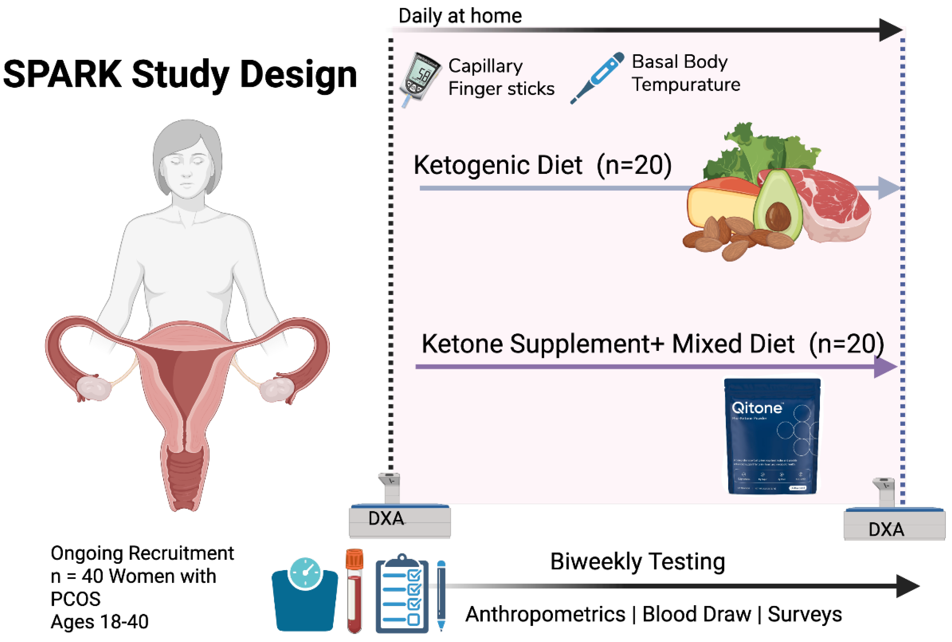
Figure 2.
| KD Body Composition Changes. Changes in total, fat, and lean mass measured by DXA among participants in the KD arm of the SPARK-pilot study. Reductions in fat mass with preservation of lean mass were observed over the 12-week intervention period. BL = Baseline; WK12 = Testing week 12.
Figure 2.
| KD Body Composition Changes. Changes in total, fat, and lean mass measured by DXA among participants in the KD arm of the SPARK-pilot study. Reductions in fat mass with preservation of lean mass were observed over the 12-week intervention period. BL = Baseline; WK12 = Testing week 12.
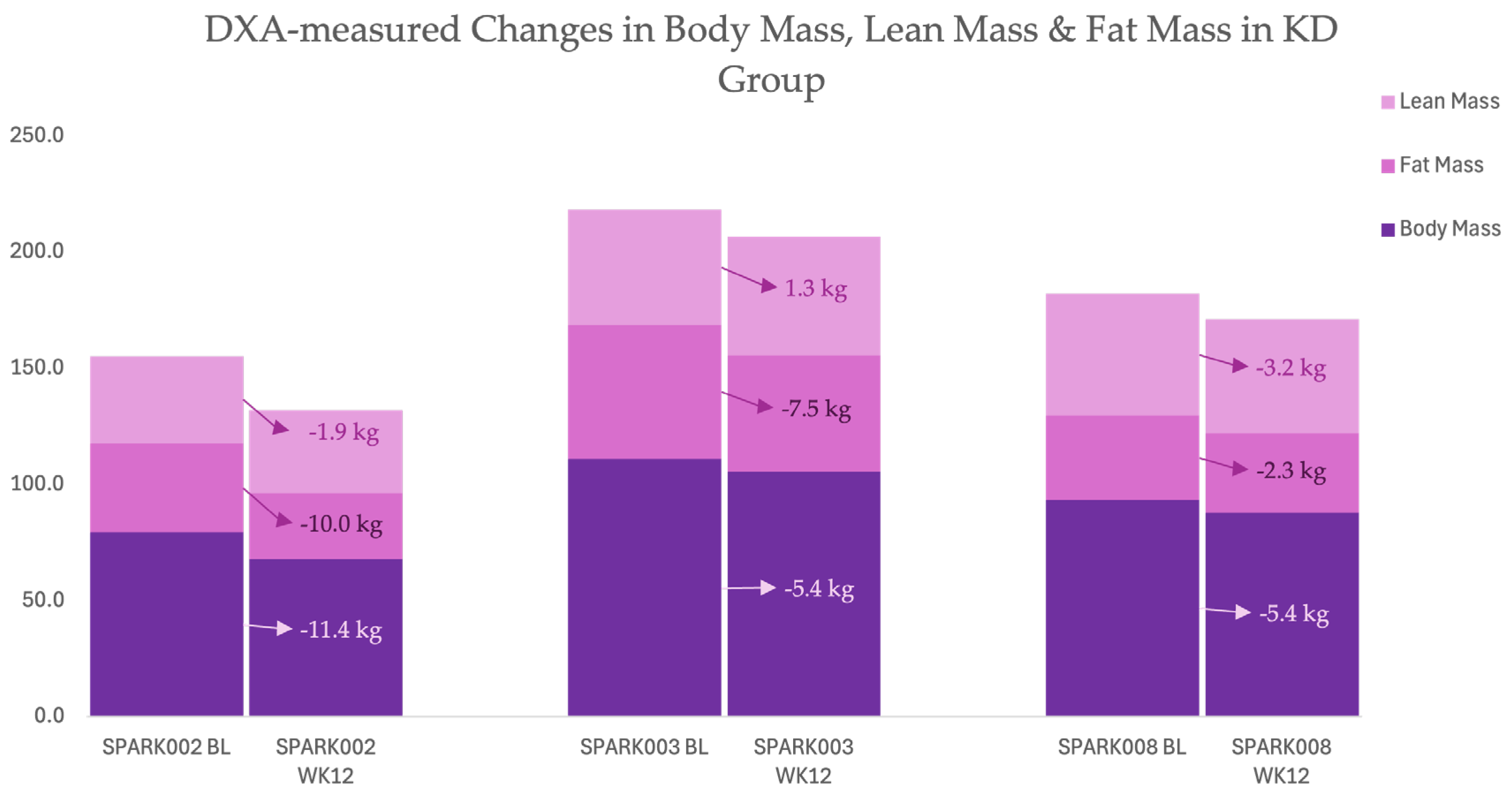
Figure 3.
| Daily fasting BHB and glucose trends with timing of menses and predicted ovulation in SPARK002. Daily BHB and glucose trends with timing of first lifetime menstrual onset in a 33-year-old woman with PMOS. Ketone levels increased to ≥0.7 mmol/L within 14 days of ketogenic-diet initiation, preceding first spontaneous menses by four days.
Figure 3.
| Daily fasting BHB and glucose trends with timing of menses and predicted ovulation in SPARK002. Daily BHB and glucose trends with timing of first lifetime menstrual onset in a 33-year-old woman with PMOS. Ketone levels increased to ≥0.7 mmol/L within 14 days of ketogenic-diet initiation, preceding first spontaneous menses by four days.
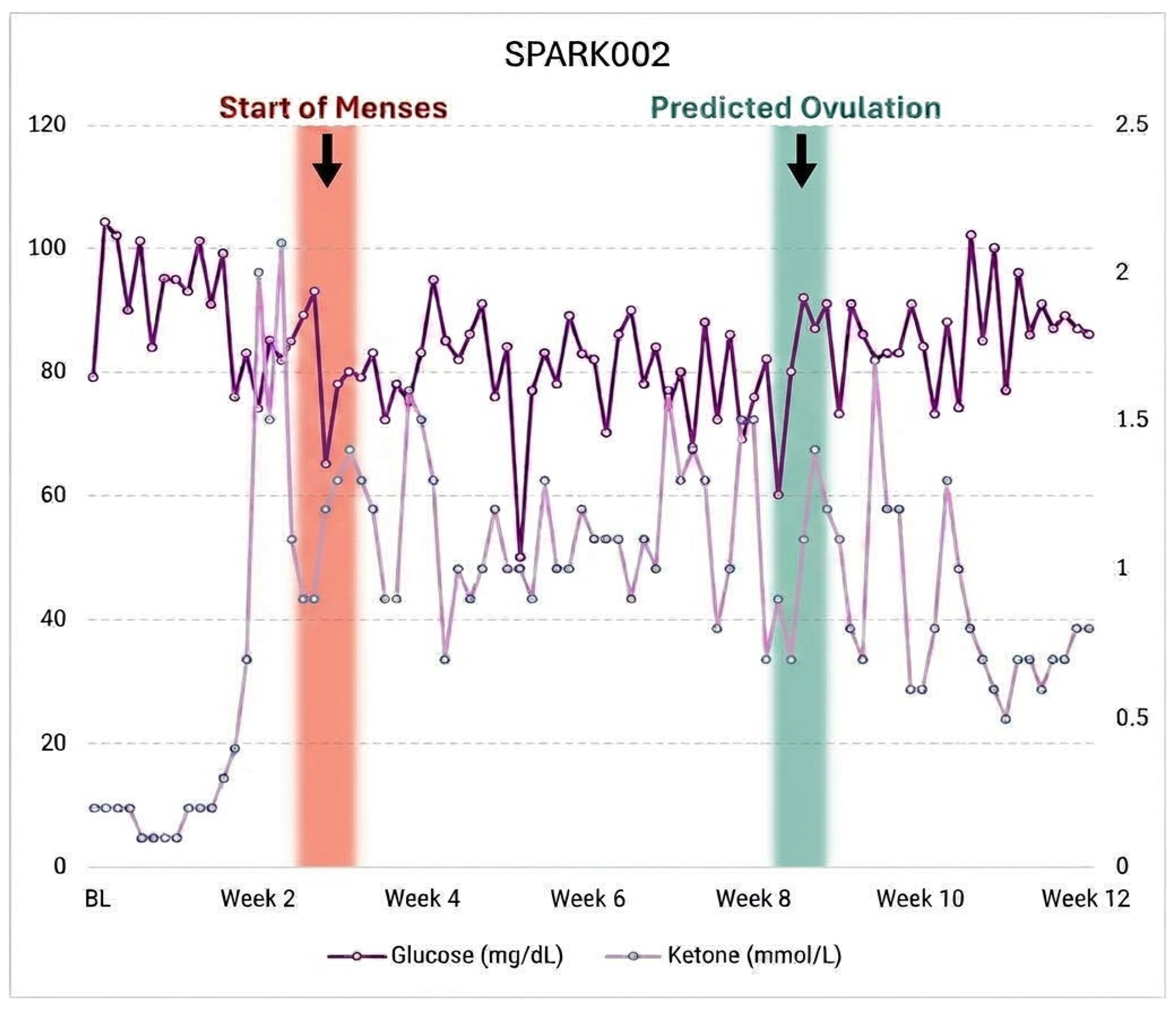
Figure 4.
| Daily BHB and glucose trends with timing of menses in SPARK003. Daily BHB and glucose trends across two menstrual cycles in a woman with oligomenorrhea. Nutritional ketosis was achieved within 48 hours and maintained throughout; shaded bars indicate week 4 and week 11 menses.
Figure 4.
| Daily BHB and glucose trends with timing of menses in SPARK003. Daily BHB and glucose trends across two menstrual cycles in a woman with oligomenorrhea. Nutritional ketosis was achieved within 48 hours and maintained throughout; shaded bars indicate week 4 and week 11 menses.
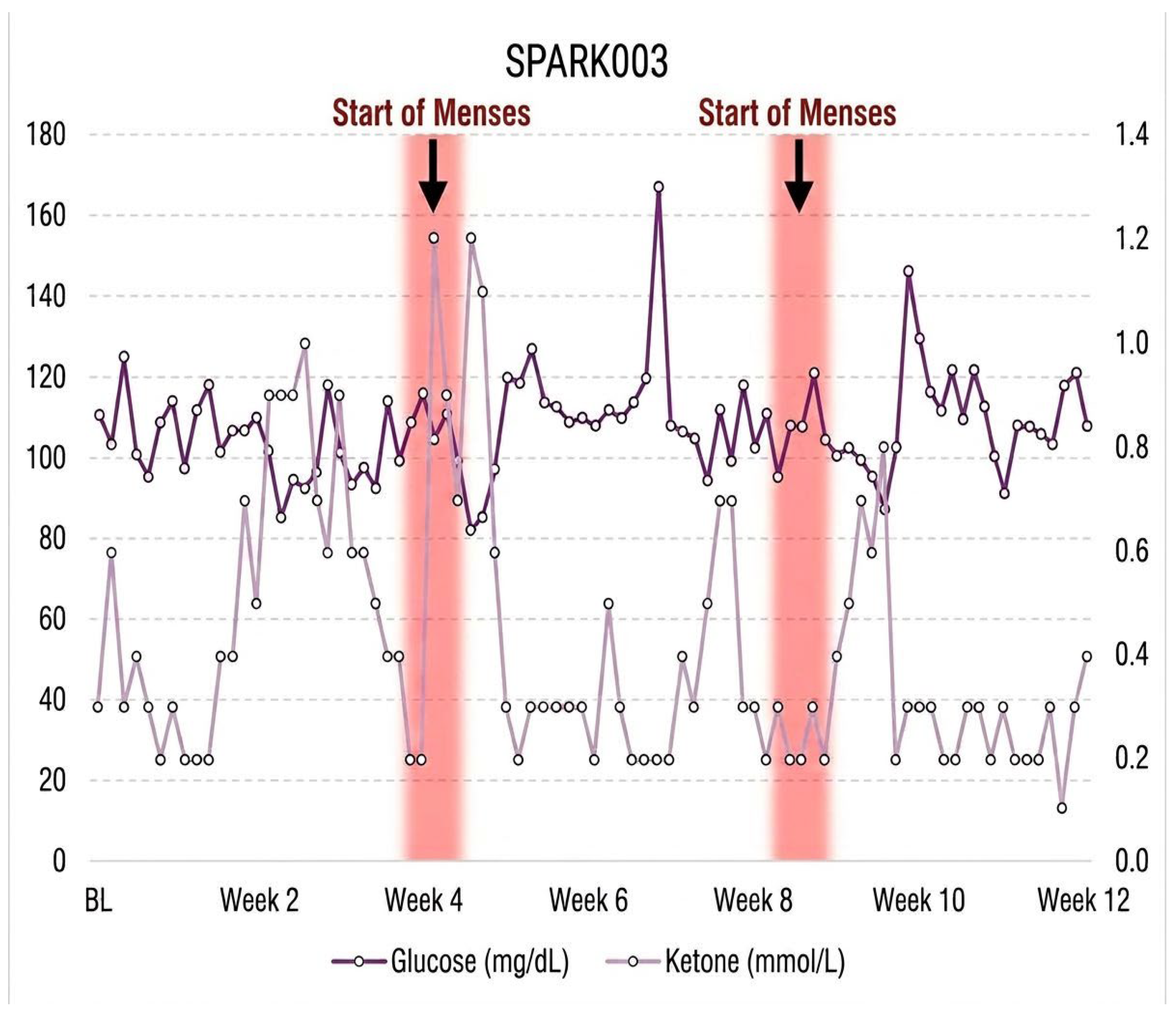
Figure 5.
| Daily BHB and glucose trends with timing of menses and predicted ovulation in SPARK008. Daily BHB and glucose trends during ketogenic-diet intervention. Menstrual onset at week 4 and predicted ovulation at day 16 were temporally associated with declining βHB concentrations following initial ketosis (≥0.6 mmol/L).
Figure 5.
| Daily BHB and glucose trends with timing of menses and predicted ovulation in SPARK008. Daily BHB and glucose trends during ketogenic-diet intervention. Menstrual onset at week 4 and predicted ovulation at day 16 were temporally associated with declining βHB concentrations following initial ketosis (≥0.6 mmol/L).
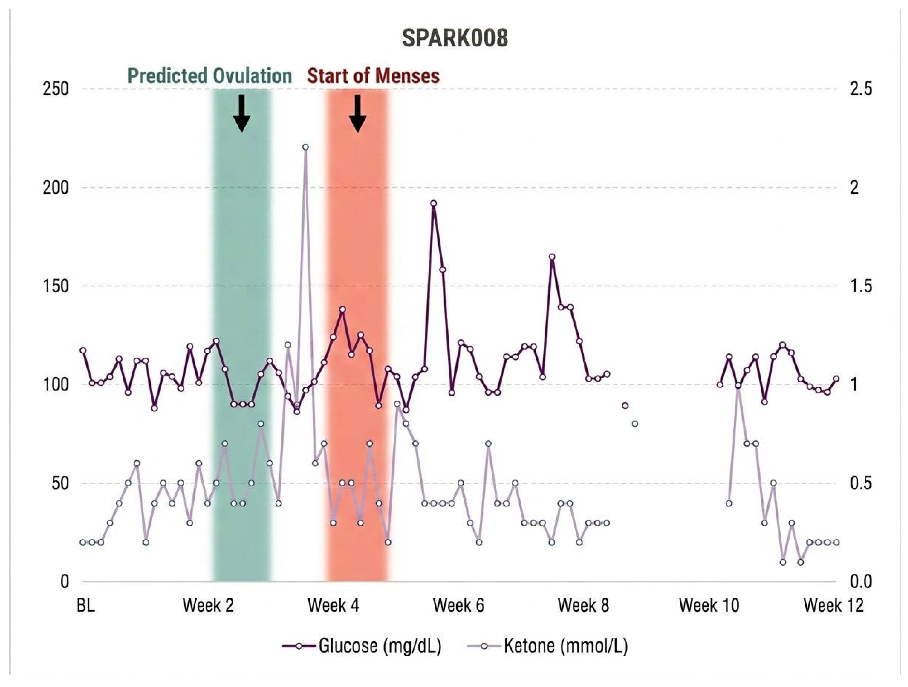
Figure 8.
|Daily BHB and glucose trends with timing of menses in SPARK005. Daily BHB and glucose trends during 12 weeks of ketone supplementation. Shaded bar denotes week 7 spontaneous resumption of menses following intermittent ketosis (0.5–0.8 mmol/L).
Figure 8.
|Daily BHB and glucose trends with timing of menses in SPARK005. Daily BHB and glucose trends during 12 weeks of ketone supplementation. Shaded bar denotes week 7 spontaneous resumption of menses following intermittent ketosis (0.5–0.8 mmol/L).
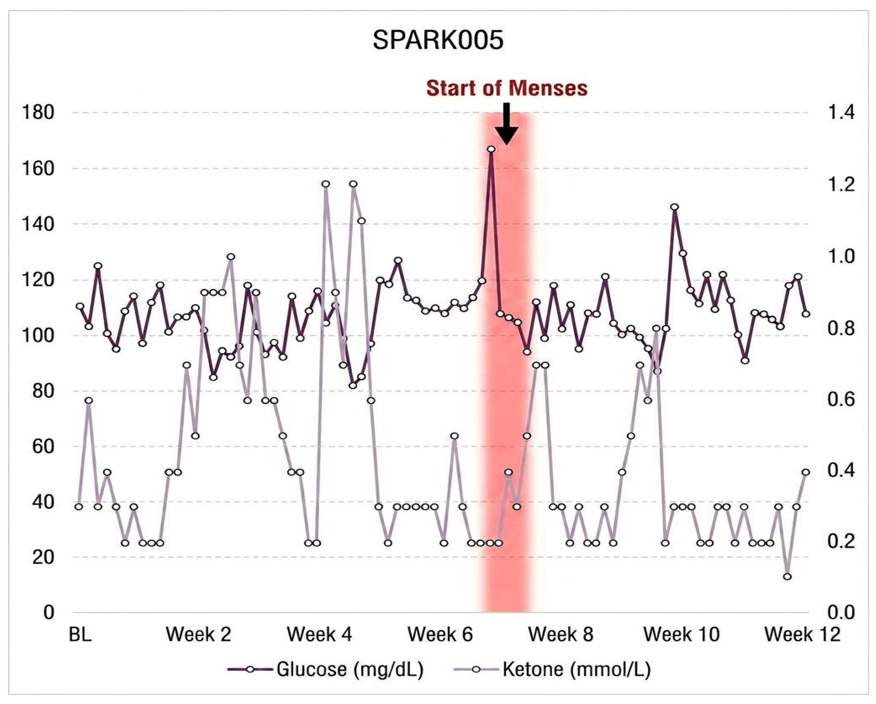
Figure 10.
| Daily BHB and glucose trends with timing of menses and predicted ovulation in SPARK007. Daily BHB and glucose trends across normalized 4-week cycles during ketone supplementation. Intermittent ketosis (0.6–1.0 mmol/L) corresponded with ovulatory recovery and regular cycle patterning.
Figure 10.
| Daily BHB and glucose trends with timing of menses and predicted ovulation in SPARK007. Daily BHB and glucose trends across normalized 4-week cycles during ketone supplementation. Intermittent ketosis (0.6–1.0 mmol/L) corresponded with ovulatory recovery and regular cycle patterning.
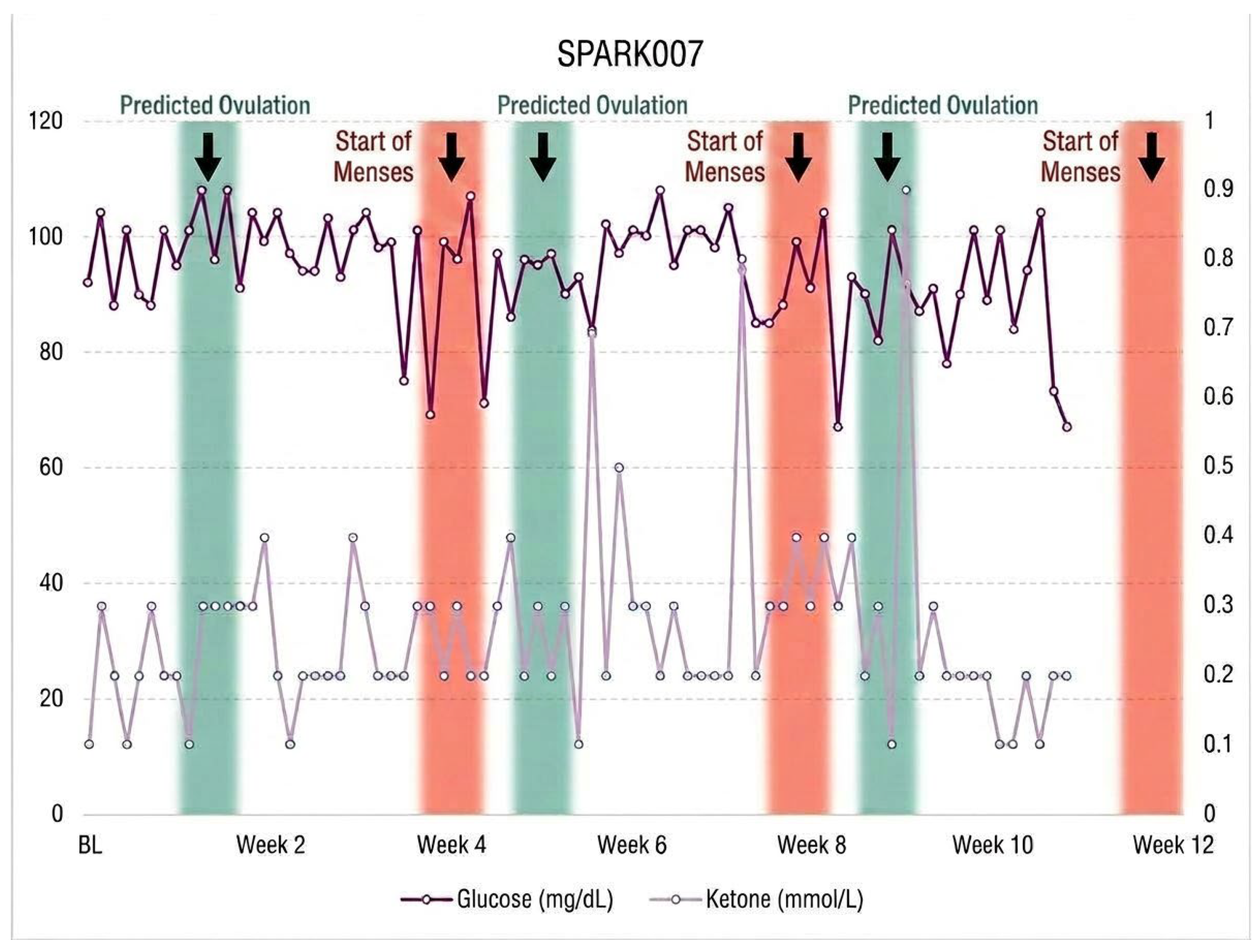
Figure 11.
| Longitudinal Delta Changes in Metabolic and Hormonal Biomarker. These graphs illustrate the biweekly change (Δ) from baseline across 12 weeks for serum Testosterone (ng/mL), Insulin (µIU/mL), and Sex Hormone-Binding Globulin (nmol/L) for seven participants (SPARK002–008).
Figure 11.
| Longitudinal Delta Changes in Metabolic and Hormonal Biomarker. These graphs illustrate the biweekly change (Δ) from baseline across 12 weeks for serum Testosterone (ng/mL), Insulin (µIU/mL), and Sex Hormone-Binding Globulin (nmol/L) for seven participants (SPARK002–008).
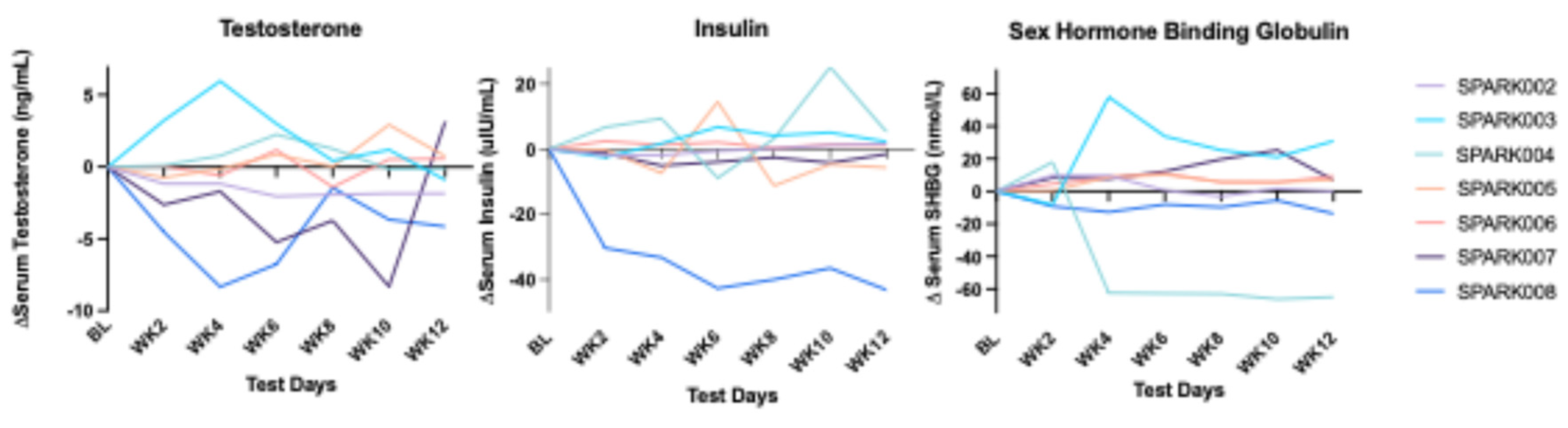

Table 1.
| Baseline Characteristics.
| Intervention | Age | Height | Weight | Lean Mass‡ | Fat Mass‡ | |
| KD/KS | Years | (cm) | (kg) | (kg) | (kg) | |
| SPARK002 | KD | 33 | 147.3 | 79.3 | 38.1 | 39.3 |
| SPARK003 | KD | 24 | 156.97 | 110.95 | 50.3 | 58.4 |
| SPARK004 | KS | 21 | 162.56 | 54.43 | 31.1 | 22.5 |
| SPARK005 | KS | 21 | 153.16 | 87.45 | 48.4 | 31.7 |
| SPARK006 | KS | 21 | 158.4 | 54.4 | 39.6 | 12.4 |
| SPARK007 | KS | 22 | 167.1 | 92.15 | 52.3 | 36.7 |
| SPARK008 | KD | 32 | 162.56 | 93.27 | 53.1 | 36.9 |
| ‡ = measured using dual x-ray absorptiometry | ||||||
Table 2.
| Individual Menstrual Changes. Individual menstrual outcomes during ketogenic diet (KD) and ketone supplementation (KS) interventions in women with PMOS (n = 7).
Table 2.
| Individual Menstrual Changes. Individual menstrual outcomes during ketogenic diet (KD) and ketone supplementation (KS) interventions in women with PMOS (n = 7).
| ID | Intervention |
Last Menses (Before BL) |
Week of First Menses |
Cycle Outcome (12 weeks) |
Birth Control |
| SPARK002 | KD | Never | 2.5 | Spontaneous menses onset; regular interval established | N |
| SPARK003 | KD | 5 months | 4, 11 | Two complete spontaneous cycles | Y |
| SPARK004 | KS | 6 months | 5 | Spotting only; no full menses | N |
| SPARK005 | KS | BL | 7 | Spontaneous menstrual resumption; regular interval | N |
| SPARK006 | KS | 5 years | 6 | Spontaneous menstrual resumption after 5-year absence | N |
| SPARK007 | KS | BL | 2 | Spontaneous menses; normalized to 4-week interval | N |
| SPARK008 | KD | BL | 2 | Spontaneous menses; normalized to 4-week interval | N |
Disclaimer/Publisher’s Note: The statements, opinions and data contained in all publications are solely those of the individual author(s) and contributor(s) and not of MDPI and/or the editor(s). MDPI and/or the editor(s) disclaim responsibility for any injury to people or property resulting from any ideas, methods, instructions or products referred to in the content. |
© 2026 by the authors. Licensee MDPI, Basel, Switzerland. This article is an open access article distributed under the terms and conditions of the Creative Commons Attribution (CC BY) license (http://creativecommons.org/licenses/by/4.0/).
Copyright: This open access article is published under a Creative Commons CC BY 4.0 license, which permit the free download, distribution, and reuse, provided that the author and preprint are cited in any reuse.