

Submitted:
19 May 2026
Posted:
20 May 2026
You are already at the latest version
Abstract
1) Background: Coumestrol is a bioactive compound that inhibits HASPIN activity and prevents tau and H3 phosphorylation. Oral ingestion of CBSs increases blood testosterone levels, which decline with age causing late-onset hypogonadism. Oral ingestion of coumestrol-rich bean sprouts (CBSs) has been shown to suppress the onset of Alzheimer’s disease in 5xFAD mice and the onset of colon cancer in APCmin/+ mice. 2) Methods: We investigated the effect of oral ingestion of CBSs on the progression of aging in male senescence-accelerated prone 1 (SAMP1) mice allowed voluntary exercise or no exercise. The SAMP1 mice were divided into two groups fed either a standard diet or a diet including bean sprouts from 12 to 18 weeks of age. Each group was divided into two groups with voluntary exercise or no exercise. 3) Results: Voluntary exercise accelerated aging-related declines in blood testosterone levels, nerve growth factor levels, and spatial working memory, and oral ingestion of CBSs suppressed these age-related phenotypes, regardless of exercise. 4) Conclusion: Ingestion of CBSs prevented aging-related phenotypes in the experimental mice. A detailed analysis of the molecular mechanisms of coumestrol will be useful for understanding aging and preventing age-related diseases such as cancer, Alzheimer’s disease, and LOH.
Keywords:
cancer
; coumestrol
; dementia
; AGA
; HASPIN
; neurotrophin
; NGF
; phytoestrogen
; testosterone
1. Introduction
In developed countries, the proportion of older people in relation to the total population is increasing because of longer life expectancies resulting from improvements in medical technology and social infrastructure. In 2024, the World Health Organization predicted that the number of people aged 65 and over worldwide will have increased from 500 million in 2005 to 1.6 billion by 2050 [1]. In an aging society, quality of life and extending one’s healthy lifespan are critical. Aging is explained by 12 factors, and it is known to be composed of a combination of these factors [2]. Moderate-exercise equipment and various health and nutritional supplements are increasingly being developed as our understanding of aging advances [3,4].
Physical activity and exercise have beneficial effects on mental health and quality of life [5]. Among older people, regular physical activity, such as walking, reduces the risk of becoming bedridden [6]. However, excessive exercise strains the body and damages cells through the generation of reactive oxygen species, and it can lead to accumulated fatigue and accelerated aging [7,8,9]. Overtraining syndrome (OTS) is known be primarily driven by chronic immune activation and dysregulation of cytokines, especially interleukin-6 (IL-6), tumor necrosis factor-α (TNF-α), and interleukin-1β (IL-1β) [7]. A complete understanding of the relationship between the quality and quantity of exercise and aging is lacking and warrants further research. However, research on aging requires long investigation periods in order to compile results. By using the senescence-accelerated prone 1 (SAMP1) mouse, a senescence-accelerated model in which aging progresses more rapidly, researchers can analyze aging over a shorter period [10]. SAMP1 is an inbred strain established through the repeated mating of siblings from ARK/J mice. Its characteristics include rapid aging at age 4–6 months and an early onset of age-related pathologies such as aging amyloidosis, learning and memory impairment, senile osteoporosis, and cataracts in a strain-specific manner [10].
Coumestrol, synthesized in plants, acts as a phytoalexin. Coumestrol is a bioactive compound that has effects on various biomolecules [11]. Coumestrol acts as a phytoestrogen for mammals. Moreover, it inhibits the serine–threonine kinase HASPIN [12], which regulates chromosome and spindle function during mitosis and meiosis and plays a role in chromosome segregation during cell division [13,14]. HASPIN is critical for normal cell division. At the same time, inhibition of HASPIN activity greatly suppresses cancer cell division by affecting active cell division [15]. Moreover, HASPIN phosphorylates tau protein [16]. Several chemical compounds that inhibit HASPIN activity have been isolated, and functional analysis of HASPIN is currently underway [17]. Coumestrol is synthesized in many plants, but it has been found to be abundant in beans such as Glycine max, Vigna radiate, and Vigna mungo. It was found that cultivating bean sprouts at 24 °C for about a week increases the concentration of the HASPIN inhibitor coumestrol in vivo [11]. Oral ingestion of coumestrol-rich bean sprouts (CBSs) increases blood testosterone levels in mice and humans [16,18]. Additionally, it suppressed the onset of disease in both the 5xFAD mouse model of Alzheimer’s disease and the APCmin/+ mouse model of familial colorectal cancer [16,19]. With age, many people develop cancer or dementia, leading to a decline in quality of life and a shortened lifespan. Although various cancer treatments have been developed, further advancements are needed. Moreover, there is no cure for dementia, and the development of a treatment is urgently needed. Therefore, understanding the mechanisms of action of ingested CBSs with respect to biomolecules is important in the context of an aging society.
Aging induces alteration of sex hormones commensurate with a loss of reproductive function [2]. The decline in testosterone levels is a marker of aging [20]. Late-onset hypogonadism (LOH) syndrome, caused by low testosterone levels, is associated with various age-related diseases [21], and it can impair the daily lives of middle-aged men and have societal effects. Furthermore, systemic comorbidities, such as metabolic syndrome, depression, and frailty, are common in aged men with LOH symptoms. LOH is diagnosed using the Aging Males’ Symptoms scale [22]. The prevalence of symptomatic androgen deficiency in 30–79-year-old men is 5.6%, and it increases substantially with age [23]. As populations age, the number of patients affected by LOH is expected to increase. Understanding and treating LOH constitute an important issue with regard to maintaining the quality of life of middle-aged and older men. Preventing aging-related declines in testosterone levels will help prevent the onset of LOH. Given its effects on suppressing declines in blood testosterone levels, the oral ingestion of CBSs may prevent LOH.
In this study, we investigated the anti-aging effects of CBSs and voluntary exercise. We conducted this study using the SAMP1 mouse model of aging to complete the analysis over a shorter period. Specifically, we measured the progression of age-related dementia and changes in testosterone and neurotrophic factor blood levels when SAMP1 mice were allowed voluntary exercise and fed CBSs.
2. Materials and Methods
2.1. Ethics
The study was conducted in accordance with the Declaration of Helsinki. All animal experiments conformed to the Guide for the Care and Use of Laboratory Animals and were approved by the Committee of Laboratory Animal Experimentation, and Research Ethics Committee of Nagasaki International University, Nagasaki, Japan (no. 167).
2.2. Bean Sprout Cultivation
Mung bean sprouts (Vigna radiata), which were unpalatable and unsuitable for consumption, were purchased from Choho Sangyo Co., Ltd. (Nagasaki, Japan) and cultivated for 3 days at 24 ℃ to ensure they contained large amounts of coumestrol (approximately 150 μg/g dry weight). Sprout cultivation and coumestrol measurement were performed as reported previously [11]. The bean sprouts were cultivated under natural light in a temperature-controlled room on netting with water mist applied once daily. The cultivated bean sprouts (BSs) were dried at 80 °C for 12 h and then ground in a mill.
2.3. Animals
Male SAMP1 and female C57BL/6 mice for use in the experiments were purchased from Japan SLC, Inc. (Hamama-tsu, Japan). The mice were provided with food and water ad libitum and maintained under specific pathogen-free conditions using materials that contribute to environmental enrichment in the animal experimentation facility of Nagasaki International University, with temperature and lighting controlled during the experiment. The mice were sacrificed via cervical dislocation immediately before the experimental end point. The mice were fed the specialized laboratory rodent diet LabDiet® 5L37 (Japan SLC, Inc.) as a standard diet (ST), which is also available internationally as a LabDiet® product (Land O’ Lakes, Inc., Arden Hills, MN, USA). Additionally, a BS-enhanced feed (hereafter referred to as the BS diet) was prepared by soaking the solid ST in water, mixing it with 15% coumestrol-rich bean sprout (CBS) powder, and treating it at 80 °C for 10 h. The nutritional composition of each diet is provided in Table S1. The BS diet contained approximately 22.5 μg/g of coumestrol. The SMAP1 mice were kept in individual cages (measuring 240 × 170 × 120 mm) to measure the amount of food each mouse consumed and prevent fighting. The SAMP1 mice were fed the ST until 12 weeks post-birth; subsequently, they were fed either the ST or BS diet. From 12 to 18 weeks of age, the ST- and BS-fed mice were divided into groups with no exercise (ST-NE and BS-NE, respectively) or voluntary exercise (ST-VE and BS-NE, respectively) (Figure 1). Seven female C57BL/6 mice were housed together in a large cage (350 × 300 × 160 mm). To observe the lifespan and appearance of the mice, some of the SAMP1 mice and female C57BL/6 mice were fed the ST or BS diet continuously and prevented from exercising, starting at 12 weeks of age, until they died of old age.
2.4. Non-Exercise and Voluntary Exercise Settings
The NE mice were held in their individual cages throughout the experiment. Conversely, the VE mice were provided access to a running wheel with a diameter of 150 mm (Marukan Co., Ltd., Osaka, Japan) for 90 min a day for 5 days a week in a quiet environment to allow free and voluntary exercise (Figure 2A, Supplemental Video S1); this is a common experimental method [24]. The running distance was automatically measured using a wheel machine.
2.5. Y-Maze Test
The Y-maze test was performed on 16–18-week-old mice to examine spontaneous locomotor activity and spatial working memory [25]. Each mouse was placed at the end of one arm of the test device, in which three arms of the same size were connected at 120°. The mouse was allowed to move freely for 8 min, and its behavior was recorded. Specifically, we recorded the number of times each arm was entered. Spontaneous alternation is a behavior consisting of exploring novel environments. In this test, we analyzed the rate at which mice entered different arms consecutively. If they entered the same arm many times, we determined that they did not remember their own exploratory behavior and hence had low spatial working memory ability. Spontaneous locomotor activity was calculated from the total number of arm entries. Six mice were used for each condition.
2.6. Western Blotting
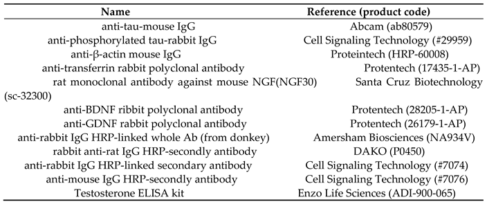
Blood samples were taken and stored at 4 °C for 1 day, after which the serum was separated via centrifugation at 15,000 × g. For each sample, 1 μL of serum was separated using sodium dodecyl sulfate–polyacrylamide gel electrophoresis, and it was then electroblotted onto polyvinylidene fluoride membranes and finally blocked with Blocking Solution in phosphate-buffered saline (Nacalai Tesque, Inc., Kyoto, Japan). After being washed in tris-buffered saline with Tween 20 (50 mM Tris-HCl, pH 7.4; 138 mM NaCl; 2.7 mM KCl; and 0.1% Tween 20), the membranes were reacted with primary antibodies overnight at 4 °C and then with secondary antibodies for 120 min at room temperature. The antigen–antibody complexes were detected using ECL Select (Cytiva Amersham, Marlborough, MA, USA). The signals were observed using an ImageQuant LAS 4000 imager (GE, Boston, MA, USA). The antibodies used in the experiment are detailed in Table 1. The image-processing program Image Lab (Bio-Rad, Hercules, CA, USA) was used to analyze signal intensity [26].
2.7. Enzyme-Linked Immunosorbent Assay
Testosterone levels were measured in 20 µL aliquots of serum via an enzyme-linked immunosorbent assay (ELISA) with a commercial kit used according to the manufacturer’s protocol (ADI-900-065; Enzo Life Sciences, Farmingdale, NY, USA). The assay detection sensitivity was 5.67 pg/mL, with an average coefficient of variation of 9.5% across sample concentrations. Each sample was analyzed in duplicate using two assay blocks simultaneously. If there was a large discrepancy between the two measurements, the assay was repeated for the sample in question.
2.8. Statistical Analysis
Data are expressed as the mean ± standard deviation (SDs). Statistical analysis was performed using Student’s t-test. In all analyses, P < 0.05 was taken to indicate statistical significance.
3. Results
3.1. Physical Examination
Voluntary exercise slows aging [5]. Among the VE mice provided regular access to a running wheel (Figure 2A and Supplemental Video 1) [24], no significant differences in running distance, body weight, or food ingestion were observed (Figure 2B–D). Calorie intake in k Cal/days was 22.04 ± 4.62, 23.77 ± 3.82, 23.77 ± 2.63, and 19.79 ± 2.66 for the ST-NE, ST-VE, BS-NE, and BS-VE groups, respectively (Figure 2E). The calorie intake of the BS-VE group was significantly (83%) lower than that of the BS-NE group, although all experimental groups consumed approximately the same amount of calories from their feed. We examined the appearance of the SAMP1 mice allowed to engage in voluntary exercise at 18 weeks old. The mice in the BS-VE group had better-groomed coats than those in the ST-VE group (Figure 3). Additionally, among 2-year-old female C57BL/6 mice who did not engage in any exercise, those fed the BS diet were found to have better-groomed coats than those fed the ST (Figure S1). Furthermore, the SAMP1 mice were kept without access to exercise and continuously fed either the ST or BS diet. The mice fed the BS diet (705.4 ± 44.9 days) lived significantly longer than those fed the ST (545.8 ± 106.7 days)(P=0.027) (Figure 4). These results suggest that voluntary exercise induced an aging-related phenotype in the SAMP1 mice, which was suppressed by oral ingestion of CBSs.
3.2. Examination of Spatial Working Memory
Next, we subjected each mouse in each experimental group to the Y-maze test to examine spatial working memory (Figure 5). Among the ST-fed mice, the ST-VE group (49.7 ± 6.5) had significantly lower spatial working memory scores than the ST-NE group (62.8 ± 5.9), suggesting that voluntary exercise reduced spatial working memory in the SAMP1 mice. The result for the SAMP1 ST-NE group in this study is almost identical to that in our previous report regarding C57BL/6 mice [19]. Among the VE mice, the BS-VE group (65.2 ± 8.3) had significantly higher spatial working memory scores than the ST-VE group, suggesting that the exercise-induced decline in spatial working memory was suppressed by CBS ingestion. Overall, the BS-NE group (77.7 ± 10.3) had the highest corrected scores and the ST-VE group the lowest, suggesting that the mice in the BS-NE group experienced less aging. Among the NE mice, the BS-NE group had significantly higher corrected scores than the ST-NE group, suggesting that CBS ingestion suppressed the aging-related decline in spatial working memory. Finally, although it is difficult to observe overexpression of endogenous phosphorylated tau even in mice with Alzheimer’s disease [27], we used Western blotting to observe whether changes could be observed in SAMP1 mice, but no significant differences among the SAMP1 groups were found (Figure S2).
3.3. Total Testosterone Levels in Serum
Blood testosterone levels decrease with age [20], but CBS ingestion may increase serum testosterone levels [16,20]. In SAMP1 mice, the effects of voluntary exercise and food on serum testosterone levels were examined using ELISA (Figure 6). The changes in serum testosterone levels were similar to the trends in appearance and spatial working memory. Among the ST-fed mice, the ST-VE group (557 ± 478 pg/ mL) had significantly lower testosterone levels than the ST-NE group (1470 ± 667 pg/ mL). Among the VE mice, the BS-VE group (2009 ± 1266 pg/ mL) had significantly higher testosterone levels than the ST-VE group. Thus, voluntary exercise resulted in lower serum testosterone levels, and this decline was suppressed by CBS ingestion. Overall, serum testosterone levels were the highest in the BS-NE group (2081 ± 1403 pg/ mL) and lowest in the ST-VE group. These results suggest that the aging-induced decline in serum testosterone levels was suppressed by CBS ingestion.
3.4. Serum Neurotrophins
Neurotrophin expression decreases with age [28]. Serum neurotrophin nerve growth factor (NGF), brain-derived neurotrophic factor (BDNF), and glial-cell-line-derived neurotrophic factor (GDNF) expression were analyzed using Western blotting. No significant differences in BDNF and GDNF levels were observed among the experimental groups (Figure 7A-D, Table 2). However, NGF expression declined with aging, which was suppressed by CBS ingestion (Figure 7A, B, E), similar to the trends observed for spatial working memory and serum testosterone levels. In the ST-fed mice, NGF expression was non-significantly lower in the ST-VE group (0.60 ± 0.21) relative to the ST-NE group (0.85 ± 0.35). Among the VE mice, NGF levels were significantly higher in the BS-VE group (1.17 ± 0.53) relative to the ST-VE group. The BS-NE group (1.53 ± 0.88) exhibited significantly higher NGF expression than the ST-VE group. Overall, serum NGF levels were highest in the BS-NE group and lowest in the ST-VE group. These results suggest that the voluntary-exercise- and age-related decline in NGF levels was suppressed by CBS ingestion.
4. Discussion
To investigate the relationship between disease onset and aging in detail, experimental animals must be reared and allowed to age over long periods. To overcome this limitation, we used the senescence-accelerated mouse strain SAMP1 to investigate the impacts of voluntary exercise and oral ingestion of CBS on aging. The results revealed that when SAMP1 mice were allowed to exercise on a running wheel, which provided loads within the typical experimental range, fur loss, spatial working memory decline, and testosterone loss, all considered signs of aging, progressed earlier. The aging-related phenotype in SAMP1 mice was accelerated by voluntary exercise. Exercise generates reactive oxygen species (ROS), which stress cells [7,8,9]. In SAMP1 mice, ROS may be generated during voluntary exercise, causing muscle microtrauma, which induces chronic inflammatory cytokine release and thus the aging-related phenotype. It is unclear why SAMP1 mice exhibit an early aging-related phenotype, but it may be due to their susceptibility to ROS. Since coumestrol has antioxidative activity, ingestion of CBSs may suppress the aging phenotype in SAMP1 mice.
First, we established a mouse-breeding approach in which an early-aging-related phenotype could be observed by allowing SAMP1 mice to exercise. Next, we observed the effect of oral ingestion of bean sprouts rich in the HASPIN inhibiter coumestrol on the progression of the aging-related phenotype due to exercise. These results indicated that oral ingestion of CBSs significantly suppressed the loss of fur, spatial cognitive function, testosterone, and NGF with voluntary exercise. These tendencies of anti-aging-related phenotypes were also observed under no-exercise conditions. Furthermore, suppression of hair loss was observed when CBSs were administered to C57BL/6 female mice. Low-calorie diets have an anti-aging effect [29]. It has been reported that reducing calorie intake can extend lifespan [30]. In this experiment, the calorie intakes of mice in the ST-NE group and BS-NE group were almost the same. These results indicated that oral ingestion of CBSs can extends lifespan. By getting the SAMP1 mice to exercise, we were able to determine significantly the effects of CBSs on aging-related phenotypes in a short period using a small number of mice. Making SAMP1 mice exercise is a more useful strategy for conducting a detailed analysis of the anti-aging effects than using a large number of wild-type mice.
Coumestrol is a phytoestrogen that affects various biomolecules, including estrogen receptors [11]; it also inhibits HASPIN kinase [12]. HASPIN, cloned as a gene specifically expressed in haploid germ cells [31], is a serine–threonine kinase conserved in eukaryotes that phosphorylates multiple proteins during spermatogenesis [32,33]. HASPIN is also expressed during somatic cell division and meiosis and has been shown to regulate cell division by phosphorylating the histones H3 and TH2A [13,14]. A HAPSIN inhibitor suppressed proliferation of various cancer cells and the development of polyps in a familial colorectal cancer mouse model (APCmin/+) [16]. HASPIN also phosphorylates tau protein expressed in neurons. Oral ingestion of CBSs suppressed Alzheimer’s disease in 5xFAD mice [19]. Precise regulation of HASPIN activity is thought to be important for maintaining cellular function. One of the factors contributing to aging is a disruption of epigenetic regulation [2]. The genomic region corresponding to HASPIN expression is epigenetically regulated as differentially methylated regions [34]. Transcriptional regulation of the HASPIN gene is mediated by a 193 bp DNA region upstream of the transcription initiation site, and HASPIN expression is suppressed by DNA methylation in differentiated cells [35]. Changes in epigenetic alterations, such as genomic DNA methylation and histone acetylation and methylation, are reported to occur with aging and affect gene expression [36]. Such alterations can cause various diseases, including cancer, lifestyle diseases, and mental disorders [36]. Age-related disturbances in epigenetic alterations lead to HASPIN expression in cells that do not require it, a process that may contribute to the development of cancer and Alzheimer’s disease, the incidence rates of which increase with age. Although HASPIN plays an important role in cell division, in haspin knockout mice, HASPIN function is compensated for by other mechanisms, and no significant defects are observable in normal cells [37]. In this study, CBSs may have inhibited abnormal HASPIN activity, eliciting the suppressed anti-aging-related phenotype without any observed side effects. Thus, CBS consumption may inhibit the progression of these diseases by inhibiting abnormal HASPIN expression. Inhibition of HASPIN is considered beneficial only in specific biological or disease contexts, not universally. However, the interest in HASPIN inhibition derives from its very specialized role in cell division, a role that creates an unusual opportunity to selectively suppress pathological cell proliferation while sparing most normal cellular functions.
Oral ingestion of CBSs increases serum testosterone levels of aging markers in both humans and mice [2,16,18]. Coumestrol is a phytoestrogen and polyphenol that reacts with estradiol receptors. Coumestrol also affects the production of steroid hormones by inhibiting aromatase and hydroxysteroid dehydrogenase [38]. It is a low-molecular-weight compound consisting of a steroid skeleton and may cross the blood–brain barrier [39]. Additionally, classical aging biomarkers should be assessed to support the conclusion of accelerated aging [40]. Although accurate analysis of multiple factors will take time because of the low volume of blood in mice, future research should analyze blood gonadotropins, which mediate testosterone secretion [22]. Further accurate analysis of blood factors is needed to clarify coumestrol’s potential mechanisms of action with respect to, for instance, the brain or gonads.
In this study, serum NGF levels were higher in the BS-fed mice than in the ST-fed mice. Estrogen has been reported to increase NGF levels [41,42]. The CBS-induced increase in serum NGF levels may have been due to coumestrol’s effects consistent with its role as a female hormone. Moreover, coumestrol has antioxidant properties, activates the longevity gene Sirt1 [43], and inhibits cholinesterase [44], among additional effects on other biomolecules [11]. Thus, the aging-related phenotypes observed in this study, including increased serum testosterone and NGF levels, may have been induced through a number of coumestrol’s functions. Additionally, bean sprouts are 95% water, but they also contain vitamin B1, B2, C, calcium, folic acid, iron, and dietary fiber [45]. Although we found no reports indicating that each compound markedly prevents the age-related decline in blood testosterone and NGF levels, it is necessary to consider the influence of molecules found in CBSs and the production of metabolic products by the intestinal microbiota. Although the experiment was conducted using SAMP1 mice, further analyses of the molecular mechanisms underlying the effects of CBSs and coumestrol can be expected to provide important insights into aging and anti-aging.
5. Conclusions
This study on SAMP1 mice reveals that voluntary exercise on a running wheel accelerated aging and that oral ingestion of CBSs suppressed the progression of aging-related phenotypes in mice who did and did not engage in exercise. These findings support the potential of CBSs as an anti-aging food.
Supplemental Materials: The following supporting information can be downloaded at the website of this paper posted on Preprints.org, Figure S1: Aged female C57BL/6 mice fed bean sprouts. Figure S2: Expression of tau and phosphorylated tau in the hippocampus of SAMP1 mice. Figure S3: Uncropped Western blot figures using anti-neurotrophin antibodies. Video S1: Observation of free and voluntary exercise for each mouse with access to a running wheel.
Author Contributions
Conceptualization, H.T.; methodology, H.T.; software, H.T. and K.K.; validation, K.K., A.K., R.Y., M.K., A.T., and H.T.; formal analysis, H.T. and K.K.; investigation, K.K., A.K., R.Y., M.K., A.T., and H.T; resources, H.T.; data curation, H.T. and K.K.; writing—original draft preparation, H.T.; writing—review and editing, H.T.; visualization, H.T. and K.K.; supervision, H.T.; project administration, H.T.; funding acquisition, H.T. All authors have read and agreed to the published version of the manuscript.
Funding
This work was supported in part by JSPS KAKENHI Grant 24K12454 for Scientific Research from the Ministry of Education, Science, and Culture, Japan.
Institutional Review Board Statement
All animal experiments conformed to the Guide for the Care and Use of Laboratory Animals and were approved by the Institutional Committee of Laboratory Animal Experimentation and Research Ethics of Nagasaki International University, Nagasaki, Japan (No. 167). This study did not involve any human experiments performed by any of the authors.
Informed Consent Statement
Not applicable.
Data Availability Statement
The datasets used and analyzed during this study are available from the corresponding author on reasonable request.
Acknowledgments
We thank Ms. Taeko Hisano for cultivating the bean sprouts, producing the feed, and breeding the mice.
Conflicts of Interest
The authors declare no conflicts of interest.
References
- World Population Prospects. 2024. United Nations. Available online: https://population.un.org/wpp/graphs?loc=900&type=Demographic%20Profiles&category=Line%20Charts (Accessed on 13 January 2026).
- López-Otín, C.; Blasco, M.A.; Partridge, L.; Serrano, M.; Kroemer, G. Hallmarks of aging: An expanding universe. Cell 2023, 186, 243–278. [Google Scholar] [CrossRef]
- Rodrigues, F.; Domingos, C.; Monteiro, D.; Morouço, P. A Review on Aging, Sarcopenia, Falls, and Resistance Training in Community-Dwelling Older Adults. Int. J. Environ. Res. Public Health 2022, 19, 874. [Google Scholar] [CrossRef]
- Kwa, F.; Lozano-Lorca, M. Editorial: Preventative medicine: nutritional and lifestyle interventions for healthy ageing and chronic diseases. Front. Nutr. 2025, 12, 723877. [Google Scholar] [CrossRef]
- Yoshimura, Y.; Wakabayashi, H.; Yamada, M.; Kim, H.; Harada, A.; Arai, H. Interventions for Treating Sarcopenia: A Systematic Review and Meta-Analysis of Randomized Controlled Studies. J. Am. Med. Dir. Assoc. 2017, 18, 553.e1–553.e16. [Google Scholar] [CrossRef]
- Inoue, M.; Iso, H.; Yamamoto, S.; Kurahashi, N.; Iwasaki, M.; Sasazuki, S.; Tsugane, S. Daily total physical activity level and premature death in men and women: results from a large-scale population-based cohort study in Japan (JPHC study). Ann. Epidemiol. 2008, 18, 522–530. [Google Scholar] [CrossRef]
- Smith, L.L. Cytokine hypothesis of overtraining: a physiological adaptation to excessive stress? Med. Sci. Sports Exerc. 2000, 32, 317–331. [Google Scholar] [CrossRef]
- Powers, S.K.; Radak, Z.; Ji, L.L.; Jackson, M. Reactive oxygen species promote endurance exercise-induced adaptations in skeletal muscles. J. Sport Health sci. 2024, 13, 780–792. [Google Scholar] [CrossRef] [PubMed]
- Fang, S.; Jiang, M.; Jiao, J.; Zhao, H.; Liu, D.; Gao, D.; Wang, T.; Yang, Z.; Yuan, H. Unraveling the ROS-Inflammation-Immune Balance: A New Perspective on Aging and Disease. Aging Dis. 2025, 17, 305–325. [Google Scholar] [CrossRef]
- Takeda, T.; Matsushita, T.; Kurozumi, M.; Takemura, K.; Higuchi, K.; Hosokawa, M. Pathobiology of the senescence-accelerated mouse (SAM). Exp. Gerontol. 1997, 32, 117–127. [Google Scholar] [CrossRef]
- Ohta, T.; Uto, T.; Tanaka, H. Effective methods for increasing coumestrol in soybean sprouts. PLoS ONE 2021, 16, e0260147. [Google Scholar]
- Kim, J.E.; Lee, S.Y.; Jang, M.; Choi, H.K.; Kim, J.H.; Chen, H.; Lim, T.G.; Dong, Z.; Lee, K.W. Coumestrol Epigenetically Suppresses Cancer Cell Proliferation: Coumestrol Is a Natural Haspin Kinase Inhibitor. Int. J. Mol. Sci. 2017, 18, 2228. [Google Scholar] [CrossRef]
- Wang, F.; Dai, J.; Daum, J.R.; Niedzialkowska, E.; Banerjee, B.; Stukenberg, P.T.; Gorbsky, G.J.; Higgins, J.M. Histone H3 Thr-3 phosphorylation by Haspin positions Aurora B at centromeres in mitosis. Science 2010, 330, 231–235. [Google Scholar] [CrossRef]
- Hada, M.; Kim, J.; Inoue, E.; Fukuda, Y.; Tanaka, H.; Watanabe, Y.; Okada, Y. TH2A is phosphorylated at meiotic centromere by Haspin. Chromosoma 2017, 126, 769–780. [Google Scholar] [CrossRef]
- Huertas, D.; Soler, M.; Moreto, J.; Villanueva, A.; Martinez, A.; Vidal, A.; Charlton, M.; Moffat, D.; Patel, S.; McDermott, J.; et al. Antitumor activity of a small-molecule inhibitor of the histone kinase Haspin. Oncogene 2012, 31, 1408–1418. [Google Scholar] [CrossRef] [PubMed]
- Tanaka, H.; Matsuyama, S.; Ohta, T.; Kakazu, K.; Fujita, K.; Fukuhara, S.; Soda, T.; Miyagawa, Y.; Tsujimura, A. The Natural HASPIN Inhibitor Coumestrol Suppresses Intestinal Polyp Development, Cachexia, and Hypogonadism in a Mouse Model of Familial Adenomatous Polyposis (ApcMin/+). Biology 2024, 13, 736. [Google Scholar]
- Malosse, K.; Josselin, B.; Ruchaud, S.; Anizon, F.; Giraud, F.; Moreau, P. Synthesis of 1H-Pyrrolo [3,2-g]isoquinoline Derivatives as Ligands Targeting Haspin Kinase. Molecules 2025, 30, 4388. [Google Scholar] [CrossRef] [PubMed]
- Kakazu, K.; Tsujimura, A.; Miyagawa, Y.; Takezawa, K.; Kobayashi, K.; Yoshimura, R.; Nakajima, K.; Kamiya, S.; Tanaka, H. Oral Ingestion of Bean Sprouts Containing the HASPIN Inhibitor Coumestrol Increased Blood Testosterone Levels in Men. Biology 2025, 14, 907. [Google Scholar] [CrossRef]
- Tanaka, H.; Matsushita, H.; Tokuhiro, K.; Fukunari, A.; Ando, Y. Ingestion of Soybean Sprouts Containing a HASPIN Inhibitor Improves Condition in a Mouse Model of Alzheimer’s Disease. Biology 2023, 12, 320. [Google Scholar] [CrossRef] [PubMed]
- Harman, S.M.; Metter, E.J.; Tobin, J.D.; Pearson, J.; Blackman, M.R. Longitudinal effects of aging on serum total and free testosterone levels in healthy men. Baltimore Longitudinal Study of Aging. J. Clin. Endocrinol. Metab. 2001, 86, 724–731. [Google Scholar] [CrossRef]
- Nieschlag, E. Late-onset hypogonadism: a concept comes of age. Andrology 2020, 8, 1506–1511. [Google Scholar] [CrossRef]
- Ide, H.; Akehi, Y.; Fukuhara, S.; Ohira, S.; Ogawa, S.; Kataoka, T.; Kumagai, H.; Kobayashi, K.; Komiya, A.; Shigehara, K.; et al. Summary of the clinical practice manual for late-onset hypogonadism. Int. J. Urol. 2023, 30, 422–430. [Google Scholar] [CrossRef]
- Araujo, A.B.; Esche, G.R.; Kupelian, V.; O’Donnell, A.B.; Travison, T.G.; Williams, R.E.; Clark, R.V.; McKinlay, J.B. Prevalence of symptomatic androgen deficiency in men. J. Clin. Endocrinol. Metab. 2007, 92, 4241–4247. [Google Scholar] [CrossRef]
- Bastioli, G.; Mancini, M.; Patel, J.C.; Gamallo-Lana, B.; Arnold, J.C.; Mar, A.C.; Rice, M.E. Voluntary exercise increases striatal dopamine release and improves motor performance in aging mice. npj Park. Dis. 2025, 11, 345. [Google Scholar] [CrossRef] [PubMed]
- Oakley, H.; Cole, S.L.; Logan, S.; Maus, E.; Shao, P.; Craft, J.; Guillozet-Bongaarts, A.; Ohno, M.; Disterhoft, J.; Van Eldik, L.; et al. Intraneuronal beta-amyloid aggregates, neurodegeneration, and neuron loss in transgenic mice with five familial Alzheimer’s disease mutations: potential factors in amyloid plaque formation. Neurosci. 2006, 26, 10129–10140. [Google Scholar] [CrossRef]
- Taylor, S.C.; Berkelman, T.; Yadav, G.; Hammond, M. A defined methodology for reliable quantification of Western blot data. Mol. Biotechnol. 2013, 55, 217–226. [Google Scholar] [CrossRef]
- Guo, Q.; Li, H.; Cole, A.L.; Hur, J.Y.; Li, Y.; Zheng, H. Modeling Alzheimer’s disease in mouse without mutant protein overexpression: cooperative and independent effects of Aβ and tau. PLoS ONE 2013, 8, e80706. [Google Scholar] [CrossRef]
- Obata, H.; Naito, K.; Kikui, A.; Nakamura, S.; Suzuki, K.; Kawakita, S.; Suzuki, T.; Goto, K.; Nagura, N.; Sugiyama, Y.; et al. Age-related differences for expression of the nerve-specific proteins after peripheral nerve injury. Exp. Ther. Med. 2022, 24, 682. [Google Scholar] [CrossRef]
- Vermeij, W.P.; Dollé, M.E.; Reiling, E.; Jaarsma, D.; Payan-Gomez, C.; Bombardieri, C.R.; Wu, H.; Roks, A.J.; Botter, S.M.; van der Eerden, B.C.; et al. Restricted diet delays accelerated ageing and genomic stress in DNA-repair-deficient mice. Nature 2016, 537, 427–431. [Google Scholar] [CrossRef]
- Di Francesco, A.; Deighan, A.G.; Litichevskiy, L.; Chen, Z.; Luciano, A.; Robinson, L.; Garland, G.; Donato, H.; Vincent, M.; Schott, W.; et al. Dietary restriction impacts health and lifespan of genetically diverse mice. Nature 2024, 634, 684–692. [Google Scholar] [CrossRef]
- Tanaka, H.; Yoshimura, Y.; Nozaki, M.; Yomogida, K.; Tsuchida, J.; Tosaka, Y.; Habu, T.; Nakanishi, T.; Okada, M.; Nojima, H.; Nishimune, Y. Identification and characterization of a haploid germ cell-specific nuclear protein kinase (Haspin) in spermatid nuclei and its effects on somatic cells. J. Biol. Chem. 1999, 274, 17049–17057. [Google Scholar] [CrossRef]
- Higgins, J.M. Haspin-like proteins: a new family of evolutionarily conserved putative eukaryotic protein kinases. Protein Sci. 2001, 10, 1677–1684. [Google Scholar] [CrossRef] [PubMed]
- Maeda, N.; Tsuchida, J.; Nishimune, Y.; Tanaka, H. Analysis of Ser/Thr Kinase HASPIN-Interacting Proteins in the Spermatids. Int. J. Mol. Sci. 2022, 23, 9060. [Google Scholar] [CrossRef]
- Sato, S.; Maeda, C.; Hattori, N.; Yagi, S.; Tanaka, S.; Shiota, K. DNA methylation-dependent modulator of Gsg2/Haspin gene expression. J. Reprod. Dev. 2011, 57, 526–533. [Google Scholar] [CrossRef] [PubMed]
- Tokuhiro, K.; Miyagawa, Y.; Yamada, S.; Hirose, M.; Ohta, H.; Nishimune, Y.; Tanaka, H. The 193-base pair Gsg2 (haspin) promoter region regulates germ cell-specific expression bidirectionally and synchronously. Biol. Reprod. 2007, 76, 407–414. [Google Scholar] [CrossRef] [PubMed]
- Kočar, E.; Šket, R.; Vasle, A.H.; Avguštin, G.; Benedik, E.; Seljak, B.K.; Simić, P.; Martinko, A.; Morrison, S.A.; Sorić, M.; et al. Measuring biological age: Insights from omics studies. Ageing Res. Rev. 2025, 114, 102988. [Google Scholar] [CrossRef]
- Shimada, M.; Goshima, T.; Matsuo, H.; Johmura, Y.; Haruta, M.; Murata, K.; Tanaka, H.; Ikawa, M.; Nakanishi, K.; Nakanishi, M. Essential role of autoactivation circuitry on Aurora B-mediated H2AX-pS121 in mitosis. Nat. Commun. 2016, 7, 12059. [Google Scholar] [CrossRef]
- Blomquist, C.H.; Lima, P.H.; Hotchkiss, J.R. Inhibition of 3alpha-hydroxysteroid dehydrogenase (3alpha-HSD) activity of human lung microsomes by genistein, daidzein, coumestrol and C(18)-, C(19)- and C(21)-hydroxysteroids and ketosteroids. Steroids 2005, 70, 507–514. [Google Scholar] [CrossRef]
- Qian, X.; Cheng, Y.H.; Mruk, D.D.; Cheng, C.Y. Breast cancer resistance protein (Bcrp) and the testis--an unexpected turn of events. Asian J. Androl. 2013, 15, 455–460. [Google Scholar] [CrossRef]
- Abanus, H.; Vural, M.; Katkat, F.; Paşaoğlu, E.; Olgun, A.; Mert, B. Cellular Senescence in the Ascending Aorta and Complexity of Coronary Atherosclerosis. J. Aging Res. 2026, 2026, 9142131. [Google Scholar] [CrossRef]
- Nakanishi, M.; Yamasaki, S.; Stanyon, D.; Miyashita, M.; Nakashima, T.; Miyamoto, Y.; Ogawa, A.; Ando, S.; Nishida, A. Associations among age at menopause, depressive symptoms, and cognitive function. Alzheimers Dement. 2025, 21, e70063. [Google Scholar] [CrossRef]
- Mirzaeian, L.; Bahrehbar, K.; Emamdoust, M.; Amiri, M.; Azari, M.; Taghi Ghorbanian, M. Investigating the influence of estrous cycle-dependent hormonal changes on neurogenesis in adult mice. Steroids 2024, 212, 109513. [Google Scholar] [CrossRef]
- Seo, D.B.; Jeong, H.W.; Lee, S.J.; Lee, S.J. Coumestrol induces mitochondrial biogenesis by activating Sirt1 in cultured skeletal muscle cells. J. Agric. Food Chem. 2014, 62, 4298–4305. [Google Scholar] [CrossRef] [PubMed]
- Koirala, P.; Seong, S.H.; Jung, H.A.; Choi, J.S. Comparative Evaluation of the Antioxidant and Anti-Alzheimer’s Disease Potetial of Coumestrol and Puerarol Isolated from Pueraria lobata Using Molecular Modeling Studies. Molecules 2018, 23, 785. [Google Scholar] [CrossRef] [PubMed]
- Subramani, T.; Ruckmangathan, S.; Ganapathyswamy, H.; Palanisamy, S.; Palaniappan, M.; Sankar, S.L.; Abbas, M.; Kalathil, S.; Belay, M. Effect of optimized germination on nutritional functional and phytochemical characteristics of green gram. Sci. Rep. 2026, 16, 13821. [Google Scholar] [CrossRef] [PubMed]
Figure 1.
Schematic of the experimental regimes implemented for the SAMP1 mice. The mice were kept in individual cages and fed a standard diet with no exercise until 12 weeks of age. Then, they were divided into groups fed either a standard diet or a bean-sprout-containing diet with either no exercise or voluntary exercise from 12 to 18 weeks of age. The Y-maze test was conducted on mice between the ages of 16 and 18 weeks. Otherwise, to allow observation of lifespan, the mice were fed a standard diet or bean-sprout-containing diet with no exercise until death by old age.
Figure 1.
Schematic of the experimental regimes implemented for the SAMP1 mice. The mice were kept in individual cages and fed a standard diet with no exercise until 12 weeks of age. Then, they were divided into groups fed either a standard diet or a bean-sprout-containing diet with either no exercise or voluntary exercise from 12 to 18 weeks of age. The Y-maze test was conducted on mice between the ages of 16 and 18 weeks. Otherwise, to allow observation of lifespan, the mice were fed a standard diet or bean-sprout-containing diet with no exercise until death by old age.
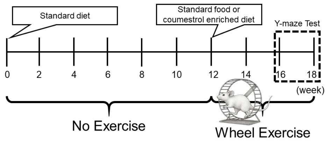
Figure 2.
Exercise and food consumption in SAMP1 mice from 12 to 18 weeks of age. The mice in the voluntary exercise group were placed on a running wheel, with the ability to rest, for 90 min (A). During the voluntary exercise time, the running distance was automatically measured by the wheel machine (B). The food ingestion (C), body weight (D), and calorie intake (E) for mice in all the experimental groups were measured. BS diet, bean-sprout-containing diet; NE, no exercise; ST, standard diet; VE, voluntary exercise. Values are means ± standard deviation, with n=6.
Figure 2.
Exercise and food consumption in SAMP1 mice from 12 to 18 weeks of age. The mice in the voluntary exercise group were placed on a running wheel, with the ability to rest, for 90 min (A). During the voluntary exercise time, the running distance was automatically measured by the wheel machine (B). The food ingestion (C), body weight (D), and calorie intake (E) for mice in all the experimental groups were measured. BS diet, bean-sprout-containing diet; NE, no exercise; ST, standard diet; VE, voluntary exercise. Values are means ± standard deviation, with n=6.
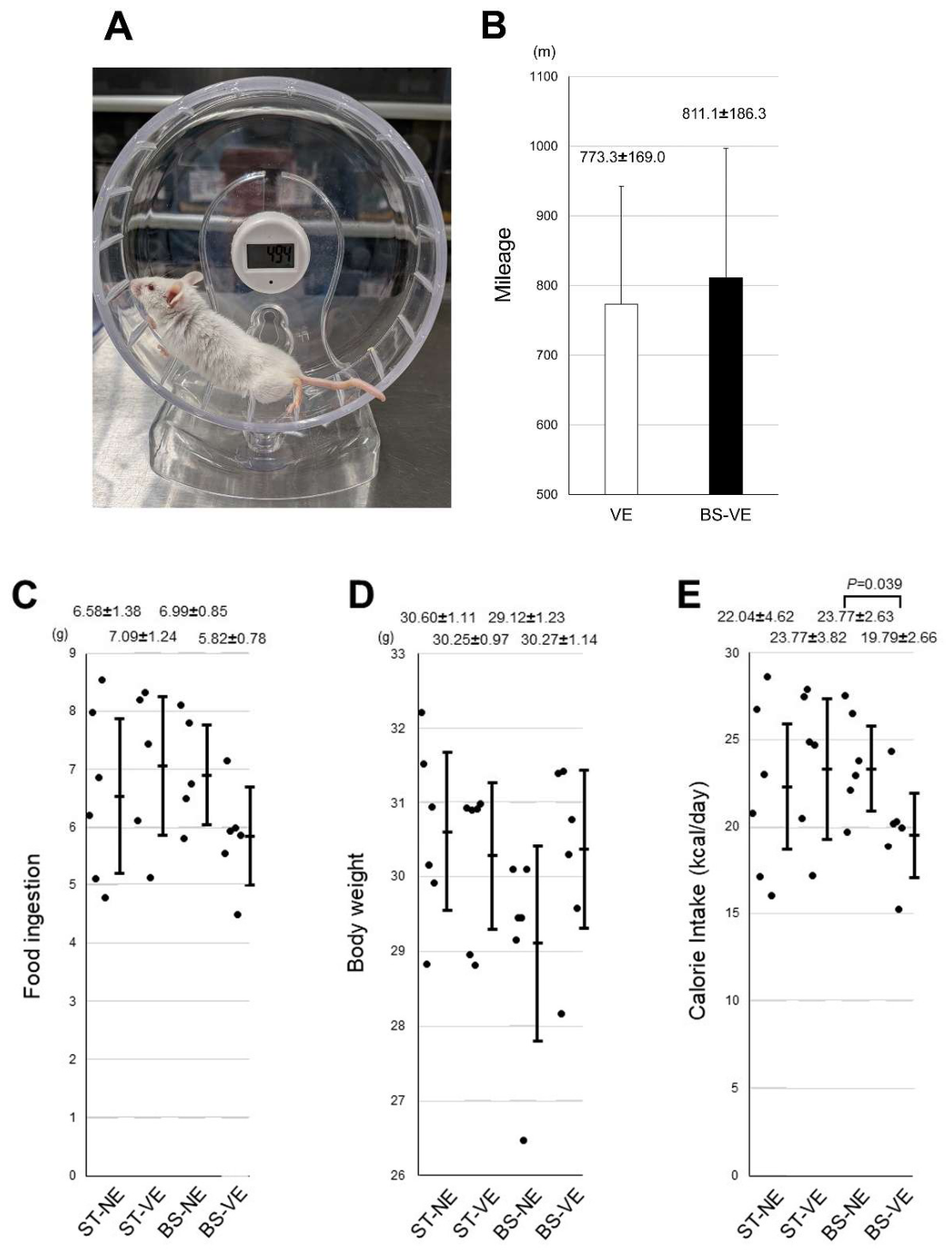
Figure 3.
Appearance of 18-week-old SAMP1 mice allowed voluntary exercise. The mice were fed a standard diet (upper set) or bean-sprout-containing diet (lower set) and had access to voluntary exercise. The mice fed the bean-sprout-containing diet had better-groomed coats than those fed the standard diet.
Figure 3.
Appearance of 18-week-old SAMP1 mice allowed voluntary exercise. The mice were fed a standard diet (upper set) or bean-sprout-containing diet (lower set) and had access to voluntary exercise. The mice fed the bean-sprout-containing diet had better-groomed coats than those fed the standard diet.
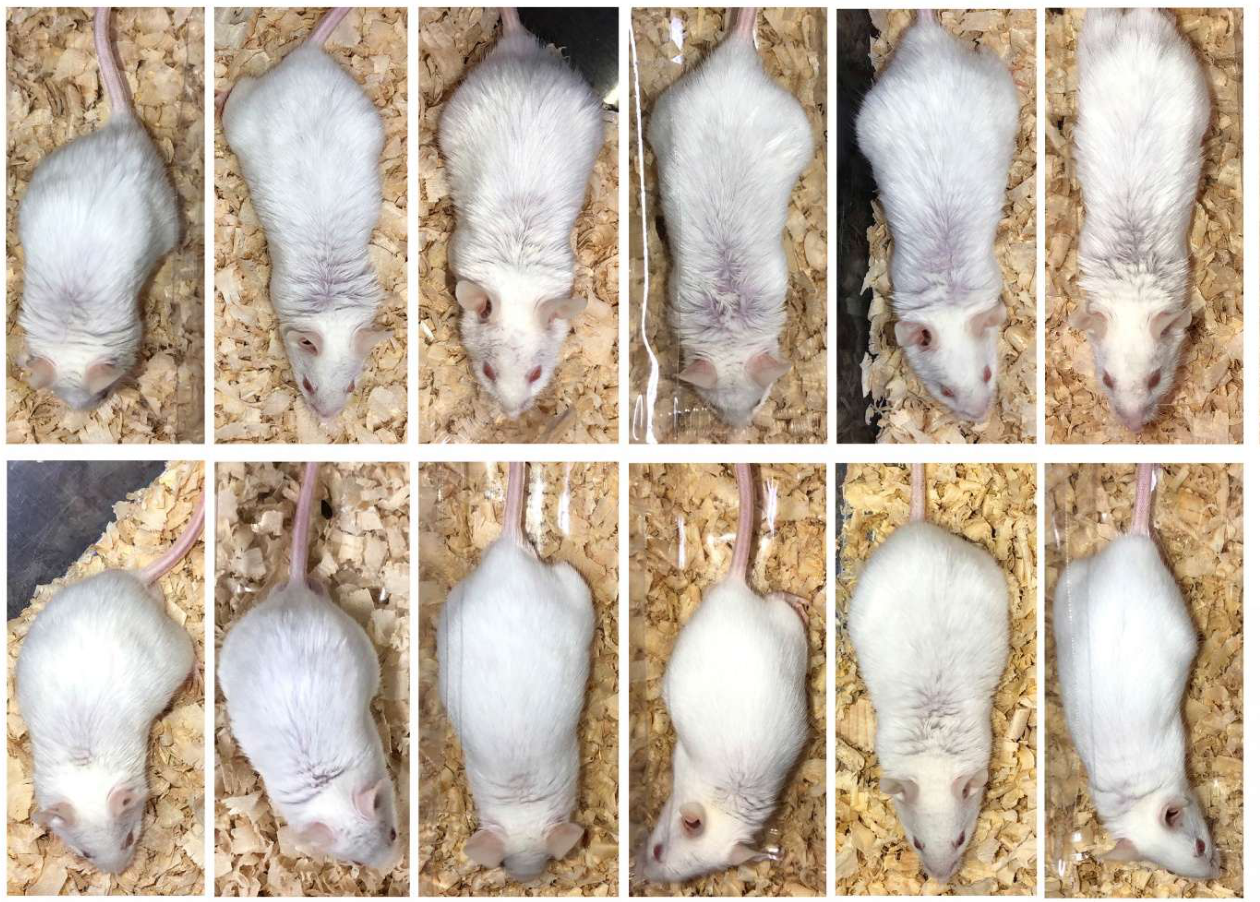
Figure 4.
Lifespan of the SAMP1 mice. The mice were fed a standard diet or bean-sprout-containing diet with no exercise. The mice fed the bean-sprout-containing diet had longer lifespans. To facilitate observation of lifespan, the mice were fed a standard diet or bean-sprout-containing diet with no exercise until death by old age.
Figure 4.
Lifespan of the SAMP1 mice. The mice were fed a standard diet or bean-sprout-containing diet with no exercise. The mice fed the bean-sprout-containing diet had longer lifespans. To facilitate observation of lifespan, the mice were fed a standard diet or bean-sprout-containing diet with no exercise until death by old age.
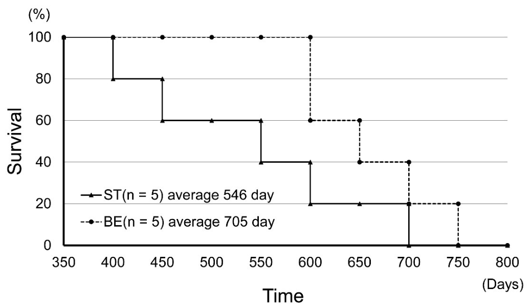
Figure 5.
Spatial working memory of the SAMP1 mice. The Y-maze test was conducted on mice fed either a standard diet or bean-sprout-containing diet, either with or without access to voluntary exercise. n = 6. P-values < 0.05 were considered statistically significant. BS, bean-sprout-containing diet; NE, no exercise; ST, standard diet; VE, voluntary exercise. Values are means ± standard deviations, with n=6.
Figure 5.
Spatial working memory of the SAMP1 mice. The Y-maze test was conducted on mice fed either a standard diet or bean-sprout-containing diet, either with or without access to voluntary exercise. n = 6. P-values < 0.05 were considered statistically significant. BS, bean-sprout-containing diet; NE, no exercise; ST, standard diet; VE, voluntary exercise. Values are means ± standard deviations, with n=6.
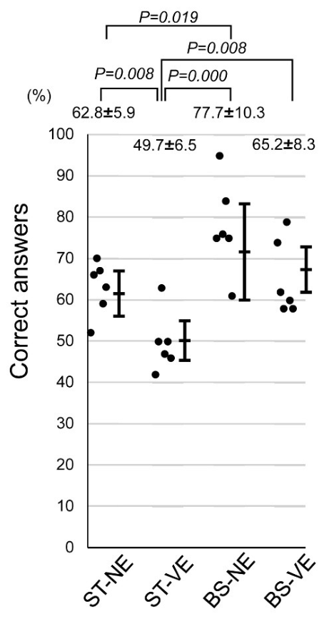
Figure 6.
Serum testosterone levels in SAMP1 mice. We collected blood samples from mice fed either a standard diet or bean-sprout-containing diet, either with or without access to voluntary exercise, to measure serum testosterone levels. n = 6. P-values < 0.05 were considered statistically significant. BS, bean-sprout-containing diet; NE, no exercise; ST, standard diet; VE, voluntary exercise. Values are means ± standard deviations, with n=6.
Figure 6.
Serum testosterone levels in SAMP1 mice. We collected blood samples from mice fed either a standard diet or bean-sprout-containing diet, either with or without access to voluntary exercise, to measure serum testosterone levels. n = 6. P-values < 0.05 were considered statistically significant. BS, bean-sprout-containing diet; NE, no exercise; ST, standard diet; VE, voluntary exercise. Values are means ± standard deviations, with n=6.
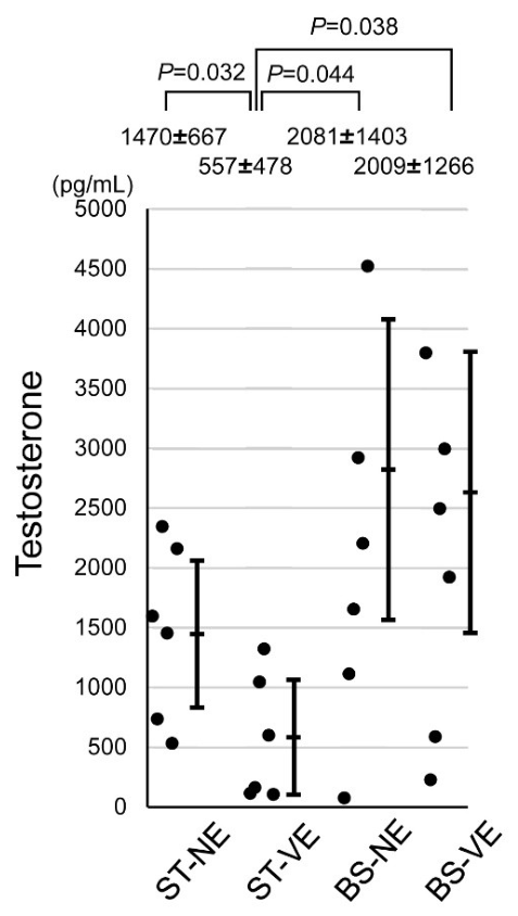
Figure 7.
Serum neurotrophin levels in the SAMP1 mice. We collected serum from and conducted Western blotting on mice fed either a standard diet or bean-sprout-containing diet, either with or without access to voluntary exercise, to measure neurotrophin levels. The ST-NE* mice were used to confirm the reproducibility of signals on each filter(A, B). The numbers represent individual mouse identification numbers. BDNF, GDNF, and NGF expression in mice measured using Western blotting (C, D, E). The signal intensity corresponds to the neurotrophin measured and quantified using the image-processing program Image Lab. Transferrin was used as a marker of serum lysates. The original Western blotting results are shown in the supplemental figures. n = 6. P-values < 0.05 were considered statistically significant. BS, bean-sprout-containing diet; NE, no exercise; NGF, nerve growth factor; ST, standard diet; VE, voluntary exercise. Values are means ± standard deviations, with n=6.
Figure 7.
Serum neurotrophin levels in the SAMP1 mice. We collected serum from and conducted Western blotting on mice fed either a standard diet or bean-sprout-containing diet, either with or without access to voluntary exercise, to measure neurotrophin levels. The ST-NE* mice were used to confirm the reproducibility of signals on each filter(A, B). The numbers represent individual mouse identification numbers. BDNF, GDNF, and NGF expression in mice measured using Western blotting (C, D, E). The signal intensity corresponds to the neurotrophin measured and quantified using the image-processing program Image Lab. Transferrin was used as a marker of serum lysates. The original Western blotting results are shown in the supplemental figures. n = 6. P-values < 0.05 were considered statistically significant. BS, bean-sprout-containing diet; NE, no exercise; NGF, nerve growth factor; ST, standard diet; VE, voluntary exercise. Values are means ± standard deviations, with n=6.
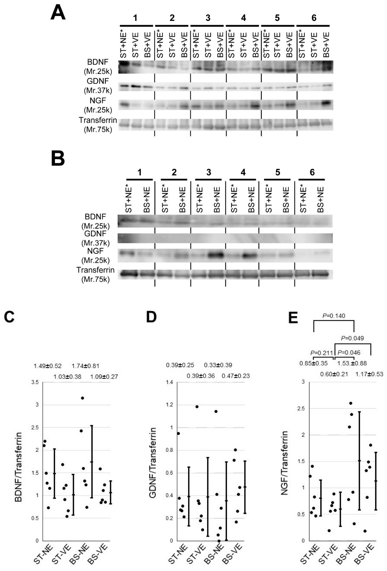

Table 1.
The antibodies and enzyme-linked immunosorbent assay kit used in the study.
 |
BDNF, brain-derived neurotrophic factor; ELISA, enzyme-linked immunosorbent assay; HRP, horseradish peroxidase; IgG, immunoglobulin G; GDNF, glial-cell-line-derived neurotrophic factor; NGF, nerve growth factor.
Table 2.
Serum neurotrophin concentrations, shown as the mean ± standard deviation.
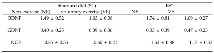 |
BDNF, brain-derived neurotrophic factor; BS, diet including 15% coumestrol-rich bean sprout powder in standard; GDNF, glial-cell-line-derived neurotrophic factor; N.D., no data; NGF, nerve growth factor. The numbers indicate the signal intensity relative to transferrin in each lane. Values are means ± standard deviations, with n=6.
Disclaimer/Publisher’s Note: The statements, opinions and data contained in all publications are solely those of the individual author(s) and contributor(s) and not of MDPI and/or the editor(s). MDPI and/or the editor(s) disclaim responsibility for any injury to people or property resulting from any ideas, methods, instructions or products referred to in the content. |
© 2026 by the authors. Licensee MDPI, Basel, Switzerland. This article is an open access article distributed under the terms and conditions of the Creative Commons Attribution (CC BY) license (http://creativecommons.org/licenses/by/4.0/).
Copyright: This open access article is published under a Creative Commons CC BY 4.0 license, which permit the free download, distribution, and reuse, provided that the author and preprint are cited in any reuse.


