

Submitted:
19 May 2026
Posted:
20 May 2026
You are already at the latest version
Abstract
Accurate donor-recipient allograft size matching remains a critical determinant of outcomes in lung transplantation, yet current approaches rely predominantly on predicted total lung capacity (pTLC) and height-based metrics derived from population-based equations. These simplified surrogates fail to capture individual anatomical variability, disease-specific alterations in thoracic geometry, and the spatial relationship between donor lungs and recipient chest cavities. In this review, we examine the limitations of conventional size matching and synthesize emerging evidence supporting imaging-based approaches, including computed tomography (CT) volumetry, radiomics, and machine learning. CT-derived volumetric analysis enables individualized anatomical assessment and has been associated with clinically relevant prediction of primary graft dysfunction and mortality. Advanced computational methods may further support extraction of imaging-derived features and integration with clinical data, although these approaches remain investigational. Collectively, these developments signal a paradigm shift from crude population-based metrics toward imaging-driven and computational approaches in the modern era. With rigorous validation and careful clinical integration, imaging-based approaches may complement conventional size metrics and support more individualized donor-recipient assessment.
Keywords:
lung transplantation
; size matching
; CT volumetry
; AI and machine learning
; segmentation
; outcomes
Introduction
Lung transplantation remains the definitive therapy for patients with end-stage pulmonary disease [1,2]. However, the limited availability of donor lungs remains a major barrier, with waitlist demand continuing to outpace organ availability [3,4]. This constraint underscores the need to use each donor organ effectively while maintaining acceptable post-transplant outcomes. A key modifiable factor is donor-recipient allograft size matching, as mismatch introduces physiologic strain that adversely affects both perioperative outcomes and long-term survival [5]. Early size-matching strategies relied on simple measures such as donor-recipient height differences [6,7]. This approach offered a simple and pragmatic means of approximating donor-recipient size; however, it is a crude surrogate that fails to reliably match donor and recipient size [7]. Given these limitations, size matching subsequently evolved to using predicted total lung capacity (pTLC) [6,7]. pTLC is typically calculated using European Respiratory Society reference equations: 7.99 × Height (m) − 7.08 for males and 6.60 × Height (m) − 5.79 for females, with size matching expressed as pTLC ratio, defined as the ratio between donor to recipient pTLC [8]. Recipients are commonly classified as undersized, size-matched, or oversized based on pTLC ratio thresholds [9,10]. While this provides a practical method for estimating lung volumes in both donors and recipients, these formulas are population-derived, do not reflect true chest cavity size, and are often unreliable in cases of chronic lung disease [11,12].
The relationship between size mismatch and transplant outcomes is disease-specific, with both undersizing and oversizing associated with adverse outcomes depending on the underlying pathology [13,14]. Undersizing results in increased mechanical stress and overdistension of smaller grafts within a relatively larger thoracic space [5,15]. This has been linked to increased perioperative morbidity, including primary graft dysfunction (PGD), prolonged mechanical ventilation, airway complications, and longer hospital length of stay [15]. Conversely, oversizing may impair chest wall mechanics, limit graft expansion, and increase intrathoracic pressures, potentially contributing to complications such as PGD and the need for delayed chest closure or graft reduction. The impact of oversizing has been associated with increased rates of PGD, higher perioperative mortality, and worse long-term survival [16,17]. These limitations are particularly important in the setting of limited donor organ availability and underscore the need for more accurate approaches to donor-recipient size matching to optimize outcomes for each transplanted allograft.
Novel approaches to lung size matching have emerged to address these limitations. Advances in imaging have enabled the use of computed tomography (CT) derived volumetry to directly quantify lung volume and thoracic dimensions, providing a patient-specific assessment of donor-recipient compatibility [11,12]. Unlike pTLC-based estimates, CT volumetry reflects true anatomic constraints and may better capture donor-recipient allograft size matching [12]. Additionally, CT volumetry combined with emerging artificial intelligence (AI)-based models may offer a more precise, data-driven approach to donor-recipient size matching by directly assessing lung volume and thoracic constraints. This review examines current lung sizing strategies, their limitations, and emerging imaging-based approaches to improve donor-recipient matching.
Clinical Consequences of Size Mismatch
Size mismatch in lung transplantation has broad clinical implications, affecting not only graft function, but also perioperative morbidity, resource utilization, and long-term outcomes [14,15,16]. The clinical impact varies by direction, degree of mismatch, and underlying disease. PGD is one of the most immediate and clinically significant complications following lung transplantation, occurring in 15–25% of recipients and strongly associated with increased ICU length of stay and both 90-day and 1-year mortality [16,18,19]. Size mismatch imposes mechanical and physiologic stress on the transplanted lung, increasing susceptibility to early graft dysfunction. Excessive mismatch may influence PGD risk in both directions, with prior studies reporting increased risk among recipients of both undersized and oversized allografts [5,16,20,21]. Beyond early graft injury, size mismatch has additional important post-transplant implications. Undersizing has been associated with increased airway complications, including airway dehiscence and higher tracheostomy rates, while larger degrees of mismatch have been linked to increased risk of CLAD and reduced long-term survival [6,15,20]. Notably, size discrepancies greater than 20% have emerged as independent predictors of the composite outcome of CLAD and mortality, underscoring the importance of appropriate donor-recipient matching [14,20].
The mechanisms underlying these complications differ depending on the direction of mismatch, in addition to specific disease processes. Undersizing results in increased mechanical stress and overdistension of smaller grafts within a relatively larger thoracic space, contributing to ventilator dependence, airway complications, prolonged recovery, and increased healthcare resource utilization, including longer hospitalizations and higher costs, with index hospitalization charges approximately $18,000 greater in undersized recipients ($176,247 vs. $158,492) [5,14]. Conversely, oversizing impairs chest wall mechanics, limits graft expansion, and increases intrathoracic pressures, leading to restricted ventilation, primary graft dysfunction, and the need for delayed chest closure or graft reduction. The clinical impact of mismatch appears most pronounced at greater degrees of discrepancy, where excessive undersizing or oversizing is associated with worse early outcomes and reduced long-term survival [14].
These effects are particularly evident in restrictive lung disease, where both undersizing and excessive oversizing have been associated with increased mortality [15,16]. Emerging data further suggest that CT-based volumetric assessment may better predict complications such as PGD and mortality, highlighting the limitations of traditional size-matching approaches [5]. However, the relationship between early graft injury and long-term outcomes is complex and non-linear. Recent analyses demonstrate that even severe early injury, such as PGD grade 3 (PGD3), may not alone predict long-term survival after adjustment for recipient, donor, and post-transplant factors. Instead, downstream complications, including dialysis and treated rejection, appear to play a more dominant role in determining long-term outcomes among patients who survive the index hospitalization [19]. These findings suggest that the impact of donor-recipient allograft size mismatch on long-term outcomes is indirect and incompletely captured by conventional global metrics, which do not account for the complex, multifactorial pathways linking early physiologic stress to long-term survival.
Disease-Specific Complexity
The clinical impact of size mismatch varies with underlying disease [17,22]. A central limitation of current approaches is that pTLC-based sizing does not account for true thoracic dimensions, which vary substantially across disease states [11]. Patients with chronic obstructive pulmonary disease (COPD) typically have hyperinflated lungs within expanded thoracic cavities, whereas those with pulmonary fibrosis and other restrictive diseases have contracted lungs within a diminished chest cage. These disease-specific patterns of thoracic remodeling are dynamic and may partially reverse following transplantation, highlighting the limitations of static, equation-based estimates. As a result, identical pTLC ratios may have opposing physiologic and clinical implications depending on the underlying diagnosis, underscoring that a uniform approach to size matching is inherently limited [12,13].
In COPD, oversizing is generally well tolerated and may be beneficial. Oversized grafts (pTLC ratio ≥ 1.1) have been associated with improved graft survival, with incremental increases in pTLC ratio linked to reduced early mortality [20]. In contrast, patients with pulmonary fibrosis and other restrictive lung diseases demonstrate the opposite pattern, where oversizing is associated with worse outcomes [12,16]. Oversized grafts in this population have been associated with higher rates of primary graft dysfunction, increased in-hospital mortality, and reduced long-term survival compared with size-matched or undersized graft. Notably, both undersizing (pTLC <0.8) and moderate oversizing (pTLC 1.1–1.2) have been associated with increased mortality in restrictive disease, suggesting a narrow optimal range for donor–recipient size matching [16]. Collectively, these findings highlight that the relationship between graft size and outcomes is highly disease-specific and that reliance on pTLC-based metrics alone may fail to capture these critical physiologic differences.
Emergence of CT Volumetry
Conventional approaches to lung size matching rely on simplified anthropometric estimates such as pTLC and height-based ratios. More recently, imaging-based and computational methods have expanded this framework to enable increasingly individualized assessment of donor-recipient compatibility (Table 1). CT volumetry represents a shift away from equation-based estimates toward direct, patient-specific measurement of lung volume [11,12]. This is particularly important, as size matching is fundamentally an anatomic and physiologic problem, yet current approaches rely on equations derived from variables such as height, sex, and age that do not fully capture individual thoracic dimensions or disease-specific alterations in lung mechanics. By incorporating direct assessment of lung volume and thoracic cage dimensions, CT volumetry enables a more precise evaluation of donor-recipient compatibility, particularly as identical pTLC ratios may have markedly different physiologic implications across disease states. Accordingly, CT volumetry may offer improved accuracy compared with pTLC, particularly when accounting for variation in underlying disease [11,12].
These differences are most apparent in disease-specific contexts. Recent studies in patients with restrictive lung disease suggest that CT-derived volumes may more accurately reflect true lung size and thoracic cage dimensions compared with pTLC-based estimates [23]. Regardless of whether sizing is based on height, pTLC, or CT-derived volumes, mismatch in either direction is associated with adverse outcomes [5,12,16]. Undersizing has been linked to increased risk of primary graft dysfunction, respiratory insufficiency, and mortality, while oversizing has similarly been associated with higher rates of PGD, impaired chest wall mechanics, and worse survival [5,6,15]. These associations have been demonstrated across registry analyses, single-center cohorts, and CT-based studies, particularly in restrictive lung disease populations. Collectively, these findings suggest that the relationship between size mismatch and outcomes is not unidirectional, but instead reflects a narrow optimal window of compatibility that current one-dimensional sizing metrics incompletely capture [11,12].
From a practical standpoint, CT volumetry is increasingly feasible, particularly on the recipient side, because chest CT imaging is commonly obtained during transplant evaluation. Donor CT imaging is also becoming more readily available with evolving donor assessment protocols. Advances in segmentation software allow for reliable volumetric assessment in the vast majority of cases. Importantly, early clinical experience suggests that CT-based assessment may facilitate more confident donor selection, including the acceptance of organs that might otherwise be declined based on pTLC estimates alone. As such, CT volumetry has the potential to improve donor-recipient matching and expand the effective donor pool.
Advanced Segmentation and Radiomics
Beyond simple volumetric measurements, advanced image segmentation and radiomic analysis enable the extraction of high-dimensional quantitative features from routine CT imaging, including lobar volumes, pulmonary vessel volume, parenchymal density patterns, and markers of small airway disease [24,25,26]. These advances enable a transition toward multidimensional characterization of lung structure and function (Figure 1).
Machine learning-based radiomics has demonstrated superior performance compared to conventional imaging assessment in detecting allograft injury and predicting clinically relevant transplant outcomes. In preclinical models, radiomic analysis has shown markedly improved accuracy for detecting allograft rejection compared to standard uptake value measurements [27]. In clinical lung transplantation, CT-based machine learning tools have successfully quantified features such as ground-glass opacity, reticulation, and pulmonary vessel volume, with the latter emerging as a strong predictor of restrictive allograft syndrome and graft failure [25].
More recently, deep learning approaches have begun to integrate donor lung CT imaging directly into predictive models of transplant outcomes. When combined with clinical data, these models demonstrate improved performance compared to clinical variables alone, highlighting the incremental value of imaging-derived features in risk stratification [28]. Importantly, CT-based machine learning approaches have also been shown to identify structural abnormalities not captured by conventional assessment and to stratify recipients at markedly increased risk of adverse outcomes, including intensive care unit (ICU) stay and chronic lung allograft dysfunction [29]. Together, these findings highlight the subjectivity and limitations of current donor evaluation practices and suggest that imaging-based phenotyping may provide complementary information for risk stratification.
Machine Learning and Artificial Intelligence Applications
Machine learning and AI offer transformative potential across lung transplantation, particularly in donor-recipient matching and outcome prediction. Recent literature has demonstrated that machine learning approaches, including random forests, support vector machines, and neural networks, consistently outperform traditional statistical methods in modeling complex transplant-related outcomes [30,31,32,33,34]. The advantages of machine learning are particularly relevant in transplantation, where outcomes are driven by highly nonlinear interactions between donor, recipient, and procedural factors. In thoracic surgery, machine learning models have demonstrated strong predictive performance for postoperative outcomes following complex procedures such as esophagectomy, with neural networks and ensemble methods showing improved performance over conventional regression approaches [35,36]. These findings highlight the limitations of traditional models in capturing multifactorial risk, which directly parallels the inadequacy of simplified metrics such as pTLC in donor-recipient size matching.
Importantly, machine learning frameworks enable integration of diverse data types, including clinical variables, imaging features, and emerging molecular markers. For example, random survival forest models have demonstrated superior discrimination and calibration compared to conventional Cox regression in predicting post-transplant survival [37]. Similarly, hybrid models incorporating multimodal data have demonstrated reliable predictive performance across short- and long-term outcomes, with good calibration and demonstrated clinical utility in decision curve analyses [38]. Recent work has begun to explore AI-assisted evaluation of donor lung CT imaging for donor assessment and transplant decision-making, further underscoring the rapid emergence of imaging-based approaches [39]. Collectively, these advances underscore the potential of AI-driven approaches to move beyond static, one-dimensional metrics toward more dynamic, personalized models of donor-recipient compatibility.
Integration of Multi-Modal Data
Building upon the limitations of one-dimensional size metrics, the future of lung size matching likely lies in integrating CT volumetry, radiomic features, and machine learning into unified, multidimensional predictive models. Such approaches enable a shift from simplified global estimates toward more comprehensive assessments of donor-recipient compatibility. AI-driven models can incorporate imaging, clinical variables, and emerging molecular data to improve donor evaluation, organ allocation, and outcome prediction [33].
Across the transplant continuum, these tools enable more precise and individualized decision-making. In the pre-transplant phase, machine learning enhances donor-recipient matching through objective imaging analysis and risk stratification. Throughout the peri- and post-transplant periods, predictive models identify patients at risk for complications such as PGD and CLAD, facilitating earlier intervention and personalized management strategies [33,40,41].
Multimodal frameworks enable the integration of heterogeneous data types, overcoming the limitations of traditional models that rely on isolated parameters. Techniques such as transfer learning support model development in data-limited settings, while advances in automated segmentation and feature extraction facilitate scalable implementation of imaging-based analytics [28,34,42].
Multimodal integration enables lung size matching to incorporate complementary dimensions of graft suitability, including regional lung volume distribution, parenchymal quality, vascular architecture, and recipient-specific physiologic demand. These approaches may eventually complement conventional size metrics by incorporating additional anatomic and clinical information, although their role in routine donor-recipient matching remains to be defined.
Barriers to Clinical Implementation
Despite promising results, several barriers limit the clinical adoption of AI-driven approaches in lung transplantation. These challenges span technical, methodological, and clinical domains (Table 2). From a technical standpoint, the availability and quality of data remain major limitations. Many models are developed using small, single-center datasets with substantial heterogeneity in data acquisition and processing. For CT-based approaches, variability in imaging protocols, the need for standardized segmentation, and reliance on specialized software further complicate implementation [12,23,33,43]. Although CT-derived lung volumes can be obtained in most recipients, donor imaging is not as readily available, necessitating reliance on predictive equations rather than direct measurement, which may introduce additional variability [44,45].
Methodologically, limitations in model development and validation hinder generalizability. Many studies lack external validation, use inconsistent performance metrics, and provide limited insight into model interpretability. More broadly, the lack of standardized approaches to model development, reporting, and validation limits comparability and clinical translation [36]. These challenges are further compounded by variability in institutional workflows and differences in resource availability across transplant centers, which may limit the scalability of AI-driven approaches. Broader clinical and ethical considerations present additional obstacles. Issues such as data privacy, regulatory compliance, interoperability across healthcare systems, and algorithmic bias must be addressed to ensure safe and equitable implementation [31,42]. Centralized donor assessment models, including regional procurement or perfusion centers, may create opportunities for more standardized imaging acquisition and advanced allograft evaluation, but their feasibility, cost, and workflow implications require careful study [46,47,48]. Collectively, these barriers underscore that despite rapid advances, translating AI-driven lung size matching into routine clinical practice will require not only technical innovation but also rigorous validation, standardization, and thoughtful integration into existing clinical workflows, as AI models must adapt in tandem with ongoing changes in transplant practice.
Conclusions
Conventional approaches to lung size matching, based on pTLC and anthropometric ratios, remain fundamentally limited by their reliance on simplified population-based estimates that fail to capture the complex anatomic and physiologic variability underlying graft function after transplantation. These limitations contribute to clinically meaningful size mismatch and are associated with adverse outcomes, particularly in patients with restrictive lung disease. Advances in CT volumetry, radiomic analysis, and machine learning offer a paradigm shift toward a more individualized and comprehensive assessment of donor-recipient compatibility. By incorporating regional lung characteristics, parenchymal features, and recipient-specific physiologic demand, these approaches may broaden size matching from a single global estimate toward a more individualized assessment. However, despite promising early results, significant challenges remain, including variability in data quality, limited availability of donor imaging, lack of standardized model development and validation frameworks, and barriers to integration within clinical workflows. Future efforts should prioritize multicenter validation, standardized methodologies, and the development of scalable tools that can be integrated into routine practice. In parallel, addressing issues related to data sharing, model interpretability, and ethical implementation will be essential to ensure equitable and reliable adoption. The transition from global size metrics toward imaging-informed assessment represents an important conceptual advance in donor-recipient matching. Realizing its clinical value will require multicenter validation, standardized imaging and segmentation workflows, interpretable models, and implementation strategies that fit the time-sensitive realities of lung transplantation.
Disclosure Information
MCH is a consultant and speaker for Atricure. DAG serves on the Thoracic Transplant Committee of the ASTS. BAW serves on the Transmedics OCS events committee.
Funding Statement
This research was generously supported through The Jewel and Frank Benson Family Endowment and The Jewel and Frank Benson Research Professorship.
Abbreviations
| AI | Artificial Intelligence |
| CLAD | Chronic Lung Allograft Dysfunction |
| COPD | Chronic Obstructive Pulmonary Disease |
| CT | Computed Tomography |
| PGD | Primary Graft Dysfunction |
| pTLC | Predicted Total Lung Capacity |
References
- Briganti, D.F.; D’Ovidio, F. Long-term management of patients with end-stage lung diseases. Best Pract. Res. Clin. Anaesthesiol. 2017, 31, 167–178. [Google Scholar] [CrossRef]
- Hartert, M.; Senbaklavacin, O.; Gohrbandt, B.; Fischer, B.M.; Buhl, R.; Vahld, C.F. Lung transplantation: A treatment option in end-stage lung disease. Dtsch. Arztebl. Int. 2014, 111, 107–116. [Google Scholar] [CrossRef]
- Young, K.A.; Dilling, D.F. The Future of Lung Transplantation. Chest 2019, 155, 465–473. [Google Scholar] [CrossRef]
- Christie, J.D.; Raemdonck, D.V.; Fisher, A.J. Lung Transplantation. N. Engl. J. Med. 2024, 391, 1822–1836. [Google Scholar] [CrossRef]
- Eberlein, M.; Arnaoutakis, G.J.; Yarmus, L.; et al. The effect of lung size mismatch on complications and resource utilization after bilateral lung transplantation. J. Heart Lung Transplant. 2012, 31, 492–500. [Google Scholar] [CrossRef]
- George, P.M.; Patterson, C.M.; Reed, A.K.; Thillai, M. Lung transplantation for idiopathic pulmonary fibrosis. Lancet Respir. Med. 2019, 7, 271–282. [Google Scholar] [CrossRef]
- Ouwens, J.P.; van der Mark, T.W.; van der Bij, W.; Geertsma, A.; de Boer, W.J.; Koëter, G.H. Size matching in lung transplantation using predicted total lung capacity. Eur. Respir. J. 2002, 20, 1419–1422. [Google Scholar] [CrossRef]
- Stocks, J.; Quanjer, P.H. Reference values for residual volume, functional residual capacity and total lung capacity. ATS Workshop on Lung Volume Measurements. Official Statement of The European Respiratory Society. Eur. Respir. J. 1995, 8, 492–506. [Google Scholar] [CrossRef]
- Barnard, J.B.; Davies, O.; Curry, P.; et al. Size matching in lung transplantation: An evidence-based review. J. Heart Lung Transplant. 2013, 32, 849–860. [Google Scholar] [CrossRef]
- Eberlein, M.; Reed, R.M.; Permutt, S.; et al. Parameters of donor-recipient size mismatch and survival after bilateral lung transplantation. J. Heart Lung Transplant. 2012, 31, 1207–1213.e7. [Google Scholar] [CrossRef]
- McMenimen, J.D.; Gauthier, J.M.; Puri, V.; Vazquez Guillamet, R. “Horses for courses” computed tomography or predicted total lung capacity for size matching in lung transplantation. Am. J. Transplant. 2024, 24, 928–932. [Google Scholar] [CrossRef]
- Prabhu, N.K.; Wong, M.K.; Klapper, J.A.; et al. Computed Tomography Volumetrics for Size Matching in Lung Transplantation for Restrictive Disease. Ann. Thorac. Surg. 2024, 117, 413–421. [Google Scholar] [CrossRef]
- Schaffer, J.M.; Singh, S.K.; Reitz, B.A.; Zamanian, R.T.; Mallidi, H.R. Single- vs Double-Lung Transplantation in Patients With Chronic Obstructive Pulmonary Disease and Idiopathic Pulmonary Fibrosis Since the Implementation of Lung Allocation Based on Medical Need. JAMA 2015, 313, 936–948. [Google Scholar] [CrossRef]
- Riddell, P.; Ma, J.; Dunne, B.; et al. A simplified strategy for donor-recipient size-matching in lung transplant for interstitial lung disease. J. Heart Lung Transplant. 2021, 40, 1422–1430. [Google Scholar] [CrossRef]
- Ganapathi, A.M.; Mulvihill, M.S.; Englum, B.R.; et al. Transplant size mismatch in restrictive lung disease. Transpl. Int. 2017, 30, 378–387. [Google Scholar] [CrossRef]
- Franz, M.; Tavil, S.; de Manna, N.D.; et al. Oversizing lung allografts deteriorates outcomes in patients with pulmonary fibrosis. J. Heart Lung Transplant. 2024, 43, 1126–1134. [Google Scholar] [CrossRef]
- Gouchoe, D.A.; Botros, M.M.; Satija, D.; et al. Adaptive Chest Cavity Expansion Following Lung Allograft Oversizing in Idiopathic Pulmonary Fibrosis. J. Surg. Res. 2025, 305, 295–303. [Google Scholar] [CrossRef]
- Gouchoe, D.A.; Cui, E.Y.; Satija, D.; et al. Ex Vivo Lung Perfusion and Primary Graft Dysfunction Following Lung Transplantation: A Contemporary United Network for Organ Sharing Database Analysis. J. Clin. Med. 2024, 13, 4440. [Google Scholar] [CrossRef]
- Gouchoe, D.A.; Whitson, B.A.; Rosenheck, J.; et al. Long-Term Survival Following Primary Graft Dysfunction Development in Lung Transplantation. J. Surg. Res. 2024, 296, 47–55. [Google Scholar] [CrossRef]
- Poo-Fernández, C.; Iturbe-Fernández, D.; Tello-Mena, S.; et al. The impact of donor-recipient size mismatch on lung transplant outcomes. J. Thorac. Dis. 2025, 17, 4621–4632. [Google Scholar] [CrossRef]
- Park, B.S.; Kim, D.J.; Lee, C.Y.; et al. Outcomes of Donor/Recipient Size-mismatched Lung Transplantation. Transplant. Proc. 2025, 57, 868–875. [Google Scholar] [CrossRef]
- Yu, W.S.; Park, C.H.; Paik, H.C.; et al. Changes in Thoracic Cavity Volume After Bilateral Lung Transplantation. Front. Med. 2022, 9, 881119. [Google Scholar] [CrossRef]
- Hussain, M.; Thornton, M.; Hussain, T.; et al. Evaluating the Use of CT-Derived Lung Volumes in Donor-Recipient Lung Size Matching for Lung Transplantation in Patients With Interstitial Lung Disease and/or Idiopathic Pulmonary Fibrosis. Transplant. Proc. 2023, 55, 623–628. [Google Scholar] [CrossRef]
- Calvert, A.D.; Hazelton, T.R. Role of imaging in lung transplantation evaluation. J. Thorac. Dis. 2020, 12, 5147–5158. [Google Scholar] [CrossRef]
- McInnis, M.C.; Ma, J.; Karur, G.R.; et al. Chronic lung allograft dysfunction phenotype and prognosis by machine learning CT analysis. Eur. Respir. J. 2022, 60, 2101652. [Google Scholar] [CrossRef]
- Zhang, X.; Zhang, Y.; Zhang, G.; et al. Deep Learning With Radiomics for Disease Diagnosis and Treatment: Challenges and Potential. Front. Oncol. 2022, 12, 773840. [Google Scholar] [CrossRef]
- Tian, D.; Shiiya, H.; Takahashi, M.; et al. Noninvasive monitoring of allograft rejection in a rat lung transplant model: Application of machine learning-based (18)F-fluorodeoxyglucose positron emission tomography radiomics. J. Heart Lung Transplant. 2022, 41, 722–731. [Google Scholar] [CrossRef]
- Ma, W.; Oh, I.; Luo, Y.; et al. Developing approaches to incorporate donor-lung computed tomography images into machine learning models to predict severe primary graft dysfunction after lung transplantation. Am. J. Transplant. 2025, 25, 1339–1349. [Google Scholar] [CrossRef]
- Ram, S.; Verleden, S.E.; Kumar, M.; et al. Computed tomography-based machine learning for donor lung screening before transplantation. J. Heart Lung Transplant. 2024, 43, 394–402. [Google Scholar] [CrossRef]
- Dueñas-Jurado, J.M.; Gutiérrez, P.A.; Casado-Adam, A.; et al. New models for donor-recipient matching in lung transplantations. PLoS ONE 2021, 16, e0252148. [Google Scholar] [CrossRef]
- Duncheskie, R.P.; Omari, O.A.; Anjum, F. Role of Artificial Intelligence in Lung Transplantation: Current State, Challenges, and Future Directions. Transplant. Proc. 2025, 57, 1621–1626. [Google Scholar] [CrossRef]
- Gholamzadeh, M.; Abtahi, H.; Safdari, R. Machine learning-based techniques to improve lung transplantation outcomes and complications: A systematic review. BMC Med. Res. Methodol. 2022, 22, 331. [Google Scholar] [CrossRef]
- Luo, W.; Tang, G.; Yang, F.; et al. Transforming lung transplantation with artificial intelligence: A narrative review from organ allocation to post-transplant management. J. Thorac. Dis. 2026, 18, 170. [Google Scholar] [CrossRef]
- Vercauteren, B.; Özsoy, B.; Gielen, J.; et al. Understanding Machine Learning Applications in Lung Transplantation: A Narrative Review. Transpl. Int. 2025, 38, 15640. [Google Scholar] [CrossRef]
- Bellini, V.; Valente, M.; Del Rio, P.; Bignami, E. Artificial intelligence in thoracic surgery: A narrative review. J. Thorac. Dis. 2021, 13, 6963–6975. [Google Scholar] [CrossRef]
- Satija, D.; Aly, A.; Dimitrov, T.; et al. The Cutting Edge: A Systematic Review of Artificial Intelligence and Machine Learning in Predicting Esophagectomy Outcomes. Ann. Thorac. Surg. Short. Rep. 2026. [Google Scholar] [CrossRef]
- Tian, D.; Yan, H.-J.; Huang, H.; et al. Machine Learning–Based Prognostic Model for Patients After Lung Transplantation. JAMA Netw. Open 2023, 6, e2312022. [Google Scholar] [CrossRef]
- Sharma, G.; Kamal, V.K.; Bollineni, S.; et al. Development and Validation of a Hybrid Machine Learning Model to Predict Lung Transplant Outcomes. JAMA Netw. Open 2025, 8, e2545369. [Google Scholar] [CrossRef]
- Daescu, O.; Yasmeen, A.; Raftopoulos, C.; et al. Artificial Intelligence—Assisted Evaluation of Donor Lung CTs: Optimizing Thoracic Allocation. J. Heart Lung Transplant. 2026, 45, 25–26. [Google Scholar] [CrossRef]
- Gotlieb, N.; Azhie, A.; Sharma, D.; et al. The promise of machine learning applications in solid organ transplantation. npj Digit Med. 2022, 5, 89. [Google Scholar] [CrossRef]
- Ronen, L.; Keshavjee, S.; Sage, A.T. Advancing lung transplantation through machine learning and artificial intelligence. Curr. Opin. Pulm. Med. 2025, 31, 381–386. [Google Scholar] [CrossRef]
- Olawade, D.B.; Marinze, S.; Qureshi, N.; Weerasinghe, K.; Teke, J. The impact of artificial intelligence and machine learning in organ retrieval and transplantation: A comprehensive review. Curr. Res. Transl. Med. 2025, 73, 103493. [Google Scholar] [CrossRef] [PubMed]
- Vazquez Guillamet, R.; Rjob, A.; Bierhals, A.; et al. Potential Role of Computed Tomography Volumetry in Size Matching in Lung Transplantation. Transplant. Proc. 2023, 55, 432–439. [Google Scholar] [CrossRef]
- Konheim, J.A.; Kon, Z.N.; Pasrija, C.; et al. Predictive equations for lung volumes from computed tomography for size matching in pulmonary transplantation. J. Thorac. Cardiovasc Surg. 2016, 151, 1163–1169.e1. [Google Scholar] [CrossRef] [PubMed]
- Gouchoe, D.A.; Cardenas, A.; Marklin, G.F.; et al. Promoting utilization of donors after circulatory death in lung transplantation. JHLT Open 2026, 12, 100491. [Google Scholar] [CrossRef]
- Walsh, M.G.; Cui, E.Y.; Dimitrov, T.; et al. External Perfusion Centers Can Bridge the Gap of Experience During Ex Vivo Lung Perfusion: A United Network for Organ Sharing Database Study. ASAIO J. 2025, 72, 418–425. [Google Scholar] [CrossRef] [PubMed]
- Gouchoe, D.A.; Satija, D.; Cui, E.Y.; et al. Extended ischemic times during ex vivo lung perfusion is not associated with increased mortality. Artif. Organs 2024, 48, 1458–1466. [Google Scholar] [CrossRef]
- Gouchoe, D.A.; Satija, D.; Cui, E.Y.; et al. The impact of ex vivo lung perfusion location on lung transplant outcomes. Artif. Organs 2024, 48, 1467–1475. [Google Scholar] [CrossRef] [PubMed]
Figure 1.
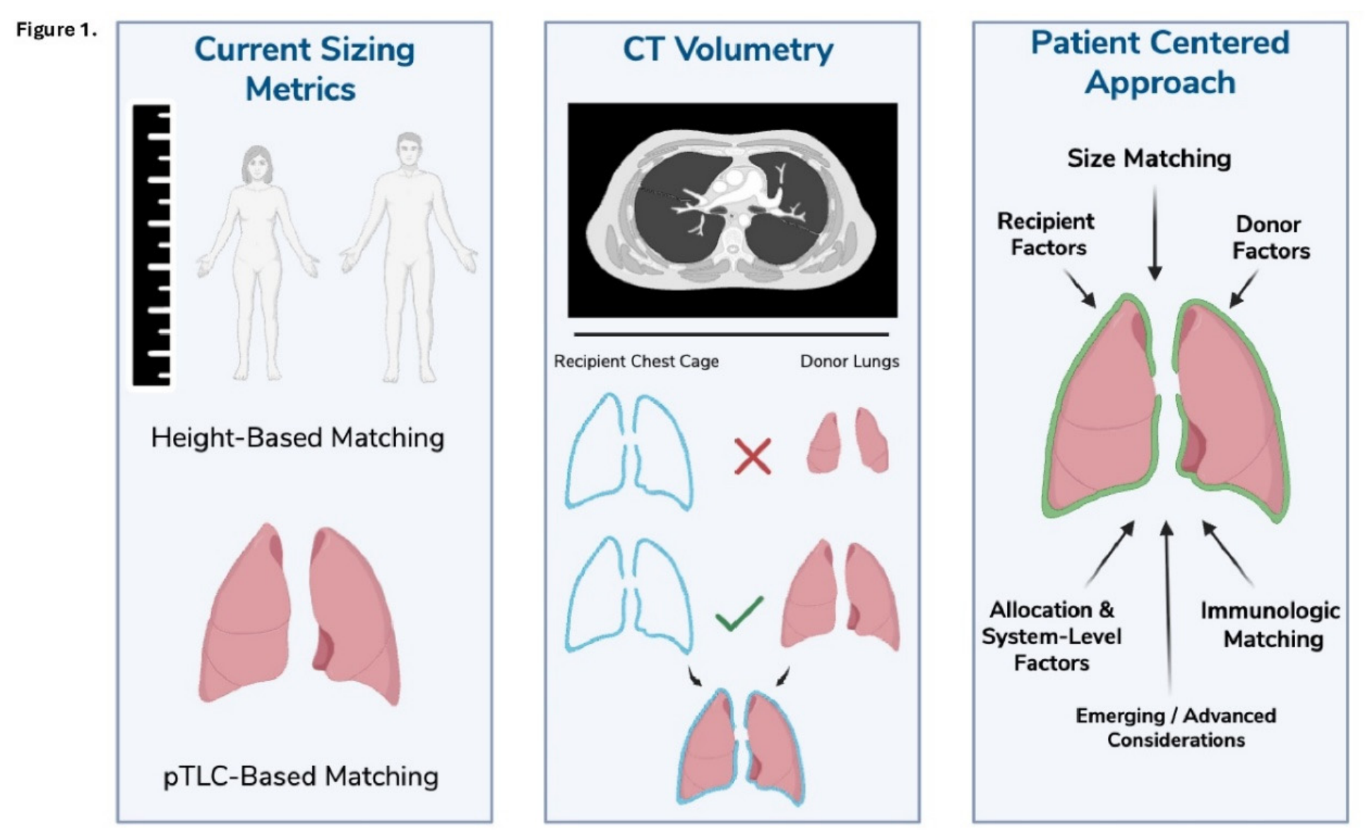

Table 1.
Evolution of size matching approaches from conventional metrics to data-driven methods.
| Approach | Data Utilized | Strengths | Limitations | Clinical Status |
|---|---|---|---|---|
| pTLC/Height | Anthropometric | Simple; widely available | Poor individualization | Standard |
| CT volumetry | Imaging | Patient-specific anatomic assessment | Donor imaging limited availability | Emerging |
| Radiomics | Imaging features | Captures regional heterogeneity | High complexity; limited standardization | Investigational |
| Machine-Learning Integration | Multimodal (imaging + clinical) | Enables personalized prediction | Requires validation and interpretability | Experimental |
Table 2.
Key barriers to clinical implementation of AI-driven approaches in lung transplantation.
| Domain | Barrier | Example |
|---|---|---|
| Technical | Data heterogeneity | CT protocol variability |
| Methodologic | Lack of validation | Single-center studies |
| Clinical | Workflow integration | EMR compatability |
| Ethical | Bias, consent | Underrepresented populations |
Disclaimer/Publisher’s Note: The statements, opinions and data contained in all publications are solely those of the individual author(s) and contributor(s) and not of MDPI and/or the editor(s). MDPI and/or the editor(s) disclaim responsibility for any injury to people or property resulting from any ideas, methods, instructions or products referred to in the content. |
© 2026 by the authors. Licensee MDPI, Basel, Switzerland. This article is an open access article distributed under the terms and conditions of the Creative Commons Attribution (CC BY) license (http://creativecommons.org/licenses/by/4.0/).
Copyright: This open access article is published under a Creative Commons CC BY 4.0 license, which permit the free download, distribution, and reuse, provided that the author and preprint are cited in any reuse.