

Submitted:
18 May 2026
Posted:
19 May 2026
You are already at the latest version
Abstract
Old murine bone marrow-derived monocytes and macrophages (BMMs) display enhanced CD38 protein, a nicotinamide adenine dinucleotide (NAD+) glycohydrolase, and reduced NAD+ level after infection with oral pathogens compared with young controls. We aimed to determine whether treatment with a CD38-specific inhibitor (78c) in mice with experimental periodontitis could alleviate alveolar bone loss and enhance NAD+ levels in tissues compared with vehicle treatment. Twenty young (2-month-old) and twenty old (18-month-old) C57BL/6J mice with experimental periodontitis were treated with either vehicle or 78c twice daily via intraperitoneal injection for 4 weeks. The liver, spleen, and right maxillary tissues were harvested to analyze NAD+ levels. The left maxillary tissues were scanned by micro-CT, processed for tissue sectioning, and stained with hematoxylin and eosin (H&E) and tartrate-resistant acid phosphatase (TRAP). Treatment with 78c significantly enhanced NAD+ levels in the liver and spleen of both young and old mice, and significantly increased NAD+ in the right maxilla of old mice compared with vehicle treatment. Additionally, treatment with 78c alleviated alveolar bone loss in both young and old mice. Our results support the notion that 78c is a promising therapeutic strategy for treating periodontal disease associated with aging.
Keywords:
CD38
; NAD+
; age
; cytokine
; osteoclastogenesis
; periodontitis
1. Introduction
Periodontitis, an inflammatory bone loss disease, is the 6th most prevalent disease, affecting 734 million people worldwide [1]. The incidence of periodontitis in the United States is 47% in adults [2]. With aging, individuals increase both prevalence and severity of periodontal disease, which are associated with comorbid systemic diseases, poor physical functioning, inflammatory dysregulation, and limited ability to self-care in older populations [3,4]. One of the characteristics of periodontitis is that oral bacterial pathogens activate toll-like receptor (TLR) pathways leading to high levels of pro-inflammatory cytokine (such as IL-1β, IL-6, and ΤNF-α) expression in the periodontal tissues. Another major characteristic of periodontitis is that periodontal inflammation promotes osteoclastogenesis and subsequent alveolar bone loss.
Cluster of Differentiation 38 (CD38) is a type II transmembrane protein ubiquitously expressed in most tissues and cells in mice and humans [5]. Predominantly, CD38 is highly expressed in inflammatory cells, including B cells, plasma cells, natural killer cells, dendritic cells, T cells, monocytes, macrophages, and neutrophils [5]. CD38 is a nicotinamide adenine dinucleotide (NAD+) glycohydrolase, which breaks down NAD+ and generates nicotinamide (NAM), ADP-ribose (ADPR), and cyclic ADP-ribose (cADPR) [5,6,7,8]. NAD+ can be reduced to NADH via dehydrogenases or can be phosphorylated to NADP+ by NAD+ kinase [9]. The NAD+/NADH couple controls cellular energy generation, glycolysis, and mitochondrial oxidative phosphorylation. In contrast, NADP+/NADPH regulates redox homeostasis and supports the biosynthesis of fatty and nucleic acids [9]. Therefore, it is essential to maintain normal NAD+ levels to support cellular energy generation, cell metabolism, and redox homeostasis. Our previous study [10] demonstrated that old murine bone marrow-derived monocytes and macrophages (BMMs, derived from 18-month-old C57BL/6J mice) enhanced CD38 protein expressions and reduced NAD+ levels after infection with oral pathogens, including Aggregatibacter actinomycetemcomitans (Aa) and Porphyromonas gingivalis (Pg), compared with young controls. The decreased NAD+ in the aging population affects many aging-associated immune dysfunctions, including mitochondrial dysfunction, intracellular accumulation of oxidatively damaged macromolecules (DNA, lipids, and proteins), dysregulated energy metabolism, impaired cellular “waste disposal”, impaired adaptive stress response, compromised DNA repair, dysregulated neuronal Ca2+ handling, stem cell exhaustion, and inflammation [11]. Therefore, the decline of NAD+ contributes to the pathogenesis of various aging-associated diseases, including infection, neurodegenerative diseases [11,12,13], cancer [6], and type II diabetes [6,12,14].
Oral bacterial pathogens, including Aa, a major oral pathogen associated with 90% of localized aggressive periodontitis and 30% to 50% of severe adult periodontitis [15], activate TLRs and their downstream signaling pathways [16,17,18,19,20], including nuclear factor kappa-B (NF-κB), phosphoinositide 3-kinase (PI3K), and mitogen-activated protein kinases (MAPKs) [including extracellular signal-regulated kinases (ERKs), c-Jun N-terminal kinase (JNK), and p38 MAPK] [21,22], leading to the production of pro-inflammatory cytokines [including IL-1β, IL-6, ΤNF-α, and receptor activator of NF-κB ligand (RANKL)]. These pro-inflammatory mediators subsequently cause periodontal tissue damage and alveolar bone loss. Our previous study [23] demonstrated that treatment with a CD38-specific inhibitor (78c) dose-dependently suppressed CD38 mRNA levels, reversed the decline of NAD+, and reduced pro-inflammatory cytokine (IL-1β, IL-6, TNF-α) levels in murine BMMs infected with oral pathogens (Aa or Pg) compared with vehicle controls. Mechanistically, we demonstrated that treatment with 78c inhibited NF-κB, PI3K, and MAPKs induced by oral pathogens [23].
Additionally, our previous study [23] demonstrated that treatment with 78c suppressed osteoclastogenesis and bone resorption in murine BMMs induced by RANKL. Mechanistically, we demonstrated that treatment with 78c inhibited osteoclastogenic transcriptional factors, including nuclear factor of activated T cells cytoplasmic calcineurin-dependent 1 (Nfatc1), cathepsin K (Ctsk), acid phosphatase 5 (Acp5), osteoclast stimulatory transmembrane protein (Ocstamp), and dendritic cell-specific transmembrane protein (Dcstamp). Additionally, treatment with 78c suppressed podosome (basic cell adhesion unit) components, including filamentous actin (F-actin), PI3K, Pyk2, Src, integrins, paxillin, and talin, and subsequently inhibited cellular adhesion and fusion, leading to the formation of multinucleated osteoclasts [23]. In this study, we hypothesized that treatment with the CD38-specific inhibitor (78c) in mice with experimental periodontitis could alleviate alveolar bone loss and enhance NAD+ levels in tissues.
2. Results
2.1. Old Mice with Experimental Periodontitis Experienced a Significant Loss in Body Weight Compared to Young Mouse Controls
To initiate chronic periodontitis in mice, researchers usually administer an oral pathogen into the oral cavity [24]. However, the C57BL/6 mice were resistant to the conventional oral gavage of an oral pathogen model [25]. Therefore, we modified the conventional oral gavage model by puncturing the palatal gingival tissues with a 27-gauge, ½-inch needle once/week, followed by oral administration of the oral pathogen Aa 3 times/week (Figure 1A). This modified procedure causes minor mucosal injury and enhances bacterial colonization in the gingival tissues. Mice were untreated (n=3), treated with vehicle (n=10), or treated with 78c (n=10) via intraperitoneal injection twice daily. After 4 weeks of treatment, vehicle-treated young mice lost an average of 0.9g and 78c-treated young mice lost an average of 1.0 g (Figure 1B). In contrast, vehicle-treated old mice lost an average of 6.7g, and 78c-treated old mice lost an average of 7.5g. There were significant differences in weight loss between the young mice and the old mice groups (n=10, ***p<0.001). These results supported that old mice displayed poor recovery after periodontal inflammation compared to young controls.
2.2. Untreated Old Mice Showed Lower NAD+ Levels in the Liver Compared to Young Controls: Treatment with 78c Elevated NAD+ Levels in the Tissues of Both Young and Old Mice
In untreated mice (Figure 2A), old mice displayed a 2.9-fold reduction of NAD+ in the liver compared with young controls (n=3, ***p<0.001). The NAD+ levels in the spleen and the right maxilla of untreated mice were too low to be detectable. Treatment with 78c significantly enhanced NAD+ levels both in the liver of young mice (2.0-fold) and old mice (2.5-fold) compared with vehicle-treated controls, respectively. Additionally, treatment with 78c significantly increased NAD+ levels in the spleens of both young and old mice compared with vehicle-treated controls (n=10, ***p<0.001). In the right maxillary tissues, 78c-treated old mice significantly increased NAD+ by 3.4-fold compared with vehicle-treated controls (n=10, *p<0.05). We also observed an increase in NAD+ in the maxilla tissues of young mice treated with 78c. However, it was not significantly different from vehicle-treated controls.
2.3. BMMs Derived from 78c-Treated Old Mice Showed a Significant Decrease in Osteoclastogenesis Compared to Vehicle-Treated Controls
To evaluate the effect of 78c in osteoclastogenesis in mice, we harvested bone marrow cells from old mice after 4-weeks of treatment with either 78c or vehicle, and cultured the cells in the presence of 10% L929 conditioned media [23] (containing macrophage colony-stimulating factor, M-CSF) and RANKL (50ng/mL) for 5 days. In line with our previous study [23], bone marrow cells derived from 78c-treated old mice showed reduced osteoclastogenesis compared with vehicle-treated controls (Figure 3). BMMs derived from 78c-treated old mice showed small osteoclasts and significantly reduced the number of osteoclasts/well by 1.8-fold compared with BMMs derived from vehicle-treated controls (n=5, ***p<0.001).
2.7. Treatment with 78c Significantly Reduced Alveolar Bone Loss in Both Young and Old Mice with Experimental Periodontitis Compared with Vehicle Treatment
To determine the effect of 78c on alveolar bone loss, the left maxillary tissues were scanned by micro-CT. As shown in Figure 4 A&B, vehicle-treated young mice significantly increased alveolar bone loss at the 1st, 2nd, and 3rd molars compared with untreated controls. Treatment with 78c significantly reduced alveolar bone loss at the 1st and 2nd molars compared with vehicle treatment. Vehicle-treated old mice also significantly increased alveolar bone loss at the 1st molar, and treatment with 78c significantly attenuated alveolar bone loss at the 2nd molar compared with vehicle treatment. These results support the notion that 78c has a therapeutic effect on alleviating alveolar bone loss in mice.
2.8. Old Mice Displayed More Polymorphonuclear Leukocytes (PMN) in the Periodontal Epithelium Compared with Young Controls, and Treatment with 78c Reduced the mRNA Levels of CD38, IL-1β, and TNF in the Gingival Tissues Compared with Vehicle Treatment
To determine whether treatment with 78c could alleviate periodontal inflammation compared with vehicle treatment, the left maxillary tissues were processed for histological processing and sectioning. Hematoxylin & eosin (H&E) staining of periodontal tissues of young mice (Figure 5) did not show any significant inflammation in the periodontal tissues. In contrast, H&E staining of periodontal tissues of old mice (Figure 6) displayed some PMN in the epithelium in untreated mice, vehicle-treated mice, and 78c-treated mice. The mRNA levels of CD38, IL-1β, IL-6, and TNF were undetectable in the right maxillary tissues of both young and old mice after 4 weeks of treatment (data not shown). These results support the idea that old mice have an increased inflammatory response compared with young mice, and that periodontal inflammation mostly resolved after 4 weeks of treatment. Although we observed a few small TRAP-stained positive cells in the periodontal tissues of both young and old mice, they are not multinucleated osteoclasts.
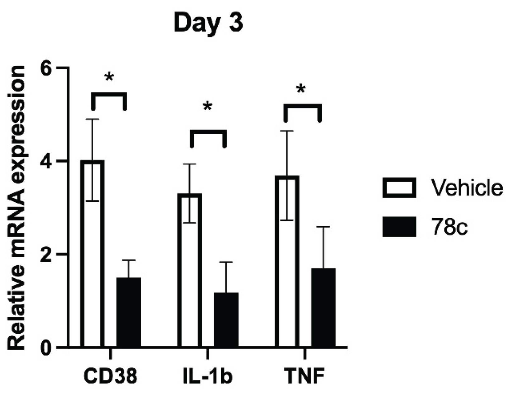
To determine the effect of 78c on CD38 and pro-inflammatory cytokine levels in the periodontal tissues, we performed another short-term study (Figure 7A). Old mice were punctured once with a 27-gauge needle, then administered Aa orally every other day for 3 days. Mice were treated with either vehicle or 78c twice daily by intraperitoneal injection. The left gingival tissues were harvested on day 3. As shown in Figure 7B, treatment with 78c significantly reduced the mRNA levels of CD38, IL-1β, and TNF in the left gingival tissues compared with vehicle-treated controls. The IL-6 mRNA levels were undetectable in the gingival tissues (data not shown). These data support that treatment with 78c inhibited CD38 and suppressed IL-1β and TNF levels in old mice with experimental periodontitis.
3. Discussion
In this study, treatment with the CD38-specific inhibitor (78c) significantly enhanced NAD+ levels in the liver and spleens of both young and old mice, which is in accordance with a previous study [26] that showed that treatment with 78c in old mice increased NAD+ in the tissues. We observed a large variation in NAD+ levels in the maxillary tissues compared with those in the liver or spleen. This might be caused by differences in the components of the maxillary tissues (including bone and muscles) compared with those in the liver and spleen.
Additionally, we demonstrated that treatment with 78c alleviated alveolar bone loss in both young and old mice and suppressed the mRNA levels of CD38, IL-1β, and TNF. These results are consistent with our previous study [23], which showed that 78c suppressed RANKL-induced osteoclastogenesis and bone resorption, reduced CD38 expression, and alleviated pro-inflammatory cytokine expression in murine BMMs infected with Aa. Previously, we demonstrated that 78c reduced podosome (a basic cell adhesion unit) components, including PI3K, Pyk2, Src, F-actin, integrins, paxillin, and talin. Therefore, treatment with 78c in mice may have alleviated alveolar bone loss mainly by suppressing RANKL-stimulated cell adhesion and fusion, thereby preventing the formation of multinucleated osteoclasts. In this study, we observed greater PMN infiltration in the periodontal epithelium of untreated old mice (Figure 6) and greater alveolar bone loss in untreated old mice compared with young controls (Figure 4). This is probably associated with increased aging-related inflammation through a process called inflammaging [27,28].
This study had some limitations. First, Aa infection is mostly associated with 90% of localized aggressive periodontitis in juveniles [15] and is not a major oral pathogen associated with aging-associated periodontitis [29]. Because Aa is a facultative anaerobic organism that can grow in conditions with or without the presence of oxygen, Aa can survive in the oral cavity of mice. In contrast, the oral pathogen Pg requires strict anaerobic conditions and dies in the oral cavity when exposed to oxygen. In the future, it is necessary to determine whether treatment with 78c in mice can alleviate inflammatory bone loss in mice infected with other oral pathogens associated with aging-associated periodontitis, including Pg, Tannerella forsythia, or Trponema denticola [29]. Second, because the old mice could not tolerate acute inflammation induced by ligature placement (ligatures placed around the 2nd molar), we used needle puncture and oral inoculation of Aa to induce chronic periodontal inflammation in mice. This inflammation was mild, and the mice resolved it after 4 weeks of treatment. We did not observe a significant difference in H&E staining or TRAP staining between young and old mice treated with 78c or vehicle (Figure 5 and Figure 6). Future studies are required to modify the existing experimental periodontitis approach to detect differences in the inflammatory response and TRAP-stained osteoclasts between the 78c-treated and vehicle-treated groups.
Currently, the gold-standard treatments of periodontitis include scaling and root surface debridement to remove bacterial plaque. There are still patients or sites that show poor response to these treatments. This could be due to sustained dysbiosis, bacterial invasion of periodontal tissues, or a non-resolving chronic inflammatory response. In this study, we demonstrated that treatment with the CD38-specific inhibitor (78c) reduced the mRNA levels of CD38, IL-1β, and TNF in the gingival tissues; enhanced NAD+ levels in the liver and spleen; and alleviated alveolar bone loss in mice with experimental periodontitis. Additionally, previous studies also showed that treatment with 78c increased the lifespan and healthspan of naturally aged mice [30] and improved several physiological and metabolic parameters of aging, including glucose tolerance, muscle function, exercise capacity, and cardiac function in mouse models of natural and accelerated aging [26,30]. Our and other studies’ results support the notion that treatment with 78c can serve as a promising therapeutic strategy for aging-associated periodontitis to alleviate periodontal inflammatory bone loss, enhance NAD+, and subsequently promote the lifespan and healthspan of human beings.
4. Materials and Methods
4.1. Animals and Reagents
Old (18-month-old) and young (2-month-old) male C57BL/6J mice were purchased from Jackson Laboratory (Bar Harbor, ME, USA). Mice were housed under a 12 h light/12 h dark cycle in specific pathogen-free conditions and had free access to food and water. All animal-related work was conducted in accordance with the guidelines laid down by the National Institutes of Health (NIH) in the United States regarding the usage of animals for experimental procedures and approved by the Institutional Animal Care and Use Committee at the Medical University of South Carolina (IACUC-2021-01287). 78c was purchased from Cayman Chemical (Ann Arbor, MI, USA). Dimethyl sulfoxide (DMSO), PEG400, and (2-hydroxypropyl)-γ-cyclodextrin were purchased from Fisher Scientific (Pittsburgh, PA, USA). 78c was first dissolved in DMSO (100 mM), followed by dilution in 15% PEG400 and 80% of 15% (2-hydroxypropyl)-γ-cyclodextrin in citrate buffer (pH 6.0) with a final 5% DMSO [26]. The vehicle consisted with 5% DMSO, 15% PEG400, and 80% of 15% (2-hydroxypropyl)-γ-cyclodextrin in citrate buffer (pH 6.0) [26]. 78c or vehicle was sterilized by filtering through a sterile bottle-top filter with 0.22 μm membrane (Fisher Scientific), aliquoted, and stored at -20 °C.
4.2. Bacterial Culture
The oral bacterial pathogen Aggregatibacter actinomycetemcomitans (Aa, ATCC 43718) was obtained from the American Type Culture Collection. Aa was streaked in brain–heart infusion agar (Fisher Scientific) and cultured in brain–heart infusion broth (Fisher Scientific) at 37 °C with 10% CO2. The Aa culture media was centrifuged, and the Aa cell pellets were washed with PBS and resuspended in PBS containing 1.5% carboxymethylcellulose (Fisher Scientific). The bacterial concentration was adjusted to 1x10¹⁰ colony forming units (CFU)/ml by measuring bacterial optical density at 600 nm of 10x diluted of bacteria (OD600 = 1 was equal to 1 × 10⁹ CFU/mL of Aa).
4.3. Animal Treatment
Mice drank antibiotic water (containing sulfamethoxazole 700 µg/ml and trimethoprim 300 µg/ml, Fisher Scientific) for 7 days to reduce indigenous oral microflora, then normal water for 1 day to remove residual antibiotics. To initiate experimental periodontitis, the mice were first sedated by inhaling isoflurane. Then, the palatal gingival tissues of mice were punctured (3 punctures on the left and 3 punctures on the right) by a 27 gauge ½ inch needle (BD Diagnostic System, Franklin Lakes, NJ, USA) once per week to cause minor mucosal damage followed by oral inoculation of the oral pathogen Aa (5x10⁸ colony forming units, 50 μl) via a wide pore pipitte tip (3 times/week for indicated days in figures). This procedure allows Aa to easily attach to the gingival mucosa and subsequently cause periodontal inflammation. The mice were either untreated, treated with vehicle, or treated with 78c (10mg/kg) by intraperitoneal injection twice daily for the indicated days, as shown in the figures. Soft foods were provided for the mice after Aa administration. After euthanizing the mice, the liver, spleen, bone marrow, maxilla, or gingival mucosa were harvested.
4.4. NAD+ Assay
The liver, spleen, and right maxillary tissues were stored at -80 °C. The tissues were homogenized using a mortar and pestle. NAD+ levels were determined using a NAD+/NADH assay kit (Sigma-Aldrich, St. Louis, MO, USA) in 20 mg of tissue, according to the manufacturer’s instructions.
4.5. Osteoclastogenesis Assay
Murine bone marrow cells were harvested from five old vehicle-treated mice or five 78c-treated mice after 4 weeks of treatment by flushing bone marrow from tibia and femur with complete minimal essential media (MEM)-α (Fisher Scientific), supplemented with 10% FBS, 100 U/ml penicillin, and 100 µg/ml streptomycin. Bone marrow cells were cultured in complete MEM-α media supplemented with 20% L929 conditioned media (containing macrophage colony-stimulating factor, M-CSF) [23] for 2 days to allow bone marrow stromal cells to attach onto the plates. The suspended bone marrow cells were plated in new petri dishes and cultured in complete MEM-α media supplemented with 20% L929-conditioned media until cells were attached. The attached bone marrow-derived monocytes (BMMs) were plated in a 96-well plate and were cultured in complete MEM-α media supplemented with both 10% conditioned L929 media and recombinant human RANKL (50 ng/mL, PeproTech, Cranbury, NJ, USA). The media was changed every two days. After RANKL treatment for 5 days, osteoclasts were stained by Tartrate-Resistant Acid Phosphatase (TRAP) staining using a leukocyte acid phosphatase kit (Millipore Sigma).
4.6. Micro-Computed Tomography (Micro-CT) Scanning and Alveolar Bone Loss Assessment
The left maxillary tissues were fixed with 10% formalin for 2 days and stored in 70% ethanol at 4 °C. The tissues were scanned with a cone-beam µ-CT40 system (Scanco Medical AG, Switzerland). Three-dimensional micro-CT images were visualized with the GE Healthcare MicroView software. The alveolar bone loss was assessed by measuring the distance from the cementoenamel junction (CEJ) to the alveolar bone crest (ABC) using Adobe Photoshop CS5.1 software.
4.7. Tissue Processing and Staining
The left maxillary tissues were decalcified in 20% EDTA for 4 weeks, then embedded in paraffin. Five µm sagittal paraffin tissue sections were cut, stained with hematoxylin and eosin (H&E) for general histology or tartrate-resistant acid phosphatase (TRAP) staining using a leukocyte acid phosphatase kit (Millipore Sigma). The images were taken by an Olympus BX43 microscope (Olympus Corporation of the Americas, Center Valley, PA, USA).
4.8. RNA Extraction and Real-Time PCR
Total RNA was isolated from maxilla or gingival tissues using TRIZOL reagent (ThermoFisher Scientific, Waltham, MA, USA). Complementary DNA was synthesized using a TaqMan reverse transcription kit (Life Technologies, Carlsbad, CA, USA) using the total RNA (1 μg). Real-time PCR was performed using a StepOnePlus Real-Time PCR System (Life Technologies). PCR conditions used were as follows: 50 °C for 2 min, 95 °C for 10 min, and 40 cycles of 95 °C for 15 s, and 60 °C for 1 min. The following amplicon primers were obtained from Life Technologies: CD38 (Mm00483143_m1), IL-1β (Mm00434228_m1), IL-6 (Mm00446190_m1), TNF (Mm00443258_m1), and β-actin (Mm02619580_g1). Amplicon concentration was determined by comparing threshold cycle values to standard curves for each primer. Sample mRNA levels were normalized to an endogenous control, β-actin, and expressed as fold changes relative to control groups.
4.9. Statistical Analysis
The data were analyzed using a one-way ANOVA with Dunnett’s or Tukey’s multiple-comparison tests when comparing more than three groups. When comparing two groups of data, the data were analyzed using the unpaired Student’s t-test with Welch’s correction. All statistical tests were performed using GraphPad Prism software (Version 10.6.1, GraphPad Software Inc., La Jolla, CA, USA). Values are expressed as means ± standard error of the means (SEM) of three independent experiments. A p-value of 0.05 or less was considered significant.
Author Contributions
Conceptualization, H.Y.; methodology, J.Y., S.P., and H.Y.; investigation, J.Y, S.P., W.H., and H.Y.; writing –original draft preparation, J.Y., H.Y.; writing-review and editing, J.Y, S.P., W.H., and H.Y.; funding acquisition, H.Y., W.H., S.P.; All authors have read and agreed to the published version of the manuscript.
Funding
This work was supported by the National Institutes of Health (grants R21DE033977 (NIDCR), 2P20GM103499-20 (SC INBRE, NIGMS), and UL1 TR001450).
Institutional Review Board Statement
This study was approved on March 18, 2024 by the IACUC of the Medical University of South Carolina (Charleston, SC, USA) under protocol IACUC-2021-01287.
Informed Consent Statement
Not applicable.
Data Availability Statement
The raw data supporting the conclusions of this article will be made available by the authors on request.
Acknowledgments
We thank Kasey Larson at the Writing Center at the Medical University of South Carolina for manuscript review and editing.
Conflicts of Interest
The authors declare no potential conflicts of interest.
References
- Richards, D. Review finds that severe periodontitis affects 11% of the world population. Evid. Based Dent. 2014, 15, 70–71. [Google Scholar] [CrossRef] [PubMed]
- Eke, P.I.; Dye, B.A.; Wei, L.; Thornton-Evans, G.O.; Genco, R.J. Prevalence of periodontitis in adults in the United States: 2009 and 2010. J. Dent. Res. 2012, 91, 914–920. [Google Scholar] [CrossRef]
- Clark, D.; Kotronia, E.; Ramsay, S.E. Frailty, aging, and periodontal disease: Basic biologic considerations. Periodontology 2000 2021, 87, 143–156. [Google Scholar] [CrossRef]
- Wu, Y.; Dong, G.; Xiao, W.; Xiao, E.; Miao, F.; Syverson, A.; Missaghian, N.; Vafa, R.; Cabrera-Ortega, A.A.; Rossa, C., Jr.; et al. Effect of Aging on Periodontal Inflammation, Microbial Colonization, and Disease Susceptibility. J. Dent. Res. 2016, 95, 460–466. [Google Scholar] [CrossRef] [PubMed]
- Piedra-Quintero, Z.L.; Wilson, Z.; Nava, P.; Guerau-de-Arellano, M. CD38: An Immunomodulatory Molecule in Inflammation and Autoimmunity. Front. Immunol. 2020, 11, 597959. [Google Scholar] [CrossRef]
- Hogan, K.A.; Chini, C.C.S.; Chini, E.N. The Multi-faceted Ecto-enzyme CD38: Roles in Immunomodulation, Cancer, Aging, and Metabolic Diseases. Front. Immunol. 2019, 10, 1187. [Google Scholar] [CrossRef]
- Kar, A.; Mehrotra, S.; Chatterjee, S. CD38: T Cell Immuno-Metabolic Modulator. Cells 2020, 9. [Google Scholar] [CrossRef]
- Benzi, A.; Grozio, A.; Spinelli, S.; Sturla, L.; Guse, A.H.; De Flora, A.; Zocchi, E.; Heeren, J.; Bruzzone, S. Role of CD38 in Adipose Tissue: Tuning Coenzyme Availability? Nutrients 2021, 13. [Google Scholar] [CrossRef]
- Xiao, W.; Wang, R.S.; Handy, D.E.; Loscalzo, J. NAD(H) and NADP(H) Redox Couples and Cellular Energy Metabolism. Antioxid. Redox Signal. 2018, 28, 251–272. [Google Scholar] [CrossRef] [PubMed]
- Cao, K.; Chowdhury, N.; Wellslager, B.; Hill, W.D.; Yilmaz, Ö.; Yu, H. Inhibition of CD38 by 78c Enhanced NAD(+), Alleviated Inflammation, and Decreased Oxidative Stress in Old Murine Macrophages Induced by Oral Pathogens. Int. J. Mol. Sci. 2025, 26. [Google Scholar] [CrossRef]
- Lautrup, S.; Sinclair, D.A.; Mattson, M.P.; Fang, E.F. NAD(+) in Brain Aging and Neurodegenerative Disorders. Cell Metab. 2019, 30, 630–655. [Google Scholar] [CrossRef]
- Verdin, E. NAD⁺ in aging, metabolism, and neurodegeneration. Science 2015, 350, 1208–1213. [Google Scholar] [CrossRef] [PubMed]
- Zhu, X.H.; Lu, M.; Lee, B.Y.; Ugurbil, K.; Chen, W. In vivo NAD assay reveals the intracellular NAD contents and redox state in healthy human brain and their age dependences. Proc. Natl. Acad. Sci. U S A 2015, 112, 2876–2881. [Google Scholar] [CrossRef]
- Zapata-Pérez, R.; Wanders, R.J.A.; van Karnebeek, C.D.M.; Houtkooper, R.H. NAD(+) homeostasis in human health and disease. EMBO Mol. Med. 2021, 13, e13943. [Google Scholar] [CrossRef]
- Raja, M.; Ummer, F.; Dhivakar, C.P. Aggregatibacter actinomycetemcomitans - a tooth killer? J. Clin. Diagn. Res. 2014, 8, ZE13–16. [Google Scholar] [CrossRef]
- Cai, J.; Chen, J.; Guo, H.; Pan, Y.; Zhang, Y.; Zhao, W.; Li, X.; Li, Y. Recombinant fimbriae protein of Porphyromonas gingivalis induces an inflammatory response via the TLR4/NF-κB signaling pathway in human peripheral blood mononuclear cells. Int. J. Mol. Med. 2019, 43, 1430–1440. [Google Scholar] [CrossRef]
- Hodgkinson, C.P.; Laxton, R.C.; Patel, K.; Ye, S. Advanced glycation end-product of low density lipoprotein activates the toll-like 4 receptor pathway implications for diabetic atherosclerosis. Arterioscler. Thromb. Vasc. Biol. 2008, 28, 2275–2281. [Google Scholar] [CrossRef]
- Kanaya, S.; Nemoto, E.; Ogawa, T.; Shimauchi, H. Porphyromonas gingivalis fimbriae induce unique dendritic cell subsets via Toll-like receptor 2. J. Periodontal Res. 2009, 44, 543–549. [Google Scholar] [CrossRef]
- Liu, Z.; Ma, Y.; Cui, Q.; Xu, J.; Tang, Z.; Wang, Y.; He, C.; Wang, X. Toll-like receptor 4 plays a key role in advanced glycation end products-induced M1 macrophage polarization. Biochem. Biophys. Res. Commun. 2020, 531, 602–608. [Google Scholar] [CrossRef] [PubMed]
- Lima, H.R.; Gelani, V.; Fernandes, A.P.; Gasparoto, T.H.; Torres, S.A.; Santos, C.F.; Garlet, G.P.; da Silva, J.S.; Campanelli, A.P. The essential role of toll like receptor-4 in the control of Aggregatibacter actinomycetemcomitans infection in mice. J. Clin. Periodontol. 2010, 37, 248–254. [Google Scholar] [CrossRef] [PubMed]
- Watanabe, K.; Yilmaz, O.; Nakhjiri, S.F.; Belton, C.M.; Lamont, R.J. Association of mitogen-activated protein kinase pathways with gingival epithelial cell responses to Porphyromonas gingivalis infection. Infect. Immun. 2001, 69, 6731–6737. [Google Scholar] [CrossRef]
- Yilmaz, O.; Jungas, T.; Verbeke, P.; Ojcius, D.M. Activation of the phosphatidylinositol 3-kinase/Akt pathway contributes to survival of primary epithelial cells infected with the periodontal pathogen Porphyromonas gingivalis. Infect. Immun. 2004, 72, 3743–3751. [Google Scholar] [CrossRef]
- Lory, W.; Chowdhury, N.; Wellslager, B.; Pandruvada, S.; Huang, Y.; Yilmaz, Ö.; Yu, H. CD38 Inhibitor 78c Attenuates Pro-Inflammatory Cytokine Expression and Osteoclastogenesis in Macrophages. Cells 2024, 13. [Google Scholar] [CrossRef] [PubMed]
- Graves, D.T.; Kang, J.; Andriankaja, O.; Wada, K.; Rossa, C., Jr. Animal models to study host-bacteria interactions involved in periodontitis. Front Oral. Biol. 2012, 15, 117–132. [Google Scholar] [CrossRef]
- Baker, P.J.; Dixon, M.; Roopenian, D.C. Genetic control of susceptibility to Porphyromonas gingivalis-induced alveolar bone loss in mice. Infect. Immun. 2000, 68, 5864–5868. [Google Scholar] [CrossRef]
- Tarragó, M.G.; Chini, C.C.S.; Kanamori, K.S.; Warner, G.M.; Caride, A.; de Oliveira, G.C.; Rud, M.; Samani, A.; Hein, K.Z.; Huang, R.; et al. A Potent and Specific CD38 Inhibitor Ameliorates Age-Related Metabolic Dysfunction by Reversing Tissue NAD(+) Decline. Cell Metab. 2018, 27, 1081–1095.e1010. [Google Scholar] [CrossRef]
- Franceschi, C.; Campisi, J. Chronic inflammation (inflammaging) and its potential contribution to age-associated diseases. J. Gerontol. A Biol. Sci. Med. Sci. 2014, 69 Suppl 1, S4–9. [Google Scholar] [CrossRef]
- Franceschi, C.; Garagnani, P.; Parini, P.; Giuliani, C.; Santoro, A. Inflammaging: a new immune-metabolic viewpoint for age-related diseases. Nat. Rev. Endocrinol. 2018, 14, 576–590. [Google Scholar] [CrossRef] [PubMed]
- Feres, M.; Teles, F.; Teles, R.; Figueiredo, L.C.; Faveri, M. The subgingival periodontal microbiota of the aging mouth. Periodontology 2000 2016, 72, 30–53. [Google Scholar] [CrossRef]
- Peclat, T.R.; Thompson, K.L.; Warner, G.M.; Chini, C.C.S.; Tarragó, M.G.; Mazdeh, D.Z.; Zhang, C.; Zavala-Solorio, J.; Kolumam, G.; Liang Wong, Y.; et al. CD38 inhibitor 78c increases mice lifespan and healthspan in a model of chronological aging. Aging Cell 2022, 21, e13589. [Google Scholar] [CrossRef] [PubMed]
Figure 1.
Old C57BL/6J mice significantly lost body weight during experimental periodontitis compared with young controls. (A) Schematic diagram of mice treatment. Young or old C57BL/6J mice drank antibiotic water (containing 700 μg/ml sulfamethoxazole and 300 μg/ml trimethoprim) for a week to reduce indigenous oral microflora. The experimental periodontitis was initiated by puncturing the palatal gingival tissues with a 27-gauge ½ inch needle (3 punctures on the right and 3 punctures on the left once/week) to allow minor mucosal injury, which was then followed by oral inoculation of the oral pathogen Aggregatibacter atinomycetemcomitans (Aa) three times/week. Mice were untreated (n=3), treated with vehicle (n=10), or treated with 78c (n=10, 10mg/kg) by intraperitoneal injection twice daily for 4 weeks. (B) Mice’s body weight was measured before and after the 4-week Aa inoculation period (**p<0.01, ***p<0.001).
Figure 1.
Old C57BL/6J mice significantly lost body weight during experimental periodontitis compared with young controls. (A) Schematic diagram of mice treatment. Young or old C57BL/6J mice drank antibiotic water (containing 700 μg/ml sulfamethoxazole and 300 μg/ml trimethoprim) for a week to reduce indigenous oral microflora. The experimental periodontitis was initiated by puncturing the palatal gingival tissues with a 27-gauge ½ inch needle (3 punctures on the right and 3 punctures on the left once/week) to allow minor mucosal injury, which was then followed by oral inoculation of the oral pathogen Aggregatibacter atinomycetemcomitans (Aa) three times/week. Mice were untreated (n=3), treated with vehicle (n=10), or treated with 78c (n=10, 10mg/kg) by intraperitoneal injection twice daily for 4 weeks. (B) Mice’s body weight was measured before and after the 4-week Aa inoculation period (**p<0.01, ***p<0.001).
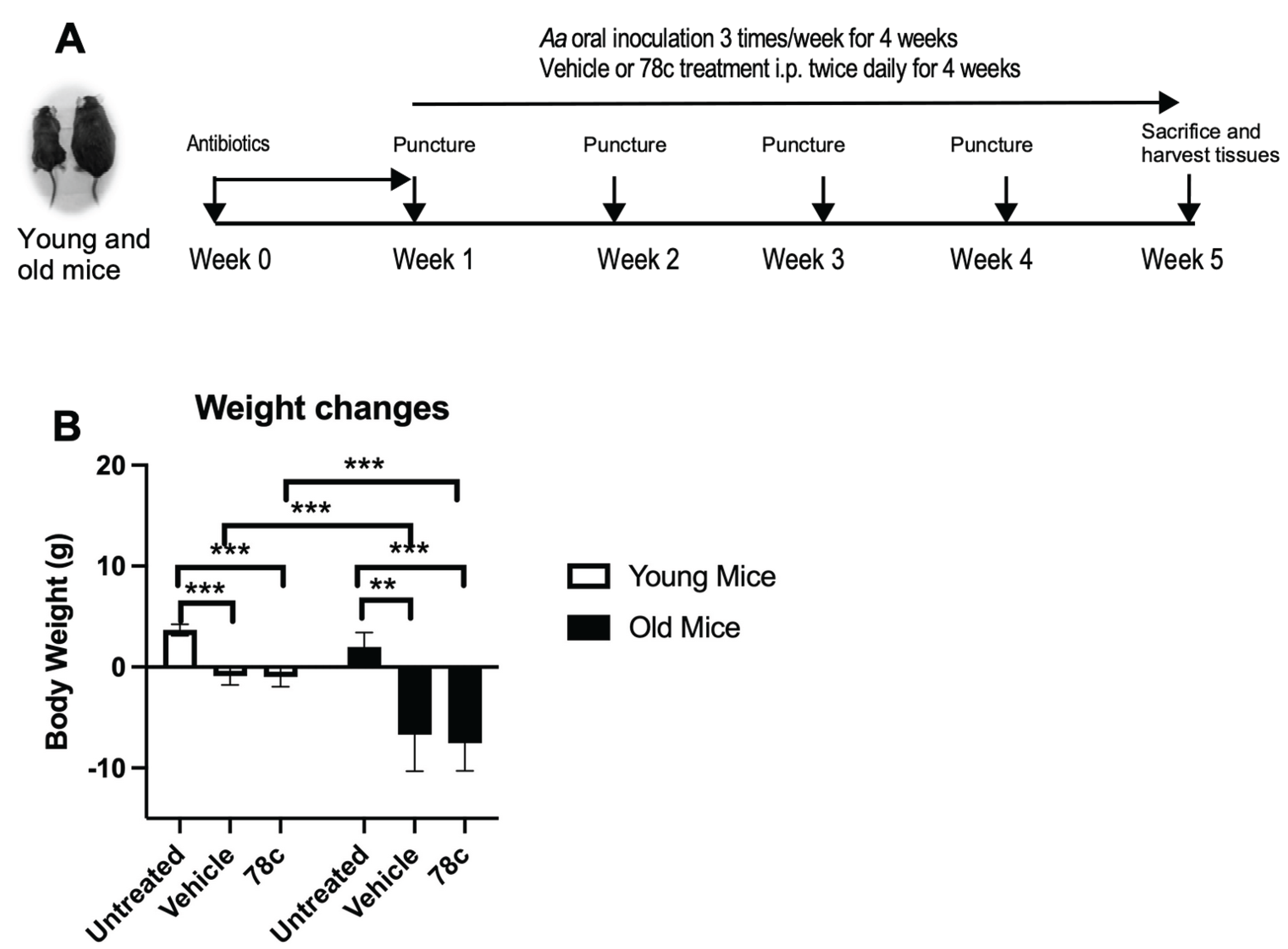
Figure 2.
NAD+ levels in the liver, spleen, and right maxillary tissues in untreated mice, vehicle-treated mice, and 78c-treated mice. C57BL/6J mice were either untreated (n=3), treated with vehicle and orally administered with the oral pathogen Aggregatibacter atinomycetemcomitans (Aa) (n=10), or treated with 78c and orally administered with Aa (n=10) for 4 weeks. (A) NAD+ levels in the liver, spleen, and right maxillary tissues of untreated mice. (B) NAD+ levels in the liver of untreated mice, vehicle-treated mice, or 78c-treated mice. (C) NAD+ levels in the spleen of untreated mice, vehicle-treated mice, or 78c-treated mice. (D) NAD+ levels in the right maxillary tissues of untreated mice, vehicle-treated mice, or 78c-treated mice. The NAD+ levels in the tissues were evaluated by a NAD/NADH assay kit (Sigma Aldrich) (*p<0.05, **p<0.01, ***p<0.001).
Figure 2.
NAD+ levels in the liver, spleen, and right maxillary tissues in untreated mice, vehicle-treated mice, and 78c-treated mice. C57BL/6J mice were either untreated (n=3), treated with vehicle and orally administered with the oral pathogen Aggregatibacter atinomycetemcomitans (Aa) (n=10), or treated with 78c and orally administered with Aa (n=10) for 4 weeks. (A) NAD+ levels in the liver, spleen, and right maxillary tissues of untreated mice. (B) NAD+ levels in the liver of untreated mice, vehicle-treated mice, or 78c-treated mice. (C) NAD+ levels in the spleen of untreated mice, vehicle-treated mice, or 78c-treated mice. (D) NAD+ levels in the right maxillary tissues of untreated mice, vehicle-treated mice, or 78c-treated mice. The NAD+ levels in the tissues were evaluated by a NAD/NADH assay kit (Sigma Aldrich) (*p<0.05, **p<0.01, ***p<0.001).
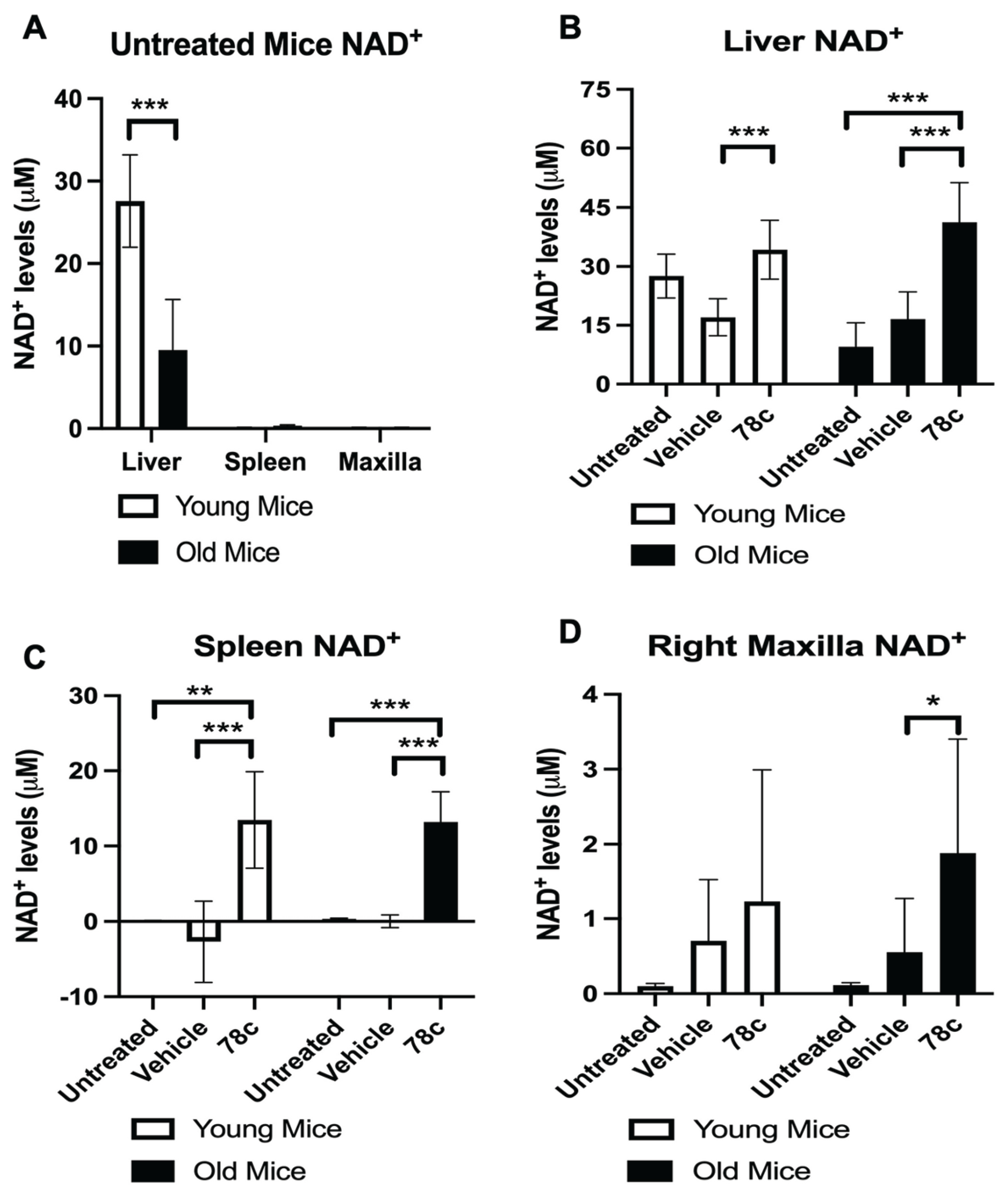
Figure 3.
Bone marrow cells derived from 78-treated old mice reduced RANKL-induced osteoclastogenesis compared with vehicle-treated controls. Bone marrow cells were harvested from vehicle-treated or 78c-treated old mice and cultured in the presence of M-CSF and RANKL for 5 days. (A) Representative images of TRAP-stained BMMs derived from vehicle-treated old mice or 78c-treated old mice. (B) Number of TRAP+ multinucleated (more than 3 nuclei) osteoclasts/well (96-well), (n=5, p<0.001).
Figure 3.
Bone marrow cells derived from 78-treated old mice reduced RANKL-induced osteoclastogenesis compared with vehicle-treated controls. Bone marrow cells were harvested from vehicle-treated or 78c-treated old mice and cultured in the presence of M-CSF and RANKL for 5 days. (A) Representative images of TRAP-stained BMMs derived from vehicle-treated old mice or 78c-treated old mice. (B) Number of TRAP+ multinucleated (more than 3 nuclei) osteoclasts/well (96-well), (n=5, p<0.001).
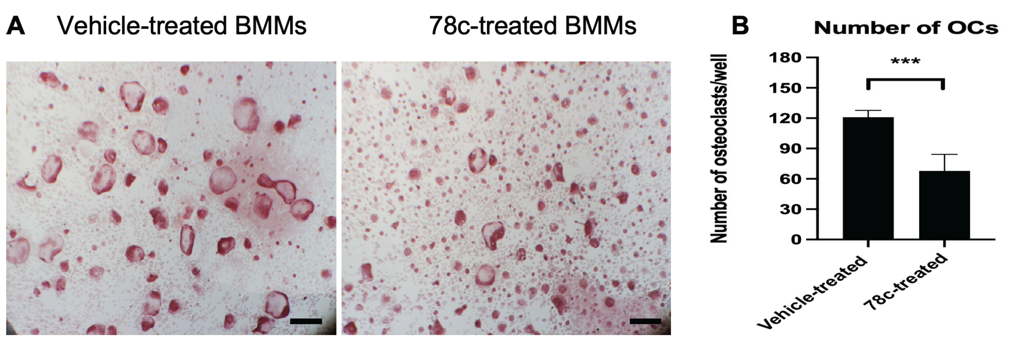
Figure 4.
Treatment with 78c alleviated alveolar bone loss in mice with experimental periodontitis. (A) Representative images of micro-CT scanning of left maxillary alveolar bone tissues were displayed. The dotted lines indicate the distances from the cementoenamel junction (CEJ) to the alveolar bone crest (ABC). (B) The distances between CEJ and ABC were quantified (n=10, *p<0.05, **p<0.01, ***p<0.001).
Figure 4.
Treatment with 78c alleviated alveolar bone loss in mice with experimental periodontitis. (A) Representative images of micro-CT scanning of left maxillary alveolar bone tissues were displayed. The dotted lines indicate the distances from the cementoenamel junction (CEJ) to the alveolar bone crest (ABC). (B) The distances between CEJ and ABC were quantified (n=10, *p<0.05, **p<0.01, ***p<0.001).
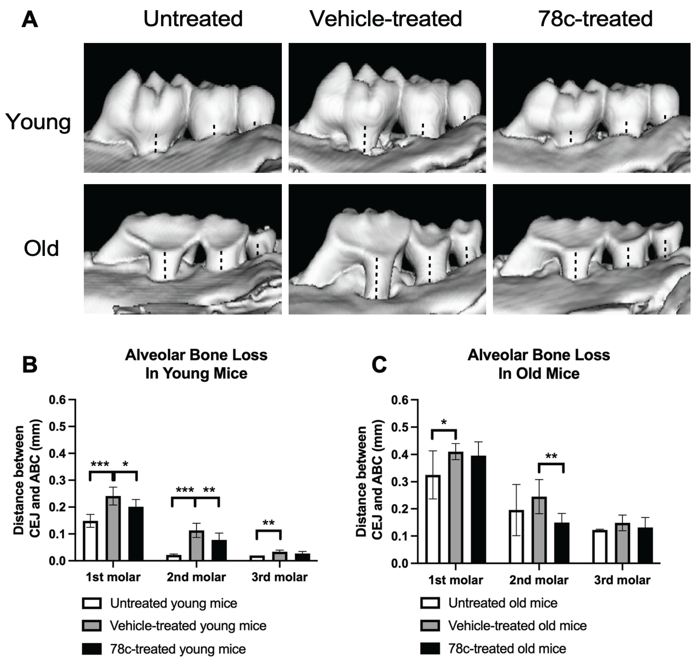
Figure 5.
Young mice exhibited no significant inflammation in the periodontal tissues after 4 weeks of treatment. (A) Representative images of hematoxylin and eosin (H&E) staining of the maxillary periodontal tissues of untreated young mice, vehicle-treated young mice, and 78c-treated young mice. Images were taken under 40x magnification or 400x magnification. (B) Representative images of tartrate-resistant acid phosphatase (TRAP) staining of the maxillary periodontal tissues of untreated young mice, vehicle-treated young mice, and 78c-treated young mice. Images were taken under 40x magnification or 400x magnification. Black arrows indicate TRAP-stained positive cells in the periodontal tissues. The black boxes indicate the magnified region in the periodontal epithelium. The scale bars represent 200 μm in the 40x images and 20 μm in the 400x images, respectively.
Figure 5.
Young mice exhibited no significant inflammation in the periodontal tissues after 4 weeks of treatment. (A) Representative images of hematoxylin and eosin (H&E) staining of the maxillary periodontal tissues of untreated young mice, vehicle-treated young mice, and 78c-treated young mice. Images were taken under 40x magnification or 400x magnification. (B) Representative images of tartrate-resistant acid phosphatase (TRAP) staining of the maxillary periodontal tissues of untreated young mice, vehicle-treated young mice, and 78c-treated young mice. Images were taken under 40x magnification or 400x magnification. Black arrows indicate TRAP-stained positive cells in the periodontal tissues. The black boxes indicate the magnified region in the periodontal epithelium. The scale bars represent 200 μm in the 40x images and 20 μm in the 400x images, respectively.
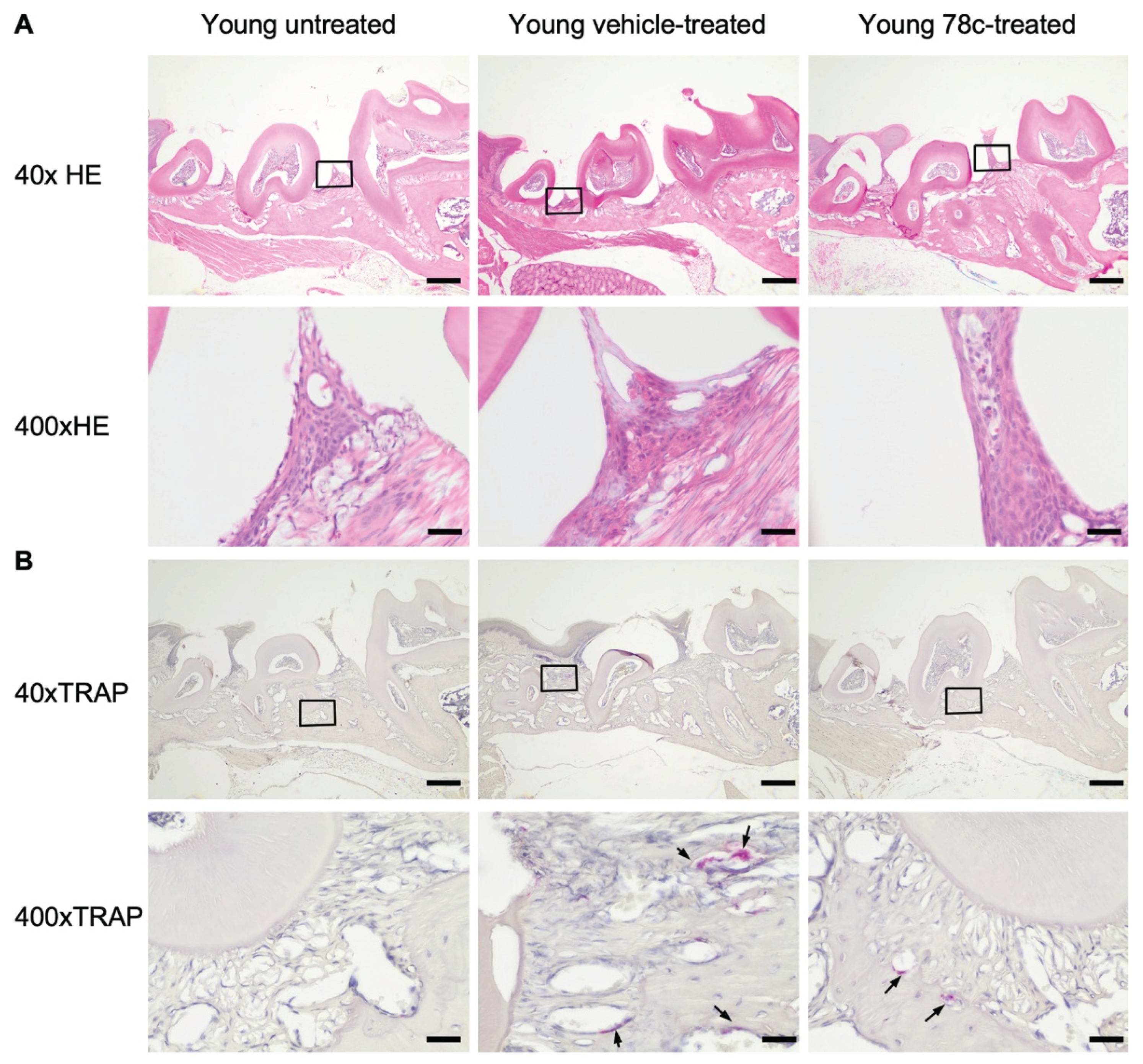
Figure 6.
Old mice displayed some polymorphonuclear leukocytes (PMN) in the periodontal epithelium after 4 weeks of treatment. (A) Representative images of hematoxylin and eosin (H&E) staining of the maxillary periodontal tissues of untreated old mice, vehicle-treated old mice, and 78c-treated old mice. Images were taken under 40x magnification or 400x magnification. Black arrows show some PMN in the periodontal epithelium. (B) Representative images of tartrate-resistant acid phosphatase (TRAP) staining of the maxillary periodontal tissues of untreated old mice, vehicle-treated old mice, and 78c-treated old mice. Images were taken under 40x magnification or 400x magnification. Black arrows indicate TRAP-stained positive cells in the periodontal tissues. The black boxes indicate the magnified region in the periodontal epithelium. The scale bars represent 200 μm in the 40x images and 20 μm in the 400x images, respectively.
Figure 6.
Old mice displayed some polymorphonuclear leukocytes (PMN) in the periodontal epithelium after 4 weeks of treatment. (A) Representative images of hematoxylin and eosin (H&E) staining of the maxillary periodontal tissues of untreated old mice, vehicle-treated old mice, and 78c-treated old mice. Images were taken under 40x magnification or 400x magnification. Black arrows show some PMN in the periodontal epithelium. (B) Representative images of tartrate-resistant acid phosphatase (TRAP) staining of the maxillary periodontal tissues of untreated old mice, vehicle-treated old mice, and 78c-treated old mice. Images were taken under 40x magnification or 400x magnification. Black arrows indicate TRAP-stained positive cells in the periodontal tissues. The black boxes indicate the magnified region in the periodontal epithelium. The scale bars represent 200 μm in the 40x images and 20 μm in the 400x images, respectively.
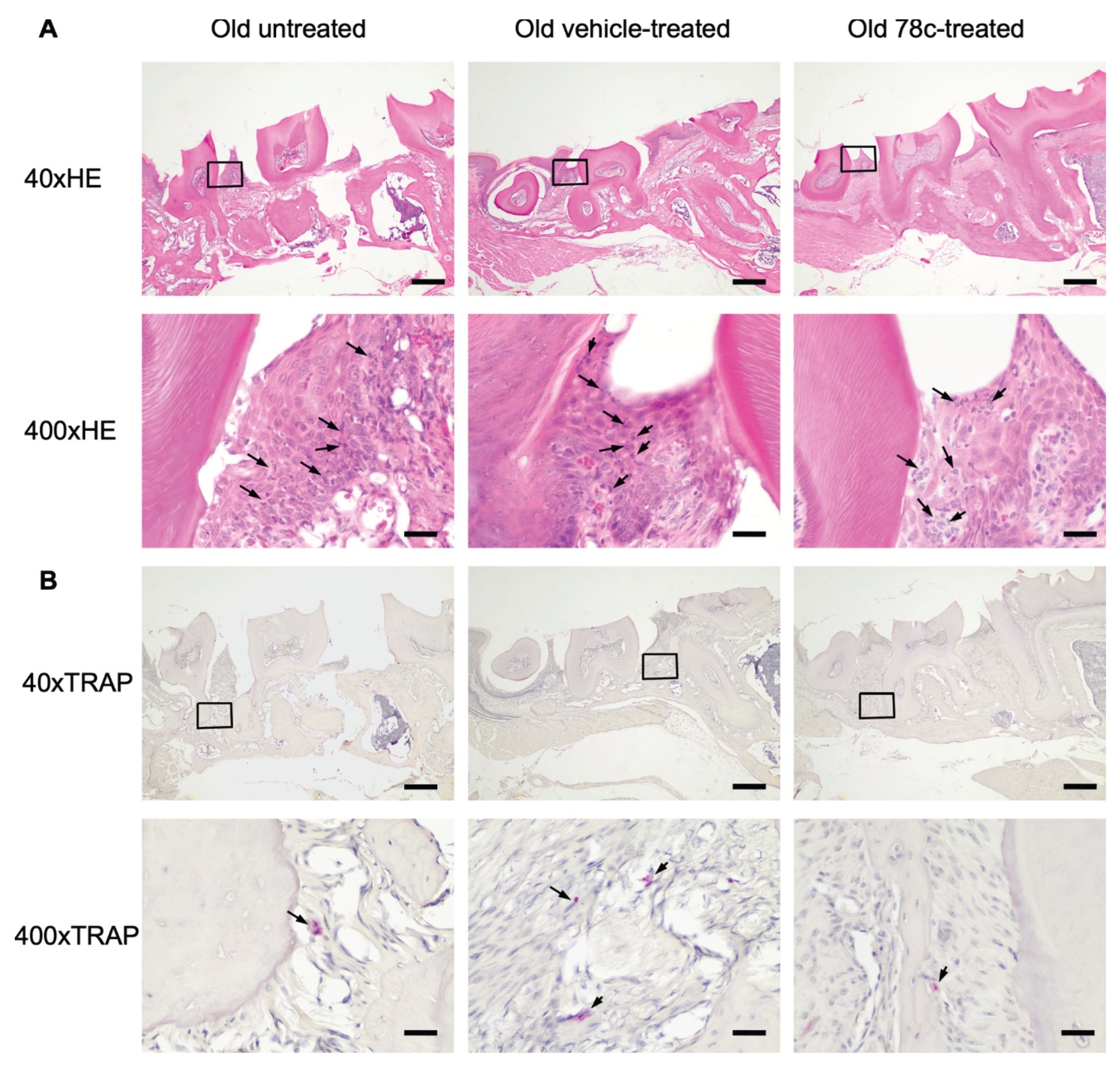
Figure 7.
Treatment with 78c in old C57BL/6J mice significantly reduced the mRNA levels of CD38, IL-1β, and TNF in the gingival tissues compared with vehicle treatment. (A) Schematic diagram of mice treatment. Old C57BL/6J mice drank antibiotic water (containing 700 μg/ml sulfamethoxazole and 300 μg/ml trimethoprim) for a week to reduce indigenous oral microflora, then drank normal water for 1 day. The palatal gingival tissues were punctured by a 27-gauge ½ inch needle (3 punctures on the right and 3 punctures on the left), followed by oral inoculation of the oral pathogen Aggregatibacter atinomycetemcomitans (Aa). Mice were untreated (n=3), treated with vehicle (n=3), or treated with 78c (n=3) by intraperitoneal injection twice daily for 3 days. (B) The relative mRNA levels of CD38, IL-1β, and TNF in the left gingival tissues 3 days after initiation of experimental periodontitis were evaluated by RT-PCR (**p<0.01, ***p<0.001).
Figure 7.
Treatment with 78c in old C57BL/6J mice significantly reduced the mRNA levels of CD38, IL-1β, and TNF in the gingival tissues compared with vehicle treatment. (A) Schematic diagram of mice treatment. Old C57BL/6J mice drank antibiotic water (containing 700 μg/ml sulfamethoxazole and 300 μg/ml trimethoprim) for a week to reduce indigenous oral microflora, then drank normal water for 1 day. The palatal gingival tissues were punctured by a 27-gauge ½ inch needle (3 punctures on the right and 3 punctures on the left), followed by oral inoculation of the oral pathogen Aggregatibacter atinomycetemcomitans (Aa). Mice were untreated (n=3), treated with vehicle (n=3), or treated with 78c (n=3) by intraperitoneal injection twice daily for 3 days. (B) The relative mRNA levels of CD38, IL-1β, and TNF in the left gingival tissues 3 days after initiation of experimental periodontitis were evaluated by RT-PCR (**p<0.01, ***p<0.001).
Disclaimer/Publisher’s Note: The statements, opinions and data contained in all publications are solely those of the individual author(s) and contributor(s) and not of MDPI and/or the editor(s). MDPI and/or the editor(s) disclaim responsibility for any injury to people or property resulting from any ideas, methods, instructions or products referred to in the content. |
© 2026 by the authors. Licensee MDPI, Basel, Switzerland. This article is an open access article distributed under the terms and conditions of the Creative Commons Attribution (CC BY) license (http://creativecommons.org/licenses/by/4.0/).
Copyright: This open access article is published under a Creative Commons CC BY 4.0 license, which permit the free download, distribution, and reuse, provided that the author and preprint are cited in any reuse.


