

Submitted:
17 May 2026
Posted:
19 May 2026
You are already at the latest version
Abstract
Fibroblast growth factor (FGF) signaling plays a pivotal role in the development, maintenance, and regeneration of musculoskeletal tissues. While its transcriptional regulation has been extensively characterized, accumulating evidence indicates that FGF activity is also modulated by a diverse array of post-transcriptional mechanisms. In this review, microRNAs, long non-coding RNAs, alternative splicing, and RNA modifications are examined as key regulators of FGF ligands and receptors across bone, cartilage, muscle, and tooth. Enhancer RNAs and RNA-binding proteins are also discussed as potential modulators of FGF transcript stability and translation. By integrating both established and emerging layers of RNA-level regulation, this review outlines a complex, tissue-specific architecture that fine-tunes FGF signaling in development and repair. To highlight this layered regulatory dimension, the concept of a pathway-specific RNA regulome is introduced, referring to the network of RNA-based mechanisms that modulate signaling cascades, such as FGF, across distinct biological processes. The therapeutic implications of targeting post-transcriptional nodes, particularly through non-coding RNAs and epitranscriptomic marks, are highlighted as promising avenues for future musculoskeletal interventions.
Keywords:
FGF signaling
; post-transcriptional regulation
; musculoskeletal development
; non-coding RNA
; RNA modifications
1. Introduction
Fibroblast growth factor (FGF) signaling is a key modulator of embryonic development and adult tissue homeostasis, with particularly well-defined roles in the formation and maintenance of musculoskeletal structures (Su et al., 2014; Teven et al., 2014; Ornitz and Itoh, 2015; Ornitz and Marie, 2015). In vertebrates, the FGF family comprises 22 members, of which 18 secreted ligands interact with four transmembrane tyrosine kinase receptors (FGFR1–FGFR4) and can also bind the atypical receptor FGFRL1. FGFRL1 lacks an intracellular kinase domain and modulates signaling through decoy or scaffolding functions (Park et al., 2022). In contrast, ligand binding to FGFR1–FGFR4 activates downstream cascades including the RAS–MAPK, PI3K–AKT, PLCγ, and STAT pathways, each of which exerts context-dependent effects on cell proliferation, differentiation, survival, and migration (Dailey et al., 2005; Ornitz and Itoh, 2015). FGF signaling is strongly influenced by co-receptors that help assemble and stabilize the active ligand–receptor complex. Heparan sulfate proteoglycans (HSPGs) act as co-receptors for the paracrine/autocrine FGFs (FGF1–10, FGF16-18, FGF20 and FGF22), increasing their local concentration and promoting productive binding to FGFRs. In contrast, the Klotho family proteins (α-Klotho and β-Klotho) function as obligate co-receptors for the endocrine FGFs (FGF19, FGF21 and FGF23), conferring ligand specificity and enabling efficient long-range, hormone-like FGF signaling. Within musculoskeletal tissues, such signaling governs critical processes such as skeletal patterning, osteogenesis, chondrogenesis, myogenesis, and tooth morphogenesis (Li et al., 2014; Su et al., 2014; Teven et al., 2014; Ornitz and Marie, 2015; Pawlikowski et al., 2017; Du et al., 2018).
During bone development, FGF signaling is required for the proper proliferation and differentiation of mesenchymal stem cells (MSCs) into osteoblasts (Chen and Deng, 2005; Su et al., 2014; Teven et al., 2014; Ornitz and Marie, 2015). Genetic knockout studies have shown that FGF18 is essential for both osteogenesis and chondrogenesis (Liu et al., 2002; Ohbayashi et al., 2002). For example, FGF2 and FGF18 have been shown to stimulate osteoprogenitor proliferation and to modulate differentiation and matrix production, via the upregulation of Bmp2 and Runx2 (Hamidouche et al., 2010; Nagayama et al., 2013). Meanwhile, FGF23 plays a distinct endocrine role in phosphate homeostasis and exerts indirect effects on bone mineralization through its actions on kidney and bone cells (Chen and Deng, 2005; Wang et al., 2008; Hori et al., 2011; Su et al., 2014; Teven et al., 2014; Guo and Yuan, 2015). Beyond bone, FGFs also play pivotal roles in cartilage, where FGFs modulate chondrocyte proliferation and hypertrophy, particularly in the growth plate, where spatial gradients of FGFR expression intersect with Indian Hedgehog (Ihh) and parathyroid hormone-related protein (PTHrP) signaling to control longitudinal bone growth (Minina et al., 2001; Su et al., 2014; Teven et al., 2014; Ornitz and Marie, 2015). FGFR3, in particular, has been implicated in chondrodysplasias due to its inhibitory role in chondrocyte expansion (Su et al., 2014; Teven et al., 2014; Ornitz and Marie, 2015; Ornitz and Legeai-Mallet, 2017), while its over overactivation leads to skeletal dysplasias (Kimura et al., 2021).
In muscle tissue, FGF signaling contributes to the activation and proliferation of satellite cells during regeneration (Fu et al., 2015; Pawlikowski et al., 2017). FGFs such as FGF2 and FGF6 are upregulated or released in response to injury and promote satellite-cell and myoblast proliferation (Floss et al., 1997) (Allen and Boxhorn, 1999), whereas sustained FGF signaling must be attenuated to permit efficient differentiation (Armand et al., 2006; Fu et al., 2015; Pawlikowski et al., 2017). Dysregulation of FGF activity in muscle has been associated with impaired regeneration, muscular dystrophies, and tumorigenesis, including rhabdomyosarcoma, in which aberrant FGFR signaling contributes to malignant growth (Crose et al., 2012; Fu et al., 2015; Pawlikowski et al., 2017; Milton et al., 2022). In odontogenesis, members of the FGF family regulate epithelial–mesenchymal interactions essential for tooth bud initiation, enamel organ formation, and root development, often in coordination with Wnt and BMP signaling pathways (Kettunen and Thesleff; Li et al., 2014; Du et al., 2018; Vaseenon et al., 2020). For example, FGF3 and FGF10 are expressed in the dental mesenchyme during early morphogenesis (Kettunen et al., 2000), while FGF4 serves as a signaling hub for cusp formation (Jernvall et al., 1994).
Although the functional consequences of FGF signaling in musculoskeletal tissues have been extensively studied, the regulatory mechanisms that determine the intensity, timing, and duration of this pathway remain incompletely understood. Historically, research has focused on ligand availability, receptor expression, and feedback antagonists such as Sprouty proteins and other extracellular or intracellular inhibitors that dampen FGF–FGFR activity (Hacohen et al., 1998; Dailey et al., 2005; Ornitz and Itoh, 2015; Xie et al., 2020; Szybowska et al., 2021). However, increasing attention has been directed toward the regulation of FGF signaling at the RNA level. This includes not only transcriptional control, but also a range of post-transcriptional mechanisms that govern RNA processing, stability, localization, and translation, some of which have been shown to fine-tune FGF output in specific developmental contexts (Li et al., 2019a; Copeland and Simoes-Costa, 2020; Ding et al., 2024). These regulatory layers are of particular interest in dynamic tissues such as bone and muscle, where rapid and reversible shifts in gene expression are required for development, adaptation, and repair (Su et al., 2014; Teven et al., 2014; Fu et al., 2015; Pawlikowski et al., 2017). In the sections that follow, the post-transcriptional regulation of FGF signaling will be examined in detail, with emphasis on mechanisms such as microRNA interference, long non-coding RNA interaction, alternative splicing, and RNA modifications (Imbriano et al., 2023; Yi and Yang, 2023). These regulatory layers collectively form what we propose here as a pathway-specific RNA regulome: a complex, tissue-specific architecture that modulates FGF signaling cascade in development and repair. This review will integrate established and emerging evidence across bone, cartilage, muscle, and tooth to highlight how RNA-level regulation fine-tunes FGF signaling in musculoskeletal biology.
Figure 1.
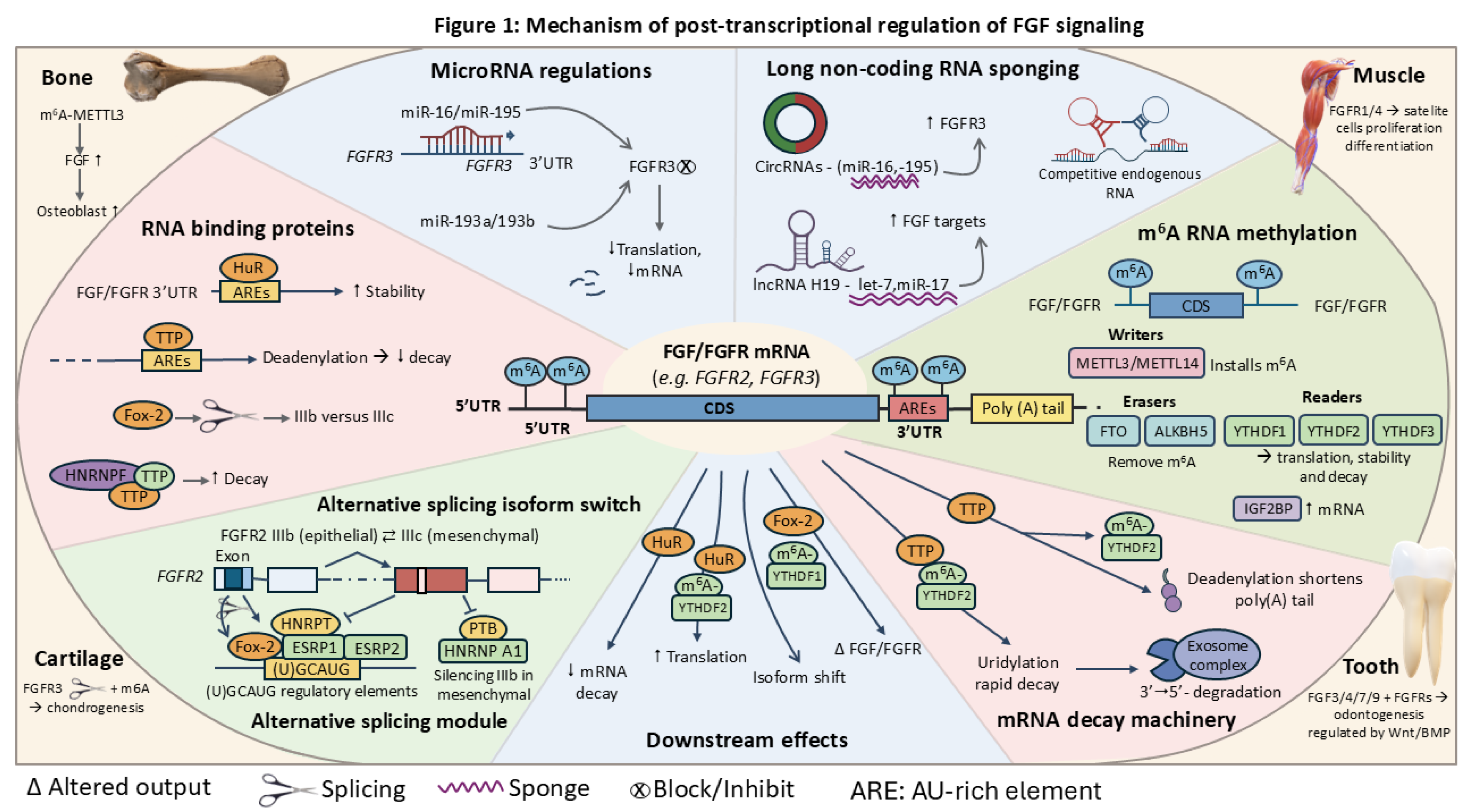
Layered post-transcriptional control of FGF/FGFR mRNAs across musculoskeletal tissues. FGF/FGFR transcripts are regulated by integrated RNA-based mechanisms acting on 5′UTR, CDS, and ARE-containing 3′UTR regions. MicroRNAs (miR-99a, miR-193a/b) repress FGFR3 translation and promote decay, counteracted by lncRNA/circRNA sponging (e.g., H19). RNA-binding proteins exert opposing effects, with HuR stabilizing and TTP–hnRNP F promoting degradation. Alternative splicing of FGFR2 (IIIb/IIIc) is controlled by Fox-2 and ESRP1/2, opposed by PTB/hnRNP A1. m6A modification, installed by METTL3/14 and interpreted by YTHDF and IGF2BP proteins, directs transcript stability, translation, or decay. These layers converge on mRNA turnover, isoform balance, and FGF/FGFR output in bone, muscle, cartilage, and tooth.
Figure 1.
Layered post-transcriptional control of FGF/FGFR mRNAs across musculoskeletal tissues. FGF/FGFR transcripts are regulated by integrated RNA-based mechanisms acting on 5′UTR, CDS, and ARE-containing 3′UTR regions. MicroRNAs (miR-99a, miR-193a/b) repress FGFR3 translation and promote decay, counteracted by lncRNA/circRNA sponging (e.g., H19). RNA-binding proteins exert opposing effects, with HuR stabilizing and TTP–hnRNP F promoting degradation. Alternative splicing of FGFR2 (IIIb/IIIc) is controlled by Fox-2 and ESRP1/2, opposed by PTB/hnRNP A1. m6A modification, installed by METTL3/14 and interpreted by YTHDF and IGF2BP proteins, directs transcript stability, translation, or decay. These layers converge on mRNA turnover, isoform balance, and FGF/FGFR output in bone, muscle, cartilage, and tooth.
2. MicroRNA-Mediated Regulation of FGF Signaling
MicroRNAs (miRNAs) are well-known critical post-transcriptional regulators of gene expression in multicellular organisms. They typically act through sequences in the 3′ untranslated regions (UTRs) of target transcripts to repress translation or promote mRNA degradation (Bartel, 2009; Ha and Kim, 2014). In the context of musculoskeletal biology, several miRNAs have been linked to cell differentiation, growth, and repair by modulating growth factor–driven pathways, including the fibroblast growth factor (FGF) signaling axis (Lian et al., 2012; Mirzamohammadi et al., 2014; Horak et al., 2016; Loh et al., 2023; Thakore and Delany, 2025).
Evidence from experimental musculoskeletal models indicates that members of the miR-16/15/195 family directly or indirectly target FGF2, a mitogenic growth factor involved in osteoblast proliferation and matrix production (Chang et al., 2017; Chen et al., 2017; Qi et al., 2020; Yang et al., 2024; Nikeghbal et al., 2025). In mesenchymal stem cells derived from bone marrow, overexpression of miR-16 reduces FGF2 expression and impairs osteogenic differentiation, an effect that can be rescued by restoring FGF2 expression or inhibiting miR-16 (Qi et al., 2020). A related miRNA, miR-195-5p, has been observed to inhibit FGF2 expression in human periodontal ligament stem cells under mechanical stretch, suggesting that miRNA–FGF interactions may respond to biomechanical stimuli during skeletal remodeling (Chang et al., 2017; Chen et al., 2017; Wang et al., 2018b; Nikeghbal et al., 2025). While earlier work focused largely on FGF2, recent studies brought forth evidence of the implication of other critical ligands such as miR-455 directly targeting FGF18 (Niu et al., 2025). The loss of this miRNA leads to dysregulated bone turnover and impaired skeletal homeostasis, providing a clear example of how specific miRNA–ligand pairs maintain the adult bone architecture through post-transcriptional control.
Additional work in ligamentogenic and craniofacial mesenchymal stem-cell models further supports this regulatory concept. These studies show that miR-15/16 family members (miR-16/miR-16-5p, miR-16a-5p and the closely related miR-195-5p) repress FGF2 expression and thereby influence lineage allocation of mesenchymal stem cells in response to mechanical or growth factor cues (Chang et al., 2017; Chen et al., 2017; Li et al., 2022a; Yang et al., 2024; Nikeghbal et al., 2025). In avian and mammalian species, differential miRNA expression patterns have also been correlated with cartilage growth plate maturation, although specific FGF targets have not always been experimentally validated in these models (Lin et al., 2016, 2018; Li et al., 2020; Yao et al., 2023).
Beyond bone, muscle tissue exhibits a distinct set of miRNAs known as myomiRs, including miR-1, miR-133, and miR-206 (Chen et al., 2005; Sweetman et al., 2006; Mitchelson and Qin, 2015; Horak et al., 2016). These miRNAs play essential roles in myogenesis and muscle repair, in part by targeting components of pro-proliferative signaling cascades that intersect with FGF pathways (Chen et al., 2005; Sweetman et al., 2006; Feng et al., 2013; Mitchelson and Qin, 2015; Ballarino et al., 2016; Galimov et al., 2016; Mok et al., 2017; Fochi et al., 2020). In embryonic and adult muscle, FGF–ERK signaling can antagonize or delay the induction of myomiRs, most clearly demonstrated for miR-206 in chick somite development. This delay maintains muscle progenitors in a proliferative state. As myomiR expression subsequently increases, these miRNAs feedback to restrict cell-cycle progression and promote myotube differentiation (Sweetman et al., 2006; Mok et al., 2017). A direct link to FGF receptor signaling has been demonstrated for miR-133, which post-transcriptionally represses FGFR1 and the ERK pathway component PP2AC in C2C12 myoblasts. this repression dampens FGF-dependent ERK1/2 activation and favors differentiation over proliferation (Feng et al., 2013). miR-206 promotes terminal myoblast differentiation by targeting multiple inhibitors of myogenesis, including Pax7, HDAC4, thereby counteracting pro-proliferative signals such as FGF2 (Chen et al., 2005; Mitchelson and Qin, 2015; Ballarino et al., 2016; Galimov et al., 2016; Fochi et al., 2020). Together with other FGF-responsive miRNAs such as miR-29a, these myomiRs ensure that proliferative FGF signals are attenuated once satellite cells have committed to differentiation (Chen et al., 2005; Ballarino et al., 2016; Galimov et al., 2016; Mok et al., 2017; Fochi et al., 2020). A similar proliferation–differentiation switch has been described for miR-1 in vertebrate muscle, where it cooperates with miR-133 to promote differentiation and repress cell-cycle genes. Its expression is modulated by pathways including FGF–ERK signaling, although direct FGF ligands or receptors by miR-1 has not been conclusively demonstrated (Chen et al., 2005; Sweetman et al., 2006; Mok et al., 2017; Fochi et al., 2020).
Cartilage provides another example of miRNA-driven modulation of FGF-related genes (Miyaki et al., 2010; Mirzamohammadi et al., 2014; Duan et al., 2020; Si et al., 2020; Li and Wu, 2021; Chaudhry et al., 2022). In porcine and human chondrocytes, miR-140 regulates cartilage homeostasis by targeting matrix degrading enzymes such as ADAMTS5, thereby limiting aggrecan degradation and indirectly influencing the extracellular environment in which FGF2 ligands function (Miyaki et al., 2010; Duan et al., 2020; Li and Wu, 2021; Chaudhry et al., 2022). More recent CRISPR–Cas9-based target validation has confirmed FGF2 as a mechanosensitive miR-140 target in human articular chondrocytes, demonstrating that miR-140 can directly regulate FGF2 expression under conditions of injury or inflammatory stress (Chaudhry et al., 2022). These findings complement in vivo studies showing that loss of miR-140 leads to defects in cartilage development and early-onset osteoarthritis, whereas miR-140 overexpression attenuates chondrocyte senescence and matrix degradation (Miyaki et al., 2010; Duan et al., 2020; Si et al., 2020; Li and Wu, 2021).
The regulatory mechanism of the FGF-miRNA axis is not limited to mammalian systems, and in fact appears to be deeply conserved across vertebrates. The same proliferation-differentiation switches that govern mammalian muscle and bone development can be observed during tissue regeneration in lower vertebrates fish models such as medaka and zebrafish. Thus fish models also provided valuable insight into miRNA regulation during fin development and regeneration, a process involving both FGF ligands and receptors (Thatcher et al., 2008; Yin et al., 2008; Ellman et al., 2013; Xie et al., 2020; Ribeiro et al., 2022; Fan et al., 2024). In zebrafish, global impairment of miRNA biogenesis or experimental manipulation of specific miRNAs, including miR-133 and miR-203, disrupts caudal fin regeneration, and FGF receptor inhibition alters miRNA expression profiles within the regeneration blastema (Thatcher et al., 2008; Yin et al., 2008; Fan et al., 2024).
FGF-dependent depletion of miR-133 promotes blastemal cell proliferation and appendage regrowth, highlighting a regulatory cascade, in which FGF signaling first reshapes the miRNA landscape, which in turn modulates downstream regenerative gene networks (Yin et al., 2008). Whether these miRNAs directly target FGF transcripts or act predominantly through parallel pathways, such as Wnt, BMP, or cell-cycle regulators, remains under investigation. Nonetheless, current evidence supports a conserved functional integration of FGF signaling with miRNA regulatory programs during vertebrate appendage regeneration (Thatcher et al., 2008; Yin et al., 2008; Ribeiro et al., 2022; Fan et al., 2024).
Altogether, these findings support the existence of tissue-specific and context-dependent miRNA networks that modulate FGF signaling across vertebrate species. The dynamic regulation of FGF ligands, receptors, and FGF-responsive downstream effectors by miRNAs enables precise temporal and spatial control of signaling outcomes during musculoskeletal development and repair (Table 1). However, only limited subsets of FGF ligands and receptors, most prominently FGF2 and FGFR1, have been validated as direct miRNA targets. Expanding functional studies to additional FGF family members and non-mammalian systems remains an important frontier for future research.
3. Single Nucleotide Variants Linked to miRNA–FGF Interaction
Single nucleotide polymorphisms (SNPs) located within the 3′ untranslated regions (3′UTRs) of mRNAs can influence post-transcriptional regulation by disrupting or enhancing microRNA (miRNA) binding sites (Saunders et al., 2007; Sethupathy and Collins, 2008; Liu et al., 2012; Moszyńska et al., 2017). These variants, often termed miRNA target-site SNPs, miRSNPs, or poly-miRTSs, can create, destroy, or quantitatively modulate miRNA recognition elements, thereby altering mRNA stability or translation efficiency (Saunders et al., 2007; Sethupathy and Collins, 2008; Liu et al., 2012; Moszyńska et al., 2017). Within the FGF pathway, miRSNPs have now been implicated in both craniofacial malformations and bone mineral density (BMD) variation, illustrating context-dependent effects on FGF ligand dosage (Lei et al., 2011; Dole and Delany, 2016; Li et al., 2016; Zhu et al., 2017; Yalaev et al., 2024). Although the majority of functional evidence has been derived from genetic studies in humans, bioinformatic surveys and GWAS-based fine-mapping suggest that similar regulatory principles are likely to apply across vertebrate species (Saunders et al., 2007; Yerges et al., 2010; Zmuda et al., 2011; Liu et al., 2012; Wu et al., 2014; Moszyńska et al., 2017; Yalaev et al., 2024).
In the context of craniofacial development, SNPs within the 3′UTRs of FGF2 (rs1048201), FGF5 (rs3733336), and FGF9 (rs546782) have been implicated in altered susceptibility to nonsyndromic orofacial clefts in a Han Chinese population (Li et al., 2016). These variants are thought to affect miRNA binding efficiency, thereby modulating transcript stability or translation. Li and colleagues systematically selected miRNA-binding site SNPs in FGFs/FGFRs. They showed that rs1048201 in the FGF2 3′UTR alters binding of miR-496, rs3733336 in the FGF5 3′UTR affects miR-145 binding, and rs546782 in the FGF9 3′UTR modulates miR-187 binding, as demonstrated by allele-specific dual-luciferase assays (Li et al., 2016). For FGF2/rs1048201, the T allele attenuated miR-496-mediated repression compared with the C allele and was associated with a slightly increased FGF2 mRNA level and a reduced risk of nonsyndromic orofacial cleft, suggesting that a marginally higher FGF2 dosage can be protective during lip and palate morphogenesis (Li et al., 2016). Similarly, rs3733336-G strengthened miR-145-dependent repression of FGF5, whereas rs546782-T weakened miR-187-mediated repression of FGF9, indicating that both increased and decreased miRNA binding can act in a protective manner, depending on the baseline requirement for each ligand during craniofacial development (Li et al., 2016). These data provide one of the clearest examples in which miRNA–FGF interactions, modified by 3′UTR SNPs, contribute directly to variation in craniofacial phenotype.
In skeletal tissues, miRSNPs influencing FGF signaling have been most clearly characterized for FGF2, where multiple 3′UTR variants have been associated with BMD. Lei et al. identified three polymorphisms (rs6854081, rs1048201, rs7683093) in the FGF2 3′UTR that lie within predicted miRNA-binding motifs, including sites for miR-146a/b, and found significant associations with femoral neck BMD in Caucasian women (Lei et al., 2011). Follow-up work in independent cohorts and meta-analyses confirmed that rs6854081 and rs1048201 are reproducibly associated with BMD, with allele-specific differences in predicted miRNA binding at these sites (Dole and Delany, 2016; Zhu et al., 2017; Tyurin et al., 2021; Yalaev et al., 2024). Zhu et al. subsequently provided direct functional evidence for rs1048201, by showing that the C allele enhances repression of FGF2 by miR-196a-3p, whereas the T allele weakens this interaction, leading to higher FGF2 expression and higher spinal BMD in Chinese subjects (Zhu et al., 2017). Together, these studies support a model in which FGF2 3′UTR miRSNPs fine-tune FGF2 abundance in osteoblast-lineage cells, thereby contributing to interindividual differences in bone mass and osteoporosis risk (Lei et al., 2011; Dole and Delany, 2016; Zhu et al., 2017; Tyurin et al., 2021; Yalaev et al., 2024).
Beyond FGF2, variants at or near FGF18 and FGFR3 loci have been linked to bone-related traits, although their roles as miRSNPs remain speculative. Candidate-gene and GWAS analyses have identified FGF18-region polymorphisms associated with vertebral trabecular BMD and with knee osteoarthritis risk, and FGFR3-region variants associated with volumetric BMD and fracture susceptibility (Yerges et al., 2010; Zmuda et al., 2011; Wu et al., 2014). However, most of these FGF18 and FGFR3 variants reside in intronic or coding regions rather than in experimentally confirmed miRNA recognition elements, and their effects on miRNA–mRNA binding have not yet been tested. In silico predictions sometimes place these SNPs close to putative miRNA seed sites, but functional validation in bone cells is lacking. In agricultural species, FGF-related loci such as FGFBP1 show polymorphisms associated with BMD and carcass traits, and FGF2-based SNP markers for growth traits in chickens have been described, but no clear miRNA target-site mechanisms have been reported yet in these settings (Hoppman et al., 2010; Wells et al., 2012; Felício et al., 2013).
In muscle regeneration, there is growing interest in the role of genetic variability in modulating satellite-cell responsiveness to FGF signals, As recently highlighted by Liu and Dong, 2025, these miRNA-mediated signals, including those within the FGF pathway, are increasingly recognized as part of a complex exosomal communication network that orchestrates skeletal muscle repair. While direct examples of FGF-focused miRSNPs in these myogenic exosomal pathwways are still emerging, variant in regulatory regions likely affect how efficiently FGFR transcripts are silenced by miRNAs such as miR-133 amd miR-206, which are known to target FGF signaling components or their downstream effectors in myoblasts and satellite cells (Chen et al., 2005; Feng et al., 2013; Pawlikowski et al., 2017; Liu and Dong, 2025). To date, however, no SNP in the 3′UTRs of FGFR1–FGFR4 has been functionally shown to alter myomiR binding in skeletal muscle, and evidence for miRNA target-site variants shaping FGF-dependent regeneration remains largely hypothetical. Population-level studies in livestock and zebrafish indicate that natural genetic variation in FGF pathway genes can influence muscle mass, body composition, or appendage regeneration (Xu et al., 2013; Wu et al., 2014), but the causal variants have generally not been mapped to specific miRNA–mRNA interfaces. However, GWAS for muscle mass in livestock have identified specific SNPs in the 3’UTRs of FGFR4 and FGF6 (Xu et al. 2013), which are prime candidates for future miRSNP testing to determine if they functionally alter the “proliferation-differentiation switch” during myogenesis.
The potential for miRSNPs to influence FGF signaling has also raised interest in personalized medicine. Functional FGF2 miRSNPs that modify BMD or craniofacial risk illustrate how allelic differences in miRNA-mediated repression can contribute to musculoskeletal phenotypes and might, in principle, influence responses to anabolic or anti-resorptive therapies (Lei et al., 2011; Dole and Delany, 2016; Li et al., 2016; Zhu et al., 2017; Tyurin et al., 2021; Yalaev et al., 2024). These functional miRSNPS constitute the regulatory architecture of the RNA regulome, illustrating how inherited sequence variation pre-determines the baseline efficiency of the FGF signaling cascade and explains inter-individual differences in development and disease susceptibility. If specific alleles confer increased or decreased sensitivity to miRNA-mediated repression, therapeutic strategies involving miRNA mimics or inhibitors may require genotype-based adjustment (Saunders et al., 2007; Moszyńska et al., 2017; Rykova et al., 2022). Moreover, the identification of functional miRSNPs in musculoskeletal disease genes could improve genetic risk prediction models by capturing regulatory variation that is not explained by coding SNPs alone (Saunders et al., 2007; Rykova et al., 2022; Bhatia et al., 2024). While very promising, this area of research is still emerging, and much of the current understanding is based on predictive algorithms rather than experimental confirmation (Saunders et al., 2007; Liu et al., 2012; Moszyńska et al., 2017; Rykova et al., 2022; Bhatia et al., 2024). Functional assays validating altered miRNA–mRNA interactions in musculoskeletal contexts remain sparse. Moving forward, integrative studies that combine fine-mapping of GWAS signals with 3′UTR reporter assays, CLIP-based mapping of miRNA–mRNA contacts, and allele-specific expression analyses in bone, cartilage, and muscle will be needed to determine how sequence variation modifies post-transcriptional FGF regulation and influences tissue phenotype.
4. Long Non-Coding RNAs and FGF Signaling
Long non-coding RNAs (lncRNAs), defined as transcripts exceeding 200 nucleotides that lack significant protein-coding capacity, have emerged as regulators of diverse biological processes, including gene expression, chromatin remodeling, and post-transcriptional regulation (Mercer et al., 2009; Fatica and Bozzoni, 2013; Kopp and Mendell, 2018; Statello et al., 2020). In musculoskeletal tissues, lncRNAs have been implicated in modulating signaling pathways that govern progenitor cell fate and tissue remodeling (Ballarino et al., 2016; Huynh et al., 2017; Silva et al., 2019; Wang et al., 2019; Sufianov et al., 2023), among which the FGF axis is increasingly recognized as a node that can be tuned either directly, via interactions with FGF ligands or receptors, or indirectly through control of downstream effector pathways (Razmara et al., 2019; Silva et al., 2019; Wang et al., 2022a; Sufianov et al., 2023; Jiang et al., 2024).
In osteogenic systems, several lncRNAs have been shown to modulate the expression or activity of FGF pathway components through competing endogenous RNA (ceRNA) mechanisms or direct protein interactions (Razmara et al., 2019; Silva et al., 2019; Li, 2022; Wang et al., 2022a; Sufianov et al., 2023; Jiang et al., 2024). For example, lncRNA MALAT1, well studied in cancer biology, has been reported to enhance osteogenic differentiation of bone marrow–derived mesenchymal stem cells (BMSCs), adipose-derived stem cells, and periodontal ligament stem cells by acting as a molecular sponge for miR-124-3p and other miRNAs, thereby upregulating IGF2BP1, RUNX2, and Wnt/β-catenin signaling (Lanzillotti et al., 2021; Li, 2022; Liu et al., 2022; Huang et al., 2023; Zhang et al., 2023; Gu et al., 2024). Rather than stabilizing FGFR1, current evidence indicates that MALAT1 promotes osteogenesis primarily by relieving miR-124-3p-mediated repression of IGF2BP1 and other osteogenic regulators, placing MALAT1 upstream of pathways that synergize with FGF-driven MAPK activity rather than directly targeting FGF receptors (Lanzillotti et al., 2021; Li, 2022; Liu et al., 2022; Huang et al., 2023; Zhang et al., 2023; Gu et al., 2024). A similar ceRNA mechanism has been described for lncRNA H19, which supports bone formation by regulating miR-22 and miR-141; in mesenchymal and dental stem cell populations, H19 sponging of these miRNAs increases SPAG9 and Wnt/β-catenin–Runx2 signaling, thereby promoting osteogenic commitment and modulating MAPK cascades that intersect with FGF signaling outputs (Li et al., 2019b; Zhou et al., 2021; Ping et al., 2022; Zhang et al., 2025b).
Figure 2.
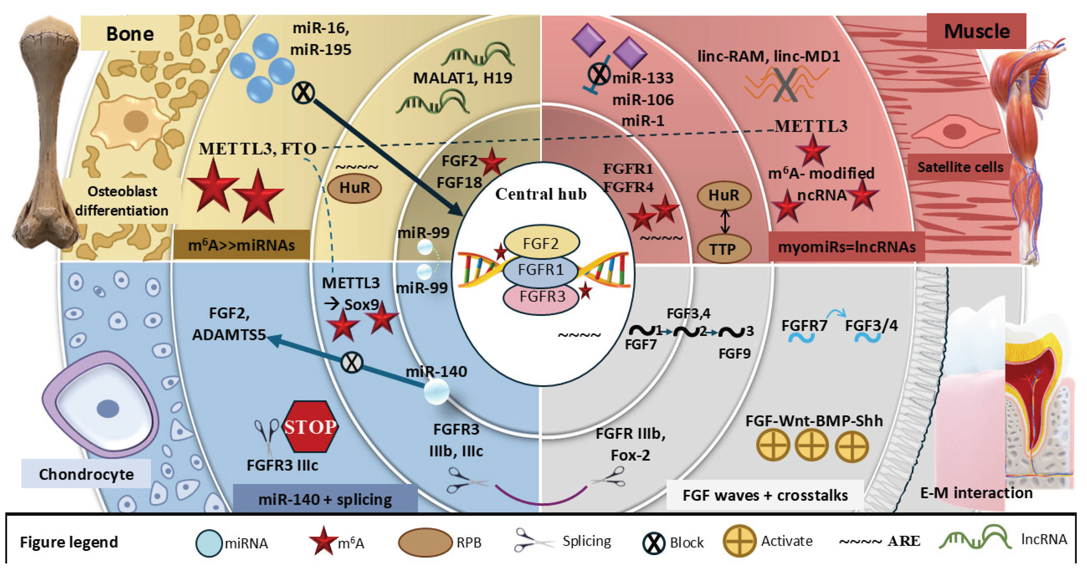
Tissue-specific RNA regulome of FGF signaling. A central FGF2–FGFR1–FGFR3 hub integrates m6A and AU-rich element regulation across four tissue contexts. In bone, METTL3/FTO-dominated m6A regulation intersects with miR-16/195, MALAT1/H19, and HuR to control osteoblast differentiation. In muscle, myomiRs (miR-1/133/206), lincRNAs, and HuR↔TTP balance regulate FGFR1/4 and satellite cell state. In cartilage, FGFR3 IIIb/IIIc splicing, miR-140 targeting, and METTL3–Sox9 signaling govern chondrocyte function. In tooth, sequential FGF3/4/7/9 signaling, FGFR2 IIIb splicing, and FGF–Wnt–BMP–Shh crosstalk drive epithelial–mesenchymal interactions. Shared regulators (METTL3, HuR, miRNAs, splicing factors) connect tissue modules.
Figure 2.
Tissue-specific RNA regulome of FGF signaling. A central FGF2–FGFR1–FGFR3 hub integrates m6A and AU-rich element regulation across four tissue contexts. In bone, METTL3/FTO-dominated m6A regulation intersects with miR-16/195, MALAT1/H19, and HuR to control osteoblast differentiation. In muscle, myomiRs (miR-1/133/206), lincRNAs, and HuR↔TTP balance regulate FGFR1/4 and satellite cell state. In cartilage, FGFR3 IIIb/IIIc splicing, miR-140 targeting, and METTL3–Sox9 signaling govern chondrocyte function. In tooth, sequential FGF3/4/7/9 signaling, FGFR2 IIIb splicing, and FGF–Wnt–BMP–Shh crosstalk drive epithelial–mesenchymal interactions. Shared regulators (METTL3, HuR, miRNAs, splicing factors) connect tissue modules.
Beyond ceRNA control of transcription factors, more direct connections between lncRNAs and FGF ligands have been uncovered in bone and tendon-related cells. lncRNA TUG1 promotes the osteogenic differentiation of tendon stem/progenitor cells by physically interacting with FGF2 and enhancing its ubiquitination, leading to reduced intracellular FGF2 protein levels and a shift from FGF2-driven proliferation toward mineralizing differentiation (Yu et al., 2020; Chen et al., 2024). In periodontal ligament stem cells, TUG1 also facilitates osteogenic differentiation by binding the RNA-binding protein Lin28A and regulating downstream pro-osteogenic transcripts (He et al., 2018), again converging on pathways that couple to FGF2 activity in the periodontal niche. More recently, NEAT1 has been shown to bind the 18-kDa FGF2 isoform, which in turn decreases FGF2-KPNB1 (importin-β) association in BMSCs, thereby modulating FGF2 nuclear localization, BMSC proliferation, and osteogenic differentiation. NEAT1 overexpression sequesters FGF2 away from KPNB1 and limits nuclear import, whereas NEAT1 knockdown enhances FGF2–KPNB1 interaction, FGF2 nuclear accumulation, and bone regeneration in vivo (Wang et al., 2025). In this context, NEAT1 is therefore a post-transcriptional regulator of BMSC aging through direct modulation of FGF2 intracellular transport. Together, these studies support a model in which osteogenic lncRNAs tune FGF signaling at multiple levels, from ligand abundance and subcellular trafficking to integration with Wnt and MAPK pathways.
In muscle tissues, lncRNAs are increasingly being recognized for their roles in satellite cell activation and differentiation (Ballarino et al., 2016; Wang et al., 2019; Martone et al., 2020; Lv et al., 2022). The lncRNA linc-MD1, for instance, modulates myoblast differentiation in mice by sponging miR-133 and miR-135, thereby derepressing MAML1 and MEF2C, two transcription factors that drive myogenic gene expression programs (Cesana et al., 2011; Legnini et al., 2014; Fu et al., 2015). Although linc-MD1 does not directly bind FGF transcripts, it operates in the same temporal window in which FGF2–ERK signaling maintains myoblast proliferation, suggesting that linc-MD1-mediated relief of miR-133/miR-135 repression is one mechanism by which cells transition from an FGF-responsive proliferative state to a differentiation-competent state (Cesana et al., 2011; Legnini et al., 2014; Fu et al., 2015; Martone et al., 2020). Another muscle-associated lncRNA, LncMyoD, is induced by MyoD and regulates cell-cycle exit and differentiation timing by interacting with MyoD and modulating chromatin accessibility at myogenic gene loci (Gong et al., 2015; Dong et al., 2020).
Importantly, several muscle lncRNAs show more direct regulatory relationships with FGF2. Linc-RAM, a MyoD-regulated lncRNA, enhances myogenic differentiation by facilitating assembly of the MyoD–BAF60c–Brg1 complex, and FGF2 represses Linc-RAM transcription through the Ras–Raf–MEK–ERK pathway; overexpression of Linc-RAM can rescue FGF2-induced inhibition of C2C12 differentiation, indicating that Linc-RAM functions as a critical intracellular mediator of FGF2’s anti-differentiation effects in satellite cells (Yu et al., 2017; Zhao et al., 2018; Hitachi and Tsuchida, 2020). In human periodontal ligament stem cells, downregulation of a “linc-RNA activator of myogenesis” transcript has been reported to participate in FGF2-mediated proliferation, further supporting the idea that FGF2 shapes lncRNA expression profiles, creating a feedback on musculoskeletal progenitor behavior (Wu et al., 2020). Additional muscle lncRNAs, such as lncMUMA and lncMREF, have been implicated in mechanical unloading–induced atrophy and regeneration, respectively, and are regulated in contexts where FGF2 is an established mitogenic cue (Cesana et al., 2011; Lv et al., 2022; Du et al., 2024), although direct binding to FGF components has yet to be fully defined.
Chondrogenic models offer fewer characterized lncRNAs directly linked to FGF, but several examples intersect with FGF-regulated matrix remodeling. In rat and human osteoarthritic cartilage, lncRNA-CIR (cartilage injury–related lncRNA) is upregulated and promotes extracellular matrix degradation by increasing the expression of MMP3, MMP13, and ADAMTS5, while also modulating autophagy (Liu et al., 2014; Li et al., 2017; Wang et al., 2018a; Tu et al., 2020). Because FGF2 can both induce matrix-anabolic responses and, under chronic or inflammatory conditions, stimulate MMP and ADAMTS expression, lncRNA-CIR-driven shifts in catabolic enzyme levels are likely to influence how chondrocytes respond to FGF2 during cartilage degeneration and attempted repair (Hu et al., 2018; Tu et al., 2020; Iantomasi et al., 2025). Additional lncRNAs such as HOTAIR, GAS5, MEG3, and XIST have been implicated in chondrocyte apoptosis, inflammatory signaling, and matrix turnover (Hu et al., 2018; Sun et al., 2019; Tu et al., 2020; Wang et al., 2021; Iantomasi et al., 2025). While these molecules have not been shown to bind FGF ligands or receptors directly, they regulate pathways (NF-κB, Wnt/β-catenin, Notch, and JAK/STAT) that functionally intersect with FGF signaling in articular cartilage and growth plate physiology (Ellman et al., 2013; Hu et al., 2018; Sun et al., 2019; Tu et al., 2020; Wang et al., 2021; Iantomasi et al., 2025).
In zebrafish, lncRNA expression patterns have been correlated with regenerative responses in fins and skeletal structures, where FGF signaling is essential for blastemal proliferation and position-dependent growth (Lee et al., 2005; King et al., 2018; Li et al., 2022b; Cudak et al., 2023). Whole-transcriptome profiling during caudal fin regeneration has revealed hundreds of differentially expressed lncRNAs, including lincRNA-154324, whose disruption impairs regenerative outgrowth (Li et al., 2022b). Although direct mechanistic links between specific lncRNAs and FGF pathway components have not been established in zebrafish fins, co-expression analyses and cis-regulatory relationships with genes in FGF-responsive domains suggest that lncRNAs may participate in wiring the transcriptional and epigenetic landscape through which FGF signals control appendage regeneration.
The functional relevance of lncRNA–FGF interactions is further underscored by the responsiveness of lncRNAs to environmental cues such as mechanical loading, oxidative stress, and cytokine exposure (Huynh et al., 2017; Silva et al., 2019; Tu et al., 2020; Sufianov et al., 2023; Deng and Wan, 2025). Mechanical strain and inflammatory mediators modulate the expression of MALAT1, H19, TUG1, NEAT1, and other lncRNAs in osteoblasts, BMSCs, chondrocytes, and tendon stem/progenitor cells, often in parallel with changes in FGF ligand production or FGF receptor expression (Huynh et al., 2017; Silva et al., 2019; Yu et al., 2020; Deng and Wan, 2025; Wang et al., 2025). These observations support a model in which lncRNAs act as context-dependent integrators, translating biomechanical and inflammatory inputs into post-transcriptional adjustments of FGF ligand abundance, receptor signaling competence, and downstream pathway sensitivity, thereby contributing to dynamic signaling adaptation under physiological and pathological conditions (Table 2). Within the framework of the pathway-specific RNA regulome, lncRNAs may occupy unique and versatile regulatory role, acting primarily at the level of mRNA stability and translation, they can simultaneously modulate FGF ligand subcellular localization, protein ubiquitination, and chromatin accessibility (LncMyoD), potentially representing a qualitatively distinct and multi-modal layer of post-transcriptional control. Despite growing evidence, direct lncRNA–FGF interactions remain limited to a handful of examples (TUG1, NEAT1, Linc-RAM). Expanding functional studies to cartilage and regenerative models will be critical to determine whether lncRNAs broadly act as modulators of FGF signaling or only in selected contexts.
5. Alternative Splicing and Isoform Regulation of FGF Components
Alternative splicing serves as a critical mechanism for expanding the functional diversity of the FGF signaling pathway across musculoskeletal tissues (Gong, 2014; Ornitz and Itoh, 2015; Ornitz and Legeai-Mallet, 2017; Xie et al., 2020). Both FGF ligands and their receptors (most prominently the FGFR1–3 pre-mRNAs) undergo alternative splicing or alternative translation initiation events that alter their biological activity, receptor-binding affinity, or cellular localization (Werner et al., 1992; Warzecha et al., 2009; Gong, 2014; Ornitz and Itoh, 2015; Xie et al., 2020; Derham and Kalsotra, 2023). These modifications allow cells to fine-tune signaling outputs in response to developmental stage, tissue type, or environmental conditions (Gong, 2014; Ornitz and Itoh, 2015; Ornitz and Legeai-Mallet, 2017; Xie et al., 2020). Among the FGFRs, FGFR1, FGFR2, and FGFR3 are known to exhibit isoform variation, particularly through mutually exclusive inclusion of exon IIIb or IIIc in the immunoglobulin-like domain III (Werner et al., 1992; Warzecha et al., 2009; Bebee et al., 2015; Derham and Kalsotra, 2023). This alteration modifies ligand-binding specificity and is especially relevant to epithelial–mesenchymal interactions that underlie bone, cartilage, and muscle development (Su et al., 2014; Bebee et al., 2015; Ornitz and Itoh, 2015; Ornitz and Marie, 2015; Ornitz and Legeai-Mallet, 2017).
In skeletal systems, the FGFR2 IIIc isoform is expressed predominantly in mesenchymal-derived cells such as osteoblasts and chondrocytes and functions as a positive regulator of ossification in these lineages (Eswarakumar et al., 2002; Su et al., 2014; Zhao et al., 2023; Zhou et al., 2023). Targeted deletion of Fgfr2IIIc in mice reduces osteoblast proliferation, delays bone collar formation, and shortens long bones, whereas Fgfr2c gain-of-function alleles that increase signaling cause craniosynostosis and bent-bone dysplasia (Eswarakumar et al., 2002, 2004; Merrill et al., 2012; Zhao et al., 2023; Zhou et al., 2023). At the biochemical level, the IIIc isoforms of FGFR1–3 preferentially bind mesenchymal FGFs such as FGF2, FGF4, FGF8, FGF9, and FGF18, whereas the IIIb isoforms bind epithelial FGFs of the FGF7 subfamily (including FGF7 and FGF10) with high affinity (Yeh et al., 2003; Olsen et al., 2004; Zhang et al., 2006; Zinkle and Mohammadi, 2019). In contrast, the FGFR2 IIIb isoform is more commonly expressed in epithelial tissues and responds to ligands like FGF7 and FGF10, which drive epithelial proliferation and branching morphogenesis in multiple organs (Miralles et al., 1999; Yeh et al., 2003; Olsen et al., 2004; Zinkle and Mohammadi, 2019). Together, reciprocal expression of FGFR2b in epithelia and FGFR2c in adjacent mesenchyme establishes epithelial–mesenchymal feedback loops that are reused during craniofacial skeletogenesis, limb bud outgrowth, and long-bone development (Miralles et al., 1999; Eswarakumar et al., 2002; Yeh et al., 2003; Su et al., 2014; Ornitz and Marie, 2015; Zinkle and Mohammadi, 2019; Zhao et al., 2023; Zhou et al., 2023). Rather than altering splicing per se, several disease-associated mutations at the D3–ligand interface of FGFR2c relax normal ligand specificity, allowing aberrant binding to FGF10 and other FGFR2b ligands and leading to craniosynostosis and limb malformations, underscoring the developmental importance of maintaining distinct IIIb and IIIc signaling domains (Yu et al., 2000; Eswarakumar et al., 2004; Ibrahimi et al., 2004; Merrill et al., 2012).
Alternative isoforms of FGF ligands have also been described, although in the musculoskeletal system FGF2 remains the best-characterized example (Warzecha et al., 2009; Xiao et al., 2009, 2010, 2014; Derham and Kalsotra, 2023). The Fgf2 gene encodes multiple high molecular weight (HMW; ≈21–24 kDa) isoforms initiated from upstream CUG codons and a low molecular weight (LMW; 18 kDa) isoform initiated at the canonical AUG (Xiao et al., 2009, 2010, 2014). The HMW isoforms contain nuclear localization sequences and accumulate predominantly in the nucleus, where they act in an intracrine manner to regulate gene expression and bone cell activity (Xiao et al., 2009, 2010, 2014; Homer-Bouthiette et al., 2014). In contrast, the LMW FGF2 isoform is exported, associates with the extracellular matrix, and participates in autocrine–paracrine signaling by activating FGFRs on neighboring cells (Xiao et al., 2009, 2010, 2014). Genetic dissection of these isoforms in mice has revealed that LMW FGF2 promotes endochondral bone formation and increases bone mass, whereas nuclear HMW FGF2 tends to suppress mineralization and elevate expression of Fgf23 and Sost, thereby exerting a negative influence on bone and phosphate homeostasis (Xiao et al., 2010, 2014; Homer-Bouthiette et al., 2014; Burt et al., 2016; Douglas Coffin et al., 2018) . Through these opposing actions, the balance between LMW and HMW FGF2 isoforms provides an additional layer of control over osteoblast proliferation, differentiation, and the coupling of osteogenesis with angiogenesis and phosphate metabolism (Xiao et al., 2010, 2014; Homer-Bouthiette et al., 2014; Burt et al., 2016; Douglas Coffin et al., 2018; Nickle et al., 2024).
In muscle tissues, most work has focused on changes in the overall abundance and signaling activity of FGFR1 and FGFR4 rather than on detailed mapping of IIIb/IIIc or kinase-domain splice choices (Templeton and Hauschka, 1992; Yu et al., 2004; Pawlikowski et al., 2017). Satellite cells and proliferating myoblasts express high levels of FGFR1 and respond to FGF1 and FGF2 with sustained ERK activation and cell-cycle progression, whereas differentiation is accompanied by downregulation of FGFR1 signaling and increased expression of myogenic regulatory factors (Templeton and Hauschka, 1992; Yu et al., 2004; Pawlikowski et al., 2017). Unlike FGFR1–3, FGFR4 does not undergo the canonical IIIb/IIIc ligand-binding domain switch, exhibiting instead a distinct form of isoform diversity through kinase domain splicing. A novel splice variant of FGFR4 (FGFR4-Δ16) has been identified in mouse myogenic cells; its expression correlates with that of wild-type FGFR4 during differentiation, and the two isoforms differ in glycosylation and tyrosine phosphorylation, suggesting isoform-dependent tuning of FGF responses in myotubes (Kwiatkowski et al., 2008). Notably, unlike canonical FGFRs, neither isoform undergoes homodimerization-dependent autophosphorylation; instead, FGFR4 tyrosine phosphorylation appears to require the presence of a heterologous kinase such as FGFR1, suggesting that FGFR4 signaling in myotubes may be fundamentally co-receptor-dependent rather than autonomous (Kwiatkowski et al., 2008). Although analogous studies of FGFR1 IIIb/IIIc usage in regenerating skeletal muscle are scarce, the general principle that mesenchymal tissues predominantly express IIIc isoforms (Werner et al., 1992; Bebee et al., 2015; Ornitz and Marie, 2015) implies that isoform composition is likely to change as myogenic precursors transit from a proliferative, FGF-dependent state to terminally differentiated myofibers, even if the precise splicing events in vivo remain to be fully defined (Templeton and Hauschka, 1992; Yu et al., 2004; Kwiatkowski et al., 2008; Pawlikowski et al., 2017).
Despite these insights, the regulatory mechanisms controlling FGF-related splicing in musculoskeletal contexts remain only partially defined (Table 3). In epithelial systems, the epithelial splicing regulatory proteins ESRP1 and ESRP2 are well-established determinants of FGFR2 IIIb versus IIIc exon choice, and TGF-β–induced epithelial–mesenchymal transition causes downregulation of ESRPs and a concomitant switch from epithelial to mesenchymal FGFR isoforms (Warzecha et al., 2009; Horiguchi et al., 2011; Holzmann et al., 2012; Wendt et al., 2014; Bebee et al., 2015; Ranieri et al., 2015; Derham and Kalsotra, 2023). Comparable splicing regulators have not yet been delineated in osteoblasts, chondrocytes, or satellite cells, and it is not clear whether mechanical stress, inflammatory cytokines, or local growth factor cues (including FGF itself) directly modulate FGFR splicing in these tissues. From the perspective of the pathway-specific RNA regulome, this represents a mechanistic asymmetry: while FGFR2 splicing in epithelial systems has been mapped to specific RNA-binding proteins (ESRP1/2), the equivalent regulators in osteoblasts, chondrocytes, and satellite cells remain unknown, leaving a significant gap in our understanding of how the RNA regulome operates in musculoskeletal tissues specifically. Further research across vertebrate models, combining isoform-specific reporters with perturbation of splicing factors, will be required to determine under which conditions FGF isoforms are selected and how they interact with broader signaling networks during tissue remodeling.
6. Emerging Post-Transcriptional Mechanisms Regulating FGF Signaling
While microRNAs, long non-coding RNAs, and splicing variants have received the most attention as post-transcriptional regulators of FGF signaling in musculoskeletal biology, several other RNA-level mechanisms have been observed or hypothesized to influence this pathway (Van Pelt et al., 2019; Huang et al., 2021; Shi and Grifone, 2021; Pashay Ahi, 2025). Recent work in bone, muscle, and cartilage suggests that RNA-binding proteins, epitranscriptomic RNA modifications, and untranslated-region (UTR)–dependent mechanisms can modulate the abundance or responsiveness of FGF ligands and receptors, although the evidence is still fragmentary compared with miRNAs and lncRNAs (Van Pelt et al., 2019; Huang et al., 2021; Shi and Grifone, 2021; Wang et al., 2023a; Pashay Ahi, 2025). In addition, structured 5′UTR elements such as internal ribosome entry sites (IRESs) and embedded RNA G-quadruplexes, as well as circular RNAs arising from FGF receptor loci, have begun to emerge as non-canonical post-transcriptional modulators of FGF-related signaling in musculoskeletal cells (Bonnal et al., 2003; Ainaoui et al., 2015; Pan et al., 2021; Du et al., 2022; Yan et al., 2022).
RNA-binding proteins (RBPs) regulate mRNA localization, translation, and decay across many cell types (Khabar, 2017; Shi and Grifone, 2021). In skeletal muscle, RBPs such as HuR (ELAVL1), PTB (PTBP1), and RBM24 contribute to myogenic differentiation and satellite-cell activation (Miyamoto et al., 2009; Hausburg et al., 2015; Grifone et al., 2020, 2021; Shi and Grifone, 2021). These proteins stabilize or repress specific transcripts during the transition from quiescent satellite cells to proliferating myoblasts and differentiated myotubes (Miyamoto et al., 2009; Hausburg et al., 2015; Grifone et al., 2020, 2021; Shi and Grifone, 2021).
HuR and the destabilizing factor TTP (ZFP36) cooperate to control the turnover of AU-rich element (ARE)–containing mRNAs in myogenic and stem-cell systems, and ARE catalogs indicate that several FGFR mRNAs (including FGFR1) harbor AU-rich motifs in their 3′UTRs, which are canonical binding sites for these RBPs (Peng et al., 1998; Khabar, 2017; Otsuka et al., 2019). Direct links between RBPs and FGF pathway mRNAs in musculoskeletal tissues are beginning to emerge: a recent study showed that the m⁶A reader IGF2BP1 binds FGFR1 mRNA in skeletal myoblasts, increasing its stability and translation in an m⁶A-dependent manner and thereby sustaining ERK signaling and inhibiting myogenic differentiation (Liu et al., 2024). In osteogenic contexts, the RBP Lin28 promotes proliferation and osteogenic differentiation of dental pulp and urine-derived stem cells by blocking let-7 maturation, thereby indirectly modulating networks (e.g., IGF2BP2/HMGA2, Wnt) that intersect with FGF-driven MAPK signaling (Wei et al., 2014; Ju et al., 2019; Wang et al., 2022b; Yan et al., 2024). Consistent with a broader role for RBPs in FGF control, the poly(C)-binding protein PCBP2 was recently identified as an intrinsic aging factor in human bone marrow mesenchymal stromal cells (hBMSCs): manipulating PCBP2 levels alters FGF2 expression and demonstrates that PCBP2 regulates replicative senescence through a ROS–FGF2 axis, although the precise post-transcriptional mechanism (direct versus indirect control of FGF2 mRNA) remains to be clarified (Chen et al., 2025). Whether Lin28 or other RBPs bind FGF ligands or FGFR transcripts directly in bone or cartilage remains unclear, but the combination of predicted AREs, RBP expression, and early IGF2BP1–FGFR1 data strongly supports RBP-mediated tuning of FGF receptor output in musculoskeletal cells (Peng et al., 1998; Khabar, 2017; Otsuka et al., 2019; Shi and Grifone, 2021; Liu et al., 2024).
RNA modifications, particularly N⁶-methyladenosine (m⁶A), have emerged as regulators of stem-cell differentiation and tissue regeneration (Yan et al., 2020; Wang et al., 2023a; Sun et al., 2024; Zhou et al., 2024). METTL3, a core component of the m⁶A methyltransferase complex, is upregulated during osteogenic differentiation of bone marrow or periodontal ligament stromal cells; gain- and loss-of-function experiments show that METTL3 promotes mineralization and alkaline phosphatase activity by installing m⁶A marks on key osteogenic transcripts such as RUNX2, OSX/SP7, and SMAD1, which are then stabilized or translationally enhanced by readers such as IGF2BP1 (Yan et al., 2020; Wang et al., 2023b; Bertels et al., 2024; Sun et al., 2024; Zhou et al., 2024). So far, FGF ligands and receptors have not been mapped as m⁶A targets in bone or cartilage, but multiple studies in other tissues demonstrate that FGF pathway transcripts are epitranscriptomically modified: FGF2 mRNA carries METTL3-dependent m⁶A marks that facilitate its expression in uterine spiral arteries and in breast cancer cells via the m⁶A reader YTHDF3, and FGFR2 and FGF13 show altered m⁶A methylation and expression after spinal cord injury (Ni et al., 2022; Gong et al., 2024; Liu et al., 2025a, 2025b). Within the skeleton itself, a newer layer of evidence has come from N⁷-methylguanosine (m⁷G): deletion of the m⁷G methyltransferase METTL1 in mesenchymal lineage cells causes severe limb shortening and impaired osteoblast/chondrocyte differentiation, and m⁷G-MeRIP sequencing identified Fgfr2 as a direct METTL1 target whose loss of m⁷G reduces mRNA stability and attenuates PI3K–AKT and MAPK signaling; pharmacologic or genetic reactivation of FGFR2 signaling rescues the skeletal phenotype (Li et al., 2025). In skeletal muscle, m⁶A methylation has been shown to regulate satellite-cell function: METTL3-mediated m⁶A on MyoD mRNA supports exercise-induced muscle growth, and YTHDF2 promotes myogenic differentiation in part by controlling the maturation of miR-378, a microRNA enriched in muscle regeneration (Koopmans et al., 2023; Deng et al., 2024; Feng et al., 2024). Together with the IGF2BP1–FGFR1 axis, these findings support a model in which epitranscriptomic marks on both FGF receptors and their downstream effectors modulate the intensity and duration of FGF signaling in musculoskeletal tissues.
Beyond covalent RNA modifications, UTR-embedded structural elements provide an additional level of control over FGF translation. The 5′ leader of human FGF2 mRNA contains a highly structured internal ribosome entry site (IRES) that includes an RNA G-quadruplex; this IRES is required for cap-independent initiation at three CUG and one AUG codon and thus governs the relative production of low- and high-molecular-weight FGF2 isoforms (Bonnal et al., 2003). Given that these same isoforms exert opposing effects on bone mass and phosphate homeostasis, IRES/G-quadruplex-mediated control of FGF2 translation is likely to be relevant for osteoblast function even though it has not yet been dissected directly in skeletal cells. A related mechanism has been described for FGF1 in myogenesis: during C2C12 myoblast differentiation, promoter A–derived FGF1 transcripts are translated through an IRES that is activated by the paraspeckle proteins p54nrb/NONO and hnRNPM; these RBPs bind both the FGF1 promoter and the IRES, coupling transcriptional induction to enhanced IRES-dependent translation of FGF1 and promoting myotube formation (Ainaoui et al., 2015). Together, these studies establish G-quadruplex–containing IRES elements and their associated IRES trans-acting factors as non-canonical post-transcriptional switches for FGF output that are likely to operate in musculoskeletal lineages.
Other RNA modifications, including A-to-I RNA editing by ADAR enzymes, 3′ uridylation, and regulation of poly(A) tail length, have not yet been linked directly to FGF signaling in musculoskeletal tissues. Nonetheless, these processes are known to regulate transcript stability and translational efficiency in a variety of developmental contexts (Charlesworth et al., 2013; Buchumenski et al., 2021; Piasecka et al., 2021; Shi and Grifone, 2021). In zebrafish, ADAR1-mediated A-to-I editing is essential for early embryonic patterning and neural crest–derived craniofacial structures, illustrating the potential for editing to impact signaling pathways that shape the craniofacial skeleton, even though FGF ligands and receptors have not been identified as direct editing substrates (Buchumenski et al., 2021; Niescierowicz et al., 2022). In human muscle cells, MEF2C-dependent 3′ uridylation of miRNAs contributes to fine-tuning of myogenic gene expression, and perturbation of this process alters the spectrum and stability of myomiRs during differentiation (Piasecka et al., 2021). Poly(A) tail length and alternative polyadenylation are also emerging as regulators of myogenic transcripts: nuclear poly(A)-binding protein 1 (PABPN1) and other polyadenylation factors influence mRNA isoform usage and stability in muscle, and their dysfunction leads to myopathy (Charlesworth et al., 2013; Shi and Grifone, 2021). At present, neither uridylation nor poly(A) length control has been tied specifically to FGF or FGFR transcripts in bone, cartilage, or muscle, but the enrichment of regulatory motifs in their UTRs makes them plausible future targets of these pathways.
Enhancer RNAs (eRNAs) represent another emerging regulatory class with potential relevance to FGF gene expression. These non-coding RNAs are transcribed from active enhancers and have been shown to promote transcriptional elongation, recruit transcription factors, and alter chromatin accessibility (Tsai et al., 2018; Lee et al., 2021). In muscle differentiation, MyoD-bound enhancers at the core enhancer (CE) and distal regulatory region (DRR) produce eRNAs that are required for robust activation of MyoD and Myogenin; the DRR eRNA, for example, associates with cohesin and is recruited in trans to the Myogenin locus to promote myogenic transcription (Tsai et al., 2018; Zhao et al., 2019; Lee et al., 2021). Although no eRNAs have yet been directly assigned to FGF loci in musculoskeletal tissues, the regulatory landscapes of key FGF genes are highly enriched in tissue-specific enhancers: integrated “holo-enhancer” units have been identified around the Fgf8 locus that drive expression in limb apical ectodermal ridge and craniofacial domains, and multiple cis-regulatory elements have been mapped for Fgf10 in limb bud, craniofacial mesenchyme, and other organs (Marinić et al., 2013; Kawakami et al., 2018; Jin et al., 2019; Hörnblad et al., 2021). Fgf18 expression in perichondrium and periosteum is also tightly controlled by upstream transcription factors such as RUNX2 and Wnt/β-catenin (Liu et al., 2002; Su et al., 2014). Given that many active enhancers elsewhere in the genome are now known to produce eRNAs, it is reasonable to hypothesize that enhancer clusters surrounding Fgf8, Fgf10, and Fgf18 may generate eRNAs that participate in rapid, context-specific modulation of FGF transcription during limb growth, craniofacial morphogenesis, and skeletal repair, even though such eRNAs have not yet been experimentally characterized.
Beyond linear RNAs, circular RNAs originating from FGF receptor loci have emerged as additional post-transcriptional regulators that intersect with FGF-connected signaling. In skeletal muscle, circFgfr2, generated from exons of the Fgfr2 gene, is upregulated during development and regeneration and promotes myogenesis by sponging miR-133, thereby relieving repression of the MAP3K20–JNK/MAPK cascade (Yan et al., 2022). In rat dental follicle cells, circFgfr2 similarly enhances osteogenic differentiation through a circFgfr2/miR-133/BMP6 axis, increasing the expression of RUNX2, DLX3 and other osteogenic markers (Pan et al., 2021; Du et al., 2022). Although circFgfr2 has not yet been shown to regulate FGFR2 mRNA itself, its genomic origin at the Fgfr2 locus and its control of MAPK and BMP pathways highlight circular RNAs as FGF-linked post-transcriptional regulators in muscle and tooth–bone interfaces.
Despite the speculative nature of some of these mechanisms in the FGF context, they represent important frontiers for investigation (see examples summary in Table 4). The convergence of transcriptional and post-transcriptional control at enhancer-associated loci, the influence of m⁶A and m⁷G methylation on mRNA turnover and translational competence, and the context-specific activity of RBPs and epitranscriptomic readers all suggest that FGF signaling is likely subject to a broader and more plastic RNA-level regulatory architecture than is currently appreciated. Recent work further adds a biophysical dimension to this view: nuclear FGF2 has been shown to form phase-separated condensates with rDNA and associated RNAs in BMSCs, reorganizing rDNA chromatin and biasing lineage choice between osteogenic and adipogenic fates, underscoring how FGF-dependent protein–RNA condensates can influence musculoskeletal cell identity (Zhang et al., 2025a). Elucidating these mechanisms, particularly how RBPs such as IGF2BP1 and Lin28, RNA modifications such as METTL1-mediated m⁷G on Fgfr2, and putative eRNAs at FGF loci intersect with classic miRNA/lncRNA networks, could enhance our understanding of tissue regeneration and disease, and may offer new molecular targets for therapeutic modulation of FGF signaling in musculoskeletal disorders.
Together, these findings suggest that the pathway-specific RNA regulome of FGF signaling extends well beyond the miRNA and lncRNA layers characterized in previous sections, encompassing a broader and more plastic set of regulatory inputs, whose full integration into a coherent regulatory model remains a major challenge and opportunity in the field.
7. Translational Perspectives: Tissue-Specific Regulation and Therapeutic Implications
The post-transcriptional regulation of FGF signaling in musculoskeletal tissues reveals a multifaceted system of control that varies not only across molecular mechanisms but also between tissue types (Ornitz and Marie, 2015; Xie et al., 2020; Loh et al., 2023). Bone, cartilage, muscle, and tooth each exhibit distinct transcriptomic environments in which microRNAs, long non-coding RNAs, and alternative splicing events modulate FGF ligand availability, receptor isoform usage, and downstream signaling outcomes (Li et al., 2014; Mirzamohammadi et al., 2014; Horak et al., 2016; Huynh et al., 2017). More recently, circular RNAs and epitranscriptomic marks such as m⁶A and m⁷G have added further layers of tissue- and lineage-specific regulation to this FGF “RNA regulome” (Su et al., 2014; Yan et al., 2022; Xu et al., 2024; Pashay Ahi, 2025). These differences are shaped by developmental timing, biomechanical inputs, injury responses, and cell-type composition (Su et al., 2014; Huynh et al., 2017). Recognizing these distinctions is essential for identifying tissue-specific regulatory vulnerabilities and guiding the design of targeted therapeutic interventions (Su et al., 2014; Ornitz and Marie, 2015; Xie et al., 2020; Loh et al., 2023).
In bone, the most extensively studied mechanisms include miRNA-mediated repression of FGF2 and FGF18 and the isoform-dependent activity of FGFR1 and FGFR2 during osteoblast differentiation (Su et al., 2014; Ornitz and Marie, 2015; Wang et al., 2017; Douglas Coffin et al., 2018; Qi et al., 2020). Yang et al. provided specific evidence for FGFR1 regulation by miR-214, reinforcing the role of miRNAs in modulating FGFR activity during osteoblast differentiation. Luciferase reporter assays confirmed FGFR1 as a direct miR-214 target, and miR-214 overexpression suppressed osteoblast differentiation and mineralization through inhibition of FGFR1-mediated FGF signaling in MSCs. This outcome is significant as it directly links miR-214 to FGFR1 repression, offering strong mechanistic support for miRNA-mediated regulation of FGF/FGFR signaling in bone (Yang et al., 2016).
Similarly, miR-16/miR-16a and miR-195 family members directly target FGF2 and FGF18 in osteogenic and periodontal ligament cells, while m⁶A-dependent miR-7212-5p–FGFR3 regulation modulates osteoblast and chondrocyte differentiation as well as fracture healing (Chang et al., 2017; Wang et al., 2017; Mi et al., 2020; Qi et al., 2020). These findings establish miRNA-mediated control of FGF/FGFR signaling as a central mechanism in bone biology. Li et al., provides general support for miRNA involvement in osteoblast differentiation (Li et al., 2009), while Sun et al., offer secondary evidence of FGF2 regulation in cancer biology, together reinforcing the FGF2/FGF18 and FGFR3 regulation (Sun et al., 2015).
In muscle, regulatory emphasis has been placed on FGFR1 and FGFR4 expression in satellite cells, modulated by myomiRs such as miR-1, miR-133, and miR-206 and by FGF1/FGF2 IRES-dependent translation (Chen et al., 2005; Sweetman et al., 2006; Feng et al., 2013; Ainaoui et al., 2015; Pawlikowski et al., 2017). The circRNA circFgfr2 adds an additional feedback loop by sponging miR-133 to tune MAP3K20–JNK/MAPK activity during myogenesis and regeneration (Yan et al., 2022).
Cartilage-specific regulation remains less well-characterized, though miR-140, lncRNA-CIR, and FGFR3 gain-of-function or isoform changes have been associated with chondrocyte proliferation, hypertrophy, and matrix remodeling (Miyaki et al., 2010; Liu et al., 2014; Mirzamohammadi et al., 2014; Ornitz and Legeai-Mallet, 2017; Chaudhry et al., 2022).
In dental mesenchyme and epithelium, the role of FGF ligands such as FGF8 and FGF10 in cusp patterning and root elongation has been demonstrated (Kettunen and Thesleff; Li et al., 2014; Du et al., 2018). Early work on lncRNAs such as H19 and MALAT1, as well as circFgfr2 in dental follicle–derived cells, suggests that post-transcriptional control of FGF-coupled pathways in tooth-supporting tissues is likely more extensive than currently appreciated (Li et al., 2019b; Sufianov et al., 2023; Xu et al., 2024).
Therapeutically, these insights raise the possibility of live cell-specific detection and modulation of FGF signaling at the RNA level to correct dysregulated tissue growth or repair (Douglas Coffin et al., 2018; Xie et al., 2020; Zhu et al., 2022; Loh et al., 2023; Ahi and Khorshid, 2025; Mancino et al., 2025; Pashay Ahi, 2025). In skeletal diseases characterized by excessive FGF activity, such as craniosynostosis or FGFR3-related chondrodysplasias, RNA-based ligands and oligonucleotides that blunt FGF signaling are under active development. A prominent example is the anti-FGF2 RNA aptamer RBM-007 (umedaptanib pegol), which neutralizes FGF2, reduces aberrant FGFR3 activation in growth plate chondrocytes, and restores bone growth in achondroplasia mouse models and human iPSC-derived cartilage xenografts (Kimura et al., 2021; Nakamura, 2021).
Aptamer engineering and delivery strategies specific for bone disease are being explored as a route to translate such FGF-directed RNA agents into the clinic (Liu et al., 2023). Conversely, in disorders marked by insufficient FGF expression or impaired FGF responsiveness, such as osteoporosis or disuse-associated bone loss and muscle wasting, stabilizing FGF mRNA transcripts through RNA-binding protein modulation or inhibition of degrading miRNAs may enhance regenerative capacity (Douglas Coffin et al., 2018; Zhu et al., 2022; Frank et al., 2025; Pashay Ahi, 2025; Khorshid and Ahi, 2026). Proof-of-concept studies using miRNA mimics or antagomirs delivered in bone-targeted scaffolds or nanoparticles, such as miRNA modulators to accelerate fracture healing and improve bone mass, illustrate how RNA-based control of growth factor networks (including FGF, BMP, and Wnt) can be harnessed to enhance skeletal repair (Douglas Coffin et al., 2018; Zhu et al., 2022; Ahi and Khorshid, 2025; Frank et al., 2025).
Non-coding RNAs also offer potential as biomarkers for musculoskeletal conditions (Mirzamohammadi et al., 2014; Horak et al., 2016; Huynh et al., 2017; Siracusa et al., 2018; Frank et al., 2025). Circulating miRNAs targeting FGF pathway components or their downstream effectors have been detected in serum during fracture healing and bone loss, and several groups have proposed miRNA signatures that distinguish normal from delayed or non-union healing (Zhu et al., 2022; Frank et al., 2025). In parallel, muscle-enriched myomiRs such as miR-1, miR-133, and miR-206 increase in circulation after acute muscle injury or exercise and are being developed as biomarkers of muscle damage and disease progression, with potential to report on FGF-dependent satellite-cell activation and myofiber turnover (Chen et al., 2005; Siracusa et al., 2018).
LncRNAs, due to their tissue specificity and longer half-life, could be harnessed to monitor disease progression or response to RNA-based therapies, as suggested by emerging data on MALAT1, H19, and NEAT1 in osteoporotic bone and osteoarthritic cartilage (Huynh et al., 2017; Li et al., 2019b; Sufianov et al., 2023).
In agricultural and veterinary contexts, FGF-related loci and their RNA regulators have been implicated in growth and carcass traits in poultry and livestock, indicating cross-species relevance for applied science and breeding strategies, even though FGF-specific post-transcriptional mechanisms remain less systematically explored in these species (Xie et al., 2020; Zhu et al., 2022).
The therapeutic translation of these mechanisms, however, is not without challenges. Delivery of RNA-based agents to dense or avascular musculoskeletal tissues remains technically complex, and unintended off-target effects on global FGF signaling must be carefully managed (Elangovan et al., 2020; Paunovska et al., 2022; Zhu et al., 2022; Sparmann and Vogel, 2023; Mancino et al., 2025). For bone and cartilage, limited vascularity and dense extracellular matrix impede diffusion of oligonucleotides and nanoparticles, necessitating localized delivery (e.g., intra-articular injections, bone-targeting peptides, calcium-based or bisphosphonate-modified nanocarriers) and raising questions about dosing, durability, and immunogenicity (Elangovan et al., 2020; Paunovska et al., 2022; Zhu et al., 2022).
In skeletal muscle, systemic delivery of antisense or siRNA drugs is already being deployed clinically for exon-skipping therapies, but translation to FGF-focused or myomiR-based interventions will require careful evaluation of long-term safety and potential interactions with other anabolic pathways (Sparmann and Vogel, 2023).
Furthermore, the redundancy among FGF ligands and receptors, together with extensive crosstalk with other signaling pathways, complicates single-target approaches. Combinatorial strategies that integrate upstream RNA regulators (miRNAs, lncRNAs, circRNAs, RBPs) with downstream signaling convergence nodes (e.g., MAPK, PI3K–AKT, STAT) may therefore offer greater specificity and therapeutic effectiveness (Xie et al., 2020; Zhu et al., 2022; Loh et al., 2023; Pashay Ahi, 2025).
In summary, post-transcriptional control of FGF signaling holds significant promise as both a diagnostic and therapeutic entry point in musculoskeletal biology (Ornitz and Marie, 2015; Douglas Coffin et al., 2018; Xie et al., 2020; Zhu et al., 2022; Loh et al., 2023; Mancino et al., 2025; Pashay Ahi, 2025). Understanding how RNA-based regulatory mechanisms operate in a tissue-specific manner may inform the development of interventions that restore proper signaling balance in developmental disorders, injury, and degenerative diseases (Su et al., 2014; Ornitz and Marie, 2015; Horak et al., 2016; Huynh et al., 2017).
Future work will benefit from in vivo functional validation of candidate RNA–FGF interactions, systematic multi-omics profiling of pathway-specific “RNA regulomes” in bone, cartilage, muscle, and tooth, and continued advances in delivery platforms, from bone-targeted nanoparticles and biomaterial scaffolds to clinically validated oligonucleotide chemistries and RNA aptamers, to facilitate the translation of FGF-directed RNA therapeutics from bench to bedside (Paunovska et al., 2022; Zhu et al., 2022; Sparmann and Vogel, 2023; Mancino et al., 2025; Pashay Ahi, 2025).
8. Conclusions
Post-transcriptional regulators of fibroblast growth factor (FGF) signaling in musculoskeletal development and regeneration, spanning miRNA-mediated repression, lncRNA-dependent nuclear organization, alternative splicing, epitranscriptomic modification, and RNA-binding protein activity, act collectively and constitute a pathway-specific RNA regulome, with an integrated activity modulating FGF signaling output in tissue- and context-dependent manner.
These mechanisms provide an additional layer of specificity, allowing cells to fine-tune FGF activity in response to developmental cues, environmental changes, and tissue damage. Across bone, cartilage, muscle, and tooth, the influence of RNA-level regulation is context-dependent and regulatory mechanisms at the pathway level appear to be conserved across vertebrate species. Functional interactions between non-coding RNAs and FGF pathway components have been observed in diverse vertebrate species, reinforcing the relevance of these findings beyond a single model organism and motivating cross-species validation as the field evolves. While many post-transcriptional modulators have been identified, the field remains in its early stages, with several regulatory axes lacking direct validation or mechanistic clarity. Importantly, the potential translational impact of these findings extends to diagnostic, therapeutic, and regenerative applications. By targeting RNA-mediated control points within the pathway specific RNA regulome, it may be possible to modulate FGF signaling with greater precision and specificity than with traditional receptor-targeted approaches. Continued research into tissue-specific RNA regulation of the FGF pathway is likely to uncover novel strategies for managing musculoskeletal disorders and enhancing endogenous repair mechanisms. Systematic mapping of the pathway-specific RNA regulome across musculoskeletal tissue types, disease states, and developmental windows represents both the immediate experimental priority and the conceptual foundation on which precision post-transcriptional therapeutics for skeletal and muscle disorders will ultimately be built.
References
- Ahi, E.P.; Khorshid, M. Potentials of RNA biosensors in developmental biology. Dev. Biol. 2025, 526, 173–188. [Google Scholar] [CrossRef] [PubMed]
- Ainaoui, N.; Hantelys, F.; Renaud-Gabardos, E.; Bunel, M.; Lopez, F.; Pujol, F.; Planes, R.; Bahraoui, E.; Pichereaux, C.; Burlet-Schiltz, O.; et al. Promoter-Dependent Translation Controlled by p54nrb and hnRNPM during Myoblast Differentiation. PLoS ONE 2015, 10, e0136466. [Google Scholar] [CrossRef]
- Allen, R.E.; Boxhorn, L.K. Regulation of skeletal muscle satellite cell proliferation and differentiation by transforming growth factor-beta, insulin-like growth factor I, and fibroblast growth factor. J. Cell. Physiol. 1999, 138, 311–315. [Google Scholar] [CrossRef]
- Armand, A.S.; Laziz, I.; Chanoine, C. FGF6 in myogenesis. Biochim. Biophys. Acta-Mol. Cell Res. 2006, 1763, 773–778. [Google Scholar] [CrossRef]
- Ballarino, M.; Morlando, M.; Fatica, A.; Bozzoni, I. Non-coding RNAs in muscle differentiation and musculoskeletal disease. J. Clin. Invest. 2016, 126, 2021–2030. [Google Scholar] [CrossRef] [PubMed]
- Bartel, D.P. MicroRNAs: Target Recognition and Regulatory Functions. Cell 2009, 136, 215–233. [Google Scholar] [CrossRef]
- Bebee, T.W.; Park, J.W.; Sheridan, K.I.; Warzecha, C.C.; Cieply, B.W.; Rohacek, A.M.; Xing, Y.; Carstens, R.P. The splicing regulators Esrp1 and Esrp2 direct an epithelial splicing program essential for mammalian development. Elife 4 2015. [Google Scholar]
- Bertels, J.C.; He, G.; Long, F. Metabolic reprogramming in skeletal cell differentiation. Bone Res. 2024, 2024 121 12, 57. [Google Scholar] [CrossRef]
- Bhatia, A.; Upadhyay, A.K.; Sharma, S. Screening and analysis of single nucleotide polymorphism in the 3′-UTR microRNA target regions and its implications for lung tumorigenesis. Pharmacogenomics 2024, 25, 299–314. [Google Scholar] [CrossRef]
- Bonnal, S.; Schaeffer, C.; Créancier, L.; Clamens, S.; Moine, H.; Prats, A.C.; Vagner, S. A Single Internal Ribosome Entry Site Containing a G Quartet RNA Structure Drives Fibroblast Growth Factor 2 Gene Expression at Four Alternative Translation Initiation Codons. J. Biol. Chem. 2003, 278, 39330–39336. [Google Scholar] [CrossRef]
- Buchumenski, I.; Holler, K.; Appelbaum, L.; Eisenberg, E.; Junker, J.P.; Levanon, E.Y. Systematic identification of A-to-I RNA editing in zebrafish development and adult organs. Nucleic Acids Res. 2021, 49, 4325. [Google Scholar] [CrossRef] [PubMed]
- Burt, P.M.; Xiao, L.; Dealy, C.; Fisher, M.C.; Hurley, M.M. FGF2 High Molecular Weight Isoforms Contribute to Osteoarthropathy in Male Mice. Endocrinology 2016, 157, 4602–4614. [Google Scholar] [CrossRef] [PubMed]
- Cesana, M.; Cacchiarelli, D.; Legnini, I.; Santini, T.; Sthandier, O.; Chinappi, M.; Tramontano, A.; Bozzoni, I. A long noncoding RNA controls muscle differentiation by functioning as a competing endogenous RNA. Cell 2011, 147, 358–369. [Google Scholar] [CrossRef]
- Chang, M.; Lin, H.; Fu, H.; Wang, B.; Han, G.; Fan, M. MicroRNA-195-5p Regulates Osteogenic Differentiation of Periodontal Ligament Cells Under Mechanical Loading. J. Cell. Physiol. 2017, 232, 3762–3774. [Google Scholar] [CrossRef]
- Charlesworth, A.; Meijer, H.A.; De Moor, C.H. Specificity factors in cytoplasmic polyadenylation. Wiley Interdiscip. Rev. RNA 2013, 4, 437–461. [Google Scholar] [CrossRef]
- Chaudhry, N.; Muhammad, H.; Seidl, C.; Downes, D.; Young, D.A.; Hao, Y.; Zhu, L.; Vincent, T.L. Highly efficient CRISPR-Cas9-mediated editing identifies novel mechanosensitive microRNA-140 targets in primary human articular chondrocytes. Osteoarthr. Cartil. 2022, 30, 596–604. [Google Scholar] [CrossRef] [PubMed]
- Chen, J.F.; Mandel, E.M.; Thomson, J.M.; Wu, Q.; Callis, T.E.; Hammond, S.M.; Conlon, F.L.; Wang, D.Z. The role of microRNA-1 and microRNA-133 in skeletal muscle proliferation and differentiation. Nat. Genet. 2005, 2005 382 38, 228–233. [Google Scholar] [CrossRef]
- Chen, L.; Deng, C.X. Roles of FGF signaling in skeletal development and human genetic diseases. Front. Biosci. 2005, 10, 1961–1976. [Google Scholar] [CrossRef]
- Chen, P.; Li, B.; Lu, Z.; Xu, Q.; Zheng, H.; Jiang, S.; Jiang, L.; Zheng, X. PCBP2 as an intrinsic agi ng factor regulates the senescence of hBMSCs through the ROS-FGF2 signaling axis. Elife 2025, 13. [Google Scholar]
- Chen, T.; Wang, K.; Sun, Z. New insights into the role of ubiquitination in angiogenesis (Review). Int. J. Mol. Med. 2024, 55, 32. [Google Scholar] [CrossRef]
- Chen, Z.; Zhang, Y.; Liang, C.; Chen, L.; Zhang, G.; Qian, A. Mechanosensitive miRNAs and Bone Formation. Int. J. Mol. Sci. 2017, Vol. 18, 1684 18: 1684. [Google Scholar] [CrossRef]
- Copeland, J.; Simoes-Costa, M. Post-transcriptional tuning of FGF signaling mediates neural crest induction. Proc. Natl. Acad. Sci. U. S. A. 2020, 117, 33305–33316. [Google Scholar] [CrossRef]
- Crose, L.E.S.; Etheridge, K.T.; Chen, C.; Belyea, B.; Talbot, L.J.; Bentley, R.C.; Linardic, C.M. FGFR4 blockade exerts distinct antitumorigenic effects in human embryonal versus alveolar rhabdomyosarcoma. Clin. Cancer Res. 2012, 18, 3780–3790. [Google Scholar] [CrossRef]
- Cudak, N.; López-Delgado, A.C.; Keil, S.; Knopf, F. Fibroblast growth factor pathway component expression in the regenerating zebrafish fin. Gene Expr. Patterns 2023, 48, 119307. [Google Scholar] [CrossRef]
- Dailey, L.; Ambrosetti, D.; Mansukhani, A.; Basilico, C. Mechanisms underlying differential responses to FGF signaling. Cytokine Growth Factor Rev. 2005, 16, 233–247. [Google Scholar] [CrossRef] [PubMed]
- Deng, H.; Wan, D. Mechanical loading regulates osteogenic differentiation and bone formation by modulating non-coding RNAs. PeerJ 2025, 13, e19310. [Google Scholar] [CrossRef]
- Deng, K.; Su, Y.; Liu, Z.; Hu, S.; Ren, C.; Wei, W.; Fan, Y.; Zhang, Y.; Wang, F. Ythdf2 facilitates precursor miR-378/miR-378-5p maturation to support myogenic differentiation. Cell. Mol. Life Sci. 2024, 81, 445. [Google Scholar] [CrossRef] [PubMed]
- Derham, J.M.; Kalsotra, A. The discovery, function, and regulation of epithelial splicing regulatory proteins (ESRP) 1 and 2. Biochem. Soc. Trans. 2023, 51, 1097–1109. [Google Scholar] [CrossRef]
- Ding, W.; Ding, L.; Lu, Y.; Sun, W.; Wang, Y.; Wang, J.; Gao, Y.; Li, M. Circular RNA-circLRP6 protects cardiomyocyte from hypoxia-induced apoptosis by facilitating hnRNPM-mediated expression of FGF-9. FEBS J. 2024, 291, 1246–1263. [Google Scholar] [CrossRef] [PubMed]
- Dole, N.S.; Delany, A.M. MicroRNA variants as genetic determinants of bone mass. Bone 2016, 84, 57–68. [Google Scholar] [CrossRef]
- Dong, A.; Preusch, C.B.; So, W.K.; Lin, K.; Luan, S.; Yi, R.; Wong, J.W.; Wu, Z.; Cheung, T.H. A long noncoding RNA, LncMyoD, modulates chromatin accessibility to regulate muscle stem cell myogenic lineage progression. Proc. Natl. Acad. Sci. U. S. A. 2020, 117, 32464–32475. [Google Scholar] [CrossRef]
- Douglas Coffin, J.; Homer-Bouthiette, C.; Hurley, M.M. Fibroblast Growth Factor 2 and Its Receptors in Bone Biology and Disease. J. Endocr. Soc. 2018, 2, 657–671. [Google Scholar] [CrossRef]
- Du, J.; Wu, Q.; Bae, E.J.; Du, J.; Wu, Q.; Bae, E.J. Epigenetics of Skeletal Muscle Atrophy. Int. J. Mol. Sci. 2024, Vol. 25(Page 8362 25), 8362. [Google Scholar] [CrossRef] [PubMed]
- Du, W.; Du, W.; Yu, H. The Role of Fibroblast Growth Factors in Tooth Development and Incisor Renewal. Stem Cells Int. 2018. [Google Scholar] [CrossRef] [PubMed]
- Du, Y.; Xu, C.; Li, G.; Li, J.; Jiang, H.; Ning, Y. Circular RNA CircFgfr2 Promotes Osteogenic Differentiation of Rat Dental Follicle Cells via Sponging MicroRNA miR-133a-3p and Up-regulating Distal-less Homeobox DLX3. 2022. [Google Scholar]
- Duan, L.; Liang, Y.; Xu, X.; Xiao, Y.; Wang, D. Recent progress on the role of miR-140 in cartilage matrix remodelling and its implications for osteoarthritis treatment. Arthritis Res. Ther. 2020, 22, 194. [Google Scholar] [CrossRef]
- Elangovan, S.; Gajendrareddy, P.; Ravindran, S.; Salem, A.K. Emerging local delivery strategies to enhance bone regeneration. Biomed. Mater. 2020, 15, 062001. [Google Scholar] [CrossRef]
- Ellman, M.B.; Yan, D.; Ahmadinia, K.; Chen, D.; An, H.S.; Im, H.J. Fibroblast growth factor control of cartilage homeostasis. J. Cell. Biochem. 2013, 114, 735–742. [Google Scholar] [CrossRef]
- Eswarakumar, V.P.; Horowitz, M.C.; Locklin, R.; Morriss-Kay, G.M.; Lonai, P. A gain-of-function mutation of Fgfr2c demonstrates the roles of this receptor variant in osteogenesis. Proc. Natl. Acad. Sci. 2004, 101, 12555–12560. [Google Scholar] [CrossRef]
- Eswarakumar, V.P.; Monsonego-Ornan, E.; Pines, M.; Antonopoulou, I.; Morriss-Kay, G.M.; Lonai, P. The IIIc alternative of Fgfr2 is a positive regulator of bone formation. Development 2002, 129, 3783–3793. [Google Scholar] [CrossRef]
- Fan, J.; Liu, X.; Duan, Z.; Zhao, H.; Chang, Z.; Li, L. The Regulatory Role of miRNAs in Zebrafish Fin Regeneration. Int. J. Mol. Sci. 2024, 25, 10542. [Google Scholar] [CrossRef]
- Fatica, A.; Bozzoni, I. Long non-coding RNAs: new players in cell differentiation and development. Nat. Rev. Genet. 2013, 2013 151 15, 7–21. [Google Scholar] [CrossRef]
- Felício, A.M.; Boschiero, C.; Balieiro, J.C.C.; Ledur, M.C.; Ferraz, J.B.S.; Moura, A.S.A.M.T.; Coutinho, L.L. Polymorphisms in FGFBP1 and FGFBP2 genes associated with carcass and meat quality traits in chickens. Genet. Mol. Res. 2013, 12, 208–222. [Google Scholar] [CrossRef]
- Feng, S.; Zhou, H.; Lin, X.; Zhu, S.; Chen, H.; Zhou, H.; Wang, R.; Wang, P.; Shao, X.; Wang, J. Exercise promotes skeletal muscle growth in adolescents via modulating Mettl3-mediated m6A methylation of MyoD in muscle satellite cells. Cell. Mol. Biol. Lett. 2024, 29, 150. [Google Scholar] [CrossRef]
- Feng, Y.; Niu, L.L.; Wei, W.; Zhang, W.Y.; Li, X.Y.; Cao, J.H.; Zhao, S.H. A feedback circuit between miR-133 and the ERK1/2 pathway involving an exquisite mechanism for regulating myoblast proliferation and differentiation. Cell Death Dis. 2013, 2013 411 4, e934–e934. [Google Scholar] [CrossRef]
- Fochi, S.; Giuriato, G.; De Simone, T.; Gomez-Lira, M.; Tamburin, S.; Del Piccolo, L.; Schena, F.; Venturelli, M.; Romanelli, M.G. Regulation of microRNAs in Satellite Cell Renewal, Muscle Function, Sarcopenia and the Role of Exercise. Int. J. Mol. Sci. 2020, Vol. 21, 6732 21: 6732. [Google Scholar] [CrossRef]
- Frank, J.K.; Kampleitner, C.; Heimel, P.; Leinfellner, G.; Hanetseder, D.; Sperger, S.; Frischer, A.; Schädl, B.; Tangl, S.; Lindner, C.; et al. Circulating miRNAs are associated with successful bone regeneration. Front. Bioeng. Biotechnol. 2025, 13, 1527493. [Google Scholar] [CrossRef] [PubMed]
- Fu, X.; Wang, H.; Hu, P. Stem cell activation in skeletal muscle regeneration. Cell. Mol. Life Sci. 2015, 72, 1663–1677. [Google Scholar] [CrossRef] [PubMed]
- Galimov, A.; Merry, T.L.; Luca, E.; Rushing, E.J.; Mizbani, A.; Turcekova, K.; Hartung, A.; Croce, C.M.; Ristow, M.; Krützfeldt, J. MicroRNA-29a in Adult Muscle Stem Cells Controls Skeletal Muscle Regeneration During Injury and Exercise Downstream of Fibroblast Growth Factor-2. Stem Cells 2016, 34, 768–780. [Google Scholar] [CrossRef] [PubMed]
- Gong, C.; Li, Z.; Ramanujan, K.; Clay, I.; Zhang, Y.; Lemire-Brachat, S.; Glass, D.J. A Long Non-coding RNA, LncMyoD, Regulates Skeletal Muscle Differentiation by Blocking IMP2-Mediated mRNA Translation. Dev. Cell 2015, 34, 181–191. [Google Scholar] [CrossRef]
- Gong, R.F.; Zhang, Z.H.; Sun, T.T.; Zhao, Y.X.; Fang, W. YTHDF3 modulates the progression of breast cancer cells by regulating FGF2 through m6A methylation. Front. Cell Dev. Biol. 2024, 12, 1438515. [Google Scholar] [CrossRef]
- Gong, S.G. Isoforms of Receptors of Fibroblast Growth Factors. J. Cell. Physiol. 2014, 229, 1887–1895. [Google Scholar] [CrossRef]
- Grifone, R.; Saquet, A.; Desgres, M.; Sangiorgi, C.; Gargano, C.; Li, Z.; Coletti, D.; Shi, D.L. Rbm24 displays dynamic functions required for myogenic differentiation during muscle regeneration. Sci. Rep. 2021, 2021 111 11, 9423. [Google Scholar] [CrossRef]
- Grifone, R.; Shao, M.; Saquet, A.; Shi, D.L. RNA-Binding Protein Rbm24 as a Multifaceted Post-Transcriptional Regulator of Embryonic Lineage Differentiation and Cellular Homeostasis. Cells 2020, Vol. 9, 1891 9: 1891. [Google Scholar] [CrossRef]
- Gu, N.; Wang, Y.; Li, L.; Sui, X.; Liu, Z. The mechanism of lncRNA MALAT1 targeting the miR-124-3p/IGF2BP1 axis to regulate osteogenic differentiation of periodontal ligament stem cells. Clin. Oral. Investig. 2024, 28, 219. [Google Scholar] [CrossRef] [PubMed]
- Guo, Y.C.; Yuan, Q. Fibroblast growth factor 23 and bone mineralisation. Int. J. Oral. Sci. 2015, 71 7, 8–13. [Google Scholar] [CrossRef]
- Ha, M.; Kim, V.N. Regulation of microRNA biogenesis. Nat. Rev. Mol. Cell Biol. 2014, 2014 158 15, 509–524. [Google Scholar] [CrossRef] [PubMed]
- Hacohen, N.; Kramer, S.; Sutherland, D.; Hiromi, Y.; Krasnow, M.A. sprouty encodes a novel antagonist of FGF signaling that patterns apical branching of the Drosophila airways. Cell 1998, 92, 253–263. [Google Scholar] [CrossRef]
- Hamidouche, Z.; Fromigué, O.; Nuber, U.; Vaudin, P.; Pages, J.C.; Ebert, R.; Jakob, F.; Miraoui, H.; Marie, P.J. Autocrine fibroblast growth factor 18 mediates dexamethasone-induced osteogenic differentiation of murine mesenchymal stem cells. J. Cell. Physiol. 2010, 224, 509–515. [Google Scholar] [CrossRef] [PubMed]
- Hausburg, M.A.; Doles, J.D.; Clement, S.L.; Cadwallader, A.B.; Hall, M.N.; Blackshear, P.J.; Lykke-Andersen, J.; Olwin, B.B. Post-transcriptional regulation of satellite cell quiescence by TTP-mediated mRNA decay. Elife, 2015. [Google Scholar]
- He, Q.; Yang, S.; Gu, X.; Li, M.; Wang, C.; Wei, F. Long noncoding RNA TUG1 facilitates osteogenic differentiation of periodontal ligament stem cells via interacting with Lin28A. Cell Death Dis. 2018, 95 9, 455. [Google Scholar] [CrossRef]
- Hitachi, K.; Tsuchida, K. Regulatory Roles of Long Non-coding RNAs in Skeletal Muscle Differentiation, Regeneration, and Disorders. RNA Technol. 2020, 11, 431–463. [Google Scholar]
- Holzmann, K.; Grunt, T.; Heinzle, C.; Sampl, S.; Steinhoff, H.; Reichmann, N.; Kleiter, M.; Hauck, M.; Marian, B. Alternative Splicing of Fibroblast Growth Factor Receptor IgIII Loops in Cancer. J. Nucleic Acids 2012, 2012, 950508. [Google Scholar] [CrossRef]
- Homer-Bouthiette, C.; Doetschman, T.; Xiao, L.; Hurley, M.M. Knockout of nuclear high molecular weight FGF2 isoforms in mice modulates bone and phosphate homeostasis. J. Biol. Chem. 2014, 289, 36303–36314. [Google Scholar] [CrossRef] [PubMed]
- Hoppman, N.; McLenithan, J.C.; McBride, D.J.; Shen, H.; Bruder, J.; Bauer, R.L.; Shaffer, J.R.; Liu, J.; Streeten, E.A.; Shuldiner, A.R.; et al. A common variant in fibroblast growth factor binding protein 1 (FGFBP1) is associated with bone mineral density and influences gene expression in vitro. Bone 2010, 47, 272–280. [Google Scholar] [CrossRef]
- Horak, M.; Novak, J.; Bienertova-Vasku, J. Muscle-specific microRNAs in skeletal muscle development. Dev. Biol. 2016, 410, 1–13. [Google Scholar] [CrossRef]
- Hori, M.; Shimizu, Y.; Fukumoto, S. Minireview: Fibroblast Growth Factor 23 in Phosphate Homeostasis and Bone Metabolism. Endocrinology 2011, 152, 4–10. [Google Scholar] [CrossRef]
- Horiguchi, K.; Sakamoto, K.; Koinuma, D.; Semba, K.; Inoue, A.; Inoue, S.; Fujii, H.; Yamaguchi, A.; Miyazawa, K.; Miyazono, K.; et al. TGF-β drives epithelial-mesenchymal transition through δEF1-mediated downregulation of ESRP. Oncogene 2011, 2012 3126 31, 3190–3201. [Google Scholar] [CrossRef]
- Hörnblad, A.; Bastide, S.; Langenfeld, K.; Langa, F.; Spitz, F. Dissection of the Fgf8 regulatory landscape by in vivo CRISPR-editing reveals extensive intra- and inter-enhancer redundancy. Nat. Commun. 2021, 2021 121 12, 439. [Google Scholar] [CrossRef]
- Hu, J.; Wang, Z.; Shan, Y.; Pan, Y.; Ma, J.; Jia, L. Long non-coding RNA HOTAIR promotes osteoarthritis progression via miR-17-5p/FUT2/β-catenin axis. Cell Death Dis. 2018, 97 9, 711. [Google Scholar] [CrossRef]
- Huang, M.; Xu, S.; Liu, L.; Zhang, M.; Guo, J.; Yuan, Y.; Xu, J.; Chen, X.; Zou, J. m6A Methylation Regulates Osteoblastic Differentiation and Bone Remodeling. Front. Cell Dev. Biol. 2021, 9, 783322. [Google Scholar] [CrossRef] [PubMed]
- Huang, X.; Jie, S.; Li, W.; Liu, C. GATA4-activated lncRNA MALAT1 promotes osteogenic differentiation through inhibiting NEDD4-mediated RUNX1 degradation. Cell Death Discov. 2023, 2023 91 9, 150. [Google Scholar] [CrossRef]
- Huynh, N.P.T.; Anderson, B.A.; Guilak, F.; McAlinden, A. Emerging roles for long noncoding RNAs in skeletal biology and disease. Connect. Tissue Res. 2017, 58, 116–141. [Google Scholar] [CrossRef] [PubMed]
- Iantomasi, T.; Aurilia, C.; Donati, S.; Falsetti, I.; Palmini, G.; Carossino, R.; Zonefrati, R.; Ranaldi, F.; Brandi, M.L. Oxidative Stress, MicroRNAs, and Long Non-Coding RNAs in Osteoarthritis Pathogenesis: Cross-Talk and Molecular Mechanisms Involved. Int. J. Mol. Sci. 2025, Vol. 26, 6428 26: 6428. [Google Scholar] [CrossRef] [PubMed]
- Ibrahimi, O.A.; Zhang, F.; Eliseenkova, A. V.; Itoh, N.; Linhardt, R.J.; Mohammadi, M. Biochemical analysis of pathogenic ligand-dependent FGFR2 mutations suggests distinct pathophysiological mechanisms for craniofacial and limb abnormalities. Hum. Mol. Genet. 2004, 13, 2313–2324. [Google Scholar] [CrossRef]
- Imbriano, C.; Moresi, V.; Belluti, S.; Renzini, A.; Cavioli, G.; Maretti, E.; Molinari, S. Epitranscriptomics as a New Layer of Regulation of Gene Expression in Skeletal Muscle: Known Functions and Future Perspectives. Int. J. Mol. Sci. 2023, Vol. 24, 15161 24: 15161. [Google Scholar] [CrossRef]
- Jernvall, J.; Kettunen, P.; Karavanova, I.; Martin, L.B.; Thesleff, I. Evidence for the role of the enamel knot as a control center in mammalian tooth cusp formation: non-dividing cells express growth stimulating Fgf-4 gene. Int. J. Dev. Biol. 1994, 38, 463–469. [Google Scholar] [CrossRef] [PubMed]
- Jiang, C.; Wang, P.; Tan, Z.W.; Zhang, Y. Long non-coding RNAs in bone formation: Key regulators and therapeutic prospects. Open Life Sci. 2024, 19, 20220908. [Google Scholar] [CrossRef]
- Jin, L.; Wu, J.; Bellusci, S.; Zhang, J.S. Fibroblast growth factor 10 and vertebrate limb development. Front. Genet. 2019, 10, 417988. [Google Scholar] [CrossRef]
- Ju, C.; Liu, R.; Zhang, Y.W.; Zhang, Y.; Zhou, R.; Sun, J.; Lv, X. Bin; Zhang, Z. Mesenchymal stem cell-associated lncRNA in osteogenic differentiation. Biomed. Pharmacother. 2019, 115, 108912. [Google Scholar] [CrossRef]
- Kawakami, H.; Johnson, A.; Fujita, Y.; Swearer, A.; Wada, N.; Kawakami, Y. Characterization of cis-regulatory elements for Fgf10 expression in the chick embryo. Dev. Dyn. 2018, 247, 1253–1263. [Google Scholar] [CrossRef]
- Kettunen, P.; Laurikkala, J.; Ita¨ranta, P.; Ita¨ranta, I.; Vainio, S.; Itoh, N.; Thesleff, I. Associations of FGF-3 And FGF-10 With Signaling Networks Regulating Tooth Morphogenesis. 2000. [Google Scholar] [CrossRef]
- Kettunen, P.; Thesleff, I. Expression and Function of FGFs-4,-8, and-9 Suggest Functional Redundancy and Repetitive Use as Epithelial Signals During Tooth Morphogenesis. [CrossRef]
- Khabar, K.S.A. Hallmarks of cancer and AU-rich elements. Wiley Interdiscip. Rev. RNA 2017, 8, e1368. [Google Scholar] [CrossRef]
- Khorshid, M.; Ahi, E.P. RNA Aptamers and Epitranscriptomics: Charting Unexplored Territories in RNA Biology. Mol. Diagn. Ther. 2026. [Google Scholar] [CrossRef] [PubMed]
- Kimura, T.; Bosakova, M.; Nonaka, Y.; Hruba, E.; Yasuda, K.; Futakawa, S.; Kubota, T.; Fafilek, B.; Gregor, T.; Abraham, S.P.; et al. An RNA aptamer restores defective bone growth in FGFR3-related skeletal dysplasia in mice. Sci. Transl. Med. 2021, 13. [Google Scholar] [CrossRef] [PubMed]
- King, B.L.; Rosenstein, M.C.; Smith, A.M.; Dykeman, C.A.; Smith, G.A.; Yin, V.P. RegenDbase: a comparative database of noncoding RNA regulation of tissue regeneration circuits across multiple taxa. npj Regen. Med. 2018, 31 3, 10. [Google Scholar] [CrossRef]
- Koopmans, P.J.; Ismaeel, A.; Goljanek-Whysall, K.; Murach, K.A. The roles of miRNAs in adult skeletal muscle satellite cells. Free Radic. Biol. Med. 2023, 209, 228–238. [Google Scholar] [CrossRef]
- Kopp, F.; Mendell, J.T. Functional Classification and Experimental Dissection of Long Noncoding RNAs. Cell 2018, 172, 393–407. [Google Scholar] [CrossRef]
- Kwiatkowski, B.A.; Kirillova, I.; Richard, R.E.; Israeli, D.; Yablonka-Reuveni, Z. FGFR4 and its novel splice form in myogenic cells: Interplay of glycosylation and tyrosine phosphorylation. J. Cell. Physiol. 2008, 215, 803–817. [Google Scholar] [CrossRef]
- Lanzillotti, C.; De Mattei, M.; Mazziotta, C.; Taraballi, F.; Rotondo, J.C.; Tognon, M.; Martini, F. Long Non-coding RNAs and MicroRNAs Interplay in Osteogenic Differentiation of Mesenchymal Stem Cells. Front. Cell Dev. Biol. 2021, 9, 646032. [Google Scholar] [CrossRef]
- Lee, J.H.; Wang, R.; Xiong, F.; Krakowiak, J.; Liao, Z.; Nguyen, P.T.; Moroz-Omori, E. V.; Shao, J.; Zhu, X.; Bolt, M.J.; et al. Enhancer RNA m6A Methylation Facilitates Transcriptional Condensate Formation and Gene Activation. Mol. Cell 2021, 81, 3368. [Google Scholar] [CrossRef]
- Lee, Y.; Grill, S.; Sanchez, A.; Murphy-Ryan, M.; Poss, K.D. Fgf signaling instructs position-dependent growth rate during zebrafish fin regeneration. Development 2005, 132, 5173–5183. [Google Scholar] [CrossRef]
- Legnini, I.; Morlando, M.; Mangiavacchi, A.; Fatica, A.; Bozzoni, I. A Feedforward Regulatory Loop between HuR and the Long Noncoding RNA linc-MD1 Controls Early Phases of Myogenesis. Mol. Cell 2014, 53, 506–514. [Google Scholar] [CrossRef]
- Lei, S.F.; Papasian, C.J.; Deng, H.W. Polymorphisms in predicted miRNA binding sites and osteoporosis. J. Bone Miner. Res. 2011, 26, 72–78. [Google Scholar] [CrossRef]
- Li, C.Y.; Prochazka, J.; Goodwin, A.F.; Klein, O.D. Fibroblast growth factor signaling in mammalian tooth development. Odontology 2014, 102, 1–13. [Google Scholar] [CrossRef]
- Li, D.; Zhang, H.; Ma, L.; Han, Y.; Xu, M.; Wang, Z.; Jiang, H.; Zhang, W.; Wang, L.; Pan, Y. Associations between microRNA binding site SNPs in FGFs and FGFRs and the risk of non-syndromic orofacial cleft. Sci. Rep. 2016, 61 6, 31054. [Google Scholar] [CrossRef]
- Li, J.; Ge, L.; Zhao, Y.; Zhai, Y.; Rao, N.; Yuan, X.; Yang, J.; Li, J.; Yu, S. TGF-β2 and TGF-β1 differentially regulate the odontogenic and osteogenic differentiation of mesenchymal stem cells. Arch. Oral. Biol. 2022a, 135, 105357. [Google Scholar] [CrossRef]
- Li, J.; Wen, W.; Zhang, S.; Zhou, C.; Feng, Y.; Li, X. The Expression and Function of lincRNA-154324 and the Adjoining Protein-Coding Gene vmp1 in the Caudal Fin Regeneration of Zebrafish. Int. J. Mol. Sci. 2022b, 23, 8944. [Google Scholar] [CrossRef]
- Li, Q.; Zhang, Y.; Ji, P.; Zhang, Y.; Jiang, J.; Jin, J.; Yuan, Z.; Tian, G.; Cai, M.; Feng, P.; et al. Mettl1-mediated m7G modification of Fgfr2 regulates osteogenic and chondrogenic differentiation of mesenchymal stem cells. Int. J. Biol. Sci. 2025, 21, 5704. [Google Scholar] [CrossRef]
- Li, S.H.; Wu, Q.F. MicroRNAs target on cartilage extracellular matrix degradation of knee osteoarthritis. Eur. Rev. Med. Pharmacol. Sci. 2021, 25, 1185–1197. [Google Scholar]
- Li, X. LncRNA metastasis-associated lung adenocarcinoma transcript-1 promotes osteogenic differentiation of bone marrow stem cells and inhibits osteoclastic differentiation of Mø in osteoporosis via the miR-124-3p/IGF2BP1/Wnt/β-catenin axis. J. Tissue Eng. Regen. Med. 2022, 16, 311–329. [Google Scholar] [CrossRef]
- Li, Y.F.; Li, S.H.; Liu, Y.; Luo, Y.T. Long Noncoding RNA CIR Promotes Chondrocyte Extracellular Matrix Degradation in Osteoarthritis by Acting as a Sponge For Mir-27b. Cell. Physiol. Biochem. 2017, 43, 602–610. [Google Scholar] [CrossRef]
- Li, Y.P.; Duan, F.F.; Zhao, Y.T.; Gu, K.L.; Liao, L.Q.; Su, H. Bin; Hao, J.; Zhang, K.; Yang, N.; Wang, Y. A TRIM71 binding long noncoding RNA Trincr1 represses FGF/ERK signaling in embryonic stem cells. Nat. Commun. 2019a, 2019 101 10, 1–13. [Google Scholar] [CrossRef]
- Li, Z.; Hassan, M.Q.; Jafferji, M.; Aqeilan, R.I.; Garzon, R.; Croce, C.M.; van Wijnen, A.J.; Stein, J.L.; Stein, G.S.; Lian, J.B. Biological functions of miR-29b contribute to positive regulation of osteoblast differentiation. J. Biol. Chem. 2009, 284, 15676–15684. [Google Scholar] [CrossRef]
- Li, Z.; Li, A.; Zhang, J.; Wang, Y.; Zhang, H.; Mehmood, K.; Lian, Y.; Iqbal, M.; Li, J. Identification and expression analysis of microRNAs in tibial growth plate of chicken through thiram toxicity. Environ. Sci. Pollut. Res. 2020, 27, 6628–6636. [Google Scholar] [CrossRef]
- Li, Z.; Yan, M.; Yu, Y.; Wang, Y.; Lei, G.; Pan, Y.; Li, N.; Gobin, R.; Yu, J. LncRNA H19 promotes the committed differentiation of stem cells from apical papilla via miR-141/SPAG9 pathway. Cell Death Dis. 2019b, 10, 130. [Google Scholar] [CrossRef]
- Lian, J.B.; Stein, G.S.; Van Wijnen, A.J.; Stein, J.L.; Hassan, M.Q.; Gaur, T.; Zhang, Y. MicroRNA control of bone formation and homeostasis. Nat. Rev. Endocrinol. 2012, 2012 84 8, 212–227. [Google Scholar] [CrossRef]
- Lin, Z.; McClure, M.J.; Zhao, J.; Ramey, A.N.; Asmussen, N.; Hyzy, S.L.; Schwartz, Z.; Boyan, B.D. MicroRNA Contents in Matrix Vesicles Produced by Growth Plate Chondrocytes are Cell Maturation Dependent. Sci. Rep. 2018, 81 8, 3609. [Google Scholar] [CrossRef]
- Lin, Z.; Rodriguez, N.E.; Zhao, J.; Ramey, A.N.; Hyzy, S.L.; Boyan, B.D.; Schwartz, Z. Selective enrichment of microRNAs in extracellular matrix vesicles produced by growth plate chondrocytes. Bone 2016, 88, 47–55. [Google Scholar] [CrossRef]
- Liu, C.; Zhang, F.; Li, T.; Lu, M.; Wang, L.; Yue, W.; Zhang, D. MirSNP, a database of polymorphisms altering miRNA target sites, identifies miRNA-related SNPs in GWAS SNPs and eQTLs. BMC Genom. 2012, 13, 661. [Google Scholar] [CrossRef]
- Liu, J. lin; Liu, Y. shi; Zheng, M. jie; He, H. yu. The management of bone defect using long non-coding RNA as a potential biomarker for regulating the osteogenic differentiation process. Mol. Biol. Rep. 2022, 49, 2443–2453. [Google Scholar] [CrossRef]
- Liu, Q.; Zhang, X.; Dai, L.; Hu, X.; Zhu, J.; Li, L.; Zhou, C.; Ao, Y. Long Noncoding RNA Related to Cartilage Injury Promotes Chondrocyte Extracellular Matrix Degradation in Osteoarthritis. Arthritis Rheumatol. 2014, 66, 969–978. [Google Scholar] [CrossRef]
- Liu, S.; Chen, W.; Chen, J.; Liu, T.; Deng, M.; Xia, L.; Li, Z.; Shi, J.; Li, Y.; Peng, Y.; et al. m6A deficiency impairs uterine spiral artery remodeling to induce preeclampsia-like symptoms via FGF2. Sci. China Life Sci. 2025a, 68, 3138–3150. [Google Scholar] [CrossRef]
- Liu, S.; Dong, H. RNA signaling in skeletal muscle: the central role of microRNAs and exosomal microRNAs. Front. Cell Dev. Biol. 2025, 13, 1639123. [Google Scholar] [CrossRef]
- Liu, X.; Hu, J.; Ning, Y.; Xu, H.; Cai, H.; Yang, A.; Shi, Z.; Li, Z. Aptamer Technology and Its Applications in Bone Diseases. Cell Transplant. 2023, 32. [Google Scholar] [CrossRef]
- Liu, X.; Li, Z.; Tong, J.; Wu, F.; Jin, H.; Liu, K. Characterization of the Expressions and m6A Methylation Modification Patterns of mRNAs and lncRNAs in a Spinal Cord Injury Rat Model. Mol. Neurobiol. 2025b, 62, 806–818. [Google Scholar] [CrossRef]
- Liu, Z.; Deng, K.; Su, Y.; Zhang, Z.; Shi, C.; Wang, J.; Fan, Y.; Zhang, G.; Wang, F. IGF2BP1-mediated the stability and protein translation of FGFR1 mRNA regulates myogenesis through the ERK signaling pathway. Int. J. Biol. Macromol. 2024, 280, 135989. [Google Scholar] [CrossRef]
- Liu, Z.; Xu, J.; Colvin, J.S.; Ornitz, D.M. Coordination of chondrogenesis and osteogenesis by fibroblast growth factor 18. Genes Dev. 2002, 16, 859–869. [Google Scholar] [CrossRef]
- Loh, H.Y.; Norman, B.P.; Lai, K.S.; Cheng, W.H.; Nik Abd Rahman, N.M.A.; Mohamed Alitheen, N.B.; Osman, M.A. Post-Transcriptional Regulatory Crosstalk between MicroRNAs and Canonical TGF-β/BMP Signalling Cascades on Osteoblast Lineage: A Comprehensive Review. Int. J. Mol. Sci. 2023, Vol. 24, 6423 24: 6423. [Google Scholar] [CrossRef]
- Lv, W.; Jiang, W.; Luo, H.; Tong, Q.; Niu, X.; Liu, X.; Miao, Y.; Wang, J.; Guo, Y.; Li, J.; et al. Long noncoding RNA lncMREF promotes myogenic differentiation and muscle regeneration by interacting with the Smarca5/p300 complex. Nucleic Acids Res. 2022, 50, 10733–10755. [Google Scholar] [CrossRef]
- Mancino, C.; Franke, M.; Greco, A.; Sontam, T.; Mcculloch, P.; Corbo, C.; Taraballi, F. RNA therapies for musculoskeletal conditions. J. Control. Release 2025, 377, 756–766. [Google Scholar] [CrossRef]
- Marinić, M.; Aktas, T.; Ruf, S.; Spitz, F. An Integrated Holo-Enhancer Unit Defines Tissue and Gene Specificity of the Fgf8 Regulatory Landscape. Dev. Cell 2013, 24, 530–542. [Google Scholar] [CrossRef] [PubMed]
- Martone, J.; Mariani, D.; Desideri, F.; Ballarino, M. Non-coding RNAs Shaping Muscle. Front. Cell Dev. Biol. 2020, 7, 515976. [Google Scholar] [CrossRef] [PubMed]
- Mercer, T.R.; Dinger, M.E.; Mattick, J.S. Long non-coding RNAs: insights into functions. Nat. Rev. Genet. 2009, 2009 103 10, 155–159. [Google Scholar] [CrossRef]
- Merrill, A.E.; Sarukhanov, A.; Krejci, P.; Idoni, B.; Camacho, N.; Estrada, K.D.; Lyons, K.M.; Deixler, H.; Robinson, H.; Chitayat, D.; et al. Bent bone dysplasia-FGFR2 type, a distinct skeletal disorder, has deficient canonical FGF signaling. Am. J. Hum. Genet. 2012, 90, 550–557. [Google Scholar] [CrossRef] [PubMed]
- Mi, B.; Xiong, Y.; Yan, C.; Chen, L.; Xue, H.; Panayi, A.C.; Hu, L.; Hu, Y.; Zhou, W.; Cao, F.; et al. Methyltransferase-like 3-mediated N6-methyladenosine modification of miR-7212-5p drives osteoblast differentiation and fracture healing. J. Cell. Mol. Med. 2020, 24, 6385–6396. [Google Scholar] [CrossRef] [PubMed]
- Milton, C.I.; Selfe, J.; Aladowicz, E.; Man, S.Y.K.; Bernauer, C.; Missiaglia, E.; Walters, Z.S.; Gatz, S.A.; Kelsey, A.; Generali, M.; et al. FGF7–FGFR2 autocrine signaling increases growth and chemoresistance of fusion-positive rhabdomyosarcomas. Mol. Oncol. 2022, 16, 1272–1289. [Google Scholar] [CrossRef]
- Minina, E.; Wenzel, H.M.; Kreschel, C.; Karp, S.; Gaffield, W.; McMahon, A.P.; Vortkamp, A. BMP and Ihh/PTHrP signaling interact to coordinate chondrocyte proliferation and differentiation. Development 2001, 128, 4523–4534. [Google Scholar] [CrossRef]
- Miralles, F.; Czernichow, P.; Ozaki, K.; Itoh, N.; Scharfmann, R. Signaling through fibroblast growth factor receptor 2b plays a key role in the development of the exocrine pancreas. Proc. Natl. Acad. Sci. 1999, 96, 6267–6272. [Google Scholar] [CrossRef]
- Mirzamohammadi, F.; Papaioannou, G.; Kobayashi, T. microRNAs in cartilage development, homeostasis, and disease. Curr. Osteoporos. Rep. 2014, 12, 410–419. [Google Scholar] [CrossRef]
- Mitchelson, K.R.; Qin, W.-Y. Roles of the canonical myomiRs miR-1, -133 and -206 in cell development and disease. World J. Biol. Chem. 2015, 6, 162. [Google Scholar] [CrossRef] [PubMed]
- Miyaki, S.; Sato, T.; Inoue, A.; Otsuki, S.; Ito, Y.; Yokoyama, S.; Kato, Y.; Takemoto, F.; Nakasa, T.; Yamashita, S.; et al. MicroRNA-140 plays dual roles in both cartilage development and homeostasis. Genes Dev. 2010, 24, 1173–1185. [Google Scholar] [CrossRef]
- Miyamoto, S.; Hidaka, K.; Jin, D.; Morisaki, T. RNA-binding proteins Rbm38 and Rbm24 regulate myogenic differentiation via p21-dependent and -independent regulatory pathways. Genes To Cells 2009, 14, 1241–1252. [Google Scholar] [CrossRef]
- Mok, G.F.; Lozano-Velasco, E.; Münsterberg, A. microRNAs in skeletal muscle development. Semin. Cell Dev. Biol. 2017, 72, 67–76. [Google Scholar] [CrossRef]
- Moszyńska, A.; Gebert, M.; Collawn, J.F.; Bartoszewski, R. SNPs in microRNA target sites and their potential role in human disease. Open Biol. 2017, 7. [Google Scholar] [CrossRef] [PubMed]
- Nagayama, T.; Okuhara, S.; Ota, M.S.; Tachikawa, N.; Kasugai, S.; Iseki, S. FGF18 accelerates osteoblast differentiation by upregulating Bmp2 expression. Congenit. Anom. . 2013, 53, 83–88. [Google Scholar] [CrossRef]
- Nakamura, Y. Multiple Therapeutic Applications of RBM-007, an Anti-FGF2 Aptamer. Cells 2021, Vol. 10, 1617 10: 1617. [Google Scholar] [CrossRef]
- Ni, S.; Luo, Z.; Fan, Y.; Zhang, W.; Peng, W.; Zhang, H. Alteration of m6A epitranscriptomic tagging of ribonucleic acids after spinal cord injury in mice. Front. Neurosci. 2022, 16, 904573. [Google Scholar] [CrossRef]
- Nickle, A.; Ko, S.; Merrill, A.E. Fibroblast growth factor 2. Differentiation 2024, 139, 100733. [Google Scholar] [CrossRef]
- Niescierowicz, K.; Pryszcz, L.; Navarrete, C.; Tralle, E.; Sulej, A.; Abu Nahia, K.; Kasprzyk, M.E.; Misztal, K.; Pateria, A.; Pakuła, A.; et al. Adar-mediated A-to-I editing is required for embryonic patterning and innate immune response regulation in zebrafish. Nat. Commun. 2022, 2022 131 13, 5520. [Google Scholar] [CrossRef] [PubMed]
- Nikeghbal, D.; Rostamzadeh, S.; Qutaiba Badraldeen, S.; Soltani, S.; Mahmoudi Anzabi, R.; Ghanbaran, S.; Abedi Diznab, F. From Bench to Bedside: Translating Research on miR-138 miR-195-5p and Long Non-Coding RNA H19 into Therapeutic Applications of Orthodontic Tooth Movement. Int. J. Mol. Cell. Med. IJMCM 2025, 14, 949–963. [Google Scholar]
- Niu, L.; Swingler, T.E.; Suelzu, C.; Ersek, A.; Orriss, I.R.; Barter, M.J.; Hayman, D.J.; Young, D.A.; Horwood, N.; Clark, I.M. The microRNA-455 null mouse shows dysregulated bone turnover. JBMR Plus 9 2025. [Google Scholar] [CrossRef] [PubMed]
- Ohbayashi, N.; Shibayama, M.; Kurotaki, Y.; Imanishi, M.; Fujimori, T.; Itoh, N.; Takada, S. FGF18 is required for normal cell proliferation and differentiation during osteogenesis and chondrogenesis. Genes Dev. 2002, 16, 870–879. [Google Scholar] [CrossRef] [PubMed]
- Olsen, S.K.; Ibrahimi, O.A.; Raucci, A.; Zhang, F.; Eliseenkova, A. V.; Yayon, A.; Basilico, C.; Linhardt, R.J.; Schlessinger, J.; Mohammadi, M. Insights into the molecular basis for fibroblast growth factor receptor autoinhibition and ligand-binding promiscuity. Proc. Natl. Acad. Sci. 2004, 101, 935–940. [Google Scholar] [CrossRef] [PubMed]
- Ornitz, D.M.; Itoh, N. The Fibroblast Growth Factor signaling pathway. Wiley Interdiscip. Rev. Dev. Biol. 2015, 4, 215–266. [Google Scholar] [CrossRef]
- Ornitz, D.M.; Legeai-Mallet, L. Achondroplasia: Development, pathogenesis, and therapy. Dev. Dyn. 2017, 246, 291–309. [Google Scholar] [CrossRef]
- Ornitz, D.M.; Marie, P.J. Fibroblast growth factor signaling in skeletal development and disease. Genes Dev. 2015, 29, 1463–1486. [Google Scholar] [CrossRef]
- Otsuka, H.; Fukao, A.; Funakami, Y.; Duncan, K.E.; Fujiwara, T. Emerging evidence of translational control by AU-rich element-binding proteins. Front. Genet. 2019, 10, 438938. [Google Scholar] [CrossRef]
- Pan, X.; Cen, X.; Zhang, B.; Pei, F.; Huang, W.; Huang, X.; Zhao, Z. Circular RNAs as potential regulators in bone remodeling: a narrative review. Ann. Transl. Med. 2021, 9, 1505. [Google Scholar] [CrossRef]
- Park, J.S.; Choi, J.; Cao, L.; Mohanty, J.; Suzuki, Y.; Park, A.; Baker, D.; Schlessinger, J.; Lee, S. Isoform-specific inhibition of FGFR signaling achieved by a de-novo-designed mini-protein. Cell Rep. 2022, 41, 111545. [Google Scholar] [CrossRef]
- Pashay Ahi, E. Regulation of Skeletogenic Pathways by m6A RNA Modification: A Comprehensive Review. Calcif. Tissue Int. 2025, 2025 1161 116, 1–23. [Google Scholar] [CrossRef]
- Paunovska, K.; Loughrey, D.; Dahlman, J.E. Drug delivery systems for RNA therapeutics. Nat. Rev. Genet. 2022, 2022 235 23, 265–280. [Google Scholar] [CrossRef] [PubMed]
- Pawlikowski, B.; Vogler, T.O.; Gadek, K.; Olwin, B.B. Regulation of skeletal muscle stem cells by fibroblast growth factors. Dev. Dyn. 2017, 246, 359–367. [Google Scholar] [CrossRef]
- Van Pelt, D.W.; Hettinger, Z.R.; Vanderklish, P.W. RNA-binding proteins: The next step in translating skeletal muscle adaptations? J. Appl. Physiol. 2019, 127, 654–660. [Google Scholar] [CrossRef] [PubMed]
- Peng, S.S.Y.; Chen, C.Y.A.; Xu, N.; Shyu, A. RNA stabilization by the AU-rich element binding protein, HuR, an ELAV protein. EMBO J. Bin 1998, 17, 3461–3470. [Google Scholar] [CrossRef] [PubMed]
- Piasecka, A.; Sekrecki, M.; Szcześniak, M.W.; Sobczak, K. MEF2C shapes the microtranscriptome during differentiation of skeletal muscles. Sci. Rep. 2021, 2021 111 11, 3476. [Google Scholar] [CrossRef]
- Ping, J.; Li, L.; Dong, Y.; Wu, X.; Huang, X.; Sun, B.; Zeng, B.; Xu, F.; Liang, W. The role of long non-coding RNAs and circular RNAs in bone regeneration: Modulating miRNAs function. J. Tissue Eng. Regen. Med. 2022, 16, 227–243. [Google Scholar] [CrossRef]
- Qi, H.; Liu, Y.; Wu, L.; Ni, S.; Sun, J.; Xue, J.; Liu, Q.; Ni, X.; Fan, W. MicroRNA-16, via FGF2 Regulation of the ERK/MAPK Pathway, Is Involved in the Magnesium-Promoted Osteogenic Differentiation of Mesenchymal Stem Cells. Oxid. Med. Cell. Longev. 2020, 2020, 3894926. [Google Scholar] [CrossRef]
- Ranieri, D.; Rosato, B.; Nanni, M.; Magenta, A.; Belleudi, F.; Torrisi, M.R. Expression of the FGFR2 mesenchymal splicing variant in epithelial cells drives epithelial-mesenchymal transition. Oncotarget 2015, 7, 5440. [Google Scholar] [CrossRef]
- Razmara, E.; Bitaraf, A.; Yousefi, H.; Nguyen, T.H.; Garshasbi, M.; Cho, W.C.S.; Babashah, S. Non-Coding RNAs in Cartilage Development: An Updated Review. Int. J. Mol. Sci. 2019, Vol. 20, 4475 20: 4475. [Google Scholar] [CrossRef]
- Ribeiro, A.O.; de Oliveira, A.C.; Costa, J.M.; Nachtigall, P.G.; Herkenhoff, M.E.; Campos, V.F.; Delella, F.K.; Pinhal, D. MicroRNA roles in regeneration: Multiple lessons from zebrafish. Dev. Dyn. 2022, 251, 556–576. [Google Scholar] [CrossRef] [PubMed]
- Rykova, E.; Ershov, N.; Damarov, I.; Merkulova, T. SNPs in 3′UTR miRNA Target Sequences Associated with Individual Drug Susceptibility. Int. J. Mol. Sci. 2022, Vol. 23, 13725 23: 13725. [Google Scholar] [CrossRef]
- Saunders, M.A.; Liang, H.; Li, W.H. Human polymorphism at microRNAs and microRNA target sites. Proc. Natl. Acad. Sci. U. S. A. 2007, 104, 3300–3305. [Google Scholar] [CrossRef]
- Sethupathy, P.; Collins, F.S. MicroRNA target site polymorphisms and human disease. Trends Genet. 2008, 24, 489–497. [Google Scholar] [CrossRef]
- Shi, D.L.; Grifone, R. RNA-Binding Proteins in the Post-transcriptional Control of Skeletal Muscle Development, Regeneration and Disease. Front. Cell Dev. Biol. 2021, 9, 738978. [Google Scholar] [CrossRef]
- Si, H. bo; Yang, T. min; Li, L.; Tian, M.; Zhou, L.; Li, D. ping; Huang, Q.; de Kang, P.; Yang, J.; Zhou, Z. ke; et al. miR-140 Attenuates the Progression of Early-Stage Osteoarthritis by Retarding Chondrocyte Senescence. Mol. Ther. Nucleic Acids 2020, 19, 15–30. [Google Scholar] [CrossRef]
- Silva, A.M.; Moura, S.R.; Teixeira, J.H.; Barbosa, M.A.; Santos, S.G.; Almeida, M.I. Long noncoding RNAs: a missing link in osteoporosis. Bone Res. 2019, 71 7, 10. [Google Scholar] [CrossRef] [PubMed]
- Siracusa, J.; Koulmann, N.; Banzet, S. Circulating myomiRs: a new class of biomarkers to monitor skeletal muscle in physiology and medicine. J. Cachexia. Sarcopenia Muscle 2018, 9, 20–27. [Google Scholar] [CrossRef]
- Sparmann, A.; Vogel, J. RNA -based medicine: from molecular mechanisms to therapy. EMBO J. 2023, 42. [Google Scholar] [CrossRef] [PubMed]
- Statello, L.; Guo, C.J.; Chen, L.L.; Huarte, M. Gene regulation by long non-coding RNAs and its biological functions. Nat. Rev. Mol. Cell Biol. 2020, 2020 222 22, 96–118. [Google Scholar] [CrossRef]
- Su, N.; Jin, M.; Chen, L. Role of FGF/FGFR signaling in skeletal development and homeostasis: learning from mouse models. Bone Res. 2014, 21 2, 14003. [Google Scholar] [CrossRef]
- Sufianov, A.; Beilerli, A.; Begliarzade, S.; Ilyasova, T.; Kudriashov, V.; Liang, Y.; Beylerli, O. The role of noncoding RNAs in the osteogenic differentiation of human periodontal ligament-derived cells. Non-Coding RNA Res. 2023, 8, 89–95. [Google Scholar] [CrossRef]
- Sun, H.; Peng, G.; Ning, X.; Wang, J.; Yang, H.; Deng, J. Emerging roles of long noncoding RNA in chondrogenesis, osteogenesis, and osteoarthritis. Am. J. Transl. Res. 2019, 11, 16. [Google Scholar]
- Sun, X. hui; Geng, X. lin; Zhang, J.; Zhang, C. miRNA-646 suppresses osteosarcoma cell metastasis by downregulating fibroblast growth factor 2 (FGF2). Tumor Biol. 2015, 36, 2127–2134. [Google Scholar] [CrossRef]
- Sun, X.; Meng, X.; Piao, Y.; Dong, S.; Dong, Q. METTL3 Promotes Osteogenic Differentiation of Human Periodontal Ligament Stem Cells through IGF2BP1-Mediated Regulation of Runx2 Stability. Int. J. Med. Sci. 2024, 21, 664. [Google Scholar] [CrossRef]
- Sweetman, D.; Rathjen, T.; Jefferson, M.; Wheeler, G.; Smith, T.G.; Wheeler, G.N.; Münsterberg, A.; Dalmay, T. FGF-4 signaling is involved in mir-206 expression in developing somites of chicken embryos. Dev. Dyn. 2006, 235, 2185–2191. [Google Scholar] [CrossRef]
- Szybowska, P.; Kostas, M.; Wesche, J.; Haugsten, E.M.; Wiedlocha, A. Negative Regulation of FGFR (Fibroblast Growth Factor Receptor) Signaling. Cells 2021, Vol. 10, 1342 10: 1342. [Google Scholar] [CrossRef]
- Templeton, T.J.; Hauschka, S.D. FGF-mediated aspects of skeletal muscle growth and differentiation are controlled by a high affinity receptor, FGFR1. Dev. Biol. 1992, 154, 169–181. [Google Scholar] [CrossRef]
- Teven, C.M.; Farina, E.M.; Rivas, J.; Reid, R.R. Fibroblast growth factor (FGF) signaling in development and skeletal diseases. Genes Dis. 2014, 1, 199–213. [Google Scholar] [CrossRef]
- Thakore, P.; Delany, A.M. miRNA-based regulation in growth plate cartilage: mechanisms, targets, and therapeutic potential. Front. Endocrinol. . 2025, 16, 1530374. [Google Scholar] [CrossRef]
- Thatcher, E.J.; Paydar, I.; Anderson, K.K.; Patton, J.G. Regulation of zebrafish fin regeneration by microRNAs. Proc. Natl. Acad. Sci. U. S. A. 2008, 105, 18384–18389. [Google Scholar] [CrossRef]
- Tsai, P.F.; Dell’Orso, S.; Rodriguez, J.; Vivanco, K.O.; Ko, K.D.; Jiang, K.; Juan, A.H.; Sarshad, A.A.; Vian, L.; Tran, M.; et al. A Muscle-Specific Enhancer RNA Mediates Cohesin Recruitment and Regulates Transcription In trans. Mol. Cell 2018, 71, 129–141.e8. [Google Scholar] [CrossRef]
- Tu, J.; Huang, W.; Zhang, W.; Mei, J.; Zhu, C. The emerging role of lncRNAs in chondrocytes from osteoarthritis patients. Biomed. Pharmacother. 2020, 131, 110642. [Google Scholar] [CrossRef]
- Tyurin, A.; Shapovalova, D.; Gantseva, H.; Pavlov, V.; Khusainova, R. Association between miRNA target sites and incidence of primary osteoarthritis in women from volga-ural region of Russia: A case-control study. Diagnostics 2021, 11, 1222. [Google Scholar] [CrossRef]
- Vaseenon, S.; Chattipakorn, N.; Chattipakorn, S.C. The possible role of basic fibroblast growth factor in dental pulp. Arch. Oral. Biol. 2020, 109, 104574. [Google Scholar] [CrossRef]
- Wang, C.L.; Peng, J.P.; Chen, X.D. LncRNA-CIR promotes articular cartilage degeneration in osteoarthritis by regulating autophagy. Biochem. Biophys. Res. Commun. 2018a, 505, 692–698. [Google Scholar] [CrossRef]
- Wang, D.; Han, Y.; Peng, L.; Huang, T.; He, X.; Wang, J.; Ou, C. Crosstalk between N6-methyladenosine (m6A) modification and noncoding RNA in tumor microenvironment. Int. J. Biol. Sci. 2023a, 19, 2198. [Google Scholar] [CrossRef]
- Wang, H.; Yoshiko, Y.; Yamamoto, R.; Minamizaki, T.; Kozai, K.; Tanne, K.; Aubin, J.E.; Maeda, N. Overexpression of Fibroblast Growth Factor 23 Suppresses Osteoblast Differentiation and Matrix Mineralization In Vitro. J. Bone Miner. Res. 2008, 23, 939–948. [Google Scholar] [CrossRef]
- Wang, J.; Sun, Y.; Liu, J.; Yang, B.; Wang, T.; Zhang, Z.; Jiang, X.; Guo, Y.; Zhang, Y. Roles of long non-coding RNA in osteoarthritis (Review). Int. J. Mol. Med. 2021, 48. [Google Scholar] [CrossRef]
- Wang, R.; Shiu, H.T.; Lee, W.Y.W. Emerging role of lncRNAs in osteoarthritis: An updated review. Front. Immunol. 2022a, 13, 982773. [Google Scholar] [CrossRef]
- Wang, S.; Jin, J.; Xu, Z.; Zuo, B. Functions and Regulatory Mechanisms of lncRNAs in Skeletal Myogenesis, Muscle Disease and Meat Production. Cells 2019, Vol. 8, 1107 8: 1107. [Google Scholar] [CrossRef]
- Wang, X.; Zou, C.; Li, M.; Hou, C.; Jiang, W.; Bian, Z.; Zhu, L. METTL14 upregulates TCF1 through m6A mRNA methylation to stimulate osteogenic activity in osteoporosis. Hum. Cell 2023b, 36, 178–194. [Google Scholar] [CrossRef]
- Wang, Y.; Jia, L.; Zheng, Y.; Li, W. Bone remodeling induced by mechanical forces is regulated by miRNAs. Biosci. Rep. 2018b, 38, 20180448. [Google Scholar] [CrossRef]
- Wang, Y.; Yang, T.; Liu, Y.; Zhao, W.; Zhang, Z.; Lu, M.; Zhang, W. Decrease of miR-195 Promotes Chondrocytes Proliferation and Maintenance of Chondrogenic Phenotype via Targeting FGF-18 Pathway. Int. J. Mol. Sci. 2017, 18. [Google Scholar] [CrossRef]
- Wang, Y.; Zhao, J.; Chen, S.; Li, D.; Yang, J.; Zhao, X.; Qin, M.; Guo, M.; Chen, C.; He, Z.; et al. Let-7 as a Promising Target in Aging and Aging-Related Diseases: A Promise or a Pledge. Biomol. 2022b, Vol. 12, 1070 12: 1070. [Google Scholar] [CrossRef]
- Wang, Z.; Zhen, W.; Wang, Q.; Sun, Y.; Jin, S.; Yu, S.; Wu, X.; Zhang, W.; Zhang, Y.; Xu, F.; et al. NEAT1 regulates BMSCs aging through disruption of FGF2 nuclear transport. Stem Cell Res. Ther. 2025, 16, 30. [Google Scholar] [CrossRef]
- Warzecha, C.C.; Sato, T.K.; Nabet, B.; Hogenesch, J.B.; Carstens, R.P. ESRP1 and ESRP2 Are Epithelial Cell-Type-Specific Regulators of FGFR2 Splicing. Mol. Cell 2009, 33, 591–601. [Google Scholar] [CrossRef]
- Wei, J.; Li, H.; Wang, S.; Li, T.; Fan, J.; Liang, X.; Li, J.; Han, Q.; Zhu, L.; Fan, L.; et al. let-7 Enhances Osteogenesis and Bone Formation While Repressing Adipogenesis of Human Stromal/Mesenchymal Stem Cells by Regulating HMGA2. Stem Cells Dev. 2014, 23, 1452. [Google Scholar] [CrossRef]
- Wells, K.L.; Hadad, Y.; Ben-Avraham, D.; Hillel, J.; Cahaner, A.; Headon, D.J. Genome-wide SNP scan of pooled DNA reveals nonsense mutation in FGF20 in the scaleless line of featherless chickens. BMC Genom. 2012, 13, 257. [Google Scholar] [CrossRef]
- Wendt, M.K.; Taylor, M.A.; Schiemann, B.J.; Sossey-Alaoui, K.; Schiemann, W.P. Fibroblast growth factor receptor splice variants are stable markers of oncogenic transforming growth factor β1 signaling in metastatic breast cancers. Breast Cancer Res. 2014, 16, R24. [Google Scholar] [CrossRef]
- Werner, S.; Duan, D.-S.R.; De Vries, C.; Peters, K.G.; Johnson, D.E.; Williams, L.T. Differential Splicing in the Extracellular Region of Fibroblast Growth Factor Receptor 1 Generates Receptor Variants with Different Ligand-Binding Specificities. Mol. Cell. Biol. 1992, 12, 82–88. [Google Scholar]
- Wu, D.; Yang, G.; Zhang, L.; Xue, J.; Wen, Z.; Li, M. Genome-wide association study combined with biological context can reveal more disease-related SNPs altering microRNA target seed sites. BMC Genom. 2014, 15, 669. [Google Scholar] [CrossRef]
- Wu, X.; Cao, Z.; Chen, H.; Ou, Q.; Huang, X.; Wang, Y. Downregulation of Linc-RNA activator of myogenesis lncRNA participates in FGF2-mediated proliferation of human periodontal ligament stem cells. J. Periodontol. 2020, 91, 422–427. [Google Scholar] [CrossRef]
- Xiao, L.; Liu, P.; Li, X.; Doetschman, T.; Coffin, J.D.; Drissi, H.; Hurley, M.M. Exported 18-kDa Isoform of Fibroblast Growth Factor-2 Is a Critical Determinant of Bone Mass in Mice. J. Biol. Chem. 2009, 284, 3170–3182. [Google Scholar] [CrossRef]
- Xiao, L.; Naganawa, T.; Lorenzo, J.; Carpenter, T.O.; Coffin, J.D.; Hurley, M.M. Nuclear Isoforms of Fibroblast Growth Factor 2 Are Novel Inducers of Hypophosphatemia via Modulation of FGF23 and KLOTHO. J. Biol. Chem. 2010, 285, 2834–2846. [Google Scholar] [CrossRef]
- Xiao, L.; Ueno, D.; Catros, S.; Homer-Bouthiette, C.; Charles, L.; Kuhn, L.; Hurley, M.M. Fibroblast Growth Factor-2 Isoform (Low Molecular Weight/18 kDa) Overexpression in Preosteoblast Cells Promotes Bone Regeneration in Critical Size Calvarial Defects in Male Mice. Endocrinology 2014, 155, 965–974. [Google Scholar] [CrossRef]
- Xie, Y.; Su, N.; Yang, J.; Tan, Q.; Huang, S.; Jin, M.; Ni, Z.; Zhang, B.; Zhang, D.; Luo, F.; et al. FGF/FGFR signaling in health and disease. Signal Transduct. Target. Ther. 2020, 2020 51 5, 1–38. [Google Scholar] [CrossRef]
- Xu, C.; Xu, Z.; Li, G.; Li, J.; Ye, L.; Ning, Y.; Du, Y. CircFgfr2 promotes osteogenic differentiation of rat dental follicle cells by targeting the miR-133a-3p/DLX3 signaling pathway. Heliyon 2024, 10, e32498. [Google Scholar] [CrossRef]
- Xu, Z.; Nie, Q.; Zhang, X. Overview of Genomic Insights into Chicken Growth Traits Based on Genome-Wide Association Study and microRNA Regulation. Curr. Genom. 2013, 14, 137. [Google Scholar] [CrossRef]
- Yalaev, B.; Deev, R.; Tyurin, A.; Salakhov, R.; Smirnov, K.; Eremkina, A.; Mokrysheva, N.; Minniakhmetov, I.; Khusainova, R. MicroRNA binding site variants–new potential markers of primary osteoporosis in men and women. Front. Genet. 2024, 15, 1470310. [Google Scholar] [CrossRef]
- Yan, G.; Yuan, Y.; He, M.; Gong, R.; Lei, H.; Zhou, H.; Wang, W.; Du, W.; Ma, T.; Liu, S.; et al. m6A Methylation of Precursor-miR-320/RUNX2 Controls Osteogenic Potential of Bone Marrow-Derived Mesenchymal Stem Cells. Mol. Ther. Nucleic Acids 2020, 19, 421–436. [Google Scholar] [CrossRef]
- Yan, J.; Yang, Y.; Fan, X.; Liang, G.; Wang, Z.; Li, J.; Wang, L.; Chen, Y.; Adetula, A.A.; Tang, Y.; et al. circRNAome profiling reveals circFgfr2 regulates myogenesis and muscle regeneration via a feedback loop. J. Cachexia. Sarcopenia Muscle 2022, 13, 696–712. [Google Scholar] [CrossRef] [PubMed]
- Yan, L.; Sun, J.; Wang, Y.; Liu, X.; Hu, J.; Sun, M.; Suo, X.; Duan, R.; Yuan, C. Lin28 affects the proliferation and osteogenic differentiation of human dental pulp stem cells by directly inhibiting let-7b maturation. BDJ Open 2024, 2024 101 10, 17. [Google Scholar] [CrossRef] [PubMed]
- Yang, J.; Dong, H.; Yang, J.; Yu, H.; Zou, G.; Peng, J. miR-16a-5p antagonizes FGF-2 in ligamentogenic differentiation of MSC: a new therapeutic perspective for tendon regeneration. Sci. Rep. 2024, 2024 141 14, 23717. [Google Scholar] [CrossRef] [PubMed]
- Yang, L.; Ge, D.; Cao, X.; Ge, Y.; Chen, H.; Wang, W.; Zhang, H. MiR-214 Attenuates Osteogenic Differentiation of Mesenchymal Stem Cells via Targeting FGFR1. Cell. Physiol. Biochem. 2016, 38, 809–820. [Google Scholar] [CrossRef]
- Yao, W.; Kulyar, M.F. e. A.; Ding, Y.; Du, H.; Hong, J.; Loon, K.S.; Nawaz, S.; Li, J. The Effect of miR-140-5p with HDAC4 towards Growth and Differentiation Signaling of Chondrocytes in Thiram-Induced Tibial Dyschondroplasia. Int. J. Mol. Sci. 2023, 24, 10975. [Google Scholar] [CrossRef]
- Yeh, B.K.; Igarashi, M.; Eliseenkova, A. V.; Plotnikov, A.N.; Sher, I.; Ron, D.; Aaronson, S.A.; Mohammadi, M. Structural basis by which alternative splicing confers specificity in fibroblast growth factor receptors. Proc. Natl. Acad. Sci. 2003, 100, 2266–2271. [Google Scholar] [CrossRef]
- Yerges, L.M.; Klei, L.; Cauley, J.A.; Roeder, K.; Kammerer, C.M.; Ensrud, K.E.; Nestlerode, C.S.; Lewis, C.; Lang, T.F.; Barrett-Connor, E.; et al. Candidate gene analysis of femoral neck trabecular and cortical volumetric bone mineral density in older men. J. Bone Miner. Res. 2010, 25, 330–338. [Google Scholar] [CrossRef]
- Yi, C.; Yang, J. RNA Modifications and Epitranscriptomics. Genom. Proteom. Bioinform. 2023, 21, 675. [Google Scholar] [CrossRef]
- Yin, V.P.; Thomson, J.M.; Thummel, R.; Hyde, D.R.; Hammond, S.M.; Poss, K.D. Fgf-dependent depletion of microRNA-133 promotes appendage regeneration in zebrafish. Genes Dev. 2008, 22, 728–733. [Google Scholar] [CrossRef]
- Yu, K.; Herr, A.B.; Waksman, G.; Ornitz, D.M. Loss of fibroblast growth factor receptor 2 ligand-binding specificity in Apert syndrome. Proc. Natl. Acad. Sci. 2000, 97, 14536–14541. [Google Scholar] [CrossRef]
- Yu, S.J.; Zheng, L.; Trinh, D.K.; Asa, S.L.; Ezzat, S. Distinct transcriptional control and action of fibroblast growth factor receptor 4 in differentiating skeletal muscle cells. Lab. Investig. 2004, 2004 8412 84, 1571–1580. [Google Scholar] [CrossRef]
- Yu, X.; Zhang, Y.; Li, T.; Ma, Z.; Jia, H.; Chen, Q.; Zhao, Y.; Zhai, L.; Zhong, R.; Li, C.; et al. Long non-coding RNA Linc-RAM enhances myogenic differentiation by interacting with MyoD. Nat. Commun. 2017, 81 8, 14016. [Google Scholar] [CrossRef]
- Yu, Y.; Chen, Y.; Zheng, Y. jing; Weng, Q. hao; Zhu, S. pin; Zhou, D. sheng. LncRNA TUG1 promoted osteogenic differentiation through promoting bFGF ubiquitination. Vitr. Cell. Dev. Biol.-Anim. 2020, 56, 42–48. [Google Scholar] [CrossRef] [PubMed]
- Zhang, C.; Pan, L.; Zhang, H.; Ke, T.; Yang, Y.; Zhang, L.; Chen, L.; Tan, J. Osteoblasts-Derived Exosomal lncRNA-MALAT1 Promotes Osteoclastogenesis by Targeting the miR-124/NFATc1 Signaling Axis in Bone Marrow-Derived Macrophages. Int. J. Nanomed. 2023, 18, 781–795. [Google Scholar] [CrossRef]
- Zhang, H.; Wang, Z.; Liu, Z.; Li, X.; Sun, W.; Zhen, W.; Xu, F.; Wang, R.; Yin, Q.; Cao, S.; et al. Nuclear FGF2 orchestrates phase separation-mediated rDNA chromatin architecture to control BMSCs cell fate. Bone Res. 2025a, 2025 131 13, 80. [Google Scholar] [CrossRef]
- Zhang, S.; Guo, J.; He, Y.; Su, Z.; Feng, Y.; Zhang, L.; Jun, Z.; Weng, X.; Yuan, Y. Roles of lncRNA in the crosstalk between osteogenesis and angiogenesis in the bone microenvironment. J. Zhejiang Univ. B 2025b, 2025 262 26, 107–123. [Google Scholar] [CrossRef] [PubMed]
- Zhang, X.; Ibrahimi, O.A.; Olsen, S.K.; Umemori, H.; Mohammadi, M.; Ornitz, D.M. Receptor Specificity of the Fibroblast Growth Factor Family: THE COMPLETE MAMMALIAN FGF FAMILY. J. Biol. Chem. 2006, 281, 15694–15700. [Google Scholar] [CrossRef]
- Zhao, X.; Erhardt, S.; Sung, K.; Wang, J. FGF signaling in cranial suture development and related diseases. Front. Cell Dev. Biol. 2023, 11, 1112890. [Google Scholar] [CrossRef] [PubMed]
- Zhao, Y.; Cao, F.; Yu, X.; Chen, C.; Meng, J.; Zhong, R.; Zhang, Y.; Zhu, D. Linc-RAM is required for FGF2 function in regulating myogenic cell differentiation. RNA Biol. 2018, 15, 404–412. [Google Scholar] [CrossRef]
- Zhao, Y.; Zhou, J.; He, L.; Li, Y.; Yuan, J.; Sun, K.; Chen, X.; Bao, X.; Esteban, M.A.; Sun, H.; et al. MyoD induced enhancer RNA interacts with hnRNPL to activate target gene transcription during myogenic differentiation. Nat. Commun. 2019, 101 10, 5787. [Google Scholar] [CrossRef]
- Zhou, S.; Zhang, G.; Wang, K.; Yang, Z.; Tan, Y. METTL3 potentiates osteogenic differentiation of bone marrow mesenchymal stem cells via IGF2BP1/m6A/RUNX2. Oral. Dis. 2024, 30, 1313–1321. [Google Scholar] [CrossRef]
- Zhou, Y.; Zhu, P.; Shen, S.; Wang, Y.; Li, B.; Guo, B.; Li, H. Overexpression of fibroblast growth factor receptor 2 in bone marrow mesenchymal stem cells enhances osteogenesis and promotes critical cranial bone defect regeneration. Front. Cell Dev. Biol. 2023, 11, 1208239. [Google Scholar] [CrossRef]
- Zhou, Z.; Hossain, M.S.; Liu, D. Involvement of the long noncoding RNA H19 in osteogenic differentiation and bone regeneration. Stem Cell Res. Ther. 2021, 12, 74. [Google Scholar] [CrossRef] [PubMed]
- Zhu, D.L.; Guo, Y.; Zhang, Y.; Dong, S.S.; Xu, W.; Hao, R.H.; Chen, X.F.; Yan, H.; Yang, S.Y.; Yang, T.L. A functional SNP regulated by miR-196a-3p in the 3′UTR of FGF2 is associated with bone mineral density in the Chinese population. Hum. Mutat. 2017, 38, 725–735. [Google Scholar] [CrossRef] [PubMed]
- Zhu, Y.; Zhu, L.; Wang, X.; Jin, H. RNA-based therapeutics: an overview and prospectus. Cell Death Dis. 2022, 2022 137 13, 1–15. [Google Scholar] [CrossRef]
- Zinkle, A.; Mohammadi, M. Structural Biology of the FGF7 Subfamily. Front. Genet. 2019, 10. [Google Scholar] [CrossRef] [PubMed]
- Zmuda, J.M.; Yerges-Armstrong, L.M.; Moffett, S.P.; Klei, L.; Kammerer, C.M.; Roeder, K.; Cauley, J.A.; Kuipers, A.; Ensrud, K.E.; Nestlerode, C.S.; et al. Genetic analysis of vertebral trabecular bone density and cross-sectional area in older men. Osteoporos. Int. 2011, 22, 1079–1090. [Google Scholar] [CrossRef]

Table 1.
Examples of microRNA regulation of FGF signaling components in musculoskeletal tissues.
| miRNA | FGF target(s) | Tissue / cell type | Species | Mechanism affected | References |
|---|---|---|---|---|---|
| miR-16-5p | FGF2 | Bone marrow MSCs (osteogenic differentiation) | Human | Represses FGF2–ERK; reduces osteogenesis | Qi et al., 2020 |
| miR-16a-5p | FGF2 | Human amniotic MSCs (ligamentogenic differentiation) | Human | Represses FGF2; inhibits ligamentogenesis | Yang et al., 2024 |
| miR-16-5p (via UCA1) | FGF2 | Human amniotic MSCs (ligament regeneration) | Human | lncRNA/miR axis elevates FGF2; promotes ligament repair | Yang et al., 2025 |
| miR-195-5p | FGF2 | Periodontal ligament cells under cyclic strain | Human | Represses FGF2; reduces osteogenesis under load | Chang et al., 2017; Chen et al., 2017; Nikeghbal et al., 2025 |
| miR-23c | FGF2 | Bone marrow MSCs / damaged articular cartilage | Rat | Represses FGF2; limits chondrogenesis and repair | Shen et al., 2019 |
| miR-140-5p/-3p | FGF2 | Primary articular chondrocytes | Human / mouse | Represses FGF2; tunes mechanosensitive response | Chaudhry et al., 2022 |
| miR-195 | FGF18 | Articular chondrocytes | Human / rat | Represses FGF18; reduces anabolic signaling | Wang et al., 2017 |
| miR-21-5p | FGF18 | Knee articular cartilage (osteoarthritis) | Mouse / human | Represses FGF18; worsens OA changes | Wang et al., 2019; Chen et al., 2021 |
| miR-21-5p | Spry1 | TMJ articular cartilage (TMJOA) | Mouse / human | Represses Spry1; enhances ERK–MAPK and ECM loss | Ma et al., 2020 |
| miR-133a/b | FGFR1 | C2C12 skeletal myoblasts | Mouse | Represses FGFR1; shifts from proliferation to differentiation | Feng et al., 2013; Horak et al., 2016 |
| miR-133a-3p | FGFR1 | Primary myoblasts (lncR-133a axis) | Goat | Represses FGFR1; modulates ERK1/2 and myogenesis | Zhan et al., 2022 |
| miR-338-3p | FGFR2 | Bone marrow stromal stem cells | Mouse | Represses FGFR2; impairs osteoblast differentiation | Liu et al., 2014 |
| miR-223-3p | FGFR2 | MSC/osteoblasts, PDL-derived cells | Mouse / human | Represses FGFR2; shifts lineage toward adipogenesis | Guan et al., 2015; Xie et al., 2015; Wang et al., 2021 |
| miR-300 | FGFR2 | Bone marrow MSCs (osteoporotic vs control) | Mouse | Represses FGFR2; lncRNA sponge restores osteogenesis | Guo et al., 2020 |
| miR-607 | FGFR4 | Skeletal muscle myotubes | Human | Predicted to repress FGFR4; impairs insulin–Akt signaling | Ling et al., 2024 |
Table 2.
Examples of long non-coding RNA regulation of FGF signaling components in musculoskeletal tissues.
Table 2.
Examples of long non-coding RNA regulation of FGF signaling components in musculoskeletal tissues.
| lncRNA | FGF target(s) | Tissue / cell type | Species | Mechanism affected | References |
|---|---|---|---|---|---|
| MALAT1 | FGF2 | Periodontal ligament stem cells (PDLSCs) | Human | Upregulates FGF2; enhances PDLSC proliferation | Chen et al., 2019 |
| TUG1 | FGF2 (bFGF) | Tendon stem/progenitor cells (TSPCs) | Mouse | Binds bFGF; promotes its ubiquitination and degradation; promotes osteogenic differentiation | Yu et al., 2020 |
| TUG1 | FGFR1 | MC3T3-E1 osteoblasts (fluid shear stress) | Mouse | Sponges miR-34a; upregulates FGFR1; promotes proliferation and inhibits apoptosis | Wang et al., 2021 |
| NEAT1 | FGF2 (via KPNB1) | Bone marrow mesenchymal stem cells (BMSCs) and alveolar bone | Human / Mouse | Binds FGF2 and KPNB1; high NEAT1 impairs FGF2 nuclear import; inhibits bone regeneration | Wang et al., 2025 |
| Linc-RAM | FGF2 signaling output | C2C12 myoblasts / satellite cells | Mouse | Mediates FGF2 repression of differentiation; overexpression rescues FGF2-induced block of myogenesis | Zhao et al., 2017; Zhao et al., 2018 |
| Linc-RAM | FGF2 | Periodontal ligament stem cells (PDLSCs) | Human | Downregulated in periodontitis; FGF2 further suppresses Linc-RAM; Linc-RAM attenuates FGF2-driven proliferation | Wu et al., 2020 |
| SNHG1 | FGF2 | Osteosarcoma cell lines (e.g. MG-63, Saos-2) | Human | Sponges miR-424-5p; upregulates FGF2; promotes proliferation and migration | Li et al., 2021 |
| MAGI2-AS3 | FGFR2 (via miR-223-3p) | MC3T3-E1 osteoblasts; fracture callus | Mouse / Human | Sponges miR-223-3p; upregulates FGFR2; enhances osteogenesis and fracture healing | Dong et al., 2024 |
Table 3.
Alternative isoforms of FGF components relevant to musculoskeletal tissues.
| FGF/FGFR isoform(s) | Isoform-generating mechanism | Musculoskeletal tissue / cell context | Species / model | Functional effect (concise) | References |
|---|---|---|---|---|---|
| FGFR2 IIIc vs IIIb |
Mutually exclusive exon IIIc (mesenchymal) vs IIIb (epithelial) in D3 | Osteoblast/chondrocyte precursors in craniofacial and long bones | Mouse | FGFR2IIIc promotes ossification; loss reduces bone growth; gain-of-function FGFR2c causes craniosynostosis/bent-bone dysplasia | Eswarakumar et al., 2002; Eswarakumar et al., 2004; Merrill et al., 2012; Su et al., 2014 |
| FGFR3 IIIc vs IIIb |
Alternative use of exon IIIc vs IIIb in D3 | Growth-plate and articular chondrocytes (IIIc) vs epithelia (IIIb) | Human / mouse | FGFR3IIIc in chondrocytes inhibits proliferation and bone elongation; FGFR3IIIb mainly epithelial | Delezoide et al., 1997; Pandit et al., 2002; L’Hôte & Knowles, 2005; Su et al., 2010 |
| FGFR1–3 b/c | Alternative “b” vs “c” exons in D3 ligand-binding domain | Epithelial vs mesenchymal compartments in limb buds and craniofacial primordia | Human / mouse | b-isoforms bind FGF7/10 in epithelia; c-isoforms bind FGF2/4/8/9/18 in mesenchyme; misexpression disrupts epithelial–mesenchymal FGF loops and skeletal patterning | Yeh et al., 2003; Olsen et al., 2004; Zhang et al., 2006; Ornitz & Itoh, 2015; Chen et al., 2023 |
| FGF2 18-kDa LMW | Translation from canonical AUG start codon | Osteoblasts, BMSCs, periosteal cells; cortical and trabecular bone | Mouse | Secreted; activates FGFR1; increases osteoblast proliferation, bone mass and fracture repair | Xiao et al., 2009; Xiao et al., 2014; Coffin & Hurley, 2018; von Hövel et al., 2021 |
| FGF2 HMW (22–24 kDa) | Upstream CUG initiation → N-terminally extended nuclear isoforms | Osteoblasts, BMSCs; bone–renal axis via FGF23 | Mouse | Nuclear; induces Fgf23 and Sost; causes hypophosphatemia and low bone mass; deletion increases bone density | Xiao et al., 2010; Xiao et al., 2013; Homer-Bouthiette et al., 2014; Coffin & Hurley, 2018 |
| FGFR4 (−16) | Exon 16 skipping in intracellular kinase domain | Satellite cell–derived myogenic cultures and C2C12 myoblasts | Mouse | Co-expressed with full-length FGFR4 during differentiation; weak phosphorylation; modulates FGF signaling strength | Kwiatkowski et al., 2008 |
Table 4.
Other emerging post-transcriptional mechanisms regulating FGF signaling in musculoskeletal systems.
Table 4.
Other emerging post-transcriptional mechanisms regulating FGF signaling in musculoskeletal systems.
| Regulatory element | FGF target(s) | Tissue / cell type | Species | Mechanism affected | References |
|---|---|---|---|---|---|
| METTL1- m⁷G | Fgfr2 | Limb mesenchymal stem cells (bone/cartilage progenitors) | Mouse | m⁷G stabilizes Fgfr2; Osteo/chondrogenesis & limb growth | Li et al., 2025 |
| METTL3- m⁶A on pri-miR-7212-5p | FGFR3 (via miR-7212-5p) | Osteoblasts; femoral fracture callus | Mouse | ↑miR-7212-5p, ↓FGFR3, restrains osteoblast differentiation & fracture healing | Mi et al., 2020 |
| IGF2BP1 m⁶A reader complex | FGFR1 | Primary skeletal myoblasts | Mouse | Binds m⁶A-FGFR1, ↑mRNA stability/translation, sustains ERK signaling, delays differentiation | Liu et al., 2024 |
| ALKBH5–YTHDF2 m⁶A axis | FGF21 | Bone marrow MSCs (diabetic bone model) | Human | ALKBH5 stabilizes FGF21; YTHDF2 promotes decay of m⁶A-FGF21, shifting osteogenic program | Wang et al., 2025 |
| IRES + p54nrb/hnRNPM | FGF1 | C2C12 myoblasts during differentiation | Mouse | IRES drives cap-independent FGF1 translation in myoblasts, promoting myotube formation | Ainaoui et al., 2015 |
| IRES / RNA G-quadruplex | FGF2 (HMW & LMW isoforms) | Mammalian cells; FGF2 isoforms act in osteoblasts/BMSCs | Human / mouse | G-quadruplex-IRES controls CUG/AUG initiation and HMW:LMW FGF2 ratio, influencing bone mass | Bonnal et al., 2003; Xiao et al., 2009 |
| circFgfr2 | FGF–MAPK effector branch | Skeletal muscle satellite cells / myofibers | Mouse | Sponges miR-133, ↑MAP3K20–JNK/MAPK, supporting myogenesis & MAPK output | Yan et al., 2022 |
| circFgfr2 | FGF–BMP osteogenic network | Dental follicle–derived osteogenic cells | Rat | Sponges miR-133, ↑BMP6, enhancing osteogenesis & intersecting with FGF–BMP crosstalk | Du et al., 2019; Pan et al., 2021 |
| PCBP2–FGF2 ribonucleoprotein complex | FGF2 | Human bone marrow MSCs (replicative aging) | Human | PCBP2 level tunes FGF2 expression and ROS–FGF2 signaling; high PCBP2/FGF2 promotes hBMSC senescence, knockdown lowers FGF2 | Chen et al., 2025 |
Disclaimer/Publisher’s Note: The statements, opinions and data contained in all publications are solely those of the individual author(s) and contributor(s) and not of MDPI and/or the editor(s). MDPI and/or the editor(s) disclaim responsibility for any injury to people or property resulting from any ideas, methods, instructions or products referred to in the content. |
© 2026 by the authors. Licensee MDPI, Basel, Switzerland. This article is an open access article distributed under the terms and conditions of the Creative Commons Attribution (CC BY) license.
Copyright: This open access article is published under a Creative Commons CC BY 4.0 license, which permit the free download, distribution, and reuse, provided that the author and preprint are cited in any reuse.