
Submitted:
15 May 2026
Posted:
18 May 2026
You are already at the latest version
Abstract
A multi-analytical study was conducted to investigate the deterioration mechanisms affecting the stone materials of the Arca di Cansignorio della Scala (Verona, Italy) and to identify the residual traces of polychromy and gilding. The investigation combined macroscopic mapping, stratigraphic sampling, optical microscopy (OM), environmental scanning electron microscopy (ESEM) coupled with energy-dispersive spectroscopy (EDXS), X-ray diffraction (XRD), Fourier-transform infrared spectroscopy (FTIR), and ion chromatography (IC). The monument, mainly carved in Candoglia marble, exhibits three principal weathering typologies: (i) meteoric washing associated with marble decohesion, (ii) grey deposits (dirt accumulation areas); and (iii) sulphation-related black crust formation (dirt wetting areas). In addition, severe mechanical damage is as-sociated with early 20th-century structural consolidation using embedded iron bars, whose corrosion-induced volumetric expansion generated vertical fissures. Strati-graphic analyses revealed the presence of original azurite-based polychrome, proteina-ceous and lipidic binders, lead white preparatory layers, and multiple gold leaf applica-tions of gold leaf. The study highlights the interaction between environmental exposure, atmospheric pollution, material incompatibility resulting from past restorations cam-paigns, and the preservation state of the surviving decorative painted layers.
Keywords:
deterioration mechanisms
; stone materials
; stone polychrome
; white washing
; dirt-wetting patterns
; dirt accumulation
; iron bar damages
; sulphation black crust
; polychrome and gilding painting
1. Introduction
The Cansignorio della Scala Arca in Verona is a highly articulated Gothic funerary monument, designed by Bonino da Campione. It was built between 1374 and 1376 mainly using Candoglia marble, from Lombardy, the same lithotype of Milan Cathedral [1]. The monument has a hexagonal plan, with Gothic pillars at each corner supporting aedicules containing six warrior saints: Quirinus, Martin, Sigismund, Valentine, George, and Louis, King of France. Six additional columns support a platform made of Verona red ammonitic stone, upon which the sarcophagus of Cansignorio rests, surrounded by pairs of cherubs [2].
The present study was undertaken to characterize the mechanisms responsible for the widespread blackening of the monument’s surface and of the significant deterioration of Candoglia marble observed in recent decades [3]. In addition to exposure to natural weathering agents and to urban pollutants, severe forms of decay were observed, likely attributable to inappropriate structural interventions carried out on some statues in the past [4]. Prior to the development of a conservation plan, a thorough understanding of the causes of ongoing alteration is paramount to defining an appropriate restoration approach. Cleaning operations are particularly critical due to the extensive surface blackening, which obscures any decorative details. The initial phase of investigation was especially delicate, as the monument was covered by a dark deposit that prevented visual legibility and hindered the identification of polychrome traces.
Macroscopic observation revealed various forms of alteration on the monument’s surface, mainly due to natural atmospheric agents, with additional anthropogenic factors contributing in recent decades. In particular, the deposition of atmospheric particulate matter and its preferential accumulation in specific areas are strongly influenced by rainfall impact and wind direction [5,6,7,8,9,10,11,12,13,14]. Repeated wind-driven rainfall over time leads to enhanced washing of surfaces exposed to prevailing winds, resulting in the formation of lighter areas due to the removal of particulate deposits. Conversely, sheltered zones exhibit accumulation phenomena, resulting in greying to blackening patterns depending on their exposure to rainwater flow. The upper part of the monument, including the equestrian statue of Cansignorio, is more exposed to atmospheric agents and shows marked surface erosion. This area, together with the lantern, the upper aedicules and the pediments above the Virtues, displays strong erosion combined with a relatively uniform grey deposit, mainly of biological origin. Biological growth was favored by a moist microenvironment caused by vegetation surrounding the monument in past centuries (Figure 1 a, b). At the lower level, including the statues of saints placed in the aedicules and the sarcophagus at the centre of the monument, significant accumulation of atmospheric particulate matter was observed, ranging in colour from grey to black.
1.1. Condition Survey According to Weathering
All statues are sheltered by aedicules and thus protected from direct rainfall; however, some areas are affected by wind-driven rain. The observed whitening corresponds to the northeast quadrant of the wind rose, consistent with prevailing winds. Protected areas exhibit greying to blackening patterns, requiring the investigation into the causes of this different behavior (Figure 2 a-f).
Saint Martin (south-southwest orientation) is almost entirely blackened, except for limited washed areas on the right arm and left elbow, which correspond to vertical washing patterns. Beneath the superficial deposits, a yellowish patina typical of natural aging is visible. Saint Quirinus (southeast) shows similar blackening, except on the northeast-facing side. The statue was reinforced with a lead shoe applied during a previous restoration and is encircled at the waist by an iron band, indicating serious past structural instability; vertical fissures are also visible in the lower legs. Saint Sigismund (east) exhibits strong blackening on the east-facing side and white washed areas on the northeast-facing side. The morphology of these washed areas clearly reflects oblique rain impact influenced by wind direction. Saint Valentine (north) shows more extensive washing areas associated with the northeast winds; it also underwent structural consolidation in 1913 [4]. Finally, Saint Louis (north-west) and Saint George (west-south) display similar patterns related to wind exposure.
1.2. Condition Survey of Past Structural Consolidation of Saints Quirinus and Valentine
The alteration forms observed on Saints Quirinus and Valentine are directly linked to structural consolidation carried out between 1913–14. The intervention aimed to restore structural stability which had been compromised by extensive cracking and the subsequent disintegration of the feet. The process involved internal reinforcement by drilling holes into the legs (82 cm length, 3 cm diameter) and inserting iron bars. Additionally, lead shoes were applied to stabilize detached fragments. However, just one year later, rust stains began to appear [4]. The disruptive effects of this phenomenon are likely attributable to atmospheric moisture coming into contact with the iron bars, both at the base and along the edges of the lead shoes during weathering events. In this case, the imperfect sealing of the lead likely facilitated rainwater infiltration into the already fractured feet. This led to water stagnation into the lead shoes and its subsequent capillary rise into the marble, eventually reaching and affecting the iron bars along their entire height [15]. The resulting vertical cracking of the marble, which extends up to the calves, was caused by the volumetric expansion (swelling) of the iron triggered by oxidation (Figure 3 a-d).
This deduction was supported by observations made during the dismantling of the lead shoes: the surface of the iron showed marked corrosion characterized by swelling and flaking of the bar (Figure 3 e, f). Once contact occurs between atmospheric moisture and iron, oxidation becomes inevitable; its effects can manifest within a few years or in the most favourable scenarios, after a few decades. In conclusion, such an intervention is conceptually flawed for artifacts intended for outdoor exposure.
The removal of the shoes confirmed the previous hypothesis: the iron was heavily oxidized and, most significantly, exhibited marked swelling. Furthermore, the cement grout that was supposed to have been poured between the metal and the marble was virtually non-existent; instead, cement was only visible as an interlayer between the marble and the iron base plate (Figure 4 a, b).
2. Materials and Methods
To determine the chemical and mineralogical composition of decayed stone, X-ray diffraction (XRD) and ion chromatography (IC) were carried out. Ion chromatography provides also the qualitative and quantitative determination of the water- soluble anions most harmful for stone decay.
The morphology of the decayed layers was investigated using environmental scanning electron microscope (ESEM), while energy dispersive spectroscopy (EDS) was used to identify elemental composition. Optical microscopy (OM) of cross-sections allowed observation of the stratigraphy of surface layers. Optical microscopy (OM) of thin section provided observation of the stone micro structure. Fourier transform infrared spectroscopy (FTIR) was used to identify organic binding media of painted layers.
Detailed sampling was carried out in selected areas representative of different deterioration patterns, as well as traces of polychromy and gilding. Table 1 reports a selection of the collected samples.
3. Results
3.1. Analyses on Areas Subject to Rainwater Runoff
In areas subject to rainwater runoff, no atmospheric particulate deposit forms. However, there may be noticeable marble decohesion accompanied by the formation of biological patinas [16,17,18]. These organisms, by colonizing the crystalline interstices, impart a non-uniform, discontinuous grey coloration. In these areas, gypsum formation is extremely limited. Oxidation is hindered because SO2 comes into contact with the calcium carbonate without the aid of carbonaceous particles or metals from atmospheric pollution; these catalysts deposit only in sheltered areas, which subsequently turn black [19]. Consequently, SO2 oxidation reactions rarely occur in washed-out areas, and any gypsum that might form would be immediately removed by the mechanical action of water. Instead, the solubilization process remains active, driven by the action of carbonic acid (H2CO3) formed by the combination of carbon dioxide and rainwater. This process leads to the solubilization of calcium carbonate, transforming it into calcium bicarbonate, which is approximately 100 times more soluble.
Macroscopic observations on nine samples taken from white washout areas reveal large crystals (approx. 2.5 mm). Under optical microscopy, thin sections reveal numerous intergranular cracks, confirming the poor state of preservation. Modal analysis indicates a porosity of 20% due to these microcracks, while the remaining 80% consists of calcite granoblasts. A significant example is sample 4, taken from an area of severe leaching near the base of the statue of St. Quirinus (Figure 5 a). The intercrystalline decohesion, clearly visible in the cross-section, shows the deep penetration of atmospheric particulate matter at the intergranular level (Figure 5 b). In Sample 13, taken from the base of the horse (Figure 5 c), a widespread presence of biological material was observed (Figure 5 d). This material formed a discontinuous black layer mixed with atmospheric particles that had penetrated the crystalline interstices (Figure 5 e). Biological analysis identified dead algae on the surface. Furthermore, biological algae (green endolithic) had developed within the stone materials, following the preferential pathways of the intergranular cracks (Figure 5 f).
Many samples were analysed to investigate on the mineralogical composition and texture. The stone is characterized by a compact, isotropic, and granular texture. It is composed of calcite grains ranging in size from 200 to 2500 μm, with a peak frequency around 1000 μm. The grains exhibit sub-regular development and boundaries, resulting in a polygonal granoblastic texture (Figure 5 g). Additionally, the presence of minor quartz crystals and opaque minerals was noted. Optical microscopy analysis revealed the presence of intergranular micro-fissures that undermine the structural integrity of the marble. The mineralogical composition, comprising calcite, quartz, and muscovite—attributes these marbles to the “dioritic- kinzigitic” formation of the Ivrea-Verbano area; therefore, in all cases, they are identified as Candoglia marble. The quarries are located in the lower Ossola Valley, on the left bank of the Toce River, above the town of Candoglia. The earliest quarries, possibly dating back to Roman times, are situated at lower altitudes. Over the centuries, others were opened at increasingly higher elevations; currently, the main quarry (500 m a.s.l.) and the Cornovo quarry (900 m a.s.l.) remain operational today. The former, an underground gallery mine, yields excellent-quality material, while the latter, an open-pit quarry, provides highly veined marble.
3.2. Analyses on Sheltered Areas
In areas sheltered from rainfall and away from runoff, grey–black surface deposits classified as “dirt accumulation” are observed. These zones are characterized by the deposition and build-up of atmospheric particulate matter, primarily composed of silicate and carbonate dust derived from the natural weathering of rocks, thus reflecting the composition of the source materials. In addition to this essentially natural contribution, small amounts of yellow to brown iron oxide particles are present, contributing to the yellowing of the original marble surface. Furthermore, the significant presence of carbonaceous particulate matter—typical of combustion processes associated with urbanization and industrial activity—has led to a pronounced darkening of the surfaces.
Samples were collected from these areas and analyzed using optical microscopy (OM) and environmental scanning electron microscopy (ESEM), yielding the following results. Sample 3, taken from the hilt of the sword of Saint Quirinus (Figure 6 a, b), shows the following stratigraphy (Figure 6 c, d): a) in contact with marble surface a light grey layer composed of gypsum, silicate particles, and minor amounts of iron oxides particles; b) a light grey layer, 150–250 µm thick, consisting of gypsum, silicate particles, and iron oxides particles; c) a dark grey deposit, 0–70 µm thick, characterized by the presence of numerous cenospheres produced by the incomplete combustion of mineral oils.
The semi-quantitative microanalysis performed on the dust deposited on the right shoulder of Saint Sigismund (Figure 7 a) revealed the presence of calcium (10%), sulphur (6.2%), carbon (34%), silicon (4.5%), and iron (2%). X-ray diffraction analysis further identified gypsum (Ca and S, a newly formed product) and quartz (Si, representing an atmospheric particulate contribution) in approximately equal proportions, along with smaller amounts of calcite and dolomite derived from the substrate stone (Figure 7 b). A quantitative determination of soluble salts carried out on the same dust sample indicated a sulphate content of 13.41%. This value is consistent with data reported for numerous other monuments, particularly in samples collected from areas affected by greying phenomena [20] (dirt accumulation).
In areas sheltered from rainfall but located adjacent to washed zones, pronounced blackening (dirt wetting) associated with the presence of a thick black crust is observed. As an example, a cross-section at OM and ESEM analysis is reported. Sample 2 (Figure 8 a), taken from a black flake characterized by a substantial accumulation of carbonaceous particles, exhibits the following stratigraphy (Figure 8 b, c): a) a brownish layer composed of calcium carbonate and gypsum, containing numerous silicate and iron oxide particles, along with minor amounts of carbonaceous material; this corresponds to the lime surface finish; b) a thick grey crust (200–450 µm) consisting of gypsum, silicates, iron oxides, and minor carbonaceous particles of atmospheric origin; c) a thin dark grey surface layer (60–110 µm) characterized by the presence of numerous dispersed cenospheres, produced by the incomplete combustion of mineral oils.
A typical thick dendritic black crust sampled from the horse’s leg (Figure 8 d) shows a higher sulphate content (65%) compared to the sample described above. This is attributable to its location within an area subject to partial wetting, in close proximity to a leaching zone, where the efficiency of transformation processes is particularly high. The degree of deterioration appears more severe compared to that of the sheltered areas previously analysed. To explain this different behaviour, quantitative compositional analyses were performed in addition to optical and scanning electron microscopy observations. Table 2 reports the quantitative determination of ionic species by ion chromatography (IC). The results indicate that the highest sulphate concentrations are found in the areas exhibiting the most intense blackening, thus demonstrating a greater efficiency of the deterioration processes under these conditions.
The analysis of samples collected from areas with varying degrees of atmospheric exposure indicates a low concentration of soluble anions in zones subjected to rainwater washout (leaching), such as the base of Saint Quirinus. In contrast, in areas sheltered from direct rainfall, the sulphate content increases significantly: a) in areas sheltered from rainfall and away from runoff, where grey–black surface deposits occur sulphates are ~13% (e.g., the back of Saint Sigismund); b) in areas sheltered from rainfall but located adjacent to washed zones, where pronounced blackening occurs sulphate is up to 35% (e.g., beneath the arm of Saint George). Nitrates detected in these samples may originate from the deterioration of proteinaceous organic matter, such as animal remains or guano deposits. These salts are highly mobile; due to their deliquescent nature, they absorb atmospheric humidity and retain it within the pore structure of the stone, thereby further accelerating degradation processes. The black dendritic crust from the horse's leg (Figure 8 d) contains an even higher quantity of sulphates (65%). This is due to its location in a partial wetting zone, close to the leaching area, which results in very high transformation efficiency.
3.3. Analyses on Polychrome and Gilding Remains on Statues
Regarding traces of polychromy and gilding, the massive presence of dark deposits initially prevented their identification. In order to investigate potential traces of polychromy, a subject of the famous dispute following the restoration carried out at the beginning of 20th century, a detailed sampling on the six warrior Statues, the Apostle niches, the six Virtues and other decorative panels of the monument was carried out [21,22]. At that time, controversy arose regarding the alleged "discoloration" of the surfaces—a claim dismissed by the restorers. This detail, and the presence of discoloration on adjacent tombs, prompted a careful stratigraphic investigation. On the back of the statue of Saint Sigismund, Sample 6 (Figure 9 a, b, c) revealed the following stratigraphy (from innermost to outermost): a) a grey layer in direct contact with the marble, containing gypsum, various silicates, and numerous carbonaceous particles, b) a blue painted layer (150–170 μm). Scattered crystals with morphological characteristics and copper content suggest azurite (confirmed by XRD). The discontinuity of this layer indicates a poor state of conservation, c) a dark grey-to-black layer (60–160 μm) with an abundance of gypsum, silicates, and both fine and coarse carbonaceous particles, d) The outermost layer (0–30 μm) containing gypsum, silicates, and iron oxides.
Layers c and d correspond to the deposition of atmospheric pollutants accumulated since the second half of the 20th century. In the border zone between layers b and c, orange particles of iron oxides are observed. The colouring of the garment is achieved using azurite applied over a preparatory layer composed of gypsum mixed with an organic binder.
In addition to Saint Sigismund, traces of azurite were identified on the armour of Saint Louis, which features a decoration with lilies. Although the original colour has been lost, it can be clearly reconstructed from the negative imprint. The chromatic reconstruction of the missing area was carried out by sampling the paint along one edge (Figure 10, sample 57). The stratigraphic analysis yielded the following results: a) the underlying layer, in contact with the stone substrate, consists of carbonated lime, fine particles of carbon black and ochre, and occasional particles of Scheele’s green. FTIR analysis identified the presence of calcium oxalate, formed through the degradation of proteinaceous organic substances, b) this is followed by a reddish-brown preparatory layer rich in organic binder (90–110 µm), partially transformed into calcium oxalate, on which a thin gold leaf (0–7 µm) was applied. Over the gilding, there is a brown layer (0–30 µm thick) composed of calcium carbonate and gypsum, associated with fine carbonaceous particles and iron oxides (atmospheric pollutants). Some oxalates are also present. Therefore, in addition to the blue armor with golden lily decoration, the crown and sword were also gilded. Additional details observable on the statue of Saint Louis includes traces of gilding on the sphere, glove, and chain, which were revealed during laser cleaning, as illustrated in the figures.
Research on gilding also yielded positive results for the statue of Saint George, where residual traces were identified near the edge of the robe. The stratigraphy, which is rather complex, is particularly noteworthy in this context. In sample 40 (Figure 11 a), a brown layer in direct contact with the stone substrate is observed; this layer contains calcium oxalates, likely formed through the degradation of an organic, probably protein-based, binder. This is followed by a preparatory layer (0–90 µm) with a light orange pigmentation, composed of white lead and red ochre particles, in which calcium oxalates—again probably derived from protein degradation—are also present. Above this, a thin gold leaf (2–3 µm) can be observed. Over the gilding, a heavily blackened layer (20–30 µm) is visible, containing white lead (appearing white in ESEM), largely altered—likely into plattnerite (lead oxide)—as well as calcium oxalates, probably resulting from protein degradation, and ochre (Figure 11 b, c). The uppermost layer is whitish (25–45 µm) and consists of secondary gypsum, calcium oxalates, cenospheres, and iron oxides.
Under the golden border of the dress (Figure 12 a) there are metal links whose nature has been investigated under the microscope: in contact with the stone, residues of a layer of calcium carbonate and silicate particles can be seen, followed by a thick metallic layer (230-380 µm) of lead and tin in a ratio of 82 to 1. On the external surface, a whitish deposit with a thickness of 0-40 µm can be observed, consisting of calcium carbonate, gypsum with dispersed silicate particles (Figure 12 b) and calcium oxalate identified by FTIR.
3.4. Analyses on Polychromy Remains on Niches of the APOSTLES
Samples taken from the niches of apostles show a different stratigraphy depending on the state of conservation of surfaces. Sample 24 (Figure 13 a) taken from the tip of the star and from the niche exposed to the southwest shows the complete stratigraphy (Figure 13 b). The first layer is a yellowish preparation based on gypsum and an organic binder of probable protein origin, deteriorated into calcium oxalate. The second layer (10-35 µm) based on red “morellone” (constituting the preparation for the subsequent layer of azurite) is composed of haematitic ochre earth dispersed in an organic binder of probable oily origin and partially deteriorated into calcium oxalate and with large inclusions of carbon black. Calcium carbonate is also found. This layer is the one visually observed in some areas and indicates areas where the final layer of azurite has been lost. Above is the visible blue paint layer (20-45 µm) consisting of azurite and carbon black in a lipoprotein binder or a mixture of oil and glue partly deteriorated into calcium oxalates. Above the coloration is a coherent brown deposit layer (0-25 µm) in which oil partly degraded into calcium oxalates, gypsum, calcium carbonate, carbonaceous particles and rare and minute tin flakes are found.
While we will now examine the star-shaped bottom of sample 25 (Figure 13 c), coming from a niche partially exposed to the action of meteoric washout. The stratigraphic observation in this case highlights a very precarious state of conservation:
a) the first layer in contact with the stone is whitish (30-60 µm) based on gypsum and organic binder degraded into calcium oxalate with sporadic hematite particles (Figure 13 d).
b) the second layer (0-40 µm) contains artificial ultramarine blue, calcium oxalate probably derived from the degradation of phosphoproteins, gypsum and calcium carbonate,
c) the third layer is brown (30-120 µm) and is composed of an organic binder (perhaps phosphoprotein) largely degraded into calcium oxalate, ochre earths and red hematite particles, d) the fourth layer is brown with whitish tones (0-120 µm) and contains needle-shaped gypsum crystals, clearly visible under ESEM, mixed with cenospheres, calcium carbonate, and iron oxides. This layer, rich in carbonaceous particles and gypsum crystals, is also present beneath the first layer, thus indicating that it is newly formed material that has penetrated beneath the colored flakes.
3.5. Analyses of Polychromy and Gilding Remains on the Virtues: Prudence, Charity, Hope, Justice, Fortitude, and Temperance (Counterclockwise)
Particular attention was devoted to the virtues due both to their differing levels of exposure to meteoric washout and to the presence of residual polychromy observed during the initial survey. This prompted a detailed stratigraphic investigation aimed at reconstructing the original appearance as far as possible.
Prudence, for example, shows deposits of atmospheric particulate matter, especially in the sheltered area of the shell, while significant surface erosion caused by meteoric washout is evident on the figure itself (Figure 14 a). Sample 30 (Figure 14 b), taken from a blue fragment, reveals the following stratigraphy: a) the first layer contains azurite crystals dispersed in an oil-based medium partially degraded into calcium oxalates, along with brown ochre particles. Malachite particles are sporadically present, likely formed through the alteration of azurite. A fragment of twisted gold leaf embedded within the layer was also observed; b) the second layer (50–200 µm), light brown in colour, consists of gypsum and calcium oxalates derived from the degradation of an organic binder, probably protein-based (0.84% phosphorus) (Figure 14 c); c) the surface layer, a coherent deposit of overall translucent brown appearance, is composed of secondary gypsum, black carbonaceous particles (cenospheres), orange ochre particles, and iron oxides resulting from atmospheric deposition (Figure 14 c). The twisted gold leaf was originally applied over the azurite. Close observation during sampling and subsequent cleaning revealed traces of gilding along the edges of the shell, suggesting that the background was painted in azurite blue, while the shell’s edges were highlighted with fine gilded lines.
The statue of Charity, located on the south-side, appears less affected by washout and consequently retains a more extensive black surface layer, which is not limited to the niche but also extends over the figure’s shoulders. Sample 33 was taken from the base of the shell in a very dark area (Figure 14 d). Stratigraphic analysis reveals a layer of poorly compacted azurite, partially transformed into copper oxalates due to binder degradation. The outer surface is covered with numerous carbonaceous particles and newly formed gypsum, indicating significant atmospheric weathering, which also contributes to the reduced cohesion of the azurite layer. The three layers can be described as follows (Figure 14 e, f): a) The first layer consists of azurite with rare red hematite particles dispersed in a mixture of gypsum and an organic binder now degraded into copper oxalates; b) the second layer (0–200 µm) is yellowish, poorly pigmented, and composed of an oil-based binder degraded into calcium oxalates, with few orange ochre particles; c) the outer black crust (50–170 µm) is composed of secondary gypsum, globular and porous carbonaceous particles (cenospheres), and red iron oxide particles.
The statue of Hope, in the areas most sheltered from rainfall, shows an extent of blackening similar to that observed on Charity). Sample 35 (Figure 14 g) was taken from a moderately affected area; stratigraphic analysis clearly reveals the absence of the substantial carbonaceous deposits identified in sample 33. The stratigraphy is composed as follows. Above an initial yellowish preparation layer containing gypsum and calcium carbonate, a blue layer approximately 150 µm thick is present, consisting of azurite in which calcium oxalates are detected as degradation products of a probable organic binder. Above the pictorial layer, a thin brown layer (20–40 µm) is observed, composed of glue, gypsum, and silicates (Figure 14 h). During the cleaning of the lower part of the shell (Figure 15 a, b), traces of gilding were unexpectedly revealed on the wings. This suggests that the background was originally painted in azurite blue, while the wings were gilded.
Let us now examine the yellow films observed in the apostles’ niches. In the lower part of the section, the white crystalline limestone substrate (Candoglia marble) is visible. ESEM imaging reveals the presence of dark areas likely attributable to biological deposits. Above this, a light orange layer 5–50 µm thick can be observed, containing particles of limonitic ochre and hematite, along with calcium oxalates—degradation products of a probable organic binder—and calcium carbonate. This layer may represent the remains of an earlier limewash. This hypothesis is supported by ESEM observation (Figure 16), which highlights a dark superficial layer intentionally applied above it.
We now turn to the yellow films present in the niches and on the robes of the Virtues. The seat of Hope exhibits a yellowish film attributable to a white lead surface finish, as identified in sample 31 (Figure 16a). The underlying substrate consists of Candoglia marble, on whose surface lead is detected, likely corresponding to white lead, concentrated particularly within fractures (Figure 16 b, c). Above this, an orange layer 30–100 µm thick is present, composed of calcium oxalates—derived from the degradation of a probable phosphoprotein binder—and containing orange ochre particles. Finally, a thin translucent layer (5–10 µm) is observed, comprising calcium oxalates (again likely resulting from phosphoprotein degradation), silicates, and brown iron oxide particles.
4. Discussion
4.1. Weathering Typologies
The characteristic weathering patterns observed on different areas of the monument surfaces, caused by localized runoff concentration, are mainly associated with prevailing winds from the northeast quadrant. Areas classified as “white washing areas” are exposed to deflected airflow and rainfall, which remove airborne particles and surface deposits. In contrast, in sheltered areas, dirt accumulates either as incoherent stratifications, loose powder adhering to the surface, or incrustations strongly bonded to the stone. These sheltered surfaces appear black due to the accumulation of carbonaceous particles and other atmospheric pollutants, and they represent zones where the transformation of calcium carbonate into gypsum occurs. Close observation of sheltered areas reveals two distinct deterioration morphologies, identified as dirt accumulation and dirt wetting. Dirt accumulation occurs in zones distant from rain-washed surfaces and is characterized by black superficial deposits formed through the deposition of atmospheric particles and the conversion of calcium carbonate into gypsum [23,24,25]. Dirt wetting, on the other hand, occurs at the interface between runoff paths and sheltered areas. In this case, the crust is thick and compact, with a rough and spongy appearance, and develops directly on the original stone surface.
Our observations indicate that dirt accumulation, dirt wetting, and white washing are all present on the monument surfaces (Figure 17a, b). Quantitative analyses performed on stone samples collected from different areas show that sulphates occur in varying amounts depending on the degree of sheltering, regardless of differences in stone texture and structure (Table 2).
In white-washing areas, optical microscope observations reveal the presence of a thin calcium carbonate layer covering the stone surface. The proposed mechanism involves the recrystallization of calcite previously dissolved by acidic water, according to the well-known reactions described below:
Gas-phase carbon dioxide dissolution
CO2 (g)+H2O (l) ↔H2CO3 (l)
Leaching action of acidic water in run-off areas
re-precipitation of bicarbonate and whitish new calcite layer formation
CaCO3 (s) +CO2 (g)+H2O↔Ca(HCO3)2 (l)
Ca(HCO3)2 (l)⟹CaCO3(s)+CO2 (g)+H2O
Calcium bicarbonate is approximately one hundred times more soluble (1.1 g/L) than calcium carbonate (1.4 × 10⁻² g/L). Under these conditions, the relatively high solubility of calcium sulphate prevents the development of a thick gypsum layer. Some authors [26] provide a general discussion of the factors affecting the acid dissolution rate of calcareous materials: below pH 4, dissolution is transport-controlled, whereas between pH 4 and 6 the reaction appears to be governed by surface kinetics and is influenced by additional factors such as hydrodynamic flow and CO2 partial pressure. In polluted atmospheres, CO2 concentrations may increase significantly, shifting the equilibrium toward carbonic acid formation. This process increases the acidity of the solution flowing over the surface and accelerates the transformation of carbonate into bicarbonate, ultimately enhancing erosion rates. The areas most affected by this phenomenon are those highly exposed to rainfall, including the horse and the statue of Cansignorio, the lantern with the apostles, the Virtues, and other surfaces of the upper structure. In these zones, an advanced stage of marble decohesion is evident, resulting from the natural ageing process, which combines thermal cycling induced by solar radiation with gradual dissolution caused by rainwater.
Dirt accumulation and dirt wetting patterns reflect a different deterioration mechanism: sulphation. Gypsum formation occurs according to the following steps: (a) dissolution of SO2 in water droplets (Eq. 4), followed by oxidation in the presence of catalysts (Eq. 5):
catalysts*
SO2 (g) +H2O⟹SO2 •H2O (l)
SO2 •H2O (l) +½ O2 ⟹ H2SO4 (l)
*(Carbonaceous particles, oxides of Fe, Cr, Mn ect.)
b) reaction of sulphuric acid with calcium carbonate
CaCO3 (s)+H2SO4 (l) + 2H2O ⟹CaSO4 •2H2O (s) +H2O+CO2 (g)
Dirt wetting areas occur at the interface between running water paths and more sheltered zones. The thick, hard black crusts observed in these areas have a rough, spongy appearance and develop directly on the original stone surface. Observations indicate that sulphate formation is more pronounced in black, dendrite-shaped crusts, which typically form at the boundary between white-washing areas and sheltered zones. The higher sulphate concentration in dirt wetting areas can be attributed to their prolonged moisture retention after rainfall events compared to dirt accumulation areas. Wetness duration plays a key role in promoting the dissolution of gaseous sulphur dioxide and the deposition of carbonaceous particles, which, as is well known, enhance the oxidation process leading to sulphuric acid formation and subsequent gypsum crystallization. In dirt accumulation areas, the sulphation process is less efficient due to insufficient surface wetness.
4.2. Polychromy and Gilding Decorations
We now summarize the areas where polychromy and gilding were identified. On the warrior saints—particularly the armour of Saints Quirinus, Sigismund and Louis—the use of azurite applied over a preparatory layer of organically bound gypsum was detected (Figure 10b). Of particular note is the extensive use of gilding, which appears not only on the fleurs-de-lis decorating Saint Louis’s armour, but also along the edges of Saint George’s robes, as well as on the gloves and knee guards of the armour. On the edges of Saint George’s robe, the gilding was applied over a preparatory layer of white lead. Even more remarkable is the presence of an additional white lead layer above the gilding; in some samples, this layer has darkened due to the oxidation of white lead into brown lead oxides (Figures b, c). Saint George exhibits the most extensive use of gilding. The links of Saint George’s armour are made of lead sheet and would therefore originally have appeared metallic grey (Figure 12a, b).
In the niches above the pairs of apostles, the starry sky was executed according to the following stratigraphy: (a) a gypsum and glue layer; (b) a preparatory layer of morellone for the background of the sky; (c) a layer of azurite in an organic binder forming the blue sky. In areas where the surface azurite layer has been lost, the dark red morellone preparation becomes visible. In some niches protected from rain exposure, the azurite remains in good condition; elsewhere, however, surface erosion has caused the blue layer to become patchy, in some cases exposing the underlying morellone layer. In one instance, artificial ultramarine blue was identified together with numerous needle-shaped gypsum crystals. Their presence indicates severe degradation and likely reflects the loss of the original azurite layer followed by a later restoration intervention using ultramarine blue (Figure 13b, d). Finally, the stars were made of tin foil coated with a yellow layer and subsequently gilded. It can therefore be concluded that azurite was generally applied over a preparatory gypsum layer, except in the starry skies, where an additional preparatory layer of morellone was used to darken the nocturnal background. Gilding, by contrast, was consistently applied over a preparatory layer of white lead.
The shell backgrounds of Prudence, Charity, and Hope are characterized by a gypsum preparatory layer beneath the azurite paint. Over time, atmospheric particulate matter gradually accumulated on these surfaces, forming stratified deposits whose composition reflects the changing concentration of pollutants released into the atmosphere. The stratigraphy ranges from an initial, predominantly ochre-toned layer, indicative of relatively unpolluted conditions, to progressively darker deposits enriched with carbonaceous particulates associated with fuel combustion during periods of industrialization and urbanization. The black coloration of cenospheres further contributed to this gradual darkening.
In the case of Prudence, a fragment of twisted gold leaf embedded within the azurite paint layer was also identified (Figure 14b). Initially, its exact stratigraphic position was unclear. However, careful observation during sampling and subsequent cleaning revealed traces of gilding along the edges of the shell, demonstrating that the gilding was confined to its borders. During the cleaning of the lower part of the shell (Figure 15a, b), unexpected traces of gilding also emerged on the wings, indicating that the shell background itself was originally blue, due to the azurite layer, while Hope’s wings had originally been gilded (Figure 15b).
The yellow, and occasionally reddish, films widely observed at the bottoms of the niches appear to derive from applications of lime whitewash mixed with organic materials (Figure 16a), incorporating particles of hematite and limonite ochres. On the statue of Hope, the yellow-toned robes were created by applying ochre pigments over a preparatory layer of white lead. Similarly, within Hope’s niche, certain elements, such as the seat, were coated with a thin layer of white lead finished with a yellowish surface treatment.
The panels beneath the sarcophagus were subjected to a series of laser-cleaning tests to ensure that no pigment or gilding was inadvertently removed [27,28]. The panel that drew particular attention due to the findings is that depicting Christ’s Entry into Jerusalem. Traces of gilding were identified in the beards and hair of Christ and the other figures, as well as on the garments, which also feature floral and geometric gilded motifs. Traces of a light blue pigment with a slight greenish hue are visible on the inner lapels of the garments. Finally, the palm leaves also show evidence of gilding, and, unexpectedly, traces were even found on the donkey’s harness.
On the southeast side of the sarcophagus, in the panel depicting the Temptation of Christ by the Devil, significant traces of gilding were found on the devil figure and along the edges of the garments, where gold embroidery with geometric and floral motifs is present. Finally, traces of both pigment and gilding were also observed in the panel depicting the Coronation of the Virgin.
5. Conclusions
Quantitative data obtained from sampling in different weathering typologies have made it possible to develop a simplified model of deterioration morphologies. In rain-washed areas, the formation of a surface crust is inhibited because the stone is continuously exposed to the washing action of rainwater, which removes both soluble compounds and deposited soot. Consequently, crust formation does not occur in these areas. Dirt accumulation and moisture retention are confined to blackened zones and represent two distinct deterioration morphologies, both dependent on the distance from rain-washed surfaces. In these areas, marble undergoes severe deterioration due to the heterogeneous texture of calcite grains, which, over time, allows water to penetrate intergranular spaces and promotes reactions with acidic sulphur-bearing solutions, leading to the formation of gypsum around the grains. This marks the onset of the progressive decay of calcitic marble. Gypsum crystals penetrate the marble, creating an intimate mixture of original calcite, gypsum, carbonaceous particles, and natural or anthropogenic atmospheric dust.
Gypsum crystals found within intergranular spaces may originate through different mechanisms: (i) primary formation caused by the penetration of sulphuric acid solutions from the atmosphere and their subsequent reaction with calcite; (ii) secondary formation resulting from the penetration of gypsum particles previously formed in the atmosphere; (iii) gypsum initially formed on the surface and subsequently transported into the marble during wetting phases [29]. The presence of gypsum crystals within the marble can generate mechanical stress during drying phases, since crystallization is associated with volumetric expansion.
In summary, marble decay occurs through several stages: (i) long-term exposure to natural thermal fluctuations in the atmosphere causes progressive crystal decohesion (physical alteration); (ii) penetration of sulphur-bearing solutions into intergranular spaces created by thermal stress, followed by the transformation of calcium carbonate into gypsum (chemical alteration); (iii) crystallization of gypsum within the pores during drying phases causes expansion and, consequently, mechanical stress (mechanical alteration). The first stage is extremely slow and occurs over a timescale of several centuries. The second and third stages became increasingly significant from the mid-twentieth century onward, following an approximately exponential relationship between time and the extent of damage. As a result, the timescale of deterioration has been drastically reduced to only a few decades.
A crucial issue was the identification of polychromy and gilding decorations, which are mentioned in archival documents but were obscured by extensive surface blackening. Careful macroscopic observation combined with stratigraphic microscopic analyses revealed traces of the original decorative layers, thereby supporting the hypothesis that selective cleaning strategies should be adopted to preserve these fragile painted surfaces.
Funding
This research received funds from the Superintendence for the Architectural and Landscape Heritage of Verona, Vicenza and Rovigo.
Institutional Review Board Statement
Not applicable.
Informed Consent Statement
Not applicable.
Data Availability Statement
The data presented in this study are available on request from the corresponding author due to privacy reasons.
Acknowledgments
The author would like to thank the Superintendence for the Architectural and Landscape Heritage of Verona, Vicenza and Rovigo, Italy; the C.S.G. Palladio, Vicenza, Italy.
Conflicts of Interest
The author declares no conflicts of interest.
References
- Gaudini, G. Nel cuore di Verona, il restauro e la valorizzazione del Cimitero Scaligero come momento di riappropriazione della memoria e della cultura della sua più grande epoca, in L’intervento di conservazione restauro e valorizzazione dell’Arca di Cansignorio della Scala a Verona; Gaudini, G., Napione, E., Eds.; Soprintendenza per i Beni Architettonici e Paesaggistici per le province di Verona, Rovigo e Vicenza, 2011; pp. 18–20. [Google Scholar]
- Napione, E. L’arca Cansignorio attraverso i secoli: cronistoria degli interventi conservativi documentati, in L’intervento di conservazione restauro e valorizzazione dell’Arca di Cansignorio della Scala a Verona; Gaudini, G., Napione, E., Eds.; Soprintendenza per i Beni Architettonici e Paesaggistici per le province di Verona, Rovigo e Vicenza, 2011; pp. 144–155. [Google Scholar]
- Amoroso, G.G.; Fassina, V. Stone Decay and Conservation; Elsevier: Amsterdam, The Netherlands, 1983; p. 453. [Google Scholar]
- Gaudini, G. L’intervento sull’Arca Cansignorio della Scala, in L’intervento di conservazione restauro e valorizzazione dell’Arca di Cansignorio della Scala a Verona; Gaudini, G., Napione, E., Eds.; Soprintendenza per i Beni Architettonici e Paesaggistici per le province di Verona, Rovigo e Vicenza, 2011; pp. 94–143. [Google Scholar]
- Schaffer R. J., The weathering of natural building stones, Building Research, special report n. 18, London, 1932304.
- Torraca, G. Lectures on Materials Science for Architectural Conservation; The Getty Conservation Institute, 2009; p. 204. [Google Scholar]
- Siegesmund, S.; Snethalage, R. (Eds.) Stone in Architecture, Properties, Durability; Springer, 2014; p. 552. [Google Scholar] [CrossRef]
- Charola, A.E. Stone deterioration in historic buildings and monuments. In Proceedings of the 10th International Congress on Deterioration and Conservation of Stone; Kwiatkowski, D., Löfvendahl, R., Eds.; ICOMOS Sweden: Stockholm, 2004; pp. 3–14. [Google Scholar]
- Steiger, M.; Charola, A. E. Weathering and deterioration, in Stone in Architecture; Siegesmund, S., Snethalage, R., Eds.; pp. 227–316.
- Weiss, T.; Strohmeyer, D.; Kirchner, D.; Sippel, J.; Siegesmund, S. Weathering of stones caused by thermal expansion, hygric properties and freeze-thaw cycles. In Proceedings of the 10th Int. Congress on Deterioration and Conservation of Stone; Kwiatkowski, D., Löfvendahl, R., Eds.; ICOMOS Sweden: Stockholm, 2004; pp. 83–90. [Google Scholar]
- Verges Belmin, V. Deterioration of stone in monuments. [CrossRef]
- Dohene, E.; Price, C. A. Stone conservation: an overview of current research; The Getty Conservation Institute, 2011; p. 164. Available online: https://www.getty.edu/publications/virtuallibrary/9781606060469.html.
- Allison, H. Stone Conservation-principles and practice; Taylor and Francis, 2006; p. 351. [Google Scholar]
- Dohene, E. Salt weathering: A selective review. In Natural Stone, Weathering Phenomena, Conservation Strategies and Case Studies; Geological Society Special Publication n. 205; Siegesmung, S., Weiss, T., Vollbrecht, A., Eds.; Geological Society Publications: London, UK, 2002; pp. 51–64. [Google Scholar]
- Iroundi, F.; Podevano-Priego, C.; Merroun, M.L.; Gonzalez-Munoz, M. T.; Rodriguez-Navarro, C. Insights into the influence of reinforcing iron bars corrosion on historical buildings: Towards optimizing limestone performance. J. Build. Eng. 2025, 111, 113203. [Google Scholar] [CrossRef]
- Negi, A.; Sarethy, I.P. Microbial biodeterioration of cultural heritage: events, colonization, and analyses. Microb. Ecol. 2019, 78, 1014–1029. [Google Scholar] [CrossRef] [PubMed]
- ICOMOS-ISCS. Illustrated glossary on stone deterioration patterns. Available online: https://www.icomos.org/publications/monuments_and_sites/15/pdf/Monuments_and_Sites_15_ISCS_Glossary_Stone.pdf.
- Verges Belmin, V. Verges Belmin, Internet-accessible multilingual illustrated glossary on stone deterioration. 10th Internation Congress on Deterioration and Conservation of Stone, Stockholm, 2004; II volume, pp. 609–706. [Google Scholar]
- Viles, H.A.; Camuffo, D.; Fitz, S.; Fitzner, B.; Lindqvist, O.; Livingston, R.A.; Maravelaki, P.-N.V.; Sabbioni, C.; Warscheid, T. Group Report: What is the state of our knowledge of the mechanisms of deterioration and how good are our estimates of rates of deterioration? In Saving our architectural heritage, the conservation of historic stone structures; Baer, N.S., Snethlage, R., Eds.; Wiley & Sons; pp. 95–112.
- Fassina, V. Basic chemical mechanisms outdoor, in Basic Environmental mechanisms affecting cultural heritage. Understanding deterioration mechanisms for conservation purposes. In COST Action D42: Chemical Interactions between cultural artefacts and indoor environment (ENVIART); Camuffo, D., Fassina, V., Havermans, J., Eds.; Nardini, Firenze, 2010; pp. 75–105. [Google Scholar]
- Napione, E. Il racconto dell’arca i colori ritrovati, in L’intervento di conservazione restauro e valorizzazione dell’Arca di Cansignorio della Scala a Verona; Gaudini, G., Napione, E., Eds.; Soprintendenza per i Beni Architettonici e Paesaggistici per le province di Verona, Rovigo e Vicenza, 2011; pp. 50–65. [Google Scholar]
- Litta, P. Famiglie celebri italiane; Della Scala di Verona: Milano, 1819. [Google Scholar]
- Robinson, G.; Baker, M.C. Wind-driven rain and buildings. Technical paper n. 445, Divison of Building Research; National Research Council of Canada: Ottawa, 1975. [Google Scholar]
- Fassina, V.; Favaro, M.; Naccari, A. Principal decay patterns on Venetian monuments. In Natural Stone, Weathering Phenomena, Conservation Strategies and case Studies; Special publication N. 205; Siegesmund, S., Weiss, T., Vollbrecht, A., Eds.; Geological Society: London, 2002; pp. 381–391. [Google Scholar]
- Fassina, V. The weathering mechanisms of marble and stone of Venetian monuments in relation to the enviroment. In Proceedings of the 10th Triennial meeting of ICOM, Committee for Conservation, Washington, 22-28 August 1993; 1993; pp. 345–351. [Google Scholar]
- Charola, A.E.; Ware, R. Acid deposition and the deterioration of stone: a brief review of a broad topic. In Natural Stone, Weathering Phenomena, Conservation Strategies and Case Studies; Special Publication N. 205; Siegesmund, S., Weiss, T., Vollbrecht, A., Eds.; Geological Society: London; pp. 393–406.
- Siano, S.; Fabiani, F.; Pini, R.; Salimbeni, R.; Giamello, M.; Sabatini, G. Determination of damage thresolds to prevent side effects in laser cleaning of Pliocene sandstone of Siena. In Proceedings of Lacona III, Florence, April 26-29 1999; Journal of Cultural Heritage; Salimbeni, R., Bonsanti, G., Eds.; Florence, 2000; Volume 1, pp. S47–S53. [Google Scholar]
- Sabatini, G.; Giamello, M.; Pini, R.; Siano, S.; Salimbeni, R. Laser cleaning methodologies for stone facades and monuments: laboratory analyses on lithotypes of Siena architecture. In Proceedings of Lacona III, Florence, April 26-29 1999; Journal of Cultural Heritage; Salimbeni, R., Bonsanti, G., Eds.; Florence, 2000; Volume 1, pp. S9–S19. [Google Scholar]
- Fassina, V.; Gaudini, G.; Cavaletti, R. Condition Assessment of Marble Sculptures and Cleaning Procedures on Polychromed and Gilded Decorations of the Arca Scaligera of Cansignorio della Scala in Verona. In 11th International Congress on Deterioration and Conservation of Stone; Lukaszewicz, J. W., Niemcewicz, P., Eds.; 2008; pp. 89–96. [Google Scholar]
Figure 1.
Arca di Cansignorio della Scala, 14th century, before restoration (left), with the location and orientation of the statues relative to the cardinal points.
Figure 1.
Arca di Cansignorio della Scala, 14th century, before restoration (left), with the location and orientation of the statues relative to the cardinal points.
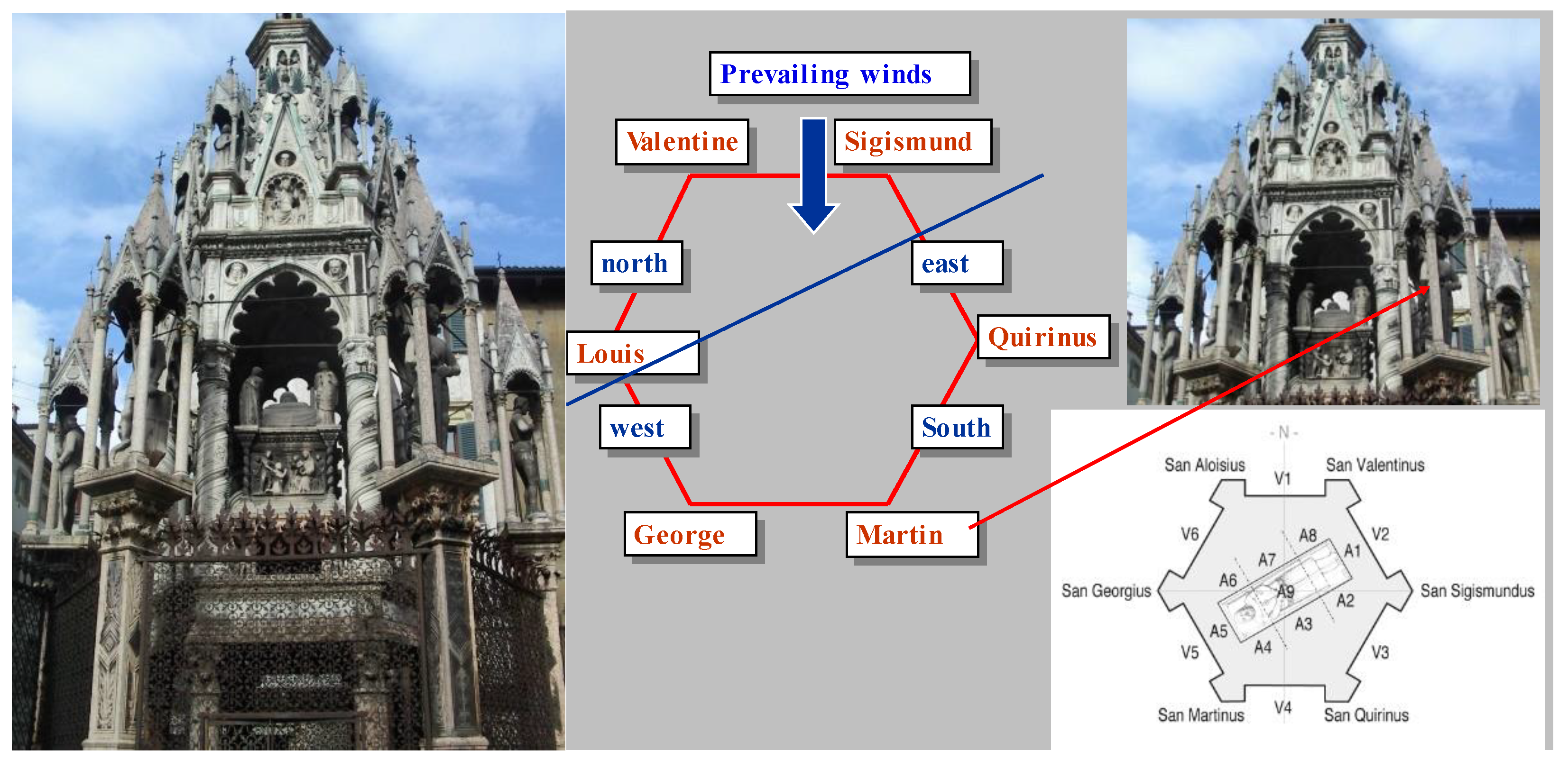
Figure 2.
a-f. From left to right: Saints Martin, Quirinus, Sigismund, Valentine, Louis the king, George.
Figure 2.
a-f. From left to right: Saints Martin, Quirinus, Sigismund, Valentine, Louis the king, George.
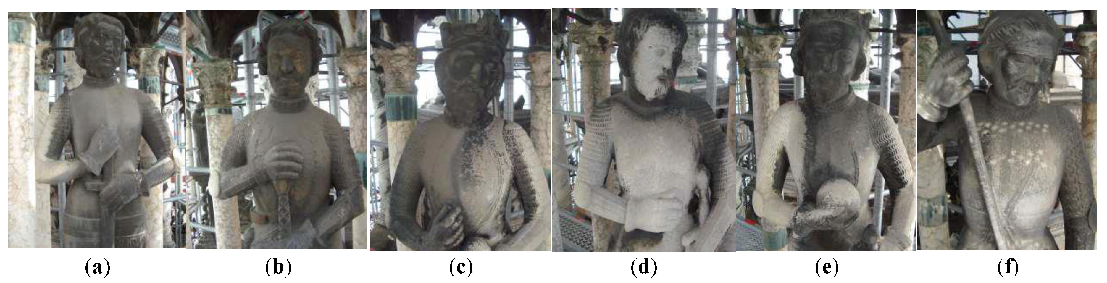
Figure 3.
From left to right: a, b) Saint Quirinus, details of the left and right legs showing vertical fissures; c, d) Saint Valentine, detail of the left leg, lead shoes, and vertical fissures; e, f) the inner substrate as revealed after the dismantling of the lead shoes.
Figure 3.
From left to right: a, b) Saint Quirinus, details of the left and right legs showing vertical fissures; c, d) Saint Valentine, detail of the left leg, lead shoes, and vertical fissures; e, f) the inner substrate as revealed after the dismantling of the lead shoes.
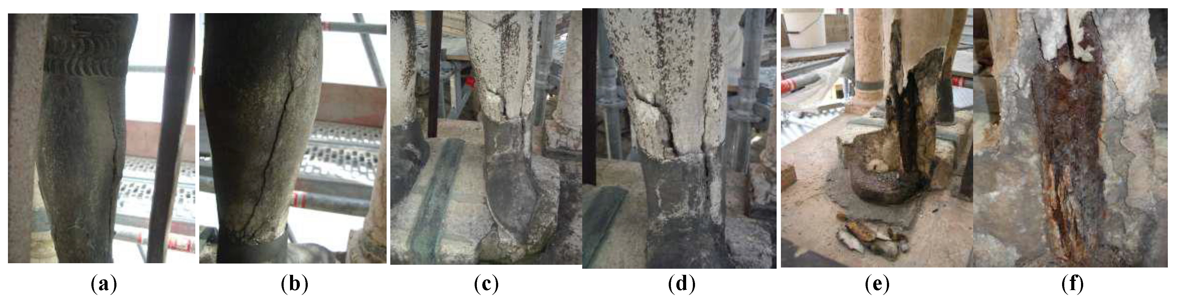
Figure 4.
a) Iron oxide stains visible on the marble fragments removed from the foot and calf; however, no traces of cement grout are present; b) cement drippings on the marble bedding that housed the iron base plate supporting the internal reinforcement bar.
Figure 4.
a) Iron oxide stains visible on the marble fragments removed from the foot and calf; however, no traces of cement grout are present; b) cement drippings on the marble bedding that housed the iron base plate supporting the internal reinforcement bar.
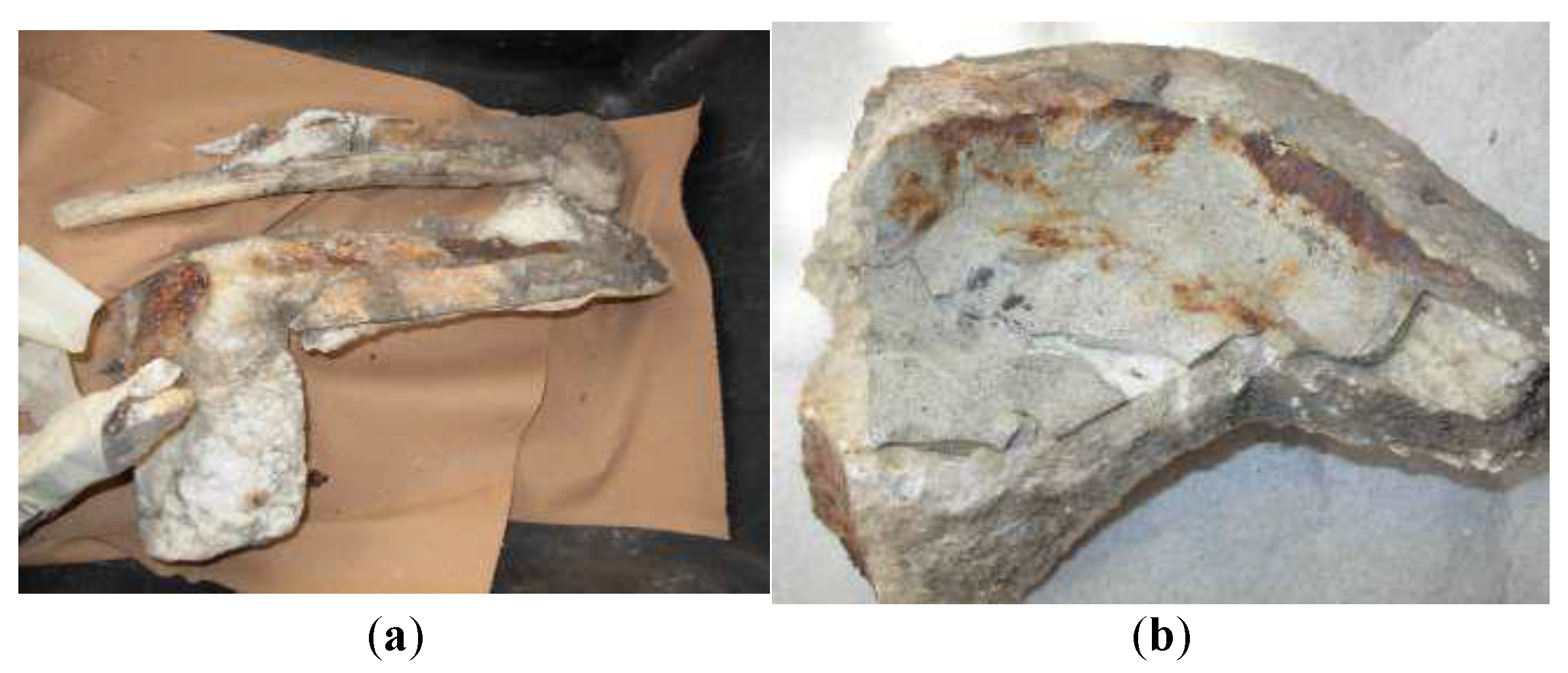
Figure 5.
a) Location of sample 4; b) cross-section observed by OM (80× magnification). Along the intergranular fissures, atmospheric particulate matter, iron oxide-based particles, and carbonaceous cenospheres are visible within the spaces between intergranular calcite crystals; c) location of sample 13; d) black surface layer consisting of atmospheric particulate matter mixed with biodeteriogens, largely dead and primarily composed of algae (OM, 80× magnification, parallel nicols); e) black biological residue on the surface observed in optical microscopy images of the polished section (OM, 80× magnification, parallel nicols); f) enlargement of the previous image: the growth of endolithic biological compounds was not confined to the surface, but penetrated deeply into the stone material following the preferential pathways provided by intergranular cracks; g) thin section (80× magnification, crossed nicols) showing the detachment of calcite crystals.
Figure 5.
a) Location of sample 4; b) cross-section observed by OM (80× magnification). Along the intergranular fissures, atmospheric particulate matter, iron oxide-based particles, and carbonaceous cenospheres are visible within the spaces between intergranular calcite crystals; c) location of sample 13; d) black surface layer consisting of atmospheric particulate matter mixed with biodeteriogens, largely dead and primarily composed of algae (OM, 80× magnification, parallel nicols); e) black biological residue on the surface observed in optical microscopy images of the polished section (OM, 80× magnification, parallel nicols); f) enlargement of the previous image: the growth of endolithic biological compounds was not confined to the surface, but penetrated deeply into the stone material following the preferential pathways provided by intergranular cracks; g) thin section (80× magnification, crossed nicols) showing the detachment of calcite crystals.
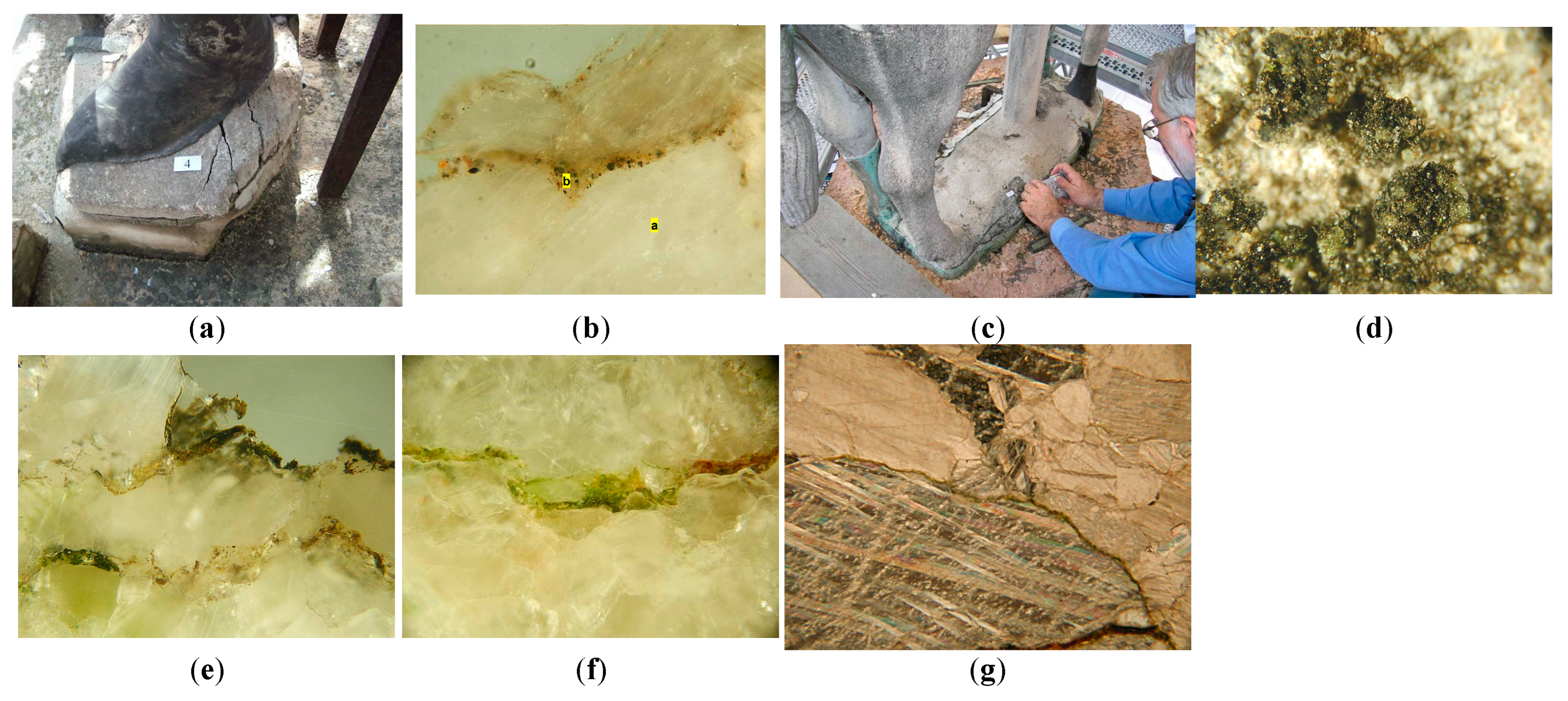
Figure 6.
a) Sampling location; b) surface observation at 33× magnification; c) OM observation of the cross-section at 33× magnification, where layer c shows carbonaceous cenosphere particles; d) ESEM observation of the cross-section of the same specimen area: layer a appears more homogeneous and light grey, whereas layer b is less homogeneous and shows black carbonaceous particles, indicating a different fossil-fuel combustion period.
Figure 6.
a) Sampling location; b) surface observation at 33× magnification; c) OM observation of the cross-section at 33× magnification, where layer c shows carbonaceous cenosphere particles; d) ESEM observation of the cross-section of the same specimen area: layer a appears more homogeneous and light grey, whereas layer b is less homogeneous and shows black carbonaceous particles, indicating a different fossil-fuel combustion period.
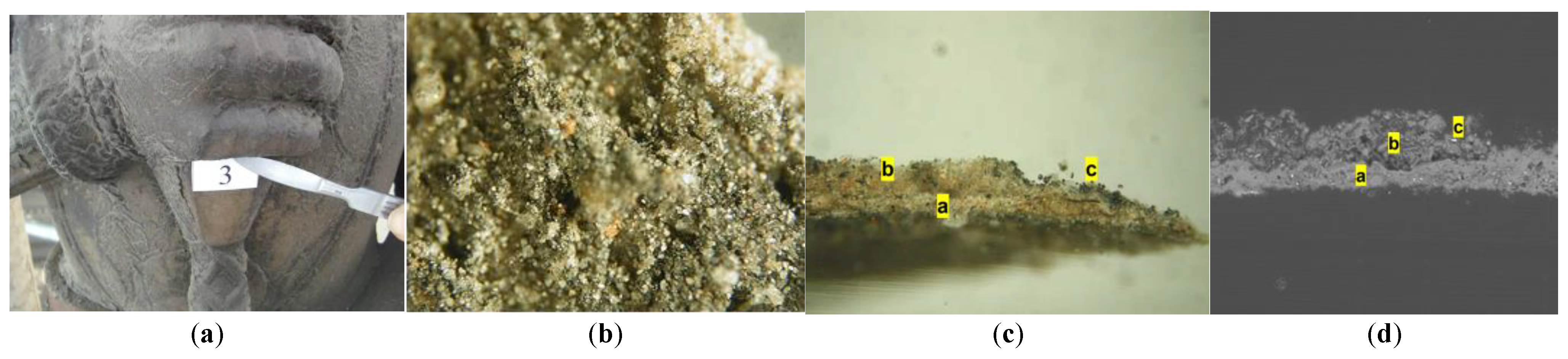
Figure 7.
a) Sampling point; b) EDX analysis showing gypsum formation (Ca and S) resulting from the reaction with the calcareous substrate, together with silicate airborne dust particles (Mg, Al, and Si).
Figure 7.
a) Sampling point; b) EDX analysis showing gypsum formation (Ca and S) resulting from the reaction with the calcareous substrate, together with silicate airborne dust particles (Mg, Al, and Si).
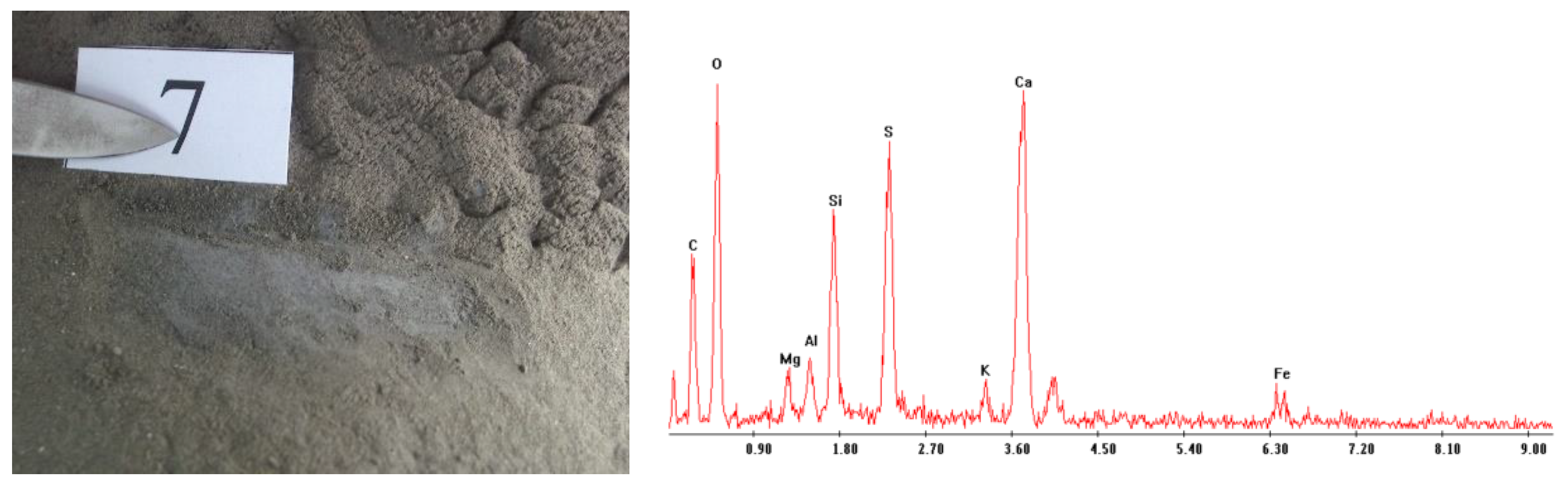
Figure 8.
a) Sampling location of the black flake; b) optical microscopy (OM) cross-section at 33× magnification: layer a corresponds to the lime finish, layer b represents the accumulation of airborne particles predating the urban pollution period, and layer c is a darkened layer rich in carbonaceous particles, typical of the recent urban-industrial period; c) ESEM cross-section at 28× magnification; d) typical dendritic black crust.
Figure 8.
a) Sampling location of the black flake; b) optical microscopy (OM) cross-section at 33× magnification: layer a corresponds to the lime finish, layer b represents the accumulation of airborne particles predating the urban pollution period, and layer c is a darkened layer rich in carbonaceous particles, typical of the recent urban-industrial period; c) ESEM cross-section at 28× magnification; d) typical dendritic black crust.
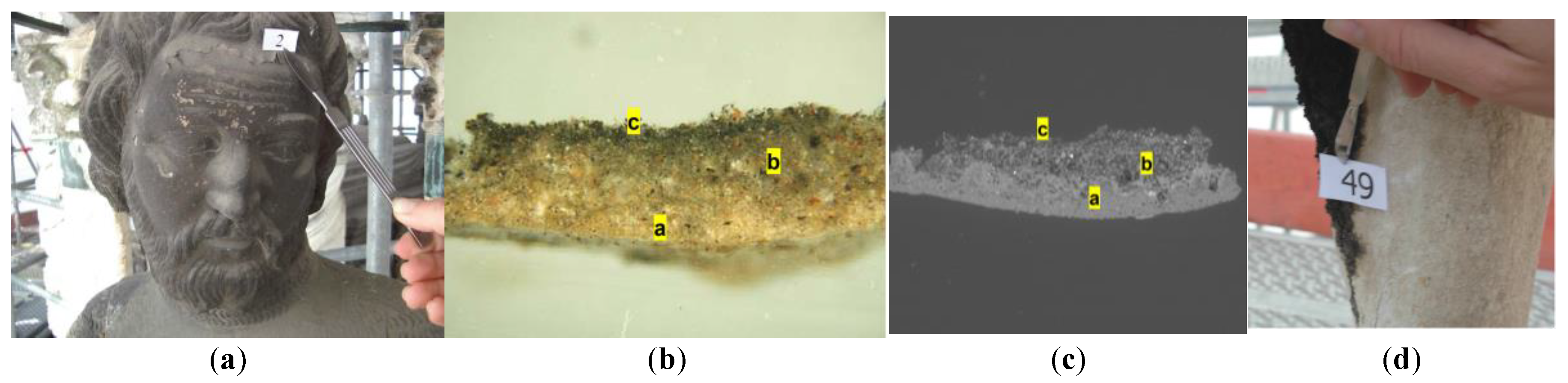
Figure 9.
a) Saint Sigismund, back; b) optical microscope cross-section (33× magnification): beneath the atmospheric deposit, an azurite-based paint layer is visible. The discontinuity of the azurite layer promotes crystal decohesion, indicating a poor state of conservation; c) the same cross-section observed by SEM (84× magnification) confirms the poor condition of the painted layer. Layer c is very compact and shows the accumulation of atmospheric particles.
Figure 9.
a) Saint Sigismund, back; b) optical microscope cross-section (33× magnification): beneath the atmospheric deposit, an azurite-based paint layer is visible. The discontinuity of the azurite layer promotes crystal decohesion, indicating a poor state of conservation; c) the same cross-section observed by SEM (84× magnification) confirms the poor condition of the painted layer. Layer c is very compact and shows the accumulation of atmospheric particles.
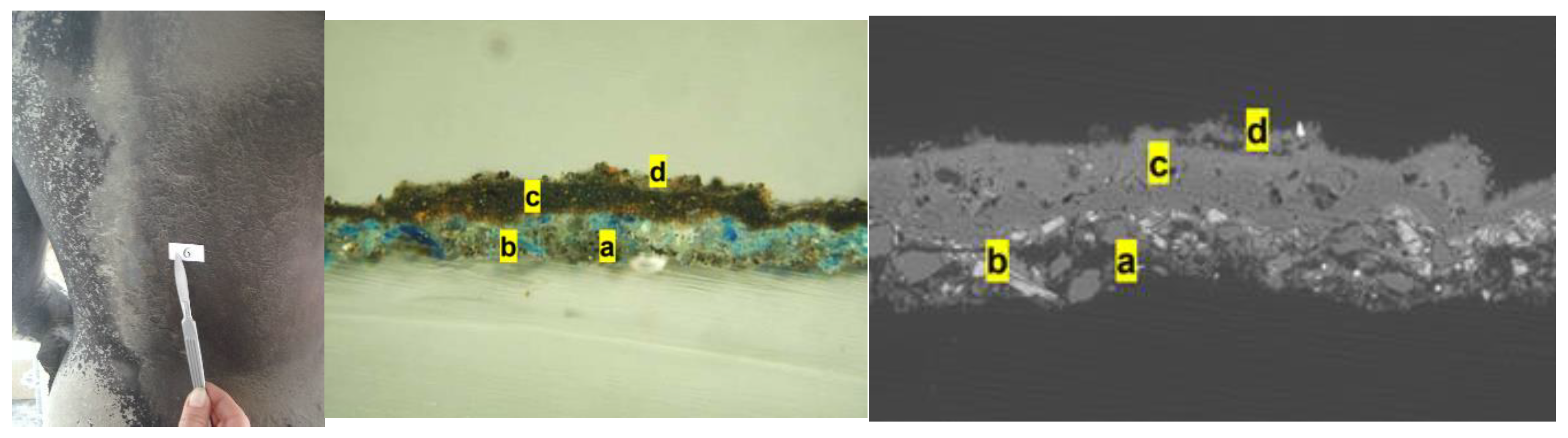
Figure 10.
a) Sample 57; b) OM cross-section at 240× magnification: dark grey layer (A), glue layer (B) used to fix the thin gold lamina (C), and a grey surface layer containing gypsum and carbonaceous particles (D); c) ESEM analysis confirms the composition and highlights the white line of the gold lamina (C).
Figure 10.
a) Sample 57; b) OM cross-section at 240× magnification: dark grey layer (A), glue layer (B) used to fix the thin gold lamina (C), and a grey surface layer containing gypsum and carbonaceous particles (D); c) ESEM analysis confirms the composition and highlights the white line of the gold lamina (C).
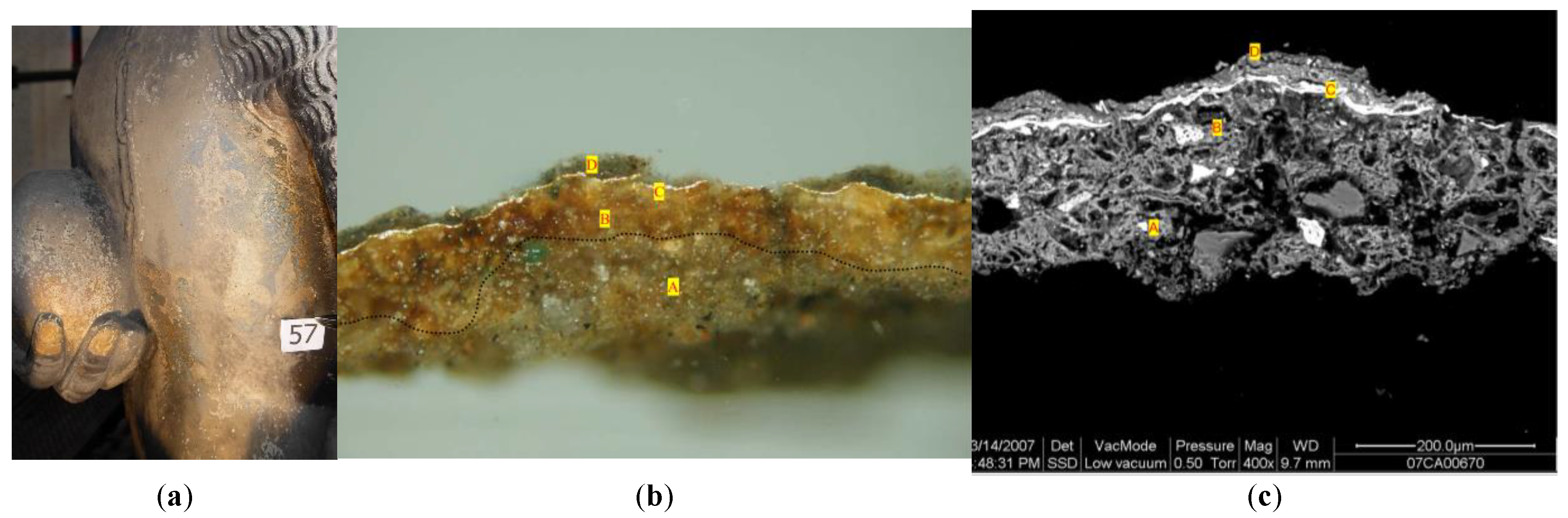
Figure 11.
a) Sample 40; b) cross-section at 480× magnification: layer B is a preparatory orange layer containing lead white and ochraceous oxide particles; layer C is a gold lamina-based layer; layer D is white lead oxidized into lead dioxide (platnerite); layer E is formed by gypsum, iron oxide, and carbonaceous particles; c) layer B shows white lead particles both in the preparatory layer and in layer D. This layer was initially applied as a white finishing layer and, over time, darkened. In the external layer E, numerous gypsum crystals resulting from pollution are present.
Figure 11.
a) Sample 40; b) cross-section at 480× magnification: layer B is a preparatory orange layer containing lead white and ochraceous oxide particles; layer C is a gold lamina-based layer; layer D is white lead oxidized into lead dioxide (platnerite); layer E is formed by gypsum, iron oxide, and carbonaceous particles; c) layer B shows white lead particles both in the preparatory layer and in layer D. This layer was initially applied as a white finishing layer and, over time, darkened. In the external layer E, numerous gypsum crystals resulting from pollution are present.
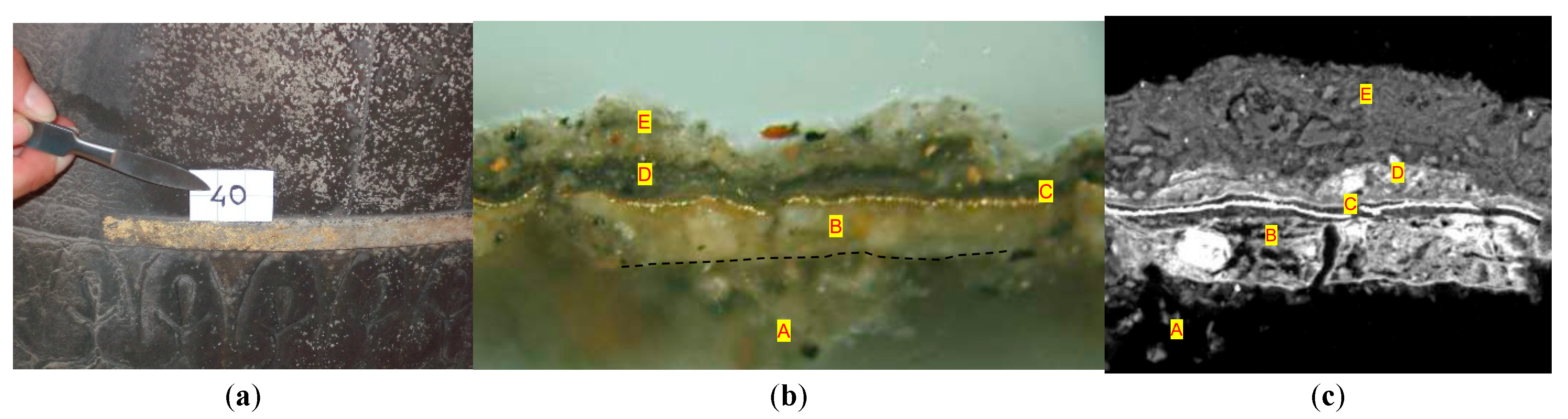
Figure 12.
a) Location of sample 59, b) Lead sheet in cross-section.
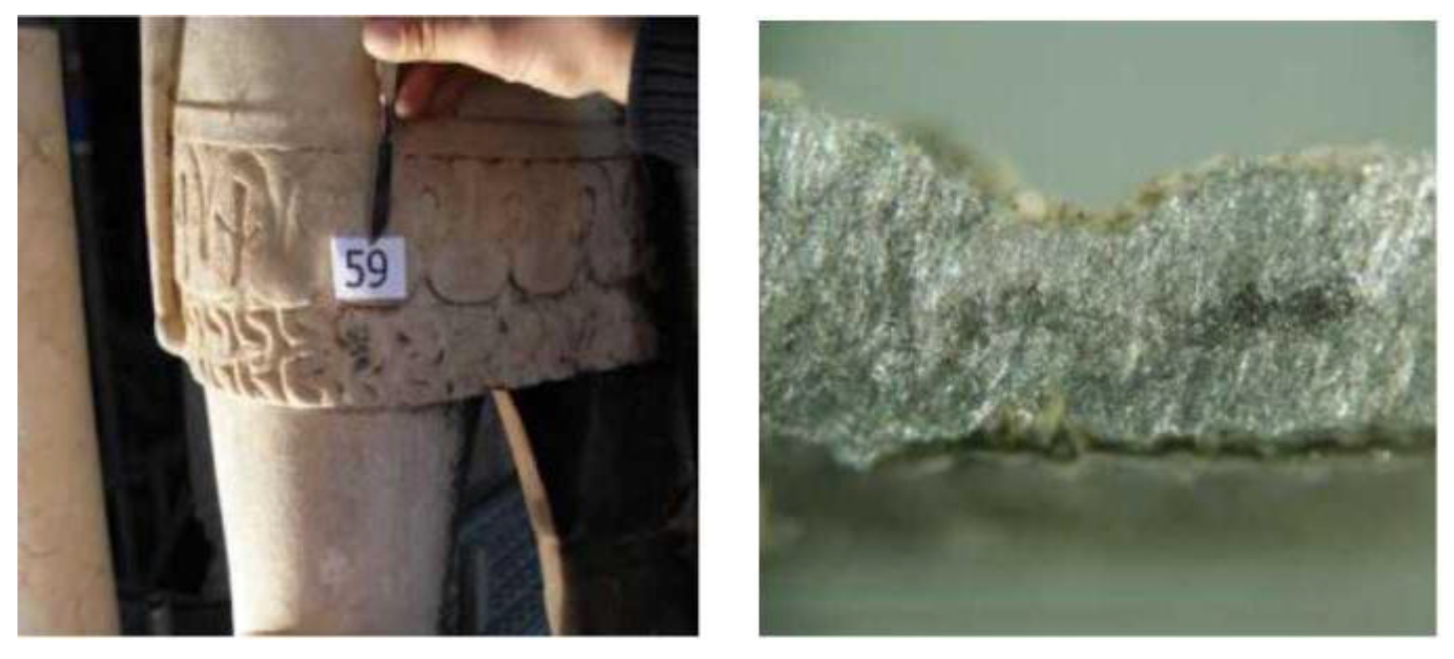
Figure 13.
a) Sample 24; b) cross-section observed by OM (480× magnification): azurite (C) above the morellone layer (B), with an atmospheric particulate deposit; c) Sample 25; d) cross-section observed by OM (240× magnification): artificial ultramarine blue (A) over a white layer of gypsum crystals, which are responsible for the poor state of conservation.
Figure 13.
a) Sample 24; b) cross-section observed by OM (480× magnification): azurite (C) above the morellone layer (B), with an atmospheric particulate deposit; c) Sample 25; d) cross-section observed by OM (240× magnification): artificial ultramarine blue (A) over a white layer of gypsum crystals, which are responsible for the poor state of conservation.
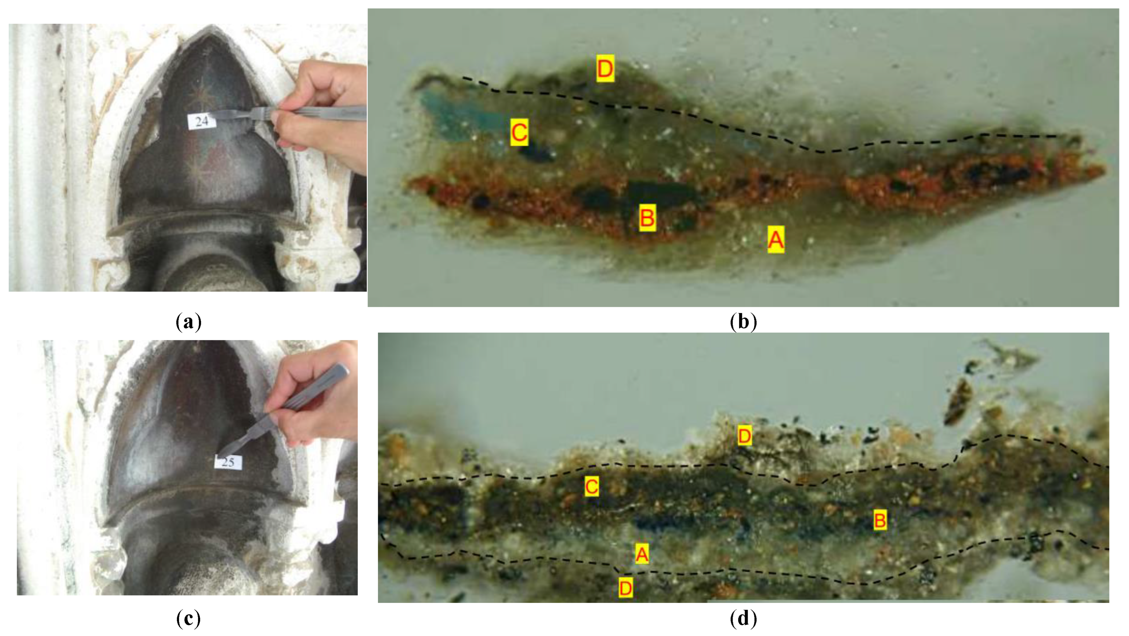
Figure 14.
a) Prudence, south-west view, detail showing traces of blue pigment; b) cross-section observed under OM (480× magnification); c) ESEM analysis showing white crystalline particles identified as azurite; d) Charity; e) cross-section observed under OM (240× magnification), where azurite particles appear widely dispersed, indicating a poor state of conservation; f) ESEM image showing detached white azurite particles; g) Hope, east view, showing extensive dark deposits; sample 35 from a partially washed-out area; h) cross-section observed under OM (240× magnification); i) ESEM image showing white azurite particles in layer B.
Figure 14.
a) Prudence, south-west view, detail showing traces of blue pigment; b) cross-section observed under OM (480× magnification); c) ESEM analysis showing white crystalline particles identified as azurite; d) Charity; e) cross-section observed under OM (240× magnification), where azurite particles appear widely dispersed, indicating a poor state of conservation; f) ESEM image showing detached white azurite particles; g) Hope, east view, showing extensive dark deposits; sample 35 from a partially washed-out area; h) cross-section observed under OM (240× magnification); i) ESEM image showing white azurite particles in layer B.
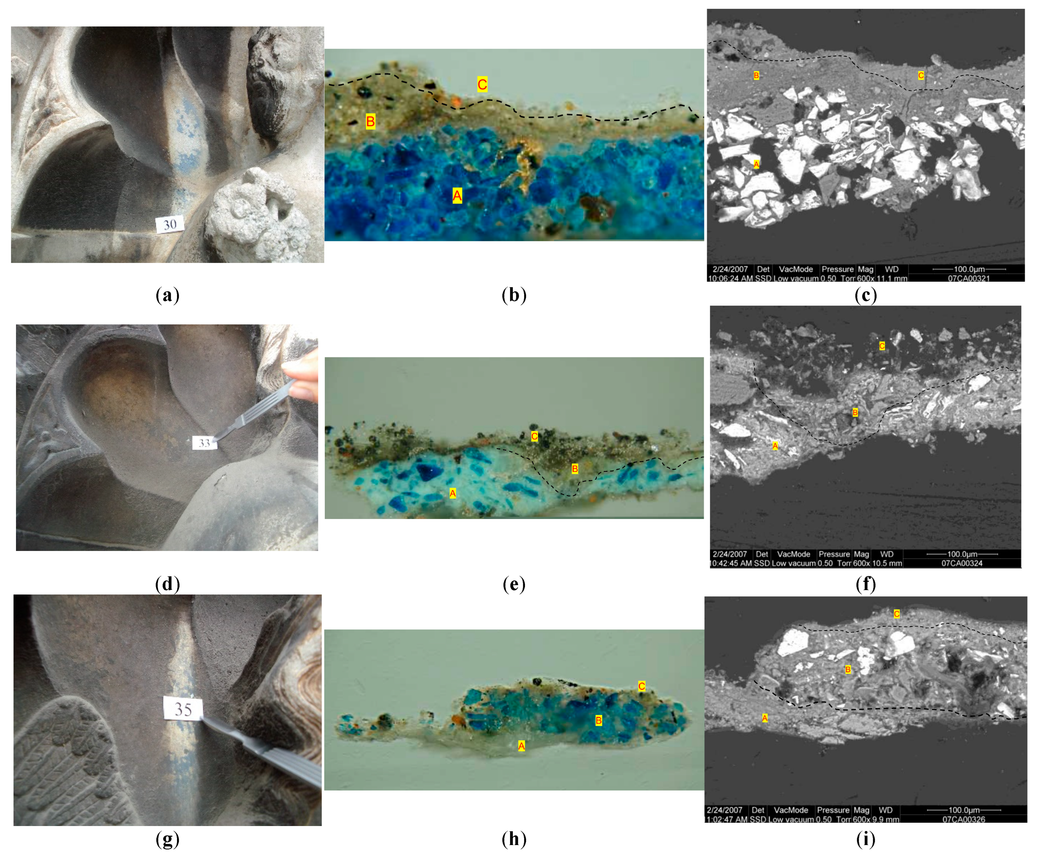
Figure 15.
a) Initial stage of cleaning; b) advanced stage of cleaning, showing traces of azurite on the ground of the shell and gold remains on the wings.
Figure 15.
a) Initial stage of cleaning; b) advanced stage of cleaning, showing traces of azurite on the ground of the shell and gold remains on the wings.
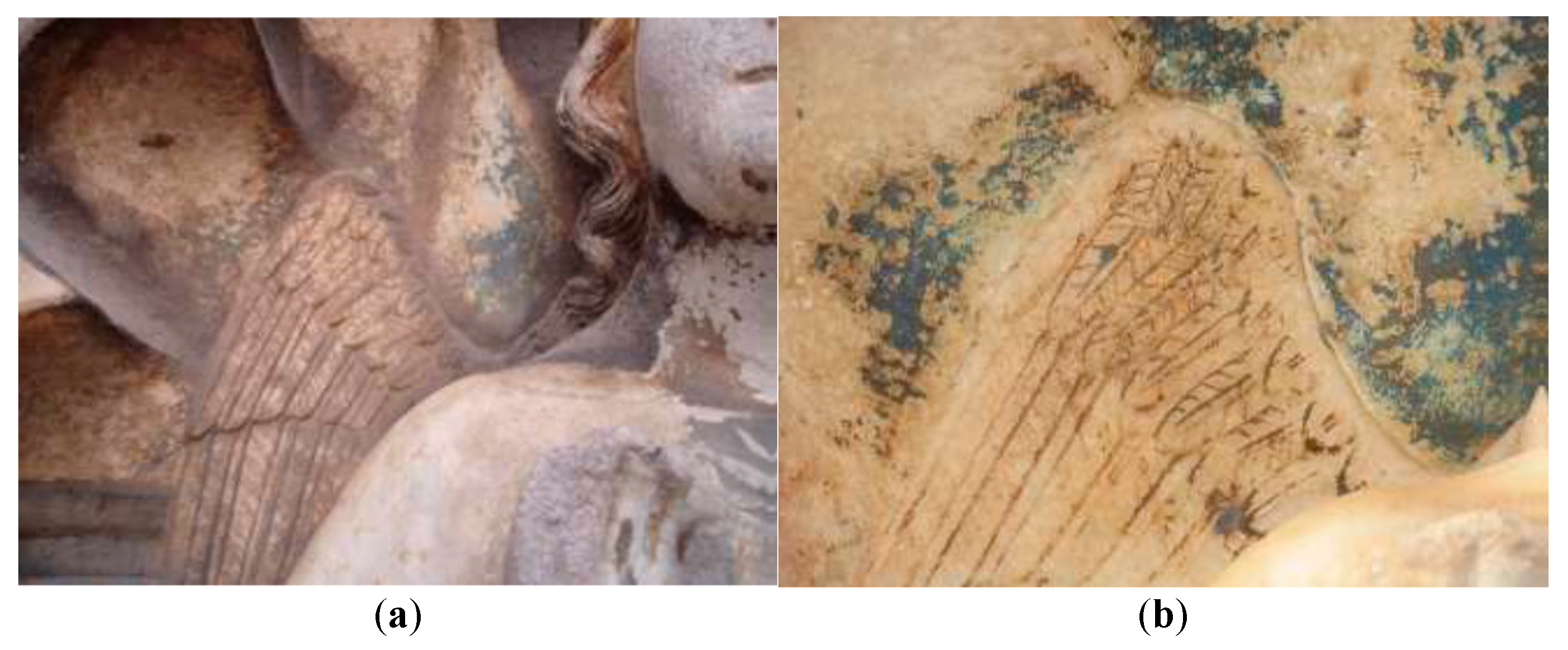
Figure 16.
a) Sample location; b) cross-section observed under OM (240× magnification); c) ESEM image showing: (A) a layer exhibiting decohesion of calcite crystals; between layers A and B, the white layer is attributed to lead white.
Figure 16.
a) Sample location; b) cross-section observed under OM (240× magnification); c) ESEM image showing: (A) a layer exhibiting decohesion of calcite crystals; between layers A and B, the white layer is attributed to lead white.
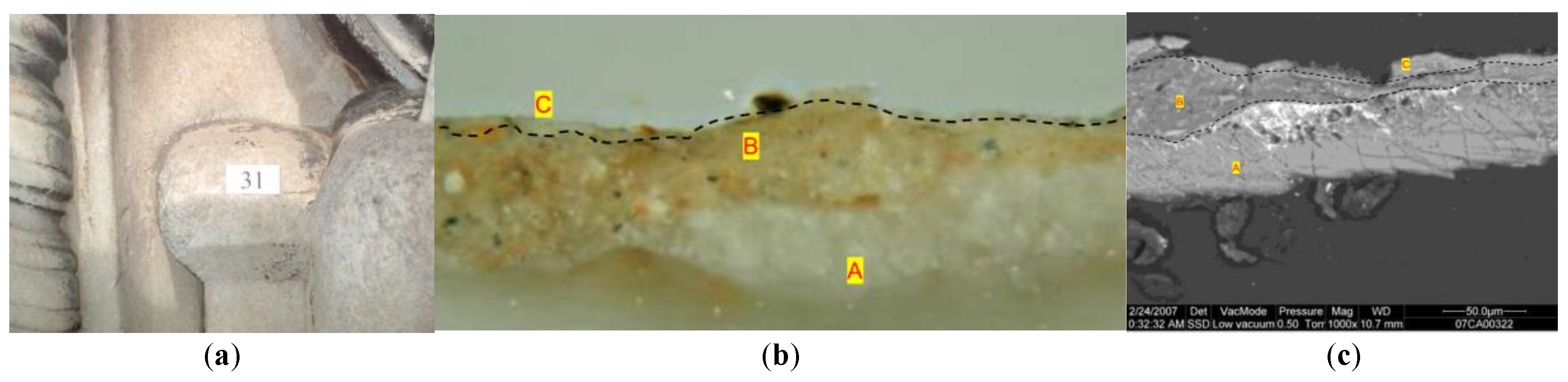
Figure 17.
Dirt wetting, dirt accumulation and white washing patterns. a) Saint Louis, b) Saint Sigismund.
Figure 17.
Dirt wetting, dirt accumulation and white washing patterns. a) Saint Louis, b) Saint Sigismund.
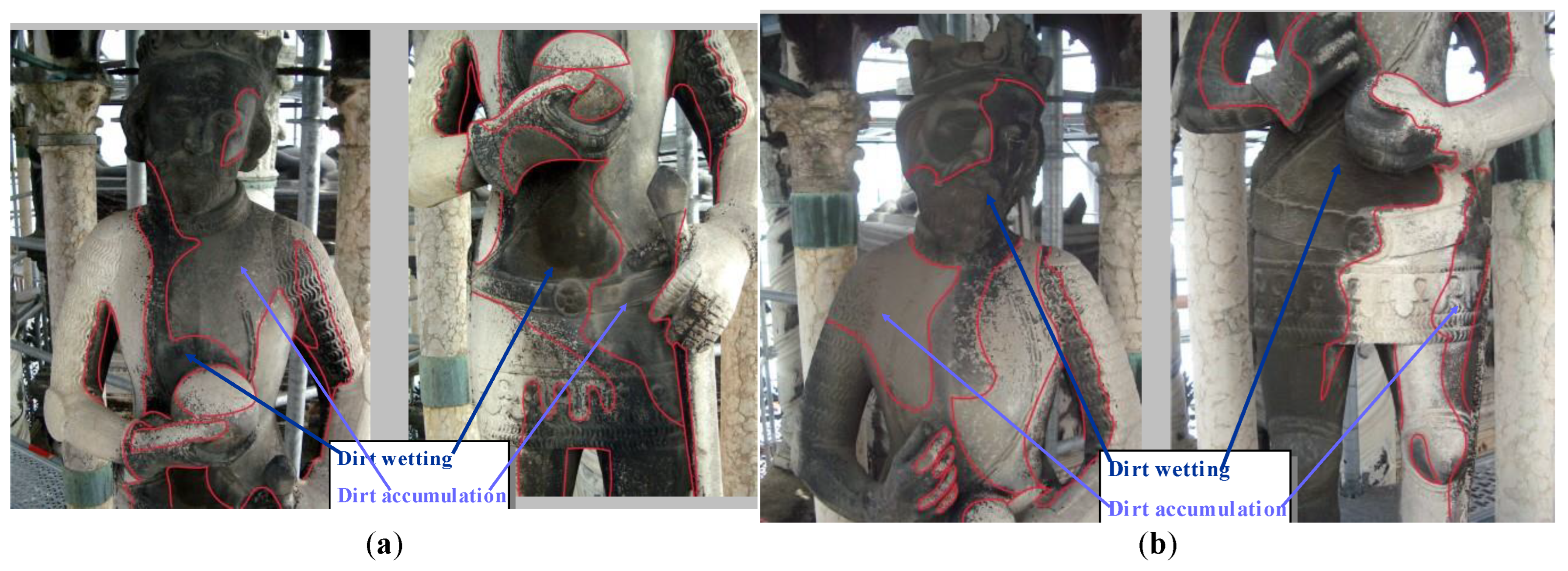

Table 1.
Selection of collected samples.
| Sample number | Sample description and location | Type of analysis |
|---|---|---|
| 4 | St. Quirinus, grey fragment from the basement | OM thin and cross-sections, IC |
| 13 | Cansignorio horse, grey fragment from the basement | OM thin and cross-sections |
| 3 | St. Quirinus, from the hilt sword | OM and ESEM-EDS cross-sections |
| 7 | St. Sigismund, incoherent deposit over the right shoulder | XRD, IC, EDS |
| 2 | St. Martin, black scales from the front | OM cross-section |
| 6 | St. Sigismund, grey deposit from the back | OM cross-section, XRD |
| 9 | St. Louis, in front over | OM and ESEM-EDS cross-sections |
| 57 | St. Louis, gold fragment | OM and ESEM-EDS cross-sections |
| 40 | St. Louis, gold fragment | OM and ESEM-EDS cross-sections |
| 59 | St. George, lead armour | OM and ESEM-EDS cross-sections |
| 24 | Niches of apostles, star of the sky | OM and ESEM-EDS cross-sections |
| 25 | Niches of apostles, blue fragment | OM and ESEM-EDS cross-sections |
| 30 | Prudence, blue fragment | OM and ESEM-EDS cross-sections |
| 33 | Charity, blue fragment | OM and ESEM-EDS cross-sections |
| 35 | Hope, blue fragment | OM and ESEM-EDS cross-sections |
| 31 | Hope, yellow fragment | OM and ESEM-EDS cross-sections |
Table 2.
Concentration of soluble salts determined by ion chromatography.
| Sampling location in different exposure to rainwater | Chlorides (%) | Nitrates (%) | Sulphates (%) |
|---|---|---|---|
| St. Quirinus – back of the base | 0.02 | <0.01 | 0.09 |
| St. Sigismund-in the middle of the back | 0.27 | 0.67 | 13.41 |
| St. George-on the underside of the right arm | 0.03 | 0.28 | 35.00 |
| Horse’s leg-thick dendritic black crust | 0.09 | 0.37 | 65.12 |
Disclaimer/Publisher’s Note: The statements, opinions and data contained in all publications are solely those of the individual author(s) and contributor(s) and not of MDPI and/or the editor(s). MDPI and/or the editor(s) disclaim responsibility for any injury to people or property resulting from any ideas, methods, instructions or products referred to in the content. |
© 2026 by the authors. Licensee MDPI, Basel, Switzerland. This article is an open access article distributed under the terms and conditions of the Creative Commons Attribution (CC BY) license (http://creativecommons.org/licenses/by/4.0/).
Copyright: This open access article is published under a Creative Commons CC BY 4.0 license, which permit the free download, distribution, and reuse, provided that the author and preprint are cited in any reuse.