
Preprint
Hypothesis

This version is not peer-reviewed.

The Fascial Capacitor Model: A Biophysical Hypothesis for the Origin of the Local Twitch Response Within Stacking Fascia

A peer-reviewed version of this preprint was published in:
International Journal of Molecular Sciences 2026, 27(13), 5901. https://doi.org/10.3390/ijms27135901

Submitted:

18 May 2026

Posted:

18 May 2026

You are already at the latest version

Abstract

Background: The local twitch response (LTR) elicited during ultrasound-guided fascial hydrorelease (FHR) is conventionally attributed to dysfunctional motor endplates. However, in a related observational paper under concurrent submission, 89/89 evaluable archived LTR events were observed within stacking fascia at sites incompatible with direct endplate excitation. Hypothesis: We propose the Fascial Capacitor Model: stacking fascia functions as a multilayer biological capacitor in which collagen sublayers act as electrodes and the interposed densified hyaluronic-acid (HA)-rich loose layer acts as the dielectric, with the LTR reinterpreted as a transient electrophysiological discharge when a needle bridges its layers. This biophysical model is explicitly grounded in the established molecular and histological architecture of human deep fascia. Supporting evidence: Each premise is independently supported by primary literature from at least eight research lines spanning roughly seventy years. Voltage gap: The apparent gap between estimated bulk discharge voltages and motor neuron threshold is resolved by reconsidering needle-tip geometry and stimulation modality, anchored by the ±6 V triboelectric measurements of Ouyang et al. (2022). Implications: The model is the immediate-phase complement to the Fascial Memory Reset Hypothesis (Int J Mol Sci 2026, 27, 3720), explains intra-procedural symptom relief, and yields falsifiable predictions. A direct empirical validation programme using insulating-needle SEA recording is in preparation at the corresponding author’s institution.

Keywords: 
;  ;  ;  ;  ;  ;  ;  

1. Introduction

The local twitch response (LTR) — a brief, involuntary contraction of a muscle band elicited by needle-based interventions including local injections, dry needling, acupuncture, and ultrasound-guided fascial hydrorelease — has, for more than five decades, served as the operational signature of trigger-point and related neuromuscular interventions [27,28]. Travell and Simons positioned the LTR as a defining clinical feature of myofascial pain syndrome [27]; Hong's controlled comparison of lidocaine injection versus dry needling established the LTR as a predictor of therapeutic effect [28]; and the modern reformulation of trigger-point physiology within the framework of pain neuroscience continues to treat the LTR as a meaningful endpoint [29]. The dominant mechanistic explanation has long been the integrated trigger-point hypothesis, in which the LTR arises from dysfunctional motor endplates with persistent acetylcholine release, recorded as spontaneous electrical activity (SEA) in the immediate vicinity of the endplate zone [30].
However, a growing body of imaging-guided observations is difficult to reconcile with a strictly endplate-centred account. In the related observational paper (Paper 1 [34]; in submission to the same IJMS Special Issue), we report a retrospective analysis of a large clinical video archive in which all 89 documented LTR events elicited during ultrasound-guided fascial hydrorelease (FHR) occurred within structures classified as stacking fascia — the ultrasound-visible phenotype of multilayered, densified deep fascia — at sites that were anatomically incompatible with proximity to a motor endplate (e.g., intermuscular fascia, perineural fascia of the sciatic nerve, ligamentous and aponeurotic layers far from any endplate band). This 100% concordance in this retrospective series raises a structural question that the endplate hypothesis is not designed to answer: what is it about densified, multilayered deep fascia in particular that converts a brief mechanical perturbation into a synchronous, single-shot muscle contraction?
The present paper proposes a biophysical answer. We argue that the histologically established architecture of stacking fascia — alternating sublayers of densely packed type I collagen separated by glycosaminoglycan (GAG)-rich loose layers whose HA component aggregates pathologically under chronic load — is structurally isomorphic, by analogy, to a multilayer ceramic capacitor (MLCC), and that the LTR is the macroscopic discharge of this biological capacitor when a needle bridges its layers. The model is presented as a Hypothesis (in the IJMS Hypothesis and Theory sense): an explicitly testable, falsifiable framework, calibrated to the molecular-biology scope of this Special Issue, offered as a complement to — not a replacement for — the established Travell–Simons–Hong endplate framework. Our group previously proposed the Fascial Memory Reset hypothesis in this journal (Int J Mol Sci 2026, 27, 3720) [33], framing the mid- to long-term effects of FHR on a mechano-epigenetic time scale; the present capacitor model addresses the immediate-phase electrophysical event on a complementary seconds-to-minutes time scale.
The hypothesis that stacking fascia spontaneously discharges electrical activity, and that this discharge underlies a range of myofascial phenomena including referred pain, motor dysfunction, and sympathetic-mediated vasoconstriction, was first proposed by the corresponding author at the 20th MPS Research Meeting (Fukuoka, Japan, October 15, 2017) [35]. The present paper develops the biophysical mechanism — the Fascial Capacitor Model — that underpins this 2017 hypothesis.
The remainder of the paper is organized as follows. Section 2 introduces the Fascial Capacitor Model itself, including a five-stage mechanistic cascade and order-of-magnitude quantitative estimates. Section 3 surveys the convergent evidence from eight independent research lines that support each premise of the model. Section 4 directly addresses the voltage gap objection. Section 5 places the present hypothesis in relation to the Fascial Memory Reset Hypothesis across two timescales. Section 6 considers the clinical correlate of intra-procedural symptom relief. Section 7 outlines the direct empirical validation programme now in preparation. Section 8 concludes.

2. The Fascial Capacitor Model

Structural analogy. A multilayer ceramic capacitor (MLCC) stores charge in an alternating stack of conductive electrodes and dielectric insulators. Stecco and colleagues, in their morphometric analysis of human deep fascia, showed that the aponeurotic fascia is built of two to three sublayers of densely packed type I collagen (mean thickness ≈ 277 µm) separated by thinner layers of loose connective tissue (mean thickness ≈ 44 µm), with adjacent collagen layers crossing at approximately 78° [7]. We propose that this histologically confirmed architecture is structurally isomorphic to an MLCC: each collagen sublayer behaves as a relatively conductive electrode (a known piezoelectric medium [1,2,3,4]), and each interposed glycosaminoglycan-rich loose layer, when its hyaluronic acid (HA) component aggregates pathologically (densification [8,11,12,13]), behaves as a dielectric that impedes charge dissipation. Within this framework, “stacking fascia” — the ultrasound-visible (sonographic) phenotype of multilayered, densified deep fascia — corresponds to the physical body of the capacitor (Figure 1).
A five-stage cascade. We propose the following sequence linking chronic mechanical load to symptom generation:
1. Stacking formation. Chronic mechanical stress (repetitive movement, postural overload, post-traumatic immobilization) drives an increase in collagen density and HA hyper-aggregation, with cross-linking and reduced water content. The fasciacyte, a fibroblast subtype specialized for inter-layer HA production [10], is a candidate cellular driver of this dielectric remodeling.
2. Capacitor formation. The alternating arrangement of densified HA between collagen sublayers completes a multilayer capacitor-like structure with measurable charge-storage capacity.
3. Charge accumulation and steady-state field. Daily mechanical loading generates charge through (a) collagen piezoelectricity [1,2,3,4] and (b) streaming potentials in the GAG matrix [5,6]. In healthy hydrated connective tissue the dissipative RC time constant (resistance–capacitance time constant) is of the order of nanoseconds, so charges do not accumulate over time — consistent with the absence of capacitor-like behaviour in healthy fascia. Two complementary mechanisms allow a quasi-steady electrical state to nevertheless emerge specifically in pathologically densified fascia. First, dynamic equilibrium: the body is never mechanically silent, and the continuous low-amplitude loading produced by respiration, cardiac pulsation, postural maintenance, and the friction that accompanies impaired inter-layer gliding sustains a continuous "generation rate". When this generation rate exceeds the dissipation rate, it supports a non-zero steady-state field. Second, pathological extension of the RC time constant: HA hyper-aggregation and densification of the loose layer dramatically increase its local insulating capacity, lengthening RC by orders of magnitude and allowing genuine charge accumulation across the multilayer structure. Both mechanisms operate concurrently in densified fascia; together they explain why a capacitor-like quasi-steady electrical state arises specifically in stacking fascia and not in healthy tissue. The result is detectable as the impaired gliding and increased viscous coupling characteristic of densified fascia.
4. Multimodal afferent activation. The dense innervation of deep fascia [31] is activated through two pathways operating in parallel. First, an electrical pathway: the resulting local electric field acts on voltage-sensitive elements of the free nerve endings (voltage-gated channels and direct depolarization at the tissue–axon interface). Second, a mechanical pathway: the deformation, densification, and chemical microenvironment of stacking fascia mechanically gate Piezo1 and Piezo2 channels [17], which are opened by membrane tension rather than by voltage. TRPV1-positive free nerve endings contribute through their polymodal sensitivity to the chemical and thermal microenvironment of densified fascia. Critically, these pathways are mechanistically distinct — Piezo channels respond to deformation, not to electric fields — but they converge on the same afferent population. The combined output can be recorded as spontaneous electrical activity (SEA) of the type originally described by Simons [30], but re-interpreted here as arising from fascia per se rather than from the motor endplate.
5. Positive feedback. Afferent activation drives pain, referred pain, allodynia, muscle guarding, and sympathetically mediated vasoconstriction. The ensuing ischemia and immobility further promote densification and additional layering, increasing the effective capacitance — a positive feedback loop that may underlie chronicity.
Quantitative estimates. Using a parallel-plate approximation, C = n · ε0 · εᵣ · A/d, with n = 2 dielectric layers (a conservative approximation based on the two-to-three-sublayer architecture reported by Stecco et al. [7]; this treats the alternating sublayer arrangement as effectively parallel during the brief discharge event, an order-of-magnitude approximation appropriate to the Hypothesis-paper scope), ε0 = 8.854 × 10⁻¹² F m−1, εᵣ ≈ 30 for densified HA (estimated from tissue dielectric measurements [32]), d ≈ 44 µm and A ≈ 5 cm2 yields a capacitance of the order of C ≈ 6 nF. Taking a collagen piezoelectric coefficient d14 ≈ 1 pC N−1 [2], a steady-state mechanical stress σ ≈ 5 kPa across A ≈ 5 cm2 gives a bulk steady-state voltage of order V ≈ 0.4 mV, with stored energy E = CV2/2. Under the transient pressure pulse delivered by FHR injection (~200–500 kPa), the same calculation gives a transient bulk voltage of order V ≈ 25 mV. Importantly, these are order-of-magnitude estimates intended to bound the plausibility of the model, not point predictions, and they refer to bulk-tissue voltages — see Section 4 for the geometric and modal corrections that apply at the needle tip.

3. Convergent Molecular and Biophysical Evidence from Independent Research Groups

A useful test of any biophysical hypothesis is whether each of its independent premises is supported by primary research from groups working without reference to the hypothesis itself. The Fascial Capacitor Model rests on at least eight such independent lines of evidence, accumulated over approximately seventy years.
Collagen piezoelectricity (Fukada–Yasuda research line, Japan). The piezoelectric effect of biological collagen was first demonstrated by Fukada and Yasuda in bone [1] and subsequently characterized in tendon and skin [2]. The piezoelectric coefficient d14 is now accepted to lie in the range 0.2–2 pC N−1.
Collagen nanoscale electromechanics (Minary-Jolandan and Yu, United States). Atomic-force-microscopy measurements of individual type I collagen fibrils confirmed shear piezoelectricity at the nanoscale and excluded a purely surface-charge artefact [3,4], directly relevant to the single-fibril scale at which the proposed mechanism operates.
Streaming potentials in GAG matrices (Grodzinsky group, MIT). The same group that defined the streaming-potential framework for articular cartilage [5] later extended its biomechanical formalism to connective tissue more broadly [6], providing an empirically grounded route by which load-driven fluid flow through a charged HA-rich matrix can generate millivolt-range potentials.
Fasciacyte, HA and densification (Stecco group, Padova). The Padova group identified the fasciacyte as the HA-producing fibroblast subtype of fascia [10], characterized the morphometry and innervation of human deep fascia [7,8], and clarified the role of HA hyper-aggregation in myofascial pain [9]. Pavan and colleagues drew the explicit operational distinction between densification (a reversible HA-driven viscosity change of the loose layer) and fibrosis (an irreversible structural change of the collagen layer) [13]. Fede et al. extended this work to a molecular-biology level in IJMS itself [11] and across multiple organ systems [12].
YAP/TAZ mechanotransduction (Dupont/Piccolo group, Padova). The seminal demonstration that YAP/TAZ transduce extracellular matrix stiffness into nuclear transcriptional programs [14] supplies the mechano-epigenetic substrate by which fascial densification is plausibly maintained over chronic time scales [15]. At the molecular level, YAP/TAZ activation in stiffened extracellular matrix drives transcription of CTGF, ANKRD1, and downstream regulators of fibroblast-to-myofibroblast transition, providing a molecularly defined substrate for the persistent mechanical phenotype of densified fascia. In vivo expression of YAP in human deep fascia has now been directly demonstrated in IJMS itself [16], making the present journal the most current scientific substrate for the molecular biology of stacking fascia.
Piezo channels as molecular mechanosensors (Coste/Patapoutian group, Scripps). The identification of Piezo1 and Piezo2 as bona fide mechanosensitive cation channels [17] supplies a molecularly defined mechanosensor in fascial fibroblasts and free nerve endings, capable of converting the mechanical component of fascia-derived perturbations into afferent signaling. Piezo channels are gated by membrane tension rather than by voltage, and therefore operate as a parallel pathway alongside, rather than downstream of, any direct electrical events generated by capacitor discharge.
The connective-tissue continuum as a signaling medium (Langevin group, Harvard/NCCIH). Langevin and colleagues established a foundational body of evidence on the connective-tissue continuum as a mechanically responsive signaling medium [24,25,26]. They showed that mechanical signaling propagates through the connective-tissue continuum to coordinate cellular responses across distance [24], and that fibroblasts in connective tissue dynamically remodel their cytoskeleton in response to subcutaneous tissue stretch [25] and to acupuncture-needle stimulation [26]. These findings together support treating the fascial network as a tissue whose biophysical state — mechanical and biochemical, and, by extension within the present model, also electrical — is physiologically meaningful, not merely incidental.
Fascial molecular biology in IJMS (Fede, Pirri, and colleagues). Most recently, IJMS has itself published primary mechanobiological work on fascia [11,16], establishing precedent and scope for the present model.
Each individual building block of the Fascial Capacitor Model — piezoelectric charge generation, streaming-potential charge generation, HA-driven dielectric behavior, mechano-epigenetic maintenance, and molecularly defined mechanosensors — is independently supported by primary literature from groups working in unrelated paradigms. The model presented here is therefore best understood as a synthesis of converging evidence, rather than as a speculative extrapolation.

4. Addressing the Voltage Gap

A natural objection to the model is that the estimated bulk voltage produced by capacitor discharge (of the order of microvolts to ~25 mV; Section 2) appears smaller than, or only marginally comparable with, the canonical depolarization required to fire an α-motor neuron action potential (~15–20 mV above resting potential). We address this objection directly, because it rests on three assumptions that are each not fully aligned with current neurophysiological understanding when the actual neurophysiology and biophysics of needling are considered.
Assumption 1: that the local twitch is direct depolarization of a motor neuron.
This view is not fully consistent with current neurophysiological evidence. The local twitch response (LTR) is a spinal reflex mediated by Aδ and group III/IV afferent endings, as documented from the original animal models onward [27,28] and confirmed in recent reviews [29]. The relevant excitation threshold is therefore not the somatic threshold of an α-motor neuron, but the much lower threshold of afferent free nerve endings embedded in densified fascia [31]. In sensitized tissue these thresholds are further reduced, as exemplified by Piezo2-dependent mechanosensitization of nociceptors in inflammatory and osteoarthritic conditions [18]. Furthermore, subthreshold inputs in noisy biological systems are well known to be amplified by stochastic resonance, in which non-zero levels of background fluctuations can cooperatively cross the firing threshold of nonlinear neural elements [20]. The “15–20 mV” figure is, in this context, applied to the wrong cell type.
Assumption 2: that the relevant voltage is the bulk-tissue voltage.
This appears incomplete in light of current neurophysiology. At the needle tip, two well-established geometric and circuit effects amplify the bulk value by approximately one to two orders of magnitude:
• Tip-field concentration. For a needle with a tip radius of curvature of the order of 10–25 µm, classical electrostatics for sharp conductors predicts that the electric field strength scales steeply with 1/r near the tip, becoming dominant at sub-µm distances. For tip geometries in this range, finite-element and analytical analyses of needle-tip and sharp-electrode field profiles report amplification factors of approximately 10–30-fold at the tip surface relative to the bulk applied potential. The classical 1/r behaviour is sufficient on its own to render bulk-tissue voltages inadequate as a measure of the field actually experienced by free nerve endings adjacent to the inserted needle.
• Effective voltage gradients across multiple stacked units in series. When a needle simultaneously contacts N collagen–HA–collagen sandwich units, those units behave approximately as series capacitors during fast discharge, so the open-circuit voltage scales approximately as N · (Q/C). With allowance for incomplete electrical isolation between layers, a realistic series multiplier is approximately 2–4-fold when several units are engaged.
Even at shallow penetration depths, tip-field concentration provides substantial voltage amplification at the needle tip; additional contribution from layer summation arises when multiple units are bridged by the inserted needle. The depth of needle insertion at the moment of LTR is typically only into the upper portion of the stacking fascia, consistent with our model in which surface tip-field amplification (and triboelectric contact, see below) constitutes the dominant trigger of discharge. Combined, the tip-field and series effects can yield an effective gain of approximately 30–90-fold (an order-of-magnitude estimate); applied to the 25 mV bulk transient of Section 2, the surface field at the needle tip is, as an order-of-magnitude plausibility bound, of the order of ~1 V, likely exceeding afferent activation thresholds.
Assumption 3: that only direct voltage-mediated depolarization is relevant.
This assumption likewise overlooks important features of current neurophysiology. Needle insertion is intrinsically a multi-modal stimulus.
• Triboelectric contact electrification has been directly measured at the acupuncture-needle/tissue interface. Ouyang et al. [21] reported open-circuit voltages of the order of ±6 V from triboelectric contact between acupuncture needles and tissue, well above any neural depolarization threshold and independent of any prior charge stored in the fascia. The triboelectric series for stainless steel against biological polymers has been quantified [23]. The clinical FHR maneuver — insertion plus micro-rotation of a stainless steel needle through a charged collagen/HA matrix — is the canonical geometry of such a generator. Importantly, the Fascial Capacitor Model is not contingent on triboelectric contact alone; the piezoelectric and streaming-potential mechanisms outlined in §2 operate independently and provide a baseline charge accumulation mechanism even in the absence of triboelectric contact.
• Mechanosensitive channels respond to deformation, not voltage. Piezo1 and Piezo2 are gated by membrane tension [17], with documented contributions to nociceptor sensitization [18]. Their activation by the mechanical component of needle insertion proceeds in parallel with any electrical event.
• Tip-localized electroporation provides an alternative pathway. Reported electroporation thresholds [19] are reachable in a sub-µm halo at the tip when the gain factors above are applied, providing a route to Ca2+ entry without classical voltage-gated depolarization.
Once the actual neurophysiology of the LTR (afferent reflex, not direct motor neuron depolarization), the actual geometry of the needle tip (~30–90-fold gain), and the actual multi-modality of needle stimulation (triboelectric + piezoelectric + mechanical + chemical) are taken into account, what initially appears as a three-order-of-magnitude “voltage gap” is more accurately described as arising from a mismatch between the level at which the objection is formulated and the level at which the model makes predictions. The model is not required to reach 15–20 mV at the bulk tissue scale, and the relevant stimulus is not a voltage in isolation.

5. Integration Across Two Timescales with the Fascial Memory Reset Hypothesis

The model presented here is the immediate-phase complement to the Fascial Memory Reset Hypothesis that one of us proposed previously [33]. The two operate on fundamentally different time scales and address fundamentally different observables, but converge on the same anatomical substrate.
Time scale Phase Dominant mechanism Observable
Seconds Immediate Capacitor discharge across densified fascia Local twitch; intra-procedural symptom change
Minutes–days Early Restoration of inter-layer hydration; washout of algogenic substances Reduced local pain; restored gliding
Days–weeks Intermediate Mechano-epigenetic remodeling; YAP/TAZ deactivation Phenotypic reversion of fasciacyte/fibroblast lineage
Weeks–months Long-term ECM remodeling; structural normalization of stacking fascia Durable resolution of symptoms
Within this framework, the local twitch is the immediate electrophysical signature of FHR action — interpreted here as the transient discharge of a multilayer fascial capacitor — while the Fascial Memory Reset Hypothesis describes the subsequent mechano-epigenetic remodeling that determines durability of clinical effect. The two hypotheses are explicitly complementary, not redundant: each addresses observations the other cannot, and together they describe both the millisecond-scale spike (capacitor discharge → LTR) and the long-term substrate reset (mechano-epigenetic remodeling) on a single anatomical structure (stacking fascia).

6. Clinical Correlate: Intra-Procedural Symptom Relief

The related observational paper (Paper 1) reports the objective electromechanical sign (local twitch). In clinical practice, a substantial subset of patients additionally report a striking subjective observation: immediate symptom improvement — reduction of pain, restoration of range of motion, release of a sensation of “pulling” or “blockage” — during the FHR injection itself, within seconds of needle entry into stacking fascia and bolus delivery.
Two conventional explanations have difficulty accounting for the time scale of this observation. Pharmacological explanations are difficult to reconcile with this timescale, given that the injected agent is a buffered Ringer-type solution without anesthetic, anti-inflammatory or neuromodulatory action, and that the relief occurs on a time scale (seconds) inconsistent with diffusion-limited pharmacology. Pure mechanical fluid-dissection explanations are insufficient because they predict a graded effect that should develop over the time required for the bolus to spread and stabilize, not an effectively instantaneous transition.
The capacitor discharge model naturally accommodates this temporal profile through a two-phase causal sequence. Phase 1 — the spike: needle entry short-circuits the multilayer capacitor, releasing accumulated charge as a transient high-amplitude current pulse that drives afferent free nerve endings and produces the local twitch via the spinal reflex pathway described above. Phase 2 — the reset: once the capacitor has discharged, the pre-existing steady-state field that had been continuously driving sensitized afferent input is no longer present; the field is, in effect, "reset" to zero, removing the persistent driver of pain. We therefore propose that the local twitch (objective sign) and intra-procedural symptom relief (subjective sign) are two complementary observations of one and the same discharge event — the macroscopic electromechanical readout in Phase 1, and the resulting collapse of the sensitizing field in Phase 2. The "Reset" terminology of the related long-term-phase hypothesis [33] thereby acquires a natural extension into the immediate-phase time scale.

6.1. Broader Mechanistic Implications

While the present paper focuses on local twitch response as the most readily observable manifestation of capacitor discharge, the 2017 framework [35] proposed three distinct downstream consequences of fascial spontaneous discharge:
1. Sensory pathway: Discharge-induced ectopic input into adjacent sensory afferents may produce referred pain patterns characteristic of myofascial pain syndrome.
2. Motor pathway: Ectopic input into adjacent motor pathways may contribute to disordered movement and muscle weakness frequently reported in chronic myofascial conditions.
3. Sympathetic pathway: Ectopic input into adjacent sympathetic fibers may underlie vasoconstriction and the chronic pain associated with sympathetically-maintained pain syndromes.
These three pathways jointly may explain a substantial portion of the clinical heterogeneity of myofascial pain syndrome under a single mechanistic framework. Each represents a falsifiable prediction warranting dedicated investigation in subsequent papers.

7. Future Directions: Direct Validation by Insulating-Needle SEA Recording

The Fascial Capacitor Model makes a defined set of testable predictions. The most direct test is an electrophysiological recording study now underway at the corresponding author’s institution (Kimura Pain Clinic). The broader research protocol has received institutional review approval (Ethics Committee of Isesaki Municipal Hospital, approval no. 2025-8, approved 25 July 2025). In this study, insulated disposable needle electrodes (e.g., NM-125I) are advanced under continuous ultrasound guidance into stacking fascia at sites away from any motor endplate, including intermuscular fascia, ligamentous fascia, and the perineural fascia of the sciatic nerve. Ultrasound visualization is used throughout to ensure avoidance of inadvertent intraneural placement, which is independently associated with structural nerve injury [22]. Spontaneous electrical activity (SEA) is recorded before and after FHR using a clinical EMG system (e.g., Neuropack MEM-8301). The design directly parallels — but is anatomically distinct from — the classical intramuscular SEA recordings of Simons and Hong [28,30], allowing the predictions of the fascial capacitor framework (SEA from fascia per se; SEA suppression coincident with the local twitch and field collapse) to be evaluated against the predictions of the endplate framework (SEA only near motor endplates). Initial recordings have been obtained, with full analysis to be reported in the planned validation study; a formal study is in preparation and will be reported separately. Beyond the local twitch response, the broader 2017 framework [35] generates testable predictions about referred pain, motor dysfunction, and sympathetic vasoconstriction. Validation studies for each of these pathways are planned as separate investigations.

7.1. Limitations

We acknowledge several limitations of the present work, presented below in a form that anticipates how each limitation may be addressed by the planned validation programme. First, voltage quantification. The numerical estimates given in §2 (C ≈ 6 nF; bulk steady-state V ≈ 0.4 mV; bulk transient V ≈ 25 mV under FHR pulse) are order-of-magnitude approximations extrapolated from collagen piezoelectric coefficients and stacking-fascia morphometry; precise values can only be established by direct in vivo electrophysiological measurement, which is the central objective of the planned insulating-needle SEA validation study (§7). Second, the boundary of the capacitor analogy. The MLCC analogy is one of structural isomorphism — collagen sublayers as electrodes, densified HA-rich loose layers as dielectrics — rather than complete functional identity; the actual electrophysical generation in fascia is a composite of triboelectric (Ouyang-type), streaming-potential (Grodzinsky-type), and piezoelectric (Fukada–Yasuda-type) mechanisms whose relative contributions cannot yet be quantitatively decomposed without independent experimental dissection. Third, uncontrolled comparison. The present hypothesis is motivated by the empirical observation reported in Paper 1 that 100% of recorded LTRs occurred within stacking fascia; however, a formal controlled comparison with FHR delivered to anatomically non-stacking-fascia sites is outside the scope of either paper and will be required as a future prospective study. Fourth, alternative hypotheses not yet excluded. Plausible alternative mechanisms — including reflex amplification of conventional motor-endplate stimulation in geometrically remote fibres, mechanical microtrauma-induced contraction, and Piezo-channel-driven afferent activation independent of any electrical event — cannot be fully excluded by the present analysis; the planned insulating-needle SEA study is specifically designed to discriminate between these alternatives by recording electrical activity from fascia per se in anatomical sites where motor endplates are absent. Fifth, qualitative clinical correlate. The intra-procedural symptom relief observation discussed in §6 remains a qualitative clinical impression and awaits formal patient-reported outcome (PRO) measurement in a prospective trial. Sixth, single-institution data. The empirical observations supporting the hypothesis derive from a single specialty pain clinic over approximately ten years; multicenter replication of both the structural observation (Paper 1) and the SEA recording prediction (planned) will be needed to establish generalizability. Seventh, microscopic basis beyond classical electromagnetism. The present model describes capacitor-like behaviour at a tissue-level (classical) scale. The microscopic ionic and electronic processes underlying charge accumulation and rapid release at the needle–tissue interface — including Fowler–Nordheim-type field electron emission under high tip fields, and Grotthuss-type proton hopping conduction through hydrogen-bond networks in the densified HA matrix — may involve quantum-biological dynamics whose macroscopic readout could be the avalanche-like discharge event proposed here. These nanoscale dynamics are beyond the scope of the present hypothesis but are an obvious target for subsequent biophysical investigation.

8. Conclusions

We have proposed the Fascial Capacitor Model as a biophysical hypothesis for the origin of the local twitch response (LTR) within stacking fascia. The model interprets stacking fascia as a histologically defined multilayer biological capacitor — collagen sublayers as electrodes, densified HA-rich loose layers as the dielectric — and the LTR as the macroscopic readout of a transient electrophysical discharge across this structure when a needle bridges its layers. The hypothesis is motivated by, and informed by the empirical observation reported in Paper 1 that 100% of recorded LTR events occurred within stacking fascia at sites incompatible with a strictly endplate-centred account.
The model rests on convergent evidence from at least eight independent research lines (Fukada–Yasuda; Minary-Jolandan and Yu; Grodzinsky; Stecco; Dupont/Piccolo; Coste/Patapoutian; Langevin; Fede/Pirri), and its principal numerical objection — the “voltage gap” — has been shown to depend on three assumptions that are not fully aligned with current neurophysiological understanding, anchored by direct measurement of ±6 V triboelectric voltages at the acupuncture-needle/tissue interface [21]. The model is falsifiable: it predicts (i) the existence of SEA recordable from fascia per se, away from motor endplates; (ii) the suppression of such SEA coincident with the local twitch and intra-procedural symptom relief; and (iii) the dependence of both observations on the multilayered, densified architecture of stacking fascia rather than on proximity to an endplate. Each of these predictions diverges from the predictions of the integrated trigger-point hypothesis and is therefore independently testable.
The Fascial Capacitor Model is positioned as the immediate-phase complement to the Fascial Memory Reset Hypothesis [33], which addresses durability of clinical effect on the mechano-epigenetic timescale. Direct empirical validation by insulating-needle SEA recording is in preparation at the corresponding author’s institution, and a parallel programme of terminological consensus on “stacking fascia” is under discussion by the FRS Task Group. The present paper is offered to the IJMS Special Issue “Fascial Anatomy and Histology: Advances in Molecular Biology” as a stand-alone theoretical paper that accompanies the empirical observation paper, in the hope that the two together will help bring the molecular and biophysical biology of fascia into closer dialogue with the clinical phenomenology of fascial therapy.
Together with the empirical observation paper (Paper 1) and the Fascial Memory Reset Hypothesis (Paper on long-term mechano-epigenetic remodeling [33]), the present paper completes a three-part research programme — observation, immediate-phase mechanism, and long-term substrate — whose convergent test now awaits the planned insulating-needle SEA validation study.

Author Contributions

Conceptualization, H.K. (foundational hypothesis first presented publicly at the 20th MPS Research Meeting, Fukuoka, 15 October 2017); methodology, H.K.; formal analysis, H.K. and T.K.; investigation, H.K.; writing — original draft preparation, H.K.; writing — critical revision and substantive editing for important intellectual content, T.K. (including biophysical rigor on RC dynamics and steady-state-field formation, separation of voltage-gated and Piezo-mediated mechanosensitive activation pathways, and the immediate-phase spike–reset causal framework linking local twitch and intra-procedural symptom relief); validation, T.K.; visualization, H.K.; supervision, H.K.; project administration, H.K. All authors have read and agreed to the published version of the manuscript.

Funding

This research received no external funding.

Institutional Review Board Statement

Not applicable. This is a Hypothesis (Hypothesis and Theory) paper containing no new human or animal data. Empirical observations referenced in Paper 1 (concurrent submission) were collected under the IRB approvals reported therein.

Data Availability Statement

No new data were generated for this Hypothesis paper. Data underlying the empirical observation that motivates the hypothesis are reported in the related observational paper (Paper 1; in submission to the same Special Issue).

Conflicts of Interest

H.K. (corresponding author) is the proprietor of Kimura Pain Clinic, where ultrasound-guided fascial hydrorelease (FHR) is performed clinically. T.K. is an academic faculty member at Hirosaki University Graduate School of Medicine and reports no financial or commercial relationships with any product or service referenced in this paper. Neither author declares financial or commercial relationships with any manufacturer of needle electrodes, EMG instrumentation, ultrasound systems, or pharmaceutical agents referenced in this paper. The authors declare no further conflicts of interest.

Acknowledgments

The authors thank Drs. Tadanao Hiroki (Department of Anesthesiology, Isesaki Municipal Hospital, and Department of Anesthesiology, Gunma University Graduate School of Medicine) and Hideaki Obata (Department of Anesthesiology, Saitama Medical Center, Saitama Medical University), co-authors of the related observational paper (Paper 1), for valuable strategic discussion during the conceptualization phase of the present Hypothesis paper. The authors thank the editorial team of the IJMS Special Issue “Fascial Anatomy and Histology: Advances in Molecular Biology”, including Associate Publisher Jessie Li, for the invitation and APC waiver. The authors acknowledge members of the FRS Task Group for ongoing discussion of stacking-fascia terminology, and clinical collaborators at Kimura Pain Clinic for assistance with the empirical observations that motivated the present hypothesis.

AI Use Disclosure

During the preparation of this manuscript, the authors used a generative AI assistant (Anthropic Claude) for literature search support, logical consistency checking, and English language drafting assistance. All scientific content, hypotheses, interpretations, and conclusions are entirely the authors' own; the authors take full responsibility for the accuracy and integrity of the manuscript. The AI tool is not listed as an author and does not satisfy the criteria for authorship.

References

  1. Fukada E, Yasuda I. On the piezoelectric effect of bone. J Phys Soc Jpn 1957, 12, 1158–1162.
  2. Fukada E, Yasuda I. Piezoelectric effects in collagen. Jpn J Appl Phys 1964, 3, 117–121.
  3. Minary-Jolandan M, Yu MF. Nanoscale characterization of isolated individual type I collagen fibrils: polarization and piezoelectricity. Nanotechnology 2009, 20, 085706. [CrossRef]
  4. Minary-Jolandan M, Yu MF. Uncovering nanoscale electromechanical heterogeneity in the subfibrillar structure of collagen fibrils responsible for the piezoelectricity of bone. ACS Nano 2009, 3, 1859–1863. [CrossRef]
  5. Grodzinsky AJ. Electromechanical and physicochemical properties of connective tissue. CRC Crit Rev Biomed Eng 1983/1987, 9, 133–199.
  6. Ahn AC, Grodzinsky AJ. Relevance of collagen piezoelectricity to “Wolff’s Law”: a critical review. Med Eng Phys 2009, 31, 733–741. [CrossRef]
  7. Stecco C, Porzionato A, Macchi V, Tiengo C, Parenti A, Aldegheri R, Delmas V, De Caro R. Histological study of the deep fasciae of the limbs. Ital J Anat Embryol 2006, 111, 105–110.
  8. Stecco C, Macchi V, Porzionato A, Duparc F, De Caro R. The fascia: the forgotten structure. Ital J Anat Embryol 2011, 116, 127–138. [CrossRef]
  9. Stecco C, Stern R, Porzionato A, Macchi V, Masiero S, Stecco A, De Caro R. Hyaluronan within fascia in the etiology of myofascial pain. Surg Radiol Anat 2011, 33, 891–896. [CrossRef]
  10. Stecco C, Fede C, Macchi V, Porzionato A, Petrelli L, Biz C, Stern R, De Caro R. The fasciacytes: a new cell devoted to fascial gliding regulation. Clin Anat 2018, 31, 667–676. [CrossRef]
  11. Fede C, Pirri C, Fan C, Petrelli L, Guidolin D, De Caro R, Stecco C. A closer look at the cellular and molecular components of the deep/muscular fasciae. Int J Mol Sci 2021, 22, 6845. [CrossRef]
  12. Fede C, Petrelli L, Pirri C, Neuhuber W, Tiengo C, Biz C, De Caro R, Schleip R, Stecco C. Innervation of human superficial fascia. Bioengineering 2022, 9, 159. [CrossRef]
  13. Pavan PG, Stecco A, Stern R, Stecco C. Painful connections: densification versus fibrosis of fascia. Curr Pain Headache Rep 2014, 18, 441. [CrossRef]
  14. Dupont S, Morsut L, Aragona M, Enzo E, Giulitti S, Cordenonsi M, Zanconato F, Le Digabel J, Forcato M, Bicciato S, Elvassore N, Piccolo S. Role of YAP/TAZ in mechanotransduction. Nature 2011, 474, 179–183. [CrossRef]
  15. Liu F, Lagares D, Choi KM, Stopfer L, Marinković A, Vrbanac V, Probst CK, Hiemer SE, Sisson TH, Horowitz JC, et al. Mechanosignaling through YAP and TAZ drives fibroblast activation and fibrosis. Am J Physiol Lung Cell Mol Physiol 2015, 308, L344–L357. [CrossRef]
  16. Pirri C, Caroccia B, Angelini A, Piazza M, Petrelli L, Caputo I, Montemurro C, Ruggieri P, De Caro R, Stecco C. A New Player in the Mechanobiology of Deep Fascia: Yes-Associated Protein (YAP). Int J Mol Sci 2023, 24, 15389. [CrossRef]
  17. Coste B, Mathur J, Schmidt M, Earley TJ, Ranade S, Petrus MJ, Dubin AE, Patapoutian A. Piezo1 and Piezo2 are essential components of distinct mechanically activated cation channels. Science 2010, 330, 55–60. [CrossRef]
  18. Obeidat AM, Wood MJ, Adamczyk NS, Ishihara S, Li J, Wang L, Ren D, Bennett DA, Miller RJ, Malfait AM, Miller RE. Piezo2-expressing nociceptors mediate mechanical sensitization in experimental osteoarthritis. Nat Commun 2023, 14, 2479 (DOI: 10.1038/s41467-023-38241-x; PMC10148822). [CrossRef]
  19. Sözer EB, Pocetti CF, Vernier PT. Asymmetric Patterns of Small Molecule Transport After Nanosecond and Microsecond Electropermeabilization. J Membr Biol 2018, 251, 197–210 (DOI: 10.1007/s00232-017-9962-1; PMC5910485). [CrossRef]
  20. McDonnell MD, Abbott D. What is stochastic resonance? Definitions, misconceptions, debates, and its relevance to biology. PLoS Comput Biol 2009, 5, e1000348 (DOI: 10.1371/journal.pcbi.1000348; PMC2660436). [CrossRef]
  21. Ouyang H, et al. Surface Plasmon Effect Dominated High-Performance Triboelectric Nanogenerator for Traditional Chinese Medicine Acupuncture. Research (Wash D C) 2022, 2022, Article ID 9765634 (DOI: 10.34133/2022/9765634; PMC9575470). [CrossRef]
  22. Sala-Blanch X, Ribalta T, Rivas E, Carrera A, Gaspa A, Reina MA, Hadzic A. Structural injury to the human sciatic nerve after intraneural needle insertion. Anesth Analg 2008, 109, 1604–1607 (PMID 18806064). [CrossRef]
  23. Zou H, Zhang Y, Guo L, Wang P, He X, Dai G, Zheng H, Chen C, Wang AC, Xu C, Wang ZL. Quantifying the triboelectric series. Nat Commun 2019, 10, 1427 (PMID 30967538). [CrossRef]
  24. Langevin HM, Churchill DL, Cipolla MJ. Mechanical signaling through connective tissue: a mechanism for the therapeutic effect of acupuncture. FASEB J 2001, 15, 2275–2282. [CrossRef]
  25. Langevin HM, Bouffard NA, Badger GJ, Iatridis JC, Howe AK. Dynamic fibroblast cytoskeletal response to subcutaneous tissue stretch ex vivo and in vivo. Am J Physiol Cell Physiol 2005, 288, C747–C756. [CrossRef]
  26. Langevin HM, Bouffard NA, Churchill DL, Badger GJ. Connective tissue fibroblast response to acupuncture: dose-dependent effect of bidirectional needle rotation. J Altern Complement Med 2006, 13, 355–360. [CrossRef]
  27. Travell JG, Simons DG. Myofascial Pain and Dysfunction: The Trigger Point Manual, 2nd ed.; Williams & Wilkins: Baltimore, MD, USA, 1999.
  28. Hong CZ. Lidocaine injection versus dry needling to myofascial trigger point: the importance of the local twitch response. Arch Phys Med Rehabil 1994, 73, 256–263.
  29. Fernández-de-las-Peñas C, Plaza-Manzano G, Sanchez-Infante J, Gómez-Chiguano GF, Cleland JA, Arias-Buría JL, Navarro-Santana MJ. The importance of the local twitch response during needling interventions in spinal pain associated with myofascial trigger points: a systematic review and meta-analysis. Acupunct Med 2022, 40, 299–311 (PMID 34894759). [CrossRef]
  30. Simons DG, Hong CZ, Simons LS. Endplate potentials are common to midfiber myofascial trigger points. Am J Phys Med Rehabil 2002, 81, 212–222. [CrossRef]
  31. Tesarz J, Hoheisel U, Wiedenhöfer B, Mense S. Sensory innervation of the thoracolumbar fascia in rats and humans. Neuroscience 2011, 194, 302–308. [CrossRef]
  32. Gabriel C, Gabriel S, Corthout E. The dielectric properties of biological tissues: I. Literature survey. Phys Med Biol 1996, 41, 2231–2249.
  33. Kimura H, Kobayashi M, Obata H, et al. The Fascial Memory Reset Hypothesis: mechano-epigenetic remodeling as the durable substrate of fascial therapy. Int J Mol Sci 2026, 27, 3720.
  34. Kimura H, et al. Local Twitches During Ultrasound-Guided Fascial Hydrorelease Occur Within Stacking Fascia: A Retrospective Analysis of a Large Video Archive. Int J Mol Sci 2026 (related paper, in submission to the same Special Issue).
  35. Kimura H. Spontaneous discharge of stacking fascia: A unified hypothesis for myofascial pain syndrome. Oral presentation, 20th MPS Research Meeting, Fukuoka Prefecture Dental Association, Fukuoka, Japan, October 15, 2017.
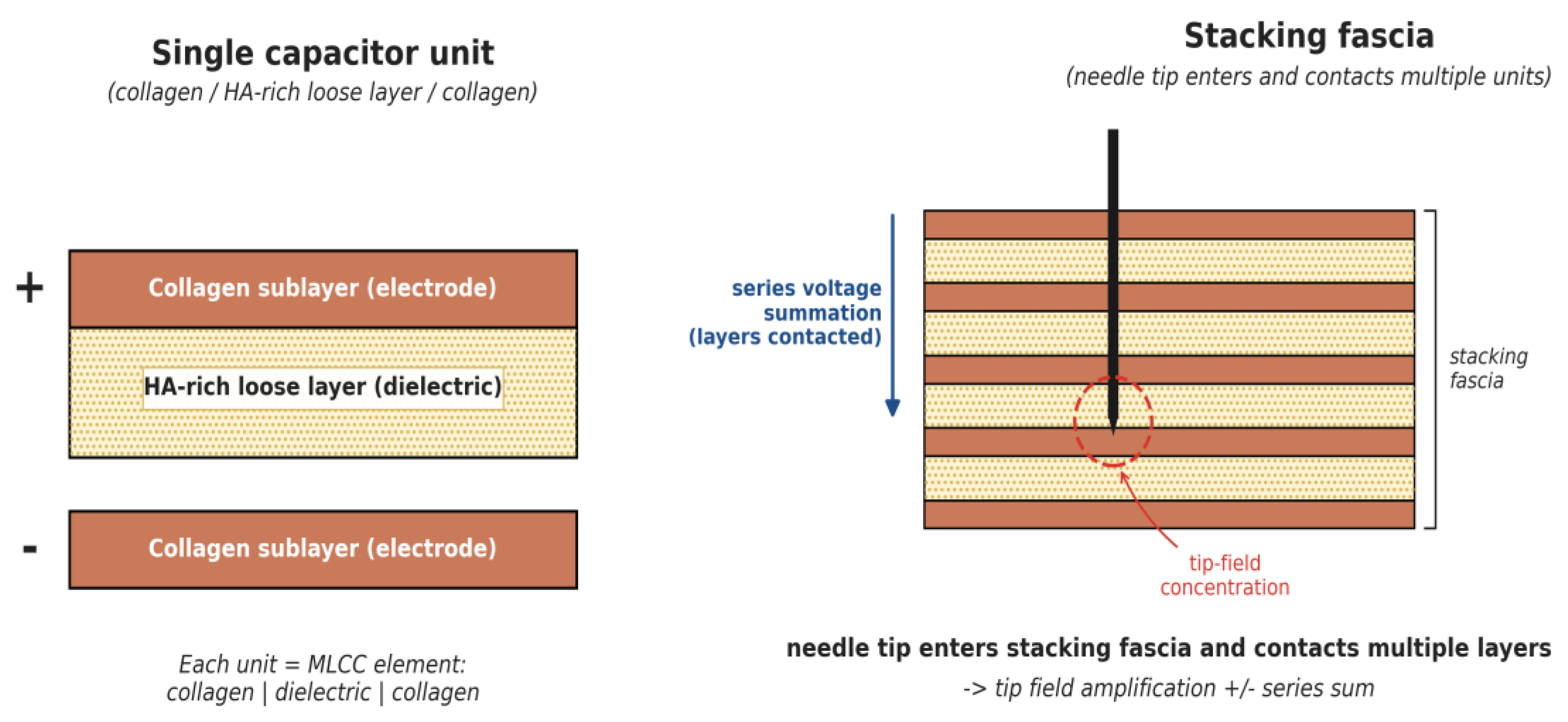
Figure 1. Schematic of the Fascial Capacitor Model. (Left) Single capacitor unit: collagen layers (electrodes) sandwich a hyaluronic-acid-rich layer (dielectric). (Right) Stacking fascia comprises multiple stacked units; a needle entering the upper portion of the stack contacts and discharges multiple layers, with electric-field concentration at the needle tip.
Preprints 214071 g001
Disclaimer/Publisher’s Note: The statements, opinions and data contained in all publications are solely those of the individual author(s) and contributor(s) and not of MDPI and/or the editor(s). MDPI and/or the editor(s) disclaim responsibility for any injury to people or property resulting from any ideas, methods, instructions or products referred to in the content.
Copyright: This open access article is published under a Creative Commons CC BY 4.0 license, which permit the free download, distribution, and reuse, provided that the author and preprint are cited in any reuse.
Prerpints.org logo

Preprints.org is a free preprint server supported by MDPI in Basel, Switzerland.

Subscribe

© 2026 MDPI (Basel, Switzerland) unless otherwise stated

Accessibility

Disclaimer

Terms of Use

Privacy Policy

Privacy Settings