

Submitted:
12 May 2026
Posted:
12 May 2026
You are already at the latest version
Preprints on COVID-19 and SARS-CoV-2
Abstract
The hexane extract of Psoralea drupacea Bunge fruits was initially evaluated for antivi-ral activity against SARS-CoV-2 based on GC-MS data indicating high bakuchiol con-tent (87.74%). Unexpectedly, the extract showed no antiviral effect in Vero E6 cells due to cytotoxicity (CC₅₀ = 7.5 μg/mL), while purified bakuchiol demonstrated moderate antiviral activity (IC₅₀ = 6.2 ± 0.8 μg/mL; SI = 2.9). Quantitative NMR revealed that the actual bakuchiol content in the extract was 44.3% — approximately half the GC-MS value — explaining the lack of efficacy at non-cytotoxic concentrations. Both the ex-tract and purified bakuchiol effectively blocked the RBD-ACE2 interaction in a com-petitive ELISA (71.3% inhibition at 50 μM for bakuchiol; IC₅₀ = 18.5 μM). Notably, the extract also inhibited the viral main protease 3CLpro (IC₅₀ = 32.0 ± 3.5 μg/mL), while purified bakuchiol showed no such activity. These findings reveal a dual mechanism: bakuchiol inhibits viral entry via RBD-ACE2 blockade, while other extract components (e.g., angelicin, psoralen) suppress viral replication via 3CLpro inhibition.
Keywords:
Psoralea drupacea
; bakuchiol
; SARS-CoV-2
; 3CLpro major protease
; protease inhibition
; RBD-ACE2
; entry inhibition
; antiviral activity
1. Introduction
The COVID-19 pandemic, caused by the SARS-CoV-2 coronavirus, has demonstrated the need for effective antiviral drugs capable not only of suppressing acute phases of infection but also of counteracting the emergence of new resistant viral variants [1,2]. Despite advances in vaccine and therapeutic development, there remains a need to expand the arsenal of antiviral agents with diverse mechanisms of action [3].
Natural compounds and their semisynthetic derivatives have historically served as an important source of drugs [4,5]. According to the World Health Organization, about 40% of modern pharmaceutical drugs are based on natural compounds or are their derivatives [6]. During the COVID-19 pandemic, interest in natural sources of antiviral agents has significantly increased, as many plants from traditional medicine have demonstrated potential in inhibiting various SARS-CoV-2 targets [7].
Among promising sources for antiviral agents, plants of the genus Psoralea (Fabaceae) stand out. They have been traditionally used in Asian medicine to treat various diseases, including skin disorders, inflammatory processes, and infections [8,9]. The most studied representative of the genus is Psoralea corylifolia L., from the fruits of which bakuchiol, a meroterpenoid of phenolic nature with a broad spectrum of biological activities including antibacterial, anti-inflammatory, antioxidant, and antiviral effects, has been isolated [10,11]. In recent years, bakuchiol has attracted particular attention from researchers due to its ability to modulate various cellular signaling pathways and exhibit activity against several viruses, including influenza virus [12]. Bakuchiol has recently been identified as a potential SARS-CoV-2 entry inhibitor capable of blocking the RBD-ACE2 interaction [13].
Psoralea drupacea Bunge is a Central Asian endemic species found in Kazakhstan and neighboring countries. The bakuchiol content in the fruits reaches 7.06%, a value comparable to that reported for P. corylifolia (5.14–6.98%). Beyond bakuchiol, the hexane extract contains furanocoumarins (angelicin, psoralen), sterols, terpenoids, and cyclitols. Although coumarins such as angelicin and psoralen are present only in minor amounts, they have previously been shown to inhibit viral proteases, suggesting they may contribute to the 3CLpro inhibition observed here. To date, however, no data have been available on the antiviral activity of P. drupacea components against SARS CoV 2.
Two key targets for antiviral intervention can be identified in the coronavirus life cycle. The early stages of infection involve the interaction of the viral spike protein with the cellular ACE2 receptor. This step ensures viral attachment to the target cell and its entry. Blocking the interaction of the RBD of the spike protein with ACE2 prevents infection at the earliest stage [14]. The replication stage, in which the main protease 3CLpro (Mpro) is a key enzyme. This enzyme processes viral polyproteins, cleaving them at 11 sites to produce functional nonstructural proteins required for viral replication [15]. 3CLpro inhibitors, such as nirmatrelvir (PF-07321332), have already proven their efficacy in clinical practice [16].
Of particular interest is the search for compounds capable of simultaneously targeting different stages of the viral life cycle, as such a multitargeted approach hinders the development of resistance and may provide a synergistic effect [17]. Plants that produce complex mixtures of secondary metabolites may serve as a source of such multitargeted agents.
One of the major challenges in natural product research is the accurate characterization of complex extracts and the interpretation of their biological activities. Crude plant extracts contain dozens or even hundreds of secondary metabolites with varying physicochemical properties, which can lead to discrepancies between different analytical methods. For example, gas chromatography-mass spectrometry (GC-MS) analysis of volatile or semi-volatile fractions is highly sensitive but can overestimate the relative abundance of highly volatile compounds due to differential evaporation during sample preparation, while non-volatile or less volatile components may be underrepresented or completely missed. Quantitative NMR (qNMR), in contrast, provides an absolute quantification of specific compounds independent of volatility or detector response, but requires prior knowledge of compound identity and non-overlapping signals. Furthermore, the biological activity of an extract in cell-based assays can be confounded by cytotoxicity, poor solubility, or aggregation of lipophilic components — issues particularly relevant for hexane extracts rich in non-polar compounds. These challenges can lead to paradoxical results, such as an extract containing a known active compound failing to show activity in cell culture, or discrepancies between the expected and actual compound content. Recognizing and resolving such discrepancies is essential for the accurate interpretation of natural product screening data and for avoiding false conclusions about the antiviral potential of plant extracts.
The aim of this work is a detailed investigation of the antiviral activity of the hexane extract of P. drupacea fruits and its major component bakuchiol against SARS-CoV-2.
2. Results
2.1. Chemical Composition of the Hexane Extract
The fruits of P. drupacea Bge. were collected in September 2015 in the South Kazakhstan region (herbarium number 3703). Sequential extraction of the ground fruits (51.00 g) with hexane yielded 8.13 g (15.94%) of the hexane extract. Subsequent extraction with chloroform, acetone, and methanol yielded 2.20 g (4.31%), 1.26 g (2.47%), and 6.30 g (12.35%), respectively.
GC-MS analysis of the hexane extract revealed the presence of 17 components, with bakuchiol as the dominant constituent (87.74% of the volatile fraction) (Table 1). Notably, the furanocoumarins angelicin (4.74%) and psoralen (1.56%) were also identified, along with the sesquiterpenoid β hydroxy-1,10-dehydrofuranofind-9-one (2.02%).
2.2. Antiviral Activity of the Hexane Extract and Purified Bakuchiol in Cell Culture
Given the high bakuchiol content indicated by GC-MS (87.74%), and since bakuchiol had been previously reported to inhibit SARS-CoV-2 entry by blocking the RBD-ACE2 interaction [13], we first evaluated the hexane extract for antiviral activity against live SARS-CoV-2 (strain nCov/Victoria/1/2020) in Vero E6 cell cultures.
Unexpectedly, the hexane extract showed no protective effect against virus-induced cytopathic effect at non-cytotoxic concentrations. Cytotoxicity testing revealed that the extract exhibited significant toxicity with a CC₅₀ of 7.5 ± 0.9 μg/mL (Figure 1A). At concentrations below this threshold (2–5 μg/mL), no antiviral protection was observed.
In contrast, purified bakuchiol demonstrated moderate protection of cells against virus-induced cytopathic effects with an IC₅₀ of 6.2 ± 0.8 μg/mL (Figure 1B). The cytotoxicity of bakuchiol was CC₅₀ = 17.8 ± 2.1 μg/mL (69.5 μM), resulting in a selectivity index (SI) of 2.9. For comparison, remdesivir, used as a control, showed no cytotoxicity in the concentration range tested (CC₅₀ > 110 μg/mL).
This negative result with the crude extract was surprising, as it contradicted the expected antiviral activity based on the presumed high bakuchiol content.
2.3. Resolution of the Discrepancy: Quantitative NMR Reveals Lower Bakuchiol Content
To resolve this discrepancy, we re-evaluated the bakuchiol content in the hexane extract using quantitative NMR with 1,3,5-trinitrobenzene as an internal standard. This method quantifies the absolute mass percentage of bakuchiol, independent of volatility or detector response.
The qNMR analysis revealed that the actual bakuchiol content was 44.3% of the total extract mass — approximately half of the 87.74% value suggested by GC-MS. This substantial overestimation by GC-MS likely resulted from differential volatility of extract components during sample preparation, leading to enrichment of bakuchiol in the injected volatile fraction.
With this corrected value, we recalculated that at the maximum non-cytotoxic concentration of the extract (5 μg/mL), the bakuchiol concentration would be only ~2.2 μg/mL (~8.6 μM) — well below the IC₅₀ for RBD-ACE2 inhibition (see Section 2.4).
2.4. Confirmation of RBD-ACE2 Inhibition by the Extract and Bakuchiol
We next directly tested whether the hexane extract and purified bakuchiol could inhibit the RBD-ACE2 interaction using a competitive ELISA. This assay measures the ability of compounds to block binding of the SARS-CoV-2 spike protein RBD to the ACE2 receptor — the primary mechanism of viral entry.
The results presented in Figure 2 demonstrate that both the hexane extract and purified bakuchiol effectively block the RBD-ACE2 interaction. At a concentration of 50 μg/mL (or 50 μM for bakuchiol), inhibition for the hexane extract was 62.5 ± 5.3% (for the Wuhan variant) and 58.2 ± 4.9% (for the Delta variant). For bakuchiol, inhibition was 71.3 ± 6.1% (for the Wuhan variant) and 65.7 ± 5.8% (for the Delta variant).
No statistically significant differences in binding inhibition were observed between the Wuhan and Delta variants (p > 0.05). The calculated IC₅₀ value for bakuchiol in this assay was 18.5 ± 2.3 μM (4.7 μg/mL).
These findings confirm that bakuchiol is an active RBD-ACE2 inhibitor, consistent with previous reports [13]. The hexane extract also showed activity, although its efficacy in cell culture was limited by the low bakuchiol concentration achievable at non-cytotoxic doses.
2.5. Discovery of 3CLpro Inhibitory Activity in the Hexane Extract
Finally, we investigated whether the hexane extract might contain additional antiviral mechanisms beyond entry inhibition. Specifically, we tested the extract and purified bakuchiol for inhibition of the SARS-CoV-2 main protease 3CLpro using a FRET-based assay.
The hexane extract exhibited a pronounced dose-dependent inhibitory effect on 3CLpro (Figure 3). The half-maximal inhibitory concentration (IC₅₀) was 32.0 ± 3.5 μg/mL, comparable to the reference compound disulfiram (IC₅₀ = 30.0 ± 2.8 μg/mL) [18].
Importantly, purified bakuchiol showed no significant inhibition of 3CLpro at concentrations up to 200 μM (data not shown). At the IC₅₀ of the hexane extract (32.0 μg/mL), the calculated concentration of bakuchiol — based on the qNMR value of 44.3% — is 14.2 μg/mL (55.4 μM). This concentration is well within the tested range of pure bakuchiol (up to 200 μM) and would have produced measurable inhibition if bakuchiol were responsible for the effect. The complete lack of activity from pure bakuchiol, combined with the presence of known protease-inhibiting coumarins (angelicin, psoralen) in the extract (Table 1), demonstrates that non-bakuchiol components are responsible for the observed 3CLpro inhibition.
To assess specificity, control experiments were performed using HIV-1 protease (an aspartyl protease). The P. drupacea hexane extract at 50 μg/mL inhibited HIV-1 protease by less than 10% (data not shown). The addition of BSA (1 mg/mL) did not affect the inhibitory activity, ruling out aggregation-based artifacts.
3. Discussion
The present study began with an unexpected observation. Based on GC-MS data indicating that bakuchiol constituted 87.74% of the volatile fraction of the P. drupacea hexane extract, and given the recent report by Zhang et al. [13] demonstrating that bakuchiol inhibits SARS-CoV-2 entry by blocking the RBD-ACE2 interaction, we anticipated that the extract would exhibit potent antiviral activity in cell culture. Surprisingly, the extract showed no protective effect against live SARS-CoV-2 in Vero E6 cells and was instead cytotoxic at concentrations above 7.5 μg/mL.
This negative result prompted a re-evaluation of the extract’s composition. Quantitative NMR revealed that the actual bakuchiol content was 44.3% — approximately half of the 87.74% value suggested by GC-MS. This discrepancy likely arose from differential volatility of extracted components during GC-MS sample preparation, leading to enrichment of bakuchiol in the injected volatile fraction. Non-volatile or less volatile components (e.g., waxes, sterols, higher terpenoids) are underrepresented in GC-MS analysis, resulting in an overestimation of the relative percentage of highly volatile compounds like bakuchiol. This observation serves as a cautionary tale for natural product researchers: GC-MS-derived relative percentages of volatile fractions should not be directly interpreted as absolute mass percentages without complementary quantitative methods such as qNMR or HPLC.
At the maximum non-cytotoxic concentration of the extract (5 μg/mL), the actual bakuchiol concentration was only ~2.2 μg/mL (~8.6 μM) — well below the IC₅₀ for RBD-ACE2 inhibition (18.5 μM). This explains the lack of antiviral activity of the crude extract in cell culture, despite the presence of an active entry inhibitor.
The IC₅₀ of the P. drupacea hexane extract against 3CLpro (32.0 ± 3.5 μg/mL) is comparable to that of disulfiram (30.0 ± 2.8 μg/mL), a known 3CLpro inhibitor [18]. This places the extract within the range of activity reported for other natural product extracts, such as Ginkgo biloba (IC₅₀ = 6.68 μg/mL) and Curcuma longa (IC₅₀ = 15.74 μg/mL), although direct comparisons are limited by differences in assay conditions [19,20].
A key finding is that purified bakuchiol, despite being the dominant component (87.74% of the volatile fraction, Table 1), shows no 3CLpro inhibition at concentrations up to 200 μM. This unequivocally demonstrates that the protease inhibitory activity resides in minor components. Based on our phytochemical analysis (Table 1), the hexane extract contains 4.74% angelicin and 1.56% psoralen — furanocoumarins previously reported to inhibit viral proteases, including 3CLpro [21,22]. Additionally, the presence of the sesquiterpenoid β-hydroxy-1,10-dehydrofuranofind-9-one (2.02%) and other unidentified compounds (totaling ~2.8%) provide further candidates for the observed activity. In silico studies have predicted that psoralen and its derivatives can bind to the active site of 3CLpro, and synergistic effects among these minor components cannot be excluded [23]. Notably, the 3CLpro inhibitory activity of the hexane extract was discovered incidentally while investigating the discrepancy between the extract’s composition and its lack of cell-based antiviral activity. This serendipitous finding reveals that P. drupacea is a source of both an entry inhibitor (bakuchiol) and, separately, 3CLpro inhibitors. Future bioactivity-guided fractionation is required to identify the specific compound(s) responsible.
The relative selectivity of the extract for 3CLpro over HIV-1 protease (<10% inhibition at 50 μg/mL) suggests a specific interaction rather than nonspecific protein denaturation. The lack of effect of BSA on inhibitory activity further rules out aggregation-based artifacts, a common concern in natural product screening [2].
Bakuchiol effectively blocked the RBD-ACE2 interaction in a competitive ELISA with an IC₅₀ of 18.5 ± 2.3 μM (4.7 μg/mL). Our findings corroborate and extend the work of Zhang et al. [13], who showed that bakuchiol binds to both RBD and ACE2 using bio-layer interferometry and inhibits pseudovirus entry. Importantly, we have now validated this entry inhibition mechanism using live SARS-CoV-2 in Vero E6 cells (IC₅₀ = 6.2 ± 0.8 μg/mL), providing physiological relevance to the biochemical binding data. The IC₅₀ value we obtained in the ELISA (18.5 μM) is consistent with the binding affinity reported by Zhang et al. (KD = 11.0 μM), validating bakuchiol as a bona fide entry inhibitor. The activity was comparable against both the Wuhan and Delta variants (p > 0.05), indicating that bakuchiol interacts with conserved regions of the RBD that are not affected by key mutations such as L452R and T478K. This is particularly important given the continued emergence of SARS-CoV-2 variants with altered RBD sequences.
The slightly lower activity of the crude extract (62.5% inhibition at 50 μg/mL, corresponding to ~22 μg/mL bakuchiol) compared to pure bakuchiol (71.3% at 12.8 μg/mL) may be explained by partial aggregation of extract components, poor solubility under ELISA conditions, or the presence of competing compounds. Such phenomena are not uncommon when comparing pure compounds with crude extracts.
Bakuchiol demonstrated moderate antiviral activity against live SARS-CoV-2 in Vero E6 cells (IC₅₀ = 6.2 ± 0.8 μg/mL). This value correlates well with the IC₅₀ for RBD-ACE2 inhibition (4.7 μg/mL), confirming that entry blockade is the primary mechanism of action of bakuchiol. However, the selectivity index of 2.9 (CC₅₀ = 17.8 ± 2.1 μg/mL) indicates a narrow therapeutic window. For systemic clinical applications, an SI > 10 is typically required, so pure bakuchiol is unlikely to be a viable systemic antiviral agent. Nevertheless, for topical applications such as nasal sprays or skin formulations, an SI of 2.9 may be acceptable, especially given the compound’s established safety profile in dermatological use [11].
The hexane extract failed to show antiviral activity in cell culture at non-toxic concentrations. This is primarily because its cytotoxicity (CC₅₀ = 7.5 μg/mL) occurs at concentrations lower than those required for 3CLpro inhibition (IC₅₀ = 32 μg/mL). Thus, the antiviral potential of the 3CLpro-inhibiting components cannot be realized in cell culture due to the extract’s inherent toxicity. This highlights a critical limitation of using crude extracts in cell-based antiviral assays: cytotoxicity can mask the activity of otherwise promising compounds.
Our findings demonstrate that P. drupacea is a source of both an entry inhibitor (bakuchiol) and, separately, 3CLpro inhibitors. This dual mechanism could potentially be exploited in combination therapy, targeting two different stages of the viral life cycle, which may reduce the likelihood of resistance development [17]. Moreover, the fact that bakuchiol retains activity against the Delta variant RBD suggests that it may be broadly active against multiple variants.
Finally, the discrepancy between GC-MS and qNMR data highlights an important methodological consideration for natural product research: relative percentages from GC-MS of volatile fractions should not be used to calculate absolute compound concentrations without verification by an orthogonal quantitative method. This is particularly critical when interpreting negative biological results or when comparing compound quantities across studies.
Several limitations should be acknowledged. First, the 3CLpro inhibitory activity of the hexane extract has been demonstrated only in a biochemical FRET-based assay using recombinant protease. The identity of the specific compound(s) responsible for this activity remains unknown, as bioactivity-guided fractionation has not yet been performed. While coumarins such as angelicin and psoralen are plausible candidates based on literature reports, direct evidence for their contribution is lacking. Second, the antiviral activity of bakuchiol in cell culture showed a narrow therapeutic window (SI = 2.9), limiting its potential for systemic use. Third, the cytotoxicity of the hexane extract precluded evaluation of its antiviral activity in cell culture; therefore, the biological relevance of its 3CLpro inhibitory activity remains unvalidated in a cellular context. Fourth, the selectivity of the extract for 3CLpro over other viral proteases was assessed only against HIV-1 protease; activity against other human or viral proteases has not been tested. Fifth, the competitive ELISA for RBD-ACE2 inhibition, while informative, does not fully recapitulate the complex membrane-associated context of viral entry, and confirmation in cell-based entry assays would be valuable. Finally, the study used only a single SARS-CoV-2 strain (nCov/Victoria/1/2020) and the Delta variant for RBD binding; activity against other variants (e.g., Omicron sublineages) remains to be tested.
Future work should focus on: (1) bioactivity-guided fractionation to identify specific 3CLpro inhibitors; (2) evaluation of bakuchiol against a broader panel of SARS-CoV-2 variants, including Omicron; (3) investigation of combination effects between bakuchiol and 3CLpro-inhibiting components; and (4) exploration of bakuchiol for topical antiviral applications.
4. Materials and Methods
4.1. Plant Material
The fruits of Psoralea drupacea Bge. were collected in September 2015 in the Republic of Kazakhstan, South Kazakhstan region, Arys district, Shagir station. The plant was identified at the Institute of Botany and Plant Introduction of Kazakhstan (herbarium number 3703). The raw materials were dried at room temperature with moderate ventilation, without direct exposure to sunlight.
4.2. Extraction of the Fruits of P. drupacea Bge.
Hexane (500.00 mL) was added to 51.00 g of finely ground fruits of P. drupacea Bge. The mixture was stirred on a magnetic stirrer for 6 hours at room temperature, then left to infuse for 24 hours. Additional stirring was carried out for 6 hours on a magnetic stirrer at 65–70 °C. The hexane extract was filtered. The procedure was repeated three times. The hexane extracts were combined, and the solvent was removed under reduced pressure. The residue was dried under vacuum. The yield of the hexane extract was 8.13 g, corresponding to 15.94% of the starting material.
After separation from hexane, the plant material was treated sequentially with chloroform (2 × 500 mL), acetone (2 × 500 mL), and methanol (2 × 500 mL). The extracts were separated and processed as described above. The following yields were obtained: chloroform extract — 2.20 g (4.31%), acetone extract — 1.26 g (2.47%), and methanol extract — 6.30 g (12.35%) per starting material. The composition of the fractions was analyzed by GC-MS.
4.3. Isolation of the Major Compound of the Hexane Extract
The hexane extract of P. drupacea Bge. fruits was separated by column chromatography on silica gel. A solution of 1.00 g of the extract in 3.00 mL of hexane was applied to 1.00 g of silica gel. The hexane was removed by evaporation under reduced pressure. The dry sorbent with the applied extract was placed on a column packed with silica gel (20.00 g) and eluted with hexane. Elution was continued with hexane–ethyl acetate mixtures of increasing polarity (5–40% at 5% intervals, then 50–100% at 10% intervals). Fractions were pooled based on TLC. The solvent was removed from the combined fractions under reduced pressure, and the residue was dried in vacuo, yielding 421.80 mg (42.18%) of bakuchiol.
4.3.1. Bakuchiol [4-[(1E)-3,7-Dimethyl-3-vinyl-1,6-octadien-1-yl]phenol] (1)
Molecular formula: C₁₈H₂₄O
HRMS [M]⁺: 256.1827
¹H NMR (400 MHz, CDCl₃) δ: 7.21 (d, J = 8.6 Hz, 2H), 6.75 (d, J = 8.6 Hz, 2H), 6.22 (d, J = 16.0 Hz, 1H), 6.02 (d, J = 16.0 Hz, 1H), 5.85 (dd, J₁ = 10.8 Hz, J₂ = 17.6 Hz, 1H), 5.09 (br t, J = 6.8 Hz, 1H), 5.02 (d, J = 5.2 Hz, 1H), 4.98 (d, J = 12.4 Hz, 1H), 1.92 (m, 2H), 1.65 (s, 3H), 1.56 (s, 3H), 1.47 (m, 2H), 1.17 (s, 3H).
¹³C NMR (100 MHz, CDCl₃) δ: 155.10 (s, C-4), 145.84 (d, C-17), 135.26 (d, C-8), 131.11 (s, C-13), 130.13 (s, C-1), 127.11 (d, C-2; C-6), 126.41 (d, C-7), 124.64 (d, C-12), 115.27 (d, C-3; C-5), 111.64 (t, C-18), 42.31 (s, C-9), 41.11 (t, C-10), 25.53 (q, C-14), 23.16 (q, C-16), 23.04 (t, C-11), 17.46 (q, C-15).
¹H and ¹³C NMR data agree with literature values.
4.4. Quantification of Bakuchiol in P. drupacea Fruits by NMR
The content of bakuchiol in the hexane extract was determined by quantitative NMR using an internal standard (1,3,5-trinitrobenzene). Briefly, 65.0 mg of hexane extract and 8.1 mg of 1,3,5-trinitrobenzene were dissolved in 0.9 mL of CDCl₃. The bakuchiol content was calculated by comparing the integrated intensity of selected bakuchiol signals to the integrated intensity of the standard signal.
4.5. GC-MS Analysis
The components of the extract were identified using a Hewlett-Packard instrument equipped with an HP 5890 Series II gas chromatograph and an HP 5971 mass selective detector (electron ionization, 70 eV). Separation was performed on an HP-5 capillary column (5% diphenyl–95% dimethylsiloxane; 30 m × 0.25 mm × 0.25 μm). Helium was used as the carrier gas at a flow rate of 1 mL/min. The temperature program was as follows: initial hold at 50 °C for 2 min, followed by a temperature increase to 300 °C at a rate of 10 °C/min, and a final hold at 300 °C for 30 min. The injector temperature was 280 °C, and the ion source temperature was 170 °C. Mass spectra were acquired at a scan rate of 1.2 scans/sec over a mass range of 29–650 m/z. Component identification was performed by comparing the full mass spectra with the NIST08 and W8N08 mass spectrometry databases and by calculating retention indices. Samples were spiked with n-alkanes (C₇–C₂₄ and C₇–C₄₀) as internal standards. Component yields were expressed as a percentage of the total volatile content.
4.6. NMR Spectroscopy
¹H and ¹³C NMR spectra were recorded on a Bruker AV 400 MHz NMR spectrometer (400.13 MHz for ¹H and 100.61 MHz for ¹³C) in CDCl₃ at 25 °C. Chemical shifts were referenced to the residual solvent signals (7.24 ppm for ¹H and 39.51 ppm for ¹³C). Signal multiplicity in the ¹³C NMR spectra was determined using the standard JMOD (attached proton test) mode.
4.7. Production of Recombinant 3CLpro and HIV 1 Proteases and Determination of Inhibitory Activity
Recombinant SARS-CoV-2 main protease (3CLpro) was produced by expression in E. coli cells followed by purification using metal affinity chromatography on Ni Sepharose, as described previously [24]. Expression was induced with IPTG. Protein purity was monitored by SDS PAGE. Protein concentration was determined by the Bradford method.
Compound activity was assessed by inhibition of cleavage of the fluorescent peptide substrate Dabcyl KTSAVLQ↓SGFRKME(Edans) NH₂ (purity >95%, CPC Scientific Inc., Hangzhou, China), which contains the 3CLpro recognition site. The principle of the method is based on FRET: upon substrate cleavage by the protease, the fluorophore (Edans) and quencher (Dabcyl) are separated, leading to an increase in fluorescence.
Fluorescence was recorded on a CLARIOstar Plus microplate fluorimeter spectrophotometer (BMG Labtech, Ortenberg, Germany) at excitation and emission wavelengths of 355 nm and 460 nm, respectively. The reaction mixture contained Tris HCl buffer (pH 7.3) with EDTA and DTT, peptide substrate (10 μM), recombinant 3CLpro (300 nM), and the test inhibitor at various concentrations. To determine IC₅₀, the inhibitor concentration was varied from 400 to 0 μM. Disulfiram was used as a positive control. All measurements were performed in triplicate in kinetic scanning mode. Recombinant HIV 1 protease expressed in E. coli was used as a negative control. The recombinant plasmid pET28 HIV was designed to express a single open reading frame encoding a SUMO HIV 1 protease fusion protein. The vector backbone and HIV 1 protease insert were amplified by PCR. The assembled pET28 HIV plasmid was transformed into chemically competent E. coli BL21(DE3) cells. Expression and purification of recombinant HIV 1 protease were performed according to established protocols [25,26].
4.8. Plasmid Construction, Production, and Purification of Recombinant Proteins
The trimeric spike protein (S trimer) was obtained as described previously [14]. Codon optimized nucleotide sequences of the spike protein of the Wuhan 1 strain (GenBank: MN908947) and the Delta variant (B.1.617.2) (GenBank: OK529678.1) were synthesized. The S protein coding fragment 1M P1213 was constructed with the protease cleavage site deleted, amino acid stabilizing substitutions K986P and V987P, and a C terminal T4 fibritin trimerization domain followed by a 10× His tag (pVEAL2 S, pVEAL2 Sdelta). The nucleotide sequence of chimeric human ACE2 Fc was synthesized and cloned into the pVEAL2 plasmid (pVEAL2 ACE2).
For stable protein expression in Chinese hamster ovary (CHO K1) cells, cells were transfected with recombinant plasmids (pVEAL2 S, pVEAL2 Sdelta, pVEAL2 ACE2) using Lipofectamine 3000 (Invitrogen, Carlsbad, CA, USA) according to the manufacturer’s instructions. To integrate the vector expression cassette into the host genome, cells were co transfected with the pCMV SB plasmid encoding SB100 transposase. Transfected cells were selected with puromycin (10 μg/mL) for 3 days. Clones were then cultured in roller bottles at 37 °C in DMEM/F 12 medium supplemented with 2% FBS.
Recombinant proteins were isolated from the culture medium of CHO K1 cells. The culture medium was centrifuged to remove cell debris and filtered through 0.22 μm filters. The trimeric S protein was purified by sequential chromatography on Ni NTA and ion exchange chromatography as described in [15]. Recombinant ACE2 Fc was purified using MabSelect SuRe resin (Cytiva, Uppsala, Sweden). Protein fractions were analyzed by SDS PAGE on 15% polyacrylamide gels, and the target protein fraction was dialyzed against PBS. The obtained protein samples were sterilized by filtration through 0.22 μm filters. Purity and homogeneity were determined using Gel Pro Analyzer, Ver. 3.1. Quantitative protein content analysis was performed using the Lowry method.
4.9. Synthesis of ACE2-HRP Conjugate
Purified ACE2 Fc protein was dissolved in water to a concentration of 2 mg/mL. Sodium periodate (100 mL of 0.088 M solution) was added to the protein, followed by incubation in the dark for 15–20 min at room temperature. The reaction was stopped by adding glycerol. After purification, ACE2 Fc was mixed with horseradish peroxidase (HRP) in a 1:1 ratio and incubated for 2 h at room temperature. Sodium cyanoborohydride (5 M) was added to the reaction solution under a fume hood, followed by incubation for 30 min at room temperature with gentle stirring. The reaction was blocked by adding 50 μL of 1 M ethanolamine, pH 9.6, with incubation for 30 min at room temperature. The conjugate was purified from excess reagents by dialysis, after which BSA was added to 5 mg/mL glycerol. ACE2 HRP was stored at −20 °C.
4.10. Competitive ELISA
The competitive ELISA protocol for assessing inhibition of S trimer binding to ACE2 was adapted from [27,28] with modifications described below. Briefly, 96 well plates were coated with S trimer proteins (Wuhan and Delta variants) at 400 ng/well in 0.01 M phosphate buffered saline (PBS, pH 7.2) and incubated for 18 h at 4 °C. Plates were then washed with PBST (0.1% Tween 20 in PBS) and blocked with 1% casein in PBS T for 1 h at 37 °C. Compound solutions in DMSO (10 mg/mL) diluted with PBS to concentrations from 5 to 500 μM were added to the wells and incubated for 1 h at 37 °C. To determine the specificity of inhibitory activity, the following procedure was performed. A solution of BSA (125 μg/mL to 1 mg/mL) with the compound at the IC₅₀ concentration was added to the wells and incubated for 1 h at 37 °C. After three washes with PBST, recombinant ACE2 labeled with HRP at a 1/500 dilution was added and incubated for 1 h at 37 °C. Wells were washed again, and TMB substrate solution was added. After 15 min, the reaction was stopped with 50 μL of 1 M HCl, and the optical density was measured at 450 nm using a Varioskan Lux multimode microplate reader (Thermo Fisher Scientific Inc., Waltham, MA, USA). All presented results are averages of at least two independent experiments. Inhibition was calculated by comparison with control wells to which no inhibitor was added (negative control). The recombinant antibody analog Nb6, known to prevent RBD ACE2 interaction, was used as a positive control [27].
4.11. Assessment of Antiviral Activity Against SARS CoV 2
Vero E6 cells were cultured in 96 well culture plates (Eppendorf, Germany) as a confluent monolayer (≥90% confluency). Growth medium consisted of DMEM (Biolot) supplemented with 10% fetal bovine serum (FBS) and antibiotics (penicillin, streptomycin; Biolot). Maintenance medium was MEM (Biolot) supplemented with 1% FBS and antibiotics.
SARS CoV 2 (strain nCov/Victoria/1/2020, passage 5 in Vero E6 cells) was obtained from the State Collection of Viral and Rickettsial Pathogens of the SRC VB “Vector”, Rospotrebnadzor. The working virus dose was 100 TCID₅₀/well.
Compound dilutions, starting from 300 μg/mL, were applied to the cell monolayer in triplicate. Eight serial three fold dilutions of each compound were prepared. Maintenance medium without virus (for determining the cytotoxic concentration of compounds) or medium containing virus at a dose of 100 TCID₅₀ (for determining the inhibitory activity of compounds) was then added to the wells. Culture plates were incubated at 37 °C for 3 days, after which they were stained with MTT (1 mg/mL). Results were recorded using a plate reader (Thermo Scientific Multiskan FC). Data analysis was performed using SOFTmax PRO 4.0 software with a four parameter logistic regression model.
The 50% cytotoxic concentration (CC₅₀) and 50% inhibitory concentration (IC₅₀) were determined. The selectivity index (SI) was calculated as the ratio of compound toxicity to inhibitory activity (SI = CC₅₀ / IC₅₀).
5. Conclusions
In the present work, we conducted the first comprehensive study of the antiviral activity of the hexane extract of Psoralea drupacea Bunge fruits and its major component bakuchiol against SARS-CoV-2. The study began with an unexpected finding: despite GC-MS data indicating high bakuchiol content (87.74% of the volatile fraction), the crude extract showed no antiviral activity in Vero E6 cells due to pronounced cytotoxicity (CC₅₀ = 7.5 μg/mL), while purified bakuchiol demonstrated moderate antiviral activity (IC₅₀ = 6.2 ± 0.8 μg/mL; SI = 2.9). Resolution of this discrepancy using quantitative NMR revealed that the actual bakuchiol content in the extract was 44.3% — approximately half the GC-MS value — explaining the lack of efficacy at non-cytotoxic concentrations.
Two key findings emerged from this investigation. First, both the hexane extract and purified bakuchiol effectively blocked the RBD-ACE2 interaction in a competitive ELISA (IC₅₀ for bakuchiol = 18.5 ± 2.3 μM), with activity maintained against the Delta variant. These results corroborate and extend independent reports identifying bakuchiol as a SARS-CoV-2 entry inhibitor [13]. Second, the hexane extract inhibited the SARS-CoV-2 main protease 3CLpro (IC₅₀ = 32.0 ± 3.5 μg/mL), comparable to disulfiram, while purified bakuchiol showed no such activity at concentrations up to 200 μM — demonstrating that non-bakuchiol components (likely the furanocoumarins angelicin and psoralen) are responsible for protease inhibition.
Collectively, these data reveal a dual mechanism of action for P. drupacea components: bakuchiol inhibits viral entry by blocking RBD-ACE2 interaction, while other extract components suppress viral replication via 3CLpro inhibition. The discrepancy between GC-MS and qNMR data serves as a cautionary methodological note for natural product research: relative percentages from volatile fractions should not be directly interpreted as absolute mass percentages without orthogonal quantitative validation. This study justifies further investigation of P. drupacea as a source of multitarget antiviral agents and stimulates the isolation and identification of individual compounds responsible for 3CLpro inhibition.
Author Contributions
Conceptualization, S.V.B. and D.N.S.; methodology, S.V.B., A.V.Z., E.D.M., E.A.V., N.A.P., D.E.M., V.O.T., T.P.K.; validation, S.V.B., A.V.Z., E.A.V.; formal analysis, S.V.B., A.V.Z.; investigation, S.V.B., A.V.Z., E.D.M., E.A.V., N.A.P., D.E.M., V.O.T.; resources, T.P.K., D.N.S.; data curation, S.V.B.; writing—original draft preparation, S.V.B.; writing—review and editing, S.V.B., D.N.S.; visualization, T.V.O., S.V.B.; supervision, D.N.S.; project administration, S.V.B.; funding acquisition, S.V.B. All authors have read and agreed to the published version of the manuscript.
Funding
This research was supported by the Russian Science Foundation (grant No. 24 75 00117).
Institutional Review Board Statement
Not applicable (the study did not involve humans or animals).
Informed Consent Statement
Not applicable.
Data Availability Statement
The data presented in this study are available on request from the corresponding author.
Acknowledgments
The authors express their gratitude to G.Zh. Baisalova (L.N. Gumilyov Eurasian National University, Astana, Kazakhstan) for providing plant material and its botanical identification. The authors thank the staff of the Chemical Research Center for Collective Use of the Siberian Branch of the Russian Academy of Sciences for spectral and analytical measurements.
Conflicts of Interest
The authors declare no conflicts of interest.
Abbreviations
The following abbreviations are used in this manuscript:
| 3CLpro | Coronavirus main protease |
| ACE2 | Angiotensin-converting enzyme 2 (SARS-CoV-2 receptor) |
| RBD | Receptor-Binding Domain |
| FRET | Fluorescence Resonance Energy Transfer |
| IC₅₀ | Half-maximal Inhibitory Concentration |
| CC₅₀ | Half-maximal Cytotoxic Concentration |
| SI | Selectivity Index |
| DTT | Dithiothreitol |
| EDTA | Ethylenediaminetetraacetic acid |
| BSA | Bovine Serum Albumin |
| DMSO | Dimethyl sulfoxide |
| HIV-1 | Human Immunodeficiency Virus type 1 |
References
- Wang, X.; Anwar, T.; Qureshi, H.; El-beltagi, H.S.; Sehar, Z.; Solieva, D. Plant-Based Traditional Remedies and Their Role in Public Health: Ethnomedicinal Perspectives for a Growing Population. J. Health Popul. Nutr. 2025, *44*. [CrossRef]
- Glenn, I.S.; Hall, L.N.; Khalid, M.M.; Ott, M.; Shoichet, B.K. Colloidal Aggregation Confounds Cell-Based Covid-19 Antiviral Screens. J. Med. Chem. 2024, *67*, 10263–10274. [CrossRef]
- Thomas, E.; Stewart, L.E.; Darley, B.A.; Pham, A.M.; Esteban, I.; Panda, S.S. Plant-Based Natural Products and Extracts: Potential Source to Develop New Antiviral Drug Candidates. Molecules 2021, *26*, 6197. [CrossRef]
- Musarra-Pizzo, M.; Pennisi, R.; Ben-Amor, I.; Mandalari, G.; Sciortino, M.T. Antiviral Activity Exerted by Natural Products against Human Viruses. Viruses 2021, *13*, 828. [CrossRef]
- Xiong, X.; Tang, N.; Lai, X.; Zhang, J.; Wen, W.; Li, X.; Li, A.; Wu, Y.; Liu, Z. Insights Into Amentoflavone: A Natural Multifunctional Biflavonoid. Front. Pharmacol. 2021, *12*, 768708.
- Newman, D.J.; Cragg, G.M. Natural Products as Sources of New Drugs over the Nearly Four Decades from 01/1981 to 09/2019. J. Nat. Prod. 2020, *83*, 770–803. [CrossRef]
- Boozari, M.; Hosseinzadeh, H. Natural Products for COVID-19 Prevention and Treatment Regarding to Previous Coronavirus Infections and Novel Studies. Phytother. Res. 2021, *35*, 864–876. [CrossRef]
- Koul, B.; Taak, P.; Kumar, A.; Kumar, A.; Sanyal, I. Genus Psoralea: A Review of the Traditional and Modern Uses, Phytochemistry and Pharmacology. J. Ethnopharmacol. 2019, *232*, 201–226.
- Li, C.-C.; Wang, T.-L.; Zhang, Z.-Q.; Yang, W.-Q.; Wang, Y.-F.; Chai, X.; Wang, C.-H.; Li, Z. Phytochemical and Pharmacological Studies on the Genus Psoralea: A Mini Review. Evid.-Based Complement. Altern. Med. 2016, *2016*. [CrossRef]
- Adarsh Krishna, T.P.; Edachery, B.; Athalathil, S. Bakuchiol – a Natural Meroterpenoid: Structure, Isolation, Synthesis and Functionalization Approaches. RSC Adv. 2022, *12*, 8815–8832. [CrossRef]
- Nizam, N.N.; Mahmud, S.; Ark, S.M.A.; Kamruzzaman, M.; Hasan, K. Bakuchiol, a Natural Constituent and Its Pharmacological Benefits [Version 2; Peer Review: 2 Approved]. F1000Research 2023, *12*, 1–24.
- Shoji, M.; Arakaki, Y.; Esumi, T.; Kohnomi, S.; Yamamoto, C.; Suzuki, Y.; Takahashi, E.; Konishi, S.; Kido, H.; Kuzuhara, T. Bakuchiol Is a Phenolic Isoprenoid with Novel Enantiomer-Selective Anti-Influenza A Virus Activity Involving Nrf2 Activation. J. Biol. Chem. 2015, *290*, 28001–28017. [CrossRef]
- Zhang, D.; Hamdoun, S.; Chen, R.; Yang, L.; Ip, C.K.; Qu, Y.; Li, R.; Jiang, H.; Yang, Z.; Chung, S.K.; et al. Identification of natural compounds as SARS-CoV-2 entry inhibitors by molecular docking-based virtual screening with bio-layer interferometry. Pharmacol. Res. 2021, *172*, 105820. [CrossRef]
- Hoffmann, M.; Kleine-Weber, H.; Schroeder, S.; Krüger, N.; Herrler, T.; Erichsen, S.; Schiergens, T.S.; Herrler, G.; Wu, N.H.; Nitsche, A.; et al. SARS-CoV-2 Cell Entry Depends on ACE2 and TMPRSS2 and Is Blocked by a Clinically Proven Protease Inhibitor. Cell 2020, *181*, 271-280.e8. [CrossRef]
- Jin, Z.; Du, X.; Xu, Y.; Deng, Y.; Liu, M.; Zhao, Y.; Zhang, B.; Li, X.; Zhang, L.; Peng, C.; et al. Structure of Mpro from SARS-CoV-2 and Discovery of Its Inhibitors. Nature 2020, *582*, 289–293. [CrossRef]
- Owen, D.R.; Allerton, C.M.N.; Anderson, A.S.; Aschenbrenner, L.; Avery, M.; Berritt, S.; Boras, B.; Cardin, R.D.; Carlo, A.; Coffman, K.J.; et al. An Oral SARS-CoV-2 Mpro Inhibitor Clinical Candidate for the Treatment of COVID-19. Science 2021, *3*, 1586–1593. [CrossRef]
- Wang, X.; Cao, R.; Zhang, H.; Liu, J.; Xu, M.; Hu, H.; Li, Y.; Zhao, L.; Li, W.; Sun, X.; et al. The Anti-Influenza Virus Drug, Arbidol Is an Efficient Inhibitor of SARS-CoV-2 in Vitro. Cell Discov. 2020, *6*, 28. [CrossRef]
- N., L.-G.; M., T.-L.; A., M.-M.; A.G., D.-S. FDA-Approved Thiol-Reacting Drugs That Potentially Bind into the SARS-CoV-2 Main Protease, Essential for Viral Replication. J. Biomol. Struct. Dyn. 2020, *39*, 3419–3427. [CrossRef]
- Xiong, Y.; Zhu, G.; Wang, H.; Hu, Q.; Chen, L.; Guan, X.; Li, H.; Chen, H.; Tang, H.; Ge, G.; et al. Discovery of Naturally Occurring Inhibitors against SARS-CoV-2 3CLpro from Ginkgo Biloba Leaves via Large-Scale Screening. Fitoterapia 2021, *150*, 104909. [CrossRef]
- Guijarro-Real, C.; Plazas, M.; Rodríguez-Burruezo, A.; Prohens, J.; Fita, A. Potential In Vitro Inhibition of Selected Plant Extracts Against SARS-CoV-2 3CLpro. Foods 2021, *10*, 1503.
- Mishra, S.; Pandey, A.; Manvati, S. Coumarin: An Emerging Antiviral Agent. Heliyon 2020, *6*, e03217. [CrossRef]
- Das, P.; Majumder, R.; Mandal, M.; Basak, P. In-Silico Approach for Identification of Effective and Stable Inhibitors for COVID-19 Main Protease (Mpro) from Flavonoid Based Phytochemical Constituents of Calendula Officinalis. J. Biomol. Struct. Dyn. 2021, *39*, 6265–6280. [CrossRef]
- Caesar, L.K.; Cech, N.B. Synergy and Antagonism in Natural Product Extracts: When 1 + 1 Does Not Equal 2. Nat. Prod. Rep. 2019, *36*, 869–888. [CrossRef]
- Belenkaya, S.V.; Merkuleva, I.A.; Yarovaya, O.I.; Chirkova, V.Y.; Sharlaeva, E.A.; Shanshin, D.V.; Volosnikova, E.A.; Vatsadze, S.Z.; Khvostov, M.V.; Salakhutdinov, N.F.; et al. The Main Protease 3CLpro of the SARS-CoV-2 Virus: How to Turn an Enemy into a Helper. Front. Bioeng. Biotechnol. 2023, *11*, 1187761. [CrossRef]
- Maseko, S.B.; Govender, D.; Govender, T.; Naicker, T.; Lin, J.; Maguire, G.E.M.; Kruger, H.G. Optimized Procedure for Recovering HIV-1 Protease (C-SA) from Inclusion Bodies. Protein J. 2019, *38*, 30–36. [CrossRef]
- Volontè, F.; Piubelli, L.; Pollegioni, L. Optimizing HIV-1 Protease Production in Escherichia Coli as Fusion Protein. Microb. Cell Fact. 2011, *10*, 1–10. [CrossRef]
- Merkuleva, I.A.; Shcherbakov, D.N.; Borgoyakova, M.B.; Shanshin, D.V.; Rudometov, A.P.; Karpenko, L.I.; Belenkaya, S.V.; Isaeva, A.A.; Nesmeyanova, V.S.; Kazachinskaia, E.I.; et al. Comparative Immunogenicity of the Recombinant Receptor-Binding Domain of Protein S SARS-CoV-2 Obtained in Prokaryotic and Mammalian Expression Systems. Vaccines 2022, *10*, 96. [CrossRef]
- Yang, L.J.; Chen, R.H.; Hamdoun, S.; Coghi, P.; Ng, J.P.L.; Zhang, D.W.; Guo, X.; Xia, C.; Law, B.Y.K.; Wong, V.K.W. Corligan prevents SARS-CoV-2 infection by targeting RBD-ACE2 binding. Phytomedicine 2021, *87*, 153591.
- Author 1, A.B. Title of Thesis. Level of Thesis, Degree-Granting University, Location of University, Date of Completion.
- Title of Site. Available online: URL (accessed on Day Month Year).
Figure 1.
Antiviral activity of bakuchiol against SARS-CoV-2 in Vero E6 cells. (A) Concentration-dependent cell protection by bakuchiol (IC₅₀ = 6.2 μg/mL). (B) Cytotoxicity of bakuchiol (CC₅₀ = 17.8 μg/mL). Data are presented as mean ± SD (n=3).
Figure 1.
Antiviral activity of bakuchiol against SARS-CoV-2 in Vero E6 cells. (A) Concentration-dependent cell protection by bakuchiol (IC₅₀ = 6.2 μg/mL). (B) Cytotoxicity of bakuchiol (CC₅₀ = 17.8 μg/mL). Data are presented as mean ± SD (n=3).
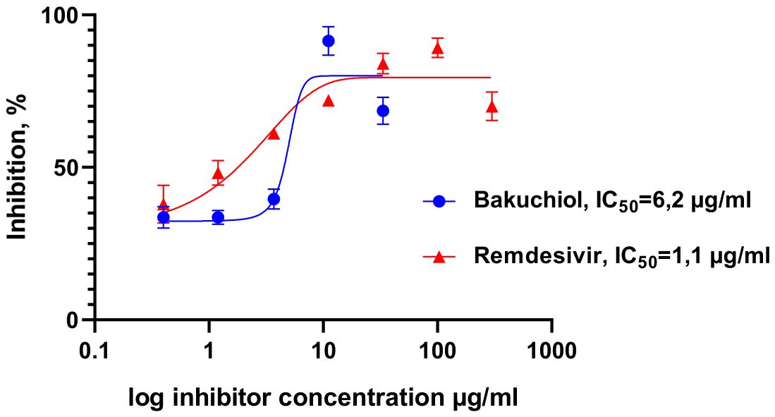
Figure 2.
Inhibition of SARS-CoV-2 spike protein (Wuhan and Delta variants) binding to ACE2-HRP. Compound concentrations: extract – 50 μg/mL, bakuchiol – 50 μM, Nb6 – 10 μg/mL. Data are presented as mean ± SD (n=3). *p < 0.05 compared to control.
Figure 2.
Inhibition of SARS-CoV-2 spike protein (Wuhan and Delta variants) binding to ACE2-HRP. Compound concentrations: extract – 50 μg/mL, bakuchiol – 50 μM, Nb6 – 10 μg/mL. Data are presented as mean ± SD (n=3). *p < 0.05 compared to control.
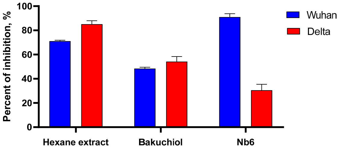
Figure 3.
Concentration–response curve for determining the half-maximal inhibitory concentration of the hexane extract against 3CLpro activity.
Figure 3.
Concentration–response curve for determining the half-maximal inhibitory concentration of the hexane extract against 3CLpro activity.
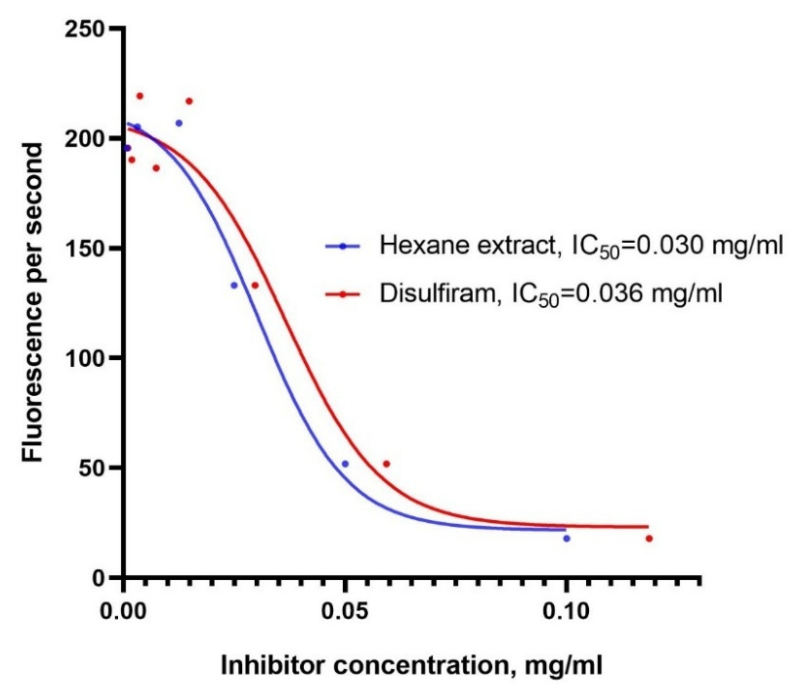

Table 1.
Chemical composition of the hexane extract of Psoralea drupacea Bunge fruits.
| No. | Compound | R. time | RI | % |
| 1 | Octadecane | 17.89 | 1800 | 0.04 |
| 2 | Angelicin | 18.33 | 1850 | 4.74 |
| 3 | n-Nonadecane | 18.87 | 1900 | 0.05 |
| 4 | Psoralen | 19.11 | 1920 | 1.56 |
| 5 | Unidentified | 19.73 | 1990 | 0.17 |
| 6 | n-Eicosane | 19.82 | 2000 | 0.07 |
| 7 | Unidentified | 20.02 | 2020 | 0.19 |
| 8 | Unidentified | 20.34 | 2055 | 0.08 |
| 9 | n-Heneicosane | 20.75 | 2100 | 0.12 |
| 10 | Bakuchiol | 21.08 | 2130 | 87.74 |
| 11 | Methyl linoleate | 21.34 | 2170 | 0.17 |
| 12 | n-Docosane | 21.62 | 2200 | 0.10 |
| 13 | β Hydroxy-1,10-dehydrofuranofind-9-one | 21.831 | 2240 | 2.02 |
| 14 | Unidentified | 21.96 | 2250 | 0.16 |
| 15 | n-Tricosane | 22.50 | 2300 | 0.18 |
| 16 | n-Tetracosane | 23.33 | 2400 | 0.13 |
| 17 | Unidentified | 23.98 | - | 2.46 |
Disclaimer/Publisher’s Note: The statements, opinions and data contained in all publications are solely those of the individual author(s) and contributor(s) and not of MDPI and/or the editor(s). MDPI and/or the editor(s) disclaim responsibility for any injury to people or property resulting from any ideas, methods, instructions or products referred to in the content. |
© 2026 by the authors. Licensee MDPI, Basel, Switzerland. This article is an open access article distributed under the terms and conditions of the Creative Commons Attribution (CC BY) license (http://creativecommons.org/licenses/by/4.0/).
Copyright: This open access article is published under a Creative Commons CC BY 4.0 license, which permit the free download, distribution, and reuse, provided that the author and preprint are cited in any reuse.


