

Submitted:
11 May 2026
Posted:
12 May 2026
You are already at the latest version
Abstract
Background/Objectives: Artificial intelligence (AI) has emerged as a promising tool to improve the detection and characterization of small bowel (SB) lesions through endoscopic imaging. This scoping review aimed to map and synthesize the available evidence regarding the diagnostic performance of AI systems applied to capsule endoscopy (CE) and device-assisted enteroscopy for the identification of SB lesions with potential malignant or premalignant relevance. Methods: A scoping review was conducted following the frameworks of Arksey and O’Malley, Levac, and Joanna Briggs Institute recommendations, and reported according to PRISMA-ScR guidelines. Searches were performed in PubMed, Scopus, and Embase. Original studies evaluating AI systems on SB endoscopic images or videos and reporting quantitative diagnostic metrics were included. Data extraction covered study characteristics, imaging modality, AI task, diagnostic performance, and methodological limitations. Results: A total of 13 studies were included, with a predominance of retrospective designs (9/13; 69.2%), followed by one prospective study (1/13; 7.7%), and one pilot study (1/13; 7.7%). Most studies originated from China (4/13; 30.8%, including an international collaboration with Denmark) and Japan (4/13; 30.8%). CE was the predominant imaging modality (9/13; 69.2%), followed by device-assisted enteroscopy (2/13; 15.4%) and upper gastrointestinal endoscopy (1/13; 7.7%). Automated lesion detection was the main application of artificial intelligence, reported in most studies (11/12; 91.7%), frequently combined with diagnostic classification tasks (8/12; 66.7%). Most models were based on convolutional neural networks, including architectures such as ResNet50 (29), Single Shot Multibox Detector (28), YOLOv5 (30), and nnU-Net (32). Diagnostic performance was consistently high, with sensitivities ranging from 81.2% to 98.6%, specificities from 88.6% to 99.8%, and area under the ROC curve values approaching 1.0 in several studies (22–30,33). Conclusion: AI improves the detection of SB lesions and significantly reduces endoscopic reading times, particularly in CE. Nevertheless, current evidence remains insufficient to support the use of AI as a screening tool for SB cancer, mainly due to the predominance of retrospective studies and the lack of robust prospective multicenter validation.
Keywords:
artificial intelligence
; small bowel
; cancer
; systematic review
; endoscopy
1. Introduction
The small bowel (SB) accounts for approximately 75% of the total length of the gastrointestinal tract [1,2]; however, neoplasms arising in this segment are rare and represent only about 3% of gastrointestinal cancers [3,4]. Despite their low incidence, SB tumors pose a significant clinical challenge, as they are frequently diagnosed at advanced stages due to their nonspecific clinical presentation and the absence of established screening strategies [1,3,4,5]. Among histological subtypes, adenocarcinoma is the most common, accounting for approximately 40% of cases, followed by neuroendocrine tumors and lymphoproliferative disorders [4,5]. This delayed diagnosis contributes to poor prognosis and highlights the importance of improving early detection strategies.
The utility of diagnostic methods varies depending on the affected anatomical segment [5,6,7]. Upper gastrointestinal endoscopy is particularly useful for detecting duodenal lesions, while colonoscopy allows identification of abnormalities in the terminal ileum [5,6]. Capsule endoscopy (CE) plays a central role in the evaluation of jejunal and ileal pathology due to its ability to visualize the entire small bowel mucosa [6,7,8]. Additionally, cross-sectional imaging techniques, such as computed tomography enterography and magnetic resonance enterography, are essential for staging and assessing disease extent, whereas device-assisted enteroscopy enables tissue sampling and histopathological confirmation [7,8,9]. However, these modalities are limited by operator dependence, the complexity of SB anatomy, the large volume of images generated and prolonged reading times, all of which increase the risk of missed lesions [4,5,8,9,10,11].
Artificial intelligence (AI) has established itself as a promising tool for endoscopic interpretation, enabling the automated analysis of large volumes of image data and potentially improving lesion detection [12,13,14]. Current AI applications in SB endoscopy have primarily focused on the detection and classification of a wide range of lesions, including vascular, inflammatory, and protruding abnormalities. However, while these findings may represent early manifestations of neoplastic processes, it remains unclear to what extent existing AI systems are specifically designed to identify or characterize malignant and premalignant lesions. Therefore, this exploratory review aims to analyse and synthesize the available literature on the use of AI in SB endoscopy, with particular attention to lesion detection and its potential implications for the identification and characterization of malignant and premalignant pathologies.
2. Methods
2.1. Scoping Review Design
This scoping review was conducted following the methodological framework described by Arksey and O’Malley [16], later expanded by Levac [17], and further guided by the Joanna Briggs Institute recommendations [18]. Reporting was structured according to the PRISMA extension for scoping reviews (PRISMA-ScR) [19] (Supplementary file 1), with the aim of identifying, mapping, and synthesizing the available evidence on the application of artificial intelligence in the early endoscopic diagnosis of small bowel cancer and its precursor lesions.
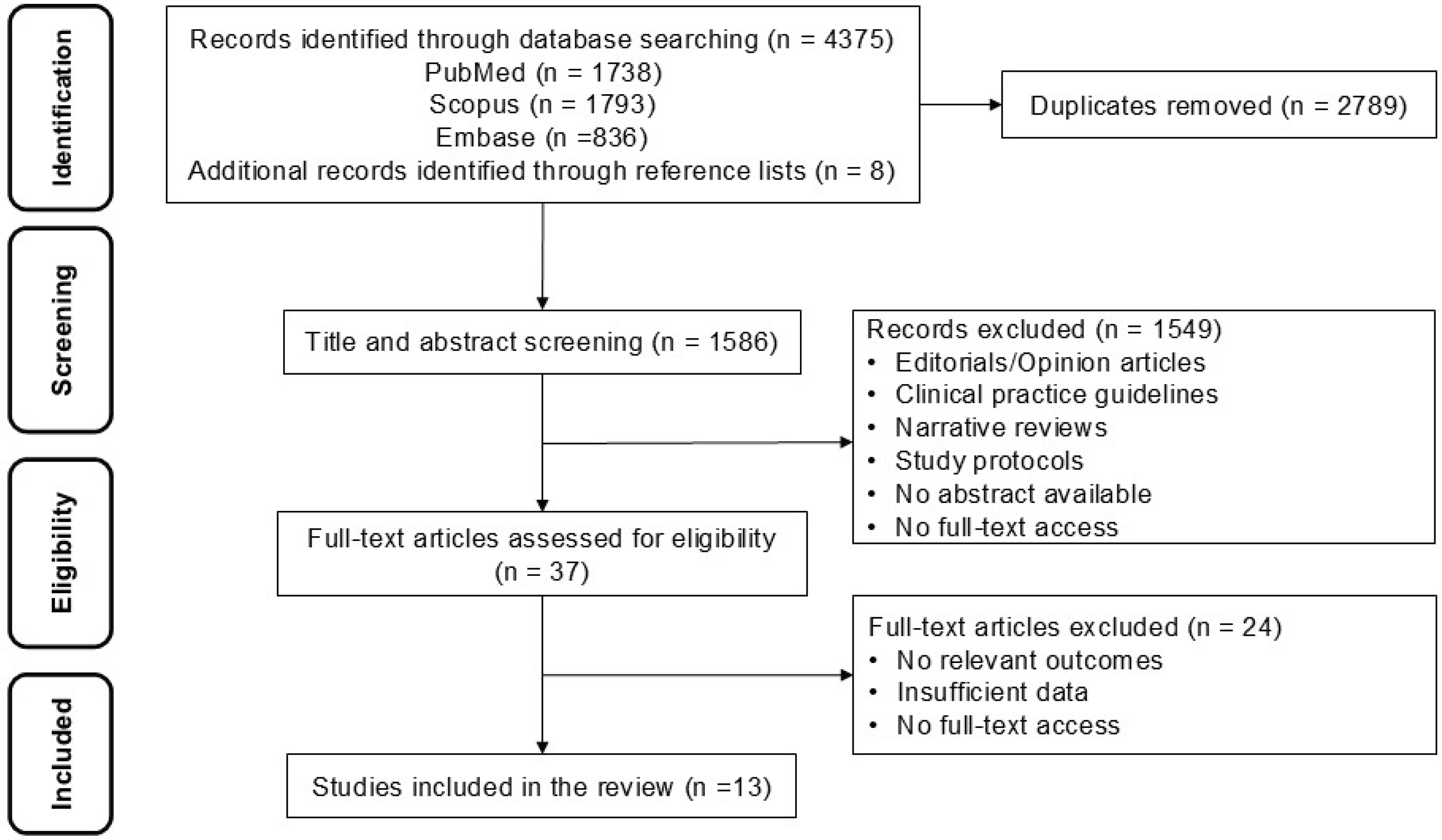
Figure 1.
Flowchart PRISMA.
2.2. Formulation of the Research Question
The research question was developed using the PCC framework (Population, Concept, and Context). The population included patients with inflammatory lesions, ulcerative lesions, and small bowel tumors. The concept encompassed artificial intelligence systems based on deep learning, machine learning, or computer-aided diagnosis. The context was limited to the use of images or videos obtained through upper gastrointestinal endoscopy and CE. Based on these elements, the guiding research question was: What is the diagnostic performance of AI systems applied to CE and device-assisted enteroscopy images for the detection, classification, or segmentation of SB lesions, and what are their potential implications for the identification of malignant or premalignant disease?
2.3. Search Strategy
The search strategy was designed to identify studies published in major biomedical databases, including PubMed, Scopus, and Embase [16,17,18]. Controlled vocabulary terms and keywords related to artificial intelligence, deep learning, convolutional neural networks, small bowel cancer, inflammatory lesions, ulcerative lesions, small bowel tumors, CE, and device-assisted enteroscopy were used. Additionally, the search strategy was refined to include terms specifically related to malignant and premalignant small bowel lesions, including adenocarcinoma, neuroendocrine tumors, lymphoma, and precursor lesions. Boolean operators were applied to optimize sensitivity and specificity in retrieving relevant studies (Supplementary file 2).
2.4. Eligibility Criteria
Original studies evaluating AI systems applied to the analysis of small bowel endoscopic images were included, provided they reported quantitative diagnostic performance metrics such as sensitivity, specificity, diagnostic accuracy, area under the ROC curve (AUC), positive predictive value, negative predictive value, or F1-score. Eligible study designs included retrospective studies, diagnostic development and validation studies, multicenter validations, prospective protocols, and comparative observational studies.
Studies using different endoscopic imaging modalities were considered, including CE, small bowel CE, device-assisted enteroscopy (DAE), double-balloon enteroscopy (DBE), and conventional white light endoscopy (WLE). Studies focusing on neoplastic, protruding, inflammatory, ulcerative, or vascular lesions of the small bowel were also included, as these represent common diagnostic scenarios for AI applications.
Narrative reviews, editorials, letters to the editor, studies without clinical validation or without quantitative diagnostic metrics, and studies focused exclusively on algorithm development without evaluation on real clinical images were excluded.
2.5. Definition of AI Diagnostic Tasks
Detection was defined as the automated identification of suspicious lesions in endoscopic images or video sequences, including protruding, ulcerative, inflammatory, vascular, or tumoral lesions. Classification referred to the assignment of a specific diagnostic category to a previously detected lesion based on morphological features or visual patterns. Segmentation was defined as the automated delineation of lesion boundaries or spatial extent within the image, with potential utility for quantifying lesion size, number, or affected surface area.
Some studies also evaluated object detection tasks, aimed at identifying multiple lesions within a single visual field, as well as multi-stage classification models integrating processes such as bowel preparation assessment and anomaly detection. Additionally, applications for automated disease monitoring and AI-assisted reading were considered, where algorithms support reducing reading time and improving diagnostic agreement with experts.
2.6. Study Selection
Study selection was conducted in two consecutive phases. First, titles and abstracts of potentially eligible studies were screened after being imported into the Rayyan platform [20], where two independent reviewers assessed each record based on standardized eligibility criteria. In the second phase, full-text articles of preselected studies were reviewed to confirm final inclusion. Discrepancies were resolved through consensus.
2.7. Data Extraction
Data extraction was performed using a standardized matrix [21]. Extracted variables included author, year of publication, country of origin, study type, type of lesion or small bowel cancer evaluated, imaging modality, type of AI task, sample size, comparison with endoscopists, main diagnostic performance metrics, key findings, and reported methodological limitations.
2.8. Evidence Synthesis and Analysis
A descriptive narrative synthesis was conducted due to heterogeneity among studies in terms of methodological design, model architectures, imaging modalities, sample sizes, and reported outcomes. Studies were grouped according to the main AI application, including automated lesion detection, histological classification, lesion segmentation, invasion depth prediction, and real-time evaluation during endoscopic procedures.
3. Results
A total of 13 studies were included (Table 1), with a predominance of retrospective designs (9/13; 69.2%) (22,23,25–28,31–33), followed by one prospective study (1/13; 7.7%) [30], and one pilot study (1/13; 7.7%) [29]. Most studies originated from China (4/13; 30.8%, including an international collaboration with Denmark) [22,30,33] and Japan (4/13; 30.8%) [26,27,28,31], followed by Portugal (3/13; 23.1%) [23,24,25], while Singapore [29] and the United States [32] each contributed one study (7.7%). CE was the predominant imaging modality (9/13; 69.2%) [22,25,26,27,28,29,30,31,33], followed by device-assisted enteroscopy (2/13; 15.4%) [23,24] and upper gastrointestinal endoscopy (1/13; 7.7%) [32].
Automated lesion detection was the main application of artificial intelligence, reported in most studies (11/12; 91.7%) [22,23,24,25,26,27,28,29,30,31,33], frequently combined with diagnostic classification tasks (8/12; 66.7%) [23,24,25,27,29,30,33]. Segmentation was evaluated in two studies (2/13; 15.4%) [26,32], mainly for delineating lesion boundaries or quantifying polyps. Other specific applications, such as bowel preparation assessment [29], multi-label detection [30], and automated disease monitoring [31], were reported in individual studies. Most models were based on convolutional neural networks, including architectures such as ResNet50 [29], Single Shot Multibox Detector [28], YOLOv5 [30], and nnU-Net [32]. Diagnostic performance was consistently high, with sensitivities ranging from 81.2% to 98.6%, specificities from 88.6% to 99.8%, and area under the ROC curve values approaching 1.0 in several studies [22,23,24,25,26,27,28,29,30,33]. Additionally, multiple studies reported significant reductions in reading time, near real-time processing, and high agreement with expert endoscopists [22,25,26,27,30].
Xie X et al. [22] reported that Saliency Segmentation–based Pattern Recognition significantly improved performance in the interpretation of CE videos for intestinal pathology, increasing sensitivity from 84.19% to 96.74%, specificity from 98.43% to 99.21%, and diagnostic accuracy from 89.47% to 97.66%. These findings suggest that this approach may help reduce the performance gap between less experienced and expert endoscopists in CE interpretation. Similarly, Martins M et al. [23] demonstrated that a convolutional neural network–based algorithm achieved high performance in the automatic detection of ulcerative lesions using device-assisted enteroscopy, with a sensitivity of 88.5%, specificity of 99.7%, and overall diagnostic accuracy of 98.9%. Likewise, Cardoso P et al. [24] showed that a convolutional neural network for the detection of protruding lesions using device-assisted enteroscopy achieved a sensitivity of 97.0%, specificity of 97.4%, positive predictive value of 94.6%, negative predictive value of 98.6%, and an overall accuracy of 97.3%, with an area under the curve of 1.00. Furthermore, the model processed the validation dataset in 10 seconds, equivalent to 157 images per second.
Hosoe N et al. [26] reported that their convolutional neural network–based algorithm for CE achieved a global sensitivity of 93.4% and a specificity of 97.8% in detecting clinically significant small bowel lesions, with particularly high performance for bleeding and angiodysplasia, reaching sensitivities of 100% for both. Additionally, the system reduced the number of images requiring review to 7.8% in the absence of massive bleeding, suggesting a reduction in reading time to approximately 10 minutes. Similarly, Aoki et al. [28] demonstrated that a convolutional neural network for the automatic detection of small bowel erosions and ulcerations via CE achieved a sensitivity of 88.2%, specificity of 90.9%, accuracy of 90.8%, and an area under the curve of 0.958, analyzing 10,440 images in 233 seconds (approximately 0.02 seconds per image), supporting its potential for rapid and efficient reading.
Li L et al. [30] developed a deep learning algorithm based on a real-time object detection model adapted for CE, capable of identifying and localizing lesions in both images and videos. In video analysis, the model achieved sensitivities of 91.9% for vascular lesions, 92.2% for ulcerative or erosive lesions, 91.4% for protruding lesions, 93.1% for parasites, 93.3% for diverticula, 95.1% for active bleeding, and 100% for villous lesions. Overall, it demonstrated a sensitivity of 94.0%, specificity of 92.3%, and accuracy of 93.1%, with a significantly reduced reading time compared to both experts and non-experts (5.62 ± 2.81 minutes), suggesting its potential as a reliable tool to support automated detection of small bowel lesions in clinical practice.
4. Discussion
This scoping review describes the utility of AI in the evaluation of small bowel lesions. The predominance of retrospective studies reflects an early stage of research, primarily focused on the development and initial validation of algorithms rather than their implementation in real clinical settings [22,23,25,26,27,28,31,32,33]. Additionally, there is a geographic concentration of studies in Asia, particularly in China and Japan [22,26,27,28,30,31,33]. Diagnostic performance may vary depending on the type of lesion, with lower sensitivity reported for conditions such as polyps or lymphoma in some studies [26,31,32], which may have relevant clinical implications. Overall, these methodological limitations reduce the strength of the evidence and highlight the need for prospective and multicenter studies to confirm their clinical utility.
Despite the refined focus on malignant and premalignant lesions, only a limited number of studies specifically addressed neoplastic pathology of the small bowel [22,26,27,28,30,31,33]. Most included studies evaluated general lesion detection, including vascular, inflammatory, or protruding lesions, which may indirectly encompass neoplastic processes but were not specifically designed to identify malignancy [22,26,27,28,30,31,33]. Explicit evaluation of small bowel tumors, such as adenocarcinoma or neuroendocrine tumors, was scarce, highlighting a significant gap in the current evidence.
Automated lesion detection represents the primary application of AI, often combined with diagnostic classification tasks [23,24,25,28,29,30,33]. From a strategic perspective, the automated detection of protruding or inflammatory lesions constitutes a critical first step in the diagnostic pathway, as these findings may represent early manifestations of neoplastic processes. Early identification at this stage is particularly relevant in small bowel pathology, where subtle mucosal changes can precede overt malignancy. However, an important gap in the current literature is that most available algorithms are designed for general lesion detection, without specifically focusing on the identification or characterization of histopathological malignancy, including clinically relevant entities such as small bowel adenocarcinoma, neuroendocrine tumors, and lymphoproliferative disorders. This gap limits the potential of AI systems to contribute directly to early cancer diagnosis and highlights an area for future research.
In this context, CE emerges as the predominant imaging modality [22,25,26,27,28,29,30,31,33], likely due to the large volume of images generated during its use, making it an ideal scenario for AI implementation. In contrast, more advanced applications, such as segmentation [26,32], bowel preparation assessment [29], and automated disease monitoring [31], remain limited and less explored.
Notably, the evaluated models demonstrated high diagnostic performance, with sensitivities and specificities generally exceeding 90% and area under the curve values approaching 1.0 in multiple studies [22,23,24,25,26,27,28,29,30,33]. These findings suggest that AI can achieve performance comparable to expert endoscopists. Furthermore, several studies reported significant reductions in reading time and near real-time processing capabilities [22,25,26,27,28,30], which is particularly relevant given that CE review is a time-consuming process prone to fatigue, potentially affecting lesion detection.
Importantly, although protruding or inflammatory lesions may represent early manifestations of neoplastic processes [24,25,27], current AI models are not specifically trained or validated to distinguish benign from premalignant or malignant lesions [26,28,29,30]. This limitation is particularly relevant in the small bowel, where tumors such as adenocarcinoma or neuroendocrine neoplasms are often diagnosed at advanced stages due to subtle or nonspecific early findings, and where only limited AI applications have specifically addressed neoplastic pathology [31,33]. Therefore, the development of AI systems specifically targeting neoplastic and premalignant lesions represents a critical unmet need and a key direction for future research.
In addition, AI may help reduce interobserver variability and narrow the performance gap between operators with different levels of experience. In this regard, some studies have demonstrated significant improvements in sensitivity and accuracy when using AI-assisted systems [22,27,30], supporting their role as a clinical support tool rather than a replacement for specialists.
Overall, the reviewed studies suggest that AI applications in small bowel endoscopy may reduce the likelihood of missing clinically relevant lesions, decrease reading time, and achieve high agreement with expert endoscopists [22,25,26,27,30]. However, the safety and applicability of these systems remain limited by important factors, including the predominance of retrospective designs, small sample sizes, single-center studies, and the use of static images instead of full-length videos [23,25,28,32,33]. Another critical issue is the lack of external validation and real-time clinical studies. Although some models, such as those based on YOLOv5, have shown promising results in prospective settings [30], more robust evidence is required to confirm their clinical impact and cost-effectiveness. Importantly, future developments should move beyond general detection tasks toward models capable of integrating morphological features with histopathological risk stratification.
Strengths and Limitations
An important limitation of this scoping review is the absence of a formal critical appraisal of the risk of bias and methodological quality of the included studies. Additionally, the heterogeneity among studies in terms of design, populations, imaging modalities, AI tasks, and reported metrics precluded quantitative synthesis or meta-analysis, limiting the findings to a descriptive approach. Most included studies were single center, with relatively small sample sizes and, in many cases, based on preselected static images, which may overestimate model performance [23,25,28,32,33]. Although a comprehensive search strategy across multiple databases was employed, the possibility of missing relevant studies cannot be excluded.
Another important limitation is the limited availability of studies specifically addressing malignant or premalignant small bowel lesions, despite targeted search efforts. This restricts the ability to draw conclusions regarding the performance of AI in early cancer detection. Despite these limitations, the available evidence suggests that AI has significant potential to improve the detection of small bowel lesions, optimize reading times, and standardize the interpretation of endoscopic studies [22,27,30]. However, its widespread implementation will depend on the availability of prospective, multicenter studies with adequate external validation. Crucially, addressing the current gap between lesion detection and histopathological characterization will be essential to fully leverage AI for early neoplasia detection and improve patient outcomes.
5. Conclusion
AI has strong potential to improve the detection and characterization of SB lesions, particularly through automated lesion detection in CE. Current evidence demonstrates high diagnostic accuracy, reduced reading times, and performance comparable to expert endoscopists. However, despite these promising results, there is still insufficient evidence to support the use of AI as a screening tool for SB cancer. The predominance of retrospective, single-center studies, the lack of external validation, and variability in performance across lesion types limit the generalizability and clinical applicability of current findings.
Supplementary Materials
The following supporting information can be downloaded at the website of this paper posted on Preprints.org.
Funding
This work was supported by Endogut Research Group.
Institutional Review Board Statement
Not applicable.
Informed Consent Statement
Not applicable.
Data Availability Statement
No new data were created or analyzed in this study.
Acknowledgments
The authors are most thankful for the Endogut Research Group.
Conflicts of Interest
The authors declare no conflicts of interest.
References
- Fantasia, S.; Cortegoso Valdivia, P.; Kayali, S.; Koulaouzidis, G.; Pennazio, M.; Koulaouzidis, A. The Role of Capsule Endoscopy in the Diagnosis and Management of Small Bowel Tumors: A Narrative Review. Cancers 2024, 16(2), 262. [Google Scholar] [CrossRef]
- Aparicio, T.; Pachev, A.; Laurent-Puig, P.; Svrcek, M. Epidemiology, risk factors and diagnosis of small bowel adenocarcinoma. Cancers 2022, 14(9), 2268. [Google Scholar] [CrossRef]
- Huang, J.; Chan, S.C.; Fung, Y.C.; Wong, M.C.S.; et al. Global incidence, risk factors, and trends of small intestine cancer: an analysis of cancer registries. Gastroenterology 2023, 164(6) Suppl 1, S-311. [Google Scholar] [CrossRef]
- Siegel, R.L.; Kratzer, T.B.; Wagle, N.S.; Sung, H.; Jemal, A. Cancer statistics, 2026. CA Cancer J. Clin. 2026, 76(1), 3–35. [Google Scholar] [CrossRef]
- Mota, J.; Almeida, M.J.; Mendes, F.; et al. From data to insights: how is AI revolutionizing small-bowel endoscopy? Diagnostics 2024, 14(3), 291. [Google Scholar] [CrossRef] [PubMed]
- Morgan, E.; Arnold, M.; Gini, A.; Lorenzoni, V.; Cabasag, C.J.; Laversanne, M.; Vignat, J.; Ferlay, J.; Murphy, N.; Bray, F. Global burden of colorectal cancer in 2020 and 2040: incidence and mortality estimates from GLOBOCAN. Gut 2023, 72(2), 338–344. [Google Scholar] [CrossRef] [PubMed]
- Benson, A.B.; Venook, A.P.; Al-Hawary, M.M.; Arain, M.A.; Chen, Y.J.; Ciombor, K.K.; Cohen, S.A.; Cooper, H.S.; Deming, D.A.; Garrido-Laguna, I.; Grem, J.L.; Hoffe, S.E.; Hubbard, J.; Hunt, S.; Kamel, A.; Kirilcuk, N.; Krishnamurthi, S.; Messersmith, W.A.; Meyerhardt, J.; Miller, E.D.; Mulcahy, M.F.; Nurkin, S.; Overman, M.J.; Parikh, A.; Patel, H.; Pedersen, K.S.; Saltz, L.B.; Schneider, C.; Shibata, D.; Skibber, J.M.; Sofocleous, C.T.; Stoffel, E.M.; Stotsky-Himelfarb, E.; Willett, C.G.; Johnson-Chilla, A.; Gregory, K.M.; Gurski, L.A. Small Bowel Adenocarcinoma, Version 1.2020, NCCN Clinical Practice Guidelines in Oncology. J. Natl. Compr. Canc Netw. 2019, 17(9), 1109–1133. [Google Scholar] [CrossRef]
- Yoon, J.Y.; Kumta, N.A.; Kim, M.K. The Role of Endoscopy in Small Bowel Neuroendocrine Tumors. Clin. Endosc. 2021, 54(6), 818–824. [Google Scholar] [CrossRef]
- Mohammed, A.; Trujillo, S.; Ghoneim, S.; Paranji, N.; Waghray, N. Small Bowel Adenocarcinoma: a Nationwide Population-Based Study. J. Gastrointest. Cancer 2023, 54(1), 67–72. [Google Scholar] [CrossRef]
- Vulasala, S.S.; Virarkar, M.; Gopireddy, D.; Waters, R.; Alkhasawneh, A.; Awad, Z.; Maxwell, J.; Ramani, N.; Kumar, S.; Onteddu, N.; Morani, A.C. Small Bowel Neuroendocrine Neoplasms-A Review. J. Comput Assist Tomogr. 2024, 48(4), 563–576. [Google Scholar] [CrossRef]
- Pennazio, M.; Rondonotti, E.; Despott, E.J.; Dray, X.; Keuchel, M.; Moreels, T.; Sanders, D.S.; Spada, C.; Carretero, C.; Cortegoso Valdivia, P.; Elli, L.; Fuccio, L.; Gonzalez Suarez, B.; Koulaouzidis, A.; Kunovsky, L.; McNamara, D.; Neumann, H.; Perez-Cuadrado-Martinez, E.; Perez-Cuadrado-Robles, E.; Piccirelli, S.; Rosa, B.; Saurin, J.C.; Sidhu, R.; Tacheci, I.; Vlachou, E.; Triantafyllou, K. Small-bowel capsule endoscopy and device-assisted enteroscopy for diagnosis and treatment of small-bowel disorders: European Society of Gastrointestinal Endoscopy (ESGE) Guideline - Update 2022. Endoscopy 2023, 55(1), 58–95. [Google Scholar] [CrossRef]
- Guleria, S.; Schwartz, B.; Sharma, Y.; Fernandes, P.; Jablonski, J.; Adewole, S.; Srivastava, S.; Rhoads, F.; Porter, M.; Yeghyayan, M.; Hyatt, D.; Copland, A.; Ehsan, L.; Brown, D.; Syed, S. The intersection of video capsule endoscopy and artificial intelligence: addressing unique challenges using machine learning. ArXiv [Preprint] 2023, arXiv:2308.13035v1. [Google Scholar]
- Oh, D.J.; Hwang, Y.; Kim, S.H.; Nam, J.H.; Jung, M.K.; Lim, Y.J. Reading of small bowel capsule endoscopy after frame reduction using an artificial intelligence algorithm. BMC Gastroenterol. 2024, 24(1), 80. [Google Scholar] [CrossRef]
- Cortegoso Valdivia, P.; Fantasia, S.; Kayali, S.; Deding, U.; Gualandi, N.; Manno, M.; Toth, E.; Dray, X.; Yang, S.; Koulaouzidis, A. Conventional small-bowel capsule endoscopy reading vs proprietary artificial intelligence auxiliary systems: Systematic review and meta-analysis. Endosc. Int. Open 2025, 13, a25442863. [Google Scholar] [CrossRef]
- Spada, C.; Piccirelli, S.; Hassan, C.; Ferrari, C.; Toth, E.; González-Suárez, B.; Keuchel, M.; McAlindon, M.; Finta, Á.; Rosztóczy, A.; Dray, X.; Salvi, D.; Riccioni, M.E.; Benamouzig, R.; Chattree, A.; Humphries, A.; Saurin, J.C.; Despott, E.J.; Murino, A.; Johansson, G.W.; Giordano, A.; Baltes, P.; Sidhu, R.; Szalai, M.; Helle, K.; Nemeth, A.; Nowak, T.; Lin, R.; Costamagna, G. AI-assisted capsule endoscopy reading in suspected small bowel bleeding: a multicentre prospective study. Lancet Digit Health 2024, 6(5), e345–e353. [Google Scholar] [CrossRef]
- Arksey, H.; O’Malley, L. Scoping studies: towards a methodological framework. Int. J. Soc. Res. Methodol. 2005, 8(1), 19–32. [Google Scholar] [CrossRef]
- Levac, D.; Colquhoun, H.; O'Brien, K.K. Scoping studies: advancing the methodology. Implement Sci. 2010, 5(1), 69. [Google Scholar] [CrossRef] [PubMed]
- Aromataris, E.; Munn, Z. JBI Manual for Evidence Synthesis; JBI, 2020. [Google Scholar]
- Tricco, A.C.; Lillie, E.; Zarin, W.; O'Brien, K.K.; Colquhoun, H.; Levac, D.; et al. PRISMA Extension for Scoping Reviews (PRISMA-ScR): Checklist and Explanation. Ann. Intern Med. 2018, 169, 467. [Google Scholar] [CrossRef]
- Ouzzani, M.; Hammady, H.; Fedorowicz, Z.; Elmagarmid, A. Rayyan—a web and mobile app for systematic reviews. Syst. Rev. 2016, 5, 210. [Google Scholar] [CrossRef] [PubMed]
- Grudniewicz, A.; Nelson, M.; Kuluski, K.; et al. Treatment goal setting for complex patients: protocol for a scoping review. BMJ Open. 2016, 6, e011869. [Google Scholar] [CrossRef] [PubMed]
- Xie, X.; Xiao, Y.F.; Yang, H.; Peng, X.; Li, J.J.; Zhou, Y.Y.; Fan, C.Q.; Meng, R.P.; Huang, B.B.; Liao, X.P.; Chen, Y.Y.; Zhong, T.T.; Lin, H.; Koulaouzidis, A.; Yang, S.M. A new artificial intelligence system for both stomach and small-bowel capsule endoscopy. Gastrointest. Endosc. 2024, 100(5), 878.e1–878.e14. [Google Scholar] [CrossRef]
- Martins, M.; Mascarenhas, M.; Afonso, J.; Ribeiro, T.; Cardoso, P.; Mendes, F.; Cardoso, H.; Andrade, P.; Ferreira, J.; Macedo, G. Deep-Learning and Device-Assisted Enteroscopy: Automatic Panendoscopic Detection of Ulcers and Erosions. Medicina 2023, 59(1), 172. [Google Scholar] [CrossRef]
- Cardoso, P.; Saraiva, M.M.; Afonso, J.; Ribeiro, T.; Andrade, P.; Ferreira, J.; Cardoso, H.; Macedo, G. Artificial Intelligence and Device-Assisted Enteroscopy: Automatic Detection of Enteric Protruding Lesions Using a Convolutional Neural Network. Clin. Transl. Gastroenterol. 2022, 13(8), e00514. [Google Scholar] [CrossRef] [PubMed]
- Mascarenhas Saraiva, M.; Afonso, J.; Ribeiro, T.; Ferreira, J.; Cardoso, H.; Andrade, P.; Gonçalves, R.; Cardoso, P.; Parente, M.; Jorge, R.; Macedo, G. Artificial intelligence and capsule endoscopy: automatic detection of enteric protruding lesions using a convolutional neural network. Rev. Esp. Enferm. Dig. 2023, 115(2), 75–79. [Google Scholar] [CrossRef]
- Hosoe, N.; Horie, T.; Tojo, A.; Sakurai, H.; Hayashi, Y.; Limpias Kamiya, K.J.; Sujino, T.; Takabayashi, K.; Ogata, H.; Kanai, T. Development of a Deep-Learning Algorithm for Small Bowel-Lesion Detection and a Study of the Improvement in the False-Positive Rate. J. Clin. Med. 2022, 11(13), 3682. [Google Scholar] [CrossRef]
- Aoki, T.; Yamada, A.; Kato, Y.; Saito, H.; Tsuboi, A.; Nakada, A.; Niikura, R.; Fujishiro, M.; Oka, S.; Ishihara, S.; Matsuda, T.; Nakahori, M.; Tanaka, S.; Koike, K.; Tada, T. Automatic detection of various abnormalities in capsule endoscopy videos by a deep learning-based system: a multicenter study. Gastrointest. Endosc. 2021, 93(1), 165–173.e1. [Google Scholar] [CrossRef] [PubMed]
- Aoki, T.; Yamada, A.; Aoyama, K.; Saito, H.; Tsuboi, A.; Nakada, A.; Niikura, R.; Fujishiro, M.; Oka, S.; Ishihara, S.; Matsuda, T.; Tanaka, S.; Koike, K.; Tada, T. Automatic detection of erosions and ulcerations in wireless capsule endoscopy images based on a deep convolutional neural network. Gastrointest. Endosc. 2019, 89(2), 357–363.e2. [Google Scholar] [CrossRef]
- Jiang, B.; Dorosan, M.; Leong, J.W.H.; Ong, M.E.H.; Lam, S.S.W.; Ang, T.L. Development and validation of a deep learning system for detection of small bowel pathologies in capsule endoscopy: a pilot study in a Singapore institution. Singap. Med. J. 2024, 65(3), 133–140. [Google Scholar] [CrossRef] [PubMed]
- Li, L.; Yang, L.; Zhang, B.; Yan, G.; Bao, Y.; Zhu, R.; Li, S.; Wang, H.; Chen, M.; Jin, C.; Chen, Y.; Yu, C. Automated detection of small bowel lesions based on capsule endoscopy using deep learning algorithm. Clin. Res. Hepatol. Gastroenterol. 2024, 48(5), 102334. [Google Scholar] [CrossRef]
- Sumioka, A.; Tsuboi, A.; Oka, S.; Kato, Y.; Matsubara, Y.; Hirata, I.; Takigawa, H.; Yuge, R.; Shimamoto, F.; Tada, T.; Tanaka, S. Disease surveillance evaluation of primary small-bowel follicular lymphoma using capsule endoscopy images based on a deep convolutional neural network (with video). Gastrointest. Endosc. 2023, 98(6), 968–976.e3. [Google Scholar] [CrossRef]
- Schupack, D.; Sood, S.; Fetzer, J.; Shalini, S.; Arunachalam, S.P.; League, J.B.; Leggett, C.; Boardman, L.; Coelho-Prabhu, N. Development of an artificial intelligence model to identify duodenal polyps in patients with familial adenomatous polyposis. IGIE 2025, 4(4), 299–306. [Google Scholar] [CrossRef] [PubMed]
- Li, B.; Meng, M.Q.; Lau, J.Y. Computer-aided small bowel tumor detection for capsule endoscopy. Artif. Intell. Med. 2011, 52(1), 11–6. [Google Scholar] [CrossRef]

Table 1.
Studies evaluating artificial intelligence (AI) applications for the detection, classification, and characterization of small bowel lesions.
Table 1.
Studies evaluating artificial intelligence (AI) applications for the detection, classification, and characterization of small bowel lesions.
| Author (Year) / Country | Study type and pathology | Imaging modality | AI task | Sample size | Main metrics | Key findings | Limitations |
|---|---|---|---|---|---|---|---|
| Xie X et al. [22] 2024 China–Denmark | Retrospective study. Pathology not specified | FAMCE / SBCE | Lesion detection and recognition using deep learning (SS Plus) | 1,069 examinations, 342 videos | Sensitivity: gastric 98.24%, SB 96.74%; specificity: gastric 96.49%, SB 99.21%; accuracy 97.66% | Improved sensitivity and specificity with reduced reading time | Limited number of endoscopists |
| Martins M et al. [23] 2023 Portugal | Retrospective study. Small bowel ulcers and erosions | Device-assisted enteroscopy (DAE) | Detection and classification using CNN | 6,772 images (250 exams) | Sens 88.5%, Spec 99.7%, PPV 96.4%, NPV 98.9%, Acc 98.9%, AUC 1.0 | High diagnostic performance with real-time analysis capability | Single-center retrospective study |
| Cardoso P et al. [24] 2022 Portugal | Diagnostic development and validation. Protruding lesions | DAE | Detection and classification using CNN | 7,925 images (72 patients) | Sens 97.0%, Spec 97.4%, PPV 94.6%, NPV 98.6%, AUC 1.0 | High sensitivity reduces missed lesions | Retrospective study with limited sample |
| Mascarenhas S et al. [25] 2022 Portugal | Retrospective study. Protruding lesions (P1/P2) | Capsule endoscopy | Detection, characterization, and classification (CNN) | 1,229 patients, 18,625 images | Sens 96.8%, Spec 96.5%, AUC up to 1.0 | High ability to detect and differentiate lesions based on bleeding risk | Single-center study with static images; limited generalizability |
| Hosoe N et al. [26] 2022 Japan | Retrospective study. Clinically significant small bowel lesions | Capsule endoscopy | Automated detection with CNN (segmentation) | 172 cases | Sens 93.4%, Spec 97.8%, AUC 0.99 | Reduced images for review and reading time (~10 min); higher performance for bleeding and angiodysplasia | Lower performance in polyps/lymphoma; lack of prospective validation |
| Aoki T et al. [27] 2021 Japan | Multicenter retrospective study. Neoplastic and vascular lesions | Capsule endoscopy | Detection and classification using CNN | 379 videos (5M images) | Overall detection 99% vs 89% (QuickView) | Improved diagnostic performance and reduced reading time without loss of detection | Patient-level analysis; limited generalizability |
| Aoki T et al. [28] 2019 Japan | Development and validation. Erosions and ulcerations | Capsule endoscopy | Automated detection (CNN SSD) | 10,440 images (validation) | Sens 88.2%, Spec 90.9%, AUC 0.958 | High accuracy with ultra-fast processing (0.02 s/image) | Static images; no etiological differentiation |
| Jiang B et al. [29] 2024 Singapore | Pilot study. Multiple small bowel pathologies | Capsule endoscopy | Classification and detection (CNN, ResNet50) | 29 patients, 36,156 images | AUC-ROC 0.969; Top-1 acc 84% | Best performance with ResNet50; potential to reduce diagnostic time | Small sample; trained on static images |
| Li L et al. [30] 2024 China | Development and prospective study. Small bowel lesions | Capsule endoscopy | Detection and localization (YOLOv5) | 298 patients (prospective phase) | Sens 94.0%, Spec 92.3%, Acc 93.1% | Performance comparable to experts with significant reduction in reading time | Limited clinical validation |
| Sumioka A et al. [31] 2023 Japan | Retrospective study. Follicular lymphoma | Capsule endoscopy | Detection and monitoring (CNN) | 26 patients, 213,574 images | Sens 81.2%, Spec 88.6%, AUC 0.91 | Useful for objective disease monitoring | Small sample; dependent on bowel preparation quality |
| Schupack D et al. [32] 2024 USA | Retrospective study. Duodenal polyps (FAP) | Upper GI endoscopy | Detection and segmentation (nnU-Net) | 870 images | Detection 78.6%, Dice 0.73 | Promising prototype for polyp detection | Static images; lower performance with high polyp burden |
| Li B et al. [33] 2011 China (Hong Kong) | Development and validation. Small bowel tumors | Capsule endoscopy | Detection and classification (CAD) | 1,200 images (10 patients) | Sens 92.33%, Spec 88.67%, Acc 90.50% | Promising results using wavelet+LBP and ensemble classifiers | Small sample; static images; requires further validation |
Notes: AI, artificial intelligence; SB, small bowel; FAMCE, fully automated magnetically controlled capsule endoscopy; SBCE, small bowel capsule endoscopy; DAE, device-assisted enteroscopy; CNN, convolutional neural network; CAD, computer-aided diagnosis; SSD, Single Shot Multibox Detector; AUC, area under the curve; Sens, sensitivity; Spec, specificity; PPV, positive predictive value; NPV, negative predictive value; Acc, accuracy; FAP, familial adenomatous polyposis.
Disclaimer/Publisher’s Note: The statements, opinions and data contained in all publications are solely those of the individual author(s) and contributor(s) and not of MDPI and/or the editor(s). MDPI and/or the editor(s) disclaim responsibility for any injury to people or property resulting from any ideas, methods, instructions or products referred to in the content. |
© 2026 by the authors. Licensee MDPI, Basel, Switzerland. This article is an open access article distributed under the terms and conditions of the Creative Commons Attribution (CC BY) license (http://creativecommons.org/licenses/by/4.0/).
Copyright: This open access article is published under a Creative Commons CC BY 4.0 license, which permit the free download, distribution, and reuse, provided that the author and preprint are cited in any reuse.