

Submitted:
06 May 2026
Posted:
11 May 2026
You are already at the latest version
Abstract
For long-term and continuous monitoring of ECG signals, textile electrodes may be an option. In this study, woven stainless steel and silver copper-plated polyester fabrics were used to create electroconductive textile-based electrodes that were simultaneously tested against commercial Ag/AgCl electrodes. The ECG signals were recorded using the static and dynamic BIOPAC MP360 ECG data capture module (BIOPAC Systems, Inc., Goleta, CA, USA). Using the algorithmic features retrieved for each electrode, sensor characterization involved ECG monitoring, surveys on the comfortability of human participants, and wash ability effect evaluation. Under both static and dynamic conditions, the obtained ECG signal waveform was observable for each electrode. Based on the signal shape, HR, and R-R interval, the ECG signals recorded while the participants were running on a treadmill machine were evaluated and compared. The findings showed that signals acquired using all electrodes had visible P, QRS, and T waves but that under both static and dynamic conditions, silver copper-plated polyester textile electrodes had a greater R-peak amplitude (1.28 mV) than did standard Ag/AgCl electrodes and stainless-steel textile electrodes. The signals were distorted slightly during running, which could have been caused by shaky skin-electrode contact.
Keywords:
textile electrodes
; ECG sensors
Introduction
The ECG signal is an electrical representation of heart activity and is the most widely recognized, known, and used signal for the diagnosis of cardiac disorders [1]. ECGs are used to measure heart rate and pace, the presence of any disorder to the heart, and the impact of drugs or devices used to monitor the heart, such as artificial pacemakers [2]. The use of appropriate electrodes is needed for high-quality ECG recording, patient comfort, and continuous and ambulatory monitoring. Wet electrodes consisting of a metal snap, silver/silver chloride (Ag/AgCl)-coated sensor, a conductive hydrogel, and an adhesive foam are routinely used for recording ECGs. Hydrogel was used to reduce the skin-electrode contact impedance. The hydrogel causes skin irritation and will dry over time, which limits the use of wet electrodes for long-term monitoring [3,4,5]. Conductive textile-based electrodes are flexible, lightweight, and gel free, which makes them comfortable and ideal for long-term monitoring [6,7]. However, there should be permanent contact with the skin, reliable performance after repeated washing, and a long lifetime [4].
The study showed that the recorded ECG signal wave patterns were similar for both silver chloride electrodes and cloth electrodes. Due to its better conductivity, the voltage range of silver chloride is wider than that of the cloth electrode [8]. Using a silver-silver chloride electrode as a reference, a higher R peak amplitude and lower skin-electrode impedance were obtained from the Weave plain electrode as the density of the conductive filaments increased, and honeycomb-weave ECG electrodes provided poorer ECG signal quality and improved comfort [9]. An acceptable ECG signal acquisition performance was obtained from an experiment performed on woven conductive silver and knitted conductive jersey dry textile electrodes by comparison with that of the standard silver-silver chloride electrode using the performance metrics signal-to-noise ratio, kurtosis, power spectral density, and baseline wander analysis [10].
An experiment showed that dry textile electrodes can achieve the required performance as wet electrodes if the weaving topology, holding pressure, and size are optimized properly [11]. Metal-blended metallic embroidering fabric electrodes were more efficient than conventional silver-silver chloride electrodes were [12]. The clear and effective ECG signals were measured by collecting ECG signals from 10 subjects using a conductive textile, flexible printed circuit, copper foil tape, and standard silver-silver chloride electrodes [13]. A study was conducted on textile electrodes of cotton, cotton-polyester, lycra, and polyester plants treated with polyethylene-di-oxythiophene–polystyrene-sulfonate (PEDOT:PSS), and the results showed that these electrodes are suitable for recording ECG signals [14].
Although several studies have been performed on the measurement of ECG signals using textile electrodes, the ability of textile electrodes to record good quality ECG signals during exercise stress tests, such as treadmill tests, has not been well explored. In addition, the performance of silver + copper platted polyester textile electrodes for ECG recording before and after repeated washing has not been well studied.
The goal of this study was to analyze the performance of silver + copper plate-type polyester textile electrodes and stainless-steel textile electrodes during exercise stress testing on a treadmill before and after repeated washing cycles by applying signal processing techniques using functional disposable silver-silver chloride as a reference. Morphological characterization and power spectral density analysis of the ECG signals acquired by these electrodes were the core of this study.
1. Methodology
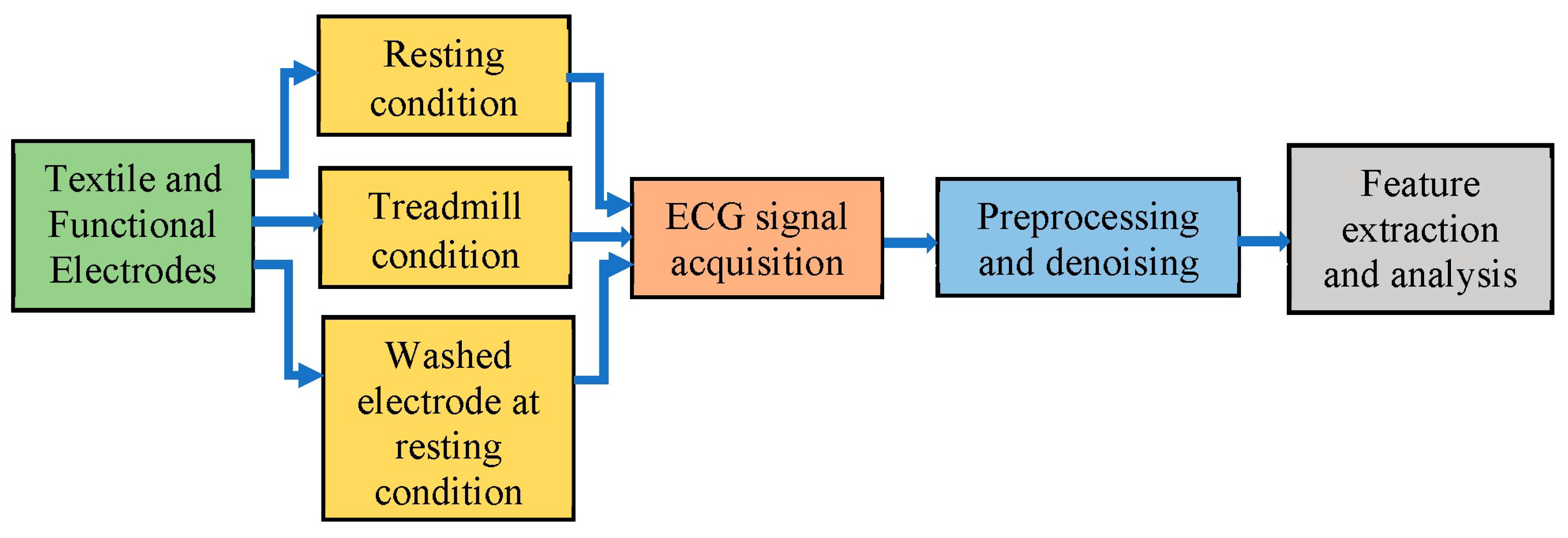
For this investigation, stainless steel electrodes and silver + copper platted polyester textile electrodes were produced by ____ processing a size of ___by _____. The performance of the fabricated textile ECG electrodes was analyzed using the extracted ECG signal feature variations from signal processing techniques, and the results were compared with those of commercially available silver-silver chloride electrodes. Figure 1 shows the proposed method.
1.1. ECG Signal Acquisition, Preprocessing and Denoising
The ECG signals were collected from five subjects using textile electrodes and conventional disposable silver-silver chloride electrodes simultaneously in the lead II conventional sitting position. The ECG signals were recorded from everyone at rest, during treadmill exercise, or during rest using washed textile electrodes. The duration of each signal is 1 minute, and the sampling frequency is 1000 Hz.
Figure 2.
Images from the data recording procedures.
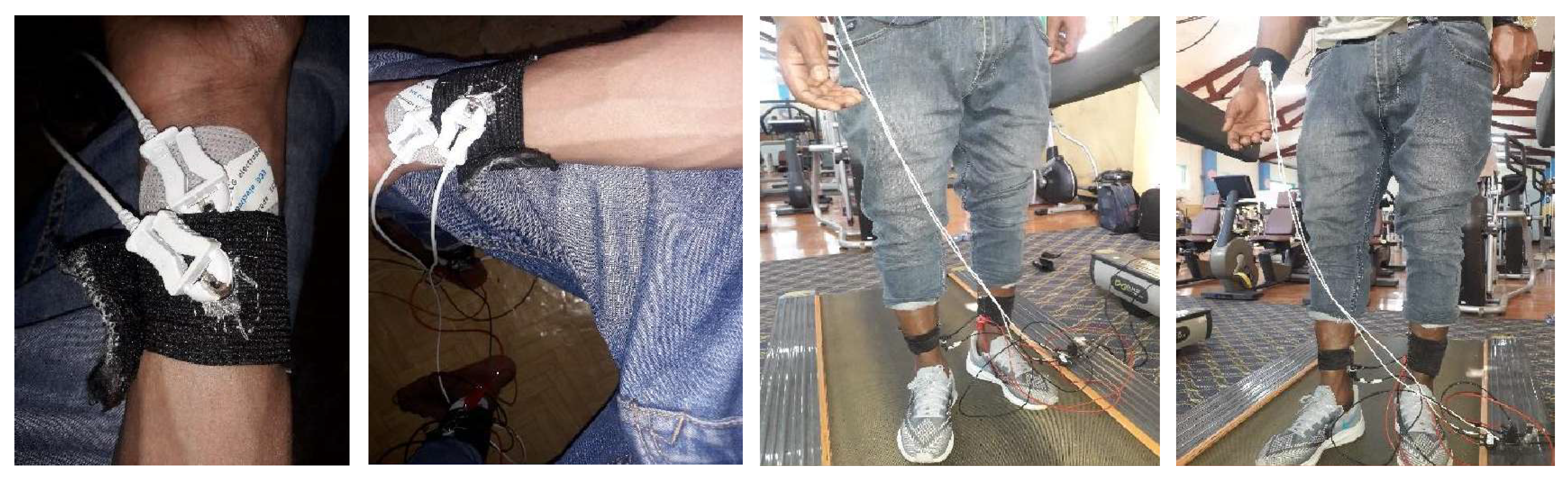
Respiratory muscle movement, electrode motion artifacts, EMG signals, power lines, and electromagnetic interference noise are expected to accompany the raw obtained signal. Due to its advantage in preserving the most significant features, the Savitzky Golay filter is selected for smoothing and baseline wander noise filtering [11,12]. The recorded signals were annotated and then soothed using a Savitzky Golay filter to remove irregularities and low-frequency noises. Wavelet-based denoising is the best and most powerful technique for filtering ECG signals [12,13,14]. The performances of different denoising techniques evaluated using the signal-to-noise ratio (SNR) and wavelet multiresolution analysis (WMRA) with the discrete mayor (dmey) mother wavelet were better. To separate the low-frequency components less than 1 Hz that are assumed to be baseline wandering noise, decomposition level 9 is the best. The low-frequency component hoses are outside the ECG frequency range, and the higher-frequency components contribute insignificant energy to the signal excluded during reconstruction, as shown in equation 1.
1.2. Feature Extraction and Analysis
In this study, Pan Tompkins QRS detection, peak detection and interval calculation algorithms were used to extract ECG features. Pan-Tompkins is the most common QRS detection method and consists of a bandpass filter, derivative filter squaring, thresholding and moving window integration algorithms sequentially [1,15,16]. Using local maximum and minimum concepts, the amplitude and location of the P, Q, R, S, and T waves were obtained. Intervals, isoelectric lines, and segments were calculated using peak locations and threshold values. Finally, the obtained ECG features from different subjects were averaged for each electrode category for better manipulation of the data and for the variations observed.
2. Results
2.1. Data Acquisition, Preprocessing and Denoising Results
Figure 3.2.
Functional electrode and the outputs of smoothing and denoising processes.
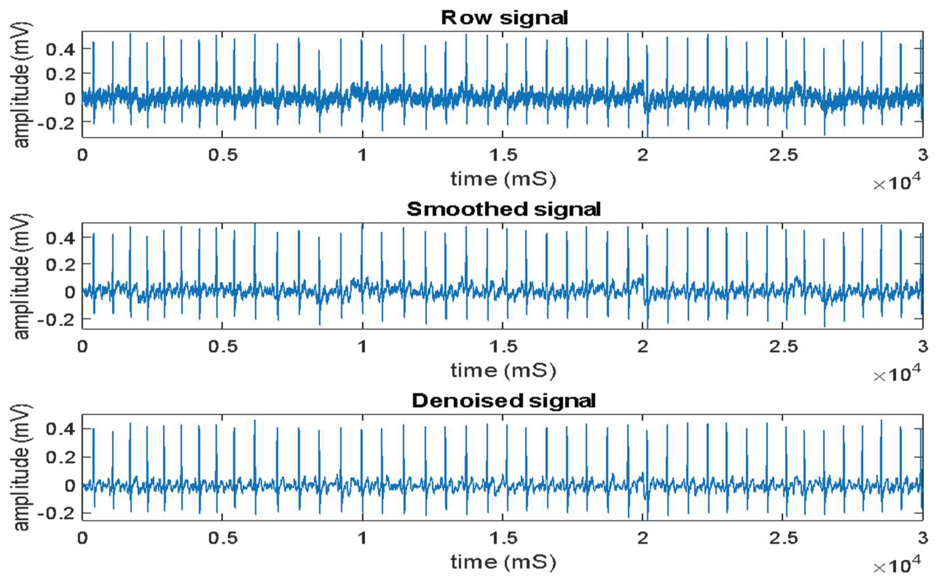
Figure.
ECG signals recorded by functional electrodes (a), polyester textile electrodes (b) and stainless-steel textile electrodes (c) during treadmill exercise.
Figure.
ECG signals recorded by functional electrodes (a), polyester textile electrodes (b) and stainless-steel textile electrodes (c) during treadmill exercise.
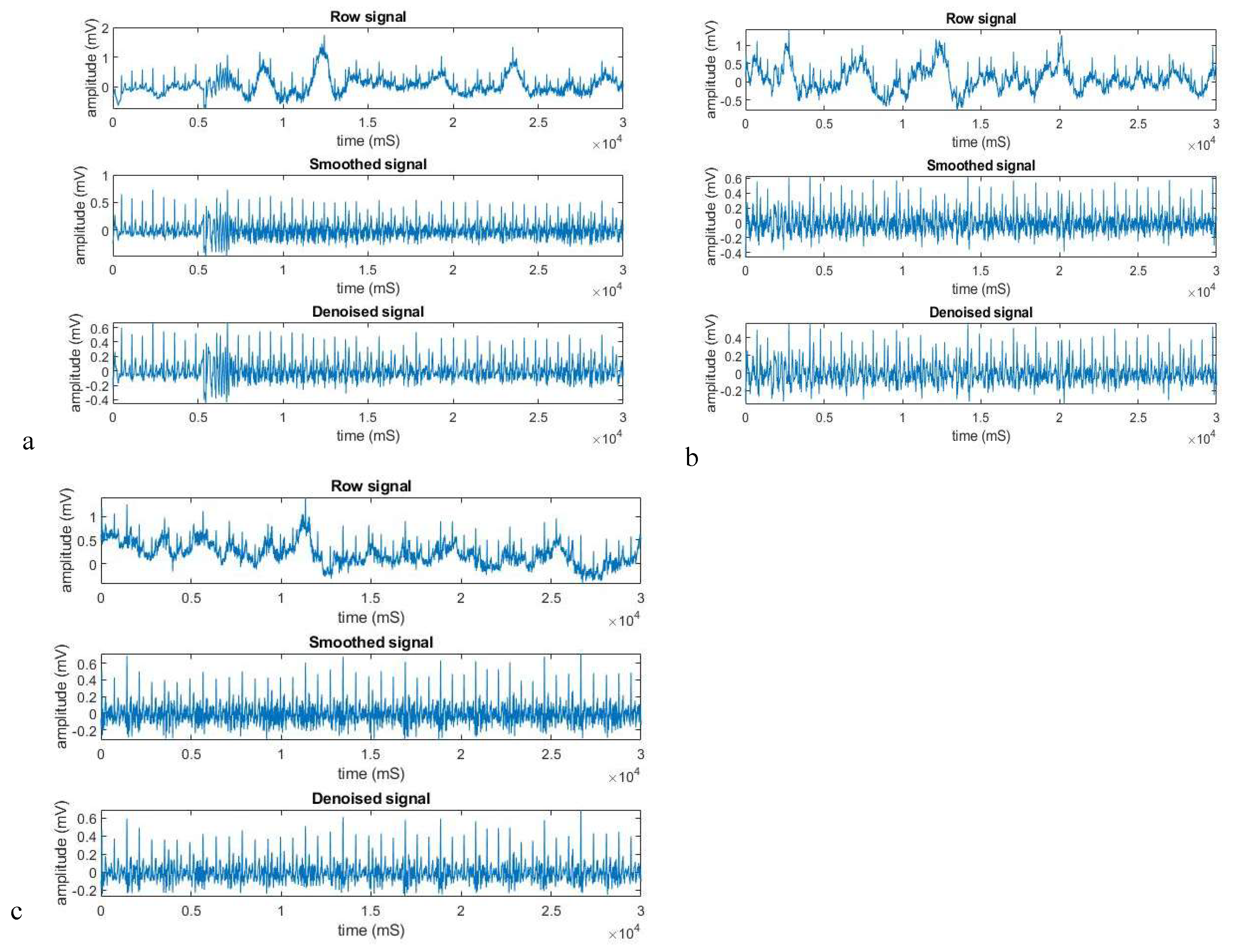
Figure.
ECG signal recorded by a functional electrode under static conditions.
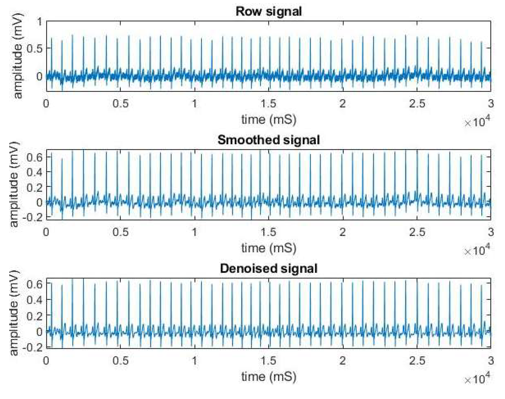
Figure.
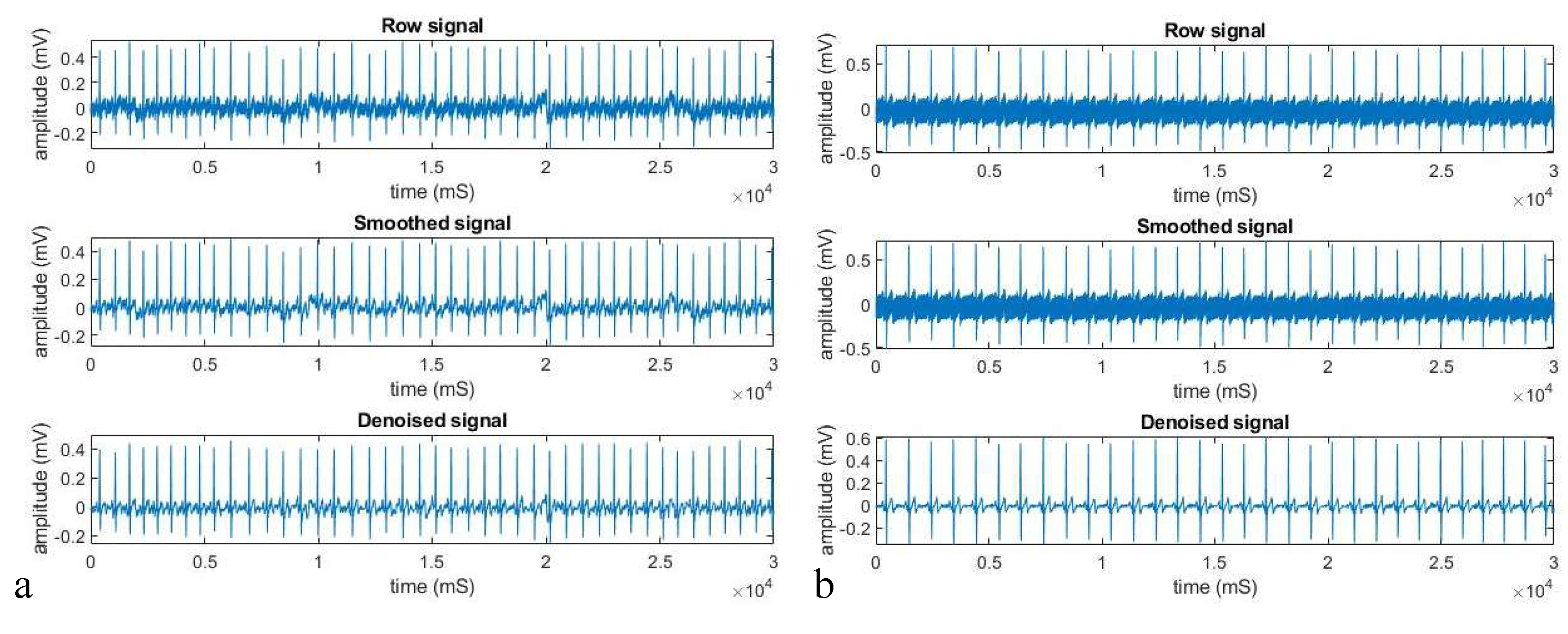
ECG signals recorded by a Polyester textile electrode before washing (a) and after repeated washing (b) under static conditions.
Figure.
ECG signals recorded by a Polyester textile electrode before washing (a) and after repeated washing (b) under static conditions.
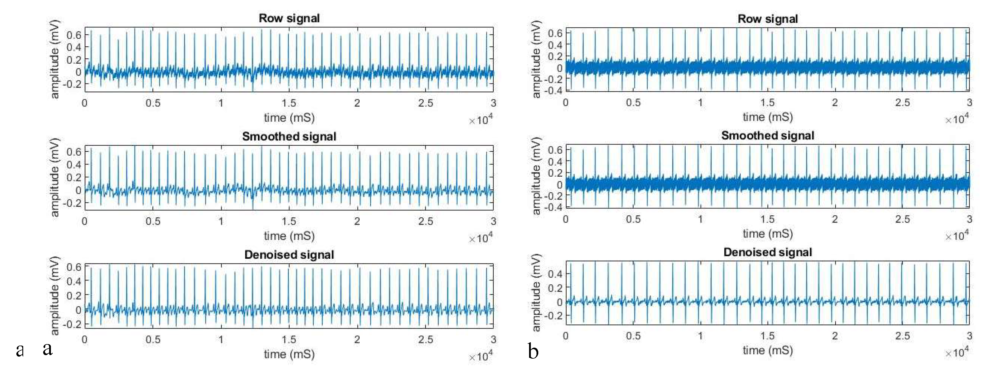
Figure.
ECG signals recorded by a Polyester textile electrode before washing (a) and after repeated washing (b) under static conditions.
Figure.
ECG signals recorded by a Polyester textile electrode before washing (a) and after repeated washing (b) under static conditions.
2.2. Feature Extraction and Analysis Results
The R, P, and T peaks were extracted from all the collected signals. The averaged extracted features in each electrode category are shown in Table 3.1.

Table 3.1.
The averaged ECG feature values of the selected electrodes under different conditions.
| ECG feature values at static condition | |||
| Feature | Functional Electrode | Polyester Electrode | Stainless steel Electrode |
| R peak | 0.6623828 | 0.59242381 | 0.56699506 |
| P peak | 0.08922754 | 0.0848366 | 0.08950509 |
| T peak | 0.182598 | 0.13889651 | 0.1462053 |
| ECG feature values at dynamic condition | |||
| R peak | 0.46834 | 0.42212 | 0.541078 |
| P peak | 0.031635 | -0.00447 | 0.098881 |
| T peak | 0.239087 | 0.216838 | 0.167139 |
| ECG feature values using washed textile electrodes at static condition | |||
| R peak | 0.922986 | 0.575616 | 0.587001 |
| P peak | 0.06638 | 0.040977 | 0.048649 |
| T peak | 0.165789 | 0.083446 | 0.081361 |
Figure 4 Extracted ECG peak values under different conditions using different electrodes
3. Discussion
For long-term monitoring, textile electrodes are comfortable [1]. To evaluate the performance of the textile-based ECG electrodes, signal processing algorithms were used. The signals were recorded using textile and conventional electrodes simultaneously and denoised, and features were extracted. The features were averaged for ease of data manipulation.
At static condition the respective P, QRS, and T amplitudes were 0.089mV, 0.66mV, and 0.18mV for Ag\AgCl electrode; 0.085mV, 0.59mV, 0.14mV for polyester textile electrode; and 0.090mV, 0.57mV, and 0.15mV for stainless-steel textile electrode. Using textile-based conductive electrodes, average peak amplitudes of 0.14, 0.96 and 0.36 mV were reported for the P, QRS and T peaks, respectively [3,17,18,19,20].
The respective dynamic condition P, QRS, and T wave amplitude values were 0.032mV, 0.47mV, and 0.24mV for Ag\AgCl electrode; -0.004mV, 0.42mV, 0.22mV for polyester textile electrode; and 0.099mV, 0.54mV, and 0.17mV for stainless-steel textile electrode [17,21].
After washing the textile (polyester and stainless-steel) electrodes the respective static condition P, QRS, and T wave amplitudes were 0.066mV, 0.92mV, and 0.17mV for Ag\AgCl electrode; 0.041mV, 0.58mV, 0.083mV for polyester textile electrode; and 0.049mV, 0.59mV, and 0.081mV for stainless-steel textile electrode. After one cycle, wash the values of 0.17, 0.83, and 0.34 mV for the P, QRS, and T peaks, respectively [3,17,18,19,20].
4. Conclusion
To evaluate the performance of the textile electrodes, ECG signals were recorded using those electrodes and the functional electrodes. Then, the signals were denoised, and vital features were extracted. The changes observed in the extracted features. Finally, the textile electrodes are acceptable for recording ECG signals.
Author Contributions
All the authors participated in conceptualizing, designing, implementing and writing this study and approved the final version of the manuscript for publication.
Funding
This research received no external funding.
Institutional Review Board Statement
Since the data recording involved fabric and had no personal information, ethical approval was not necessary.
Informed Consent Statement
The participants provided an oral informed consent, stating that "the study team has obtained my full permission to use my ECG data for their study and publication.
Data Availability Statement
Datasets used and/or analyzed during the current investigation are available at the corresponding author and can be obtained upon reasonable request.
Conflicts of Interest
The authors declare no conflict of interest.
References
- Rangayyan, R. M. Biomedical Signal Analysis, 2nd ed.; John Wiley & Sons: United States of America.
- Khandpur, R. S. Biomedical instrumentation: technology and applications, 2nd ed.; McGraw-Hill: New York, 2005. [Google Scholar]
- Tseghai, G. B.; Malengier, B.; Fante, K. A.; Nigusse, A. B.; Etana, B. B.; Van Langenhove, L. ‘PEDOT:PSS/PDMS-coated cotton fabric for ECG electrode’. 2020 IEEE International Conference on Flexible and Printable Sensors and Systems (FLEPS), Manchester, UK, Aug. 2020; pp. 1–4. [Google Scholar] [CrossRef]
- Wu, Y.-Z.; Sun, J.-X.; Li, L.-F.; Ding, Y.-S.; Xu, H.-A. ‘Performance Evaluation of a Novel Cloth Electrode’; p. 5.
- Xiao, X.; Pirbhulal, S.; Dong, K.; Wu, W.; Mei, X. ‘Performance Evaluation of Plain Weave and Honeycomb Weave Electrodes for Human ECG Monitoring’. J. Sens. 2017, vol. 2017, 1–13. [Google Scholar] [CrossRef]
- Rajanna, R. R.; Sriraam, N.; Vittal, P. R.; Arun, U. ‘Performance Evaluation of Woven Conductive Dry Textile Electrodes for Continuous ECG Signals Acquisition’. IEEE Sens. J. 2020, vol. 20(no. 3), 1573–1581. [Google Scholar] [CrossRef]
- An, X.; Tangsirinaruenart, O.; Stylios, G. K. ‘Investigating the performance of dry textile electrodes for wearable end-uses’. J. Text. Inst. 2019, vol. 110(no. 1), 151–158. [Google Scholar] [CrossRef]
- Cho, G.; Jeong, K.; Paik, M. J.; Kwun, Y.; Sung, M. ‘Performance Evaluation of Textile-Based Electrodes and Motion Sensors for Smart Clothing’. IEEE Sens. J. 2011, vol. 11(no. 12), 3183–3193. [Google Scholar] [CrossRef]
- Peng, S.; Xu, K.; Chen, W. ‘Comparison of Active Electrode Materials for Non-Contact ECG Measurement’. Sensors 2019, vol. 19(no. 16), 3585. [Google Scholar] [CrossRef] [PubMed]
- Reinel, C.; Jairo, J. P.; Henry, A. ‘Electrical performance of PEDOT:PSS-based textile electrodes for wearable ECG monitoring: a comparative study | BioMedical Engineering OnLine | Full Text’. Biomed. Eng. OnLine 2018, vol. 17(no. 38). [Google Scholar] [CrossRef]
- Savitzky, Abraham.; Golay, M. J. E. ‘Smoothing and Differentiation of Data by Simplified Least Squares Procedures.’. Anal. Chem. 1964, vol. 36(no. 8), 1627–1639. [Google Scholar] [CrossRef]
- Rezuana, B. J. ‘ECG DE-NOISING TECHNIQUES FOR DETECTION OF ARRHYTHMIA - PDF Free Download’. IRJET 2015, vol. 02(no. 09). Available online: https://healthdocbox.com/Heart_Disease/78990333-Ecg-de-noising-techniques-for-detection-of-arrhythmia.html (accessed on Jan. 23 2022).
- Biswas, U.; Hasan, K. R.; Sana, B.; Maniruzzaman, Md. ‘Denoising ECG signal using different wavelet families and comparison with other techniques’. 2015 International Conference on Electrical Engineering and Information Communication Technology (ICEEICT), Savar, Dhaka, Bangladesh, May 2015; pp. 1–6. [Google Scholar] [CrossRef]
- Aqil, M.; Jbari, A.; Bourouhou, A. ‘ECG Signal Denoising by Discrete Wavelet Transform’. Int. J. Onl. Eng. 2017, vol. 13(no. 09), 51. [Google Scholar] [CrossRef]
- Pan, J.; Tompkins, W. J. ‘A Real-Time QRS Detection Algorithm’. IEEE Trans. Biomed. Eng. 1985, vol. BME-32(no. 3), 230–236. [Google Scholar] [CrossRef] [PubMed]
- Madiraju, N. S.; Kurella, N.; Valapudasu, R. ‘FPGA Implementation of ECG feature extraction using Time domain analysis’; p. 4.
- Nigusse, B.; Malengier, B.; Mengistie, D. A.; Van Langenhove, L. ‘A Washable Silver-Printed Textile Electrode for ECG Monitoring’. Eng. Proc. 2021, vol. 6(no. 1), 63. [Google Scholar] [CrossRef]
- Kannaian, T.; Neelaveni, R.; Thilagavathi, G. ‘Design and development of embroidered textile electrodes for continuous measurement of electrocardiogram signals’. J. Ind. Text. 2013, vol. 42(no. 3), 303–318. [Google Scholar] [CrossRef]
- Das, P. S.; Kim, J. W.; Park, J. Y. ‘Fashionable wrist band using highly conductive fabric for electrocardiogram signal monitoring’. J. Ind. Text. 2019, vol. 49(no. 2), 243–261. [Google Scholar] [CrossRef]
- Arquilla, K.; Webb, A.; Anderson, A. ‘Textile Electrocardiogram (ECG) Electrodes for Wearable Health Monitoring’. Sensors 2020, vol. 20(no. 4), 1013. [Google Scholar] [CrossRef] [PubMed]
- Nigusse, B.; Malengier, B.; Mengistie, D. A.; Van Langenhove, L. ‘Evaluation of silver-coated textile electrodes for ECG recording’. 2021 IEEE International Conference on Flexible and Printable Sensors and Systems (FLEPS), Manchester, United Kingdom, Jun. 2021; pp. 1–4. [Google Scholar] [CrossRef]
Disclaimer/Publisher’s Note: The statements, opinions and data contained in all publications are solely those of the individual author(s) and contributor(s) and not of MDPI and/or the editor(s). MDPI and/or the editor(s) disclaim responsibility for any injury to people or property resulting from any ideas, methods, instructions or products referred to in the content. |
© 2026 by the authors. Licensee MDPI, Basel, Switzerland. This article is an open access article distributed under the terms and conditions of the Creative Commons Attribution (CC BY) license (http://creativecommons.org/licenses/by/4.0/).
Copyright: This open access article is published under a Creative Commons CC BY 4.0 license, which permit the free download, distribution, and reuse, provided that the author and preprint are cited in any reuse.