

Submitted:
23 September 2026
Posted:
24 September 2026
You are already at the latest version
Abstract
The paper derives the fundamental physical units in a universe over a finite holographic substrate: a finite totality whose capacity bounds the state count of every embedded observer, the holographic bound taken as the substrate itself. The units are the substrate's four horizons, the Planck length, momentum, time and energy; the constants \( c \), \( \hbar \), \( G \), \( k_B \) follow uniquely from this quartet, over realisations stated with their falsifiers:\( c \) the ratio across the two Fourier pairs, \( \hbar \) the product within each conjugate pair, \( k_B \) the mixed product, \( G \) the normalisation to the totality. The quartet admits one cancellation identity and no further independent relation; on an admissible substrate the constants are exact residues, read on the unit face as magnitudes. Beneath the quartet, dimensional analysis is a graded modular domain algebra on the framed shell, recovering the classical calculus exactly inside the sub-capacity window. The domain of \( \hbar \) is the unit flag, the generator of the lattice's unique order-four subgroup; its torsion-free surrogate is the classical mass dimension, temperature inherits the acceleration domain, and the flag cancels in every count-valued comparison. Every theorem of the paper is machine-verified (217 exact checks) and formalized in Lean 4.
Keywords:
dimensional analysis
; finite ring cosmology
; physical units
; Planck units
; physical constants
; modular domains
; quantum mechanics
; mass
; gravitational constant
; speed of light
1. Introduction
While potential damage of IT overuse has long been realized [1] and heavy consumption of short videos (reels) has been found to reduce neurophysiological markers of attention [2], playing action video games can lead to improvements. The "action" component of these games requires sensorimotor skills, hand-eye coordination, and visuospatial cognition. Research suggests that action video game experience improves both low-level perceptual skills (like contrast sensitivity) and higher-level attentional processes (like the ability to ignore distractors) [3,4]. Over the past two decades, extensive research has established that it is indeed associated with significant improvements in visual and attentional skills [5]. Interestingly, these benefits appear to generalize to novel tasks [6], a phenomenon termed "learning to learn," which differentiates AVG training from traditional task-specific perceptual learning [7].
Action video games—including first-person shooters (FPS) like Call of Duty [8] and action real-time strategy (ARSG) games like League of Legends [9] —place extreme demands on the visual system. Players must stay alert to stimuli in the peripheral region while tracking multiple moving targets and making decisions under time pressure [10]. As video game playing has become a ubiquitous activity in today's society, it is worth considering its potential consequences on perceptual and motor skills [11] on a broader scale. Although much research has already been done, one of the still ongoing debates is whether the benefits from playing video games are more a top-down influence (executive, function, selective attention, working memory, etc.) or rather a bottom-up effect (early sensory processing enhancement, visual acuity, contrast sensitivity, etc.).
In contrast to purely behavioral studies, the current empirical investigation utilized the approach to generate event-related potentials (ERPs) recorded in response to very simple visual presentations in order to investigate very basic sensory-specific improvements with a particular focus on the two separate information channels of the visual system, the magnocellular (visual field periphery) and the parvocellular (visual field center) channel. In line with this distinction, the current study used simple white circle (on black background) presentations shown in the center or in the periphery of the visual field. Thereby, peripheral presentations are processed by the magnocellular system, while central presentations are processed by the parvocellular system (see further below). This distinction together with simple visual stimuli enable the current study to focus on early sensory-related brain activity phenomena that are potentially different between gamers and non-gamers. However, before going into any detail some prior studies relevant to the current study are introduced.
1.1. Action Video Game Playing (AVGP) Behavioral Effects
Green & Bavelier (2007) [4] measured the smallest distance a distractor could be from a target without compromising target identification. This approach exploits the fact that visual processing is hindered as distractors are brought close to the target, a phenomenon known as crowding. Compared with non-players, action-video-game players could tolerate smaller target-distractor distances. Thus, the spatial resolution of visual processing is enhanced in this population. Critically, similar effects were observed in non-video-game players who were trained on an action video game, which verifies a causative relationship between video-game play and augmented spatial resolution. Dye & Bavelier (2010) [12] tested children aged 7–17 years and adults aged 18–22 years on three aspects of visual attention. Their data suggested different developmental trajectories for these components. To some extent, spatial, temporal and object-based attentional processes seem to be subserved by different neural resources which develop at different rates. However, most importantly in the current context, participants who played action games showed enhanced performance on all aspects of attention tested as compared to non-gamers.
Interestingly, AVGP has even been introduced to psychotherapy and clinical psychology. Franceschini et al. (2013) [13] demonstrated that action video game (AVG) training can significantly improve reading abilities in children with dyslexia, even without traditional phonological or linguistic exercises. Only 12 hours of AVG training (spread over nine days) improved children's reading speed and accuracy significantly more than traditional reading treatments. The authors attributed this improvement to the enhancement of visual spatial attention (which is crucial for shifting focus across letters and words. Interestingly, the gains in reading performance remained stable when tested several months later. As will be mentioned further below, there is even a theory that links dyslexia with malfunctions in the frame of the magnocellular-dorsal pathway.
1.2. Action Video Game Playing (AVGP) Physiological Effects
Besides behavioral improvements, also brain activity changes have been demonstrated. Latham et al. (2013) [14] applied electroencephalography (EEG) to assess occipital N1 latencies and interhemispheric transfer time (IHTT) in expert video game players VGPs. Participants comprised 15 right-handed male expert VGPs and 16 non-VGP controls matched for age, handedness, IQ and years of education. Expert VGPs began playing before age 10, had a minimum of 8 years experience, and maintained playtime of at least 20 hours per week over the last 6 months. Non-VGPs had little-to-no game play experience (maximum 1.5 years). Participants responded to checkerboard stimuli presented to the left and right visual fields while 128-channel EEG was recorded. Expert VGPs responded significantly more quickly than non-VGPs. Expert VGPs also had significantly earlier occipital N1s in direct visual pathways (the hemisphere contralateral to the visual field in which the stimulus was presented).
In another study [15], the exact mechanism of game-induced visual processing enhancements were targeted. By recording event-related potentials (ERPs) during focused and divided attention visual tasks, the researchers evaluated whether action video game players (AVGPs) differ from non-players (NAVGPs) at low-level visual stage pathways vs. higher-level parietal/attentional control stages. In support for higher-level attentional control (anterior N1), AVGPs showed a significantly larger anterior N1 component over parietal scalp sites during focused attention compared to NAVGPs. The anterior N1 reflects top-down control over the spatial allocation of attention. In addition, AVGPs exhibited a more pronounced P2 component (contralateral to visual stimuli) than NAVGPs. The authors interpreted this as evidence of more efficient higher-order visual perceptual processing and target identification. No group differences were found in low-level sensory pathways (P1 & Posterior N1). Thus, contrary to hypotheses predicting sensory gain improvements in early visual areas, attention-modulated occipital components (P1 and posterior N1) generated within early extrastriate pathways showed no significant differences between AVGPs and NAVGPs. Regarding behavioral effects, AVGPs responded faster to target stimuli during focused attention conditions without sacrificing accuracy. This led the authors to the conclusion that the primary neural signature driving the perceptual and attentional advantages in action video game players is not an alteration of early visual sensory processing in occipital cortex, but rather enhanced top-down parietal attentional control (anterior N1) paired with more efficient higher-level perceptual evaluation (P2).
A study by Birch-Hurst et al. (2021) [16] investigated why AVGPs are faster and more efficient at visual search tasks compared to non-gamers, specifically looking at neural markers of attention. Their key findings were that AVGPs showed a significantly earlier N2pc component (an ERP associated with the "shifting" of the attentional spotlight to a target). This confirms they are faster at physically selecting a target among distractors. They also measured active suppression of distracting items and found that while both groups used suppression, gamers were more efficient at "releasing" attention from distractors to move to the next item. The authors finally concluded that enhanced visual search performance in action video game players is driven by differences in early sensory processing in combination with changes to later cognitive and motor processes allowing for quicker transition of attention from one point in space to another. In lay terms, action video game players don't just see better, their brains are faster at allocating and reallocating spatial attention, allowing them to scan complex visual environments with superior temporal precision.
1.3. Action Video Games and the Plasticity of the Visual System
A key finding in AVG research that is highly relevant for the current study is the expansion of the "useful field of view" (UFOV). Gamers are more accurate at identifying stimuli in the periphery, even when engaged in a concurrent central task, ruling out a simple trade-off between central and peripheral attention. This improvement is not just a result of faster reaction times but reflects a genuine enhancement in the spatial distribution of visual attention [11,17,18,19]. Furthermore, action gaming is linked to enhanced temporal resolution. VGPs show a superior ability to discriminate between visual events in close temporal proximity (on the millisecond level). This ability is associated with reduced alpha-band power and increased inter-trial phase coherence in parieto-occipital regions, indicating more efficient neural processing of fast-paced information. These enhancements suggest a specific strengthening of the magnocellular pathway, which is specialized for high temporal frequencies and peripheral motion [20,21].
The specific mechanics of action games—tracking fast-moving objects, detecting low-contrast changes, and maintaining alertness to peripheral threats—directly stress the magnocellular-dorsal pathway. This constant demand is hypothesized to trigger functional and structural plasticity in the M-channel. AVG experts show altered white matter (WM) networks in prefrontal, limbic, and sensorimotor regions [22]. These networks modulate the distribution of action potentials and coordinate communication between distant brain regions. Plasticity in these areas is associated with improved cognitive control and sensorimotor function. Functional MRI studies have also revealed increased gray matter volume (GMV) in the right posterior parietal cortex, the occipital lobe, and the dorsal striatum of AVG experts [23]. These regions are critical components of the dorsal "where" pathway (fed by information from the magnocellular channel) and the attentional networks that manage spatial resources [24]. However, the effects of gaming can be nuanced; for instance, first-person shooter games have been linked to reduced gray matter in the hippocampus for players who use response-based navigation strategies, whereas spatial-based strategies may lead to hippocampal growth [25]. While this is overly important, the idea of a particular effect of gaming on the M-channel seems intriguing. The beneficial effects of action video game training on developmental dyslexia (DD) are hypothesized to rely on M-channel (magnocellular pathway) plasticity [13]. The "magnocellular hypothesis" of dyslexia proposes that a primary cause of the disorder is a deficit in the visual timing systems mediated by M-neurons [26]. This brings us back to the beginning where it was already mentioned that the human visual field has a strict division into central and peripheral, both parts associated with separate processing channels and related separate projection targets on the cortex. At this stage, it seems essential to take a closer look at those two channels.
1.4. Visual Pathways: A Brief Neuroanatomical Background
The visual system is of course heavily involved during action video game playing. Its pathway from the eyes, where respective sensory neurons (i.e., rods and cones) transduce light-related information into neural signals, comprises two important separate projections to the occipital cortex, where higher-order processing begins. Following a so-called retinotopic organization, those two pathways start from separate areas on the retina, continue separately through thalamic relay nuclei and end separately at the visual cortex [27]. First, the P-ganglion cells project to the so-called parvocellular layers on the thalamus, while the M-ganglion cells project to the magnocellular layers [28,29,30]. The segregation of these channels is a defining feature of the primate visual system. The physiological differences between M and P cells dictate the types of stimuli they are most sensitive to and the speed at which they can relay information to the brain. Finally, in addition to those two different layers there is a third layer type consisting of koniocellular cells [31,32]. Table 1 shows a summary of those three types of cells including some of their features.
In combination with these two pathways also distinct photoreceptor types are associated with them. The distribution of the three types of cones—L (Long-wave/Red), M (Medium-wave/Green), and S (Short-wave/Blue) across the human retina is highly specialized and far from uniform. The fovea is a small pit in the center of the macula responsible for our sharpest vision. The density of L and M cones is at its peak here, reaching up to 150,000 cones per mm2. A striking feature of the very center (the foveola) is the complete absence of S-cones. We are effectively "blue-blind" in the tiny central point of our gaze. Clearly, L and M cones dominate this area. On average, there are twice as many L-cones as M-cones, though this ratio varies significantly between individuals (from 1:1 to nearly 16:1) without obviously impacting color perception (even though this might cause slight nuance differences between subjective color perception. S-cones represent something like a distinct "sub-system" with a unique distribution pattern. They make up only about 5–10% of the total cone population with their highest density found in a ring-like zone just outside the foveola [33]. Unlike the somewhat random L/M mosaic, S-cones are distributed in a very regular, widely spaced hexagonal grid across the rest of the retina [34]. As one moves away from the fovea toward the edges of the retina the cone density drops off dramatically. The space between the sparse cones is filled by rods, which are absent in the central foveola, but reach their maximum density about 20° away from the center [35]. Because the cones are so spread out in the periphery, we lose the ability to see fine details and vibrant colors in our "corner of the eye" vision. In short, the human eye prioritizes spatial resolution (via L and M cones) in the center, while treating blue color information (via S-cones) as a lower-resolution "color wash" provided by a sparse underlying grid. See Table 2 summarizing photoreceptor distributions on the retina.
This non-uniform distribution of photoreceptors and ganglion cells across the retina dictates the relative dominance of M and P channels in different parts of the visual field, leading to the distinct functional properties of central and peripheral vision. The neuroanatomical differentiation of the M and P pathways is established by the morphology and physiology of their constituent neurons. Magnocellular neurons originate from large M-type (parasol) ganglion cells in the retina, which possess thick, myelinated axons facilitating rapid signal transmission [36,37]. In contrast, the parvocellular pathway originates from smaller P-type (midget) ganglion cells, which transmit information more slowly but with greater spatial precision [38]. The magnocellular system’s transient response pattern allows it to fire rapidly at the onset or offset of a stimulus, making it ideal for detecting flicker, sudden motion, and rapid changes in the environment. Parvocellular cells provide a sustained response, allowing for the continuous processing of stationary or slow-moving objects in high detail, which is essential for tasks like reading and facial recognition (see Table 3).
1.5. Visual Pathways Beyond the Cortex
From the thalamus then, projections reach first cortical layers and from there (primary visual cortex (V1)), visual processing bifurcates into the dorsal and ventral streams. The dorsal stream, often referred to as the "action" or "where" pathway, receives predominant input from the magnocellular system and extends into the posterior parietal cortex, governing spatial awareness and the visual control of action (see Figure 1). The ventral stream, or the “perception” or “what” pathway, integrates both M and P inputs but relies heavily on parvocellular signals to process form and color in the inferior temporal cortex to support object recognition and identification [24,39,40,41,42,43,44,45,46].
2. Methods
2.1. Participants
EEG-data from twenty-five participants were analyzed and used for this study. According to their playing habits, they were grouped into non-players (11; zero playing hours) and players (14; mean playing time per week = 9 hours). Both groups had a similar mean age of 24 years, while also all other demographic details did not differ. Among the 14 gamers, first-person shooters (FPS) were the most popular genre, reported by six participants (42.9%), followed by racing games (35.7%, n = 5), massively multiplayer online role-playing game (MMORPG) (14.3%, n = 2), and horror games (7.1%, n = 1).
2.2. Stimuli
To analyze early sensory-related brain activity and to compare central versus peripheral visual information processing the following four conditions were generated. The simple visual stimulus was a white circle on black background. This circle was i) placed in the center of the screen, ii) simultaneously far from the center to the left and to the right, iii) just to the left, and iv) just to the right (Figure 1).
2.3. Procedure
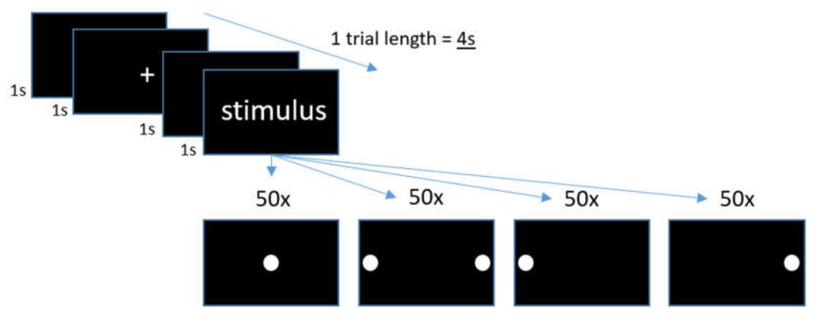
After arrival at the lab, the participants were introduced to the purpose of the study. They were given the informed consent form to sign if they agreed to participate. This investigation was conducted in accordance with the principles outlined in the Declaration of Helsinki (1975, revised in 2013). Approval was obtained from the local Institutional Review Board (IRB) (Ethics Comission of the Faculty of Psychotherapy Science, Psychology and Law; Approved on 3.2.2026; Approval ID: QDPFMFUPCOELET92112). The actiCAP with 64 electrodes embedded (from Brain Products; Gilching, Germany) was applied and connected to an amplifier (see further details below). Before the recordings started, the participants were instructed to sit still and blink with their eyes only when they saw a fixation cross, but to avoid blinking during circle presentations. Each circle condition was presented fifty times (in random order; in total 200 presentations) for 300 ms on a computer monitor placed on a table in front of the participants, who sat on a comfortable chair. One stimulus presentation was followed by a black screen for 1 s and a white fixation cross on a black background for 1 s with a final black screen for, again, 1 s. The eye-to-screen distance was about 0.7 m. The participants were instructed to simply view all 200 presentations without any specific task given to them.
Figure 2.
One single trial consisted of a black screen presented for 1s, followed by a black screen with a central + presentation (fixation point) for 1s. This was again followed by a 1s black screen until one of the four stimuli was presented also for 1s. The stimulus categories comprised i) a central white circle, ii) two peripheral white circles (left and right), iii) a left circle, and iv) a right circle.
Figure 2.
One single trial consisted of a black screen presented for 1s, followed by a black screen with a central + presentation (fixation point) for 1s. This was again followed by a 1s black screen until one of the four stimuli was presented also for 1s. The stimulus categories comprised i) a central white circle, ii) two peripheral white circles (left and right), iii) a left circle, and iv) a right circle.
2.4. Electroencephalography (EEG), Event-Related Potentials (ERPs)
For recording brain potential changes, a 64-channel actiCHamp Plus System from Brain Products (Gilching, Germany) with active electrodes embedded in an actiCAP connected to an amplifier was used. The amplifier was operated by a powerful lithium-ion battery pack. The brain potentials were sampled at a rate of 1 kHz (filtered: DC to 100 Hz). Impedance was kept equal to or below 10 kΩ. Cz was used as reference, and a mid-frontal position on the forehead was used as the ground electrode. Offline, all EEG data were down-sampled to 250 Hz, and a bandpass filter from 0.1 to 30 Hz was applied in preparation for following EEG data processing. Those data were then used to generate ERPs. For this purpose, 1.1 s long time windows (epochs) were cut out of the ongoing EEG recordings starting 100 ms before each trigger until 1 s after the trigger. Baseline correction was performed by using the 100 ms time period before each trigger. Finally, all epochs within each of the 5 term categories were averaged to generate ERPs. Those were then used to display term category-specific neurophysiological activity changes over time. All EEG data processing was conducted with the EEGDisplay (Version 6.4.9) software (by Ross Fulham).
2.5. Analyses
After generating ERPs for all four categories for each participant, the software EEGDisplay was further used to export neurophysiological data for statistical analyses. While Figure 1 shows ERPS for all four conditions, only two of those were further analyzed via descriptive and analytical statistics. For this purpose, each participant’s peak latencies for the P170 and for the N250 ERP component were automatically detected for those two presentation conditions with a focus on the central white circle presentation condition (central) and the bilateral peripheral white circles presentation condition (left + right) (see Figure 1). For respective final data export, electrode locations P5, P6, P7, P8, PO3, PO4, PO7, PO8, O1 and O2 were selected on the basis of their locations reflecting early visual information processing on the cortical level. Respective mean amplitudes (including standard deviations) were calculated across all participants for each group separately. Finally, paired-sampled t-tests were calculated to compare the mean peak latencies for the early positive as well as the later negative component between non-gamers and gamers. In order to visualize respective results, ERPs elicited by all four visual presentation conditions are shown in Figure 1. Even though ERPs are only shown for a selected set of electrodes, topographical maps are shown with all 64 electrodes included.
3. Results
3.1. Descriptive and Analytic Statistics
An analysis of peak latencies revealed distinct temporal dynamics between gamers and non-gamers across two early ERP components. For the early P170 component under the central stimulus condition, mean peak latencies were comparable between groups (170.96 ms for gamers vs. 165.18 ms for non-gamers, a 5.78 ms difference). A paired-sample t-test revealed a non-significant difference ((t(12) = 1.821, p = .094). However, the later N200 component demonstrated a substantial divergence, with gamers processing the stimulus faster (237.99 ms) than non-gamers (263.36 ms), reflecting a 25.37 ms latency advantage. Furthermore, a paired-samples t-test confirmed a highly significant difference in mean N250 latencies between groups during bilateral peripheral white circle presentations (t(12) = -4.289, p < .001). Table 1 shows mean latencies of P170 ERP amplitudes for both groups, gamers and non-gamers for the central stimulus condition.
Figure 1.
Event-related potentials (ERPs) for all four white circle presentation conditions for both groups (non-gamer, gamer). Shown are overlaid ERPs from 10 electrode locations in left and right occipital and occipito-temporal cortical regions (butterfly-curves). The two conditions “central” (brown curves) and “left + right” (blue curves; bilateral) were of particular interest and thus further analyzed. Note that ERPs elicited by central stimulus presentations show similar peak latencies at approximately 170ms post-stimulus in both groups. On the other hand, peripheral bilateral stimulus presentations (left + right) elicited earlier N200 peak latencies in the gamer group compared to non-gamer. Further, both one-sided peripheral circle presentations (“left”, “right”) also resulted in shorter ERP peak latencies in gamer compared to non-gamer. Although this difference was not statistically analyzed, is seems very obvious and matches the group difference found for the bilateral peripheral presentation condition. Crucially, peripheral visual information processing is mediated through the magnocellular pathway, while central processing goes through the parvocellular pathway. Consequently, it is concluded that the magnocellular pathway (peripheral visual processing) is particularly improved in response to action video gaming, while central visual processing does not seem to be affected.
Figure 1.
Event-related potentials (ERPs) for all four white circle presentation conditions for both groups (non-gamer, gamer). Shown are overlaid ERPs from 10 electrode locations in left and right occipital and occipito-temporal cortical regions (butterfly-curves). The two conditions “central” (brown curves) and “left + right” (blue curves; bilateral) were of particular interest and thus further analyzed. Note that ERPs elicited by central stimulus presentations show similar peak latencies at approximately 170ms post-stimulus in both groups. On the other hand, peripheral bilateral stimulus presentations (left + right) elicited earlier N200 peak latencies in the gamer group compared to non-gamer. Further, both one-sided peripheral circle presentations (“left”, “right”) also resulted in shorter ERP peak latencies in gamer compared to non-gamer. Although this difference was not statistically analyzed, is seems very obvious and matches the group difference found for the bilateral peripheral presentation condition. Crucially, peripheral visual information processing is mediated through the magnocellular pathway, while central processing goes through the parvocellular pathway. Consequently, it is concluded that the magnocellular pathway (peripheral visual processing) is particularly improved in response to action video gaming, while central visual processing does not seem to be affected.
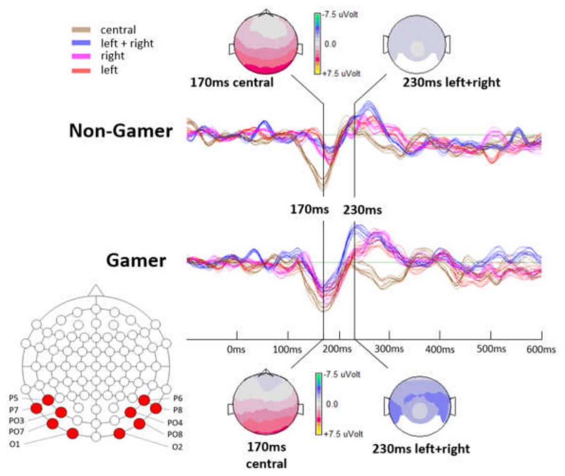
These findings point to a notable acceleration in visual processing speed among gamers, particularly during later visual evaluation stages. While early sensory encoding (P170) of contral visual field information shows minimal latency differences between gamers and non-gamers (~5.78 ms), downstream processing (N200) demonstrates a pronounced processing speed advantage for gamers of over 25 ms in case of peripheral visual information processing. This suggests that extensive gaming experience may not drastically alter early sensory arrival times related to central input, but significantly enhances the speed of peripheral visual processing. The highly significant latency reduction in the N200 component during peripheral stimulation (p < .001) further highlights enhanced attentional allocation and speed in processing peripheral visual stimuli.
4. Discussion
While much emphasis was put on possible cognitive benefits from action video game training [47], the present study has its focus more on early sensory-related effects. The findings of the present investigation provide empirical, electrophysiological support for the hypothesis that long-term action video game playing (AVGP) induces functional changes in early visual processing, specifically enhancing the processing speed of the magnocellular (M) visual pathway. By recording event-related potentials (ERPs) during passive visual stimulation (central versus peripheral stimulation), this study isolates fundamental sensory processing differences from downstream motor, task-switching, or decision-making demands. The observed double dissociation, where gamers demonstrate significantly faster latency during peripheral, bilateral stimulation without showing corresponding latency differences during central stimulation, offers a clear view into the sub-cortical and early cortical architecture modified by visual gaming experience. The present study thus is supportive of game-induced neuroplasticity being associated with enhanced low-level sensory processing rather than high-level attentional allocation.
In more detail, the findings presented in this manuscript offer electrophysiological evidence supporting the selective sensory-related enhancement of the magnocellular (M) visual pathway in response to intense action video game playing (AVGP). The observed significant latency reduction related to the N200 ERP component during bilateral peripheral stimulation - contrasted with equal P170 latencies during central stimulation - provides a compelling basis for discussing sensory-specific neural plasticity.
4.1. Magnocellular Enhancement
The primary neurophysiological finding is a mean 25.59ms processing advantage observed in action gamers regarding the N200 component elicited via bilateral peripheral white circle (on black background) presentations (237.77ms in gamers vs. 263.36ms in non-gamers; p < .001). Because our experimental design required only passive viewing of very simple stimuli without active target selection or distractor filtering, the observed 25.59ms processing advantage in the bilateral peripheral presentation condition cannot be attributed to task-driven top-down attentional allocation. Instead, it suggests a structural or functional recalibration of early visual pathways. Fast-paced video game environments constantly strain the peripheral field with rapid motion and low-contrast events, likely driving experience-dependent synaptogenesis or enhanced myelination along the magnocellular projections to the posterior parietal cortex. Similarly, Birch-Hurst et al. (2021) [16] demonstrated that gamers exhibit earlier N2pc components, which they linked to an optimized magnocellular-dorsal stream facilitating rapid attentional shifting during a visual search task. Due to simple passive viewing in our study, our findings are better interpreted as sensory-related improvements regarding processing speed. In fact, the faster latency reported here provides a functional counterpart to neuroimaging findings by other researchers [22,23], who observed altered white matter microstructural integrity and increased gray matter volume in posterior parietal and occipital networks among action game experts. Enhanced myelination or denser synaptic connectivity in dorsal pathway fibers directly supports faster action potential conduction, providing a physical explanation for the 25ms latency reduction observed in our gamer cohort.
4.2. Parvocellular Stability
In contrast, central stimulus presentations yielded no statistically significant difference in early P170 component latencies between the two groups (170.96ms in gamers vs. 165.18ms in non-gamers; p = .094). The positivity peak at 170ms post-stimulus in this study reflects the well-known visual P200 (or P2) as a positive-going ERP deflection peaking between 150 and 250 ms post-stimulus, maximal over the centro-frontal and parieto-occipital scalp regions [46]. In response to simple visual stimulation, the visual P200 reflects early, non-conscious perceptual processing, feature detection, and initial sensory evaluation before complex semantic or cognitive appraisal occurs. It functions as part of an early cognitive matching system that compares incoming visual input against representations stored in memory, while also indexing early selective attention mechanisms used to suppress irrelevant sensory features [47]. The amplitude and latency of the visual P200 vary depending on stimulus complexity, spatial probability, and the allocation of early attentional resources, making it a critical neurophysiological marker for sensory gating and early visual processing. Anyway, in the present study, visual stimulation was as simple as it can be and no task was given to the participants, which explains the early occurrence of this component already at 170ms post-stimulus.
Föcker et al. (2019) [15] already observed that while initial sensory gain (P1/N1) remained stable during spatial selection tasks (target discrimination), gamers exhibited altered N2 amplitudes related to distractor suppression. However, our finding of a significantly earlier N200 latency specifically for peripheral stimulation extends this literature, indicating that very bottom-related basic visual processing (M-stream acceleration) is a core mechanism behind the observed visual search advantages in gamers.
4.3. Divergence Between Central and Peripheral Processing Effects
The found divergence aligns directly with the functional division of the visual system that has been mentioned in the introduction section. The magnocellular pathway, which originates from large M-type parasol ganglion cells across the retina (concentrated heavily in the periphery), projects to lateral geniculate nucleus (LGN) layers 1–2 in the thalamus and onwards to cortical layer 4C alpha in the V1 region [38]. This channel is specialized for low spatial contrast, rapid transient responses, and motion detection. As such, it forms the visual foundation of the dorsal "where/how" stream [50]. This processing channel seems to be affected by playing action video games in that its processing speed is enhanced at very early processing stages, which supports any following functions involving peripheral visual field input.
The parvocellular pathway, which originates from smaller P-type midget cells concentrated in the fovea, projects to LGN layers 3–6 and V1 cortical layer 4C beta. This pathway is tuned for sustained firing, high spatial acuity, and color details, feeding primarily into the ventral "what" stream, which is also most directly linked to conscious vision (in other words visual perception) [50]. This processing channel does not seem to be affected by intense action game experience.
Because the peripheral circle presentations heavily engaged the rod-fed, parasol-ganglion-cell network of the visual field periphery, the observed acceleration in the peripheral N200 directly reflects enhanced conduction speed or more efficient neural synchronization along the M-pathway. Conversely, the central stimulus engages foveal cone density and P-cell pathways, showing no latency shift in gamers. This selective enhancement suggests that AVGP does not uniformly accelerate all visual processing, but rather targets the specific neural pathways subjected to high demand during gameplay, which seems to be more in the peripheral compared to the central visual field.
Comparing these findings with existing literature, our electrophysiological results both build upon and refine existing behavioral and neuroimaging studies regarding action-game-induced visual plasticity. First, regarding spatial and temporal attention capabilities, behavioral work established that AVGPs show an expanded useful field of view (UFOV), improved spatial contrast sensitivity, and reduced visual crowding [7]. While earlier studies attributed these advantages broadly to enhanced visual attention, our passive viewing paradigm demonstrates that latency advantages exist at early sensory levels even in the absence of active target detection or conscious cognitive strategies.
4.4. Broader Clinical and Therapeutic Applications
As already mentioned in the introduction, peripheral visual processing has been discussed as an essential function involved in reading capacity [51,52]. In this regard, the finding that action video games selectively strengthen the M-pathway carries meaningful clinical implications, particularly regarding developmental dyslexia. Under the "magnocellular deficit theory of dyslexia," impaired reading development is frequently linked to visual timing deficits, sluggish attentional disengagement, and severe letter crowding driven by a compromised M-dorsal pathway [26]. The current study now provides strong evidence that playing action video games strengthens quite selectively exactly this processing stream. Franceschini et al. (2013) [13] established that brief action video game interventions yield substantial improvements in reading speed and spatial attention in dyslexic children. Our neurophysiological data provide direct mechanistical support for these clinical gains. Action gaming exerts targeted training stress on peripheral, high-temporal-frequency visual channels. By accelerating M-channel dynamics (as indexed by earlier N200 peaks), video-game-based therapies can help remediate visual-spatial parsing and rapid attentional shifting needed for fluent reading acquisition.
5. Conclusion
This investigation demonstrates that action video game players exhibit significantly faster neural processing latencies (N200) specifically related to bilateral peripheral visual stimulation compared to non-gamers, while processing of central visual stimuli (P170) remains comparable between groups. These neurophysiological results provide evidence for targeted functional neuroplasticity within the human visual system, demonstrating that the magnocellular pathway—specialized for peripheral sight, rapid temporal processing, and motion—is selectively enhanced by action video gaming experience. Beyond understanding perceptual expertise in gaming, these findings support the translation of action-gaming paradigms into therapeutic interventions designed to strengthen dorsal stream processing and correct magnocellular deficits. Future research should focus on refining the "active ingredients" of action games to maximize therapeutic benefits while minimizing potential risks, such as addiction or the suppression of proactive cognitive control. As the video game industry continues to evolve, the opportunity to use these immersive environments for the rehabilitation of perceptual, attentional, and motor deficits will only grow, paving the way for a new era of neuro-educational technology.
Funding
This research received no external funding.
Institutional Review Board Statement
This investigation was conducted in accordance with the principles outlined in the Declaration of Helsinki (1975, revised in 2013). Approval was obtained from the local Institutional Review Board (IRB) (Ethics Commission of the Faculty of Psychotherapy Science, Psychology and Law; Approved on 3.2.2026; Approval ID: QDPFMFUPCOELET92112).
Conflicts of Interest
The authors declare no conflicts of interest.
References
- Montag, C.; Walla, P. Carpe Diem instead of losing your social mind: Beyond digital addiction and why we all suffer from digital overuse. Cogent Psychol. 2016, 3, 1157281. [Google Scholar] [CrossRef]
- Walla, P.; Zheng, Y. Intense Short-Video-Based Social Media Use reduces the P300 Event-Related Potential Component in a Visual Oddball Experiment: A Sign for Reduced Attention. Life 2024, 14(3), 290. [Google Scholar] [CrossRef] [PubMed]
- Li, R.; Polat, U.; Makous, W.; Bavelier, D. Enhancing the contrast sensitivity function through action video game training. Nat. Neurosci. 2009, 12, 549–551. [Google Scholar] [CrossRef] [PubMed]
- Green, C.S.; Bavelier, D. Action-video-game experience alters the spatial resolution of vision. Psychol. Sci. Erratum in: Psychol Sci. 2019; 30(12): 1790. 2007, 18(1), 88–94. [Google Scholar] [CrossRef] [PubMed]
- Bavelier, D.; Green, C.S. Enhancing Attentional Control: Lessons from Action Video Games. Neuron 2019, 104(1), 147–163. [Google Scholar] [CrossRef] [PubMed]
- Granek, J. A.; Gorbet, D. J.; Sergio, L. E. Extensive video-game experience alters cortical networks for complex visuomotor transformations. Cortex A J. Devoted To Study Nerv. Syst. Behav. 2010, 46(9), 1165–1177. [Google Scholar] [CrossRef] [PubMed]
- Bavelier, D.; Green, C.S.; Pouget, A.; Schrater, P. Brain plasticity through the life span: learning to learn and action video games. Annu Rev. Neurosci. 2012, 35, 391–416. [Google Scholar] [CrossRef] [PubMed]
- Ward, Infinity. Call of Duty: Modern Warfare (PlayStation 4 version) [Video game; Activision, 2019. [Google Scholar]
- Riot Games. League of Legends [Video game]; Riot Games, 2009. [Google Scholar]
- Argilés, M.; González-Fortuny, X.; Fonts, E.; Sunyer-Grau, B. Global visual attention SPAN in different video game genres. Sci. Rep. 2023, 13, 21882. [Google Scholar] [CrossRef] [PubMed]
- Green, C.; Bavelier, D. Action video game modifies visual selective attention. Nature 2003, 423, 534–537. [Google Scholar] [CrossRef] [PubMed]
- Dye, M.W.G.; Bavelier, D. Differential development of visual attention skills in school-age children. Vis. Res. 2010, 50(4), 452–459. [Google Scholar] [CrossRef] [PubMed]
- Franceschini, S.; Gori, S.; Ruffino, M.; Viola, S.; Molteni, M.; Facoetti, A. Action Video Games Make Dyslexic Children Read Better. Curr. Biol. 2013, 23(6), 462–466. [Google Scholar] [CrossRef] [PubMed]
- Latham, A.J.; Patston, L.L.M.; Westermann, C.; Kirk, I.J.; Tippett, L.J. Earlier Visual N1 Latencies in Expert Video-Game Players: A Temporal Basis of Enhanced Visuospatial Performance? PLoS ONE 2013, 8(9), e75231. [Google Scholar] [CrossRef] [PubMed]
- Föcker, J.; Mortazavi, M.; Khoe, W.; Hillyard, S.A.; Bavelier, D. Neural Correlates of Enhanced Visual Attentional Control in Action Video Game Players: An Event-Related Potential Study. J. Cogn. Neurosci. 2019, 31(3), 377–389. [Google Scholar] [CrossRef] [PubMed]
- Birch-Hurst, K.; Petrie, A.C.P.; Archer, L.; Stephenson, A.C.; Clark, K. Neural mechanisms underlying enhanced visual search performance in action video game players. J. Vis. 2021, 21(9), 2852. [Google Scholar] [CrossRef]
- Green, C. S.; Bavelier, D. Effect of action video games on the spatial distribution of visuospatial attention. J. Exp. Psychol. Hum. Percept. Perform. 2006, 32(6), 1465–1478. [Google Scholar] [CrossRef] [PubMed]
- West, G.L.; Stevens, S.A.; Pun, C.; Pratt, J. Visuospatial experience modulates attentional capture: Evidence from action video game players. J. Vis. 2008, 8(16), 13. [Google Scholar] [CrossRef] [PubMed]
- Bediou, B.; Adams, D. M.; Mayer, R. E.; Tipton, E.; Green, C. S.; Bavelier, D. Meta-analysis of action video game impact on perceptual, attentional, and cognitive skills. Psychol. Bull. 2018, 144(1), 77–110. [Google Scholar] [CrossRef] [PubMed]
- Li, R.; Polat, U.; Makous, W.; Baveliert, D. Enhancing the contrast sensitivity function through action video game training. Nat. Neurosci. 2009, 12, 549–551. [Google Scholar] [CrossRef] [PubMed]
- Li, R.; Polat, U.; Scalzo, F.; Bavelier, D. Reducing backward masking through action game training. J. Vis. 2010, 10(14), 33. [Google Scholar] [CrossRef] [PubMed]
- Gong, D.; Ma, W.; Gong, J.; He, H.; Dong, L.; Zhang, D.; Li, J.; Luo, C.; Yao, D. Action Video Game Experience Related to Altered Large-Scale White Matter Networks. Neural Plast. 2017, 2017, 7543686. [Google Scholar] [CrossRef] [PubMed]
- Kühn, S.; Gleich, T.; Lorenz, R.C.; Lindenberger, U.; Gallinat, J. Playing Super Mario induces structural brain plasticity: gray matter changes resulting from training with a commercial video game. Mol. Psychiatry 2014, 19(2), 265–71. [Google Scholar] [CrossRef] [PubMed]
- Mishkin, M.; Ungerleider, L.G.; Macko, K.A. Object vision and spatial vision: two cortical pathways. Trends Neurosci. 1983, 6, 414–417. [Google Scholar] [CrossRef]
- West, G.L.; Konishi, K.; Diarra, M.; Benady-Chorney, J.; Drisdelle, B.L.; Dahmani, L.; Sodums, D.J.; Lepore, F.; Jolicoeur, P.; Bohbot, V.D. Impact of video games on plasticity of the hippocampus. Mol. Psychiatry 2018, 23(7), 1566–1574. [Google Scholar] [CrossRef] [PubMed]
- Stein, J.; Walsh, V. To see but not to read; the magnocellular theory of dyslexia. Trends Neurosci. 1997, 20(4), 147–52. [Google Scholar] [CrossRef] [PubMed]
- Wandell, B.A.; Dumoulin, S.O.; Brewer, A.A. Visual Field Maps in Human Cortex. Neuron 2007, 56(2), 366–383. [Google Scholar] [CrossRef] [PubMed]
- Perry, V.H.; Oehler, R.; Cowey, A. Retinal ganglion cells that project to the dorsal lateral geniculate nucleus in the macaque monkey. Neuroscience 1984, 12(4), 1101–23. [Google Scholar] [CrossRef] [PubMed]
- Leventhal, A.G.; Rodiek, R.W.; Dreher, B. Retinal Ganglion Cell Classes in the Old World Monkey: Morphology and Central Projections. Science 1981, 213((4512), 1139–1142. [Google Scholar] [CrossRef] [PubMed]
- Schiller, P.H.; Malpeli, J.G. Functional specificity of lateral geniculate nucleus laminae of the rhesus monkey. J. Neurophysiol. 1978, 41, 788–797. [Google Scholar] [CrossRef] [PubMed]
- Hendry, S.H.C.; Reid, R.C. The Koniocellular Pathway in Primate Vision. Annu. Rev. Neurosci. 2000, 23, 127–153. [Google Scholar] [CrossRef] [PubMed]
- Casagrande, V.A. A third parallel visual pathway to primate area V1. Trends Neurosci. 1994, 17(7), 305–310. [Google Scholar] [CrossRef] [PubMed]
- Ahnelt, P.K.; Kolb, H.; Pflug, R. Identification of a subtype of cone photoreceptor, likely to be blue sensitive, in the human retina. J. Comp. Neurol. 1987, 255(1), 18–34. [Google Scholar] [CrossRef] [PubMed]
- Calkins, D.J. Seeing with S cones. Prog. Retin Eye Res. 2001, 20(3), 255–87. [Google Scholar] [CrossRef] [PubMed]
- Osterberg, G. Topography of the layer of rods and cones in the human retina; Levin & Munksgaard, 1935. [Google Scholar]
- Kim, U.S.; Mahroo, O.A.; Mollon, J.D.; Yu-Wai-Man, P. Retinal Ganglion Cells—Diversity of Cell Types and Clinical Relevance. Front. Neurol. 2021, 12, 661938. [Google Scholar] [CrossRef] [PubMed]
- Manookin, M.; Patterson, S.; Linehan, C. Neural Mechanisms Mediating Motion Sensitivity in Parasol Ganglion Cells of the Primate Retina. Neuron 2018, 97, 1327–1340.e4. [Google Scholar] [CrossRef] [PubMed]
- Livingstone, M.; Hubel, D. Segregation of Form, Color, Movement, and Depth: Anatomy, Physiology, and Perception. Science 1988, 240, 740–749. [Google Scholar] [CrossRef] [PubMed]
- Goodale, M.A.; Milner, A.D. Separate visual pathways for perception and action. Trends Neurosci. 1992, 15(1), 20–5. [Google Scholar] [CrossRef] [PubMed]
- Creem, S.H.; Proffitt, D.R. Defining the cortical visual systems: “What”, “Where”, and “How”. Acta Psychol. 2001, 107(1–3), 43–68. [Google Scholar] [CrossRef] [PubMed]
- Silveira, L.C.; Perry, V.H. The topography of magnocellular projecting ganglion cells (M-ganglion cells) in the primate retina. Neuroscience 1991, 40(1), 217–37. [Google Scholar] [CrossRef] [PubMed]
- Heywood, C.A.; Cowey, A.; Newcombe, F. On the role of parvocellular (P) and magnocellular (M) pathways in cerebral achromatopsia. Brain 1994, 117, 245254. [Google Scholar] [CrossRef] [PubMed]
- Livingstone, M.; Hubel, D. Segregation of Form, Color, Movement, and Depth: Anatomy, Physiology, and Perception. Science 1988, 240, 740–749. [Google Scholar] [CrossRef] [PubMed]
- Schiller, P.; Logothetis, N.; Charles, E. Functions of the colour-opponent and broad-band channels of the visual system. Nature 1990, 343, 68–70. [Google Scholar] [CrossRef] [PubMed]
- Rodieck, R.W. Visual Pathways. Annu. Rev. Neurosci. 1979, 2, 193–225. [Google Scholar] [CrossRef] [PubMed]
- Maunsell, J.H.; Nealey, T.A.; DePriest, D.D. Magnocellular and parvocellular contributions to responses in the middle temporal visual area (MT) of the macaque monkey. J. Neurosci. 1990, 10(10), 3323–34. [Google Scholar] [CrossRef] [PubMed]
- Bisoglio, J.; Michaels, T.I.; Mervis, J.E.; Ashinoff, B.K. Cognitive enhancement through action video game training: great expectations require greater evidence. Front Psychol. 2014, 5, 136. [Google Scholar] [CrossRef] [PubMed]
- Bourisly, A.K.; Shuaib, A. Neurophysiological effects of aging: A P200 ERP study. Transl. Neurosci. 2018, 9(1), 61–66. [Google Scholar] [CrossRef] [PubMed]
- Sur, S.; Sinha, V. Event-related potential: An overview. Ind. Psychiatry J. 2009, 18(1), 70–73. [Google Scholar] [CrossRef] [PubMed]
- Lund, J.S. Anatomical organization of macaque monkey striate visual cortex. Annu Rev. Neurosci. 1988, 11, 253–88. [Google Scholar] [CrossRef] [PubMed]
- Ehri, L. C. Learning to Read Words: Theory, Findings, and Issues. Sci. Stud. Read. 2005, 9(2), 167–188. [Google Scholar] [CrossRef] [PubMed]
- Nicolson, R.I.; Fawcett, A.J.; Dean, P. Developmental dyslexia: the cerebellar deficit hypothesis. Trends Neurosci. 2001, 24(9), 508–11. [Google Scholar] [CrossRef] [PubMed]
Figure 1.
Bifurcation into a dorsal (blue) and a ventral (red) processing stream. The dorsal stream is fed by the magnocellular system, while the ventral stream gets its input from the parvocellular system.
Figure 1.
Bifurcation into a dorsal (blue) and a ventral (red) processing stream. The dorsal stream is fed by the magnocellular system, while the ventral stream gets its input from the parvocellular system.
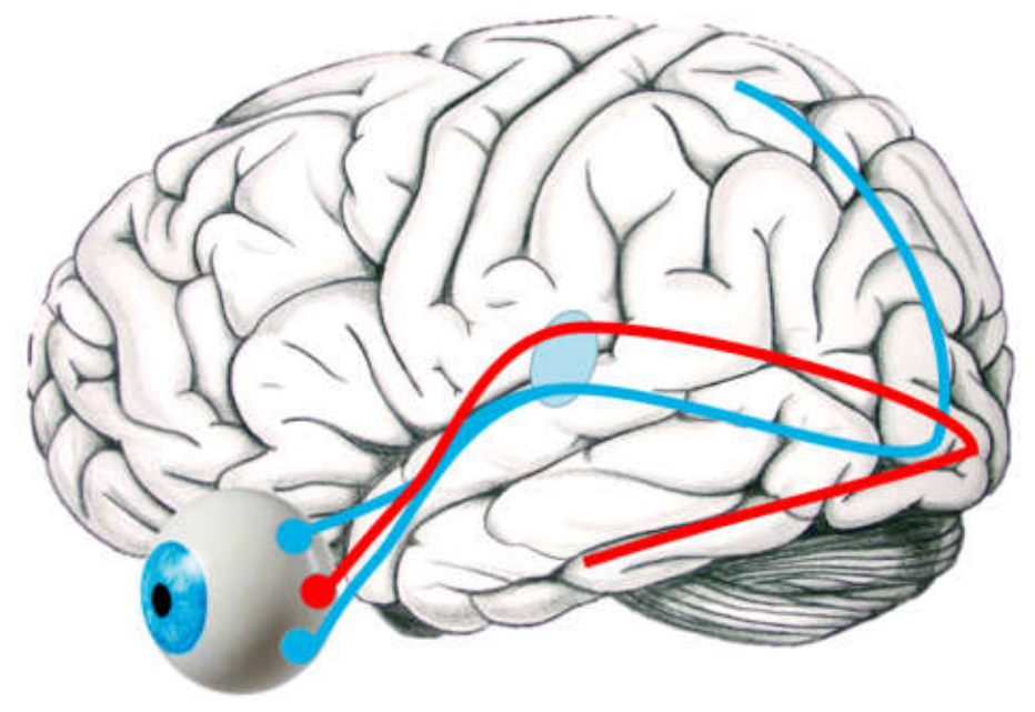
Figure 1.
The four visual stimuli that were used to analyze early visual sensory-related information processing in gamer versus non-gamers.
Figure 1.
The four visual stimuli that were used to analyze early visual sensory-related information processing in gamer versus non-gamers.
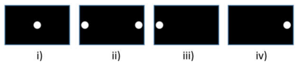

Table 1.
The three different types of cells in the thalamus.
| Type | Size | Information | Location | Response |
| Magnocellular cells (fed by rods); M-cells | large | movement, brightness nuances | LGN layers 1 and 2 | Rapid and transient |
| Parvocellular cells (fed by red and green cones); P-cells | small | color and accurate details | LGN layers 3,4,5 and 6 | Slow and sustained |
| Koniocellular cells (fed by blue cones) | small | "warmth" vs. "coolness" of a perceived environment. | LGN layers between M and P layers | Very slow |
Table 2.
Foveal vs. Peripheral Organization.
| Visual Field Location | Predominant Cell Type | Acuity vs. Sensitivity | Functional Domain |
| Central (Fovea) | Cones (S,M,L) / P-Cells | High Acuity, Low Sensitivity | Detail, Color, Text |
| Parafovea (Mid) | Mixed (P-Heavy) | Balanced | Transition Zone |
| Periphery (Far) | Rods / M-Cells | Low Acuity, High Sensitivity | Motion, Global Depth |
Table 3.
Comparing the magnocellular and the parvocellular pathway.
| Feature | Magnocellular (M) Pathway | Parvocellular (P) Pathway |
| Retinal Origin | Large M-type (Parasol) Cells | Small P-type (Midget) Cells |
| Axon Morphology | Thick, Myelinated | Thin, Less Myelinated |
| Conduction Speed | Rapid | Moderate to Slow |
| Temporal Sensitivity | High | Low |
| Spatial Resolution | Low | High |
| Contrast Sensitivity | High | Low |
| Color Sensitivity | Achromatic (Colorblind) | Chromatic |
| Primary Cortex Target | V1 Layer 4C\alpha | V1 Layers 4A and 4C\beta |
Table 1.
Mean peak amplitude latencies for the P170 and the N250 ERP components for both groups (gamers and non-gamers).
Table 1.
Mean peak amplitude latencies for the P170 and the N250 ERP components for both groups (gamers and non-gamers).
| P170 Mean (central stimulation) | N200 (bilateral peripheral stimulation) | |
| Gamer | 170.96ms (SD=11.95) | 237.77 (SD=16.82 |
| Non-gamer | 165.18ms (SD=7.86) | 263.36 (SD=14.18) |
Disclaimer/Publisher’s Note: The statements, opinions and data contained in all publications are solely those of the individual author(s) and contributor(s) and not of MDPI and/or the editor(s). MDPI and/or the editor(s) disclaim responsibility for any injury to people or property resulting from any ideas, methods, instructions or products referred to in the content. |
© 2026 by the authors. Licensee MDPI, Basel, Switzerland. This article is an open access article distributed under the terms and conditions of the Creative Commons Attribution (CC BY) license (http://creativecommons.org/licenses/by/4.0/).
Copyright: This open access article is published under a Creative Commons CC BY 4.0 license, which permit the free download, distribution, and reuse, provided that the author and preprint are cited in any reuse.