

Submitted:
06 May 2026
Posted:
07 May 2026
You are already at the latest version
Abstract
Poor drug bioavailability remains a major challenge in pharmaceutical development, primarily due to low aqueous solubility, limited membrane permeability and rapid met-abolic degradation, which significantly reduce therapeutic efficacy. In this context, nanocarrier-based drug delivery systems have emerged as an effective strategy to over-come these limitations. This review summarizes the main types of nanocarriers, including lipid-based, polymeric, inorganic and hybrid systems, and discusses their design and functional properties. Nanocarriers enhance drug performance by improving solubility, protecting active compounds from degradation, enabling controlled and sustained re-lease, and facilitating targeted delivery to specific tissues. The influence of physicochemi-cal parameters, such as particle size, surface charge and functionalization, on pharmaco-kinetics and pharmacodynamics is also addressed. Representative case studies illustrate the successful application of nanocarrier formulations in improving bioavailability and therapeutic outcomes. Despite these advances, challenges related to toxicity, large-scale production and regulatory approval remain. Overall, nanocarrier-based systems represent a versatile and promising platform for enhancing drug bioavailability and advancing modern pharmaceutical therapies.
Keywords:
nanocarriers
; drug bioavailability
; drug delivery systems
1. Introduction
A key challenge in modern pharmaceuticals is the poor bioavailability of many drug candidates, which significantly contributes to the high failure rate in drug development. Despite advances in drug discovery, over 90% of compounds fail during clinical development, with many of these failures being attributed to unfavorable biopharmaceutical properties rather than a lack of pharmacological activity [1]. Key limitations include poor aqueous solubility, inadequate membrane permeability, rapid metabolism and active efflux by transporters. These factors restrict drug absorption and reduce the amount of drug that reaches the systemic circulation [1]. These issues are particularly critical for orally administered drugs, for which dissolution in gastrointestinal fluids and transport across the intestinal epithelium are essential for therapeutic efficacy. Furthermore, conventional drug delivery systems frequently exhibit low solubility, poor absorption and instability in vivo, which further contributes to reduced bioavailability and suboptimal therapeutic outcomes. [1,2]. Bioavailability is defined as the extent to which a drug administered in its original form reaches the systemic circulation and is therefore available to exert its therapeutic effect. This process is governed by the collective pharmacokinetic processes of absorption, distribution, metabolism and excretion (ADME). Low bioavailability can result from several factors, including insufficient solubility, limited permeability across biological membranes, and extensive first-pass metabolism, all of which reduce the fraction of the administered dose that becomes systemically available. These limitations have been overcome through the extensive development of nanocarrier-based drug delivery systems. Nanoparticles can enhance drug absorption and stability, as well as enabling targeted delivery, thereby increasing bioavailability and therapeutic efficacy . Furthermore, nanocarriers can be engineered to control drug release and enhance biodistribution, minimizing side effects and improving overall treatment outcomes [3].
2. Types of Nanocarriers
Nanocarriers are nanoscale drug delivery systems typically ranging from 1 to 1000 nm in size. They are engineered to transport and deliver therapeutic agents within the body in a controlled and targeted manner. Composed of materials such as lipids, polymers, or inorganic substances, they can encapsulate, absorb, or chemically bind drugs. Modifying their size, surface properties, and composition can enhance drug solubility, protect active compounds from degradation, improve absorption across biological barriers, and enable site-specific delivery. These features contribute to increased bioavailability, reduced toxicity, and improved therapeutic efficacy compared to conventional drug delivery systems.
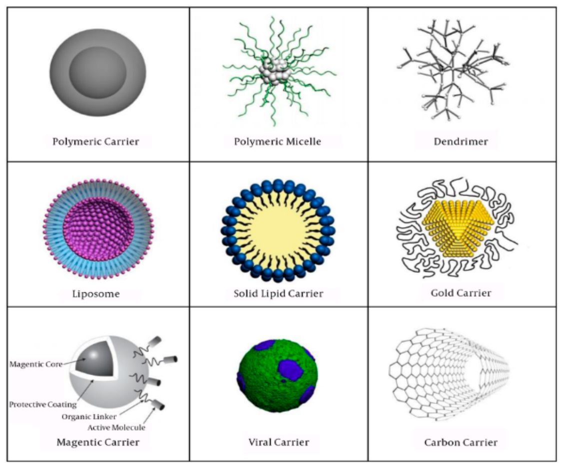
Figure 1.
Different types of Nanocarriers used in drug delivery systems [4].
Figure 1.
Different types of Nanocarriers used in drug delivery systems [4].
2.1. Lipid-Based Nanocarriers
Lipid nanocarriers or lipid nanoparticles (LNPs) are a delivery systems, which can transport drugs or genetic material (siRNA, mRNA, and DNA [5]) into the body [6]. These vesicles are usually between 10 and 100 nm in size [7]. In treating a variety of diseases, LNPs have shown remarkable therapeutic efficacy in delivering both hydrophobic and hydrophilic drugs [5]. A loaded liposome was one of the first nanodrugs to be used for the treatment of cancer. In addition to their limited biotoxicity, remarkable biocompatibility, biodegradability, and trapping effectiveness, these nanoparticles can minimize any side effects by targeted drug delivery [5,6]. The creation of lipid nanocarriers requires the selection of many parameters and characteristics. They depend on the raw materials and preparation conditions [6]. Lipid for carriers can be synthetic or/and natural [8]. Natural lipids are typically sourced from microorganisms (e.g. algae, yeast, bacteria), plants, humans (tissues, cells), and animals (tissue, cells or their products) [6]. Important lipid components in these systems are: phospholipids, cholesterol, ionizable and cationic lipids (essential for nucleic acid delivery), and PEG-lipid (e.g. provide stabilization). Cholesterol controls membrane fluidity and stability, but on the other hand, phospholipids provide the structural foundation, mainly in liposomes [7]. Choice and source of used lipids has a major impact on composition, structure, effectiveness, stability, drug-loading capacity, and biocompatibility of nanoparticles [6]. Additionally, modifiers are also added to the external part of lipid nanoparticles, such as polymers and peptides, to impart additional functional features [8]. The technique of preparation (both high and low energy) significantly affects the structure of the platform. LNPs manufacturing methods are e.g.: microemulsion, supercritical fluid; solvent evaporation; microfluidics; high-pressure and ultrasonic homogenization [6]. The majority of nanoparticles used in clinical trials are lipid-based [8]. LNPs are widely used in clinical therapies in the form of vaccines and cancer drugs (as chemotherapeutic agents delivered to tumor sites), as well as in gene therapy and genome editing applications [7].
2.1.1. Liposomes
Liposomes are a small spherical nanostructures discovered in 1965 [5,6,9]. Their core-shell composition consist of an internal aqueous core and an amphipathic (unilamellar or multilamellar) phospholipid double layer [5,6,7,8,10,11,12]. The phospholipid bilayer is around 4-5 nm (one or more bilayers are possible [12]), while aqueous space can range in size from 30 nm to micrometers [6]. Liposomes may transport small and big lipophilic and lipophobic molecules, such as nucleic acids, proteins, and imaging agents [8]. Hydrophilic active substances are typically trapped in the aqueous phase of the core, while hydrophobic drugs are encapsulated in the hydrophobic bilayers of the shell [5,8,11]. The main part of liposomes are natural or synthetic phospholipids, which are biodegradable and biocompatible because of their similarity to the lipids found in cell membranes [8]. Basically, phospholipids are made of hydrophilic head and non-polar extension (two fatty acid sequences with 10-24 carbon atoms) [13]. Phospholipids that are used to produce liposomes are e.g.: phosphatidylethanolamines (PE), phosphatidylserines (PS), phosphatidylcholines (PC), phosphatidic acid (PA), and phosphatidylglycerols (PG) [8,14]. They are typically chosen based on how they affects pharmacodynamics, encapsulation, drug stability, pharmacokinetics, and release [8]. Also, they can determined the liposome’s surface charge (neutral, anionic, or cationic) [15]. Cholesterol is another crucial component that provides stability of these structures [7,13]. Because of liposomes biocompatibility, ease of production, stability in biological environments, high drug loading, and safe ingredients (reduced drug toxicity [11]), they are an important drug delivery system [6]. If this nanocarrier display changes in their organization with pH or temperature they may become reactive. For example in healthy tissues, pH-sensitive liposomes remain stable, but in tissues with a pH lower than 7.4 they become unstable (absorbed bioactive material will leak out) [13]. Liposomes have some disadvantages like limited stability and short circulation times, but to get around this, liposomes can be made by adding anti-fouling polymers, e.g. polyethylene glycol (PEG) [8,12], which can increase the blood half-life of a liposome drug up to 45h (regardless of dose) [15]. Recently, PEG and PEG-lipid have recently been linked to negative effects, including hypersensitivity responses. In order to address these problems, new materials are being investigated, such as polysialic acids, glycoproteins, zwitterionic polymers, and polysaccharides [8]. Liposomes formulations have been applied in studies for gene delivery, malaria, several kinds of cancer (also chemotherapy), depression, and many other disorders [11,15].
2.1.2. Solid lipid nanoparticles
Solid lipid nanoparticles (SLNs) are usually made of one or more lipids which are solid both at room and body temperature [6,7,10,16]. These nanoparticles can be divided into three categories: solid solution type (homogenous matrix); drug-enriched shell model; drug-enriched core model [5,6,16]. Cold homogenization, in which a solid drug is evenly distributed throughout a lipid matrix without the use of surfactants, creates the solid solution model of SLNs. In drug-enriched shell concept, the drug is encapsulated within a core-shell structure created by thermal homogenization. During cooling and recrystallization, a lipid core forms, causing the drug to go into the lipid phase. The drug-enriched core model is formed by dispersing drug in melted lipid, leading to supersaturation upon cooling. As the lipid recrystallizes, it forms a membrane like shell around the drug rich core [16]. For the synthesis of SLNs, steroids, waxes, free fatty alcohol or acids, and (mono, di or tri)glycerides are frequently used [12,17]. The active substance may be integrated into shell, core or matrix [12]. Those nanoparticles have numerous benefits such as small size, controlled release, very good drug protection, improved physical stability of hydrophobic drugs, customizable features (by changing lipid) [6,8], low cost and nontoxicity (less toxic than polymeric nanocarriers) [10]. Compared to liposomes, these particles have better physical stability because of their solid lipid core matrix, which is stabilized by surfactants [7,17] (in concentrations from 0.5% to around 5% [16]) or polymers (hybrid polymer-lipid nanoparticles) [10]. Due to variations in formulations, preparation methods, and manufacturing temperatures, solid nanoparticles may have different characteristics [16].
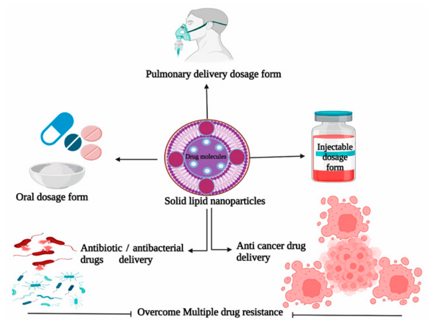
Figure 2.
Schematic representation of solid lipid nanoparticles and their target-specific interactions [16].
Figure 2.
Schematic representation of solid lipid nanoparticles and their target-specific interactions [16].
One of the main problems with this kind of lipid nanoparticles is limited drug loading capacity, but also low long term drug retention (polymorphic transition and as a result, the removal of drug from the structure). In order to eliminate these problems and improve stability, part of the solid lipids began to be replaced with liquid lipids or solubilizers. As a result of this operation, nanostructured lipid carriers were formed [6,7,8], which are discussed in the section below. SLNs has been extensively studied for use in gene transfer, antibacterial, anticancer, and vaccine applications [11].
2.1.3. Nanostructured Lipid Carriers
Nanostructured lipid carriers (NLCs) are a drug delivery systems, which are made with improved features, by changing solid part of SLNs with liquid lipids (oils) in different proportions [5,6,10,16]. For this purpose, different types of lipid are used, including fatty acids, steroids, triglycerides, partial glycerides, waxes [6]. Their models are divided based on the arrangement of lipids and drugs in structure: imperfect type (mixing of spatially different lipids); amorphous type (structureless solid matrix); multiple type (drug contains oil) [5,6,13,16]. Liquid lipid cause structural flaws in solid lipids, resulting in a less ordered crystalline arrangement [16]. Therefore, when polymorphism and lipid crystallinity problems in SLNs have been mostly eliminated, NLCs nanocarriers exhibit larger loading capacities, better stability, and better drug retention [6,7,11]. Nanostructured carriers are known as platforms of both water soluble and fat-soluble substances [13]. Despite their important qualities, NLCs have also been linked to cytotoxic effects, limited stability, irritation, and surfactant sensitization [16].
2.2. Polymeric Nanocarriers
Polymeric nanoparticles (PNCs) are a colloidal particles made from natural or synthetic materials, monomers, or premade polymers (which enables a wide range of PNCs potential shapes and properties) [15,18]. Natural PNCs are protein-based or polysaccharides polymers e.g. gelatine, collagen, albumin, alginate, chitosan, agarose, hyaluronic acid (HA) [15]. On the other hand, synthetic polymers are e.g.: PEG, chitosan, poly(glycolic acid) (PGA), poly(lactic acid) (PLA), poly(lactic-co-glycolic acid) (PLGA), poly(dimethylsiloxane) (PDMS), poly(caprolactone) (PCL), and poly(ortho esters) (POE) [15,18,19,20]. Compared to natural polymeric nanocarriers, synthetic polymers are more appealing in drug delivery system because they are more repeatable and exhibit less batch-to-batch molecular weight (MW) dispersion and variation [15]. It is possible for active substances to be attached to the PNCs surface (by adsorption of drug), chemically conjugated to the polymer, trapped in the polymer matrix, or encapsulated within the core [18,21]. This makes it possible to deliver a variety of drugs (hydrophilic, hydrophobic), but also substances with varying molecular weights, e.g.: biological macromolecules, vaccines, proteins, and tiny molecules. They are biocompatible and easy to make (simple composition parameters). The loading efficacy and release kinetics of polymeric nanoparticles can be carefully regulated, by composition, stability, responsiveness, and surface charge. PNCs are water soluble, stable during storage, biocompatible, biodegradable, and biomimetic. The main disadvantages of this carriers are increased risk of aggregation and toxicity [18]. Several methods, including emulsification, nanoprecipitation, ionic gelation and microfluidics are used to produce them [15,18]. Polymeric nanocarriers can be divided (based on how they are prepared) into nanocapsules (spaces with a polymeric shell/membrane surrounding them), and nanospheres (solid matrix systems) [15,18,21].
2.2.1. Polymeric Micelles
Polymeric micelles consist of a hydrophilic (polymeric) core and a hydrophobic coating, which makes them different from lipid micelles (hydrophobic core) [15,18]. This structure improve drug circulation time and is very beneficial especially for hydrophilic drugs (better protection) [18]. Micelles are really small, their size are usually between 20 and 100 nm [15]. Poly(ethylene glycol) (PEG) is the most widely used hydrophilic polymer because it is an excellent defender against biological degradation and offers surface stabilization. [15]. Polymeric micelles are known for possibility of loading very various range of molecules, from small to big proteins [18]. Polymeric micelles demonstrated better qualities than traditional surfactant micelles, e.g. more stability, longer blood circulation time. They also are characterized by increased bioavailability of drug across barriers, high drug loading capacity, targeting delivery and controlled and sustained release drug [15]. Polymeric micelles can be enriched with medicine by physical and chemical (covalent attachment) methods [17].
2.2.2. Dendrimers
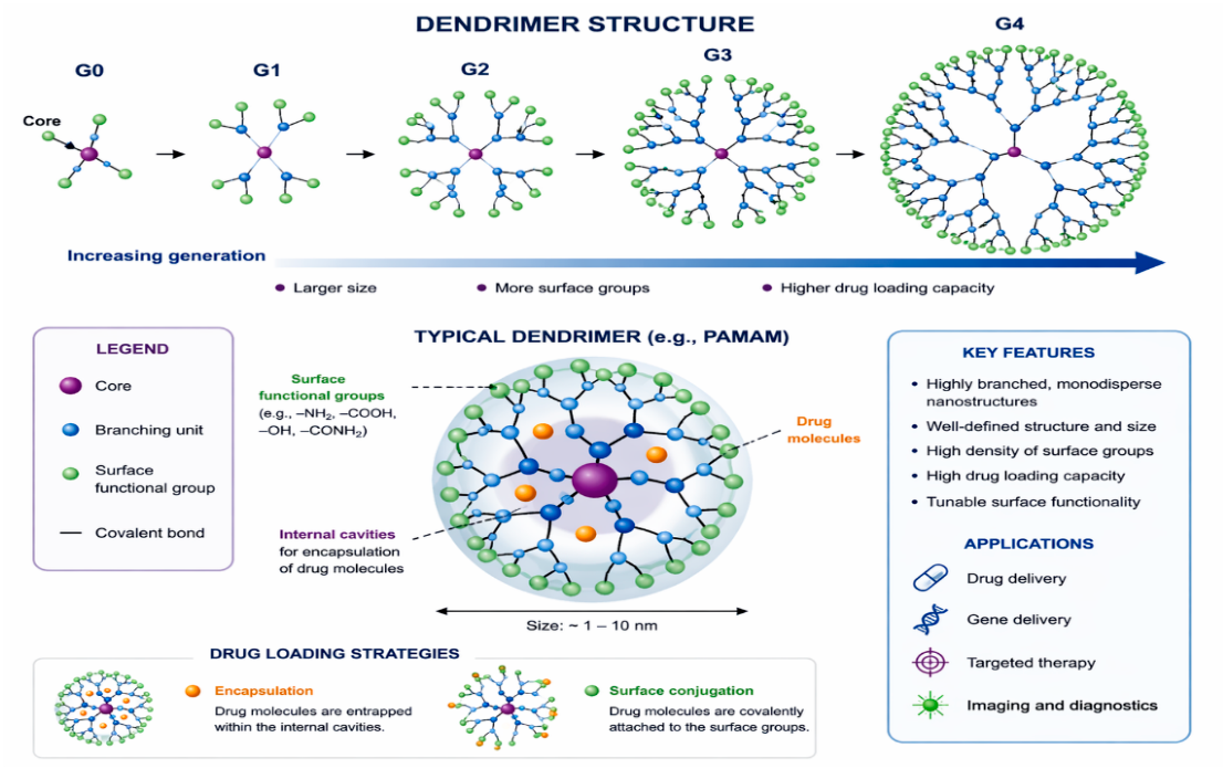
Dendrimers are a stare or tree like shaped nanoparticles made up of a three key components: central core nucleus, several branches, and end groups providing a flexible surface (which overall creates a complex 3D structure )[17,18,22,23]. The inner and outer layers are very similar, repetitive to each other [22]. Every fresh-grown branching creates a new “generation”, which can have different benefits. Lower generations (G1-G3) have a high accessibility, and lower diffusion, but higher generations (G4-G6) have a large loading capacity [24].
Figure 3.
Schematic representation of dendrimer structure and drug loading strategies.
The mass, size, shape, and surface chemistry of dendrimers can be manipulate in order to obtain structures with selected properties [18]. Additionally, they have a high surface/volume ratio, and capacity to contain a significant amount of medication (or contrast agents) inside or on their surface [19], because of functional groups on the outside [18]. They can increase bioavailability, and solubility of hydrophobic medications [23]. One of the primary obstacles is controlling dendrimers’ size and form, which can impact pharmacokinetics and distribution, but also their toxicity and possibility of interaction with the immune system [19]. Drug delivery systems consisting of dendrimers can deliver nucleic acids and small molecules, for which poly(ethylenimine) (PEI) and poly(amidoamine) (PAMAM) are often used [17,18]. Additionally, they are used in delivery of genes, molecular sensors, bioimaging, and enzyme mimics [23].
2.3. Inorganic Nanocarriers
Inorganic nanocarriers materials are used for a variety of drug delivery (e.g. anticancer, antibacterial, antioxidant [20]), photothermal therapies, and imaging applications [18]. They are using inorganic elements like silica, calcium phosphate, iron, silver, titanium, zinc, and gold [18,25]. They can be further divided into three categories i.e. magnetic (iron oxide, gold, silver), ceramic (silica, titanium oxide, alumina), and semiconductor (zirconium dioxide, zinc oxide) [20]. To obtain these particles, various substances are used, both of natural and synthetic origin, depending on the synthesis method. In the case of chemical methods, they need substances like, e.g.: dimethyl formamide, and sodium borohydride (and high temperature). In turn, safer and less toxic natural substances include, e.g. plant parts or microorganisms (bacteria, fungus), which makes a natural method of preparations simpler, faster, more stable, and eco-friendlier than the other one [25]. Furthermore, because of the characteristics of the base material, inorganic nanocarriers can have, e.g.: catalytic, thermal, physical, electrical, radioactive, plasmonic, magnetic, and optical capabilities [18,26]. The majority of them are stable, small (so they can penetrate membranes both physiological, and biological, which are typically impermeable to others macromolecules [25]), and exhibit better loading capacity [18,19,26]. However, low solubility and toxicity issues, particularly in formulations, including heavy metals, restrict their clinical use [18]. Commonly, to improve metal nanoparticles, they are coated with PEG or their surface is functionalized by substances that gives them targeting properties (e.g. in tumor cells treatment). These nanocarriers can be synthesized by various methods, e.g.: microemulsion, polyol method, laser ablation, chemical reduction, sol-gel [25].
2.3.1. Gold Nanoparticles
Gold nanoparticles (AuNPs) can have a variety of shapes, which are divided by dimensions, including: nanorods, nanowires (1D), nanoplates (2D), nanostars, nanopods (branched structures) [18,19,26]. Gold nanodrugs have photothermal and imaging (optical) capabilities because of their free electrons (on the surface with large surface to volume ratio [27]) that continuously oscillate at a frequency which depends on their size and shapes [18,19]. They also are easily functionalized, and can be packed with targeting and/or imaging agents (e.g. MRI agents, fluorophores [27]), but also with chemotherapeutics, peptides, proteins, siRNAs, and pharmaceuticals [18,25,27]. Applying modifications can give them new delivery properties, which is advantageous in diagnostics, treatment, and targeting (even simultaneously with real-time monitoring) [18,27]. Unfortunately, they may cause side effects by unwanted interactions with biological molecules despite being known for their biocompatibility. In order to minimize the toxicity of nanocarriers, scientists add biocompatible polymers or proteins [27].
2.3.2. Magnetic Nanoparticles
Magnetic nanoparticles are known for their potential (at specific sizes) as DDS, contrast agents, thermal based drugs, and in diagnostic of cancer [18,27]. They are composed of iron oxide, i.e. hematite (α-Fe2O3), maghemite (γ-Fe2O3) and magnetite (Fe3O4) [18,26]. In the case of paramagnetic nanoparticles, iron oxide is put into the core, while other substances (e.g., silica, gold, phospholipids, peptides, fatty acids, surfactants, and polymers) might be a coating [23]. Magnetic nanocarriers can be transported using an external magnetic field on particular bodily locations [19]. Also they are characterized by ease of functionalization, biodegradability, low-cost synthesis, and they also have an antibacterial properties [20] Nevertheless, these nanocarriers have major limitations, e.g., toxicity, and they cannot accumulate in high enough concentrations for efficient imaging or drug delivery. These problems can, however, be avoided by addition of stimuli-responsive agents, and surface modification. The solutions make iron nanoparticles stable, biocompatible, but more importantly, the can act directly and controlled on cancer cells without overall toxicity [27].
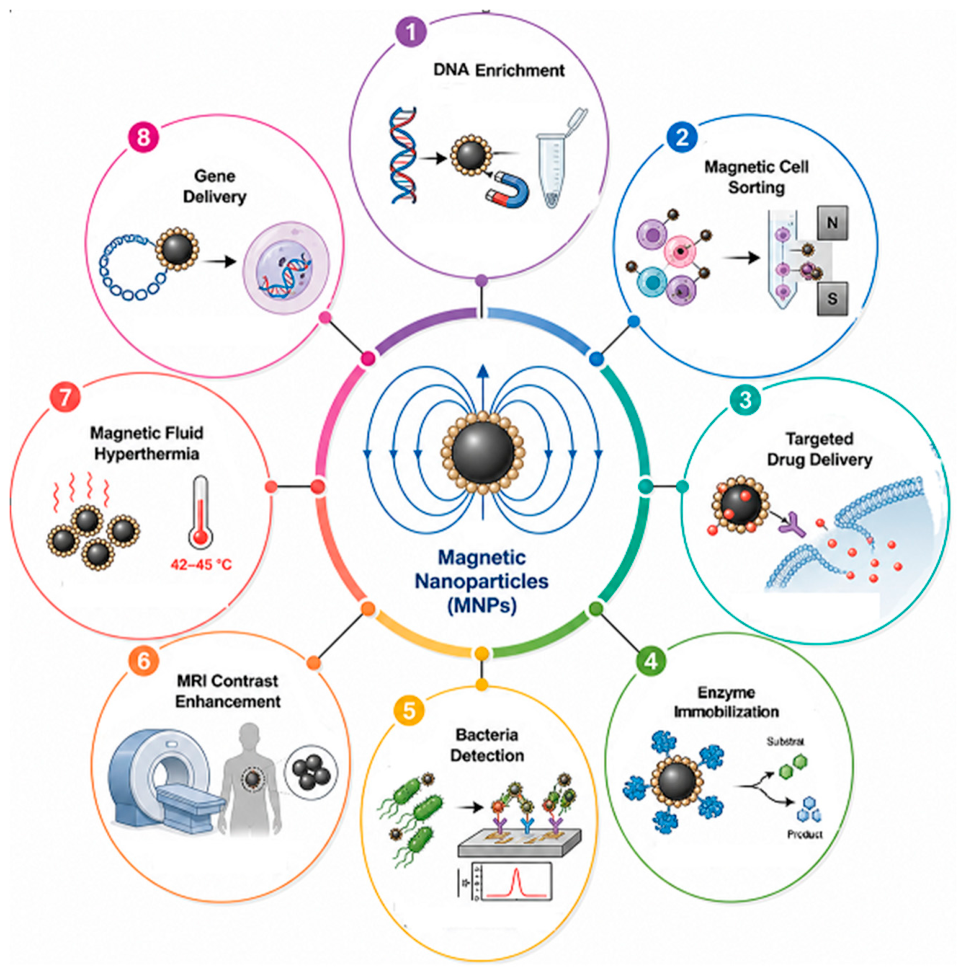
Figure 4.
Applications of magnetic nanoparticles [28].
Figure 4.
Applications of magnetic nanoparticles [28].
2.3.3. Carbon-Based Inorganic Nanocarriers
Carbon-based nanomaterials (CBNs) are a distinct class of inorganic nanocarriers, characterized by unique allotropic structures, nanoscale dimensions and exceptional physicochemical properties. Materials in this class include carbon nanotubes (CNTs), graphene, fullerenes and related derivatives. They have attracted considerable interest in the field of nanomedicine thanks to their potential as efficient drug delivery platforms. These materials, are composed of sp²-hybridized carbon atoms arranged in ordered nanostructures, which distinguishes them from classical organic or polymeric carriers and supports their classification as inorganic systems. The diversity of carbon allotropes allows for the development of nanocarriers with adjustable morphology and functionality, making them suitable for use in biomedicine [29]. Carbon nanotubes (CNTs) are among the most extensively studied carbon-based nanocarriers. Structurally, CNTs are cylindrical nanostructures formed by rolling graphene sheets into seamless tubes with nanometer-scale diameters. They are classified into single-walled (SWCNTs) and multi-walled (MWCNTs) depending on the number of graphene layers [30] .
Figure 5.
Carbon nanotube structure. a) Single-walled CNTs. B) Multiwalled CNTs [31].
Figure 5.
Carbon nanotube structure. a) Single-walled CNTs. B) Multiwalled CNTs [31].
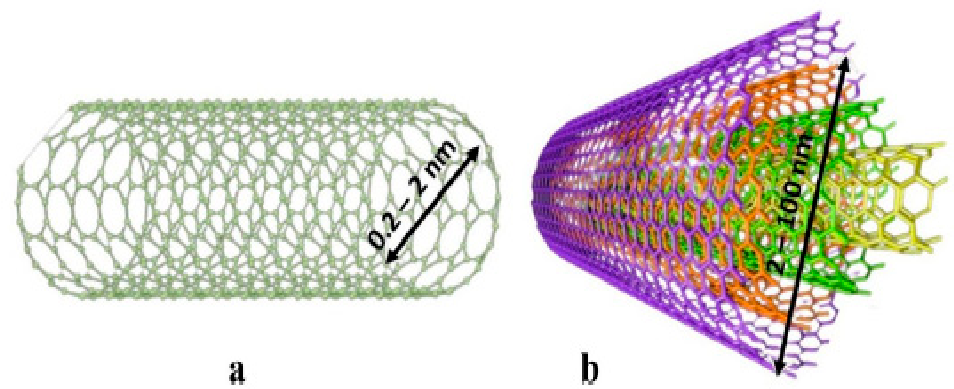
CNTs exhibit exceptional mechanical strength, electrical conductivity, and thermal stability due to their unique carbon–carbon bonding and one-dimensional structure [32]. Their extremely high surface-to-volume ratio provides a large interface for molecular interactions, which is essential for drug loading [33] CNTs can adsorb drugs through π–π stacking interactions, hydrophobic interactions, and electrostatic forces, enabling the loading of both small molecules and biomacromolecules such as nucleic acids . This high loading efficiency enhances cellular uptake and facilitates intracellular delivery [34]. Additionally, CNTs have demonstrated the ability to penetrate cellular membranes and, when appropriately functionalized, cross biological barriers such as the blood–brain barrier. Carbon-based inorganic nanocarriers also exhibit multifunctional properties that extend beyond conventional drug delivery. Their intrinsic optical and electronic characteristics allow their use in theragnostic applications, combining drug delivery with imaging and therapeutic modalities. CNT-based systems have been applied in cancer therapy to enhance drug accumulation at tumor sites and improve therapeutic efficacy while reducing systemic toxicity [34]. Furthermore, their ability to enable controlled and stimuli-responsive drug release makes them promising platforms for advanced therapeutic strategies [35] Despite these advantages, the clinical translation of carbon-based nanocarriers is limited by several challenges. Their potential cytotoxicity and long-term biocompatibility are important concerns as these properties depend on size, shape and surface chemistry . The formation of a protein corona in biological environments can also affect biodistribution, cellular uptake and the immune response [31]. Furthermore, ensuring the safety and efficacy of carbon-based nanocarriers requires careful design and surface modification to prevent aggregation and persistence in biological systems.
2.4. Hybrid Systems
In order to improve nanomaterials i.e. overcome limitations or make carriers more multifunctional, hybrid nanoparticles were created [5]. Polymer-Lipid nanocarriers (PLNs) are classified according to their structural configurations, which include different lipid and polymer combinations [6]. They consist of three main components [36]. The most known one of them is made of an exterior layer of PEG lipid and interior monolayer of lipid that surrounds a polymeric core [5,36]. PEG stabilize and keeps PLNs from being broken down by the immune system. Middle monolayer protect the core and act as a molecular barrier and can stop the loss of medication [36]. They combine the properties of both lipids and polymers and are characterized by higher biocompatibility, bioavailability, excellent physical stability, extended release (and circulation time), and higher permeability through membranes [5,6,36]. Both hydrophobic and hydrophilic medications can be delivered with hybrid PLNs [36]. Lipid-coated silver nanoparticles (lipid-AgNPs), liposome-gold nanoparticles, and lipid-aluminum nanoparticles are a few examples of hybrid lipid-inorganic nanoparticles. Cooperation of metals and lipids in nanoparticles increase biocompatibility, stability, and endocytosis efficiency [5]. Among the more advanced hybrid nanocarrier systems developed for drug delivery applications are virus-based platforms and carbon dots (CDs) - particularly carbon–organic hybrid systems.
2.4.1. Viral Carriers
Viral nanocarriers are a type of hybrid nanocarrier derived from naturally occurring viruses or engineered virus-based systems. Examples include viral nanoparticles (VNPs) and virus-like particles (VLPs). These structures consist of self-assembled protein capsids that form highly ordered, monodisperse nanostructures with a defined size and geometry . VLPs are non-infectious due to their lack of a genome, while still maintaining the structural features required for cargo delivery [37]. A key characteristic of viral nanocarriers is their innate capacity to transport biological cargo; viruses have evolved to naturally deliver nucleic acids into host cells. This capability is exploited in nanomedicine for the delivery of drugs, proteins and nucleic acids. Cargo can be encapsulated within the capsid, adsorbed onto its surface or chemically conjugated to the functional groups present on the protein shell. This enables a variety of loading strategies. Viral nanocarriers exhibit several properties that make them well-suited to biomedical applications. These include biocompatibility, biodegradability and structural uniformity, which result from genetically encoded assembly. Their multivalent protein surfaces allow targeting ligands to be attached, enabling site-specific delivery to selected tissues or cells [37,38]. Furthermore, viral capsids can be modified through genetic engineering or chemical functionalization to improve targeting efficiency and therapeutic performance. An important subgroup of viral nanocarriers are viral vectors used in gene therapy. Examples include adeno-associated viruses (AAVs), lentiviruses and adenoviruses. These systems enable highly efficient gene delivery and can achieve long-term expression of therapeutic genes in target cells. AAV-based vectors in particular are widely used due to their low pathogenicity, broad tissue tropism and ability to sustain transgene expression, making them one of the most successful platforms in clinical gene therapy [39,40].
2.4.2. Carbon Dots
Carbon dots (CDs) are a class of zero-dimensional, carbon-based nanomaterials typically measuring less than 10 nm. They are characterized by a carbonaceous core composed of sp²/sp³ hybridized carbon, as well as a surface rich in functional groups such as carboxyl, hydroxyl and amino moieties [41,42]. This core–shell structure determines their physicochemical and biological behavior, as the functionalized surface governs interactions with biomolecules and enables further chemical modification [41]. Due to their abundant surface functional groups and carbon-based composition, CDs exhibit high water solubility, low toxicity, and good biocompatibility [42,43]. The surface chemistry of CDs allows them to be extensively functionalized and conjugated with therapeutic agents, targeting ligands and biomolecules. This supports their use as nanocarriers [41,43]. Their small size and high surface-to-volume ratio facilitate efficient cellular uptake and drug loading, and functionalized systems enable targeted, controlled drug delivery [43]. Furthermore, CDs can act as multifunctional platforms, combining drug transport with real-time imaging thanks to their fluorescent properties [43].
3. Nanoparticle Bioavailability Enhancement
3.1. Bioavailability Determinants
Bioavailability is a very important factor affecting the performance of pharmacological therapy because it influences the amount of the drug that is delivered to systematic circulation (in unchanged active form) from the selected drug delivery platform (topical, oral, rectal, parenteral) [44,45,46]. Several factors influence the overall pharmaceutical bioavailability, namely the chemical and physical properties of the selected drug, the mode of transport (including the DDS platform), interactions with other substances (both in the drug formulation and in the body), absorption, distribution, metabolism, and final excretion [45,46]. Bioavailability is the degree to which absorption of the drug occurs (passive, facilitated, and active diffusion, also endocytosis) [44,47]. Drug need to pass, e.g. gastrointestinal tract (GI tract), skin ora respiratory system to bloodstream [45]. Basically, passive diffusion is a movement of particles from a higher to lower concentration. The penetration of drugs with hydrophilic properties through biological membranes is very limited (in contrast to hydrophobic substances). In facilitated diffusion movement is driven by an electrochemical gradient, and proteins are involved as carriers. Active diffusion is a movement against gradient, and drug molecules need to be pick very specifically for this kind of transport [44]. After the drug is absorbed into the systemic circulation, the next stage begins, which is distribution - the dispersion of the substance to appropriate locations, such as tissues and organs. Next, as a result of enzymes mostly in the liver, drug metabolism occurs, transforming them into more soluble metabolites, and the elimination of the substances from the body follows [45]. Drug formulation (such as added excipients, and used methods), solubility, stability, hydrophilic or hydrophobic properties are a few examples of drug’s characteristics crucial to its bioavailability [45]. Additionally, very crucial to drug’s absorption are organ blood flow, intestinal microflora, organ barriers (e.g. BBB, which is a physiological semipermeable barrier between blood and brain tissues, important to overcome for drugs that treat, e.g. depression [11]), gastric acidity, and digestive enzyme activity [26,29]. Other factors that can reduce bioavailability are, e.g. the quantity of hydrogen donors, heavy atoms, drug-other molecules interactions (which ends with the reaction or formation of a complexes and aggregates), and some substances, like tetrazole, benzoquinone groups, aminopyridine. On the other hand, factors increasing bioavailability are e.g. the quantity of hydrogen acceptors, small atoms (with minimal molecular weight), and substances (azide, salicylic acid, or amide groups) [44]. One of the very important points to address when increasing bioavailability are nanoparticles, described in the point below.
3.2. Nanoparticles for Improving Drug Bioavailability
Traditional medicines have relatively low bioavailability, which reduces their overall absorption by the body and ultimately affects the quality of their action [19]. A lot of pharmaceuticals are rejected because of their deficient physiochemical properties [15]. Currently, nanocarriers provide an attractive substitute for conventional drugs by addressing various issues related to drug administration [6]. Many of them are believed to have better qualities because of their physical properties, smaller size, and possibility of targeted delivery [19]. Drug delivery systems use nanoparticles to increase drug absorption time, drug solubility in blood (and water), and to reduce drug degradation, inactivation, as well as to eliminate drug aggregation [9]. An ideal, successful, and effective delivery system based on nanoparticles should have a number of essential features. First, it should maximize drug bioavailability while maintaining controlled and predictable release kinetics. Equally important is the ability to selectively target specific tissues, which enhances efficacy and reduces off-target effects. The system should also minimize immunological responses to ensure safety and compatibility within the body. It must also support good patient compliance and have an adequate drug loading capacity. Lastly, a perfect system should be able to deliver a variety of treatments, including complex biomolecules and substances that are usually challenging, including lipophilic and amphiphilic molecules [49]. Some of nanoparticles can enter the body’s smallest parts because of the remarkable ratio of atom/molecules surface area to their mass or volume. This is used in the case of cancerous cells, or ineffective lymphatic drainage because of the increased permeability and retention (EPR effects) [15]. However, certain nanoparticles exhibit limited ability to cross cell membranes [19]. Table 1 presents the collected information regarding the type of nanoparticles, their advantaged (also affecting bioavailability), and limitations.
3.2.1. Physical Properties of Nanocarriers
Very important role in increasing bioavailability have size, surface properties, targeting, accumulation, and shape of nanoparticles [19,55]. The size, shape, and possible surface modifications for nanocarriers are presented in Table 2. NPs are characterized by their small size and high mobility, which allows them to reach hard to access places, e.g. to penetrate through many biological barriers that were previously inaccessible to traditional drugs [23,56]. Additionally, physical properties affect drug load and release, as well as stability [23]. The smaller the particles, the greater the surface/volume ratio, which leads to a significantly faster release of the active substance from the carrier [23,57]. One of the negative aspects of smaller nanoparticles is the possibility of aggregation [21,23]. The size affect many factors, such as enzymatic degradation, targeted distribution through body, cellular uptake, and sustained drug release [57]. Surface charge (zeta potential) of nanoparticles affect their adsorption, stability, and biodistribution [23], but also aggregation [57]. The negatively charged surface of NPs affects, among other things, longer circulation and better targeting. On the other hand, positively charged NPs have very strong interactions with membranes, and also support biocompatibility and better circulation [23,56]. Surface modification are also widely used, mainly utilizing polymer materials [57]. Zwitterionic/polymer coatings improve biocompatibility, stability, and circulation time. PEG coatings on surface of NPs creates a hydrophilic barrier, and reduces interactions with other substances, and can extends blood circulation time [23]. The chitosan coating improves the solubility of the drug in a pH-sensitive places and also releases the drug in a controlled manner [56]. Those kins of coatings can also promotes accumulation in tumor tissue, which is important in cancer therapy [23].
3.2.2. Solubility, Stability and Protection from Degradation
Over 70% of recently developed drugs are water insoluble [15]. The solubility of a drug is a very important parameter that determines the bioavailability of the drug on the absorptive surface or membrane [44] and further biodistribution [15]. Basically, low solubility usually cause low bioavailability and medications with poor solubility may need larger dosages to reach therapeutic levels (which can raise toxicity) [46]. Improving the solubility of a medicine is especially crucial for substances whose absorption profile is closely related to solubility [44,46]. Its improvement affects the increase in the rate of absorption of the active substance, e.g. in intestinal environment, thereby enhancing the quality of therapy [44]. Drug can penetrate biological membranes in ionic forms, which depends on solubility, and local pH [44,45]. Local pH can be very different and in GI track ranges from acidic (stomach, pH 1-3) to neutral, and alkane (pH 5-7). To overcome this problems, carriers with coatings are being created to protect the platform from degradation caused by different pH levels (mainly by environment in stomach) [46]. In general, ionized pharmaceuticals are more hydrophilic [44]. Managing solubility is particularly important for oral administration of drugs because they must dissolve in GI tract before being absorbed into the bloodstream. Advance formulation use surfactants, prodrugs, and nanocarriers [46]. Nanoparticles (e.g. liposomes, dendrimers, SLNc, polymeric) commonly increase solubility by reducing the particle size of the drug (higher surface area, and area/volume ratio) [46] i.e. top-down processes. It include high pressure homogenization, high energy wet milling in water phase and yielding [21]. It directly affects solubility of the drugs in biological fluids, which allows for the better absorption, delivery and maintenance of the appropriate concertation of the medication, thereby achieving the therapeutic effect [45,46]. Another method is bottom-up processes, where molecules group together to form tiny particles, e.g. hydrosol method, freeze drying, supercritical technology and gas recrystallization [21]. Encapsulation of the drug by nanocarriers apart from increasing solubility, can protect them from degradation in, e.g. GI track (previously mentioned pH changes) [46,47]. A common example of this phenomenon is liposomes, which, thanks to their spherical core-shell structure, can protect both hydrophilic and hydrophobic substances from environmental factors [46]. What is worth emphasizing is that the size of the nanoparticle also affects its degradation. The smaller the molecule, the greater the likelihood of enzymatic degradation due to the larger surface area, and consequently, the drug may be released more quickly [57].
3.2.3. Targeting Strategies and Drug Loading in Nanocarrier
Targeted delivery to specific tissues, organs, and cells is achievable by nanoparticles by functionalization of their surface with ligands. These ligands attach only to the appropriate receptors located on the surface of a given cell, such as a cancer cell, thereby improving the therapeutic value of the drug and its bioavailability [46]. Targeting removes the drug’s interaction with other healthy cells, reducing systemic toxicity and side effects [46,47]. Passive targeting focuses on properties of DDS, and on characteristics of the target site. On the other hand, active targeting, is related to changes on the surfaces of nanodrugs. Such changes involve, e.g. the attachment of the ligands or antibody-antigen interactions [28]. A large drug-loading capacity is essential for an efficient delivery system. The higher the drug loading, the more medication reaches the body in a single dose (more of the substance has a chance to reach the site of action) [23]. Some nanoparticles can have limited drug loading because of smaller size [21]. Typical drug loading for polymeric nanoparticles ranges from 5% to 20%, for lipid-based NPs its 1-5% and for lipid-polymer hybrid 5-15% [53]. There are two methods. The first one is when the drug is included during the creation of the nanoparticle (incorporation method). The second one is when NPs is absorbed or adsorbed after production. The degree to which the drug dissolves in the nanocarrier matrix determines the effectiveness of drug loading. As a result, choosing the right polymer is crucial, e.g. PEG [23].
3.3. Overview of Nanocarrier Types and Their Biomedical Applications
Currently, there is a lot of experimental work being done on the development of nanoparticles, mainly in the pharmaceutical sector, but also in the food sector. The improvement of NPs bioavailability primarily concerns increasing the solubility and stability of nanostructures. Mainly, polymeric and hybrid substances are chosen, which combine the properties of different types of NPs, e.g. polymer-lipid nanoparticles (where often the core is lipid and the outer layer, shell is polymer - PEG or chitosan). It is worth noting that the vast majority of scientific papers dealing with bioavailability focus on the oral delivery of drugs/active substances, as shown in Table 3.
3.4. Routes of Administration
To deliver the drug quickly (especially at the beginning) and in the largest possible quantity, many types of drug delivery systems have been created [25]. Drug absorption (so also bioavailability) depends on the drug administration route and the reason of application [25,26]. Table 4 presents the main groups of nanoparticles and their places of use.
Parenteral administration routes can be divided, e.g. into intravenous (invasive, avoid first-pass metabolism, quick increase of drugs concentration), intramuscular, intraarticular, subcutaneous, and transdermal (more patient friendly, longer release of medication, administered through skin) can be used [16,25]. TDDS method avoids metabolism, gastrointestinal irritation, and usually is more acceptable for patients because of its non-invasive properties. On the other hand, intravenous method is suitable when drug cannot be injected into tissues, and are characterized by complete bioavailability of drug (directly injection of drug into circulation, without phase of absorption ) [25,44]. The main barrier in oral administration is gastrointestinal epithelium. The absorption of drugs through this route due to the GI track is very complicated and highly dependent on the properties of the DDS, but also enzyme activity, thickness of mucosal, and surface area [57]. Most experiments focus on improving the bioavailability of orally administered drugs [44]. Delivery through inhalation is very beneficial, e.g. large surface area of absorption and non-invasive action [69]. The amount of active substance delivered by a given DDS platform and the amount of drug that actually enters the systemic circulation are often different, with the exception of intravenous administration, which is characterized by 100% bioavailability [44,45]. Table 5 summarize routes of administration (with absorption rate and site), factors affecting absorption and possible nanoparticle’s impact on bioavailability.
Patient compliance, the medication's physicochemical characteristics (e.g. solubility, bioavailability), the target site's accessibility, and overall therapeutic efficacy are all important considerations when choosing a drug delivery method [25]. More than 50% of the lipid-based nanocarriers are being delivered by parenteral routes [49] (most effective when drug has low bioavailability), but they can also be administered as pellets, powders, tablets (after lyophilization and spray-drying), and gelatin capsules. Macromolecules such as proteins and peptide (when given orally) are very vulnerable to enzymatic degradation [16]. Liposomes can be deliver to the lungs (as aerosols, inhalation), also on the skin (topically as creams, and gels), and orally. Oral administration of liposomes are very limited because of the environment in stomach (in this case there are no difference in traditional drug and liposomes-drug structure) [14]. Inorganic, and more precisely, metallic nanoparticles can be administered transdermally, and using inhalation, injection or by oral delivery (intravenous methods) [25]. Polymer nanoparticles are delivered via injections, oral, pulmonary, and transdermal administration [69].

Table 5.
Routes of drug administration, absorption characteristics, and the impact on nanoparticles on bioavailability.
Table 5.
Routes of drug administration, absorption characteristics, and the impact on nanoparticles on bioavailability.
| Route of administration | Absorption rate and site [45] |
Factors affecting absorption [44,70] |
Possible nanoparticle impact on bioavailability, e.g.: | |
|---|---|---|---|---|
| Oral | Slow; Mucosa gastric or intestinal |
Blood flow to GI tract, surface area of absorption, gastric emptying time, pH of GI tract, gender, emesis, digestive enzymes (drug stability, dissolution time), intestinal flora, properties of drugs | Improve solubility; Protect drug from acidic degradation in stomach; Reduce first-pass metabolism; Enhance intestinal absorption. |
|
| Injection | Intravenous | Very quickly; - |
- | No absorption needed (100% bioavailability); Enable controlled drug release; Targeted delivery; Prolong circulation time (e.g. PEGylation). |
| Subcutaneous | Quickly; Subcutaneous (or adipose) tissue |
Blood flow at injection site, vasoconstriction, local pH, properties of the drug, e.g.: size (the larger the size, the lower the bioavailability) and complex composition | Controlled/sustained drug release; Improved lymphatic uptake. Reduced degradation at injection site. |
|
| Intramuscular | Very quickly; Muscle tissue |
Blood flow to muscle, gender, drug properties, muscle mass and activity | Prolonged release at injection site; Improved stability of drug; Enhance absorption depending on particle size. |
|
| Inhalation | Very quickly; Mucous membrane of the lungs |
Lung surface area, disease of the lung (lower perfusion and ventilation), drug properties (particle size) | Improved deposition in lungs; Enhanced absorption; Targeted delivery to respiratory tract; Reduced systemic side effects. |
|
| Transdermal | Quickly; Stratum corneum; epidermis; dermis |
Skin hydration and blood flow, skin barrier function, thickness of stratum corneum, drug lipophilicity, drug formulation | Enhanced skin penetration. Overcome barrier of stratum corneum; Controlled drug release; Improved bioavailability of poorly permeable drugs. |
|
| Rectal | Quickly; Mucous membrane of the rectum |
Rectal blood flow, drug formulation, pH of fluids | Improved drug stability; Enhanced mucosal absorption; Prolonged local or systemic effect. |
|
4. Nanoparticle for Improving Bioavailability- Case Studies
4.1. Case 1: Curcumin Nanoformulation
Curcumin is a hydrophobic polyphenol with significant therapeutic potential. However, its clinical application is severely limited due to its extremely low oral bioavailability. This is due to poor aqueous solubility, rapid intestinal and hepatic metabolism, and inefficient absorption, resulting in negligible systemic exposure even at very high oral doses. Clinical studies have shown that taking up to 8–12 g of conventional curcumin results in undetectable or minimal plasma concentrations in most people [71]. These limitations have been addressed by developing nanoformulation strategies to enhance solubility and absorption. Nanoparticle-based formulations such as THERACURMIN reduce particle size and improve aqueous dispersibility, leading to significantly increased systemic exposure. Human studies have shown that THERACURMIN achieves measurable plasma concentrations (C_(max) up to 275 ng/mL) at doses as low as 150–210 mg, whereas conventional formulations at gram-scale doses remain largely undetectable [72]. Similarly, micellar and micronized delivery systems substantially enhance bioavailability by improving dissolution and gastrointestinal solubilization. In a controlled human study, micellar curcumin increased systemic exposure by up to 185-fold, while micronized formulations resulted in bioavailability up to nine times higher than that of native curcumin [73]. Overall, these findings demonstrate that nanocarrier-based approaches effectively overcome the intrinsic physicochemical and pharmacokinetic limitations of curcumin by primarily increasing the dissolution rate and enhancing solubilization, as well as partially protecting the compound from rapid metabolic degradation.
4.2. Case 2: Paclitaxel Nanoformulations
Paclitaxel is a highly effective anticancer agent; however, its clinical use is significantly limited by its extremely poor solubility in water, which necessitates its formulation with toxic solvents such as Cremophor EL and ethanol. These solvents are associated with severe hypersensitivity reactions, altered pharmacokinetics and impaired drug distribution due to micelle formation. This can entrap paclitaxel in circulation, reducing its delivery to tumour tissues [74]. To overcome these limitations, a solvent-free formulation of paclitaxel bound to albumin (nab-paclitaxel, ABI-007) was developed. This nanocarrier system uses albumin to enhance the transport of the drug and its delivery to tumours via natural biological pathways, including receptor-mediated transcytosis and increased endothelial transport. Preclinical studies have demonstrated that nab-paclitaxel transports across endothelial cells approximately four times more effectively and accumulates in tumours more efficiently than conventional formulations do [74]. Clinically, nab-paclitaxel has demonstrated significantly improved therapeutic performance. In a phase III trial, nanoparticle-based paclitaxel produced higher overall response rates (33% versus 19%) and prolonged time to tumour progression (23.0 versus 16.9 weeks) than conventional paclitaxel, despite a substantially higher delivered dose [75]. Notably, the incidence of severe neutropenia was significantly reduced (9% vs. 22%), and no hypersensitivity reactions occurred without premedication [75]. These findings demonstrate that, in addition to overcoming solubility limitations, nanocarrier-based delivery improves pharmacokinetics, enhances tumour targeting and reduces formulation-related toxicity, resulting in a significantly improved therapeutic index.
4.3. Case 3: Amphotericin B Nanoformulations
Amphotericin B (AMB) is a potent, broad-spectrum antifungal agent. However, its clinical use is severely restricted due to its poor aqueous solubility and significant, dose-dependent toxicity, particularly nephrotoxicity. The conventional formulation (Fungizone®), which uses deoxycholate micelles, is associated with infusion-related reactions and renal damage, which restricts its therapeutic use despite its strong antifungal efficacy [76]. To address these limitations, lipid-based nanoformulations such as liposomes and lipid complexes were developed. These systems incorporate AMB into nanoscale carriers (e.g., AmBisome®, Abelcet®, Amphotec®), altering its distribution and interaction with biological membranes. Liposomal AMB consists of small (~80–100 nm), stable phospholipid vesicles that retain the drug within the bilayer, reducing its interaction with mammalian cell membranes while preserving antifungal activity[77]. This modification significantly decreases toxicity while enabling targeted delivery, including preferential accumulation at infection sites and within phagocytic cells. Clinically, nanoformulated AMB demonstrates comparable or improved efficacy with a markedly enhanced safety profile. Liposomal AMB has been shown to be at least as effective as conventional formulations while significantly reducing nephrotoxicity and infusion-related adverse effects. his modification significantly decreases toxicity while enabling targeted delivery, including preferential accumulation at infection sites and within phagocytic cells. Clinically, nanoformulated AMB demonstrates comparable or improved efficacy with a markedly enhanced safety profile. Liposomal AMB has been shown to be at least as effective as conventional formulations while significantly reducing nephrotoxicity and infusion-related adverse effects[76,77]. These results demonstrate that the nanocarrier-based reformulation of amphotericin B successfully addresses solubility and toxicity issues, improves pharmacokinetics and biodistribution, and increases the therapeutic index.
4.4. Case 4: Fenofibrate Nanocrystals
Fenofibrate is a lipophilic, lipid-lowering agent used to treat hypercholesterolemia and dyslipidaemia. However, its clinical performance is limited by its extremely poor aqueous solubility. This results in dissolution-limited absorption and a strong dependence on food intake. Conventional formulations exhibit significantly higher bioavailability under fed conditions, with absorption increasing by up to 35% compared to fasting due to enhanced solubilization by dietary lipids [78]. To overcome these limitations, nanocrystal formulations of fenofibrate (e.g. Tricor®) have been developed, which use particle size reduction to the nanometre range. This increases the surface area, dissolution velocity and apparent solubility of the drug without altering its chemical structure. Consequently, nano-sized fenofibrate achieves improved dispersion in gastrointestinal fluids and enhanced absorption, independent of external solubilizing factors, such as food intake [78]. Clinically, this formulation eliminates the variability observed with conventional products under fed and fasted conditions, resulting in bioequivalent plasma levels under both conditions [78]. The improved dissolution behaviour enables consistent systemic exposure and more predictable pharmacokinetics. These findings demonstrate that nanocrystal technology effectively overcomes the absorption limitations of fenofibrate due to its low solubility by enhancing the dissolution rate and apparent solubility, thereby improving bioavailability independently of food intake.
4.5. Case 5: Doxil® (Doxorubicin) Nanoformulation
Doxorubicin is a highly effective, broad-spectrum anticancer agent. However, its clinical use is significantly limited by severe, dose-dependent toxicity, particularly cardiotoxicity, and rapid distribution and non-specific tissue exposure. These limitations restrict the maximum tolerated dose and reduce therapeutic selectivity. To address these challenges, PEGylated liposomal doxorubicin (Doxil®) was developed as a nanocarrier-based formulation. This system consists of ~100 nm sterically stabilized liposomes that encapsulate doxorubicin via remote loading, enabling high and stable drug retention. PEGylation prolongs circulation time by reducing recognition and clearance by the reticuloendothelial system, while the nanoscale size allows passive tumor targeting via the enhanced permeability and retention effect. Clinically, Doxil® demonstrates markedly altered pharmacokinetics, with prolonged circulation half-life (~90 h) and detectable drug levels for over 350 h, enabling enhanced tumor accumulation compared to conventional (non-liposomal) doxorubicin [79]. This results in improved therapeutic index, primarily through reduced systemic toxicity, particularly cardiotoxicity, while maintaining anticancer efficacy. These findings demonstrate that nanocarrier-based delivery of doxorubicin effectively modifies drug distribution, prolongs systemic exposure, and enhances tumor targeting, thereby overcoming key limitations of conventional formulations.
5. Pharmacokinetics and Pharmacodynamics of Nanocarrier Drug Delivery Systems
Nanocarrier-based drug delivery systems significantly influence both pharmacokinetics (PK) and pharmacodynamics (PD), thereby improving the therapeutic performance of active pharmaceutical ingredients. While conventional formulations are primarily limited by poor absorption and rapid clearance, nanocarriers enable modulation of drug behavior at multiple stages of the ADME process. These systems enhance drug exposure, alter biodistribution profiles, and enable controlled and sustained drug release.
5.1. Absorption Enhancement
Nanocarrier-based drug delivery systems can significantly improve the absorption of drugs by overcoming the physiological and biochemical barriers that are associated with conventional formulations. Poor oral bioavailability is primarily due to drug instability in the gastrointestinal tract, limited permeability across biological membranes and extensive first-pass metabolism. Nanoparticles address these limitations through multiple complementary mechanisms. One of the primary mechanisms of absorption enhancement is protecting active pharmaceutical ingredients from degradation in the GI environment. The acidic pH of the stomach and the presence of digestive enzymes can lead to premature drug degradation, thereby limiting the amount that is available for absorption. Nanocarriers, including lipid- and polymer-based systems, provide a protective matrix that shields drugs from these harsh conditions, thereby increasing their stability and effective concentration at the absorption site [80,81]. This protective effect is particularly important for sensitive molecules, such as peptides and proteins, and for poorly soluble compounds.
In addition to improving stability, nanocarriers facilitate the transport of drugs across biological barriers. The mucus layer and the intestinal epithelium are major obstacles to oral drug delivery. Nanoparticles can be engineered to minimize interactions with mucus and facilitate diffusion through the mucin network. Studies have shown that particle size plays a critical role in this process, with particles measuring less than 200–300 nm in diameter exhibiting improved permeability through mucus and epithelial layers [55]. Furthermore, surface properties such as hydrophilicity and a near-neutral charge reduce adhesion to mucus components and promote more efficient transport [80]. These characteristics enable nanoparticles to reach the epithelial surface and increase the likelihood of absorption.
Nanocarriers also facilitate alternative uptake pathways that are inaccessible to conventional drug formulations. These include transcellular transport via enterocytes, paracellular transport through tight junction modulation and uptake via specialized M cells located in Peyer's patches. Additionally, receptor-mediated transport mechanisms involving bile acid transporters or Fc receptors can be exploited through the functionalization of nanoparticle surfaces [80,81]. Importantly, some nanocarrier systems facilitate lymphatic transport, enabling drugs to bypass first-pass hepatic metabolism and significantly increasing systemic bioavailability [81]. These mechanistic advantages translate into measurable pharmacokinetic improvements. Comparative in vivo studies have demonstrated that nanoparticle-based formulations can increase systemic exposure, as reflected by higher area under the curve (AUC), and prolong circulation half-life relative to conventional drug forms [82]. Such enhancements are particularly pronounced for lipophilic and poorly soluble drugs, where dissolution and permeability are the primary limiting factors.
5.2. Distribution Modification
Nanocarrier-based drug delivery systems can significantly impact drug distribution, a critical factor in determining therapeutic efficacy and toxicity. Unlike conventional small-molecule drugs, which typically distribute non-specifically across tissues, nanoparticles exhibit distinct biodistribution patterns that are governed by their physicochemical properties, including size, surface charge, composition and surface functionalization [83,84]. A key feature of nanoparticle-mediated distribution is their preferential accumulation in specific organs. Quantitative analyses of over 2,000 datasets have demonstrated that, following systemic administration, nanoparticles predominantly accumulate in the liver (approximately 17.6 %ID/g) and spleen (approximately 12.1 %ID/g), with significantly lower distribution to other tissues (generally less than 5 %ID/g). This phenomenon is largely attributed to uptake by the reticuloendothelial system (RES), particularly by macrophages in these organs that recognize and internalize nanoparticles coated with serum proteins (opsonins) [84,85]. Surface modification strategies, particularly PEGylation, have been extensively developed to modulate nanoparticle biodistribution. PEGylation reduces protein adsorption and macrophage recognition through steric hindrance, thereby decreasing RES uptake and prolonging systemic circulation [84,86]. Experimental studies have demonstrated that PEGylation can reduce RES uptake by up to threefold, increase plasma exposure by around sixfold, and enhance tumour accumulation by up to threefold. These changes directly reflect altered tissue distribution patterns and improved delivery efficiency to target sites.
Another critical mechanism influencing nanoparticle distribution is the enhanced permeability and retention (EPR) effect. Due to the leaky vasculature and impaired lymphatic drainage in tumour tissues, nanoparticles can preferentially extravasate and accumulate within tumours while being largely excluded from healthy tissues [84]. Although the EPR effect has been widely demonstrated in preclinical models, its magnitude and reliability in human tumors remain variable and often limited.
Figure 6.
Schematic illustration of the enhanced permeability and retention (EPR) effect, depicting the preferential accumulation of nanoparticles in tumor tissue due to leaky vasculature and impaired lymphatic drainage. This representation is simplified; in vivo EPR effects are highly heterogeneous and often limited in clinical tumors.
Figure 6.
Schematic illustration of the enhanced permeability and retention (EPR) effect, depicting the preferential accumulation of nanoparticles in tumor tissue due to leaky vasculature and impaired lymphatic drainage. This representation is simplified; in vivo EPR effects are highly heterogeneous and often limited in clinical tumors.
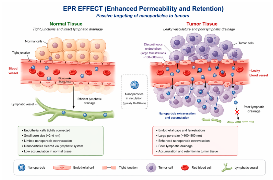
This selective accumulation depends on both nanoparticle properties and tumour microenvironment characteristics, including vascular permeability and interstitial pressure. Physicochemical parameters play a decisive role in determining biodistribution profiles. Particle size is particularly important: nanoparticles must be large enough to avoid renal filtration (with a cutoff of <5.5 nm), yet small enough (typically ~100 nm) to evade splenic filtration and enable tumour penetration [84]. Similarly, surface charge has a strong influence on distribution: highly charged nanoparticles (positive or negative) are rapidly cleared due to enhanced protein adsorption and macrophage uptake, whereas near-neutral particles exhibit prolonged circulation and reduced non-specific accumulation [84]. Surface composition and hydrophobicity also modulate protein binding and subsequent biodistribution behaviour.
Importantly, nanocarriers can enhance tissue selectivity and reduce off-target exposure. Encapsulation within nanoparticles protects drugs from rapid metabolism and renal clearance, enabling prolonged circulation and increasing the likelihood of accumulation in target tissues [85]. At the same time, the controlled modification of nanoparticle properties enables the reduction of drug distribution to non-target organs, thereby minimizing systemic toxicity and improving the therapeutic effect.
5.3. Controlled Release Kinetics
Nanocarrier-based drug delivery systems enable precise control over drug release kinetics. This is a critical factor that influences both pharmacokinetic behaviour and therapeutic efficacy. Unlike conventional formulations, which often result in rapid and uncontrolled drug release, nanocarriers provide sustained release profiles mediated by diffusion or degradation. These profiles can be tailored through material selection and structural design. One of the fundamental mechanisms governing the controlled release of drugs from nanocarrier systems is diffusion-driven release. In polymeric nanoparticles, drugs are usually either encapsulated within the polymer matrix or adsorbed onto it, and are then released via diffusion or desorption processes [58]. The rate of release depends on factors such as the composition of the polymer, the interactions between the drug and the polymer, and the structure of the nanoparticle. For instance, encapsulated drugs can gradually diffuse from the carrier into the surrounding environment, enabling prolonged exposure of the target site to the drug [58]. Another key mechanism is degradation-controlled release, which is particularly prevalent in biodegradable polymer systems, such as poly(lactic-co-glycolic acid) (PLGA). These materials undergo hydrolytic degradation into monomeric units under physiological conditions, enabling the sustained release of encapsulated drugs over time [57,86]. The rate of degradation — and thus the rate of drug release — can be modulated by adjusting the properties of the polymer, such as its molecular weight and copolymer ratio. This provides a high degree of control over the release kinetics [58,86].
Nanocarriers also enable sustained release through their structural and physicochemical design. For example, nanoemulsion-based systems deliver drugs over a prolonged period due to their small droplet size, large interfacial area and interaction with biological membranes, facilitating gradual transport of drugs across tissues. These systems can maintain drug levels over extended periods, reducing the need for frequent dosing and improving therapeutic consistency. Furthermore, nanoemulsions demonstrate high stability and resistance to coalescence, resulting in controlled and predictable release behaviour [87].
The release kinetics of nanocarriers can be further influenced by surface and structural modifications. For example, functionalization with polymers or surface coatings can modulate drug diffusion rates and interaction with biological environments. For instance, dendrimer- and polymer-modified systems have been demonstrated to reduce drug release rates and enhance stability, enabling more precise and prolonged therapeutic delivery [58]. Similarly, lipid-based and polymer-coated nanoparticles provide structural barriers that slow drug release and enhance retention within the carrier system.
Controlled release is also closely linked to improved drug stability and bioavailability. Nanocarriers protect encapsulated drugs from premature degradation and enable gradual release into the systemic circulation, thus maintaining drug concentrations within the therapeutic window. In the case of nanocrystal-based systems, the increased surface area enhances dissolution kinetics while enabling prolonged drug exposure due to improved interaction with biological membranes [88].
5.4. Pharmacodynamics Implication
Nanocarrier-based drug delivery systems can significantly impact pharmacodynamic (PD) outcomes by altering drug exposure at the target site, thereby enhancing therapeutic efficacy and reducing systemic toxicity. These effects arise from changes in pharmacokinetics, biodistribution and cellular interactions mediated by nanoparticles, which collectively determine the biological response to treatment [82,89].
One of the primary PD advantages of nanocarriers is improving therapeutic efficacy by enhancing drug accumulation in target tissues. Nanoparticles can be engineered to deliver drugs selectively to diseased sites, such as tumours, resulting in higher local concentrations of the drug compared to conventional formulations. For instance, the liposomal encapsulation of doxorubicin has been demonstrated to increase tumour accumulation and cytotoxicity, while simultaneously reducing cardiotoxic side effects and improving overall therapeutic outcomes [19]. This selective delivery enhances the action of the drug at the target site while minimizing damage to healthy tissues. Nanocarriers contribute to an improved therapeutic index by balancing efficacy and toxicity. Conventional drugs often exhibit dose-limiting toxicity due to non-specific distribution. However, nanoparticle formulations reduce off-target exposure and enable more controlled drug release. Consequently, higher effective doses can be administered without increasing systemic toxicity, thereby enhancing the safety profile of the treatment [82]. The relationship between nanoparticle-mediated targeting and reduced adverse effects is central to their pharmacodynamic benefit. Another important implication is their ability to interact directly with biological systems and modulate cellular processes. Unlike conventional drug carriers, nanocarriers are not necessarily inert; they can influence biological pathways by interacting with proteins, cell membranes and the immune system. Certain nanomaterials have been shown to reduce inflammation or release bioactive species, thereby contributing to therapeutic effects beyond simple drug delivery [82]. These additional mechanisms can enhance treatment outcomes, particularly in complex diseases such as cancer and chronic inflammatory conditions.
The physicochemical properties of nanoparticles play a critical role in determining these responses. Parameters such as size, surface charge and composition influence protein corona formation, cellular uptake and tissue targeting, thereby affecting drug efficacy. For example, nanoparticles that are optimized for prolonged circulation and reduced immune recognition can accumulate more effectively at target sites, leading to enhanced pharmacological effects [82,89]. Similarly, modifying nanoparticle properties has been associated with improved tissue penetration and therapeutic response. Importantly, the pharmacodynamic effects of nanoparticle systems cannot be fully predicted by traditional pharmacokinetic measurements alone. Plasma drug concentration does not necessarily reflect local drug activity, especially when nanoparticles enable targeted delivery and localized release. Therefore, to gain a comprehensive understanding of nanoparticle-mediated pharmacodynamics, it is necessary to integrate tissue distribution data, cellular interactions and local drug activity [82].
Nanocarrier-based drug delivery systems allow for the effective modulation of pharmacokinetics and pharmacodynamics. They enhance absorption by improving stability and permeability, and alter distribution through controlled biodistribution and targeted accumulation. Furthermore, nanocarriers provide sustained and controlled release profiles, ensuring stable drug exposure over time. These pharmacokinetic improvements directly translate into pharmacodynamic benefits, including increased drug efficacy, reduced systemic toxicity and an improved therapeutic index. Overall, nanocarriers offer an integrated approach to optimizing drug performance by linking physicochemical design with biological outcomes.
6. Challenges, Opportunities and Future Perspectives
Despite significant advances in nanocarrier-based drug delivery, major challenges remain related to safety, long-term toxicity, and translational predictability, highlighting the need for mechanism-driven design, standardized evaluation frameworks, and integration of computational approaches.
6.1. Safety and Toxicity Considerations
Despite significant progress in nanocarrier-based drug delivery, safety and toxicity remain key barriers to its clinical application. The physicochemical properties that provide therapeutic benefits, such as nanoscale size, a high surface area and adjustable surface chemistry, also pose potential biological risks. Due to their small size, nanoparticles can cross biological membranes efficiently and accumulate in tissues and organs, leading to increased systemic exposure and potential long-term toxicity[90].
The toxicity of nanoparticles is strongly dependent on their physicochemical characteristics, including size, surface charge, shape and composition. Smaller nanoparticles are more easily internalized by cells and are more reactive, which can significantly increase cytotoxic responses. The surface charge is particularly important as positively charged nanoparticles strongly interact with negatively charged cell membranes, thereby promoting membrane disruption and intracellular damage [91]. These interactions trigger key molecular mechanisms of toxicity, including the generation of reactive oxygen species (ROS), mitochondrial dysfunction, DNA damage, inflammatory signaling and apoptosis [91,92].
Figure 7.
Nanoparticle toxicity mechanisms [91].
Figure 7.
Nanoparticle toxicity mechanisms [91].
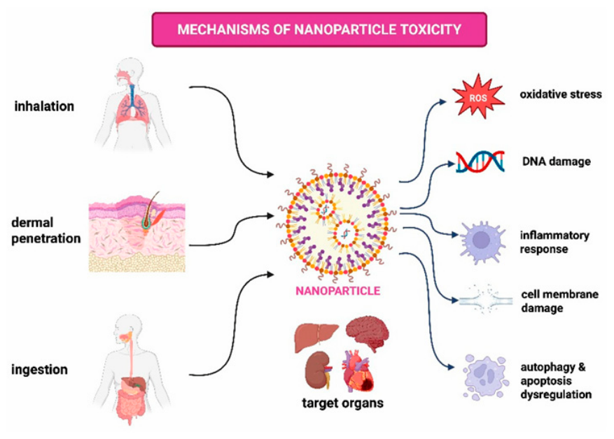
Quantitative evidence highlights the magnitude of these effects. Meta-analytical data have demonstrated significant nanoparticle-induced toxicity in metabolic organs, including increased hepatic oxidative stress (standardized mean difference, SMD = 1.42; 95% CI: 1.10–1.75), renal tubular apoptosis (SMD = 1.27; 95% CI: 0.94–1.61), and pancreatic β-cell dysfunction (SMD = 1.18; 95% CI: 0.88–1.49)[93]. These findings provide strong evidence for organ-specific toxicity and confirm that nanoparticle exposure can result in measurable biological damage.
In addition to metabolic toxicity, nanoparticle-based systems may induce application-specific adverse effects. For instance, nanoparticle-mediated therapies have been associated with cardiotoxicity, driven by oxidative stress, endothelial dysfunction, immune activation, and off-target accumulation in cardiac tissue [94]. Experimental studies further demonstrate that different nanoparticle types can elicit distinct immune responses and biodistribution patterns, with variations in T-cell activation and inflammatory markers depending on nanoparticle composition [95].
A critical translational challenge is the persistence and incomplete clearance of nanoparticles. Inefficient elimination may lead to prolonged retention in tissues, resulting in chronic inflammation and cumulative toxicity [93]. This issue is compounded by the lack of standardized toxicity assessment protocols and regulatory frameworks. Current models often fail to capture nanoparticle-specific biological interactions, while variability in synthesis and characterization leads to inconsistent and non-reproducible results [96]. These limitations significantly hinder the reliable translation of nanocarrier systems into clinical practice.
6.2. Opportunities and Future Perspectives
Despite the challenges involved, nanocarrier systems present significant opportunities for improving drug delivery and precision medicine. A key area of research is the development of targeted, multifunctional nanoparticles that can deliver drugs selectively. Functionalization strategies enable enhanced accumulation at disease sites through passive mechanisms such as the enhanced permeability and retention effect, as well as active targeting approaches. This reduces off-target toxicity while improving therapeutic efficacy [97]. Nanocarriers also enable precise modulation of intracellular signaling pathways. For example, nanoparticle-based systems improve the delivery of therapeutics targeting the PI3K/AKT/mTOR pathway, allowing regulation of autophagy and tumor progression while overcoming limitations of conventional therapies such as poor specificity and systemic toxicity[98]. These capabilities establish nanomedicine as a vital tool for overcoming drug resistance and enhancing treatment outcomes.
Future developments are increasingly focused on the rational design of nanocarriers to balance efficacy and safety. Strategies include using biodegradable and biocompatible materials, optimizing size and surface properties to reduce immune recognition and developing stimuli-responsive systems that enable controlled drug release in response to physiological triggers [96,97]. These approaches aim to mitigate toxicity while preserving therapeutic performance. Emerging technologies are expected to play a central role in advancing the field. Artificial intelligence and computational modelling provide powerful tools for predicting nanoparticle behaviour, optimizing design parameters and reducing experimental variability. Additionally, advanced biological models, such as organoids and organ-on-chip systems, offer more physiologically relevant platforms for evaluating toxicity and efficacy, thereby improving the predictive value of preclinical studies [94,96].
Overall, the future of nanocarrier-based drug delivery lies in the integration of physicochemical design, quantitative pharmacokinetic–pharmacodynamic modelling and advanced toxicity assessment. Overcoming current challenges relating to safety, reproducibility and regulatory standardization is essential for translating nanomedicine from experimental research into routine clinical practice.
7. Conclusions
Nanocarrier-based drug delivery systems are a well-established and mechanistically supported approach to overcoming key limitations in biopharmaceuticals, such as poor solubility, instability and limited permeability. Consistent evidence across lipid-, polymer-, inorganic-, and hybrid-based platforms shows that nanoscale formulation primarily improves drug bioavailability through enhanced dissolution, protection from degradation, modulation of pharmacokinetics, and facilitation of targeted delivery.
However, the effectiveness of nanocarriers is not universal, but depends heavily on their physicochemical properties, including size, surface charge, composition, and functionalization. These parameters directly determine absorption pathways, biodistribution, and release kinetics, demonstrating that rational design, rather than simply reducing the size of particles, is essential for achieving meaningful therapeutic improvement. The case studies presented in this review (involving curcumin, paclitaxel, amphotericin B, fenofibrate and doxorubicin, for example) demonstrate that nanocarrier systems can significantly increase systemic exposure, reduce formulation-related toxicity and improve the therapeutic index. However, these benefits are context- and formulation-specific.
Despite these advances, translation into clinical practice remains limited. Major barriers include safety concerns, long-term toxicity, variability in biological interactions (e.g. protein corona formation) and a lack of standardized evaluation protocols. In particular, discrepancies between preclinical models and human outcomes continue to hinder predictability and regulatory approval.
Future progress in this field requires a shift towards a mechanism-driven, quantitatively guided design approach that integrates physicochemical characterization with pharmacokinetic–pharmacodynamic modelling. Developing reproducible, scalable and biocompatible systems supported by advanced in vitro models and computational tools will be essential for improving translational success.
Overall, nanocarriers are a powerful, albeit not universally applicable, solution for enhancing drug bioavailability. Their clinical impact will depend on balancing efficacy with safety and on the ability to design systems that are tailored to specific drugs, biological barriers and therapeutic contexts.
Author Contributions
editing and writing the final draft of the article, M.G., investigation and writing, M.Sz and M.G., writing—review and editing, A.T. All authors have read and agreed to the published version of the manuscript.
Funding
The topic was funded by projects of the National Science Centre in Poland: 2022/47/O/ST8/02850 and 2023/51/B/ST8/02971.
Data Availability Statement
No new data were generated in this review article. All studies and data reported are available publicly from the references cited.
Conflicts of Interest
The authors declare no conflicts of interest.
References
- Ali, H. B.; Khan, M. B. Artificial Intelligence-driven Strategies and Modern Innovations to Surpass Biopharmaceutic Limitations in Traditional Drug Development. J. Explor Res. Pharmacol. 2025, vol. 10(no. 3). [Google Scholar] [CrossRef]
- John, J. Advancements in nano-based drug delivery systems for therapeutics: a comprehensive review. RSC Pharm. 2026, vol. 3(no. 1), 43–59. [Google Scholar] [CrossRef]
- Akomolafe, O. A.; Akinsiku, A. A. Advances in nanoparticles as drug delivery systems: A review. Sci. Afr. 2025, vol. 30, e03101. [Google Scholar] [CrossRef]
- Khodabandehloo, H.; Zahednasab, H.; Ashrafi Hafez, A. Nanocarriers Usage for Drug Delivery in Cancer Therapy. Iran. J. Cancer Prev. In Press. 2016. [Google Scholar] [CrossRef]
- Xu, L.; Wang, X.; Liu, Y.; Yang, G.; Falconer, R.J.; Zhao, C.-X. Lipid Nanoparticles for Drug Delivery. Adv. NanoBiomed Res. 2022, 2, 2100109. [Google Scholar] [CrossRef]
- Alfutaimani, A.S.; Alharbi, N.K.; S. Alahmari, A.; A. Alqabbani, A.; Aldayel, A.M. Exploring the Landscape of Lipid Nanoparticles (LNPs): A Comprehensive Review of LNPs Types and Biological Sources of Lipids. Int. J. Pharm. X 2024, 8, 100305. [Google Scholar] [CrossRef]
- Molla, A. Lipid Nanoparticles in Drug Delivery: Advances, Challenges, and Clinical Prospects. JDDMC 2025, 11, 48–54. [Google Scholar] [CrossRef]
- Karmaker, S.; Rosales, P.D.; Tirumuruhan, B.; Viravalli, A.; Boehnke, N. More than a Delivery System: The Evolving Role of Lipid-Based Nanoparticles. Nanoscale 2025, 17, 11864–11893. [Google Scholar] [CrossRef]
- Afzal, O.; Altamimi, A.S.A.; Nadeem, M.S.; Alzarea, S.I.; Almalki, W.H.; Tariq, A.; Mubeen, B.; Murtaza, B.N.; Iftikhar, S.; Riaz, N.; et al. Nanoparticles in Drug Delivery: From History to Therapeutic Applications. Nanomaterials 2022, 12, 4494. [Google Scholar] [CrossRef]
- Waheed, I.; Ali, A.; Tabassum, H.; Khatoon, N.; Lai, W.-F.; Zhou, X. Lipid-Based Nanoparticles as Drug Delivery Carriers for Cancer Therapy. Front. Oncol. 2024, 14, 1296091. [Google Scholar] [CrossRef]
- Feng, X.; Jia, P.; Zhang, D. Nanocarrier Drug Delivery System: Promising Platform for Targeted Depression Therapy. Front. Pharmacol. 2024, 15, 1435133. [Google Scholar] [CrossRef]
- Chamundeeswari, M.; Jeslin, J.; Verma, M.L. Nanocarriers for Drug Delivery Applications. Env. Chem. Lett. 2019, 17, 849–865. [Google Scholar] [CrossRef]
- Crintea, A.; Dutu, A.G.; Sovrea, A.; Constantin, A.-M.; Samasca, G.; Masalar, A.L.; Ifju, B.; Linga, E.; Neamti, L.; Tranca, R.A.; et al. Nanocarriers for Drug Delivery: An Overview with Emphasis on Vitamin D and K Transportation. Nanomaterials 2022, 12, 1376. [Google Scholar] [CrossRef]
- Daraee, H.; Etemadi, A.; Kouhi, M.; Alimirzalu, S.; Akbarzadeh, A. Application of Liposomes in Medicine and Drug Delivery. Artif. Cells Nanomed. Biotechnol. 2016, 44, 381–391. [Google Scholar] [CrossRef] [PubMed]
- Mazdaei, M.; Asare-Addo, K. A Mini-Review of Nanocarriers in Drug Delivery Systems. Br. J. Pharm. 2022, 7. [Google Scholar] [CrossRef]
- Madkhali, O.A. Perspectives and Prospective on Solid Lipid Nanoparticles as Drug Delivery Systems. Molecules 2022, 27, 1543. [Google Scholar] [CrossRef]
- Edis, Z.; Wang, J.; Waqas, M.K.; Ijaz, M.; Ijaz, M. Nanocarriers-Mediated Drug Delivery Systems for Anticancer Agents: An Overview and Perspectives. IJN 2021, Volume 16, 1313–1330. [Google Scholar] [CrossRef]
- Mitchell, M.J.; Billingsley, M.M.; Haley, R.M.; Wechsler, M.E.; Peppas, N.A.; Langer, R. Engineering Precision Nanoparticles for Drug Delivery. Nat. Rev. Drug Discov. 2021, 20, 101–124. [Google Scholar] [CrossRef]
- Yusuf, A.; Almotairy, A.R.Z.; Henidi, H.; Alshehri, O.Y.; Aldughaim, M.S. Nanoparticles as Drug Delivery Systems: A Review of the Implication of Nanoparticles’ Physicochemical Properties on Responses in Biological Systems. Polymers 2023, 15, 1596. [Google Scholar] [CrossRef]
- Eker, F.; Duman, H.; Akdaşçi, E.; Bolat, E.; Sarıtaş, S.; Karav, S.; Witkowska, A.M. A Comprehensive Review of Nanoparticles: From Classification to Application and Toxicity. Molecules 2024, 29, 3482. [Google Scholar] [CrossRef] [PubMed]
- Kumar, S.; Dilbaghi, N.; Saharan, R.; Bhanjana, G. Nanotechnology as Emerging Tool for Enhancing Solubility of Poorly Water-Soluble Drugs. BioNanoSci 2012, 2, 227–250. [Google Scholar] [CrossRef]
- Hsu, C.-Y.; Rheima, A.M.; Kadhim, M.M.; Ahmed, N.N.; Mohammed, S.H.; Abbas, F.H.; Abed, Z.T.; Mahdi, Z.M.; Abbas, Z.S.; Hachim, S.K.; et al. An Overview of Nanoparticles in Drug Delivery: Properties and Applications. South Afr. J. Chem. Eng. 2023, 46, 233–270. [Google Scholar] [CrossRef]
- Zahin, N.; Anwar, R.; Tewari, D.; Kabir, Md.T.; Sajid, A.; Mathew, B.; Uddin, Md.S.; Aleya, L.; Abdel-Daim, M.M. Nanoparticles and Its Biomedical Applications in Health and Diseases: Special Focus on Drug Delivery. Env. Sci. Pollut. Res. 2020, 27, 19151–19168. [Google Scholar] [CrossRef]
- Alami, O.; Laurent, R.; El Kazzouli, S.; El Brahmi, N.; Caminade, A.-M. Dendrimers and Graphene Oxide: An Overview on Combination, Properties and Applications. Coord. Chem. Rev. 2026, 558, 217781. [Google Scholar] [CrossRef]
- Chandrakala, V.; Aruna, V.; Angajala, G. Review on Metal Nanoparticles as Nanocarriers: Current Challenges and Perspectives in Drug Delivery Systems. Emergent mater. 2022, 5, 1593–1615. [Google Scholar] [CrossRef] [PubMed]
- Unnikrishnan, G.; Joy, A.; Megha, M.; Kolanthai, E.; Senthilkumar, M. Exploration of Inorganic Nanoparticles for Revolutionary Drug Delivery Applications: A Critical Review. Discov. Nano 2023, 18, 157. [Google Scholar] [CrossRef] [PubMed]
- Al-Thani, A.N.; Jan, A.G.; Abbas, M.; Geetha, M.; Sadasivuni, K.K. Nanoparticles in Cancer Theragnostic and Drug Delivery: A Comprehensive Review. Life Sci. 2024, 352, 122899. [Google Scholar] [CrossRef]
- Kianfar, E. Magnetic Nanoparticles in Targeted Drug Delivery: a Review. J. Supercond. Nov. Magn. 2021, vol. 34(no. 7), 1709–1735. [Google Scholar] [CrossRef]
- Debnath, S.K.; Srivastava, R. Drug Delivery With Carbon-Based Nanomaterials as Versatile Nanocarriers: Progress and Prospects. Front Nanotechnol. 2021. [Google Scholar] [CrossRef]
- Zhang, W.; Zhang, Z.; Zhang, Y. The application of carbon nanotubes in target drug delivery systems for cancer therapies. Nanoscale Res. Lett. 2011, vol. 6(no. 1), 555. [Google Scholar] [CrossRef]
- Komane, P.; Kumar, P.; Choonara, Y. Functionalised Carbon Nanotubes: Promising Drug Delivery Vehicles for Neurovascular Disorder Intervention. AAPS PharmSciTech 2023, vol. 24(no. 7), 201. [Google Scholar] [CrossRef] [PubMed]
- Dubey, R.; Dutta, D.; Sarkar, A.; Chattopadhyay, P. Functionalized carbon nanotubes: synthesis, properties and applications in water purification, drug delivery, and material and biomedical sciences. Nanoscale Adv. 2021, vol. 3(no. 20), 5722–5744. [Google Scholar] [CrossRef]
- Rahamathulla, M. Carbon Nanotubes: Current Perspectives on Diverse Applications in Targeted Drug Delivery and Therapies. Materials 2021, vol. 14(no. 21), 6707. [Google Scholar] [CrossRef]
- Wong, B.S. Carbon nanotubes for delivery of small molecule drugs. Adv. Drug Deliv. Rev. 2013, vol. 65(no. 15), 1964–2015. [Google Scholar] [CrossRef]
- Naief, M.F.; Mohammed, S.N.; Mohammed, A.M. Carbon nanotubes: A review on synthesis and drug delivery for cancer treatment. Inorg. Chem. Commun. 2024, vol. 159, 111694. [Google Scholar] [CrossRef]
- Parveen, S.; Gupta, P.; Kumar, S.; Banerjee, M. Lipid Polymer Hybrid Nanoparticles as Potent Vehicles for Drug Delivery in Cancer Therapeutics. Med. Drug Discov. 2023, 20, 100165. [Google Scholar] [CrossRef]
- Chung, Y.H.; Cai, H.; Steinmetz, N.F. Viral nanoparticles for drug delivery, imaging, immunotherapy, and theranostic applications. Adv. Drug Deliv. Rev. 2020, vol. 156, 214–235. [Google Scholar] [CrossRef]
- Steinmetz, N. F. Viral Nanoparticles in Drug Delivery and Imaging. Mol. Pharm. 2013, vol. 10(no. 1), 1–2. [Google Scholar] [CrossRef] [PubMed]
- Kotterman, M. A.; Chalberg, T. W.; Schaffer, D. V. Viral Vectors for Gene Therapy: Translational and Clinical Outlook. Annu. Rev. Biomed. Eng. 2015, vol. 17(no. 1), 63–89. [Google Scholar] [CrossRef]
- Wang, J.H.; Gessler, D.J.; Zhan, W.; Gallagher, T.L.; Gao, G. Adeno-associated virus as a delivery vector for gene therapy of human diseases. Sig Transduct. Target Ther. 2024, vol. 9(no. 1), 78. [Google Scholar] [CrossRef]
- Singh, H. Carbon dots in drug delivery and therapeutic applications. Adv. Drug Deliv. Rev. 2025, vol. 224, 115644. [Google Scholar] [CrossRef]
- Lim, S.Y.; Shen, W.; Gao, Z. Carbon quantum dots and their applications. Chem. Soc. Rev. 2015, vol. 44(no. 1), 362–381. [Google Scholar] [CrossRef]
- Iannazzo, D.; Celesti, C.; Cardo, L.; Prato, M.; Bitto, A. Carbon dots for drug delivery: Insights into their potential in nanopharmacology’. Pharmacol. Rev. 2026, vol. 78(no. 2), 100107. [Google Scholar] [CrossRef]
- Paul, A. Drug Absorption and Bioavailability. In Introduction to Basics of Pharmacology and Toxicology; Raj, G.M., Raveendran, R., Eds.; Springer Singapore: Singapore, 2019; pp. 81–88. ISBN 978-981-329-778-4. [Google Scholar]
- Stielow, M.; Witczyńska, A.; Kubryń, N.; Fijałkowski, Ł.; Nowaczyk, J.; Nowaczyk, A. The Bioavailability of Drugs—The Current State of Knowledge. Molecules 2023, 28, 8038. [Google Scholar] [CrossRef]
- Zhuo, Y.; Zhao, Y.-G.; Zhang, Y. Enhancing Drug Solubility, Bioavailability, and Targeted Therapeutic Applications through Magnetic Nanoparticles. Molecules 2024, 29, 4854. [Google Scholar] [CrossRef] [PubMed]
- Liu, Y.; Liang, Y.; Yuhong, J.; Xin, P.; Han, J.L.; Du, Y.; Yu, X.; Zhu, R.; Zhang, M.; Chen, W.; et al. Advances in Nanotechnology for Enhancing the Solubility and Bioavailability of Poorly Soluble Drugs. DDDT 2024, Volume 18, 1469–1495. [Google Scholar] [CrossRef] [PubMed]
- Beach, M.A.; Nayanathara, U.; Gao, Y.; Zhang, C.; Xiong, Y.; Wang, Y.; Such, G.K. Polymeric Nanoparticles for Drug Delivery. Chem. Rev. 2024, 124, 5505–5616. [Google Scholar] [CrossRef]
- Scioli Montoto, S.; Muraca, G.; Ruiz, M.E. Solid Lipid Nanoparticles for Drug Delivery: Pharmacological and Biopharmaceutical Aspects. Front. Mol. Biosci. 2020, 7, 587997. [Google Scholar] [CrossRef]
- Abdi Syahputra, R.; Dalimunthe, A.; Utari, Z.D.; Halim, P.; Sukarno, M.A.; Zainalabidin, S.; Salim, E.; Gunawan, M.; Nurkolis, F.; Park, M.N.; et al. Nanotechnology and Flavonoids: Current Research and Future Perspectives on Cardiovascular Health. J. Funct. Foods 2024, 120, 106355. [Google Scholar] [CrossRef]
- Krasteva, N.; Georgieva, M. Promising Therapeutic Strategies for Colorectal Cancer Treatment Based on Nanomaterials. Pharmaceutics 2022, 14, 1213. [Google Scholar] [CrossRef] [PubMed]
- Kasina, V.; Mownn, R.J.; Bahal, R.; Sartor, G.C. Nanoparticle Delivery Systems for Substance Use Disorder. Neuropsychopharmacol. 2022, 47, 1431–1439. [Google Scholar] [CrossRef] [PubMed]
- Jacob, S.; Varkey, N.R.; Boddu, S.H.S.; Gorain, B.; Rao, R.; Nair, A.B. Advances in Lipid-Polymer Hybrid Nanoparticles: Design Strategies, Functionalization, Oncological and Non-Oncological Clinical Prospects. Pharmaceuticals 2025, 18, 1772. [Google Scholar] [CrossRef]
- Bernardo, R.R.; De Oliveira, L.A.S.; Paz, G.S.; Fernandes, J. The Combination of Cutting-Edge Strategies in Nano-Delivery Systems to Overcome Drawbacks for Malignant Tumor Treatment. J. Explor Res. Pharmacol. 2025, 000, 000–000. [Google Scholar] [CrossRef]
- Wang, Y.; Pi, C.; Feng, X.; Hou, Y.; Zhao, L.; Wei, Y. The Influence of Nanoparticle Properties on Oral Bioavailability of Drugs. IJN 2020, Volume 15, 6295–6310. [Google Scholar] [CrossRef]
- Eltaib, L. Polymeric Nanoparticles in Targeted Drug Delivery: Unveiling the Impact of Polymer Characterization and Fabrication. Polymers 2025, 17, 833. [Google Scholar] [CrossRef]
- Gangavarapu, A.; Tapia-Lopez, L.V.; Sarkar, B.; Pena-Zacarias, J.; Badruddoza, A.Z.M.; Nurunnabi, M. Lipid Nanoparticles for Enhancing Oral Bioavailability. Nanoscale 2024, 16, 18319–18338. [Google Scholar] [CrossRef]
- Chenthamara, D.; Subramaniam, S.; Ramakrishnan, S.G.; Krishnaswamy, S.; Essa, M.M.; Lin, F.-H.; Qoronfleh, M.W. Therapeutic Efficacy of Nanoparticles and Routes of Administration. Biomater. Res. 2019, 23, 20. [Google Scholar] [CrossRef]
- Grundmann, D.D.R.; Lourenço, C.; Sanches, M.P.; Saatkamp, R.H.; Parize, A.L.; Delfrate, G.; Albino, L.B.; Fernandes, D.; Sonvico, F.; Stulzer, H.K. Improving Oral Bioavailability: Lipid-Polymeric Nanoparticles of Simvastatin and Coenzyme Q10. J. Mol. Liq. 2026, 453, 129525. [Google Scholar] [CrossRef]
- Marcano, R.G.D.J.V.; Campos, L.D.A.; Khalil, N.M.; Auler, M.E.; Machado, C.S.; Mainardes, R.M. Gliadin/Pectin Hybrid Nanoparticles for Improved Oral Bioavailability of Amphotericin B for the Treatment of Fungal Infections. Int. J. Biol. Macromol. 2026, 335, 149384. [Google Scholar] [CrossRef] [PubMed]
- Fanse, S.; Maurus, K.; Bhorkade, S.; Burgess, D.J. Loteprednol Etabonate Nanostructured Lipid Carriers for Enhanced Ocular Bioavailability. Colloids Surf. B Biointerfaces 2026, 264, 115661. [Google Scholar] [CrossRef]
- Singh, A.; Guo, Y.; Cruz, M.; Shi, D.; Kitts, D.D.; Pratap-Singh, A. Chitosan/PEG Nanoparticle-Embedded Oleogels Improve Blueberry Derived Resveratrol Bioavailability and Antioxidant Capacity. Food Chem. 2025, 490, 145038. [Google Scholar] [CrossRef]
- Rui, S.; Qi, W.; Liu, B.; Deng, Q.; Li, S.; Chen, J.; Feng, S.; Liu, Z.; Wang, S.; Zhao, Q. Comparison of Dendritic Mesoporous Silica Nanoparticles with Commercial Mesoporous Excipients in Improving Physical Stability and Oral Bioavailability of Insoluble Drugs. Colloids Surf. A Physicochem. Eng. Asp. 2025, 725, 137702. [Google Scholar] [CrossRef]
- Sahar, N.U.; Gul, M.; Choi, H.-I.; Ryu, J.-S.; Noh, H.-Y.; Vo, D.-K.; Nguyen, T.-H.; Ansari, M.M.; Kim, W.; Maeng, H.-J.; et al. Lipid Core-Chitosan Shell Hybrid Nanoparticles for Enhanced Oral Bioavailability of Sorafenib. Int. J. Biol. Macromol. 2025, 299, 140030. [Google Scholar] [CrossRef]
- Wei, W.; Lu, M.; Xu, W.; Polyakov, N.E.; Dushkin, A.V.; Su, W. Preparation of Protamine-Hyaluronic Acid Coated Core-Shell Nanoparticles for Enhanced Solubility, Permeability, and Oral Bioavailability of Decoquinate. Int. J. Biol. Macromol. 2022, 218, 346–355. [Google Scholar] [CrossRef] [PubMed]
- Windell, D.L.; Mourabit, S.; Moger, J.; Owen, S.F.; Winter, M.J.; Tyler, C.R. The Influence of Size and Surface Chemistry on the Bioavailability, Tissue Distribution and Toxicity of Gold Nanoparticles in Zebrafish (Danio Rerio). Ecotoxicol. Environ. Saf. 2023, 260, 115019. [Google Scholar] [CrossRef]
- Liu, M.; Peng, S.; McClements, D.J.; Chen, L.; Lin, S.; Wang, W. Enhancing Stability of Curcumin-Loaded Casein Nanoparticles by Adding Liposomal Nanoparticles. LWT 2023, 189, 115405. [Google Scholar] [CrossRef]
- Lu, Y.; Wang, J.T.-W.; Li, N.; Zhu, X.; Li, Y.; Bansal, S.; Wang, Y.; Al-Jamal, K.T. Intranasal Administration of Edaravone Nanoparticles Improves Its Stability and Brain Bioavailability. J. Control. Release 2023, 359, 257–267. [Google Scholar] [CrossRef]
- Machtakova, M.; Thérien-Aubin, H.; Landfester, K. Polymer Nano-Systems for the Encapsulation and Delivery of Active Biomacromolecular Therapeutic Agents. Chem. Soc. Rev. 2022, 51, 128–152. [Google Scholar] [CrossRef]
- Fan, L.; Huang, J.; Ma, S. Recent Advances in Delivery of Transdermal Nutrients: A Review. Exp. Dermatol. 2024, 33, e14966. [Google Scholar] [CrossRef]
- Lao, C.D. Dose escalation of a curcuminoid formulation. BMC Complement. Altern. Med. 2006, t. 6, nr 1, s. 10. [Google Scholar] [CrossRef]
- Kanai, M. Dose-escalation and pharmacokinetic study of nanoparticle curcumin, a potential anticancer agent with improved bioavailability, in healthy human volunteers. Cancer Chemother. Pharmacol. 2012, t. 69(nr 1), s. 65–70. [Google Scholar] [CrossRef]
- Schiborr, C.; Kocher, A.; Behnam, D.; Jandasek, J.; Toelstede, S.; Frank, J. The oral bioavailability of curcumin from micronized powder and liquid micelles is significantly increased in healthy humans and differs between sexes. Mol. Nutr. Food Res. 2014, t. 58(nr 3), s. 516–527. [Google Scholar] [CrossRef]
- Desai, N. Nanoparticle Albumin-Bound Paclitaxel (Abraxane®)’. In Albumin in Medicine; Otagiri, M., Chuang, V.T.G., Eds.; Springer Singapore: Singapore, 2016; pp. 101–119. [Google Scholar] [CrossRef]
- Gradishar, W. J. Phase III Trial of Nanoparticle Albumin-Bound Paclitaxel Compared With Polyethylated Castor Oil–Based Paclitaxel in Women With Breast Cancer. J. Clin. Oncol. 2005, vol. 23(no. 31), 7794–7803. [Google Scholar] [CrossRef]
- Barratt, G.; Bretagne, S. Optimizing efficacy of Amphotericin B through nanomodification. Int. J. Nanomed. 2007, vol. 2(no. 3), 301–313. [Google Scholar] [PubMed] [PubMed Central]
- Adler-Moore, J.; Proffitt, R.T. AmBisome: liposomal formulation, structure, mechanism of action and pre-clinical experience. J. Antimicrob. Chemother. 2002, vol. 49, no. suppl_1, 21–30. [Google Scholar] [CrossRef]
- Müller, R.H.; Junghanns, J.-U.A.H. ‘Nanocrystal technology, drug delivery and clinical application’. Int. J. Nanomed. 2008, 295. [Google Scholar] [CrossRef] [PubMed]
- Barenholz, Y. Doxil® — The first FDA-approved nano-drug: Lessons learned. J. Control. Release 2012, vol. 160(no. 2), 117–134. [Google Scholar] [CrossRef] [PubMed]
- Kim, K. S.; Na, K.; Bae, Y. H. Nanoparticle oral absorption and its clinical translational potential. J. Control. Release 2023, vol. 360, 149–162. [Google Scholar] [CrossRef] [PubMed]
- Kim, K. S.; Suzuki, K.; Cho, H.; Bae, Y. H. Selected Factors Affecting Oral Bioavailability of Nanoparticles Surface-Conjugated with Glycocholic Acid via Intestinal Lymphatic Pathway. Mol. Pharm. 2020, vol. 17(no. 11), 4346–4353. [Google Scholar] [CrossRef] [PubMed]
- Peralta-Cuevas, E.; Degollado-Hernández, N. Y.; Martínez-Ortiz, I. C.; Gutierrez-Onofre, A. J.; Garcia-Atutxa, I.; Villanueva-Flores, F. How do nanoparticle properties shape pharmacokinetics and pharmacodynamics? A mechanistic review. Front. Pharmacol. 2026, vol. 16, 1704814. [Google Scholar] [CrossRef]
- Kumar, M.; Kulkarni, P.; Liu, S.; Chemuturi, N.; Shah, D. K. Nanoparticle biodistribution coefficients: A quantitative approach for understanding the tissue distribution of nanoparticles. Adv. Drug Deliv. Rev. 2023, vol. 194, 114708. [Google Scholar] [CrossRef] [PubMed]
- Ernsting, M. J.; Murakami, M.; Roy, A.; Li, S.-D. Factors controlling the pharmacokinetics, biodistribution and intra-tumoral penetration of nanoparticles. J. Control. Release 2013, vol. 172(no. 3), 782–794. [Google Scholar] [CrossRef]
- Li, S.-D.; Huang, L. Pharmacokinetics and Biodistribution of Nanoparticles. Mol. Pharm. 2008, vol. 5(no. 4), 496–504. [Google Scholar] [CrossRef] [PubMed]
- Hamidi, M.; Azadi, A.; Rafiei, P. Pharmacokinetic Consequences of Pegylation. Drug Deliv. 2006, vol. 13(no. 6), 399–409. [Google Scholar] [CrossRef] [PubMed]
- Rai, V. K.; Mishra, N.; Yadav, K. S.; Yadav, N. P. Nanoemulsion as pharmaceutical carrier for dermal and transdermal drug delivery: Formulation development, stability issues, basic considerations and applications. J. Control. Release 2018, vol. 270, 203–225. [Google Scholar] [CrossRef]
- Majekodunmi, S. O. Enhancement of the solubility, bioavailability and absorption of poorly soluble drug candidates through nanotechnology—A review. West Afr. J. Pharm. 2016, vol. 27(no. 2), 78–88. [Google Scholar]
- Haripriyaa, M.; Suthindhiran, K. Pharmacokinetics of nanoparticles: current knowledge, future directions and its implications in drug delivery. Future J. Pharm. Sci. 2023, vol. 9(no. 1), 113. [Google Scholar] [CrossRef]
- Qamar, W. An insight into impact of nanomaterials toxicity on human health. PeerJ 2024, vol. 12, e17807. [Google Scholar] [CrossRef]
- Muhaimin, M.; Chaerunisaa, A. Y.; Dewi, M. K.; Khatib, A.; Hazrina, A. The Toxicological Profile of Active Pharmaceutical Ingredients–Containing Nanoparticles: Classification, Mechanistic Pathways, and Health Implications. Pharmaceuticals 2025, vol. 18(no. 5), 703. [Google Scholar] [CrossRef]
- Xuan, L.; Ju, Z.; Skonieczna, M.; Zhou, P.; Huang, R. Nanoparticles-induced potential toxicity on human health: Applications, toxicity mechanisms, and evaluation models. MedComm 2023, vol. 4(no. 4), e327. [Google Scholar] [CrossRef]
- Murali, S.; Thomas, J.; Abraham, J.; Kanna, V.; Ramachandiran, S.; Ss, S. Nanodiagnostics and nanotoxicology: A systematic review and meta-analysis on nanoparticle applications and safety in metabolic organs. OpenNano 2026, vol. 27, 100266. [Google Scholar] [CrossRef]
- Oisakede, E. O.; Oyedeji, O. O.; Olawuyi, O. F.; Alabi, J. O.; Daniel, R. I. A.; Olawade, D. B. Nanoparticle-mediated cardiotoxicity and nanomedicine interventions in cancer treatment. Nano TransMed 2026, vol. 5, 100113. [Google Scholar] [CrossRef]
- Alexander, E.; Leong, K. W. Toxicity and biodistribution comparison of functionalized nanodiamonds, quantum dot nanocarbons and gold nanoparticles. Front. Nanotechnol. 2025, vol. 7, 1512622. [Google Scholar] [CrossRef]
- Havelikar, U. Comprehensive insights into mechanism of nanotoxicity, assessment methods and regulatory challenges of nanomedicines. Discov. Nano 2024, vol. 19(no. 1), 165. [Google Scholar] [CrossRef]
- Islam, S.; Ahmed, M. M. S.; Islam, M. A.; Hossain, N.; Chowdhury, M. A. Advances in nanoparticles in targeted drug delivery–A review, Results Surf. Interfaces 2025, vol. 19, 100529. [Google Scholar] [CrossRef]
- Rahman, M. A.; Jalouli, M.; Bhajan, S. K.; Al-Zharani, M.; Harrath, A. H. A Comprehensive Review of Nanoparticle-Based Drug Delivery for Modulating PI3K/AKT/mTOR-Mediated Autophagy in Cancer. Int. J. Mol. Sci. 2025, vol. 26(no. 5), 1868. [Google Scholar] [CrossRef]
Table 1.
Comparative overview of nanoparticle systems, effects on bioavailability advantages, and limitations.
Table 1.
Comparative overview of nanoparticle systems, effects on bioavailability advantages, and limitations.
| Type of nanoparticle |
Factors contributing to enhanced bioavailability |
Other positive factors | Disadvantages |
|---|---|---|---|
| Liposomes | Ability to encapsulate both hydrophobic and hydrophilic drugs [19,20,27]; Controlled and precise targeting to specific tissues/cells [14,20,27]; Protection from enzymatic degradation [14,19]. Ease of functionalization [19,20]. |
Biocompatibility [19,20,27], biodegradability [19,20], low toxicity [27], ability to carry large molecules [14] |
High production cost, short half-life [14], low stability, relatively low encapsulation efficiency [26,50] (depending on formulation), high cost [50,51] |
| Lipid-based (SLNs, NLCs) | Ability to encapsulate both hydrophobic and hydrophilic drugs [20,52]; Enhanced stability [20,50] and bioavailability [19]; Prolonged blood circulation (ligand-based targeting) [19,52]. Surface modifications [19,20] |
Biocompatibility, biodegradability, easy functionalization, controlled release, stimuli-responsive behavior [20], loading capacity [19], non-toxic [26], safer, low cost [50] |
Potential cytotoxicity [52], low drug incorporation efficiency, risk of gelation [50] |
| Polymeric (nanospheres, nanocapsules) |
Surface functionalization (e.g. targeting ligands) [22,23,52]; Carrier for hydrophobic and hydrophilic drugs [19,51,52]; Controlled and sustained release of drug [19,51]; Protection from degradation [19,51] |
Biodegradability, biocompatibility [19,52], ease of modification, high stability [51], intracellular release [53], water solubility [54] |
Self-aggregation [52], unpredictable drug release in bloodstream [50], scalability issues [53], risk of degradation [53], toxic monomer accumulation [18] |
| Dendrimers | Targeted delivery (passive and active) [54]; Surface functionalization [51]; Carrier for hydrophobic, hydrophilic drugs [54]; High water solubility [54] |
Well defined structure (size and shape), can be produce for specific application, tunable architecture [51], high loading capacity [53] |
High production cost [51], complex synthesis, limited clinical use [53] |
| Polymeric micelles |
Increased solubility of hydrophobic drugs (core-shell structure) [53]; Target delivery (ligand-based) [51]; Prolonged circulation time [51]. |
Non-toxic [26], ease of synthesis, reduce side effects [51] |
Potential increase in systemic toxicity [51], low drug loading, instability in circulation [53] |
| Gold (AuNPs) | Surface functionalization improving targeting and accumulation [51]; High surface/volume ratio [51,54]. |
Small size [52], optical properties [19], ease of functionalization [26], multiple forms [20] |
Limited biocompatibility [26] (depending on size and surface chemistry) |
| Iron (Fe2O3; Fe3O4) | Target delivery through surface ligands or external magnetic field [19,51]; Easily functionalized [26]; Large surface area [51,54]. |
Improved magnetic properties, multiple forms [20], high stability [26], small size [52] |
Cellular toxicity, low biodegradability [26] |
Table 2.
Key physical characteristics of nanoparticles (based on data available in [58]).
Table 2.
Key physical characteristics of nanoparticles (based on data available in [58]).
| Nanomaterials | Composition | Size [nm] | Shape | Possible surface modifications |
|
|---|---|---|---|---|---|
| Polymeric | Biodegradable polymers (e.g. PLGA) |
20-50 | Spherical; Nanocapsules (core-shell); Nanospheres (solid). |
PEGylation, targeting ligands, functional groups, hydrogel coatings, stimuli-responsive modifications (pH, temperature) |
|
| Dendrimer | Branched polymers |
1-10 | Spherical (high symmetry); Branched, tree-like structures. |
||
| Organic, lipid-based |
Solid or liquid lipids |
50-1000 | Spherical (core-shell) Irregular; |
PEGylation, targeting ligands, hydrophobic or hydrophilic modifications, functional groups | |
| Liposomes | Phospholipid bilayer |
50-1000 | Spherical (core-shell); Uni/multilamellar. |
||
| Inorganic, metallic |
Metals (e.g. gold, silver) |
20 | Divided by dimensions, e.g. nanostars, nanorods | PEGylation, silica coatings, ligands, stabilizing polymers |
|
Table 3.
Overview of nanoparticle types, composition, and applications.
| Nanoparticle type | Main components | Active substances | Bioavailability enhancement | Route of administration/ Application | Ref. |
|---|---|---|---|---|---|
| Polymeric - lipid nanoparticles | Lecithin; chitosan |
Simvastatin; Coenzyme Q10 |
Solubility | Oral; Treating hypercholesterolemia |
[59] |
| Polymer based | Gliadin; pectin | Amphotericin B | Controlled release, improved oral bioavailability | Oral; Fungal infections |
[60] |
| Nanostructured lipid carriers | Gelucire 44/14; Miglyol 829 |
Loteprednol | Targeting, drug loading, encapsulation efficiency | Ocular; Ocular inflammation |
[61] |
| Polymer based | Chitosan; PEG |
Resveratrol | Stability, solubility, antioxidant capacity | Antioxidant activity in food and supplements | [62] |
| Inorganic nanoparticles | Dendritic mesoporous silica | Carvedilol; Indomethacin; Magnolia officinalis extract |
Stability, solubility, drug capacity, specific surface | Oral; Therapeutic applications |
[63] |
| Polymeric-lipid nanoparticles (+ SLNs) |
Lipid core (stearyl alcohol); chitosan shell |
Sorafenib | Solubility in water, sustained drug release, better bioavailability | Oral; Antitumor activity |
[64] |
| Polymer based nanoparticles (micellar) | Disodium glycyrrhizinate; protamine; hyaluronic acid |
Decoquinate | Solubility, targeted biodistribution, drug release, dissolution | Oral; Anti-parasitic DDS |
[65] |
| Inorganic, gold nanoparticles | Gold (core); PEG, TNFα, NHS/PAMAM (coatings) |
- | Tissue distribution studies, toxicity, accumulation, clearance, size and surface chemistry | Nanoparticle model in research | [66] |
| Protein-lipid nanoparticles | Casein; Lecithin (liposomes) |
Curcumin | Thermal and storage stability, solubility in water, protection from pH | Nutraceutical in food, supplement, pharmaceuticals | [67] |
| Polymeric nanoparticles | PLGA | Edaravone | Drug stability, half-life, brain bioavailability, reduction of systemic exposure, targeted drug delivery | Nasal; treatment of amyotrophic lateral sclerosis (ALS) |
[68] |
Table 4.
Nanocarrier systems, their administration routes and applications.
| Type of nanoparticle | Applications |
|---|---|
| Liposomes | DDS [14,20,23,27], cancer therapy [14,23], diagnostic imaging [27], gene therapy [20,27], cosmetics, food preservation, wound healing [20] |
| Lipid-based (SLNs, NLCs) |
DDS, cosmetics [19,20,23], food preservation, anticancer agents [16,20], genetic therapy [18], delivery of antibiotic [16], wound healing, tissue engineering [20] |
| Polymeric (nanospheres, nanocapsules) |
DDS [18,19,21], cancer therapy, diagnostics [18], imaging, gene therapy [19], wound healing, tissue engineering, food preservation, food packaging [20] |
| Dendrimers | DDS, gene therapy, imaging diagnostics [19,23,51] |
| Inorganic, metal nanocarriers | DDS [14,16,23], imaging, diagnostics [18,19], photothermal therapy, gene delivery, cancer therapy [27], biosensors [19], tissue engineering, cosmetics, food packaging, bioimaging [20] |
Disclaimer/Publisher’s Note: The statements, opinions and data contained in all publications are solely those of the individual author(s) and contributor(s) and not of MDPI and/or the editor(s). MDPI and/or the editor(s) disclaim responsibility for any injury to people or property resulting from any ideas, methods, instructions or products referred to in the content. |
© 2026 by the authors. Licensee MDPI, Basel, Switzerland. This article is an open access article distributed under the terms and conditions of the Creative Commons Attribution (CC BY) license (http://creativecommons.org/licenses/by/4.0/).
Copyright: This open access article is published under a Creative Commons CC BY 4.0 license, which permit the free download, distribution, and reuse, provided that the author and preprint are cited in any reuse.


