

Submitted:
03 May 2026
Posted:
05 May 2026
You are already at the latest version
Abstract
Amphotericin B (AmB) is a broad‑spectrum antifungal agent and a long‑standing standard of care; however, its clinical use is compromised by poor solubility, off‑target tissue distribution and severe dose‑limiting toxicity. AmB‑loaded poly (D, L‑lactide‑co‑glycolide) nanoparticles (PLGA‑AmB‑NPs) were developed and characterised with respect to their physicochemical properties, antifungal activity against A. fumigatus biofilms, in vivo efficacy in the Galleria mellonella infection model, and hemolytic toxicity in vitro. Blank PLGA nanoparticles (PLGA‑NPs) and PLGA‑AmB‑NPs exhibited mean diameters of 165 nm (PDI 0.075) and 120 nm (PDI 0.210), respectively, with negative zeta‑potential values consistent with colloidal stability in aqueous media. PLGA‑AmB‑NPs showed significantly enhanced activity against 24 h A. fumigatus biofilms compared with free AmB at concentrations of 5 and 10 µg/mL, while unloaded PLGA‑NPs were inactive. Infected G. mellonella larvae treated with PLGA‑AmB‑NPs displayed markedly improved survival over a 5‑day period relative to those receiving equivalent doses of free AmB. Furthermore, PLGA‑AmB‑NPs induced substantially lower hemolysis of human red blood cells than free AmB across all tested concentrations (5–20 µg/mL). PLGA‑AmB‑NPs represent a promising polymeric platform for the treatment of A. fumigatus infections.
Keywords:
Aspergillus fumigatus
; Galleria mellonella
; PLGA-NPs
; amphotericin B
; biofilm
1. Introduction
Fungal infections today represent one of the most alarming yet least addressed issues in global healthcare. The therapeutic arsenal is limited and outdated: there are fewer antifungal agents than antibiotics targeting other organisms, and the situation is further complicated by the fact that some of these drugs are toxic and have several drawbacks. Furthermore, resistance to some agents has already emerged. Thus, the development of non-toxic formulations of antifungal agents is highly relevant.
Amphotericin B (AmB) is an antifungal agent used for the treatment of mycoses caused by a wide range of pathogenic fungi, including Candida spp., Aspergillus spp., and Mucorales spp. [1]. AmB has remained a cornerstone of antifungal therapy for over six decades and is often described as the “gold standard” against a broad spectrum of life-threatening fungal infections [2]. However, in addition to acting on ergosterol in fungal cells, AmB also binds, albeit to a lesser extent, to cholesterol in kidney cell membranes, causing nephrotoxicity characterized by vasoconstriction and tubular damage [2,3,4]. In addition, conventional AmB is poorly soluble at physiological pH [1], induces the release of pro-inflammatory cytokines [3], distributes to non-target tissues [5], is degraded in the gastrointestinal tract and is not absorbed upon oral administration due to its hydrophobicity [6], exhibits limited penetration across the blood–brain barrier [7], and shows poor penetration into fungal biofilms [8].
Among AmB-susceptible pathogens, A. fumigatus is a leading cause of invasive aspergillosis in immunocompromised patients and is associated with high mortality rates [9]. In addition, A. fumigatus readily forms biofilms whose extracellular matrix limits antifungal penetration and confers increased tolerance to multiple drug classes, including polyenes [10]. Encapsulation in nanoparticles or lipid carriers helps to overcome the poor solubility of AmB and its limited delivery to “hard-to-reach” sites, reduces toxicity through more targeted distribution, and preserves drug integrity during transport to the site of action.
Currently, AmB is approved for use in several intravenous formulations [11], with its clinical application limited due to toxicity. Thus, modification of this promising antifungal agent is required to overcome these limitations. The novelty of this work lies in the combined evaluation of PLGA/PLA–PEG-based AmB nanoparticles across hemocompatibility, antibiofilm activity, and in vivo efficacy in an A. fumigatus infection model. The aim of this study was (1) to develop and characterize an AmB-loaded drug formulation using nanoparticles composed of poly(D,L-lactide-co-glycolide) (PLGA) and poly(D,L-lactide)-b-(ethylene glycol methyl ether) diblock-copolymer (PLA–PEG); (2) to investigate the hemolytic activity of the resulting formulation in vitro; (3) to evaluate in vitro antifungal activity of PLGA-AmB-NPs against biofilm formation; and (4) to assess its therapeutic efficacy in G. mellonella larvae infected by A. fumigatus.
2. Materials and Methods
2.1. Reagents
Poly(D,L-lactide-co-glycolide) with average molecular weight Mw=10000 and lactide to glycolide ratio 50:50 (PLGA) and poly(D,L-lactide)-b-(ethylene glycol methyl ether) diblock copolymer (PLA–PEG) with PEG Mn= 2000 and PLA Mn=2000 from Sigma-Aldrich (Saint Louis, MO, USA) were used as received. THF and methanol from Reachim (Moscow, Russia) were purified by distillation prior to experiments.
2.2. Preparation and Characterization of PLGA-AmB-NPs
PLGA-NPs were synthesized according to the film hydration procedure described elsewhere [12] with the following modification. 10 mg of PLGA and 10 mg of PLA-PEG were placed in a round-bottom flask and dissolved in 2 mL of THF. Then, the solvent was evaporated using a Laborota 4000 vacuum rotary evaporator by Heidolph (Schwabach, Germany) at 40 °C. The resulting film was dispersed in 2 mL of DI water. Then the solution was exposed to ultrasound using tip sonicator for 10 min with constant water cooling. As a result, a micellar suspension with PLGA-NPs was obtained.
PLGA-AmB-NPs were synthesized according to the procedure described above with addition of AmB into THF solution of polymers.
The methanol solution of AmB (75 mg/ml) was added to the polymer mixture to obtain a theoretical AmB weight fraction of 10 wt%, calculated as ω = mAmB / (mPLA–PEG + mPLGA + mAmB). As a result, a micellar solution with PLGA-AmB-NPs was obtained.
The characterization of PLGA-NP and PLGA-AmB-NPs was performed by dynamic light scattering and zeta-potential measurement using NanoBrook equipment by Brookhaven Industry Corporation (Holstville, NY, USA). The mean diameter and zeta potential of particles were determined using software supplied by the manufacturer.
2.3. Fungal Strain and Culture Condition
A. fumigatus DSM 790, obtained from the German Collection of Microorganisms (DSMZ, Braunschweig, Germany), was used. The fungus was cultivated for five days on potato dextrose agar (Sigma-Aldrich, Saint Louis, MO, USA). Conidia were collected using phosphate-buffered saline (PBS) and counted with a hemocytometer before use. Tween 20 was excluded from inoculum preparation to avoid possible interference.
2.4. Antifungal Susceptibility Testing
The minimum inhibitory concentration (MIC) of AmB, PLGA-AmB-NPs and PLGA-NPs against A. fumigatus DSM 790 was determined according to the standardized method (CLSI M38-A2 document). A. fumigatus DSM 790 was grown on potato dextrose agar (Sigma Aldrich, St. Louis, MI, USA) for 120 h. The concentration of the inoculum was 1.0 × 10⁴–2.5 × 10⁴ CFU/mL. In vitro antifungal activity was evaluated using concentrations ranging from 0.195 µg/ml to 100 µg/ml. Each experiment was performed at least three times, in triplicate, on separate dates. After 48 h, the MIC was calculated. After agitation of the plates, the growth in each well with that of the growth control (drug-free) well has been compared with the aid of a reading mirror and using a microplate reader (Thermo Multiskan EX).
2.5. Hemolytic Assay
Defibrinated human blood was obtained from de-identified healthy volunteers through the National Medical Research Centre for Therapy and Preventive Medicine, Ministry of Healthcare (Moscow, Russia). Briefly, a 1 mL aliquot of blood was centrifuged at 1600 × g for 10 min, after which the plasma was removed. The resulting red blood cell (RBC) pellet was washed at least three times with sterile 1× PBS, and following the final wash, the supernatant was discarded and the pellet was resuspended in 750 μL of sterile PBS. A 2% RBC suspension was then prepared by transferring 200 μL of the washed RBCs into a 15 mL conical tube containing 9.8 mL of sterile 1× PBS.
For the hemolysis assay, 50 μL aliquots of the 2% RBC suspension were dispensed into each well (n = 3) of a 96-well plate containing PLGA-NPs, PLGA-AmB-NPs, or free AmB at final concentrations ranging from 5 to 20 μg/mL. Wells containing 2% RBCs in 1× PBS alone served as the negative control, while 2% RBCs in 1% Triton X-100 served as the positive (100% hemolysis) control. The plate was incubated at 37 °C for 3 h and subsequently centrifuged at 1000 × g for 2 min. The supernatant was carefully transferred to a fresh 96-well plate (tissue culture-treated; Falcon 353072) and absorbance was measured at 540 nm. The percentage of hemolysis was calculated relative to the Triton X-100 positive control (Equation 1). All experiments were performed in triplicate [13].
% hemolysis = (Abs sample - Abs negative control) / (Abs positive control - Abs negative control) × 100
2.6. In Vitro Antifungal Activity of PLGA-AmB-NPs Against Biofilm Formation
The anti-biofilm activity was evaluated in 96-well plates as previously described. A. fumigatus DSM 790 was cultured on potato dextrose agar for five days. Conidia were harvested using PBS and counted with a hemocytometer. The inoculum was standardized to 1.0 × 10⁶ cells/mL. For biofilm formation assays, compound concentrations ranged from 0.63 µg/mL to 10 µg/mL. After a 24-hour incubation, cells were washed, and metabolic activity was assessed using the XTT reduction assay. XTT-menadione was added, and optical density at 450 nm was measured after incubation [14]. All experiments were conducted in triplicate on at least three separate dates.
2.7. In Vivo Activity of PLGA-AmB-NPs
To evaluate antifungal efficacy in vivo, sixth-instar G. mellonella larvae were infected via injection with A. fumigatus conidia (4 × 10⁴ to 5 × 10⁴ conidia per larva), followed by treatment with AmB, PLGA-AmB-NPs or PLGA-NPs. Control groups received sterile Phosphate-Buffered Saline from Sigma-Aldrich (Saint Louis, MO, USA). Larval survival was recorded over a 120-hour observation period. Mortality was assessed by visual inspection, with death defined as the concurrent presence of body discoloration (brown to dark brown) and unresponsiveness to mechanical stimulation with forceps [15]. All experiments were performed in triplicate.
2.8. Toxicity of PLGA-NPs on Galleria mellonella Larvae Model
In vivo toxicity assessments were conducted using sixth-instar G. mellonella (Lepidoptera: Pyralidae) larvae (Life Moscow), following previously established protocols. Larvae were maintained in wood shavings under dark conditions at 18 °C prior to experimentation. Only larvae weighing between 0.3 and 0.4 g were included; individuals exhibiting signs of melanization or discoloration (e.g., dark spots) were excluded from the study.
Larvae were administered injections of AmB at varying concentrations, either in free form or encapsulated within PLGA nanoparticles (PLGA-NPs), as well as blank PLGA-NPs. Control groups received either Phosphate-Buffered Saline from Sigma-Aldrich (Saint Louis, MO, USA) or no injection. Survival was recorded over a 120-hour observation period. Mortality was determined by visual inspection, with death defined as the combination of body discoloration (brown to dark brown) and absence of movement upon stimulation with forceps [15]. All experiments were performed in triplicate.
2.9. Statistical Analysis
The data were expressed as mean ± SEM; p < 0.05 was considered statistically significant. Statistical criteria, p, and other parameters are shown for each experiment. The Kolmogorov–Smirnov test was applied to investigate the normality of data distribution. G. mellonella survival was displayed via Kaplan–Meier curves. The statistical data analysis was performed using the GraphPad Prism 8 software (GraphPad Software Inc., La Jolla, CA, USA).
3. Results
3.1. Characterization of Nanoparticles
3.1.1. Size and Zeta Potential of NP
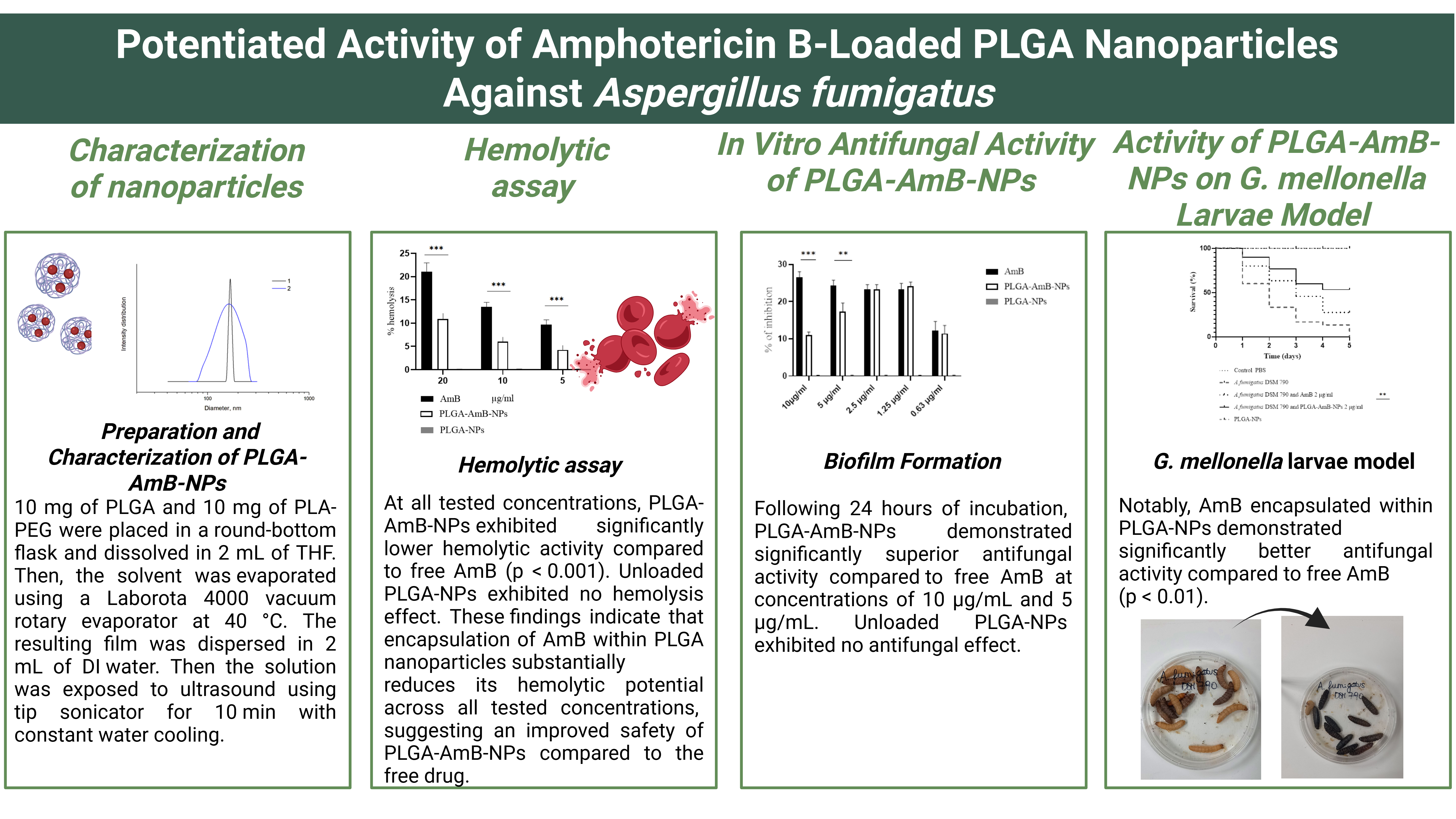
The sizes of NPs were determined in DI water. Representative intensity-weighted size distributions for blank and AmB-loaded PLGA nanoparticles are shown in Figure 1. Mean diameter of PLGA-NP was found to be 165 nm with PDI value 0.075 (Figure 1 curve 1). Incorporation of AmB into NPs did not dramatically change the sizes of the micelles. The mean diameter of PLGA-AmB-NPs was found to be 120 nm with PDI value 0.210. The broader polydispersity of loaded micelles could be attributed to disordering of the hydrophobic core of PLGA-AmB-NPs by AmB molecules. The measured zeta-potential values (-15.5 ± 1.2 mV for PLGA-NP and - 32.4 ± 1.4 mV for PLGA-AmB-NPs) indicate that the nanoparticles carry an overall net negative charge, consistent with their colloidal stability in aqueous media.
3.2. Antifungal Activity
3.2.1. Activity Against A. fumigatus Planktonic Growth
The effects of AmB, PLGA-AmB-NPs, and PLGA-NPs on A. fumigatus planktonic growth were evaluated. AmB and PLGA-AmB-NPs demonstrated comparable inhibitory activity, with MIC₅₀ and MIC₉₀ values of 1 µg/mL and 2 µg/mL, respectively. Blank PLGA-NPs exhibited no measurable inhibitory activity against planktonic cells.
3.2.2. Hemolytic Assay
The hemolytic activity of free AmB, PLGA-AmB-NPs, and PLGA-NPs was assessed against human erythrocytes across a concentration range of 5–20 µg/mL, encompassing concentrations up to 10-fold above the determined MIC₉₀ (2 µg/mL). The percentage of hemolysis was calculated relative to the Triton X-100 positive control, defined as 100% hemolysis.
At all tested concentrations, PLGA-AmB-NPs exhibited significantly lower hemolytic activity compared to free AmB (p < 0.001), Figure 2. Unloaded PLGA-NPs exhibited no hemolysis effect. These findings indicate that encapsulation of AmB within PLGA nanoparticles substantially reduces its hemolytic potential across all tested concentrations, suggesting an improved safety of PLGA-AmB-NPs compared to the free drug.
3.2.3. In Vitro Antifungal Activity of PLGA-AmB-NPs against Biofilm Formation
The antifungal efficacy of free AmB and PLGA-AmB-NPs against biofilm formation was assessed in vitro by measuring the metabolic activity of fungal cells. Following 24 hours of incubation, PLGA-AmB-NPs demonstrated significantly superior antifungal activity compared to free AmB at concentrations of 10 µg/mL and 5 µg/mL (Figure 3). Unloaded PLGA-NPs exhibited no antifungal effect.
3.2.4. Activity of PLGA-AmB-NPs, Free AmB and PLGA-NPs on G. mellonella Larvae Model
The in vivo efficacy of the formulations was assessed using the G. mellonella infection model, a well-established surrogate system for studying fungal virulence owing to the functional homology between its innate immune responses and those of vertebrates. Larvae were inoculated with A. fumigatus conidia at a dose of 4–5 × 10⁴ conidia per larva and subsequently treated with free AmB or PLGA-AmB-NPs at a concentration of 2 µg/mL, corresponding to the MIC₉₀. Larval survival was recorded daily over a 5-day observation period. Larvae administered PBS or PLGA-NPs alone served as negative controls and exhibited 0% mortality, whereas all larvae infected with A. fumigatus conidia succumbed by day 5 post-infection, corresponding to 100% mortality. Notably, AmB encapsulated within PLGA-NPs demonstrated significantly better antifungal activity compared to free AmB (p < 0.01), Figure 4.
3.2.5. In Vivo Toxicity Assessment in the G. mellonella Larva Model
PLGA-NPs, PLGA-AmB-NPs and AmB were tested in an in vivo toxicity model. The LD50 detected dose was more than 500 mg/kg for AmB and more than 800 mg/kg for PLGA-AmB-NPs (Table 1).
4. Discussion
Nanoparticles offer several advantages for drug delivery, as their small size enables penetration into small capillaries and leads to efficient accumulation at target sites [16,17]. Historically, the formulation of AmB-loaded PLGA nanoparticles has evolved considerably. Early work by Venier-Julienne et al. [18] using solvent evaporation methods resulted in very low drug loading (0.7–1.3%) due to poor miscibility between PLGA and AmB. Subsequent advances, including nanoprecipitation techniques stabilized with vitamin E TPGS, enabled improved particle size control and drug incorporation [19]. These optimized systems not only reduced toxicity relative to conventional formulations such as Fungizone®, but also demonstrated superior therapeutic efficacy in murine models of invasive aspergillosis, even outperforming parenteral liposomal formulations like AmBisome® following oral administration [20].
The physicochemical characterization in this study revealed that blank PLGA NPs had a mean diameter of 165 nm with a low polydispersity index (PDI 0.075), whereas AmB-loaded nanoparticles exhibited a smaller mean size (~120 nm) with a moderately broader distribution (PDI 0.210). Both formulations displayed a net negative surface charge, with zeta potentials of −15.5 ± 1.2 mV (blank NPs) and − 32.4 ± 1.4 mV (AmB-loaded NPs), indicating good colloidal stability. These values are consistent with previously reported PLGA-based antifungal systems, which typically fall within a size range of 50–250 nm and exhibit ζ-potentials between −20 and −30 mV [21]. The observed decrease in particle size and increased surface charge following drug encapsulation likely reflects structural reorganization within the nanoparticle core and surface composition.
Encapsulation of AmB in PLGA nanoparticles significantly enhanced antifungal activity against A. fumigatus biofilms in vitro. PLGA-AmB-NPs reduced the metabolic activity of biofilm-embedded cells more effectively than free AmB at equivalent concentrations (5 and 10 µg/mL), while blank NPs showed no antifungal activity. This improvement may be attributed to enhanced penetration of nanoparticles through the extracellular biofilm matrix and sustained drug release at the infection site. Similar findings have been reported for other PLGA-based antifungal systems, including PLGA PTB nanoparticles targeting Aspergillus biofilms [22]. Additionally, Yang et al. [23] demonstrated that AmB-loaded PLGA nanoparticles combined with ultrasound significantly disrupted C. albicans biofilms, reducing biomass and enzymatic activity while altering biofilm structure, suggesting that nanoparticle-based strategies can further be enhanced through combinatorial approaches.
In vivo efficacy was confirmed using the G. mellonella infection model, where PLGA-AmB-NP-treated larvae showed significantly improved survival compared to those receiving free AmB. These findings are in agreement with previous studies employing PLGA-based formulations in similar infection models [21], as well as mammalian studies demonstrating enhanced therapeutic outcomes and reduced toxicity [19,24]. For example, Souza et al. [24] reported significant antifungal activity of PLGA-DMSA AmB nanoparticles at relatively low doses without observable toxicity, while Tang et al. [25] developed targeted, pH-responsive PLGA-based nanoparticles that improved antifungal efficacy and reduced toxicity in both in vitro and in vivo systems.
A major advantage of PLGA-based nanoformulations is the reduction of AmB-associated toxicity. In the present study, amphotericin B-loaded PLGA nanoparticles were successfully developed and demonstrated reduced hemolytic toxicity compared with the free drug. The dose-dependent hemolysis observed for both formulations aligns with the known mechanism of AmB-induced membrane damage via interaction with cholesterol in erythrocyte membranes [24,26]. These findings are consistent with earlier reports, particularly those of Italia et al. [19,27], who showed that PLGA-based AmB nanoparticles significantly decreased hemolytic activity and renal toxicity while achieving an oral bioavailability as high as 800%, highlighting their promise for oral delivery in systemic fungal infections.
Despite these promising findings, several limitations should be acknowledged. The current study evaluated only a single A. fumigatus strain and focused on early-stage biofilm formation; thus, the efficacy of PLGA-AmB-NPs against mature biofilms and diverse clinical isolates, including azole-resistant strains, remains to be determined. Additionally, pharmacokinetics and biodistribution were not assessed, which are critical parameters in comparison with clinically approved lipid formulations. While the G. mellonella model provides valuable preliminary insights, validation in mammalian models is essential before clinical translation. Future studies should therefore focus on optimizing nanoparticle formulations, evaluating in vivo pharmacokinetics, and exploring combination therapies or targeting strategies to further enhance antifungal efficacy and safety.
5. Conclusions
In summary, AmB-loaded PLGA nanoparticles were developed and characterized; this nanoformulation was shown to enhance the antifungal activity of AmB against A. fumigatus biofilms, improve survival outcomes in the G. mellonella infection model, and significantly reduce hemolytic toxicity in vitro compared with free AmB. These results indicate that PLGA-based nanoencapsulation can increase the therapeutic index of AmB by simultaneously potentiating its antifungal efficacy and mitigating its toxicity. PLGA-AmB-NPs therefore represent a promising polymeric platform for the treatment of A. fumigatus infections, particularly those involving biofilm associated growth, and warrant further investigation in extended strain panels, azole resistant isolates and mammalian models of aspergillosis.
Author Contributions
Conceptualization, A. Orekhova and A.V. Sybachin; methodology, A. Osmolovskiy; formal analysis, V. Lyamina, D. Mamiy; investigation, A. Orekhova; resources, A. Orekhova; data curation, A. Osmolovskiy; writing—original draft preparation, V. Lyamina, D. Mamiy, A. Orekhova; writing—review and editing, V. Lyamina, D. Mamiy; visualization, V. Lyamina, D. Mamiy, A. Orekhova; supervision, O. M. Drapkina; project administration, A. Orekhova.; funding acquisition, A. Orekhova. All authors have read and agreed to the published version of the manuscript.
Funding
This research was funded by Russian Science Foundation, grant number 25-75-00066 «Development of a targeted drug delivery method for antifungal drugs based on polyglycolic acid nanoparticles» 2025-2027.
Institutional Review Board Statement
This study was conducted in accordance with the Declaration of Helsinki and approved by the local Ethics Committee of the National Medical Research Center for Therapy and Preventive Medicine (protocol code 02-02/21, date of approval 25 February 2021).
Data Availability Statement
Data are available upon request.
Acknowledgments
The authors would like to thank Lukianov Dmitrii for collaboration.
Conflicts of Interest
The authors declare no conflict of interest and the funders had no role in the design of the study; in the collection, analyses, or interpretation of data; in the writing of the manuscript, or in the decision to publish the results.
References
- Fairuz, S.; Nair, R.S.; Billa, N. Orally Administered Amphotericin B Nanoformulations: Physical Properties of Nanoparticle Carriers on Bioavailability and Clinical Relevance. Pharmaceutics 2022, 14, 1823. [CrossRef]
- Cavassin, F.B.; Baú-Carneiro, J.L.; Vilas-Boas, R.R.; Queiroz-Telles, F. Sixty Years of Amphotericin B: An Overview of the Main Antifungal Agent Used to Treat Invasive Fungal Infections. Infect Dis Ther 2021, 10, 115–147. [CrossRef]
- Laniado-Laborín, R.; Cabrales-Vargas, M.N. Amphotericin B: Side Effects and Toxicity. Revista Iberoamericana de Micología 2009, 26, 223–227. [CrossRef]
- Carolus, H.; Pierson, S.; Lagrou, K.; Van Dijck, P. Amphotericin B and Other Polyenes—Discovery, Clinical Use, Mode of Action and Drug Resistance. JoF 2020, 6, 321. [CrossRef]
- Gershkovich, P.; Wasan, E.K.; Lin, M.; Sivak, O.; Leon, C.G.; Clement, J.G.; Wasan, K.M. Pharmacokinetics and Biodistribution of Amphotericin B in Rats Following Oral Administration in a Novel Lipid-Based Formulation. Journal of Antimicrobial Chemotherapy 2009, 64, 101–108. [CrossRef]
- Serrano, D.R.; Lalatsa, A. Oral Amphotericin B: The Journey from Bench to Market. Journal of Drug Delivery Science and Technology 2017, 42, 75–83. [CrossRef]
- Petraitis, V.; Petraitiene, R.; Valdez, J.M.; Pyrgos, V.; Lizak, M.J.; Klaunberg, B.A.; Kalasauskas, D.; Basevicius, A.; Bacher, J.D.; Benjamin, D.K.; et al. Amphotericin B Penetrates into the Central Nervous System through Focal Disruption of the Blood-Brain Barrier in Experimental Hematogenous Candida Meningoencephalitis. Antimicrob Agents Chemother 2019, 63, e01626-19. [CrossRef]
- Alakkad, A.; Stapleton, P.; Schlosser, C.; Murdan, S.; Odunze, U.; Schatzlein, A.; Uchegbu, I.F. Amphotericin B Polymer Nanoparticles Show Efficacy against Candida Species Biofilms. Pathogens 2022, 11, 73. [CrossRef]
- Morrissey, C.O.; Kim, H.Y.; Duong, T.-M.N.; Moran, E.; Alastruey-Izquierdo, A.; Denning, D.W.; Perfect, J.R.; Nucci, M.; Chakrabarti, A.; Rickerts, V.; et al. Aspergillus Fumigatus —a Systematic Review to Inform the World Health Organization Priority List of Fungal Pathogens. Medical Mycology 2024, 62, myad129. [CrossRef]
- Seidler, M.J.; Salvenmoser, S.; Müller, F.-M.C. Aspergillus Fumigatus Forms Biofilms with Reduced Antifungal Drug Susceptibility on Bronchial Epithelial Cells. Antimicrob Agents Chemother 2008, 52, 4130–4136. [CrossRef]
- Angelini, J.; Ferin, S.; Martini, L.; Flammini, S.; Tascini, C.; Giuliano, S. Current Opinion on the Potential Role of Liposomal Amphotericin B in Underexplored Clinical Scenarios. Clin Microbiol Rev 2026, e00228-25. [CrossRef]
- Pigareva, V.A.; Alekhina, Y.A.; Grozdova, I.D.; Zhu, X.; Spiridonov, V.V.; Sybachin, A.V. Magneto-sensitive and Enzymatic Hydrolysis-resistant Systems for the Targeted Delivery of Paclitaxel Based on Polylactide Micelles with an External Polyethylene Oxide Corona. Polymer International 2022, 71, 456–463. [CrossRef]
- Carpenter, A.M.; Van Hoek, M.L. Development of a Defibrinated Human Blood Hemolysis Assay for Rapid Testing of Hemolytic Activity Compared to Computational Prediction. Journal of Immunological Methods 2024, 529, 113670. [CrossRef]
- Pierce, C.G.; Uppuluri, P.; Tristan, A.R.; Wormley, F.L.; Mowat, E.; Ramage, G.; Lopez-Ribot, J.L. A Simple and Reproducible 96-Well Plate-Based Method for the Formation of Fungal Biofilms and Its Application to Antifungal Susceptibility Testing. Nat Protoc 2008, 3, 1494–1500. [CrossRef]
- Orekhova, A.; De Angelis, M.; Cacciotti, A.; Reverberi, M.; Rotili, D.; Giorgi, A.; Protto, V.; Bonincontro, G.; Fiorentino, F.; Zgoda, V.; et al. Modulation of Virulence-Associated Traits in Aspergillus Fumigatus by BET Inhibitor JQ1. Microorganisms 2022, 10, 2292. [CrossRef]
- Li, J.; Burgess, D.J. Nanomedicine-Based Drug Delivery towards Tumor Biological and Immunological Microenvironment. Acta Pharmaceutica Sinica B 2020, 10, 2110–2124. [CrossRef]
- He, W.; Xing, X.; Wang, X.; Wu, D.; Wu, W.; Guo, J.; Mitragotri, S. Nanocarrier-Mediated Cytosolic Delivery of Biopharmaceuticals. Adv Funct Materials 2020, 30, 1910566. [CrossRef]
- Venier-Julienne, M.C.; Benoît, J.P. Preparation, Purification and Morphology of Polymeric Nanoparticles as Drug Carriers. Pharmaceutica Acta Helvetiae 1996, 71, 121–128. [CrossRef]
- Italia, J.L.; Sharp, A.; Carter, K.C.; Warn, P.; Kumar, M.N.V.R. Peroral Amphotericin B Polymer Nanoparticles Lead to Comparable or Superior In Vivo Antifungal Activity to That of Intravenous Ambisome® or FungizoneTM. PLoS ONE 2011, 6, e25744. [CrossRef]
- Italia, J.L.; Yahya, M.M.; Singh, D.; Ravi Kumar, M.N.V. Biodegradable Nanoparticles Improve Oral Bioavailability of Amphotericin B and Show Reduced Nephrotoxicity Compared to Intravenous Fungizone®. Pharm Res 2009, 26, 1324–1331. [CrossRef]
- Mohammadi, G.; Fathian-Kolahkaj, M.; Mohammadi, P.; Adibkia, K.; Fattahi, A. Preparation, Physicochemical Characterization and Anti-Fungal Evaluation of Amphotericin B-Loaded PLGA-PEG-Galactosamine Nanoparticles. Adv Pharm Bull 2020, 1. [CrossRef]
- Orekhova, A.; Palocci, C.; Chronopoulou, L.; De Angelis, G.; Badiali, C.; Petruccelli, V.; D’Angeli, S.; Pasqua, G.; Simonetti, G. Poly-(Lactic-Co-Glycolic) Acid Nanoparticles Entrapping Pterostilbene for Targeting Aspergillus Section Nigri. Molecules 2022, 27, 5424.
- Yang, M.; Du, K.; Hou, Y.; Xie, S.; Dong, Y.; Li, D.; Du, Y. Synergistic Antifungal Effect of Amphotericin B-Loaded Poly(Lactic-Co-Glycolic Acid) Nanoparticles and Ultrasound against Candida Albicans Biofilms. Antimicrob Agents Chemother 2019, 63, e02022-18. [CrossRef]
- Souza, A.C.O.; Nascimento, A.L.; De Vasconcelos, N.M.; Jerônimo, M.S.; Siqueira, I.M.; R-Santos, L.; Cintra, D.O.S.; Fuscaldi, L.L.; Pires Júnior, O.R.; Titze-de-Almeida, R.; et al. Activity and in Vivo Tracking of Amphotericin B Loaded PLGA Nanoparticles. European Journal of Medicinal Chemistry 2015, 95, 267–276. [CrossRef]
- Tang, X.; Dai, J.; Xie, J.; Zhu, Y.; Zhu, M.; Wang, Z.; Xie, C.; Yao, A.; Liu, T.; Wang, X.; et al. Enhanced Antifungal Activity by Ab-Modified Amphotericin B-Loaded Nanoparticles Using a pH-Responsive Block Copolymer. Nanoscale Res Lett 2015, 10, 256. [CrossRef]
- Choi, K.-C.; Bang, J.-Y.; Kim, P.-I.; Kim, C.; Song, C.-E. Amphotericin B-Incorporated Polymeric Micelles Composed of Poly(d,l-Lactide-Co-Glycolide)/Dextran Graft Copolymer. International Journal of Pharmaceutics 2008, 355, 224–230. [CrossRef]
- Le, T.; Kinh, N.V.; Cuc, N.T.K.; Tung, N.L.N.; Lam, N.T.; Thuy, P.T.T.; Cuong, D.D.; Phuc, P.T.H.; Vinh, V.H.; Hanh, D.T.H.; et al. A Trial of Itraconazole or Amphotericin B for HIV-Associated Talaromycosis. N Engl J Med 2017, 376, 2329–2340. [CrossRef]
Figure 1.
Intensity distribution of sizes of PLGA-NP (1) and PLGA-AmB-NPs (2).
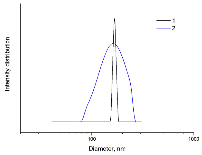
Figure 2.
In vitro hemolytic activity of free AmB, PLGA-AmB-NPs and PLGA-NPs at varying concentrations. Data are expressed as mean ± SD (n = 3), ** p < 0.01, *** p < 0.001 for PLGA-AmB-NPs vs. free AmB (one-way ANOVA with Tukey's post-hoc test).
Figure 2.
In vitro hemolytic activity of free AmB, PLGA-AmB-NPs and PLGA-NPs at varying concentrations. Data are expressed as mean ± SD (n = 3), ** p < 0.01, *** p < 0.001 for PLGA-AmB-NPs vs. free AmB (one-way ANOVA with Tukey's post-hoc test).
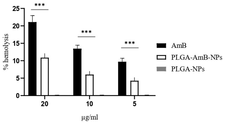
Figure 3.
Activity of free AmB, PLGA-AmB-NPs and PLGA-NPs against 24 h A. fumigatus biofilm. ** p < 0.01, *** p < 0.001 for PLGA-AmB-NPs vs. free AmB.
Figure 3.
Activity of free AmB, PLGA-AmB-NPs and PLGA-NPs against 24 h A. fumigatus biofilm. ** p < 0.01, *** p < 0.001 for PLGA-AmB-NPs vs. free AmB.
Figure 4.
PLGA-encapsulated AmB exhibited significantly superior antifungal efficacy against A. fumigatus virulence in the G. mellonella wax moth model compared to free AmB. Survival of G. mellonella larvae (n = 30 per group) was monitored over a 5-day period following injection with 4 × 10⁴ to 5 × 10⁴ conidia per larva of A. fumigatus. Statistical differences relative to the PBS control were assessed using the Kaplan–Meier method with Mantel–Cox log-rank test, where ** p < 0.01 denote significant differences compared to the A. fumigatus and AmB 2 µg/ml and A. fumigatus and PLGA-AmB-NPs 2 µg/ml. All results represent data from a minimum of three independent biological replicates.
Figure 4.
PLGA-encapsulated AmB exhibited significantly superior antifungal efficacy against A. fumigatus virulence in the G. mellonella wax moth model compared to free AmB. Survival of G. mellonella larvae (n = 30 per group) was monitored over a 5-day period following injection with 4 × 10⁴ to 5 × 10⁴ conidia per larva of A. fumigatus. Statistical differences relative to the PBS control were assessed using the Kaplan–Meier method with Mantel–Cox log-rank test, where ** p < 0.01 denote significant differences compared to the A. fumigatus and AmB 2 µg/ml and A. fumigatus and PLGA-AmB-NPs 2 µg/ml. All results represent data from a minimum of three independent biological replicates.
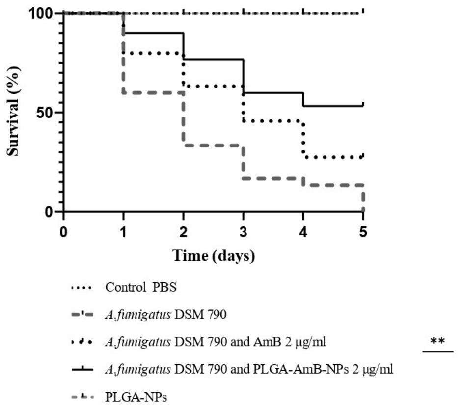

Table 1.
Survival of G. mellonella larvae following administration of PLGA-NPs, PLGA-AmB-NPs by intra-haemocoel injection. Each experiment was conducted in triplicate with 10 larvae per group. All values are the mean of three independent experiments.
Table 1.
Survival of G. mellonella larvae following administration of PLGA-NPs, PLGA-AmB-NPs by intra-haemocoel injection. Each experiment was conducted in triplicate with 10 larvae per group. All values are the mean of three independent experiments.
| Chemical | LD50 (mg/kg) | Solvent |
|---|---|---|
| PLGA-NPs | - | H2O |
| AmB | >500 | 100 H2O: 1 DMSO |
| PLGA-AmB-NPs | >800 | 100 H2O: 1 DMSO |
Disclaimer/Publisher’s Note: The statements, opinions and data contained in all publications are solely those of the individual author(s) and contributor(s) and not of MDPI and/or the editor(s). MDPI and/or the editor(s) disclaim responsibility for any injury to people or property resulting from any ideas, methods, instructions or products referred to in the content. |
© 2026 by the authors. Licensee MDPI, Basel, Switzerland. This article is an open access article distributed under the terms and conditions of the Creative Commons Attribution (CC BY) license (http://creativecommons.org/licenses/by/4.0/).
Copyright: This open access article is published under a Creative Commons CC BY 4.0 license, which permit the free download, distribution, and reuse, provided that the author and preprint are cited in any reuse.