

Submitted:
02 May 2026
Posted:
05 May 2026
You are already at the latest version
Abstract
Foodborne mycotoxins are toxic secondary metabolites produced by filamentous fungi. At sufficient concentrations, ingested mycotoxins have been found to disrupt the microbiome and exert toxic effects on gastrointestinal, hepatic, renal, and other tissues. Toddlers are particularly vulnerable to ill effects due to increased intake relative to lower body weight and immature detoxification, metabolic, and immune function. Despite this susceptibility, their adverse health effects remain under-recognized in public health sectors, and data on mycotoxin contamination in foods marketed to young children in North America remain sparse. To investigate this, 118 food products, including cereals, snacks, pasta, first foods, juices, and staple ingredients, were purchased at retail and analyzed for 34 mycotoxins using liquid chromatography–tandem mass spectrometry. The results were compared with United States Food and Drug Administration (FDA) regulatory thresholds and European Tolerable Daily Intake (TDI) limits. At least one mycotoxin was detected in 88 percent of products, with a mean of 3.8 per item and a maximum of 13; 26 of 34 analytes were identified. Multi-toxin contamination was common, many products exceeded FDA and TDI limits, and many remain unregulated. These findings highlight regulatory gaps in food safety and underscore the need for stronger child-focused oversight, particularly regarding emerging mycotoxins and multi-toxin contamination.
Keywords:
mycotoxins
; food safety
; emerging mycotoxins
; toddler foods
; multi-toxin contamination
; pediatric nutrition
; risk assessment
1. Introduction
Mycotoxins are low-molecular-weight secondary metabolites produced by filamentous fungi under favorable environmental and storage conditions [1,2]. Since the 1960s, hundreds of mycotoxins have been identified, with dietary ingestion representing the primary route of human exposure [1]. Inhalation and dermal contact may also occur in mold-contaminated indoor environments, such as water-damaged buildings, and in occupational settings, including bakeries and breweries, making complete avoidance difficult [2,3,4].
Historically, outbreaks of mycotoxin-related foodborne illness were often regionally confined before the expansion of global trade [2,5]. However, mycotoxin contamination is now widespread, with an estimated 60–80% of cereal grains and nut crops affected worldwide, posing risks to both human and animal health [1]. As a result, humans are continuously exposed to low- and, at times, high levels of dietary mycotoxins, with infants and young children among the most disproportionately affected due to lower body weight and higher relative toxin exposure [6,7,8].
The primary mycotoxin-producing genera affecting the human food supply include Aspergillus, Penicillium, Fusarium, and Alternaria species [2]. These fungi produce a chemically diverse range of toxic compounds, including aflatoxins, ochratoxins, trichothecenes (e.g., deoxynivalenol, nivalenol, HT-2 toxin, and T-2 toxin), zearalenone, fumonisins, enniatins, beauvericin, alternariol mono-ether, and alternariol [2]. Many of these metabolites have been shown to adversely affect multiple organ systems, including the gastrointestinal tract, liver, kidneys, nervous system, reproductive organs, and epithelial and immune tissues [3,4,9,10].
Within the context of organ-specific toxicities, many mycotoxins have acute and chronic health effects. For example, at low but chronic doses, the commonly occurring trichothecene mycotoxin deoxynivalenol (DON) may stimulate immune activation, inhibit protein synthesis, compromise the intestinal mucus barrier, increase intestinal permeability, and suppress appetite, thereby potentially contributing to growth stunting in children [11,12,13,14,15,16,17,18]. In higher, more acute exposures, DON has been linked to immune suppression, nausea, vomiting, abdominal pain, diarrhea, intestinal hemorrhage, and fever in both animal models and human outbreaks [5,9,11,12,13,16,19,20,21]. These effects have led some researchers to hypothesize that chronic trichothecene ingestion may contribute to inflammatory gastrointestinal (GI) disorders, including inflammatory bowel disease (IBD) [3,13,19,20,21].
Following systemic absorption, some mycotoxins have been found to cross the blood-brain barrier. DON and ochratoxin A (OTA) can cross the blood–brain barrier and have been shown to damage astrocytes and glial cells in animal and in vitro models [15,22]. Detoxification differences between developmentally typical children and their siblings with autism spectrum disorder (ASD) have raised concerns about their potential role in ASD [14,15,22,23,24,25].
Other mycotoxins also exhibit distinct organ-specific effects. Cyclopiazonic acid, an unregulated mycotoxin, has been linked to GI and neurological toxicity, including human outbreaks of Kodua poisoning [26,27,28,29,30]. Aflatoxins are well-established hepatotoxins and hepatocarcinogens, while enniatins and Alternaria toxins, including alternariol (AOH) and alternariol monomethyl ether (AME), may also contribute to liver pathology, although their roles remain less well defined [31,32,33,34,35,36,37,38]. Zearalenone (ZEA), AOH, and AME exhibit hormone-disruptive activity, and ZEA has been associated with reproductive abnormalities, including links to polycystic ovarian syndrome in animal models [2,37,39,40,41,42,43,44,45]. Fumonisins exposure has been implicated in the development of neural tube defects in infants and possibly esophageal cancer in humans [2,46,47]. Aflatoxin B1, fumonisins, and DON have recently been under investigation for their potential role in growth stunting in children [48,49].
In addition to direct toxicity, many mycotoxins disrupt intestinal microbial communities in a manner analogous to antibiotics. DON alters Bacteroides and Firmicutes populations [50,51,52]. Aflatoxins reduce lactic acid–producing bacteria. OTA targets lactobacilli, including Lactobacillus reuteri [53]. Beauvericin exhibits broad antimicrobial activity against Gram-positive and Gram-negative organisms, potentially contributing to dysbiosis [54,55,56,57].
Collectively, these findings highlight the potential for foodborne mycotoxins to negatively impact the human microbiome and overall health. Despite decades of research and the identification of many foodborne mycotoxins with known toxicity, numerous fungal metabolites are still underrecognized in clinical and public health fields [9,30,58,59].
Exposure can start in utero, as mycotoxins have been found in amniotic fluid and linked to impaired fetal growth [60]. In a prospective cohort study, Tan et al. (2023) reported that mothers in the highest tertile of DON intake were more likely to deliver infants with lower birth weight and reduced birth length relative to gestational age (all P-trends < 0.05) [61]. Postnatally, exposure may continue through contaminated breast milk, infant formula, and cow’s milk, with additional increases as solid foods are introduced [6,8,61,62,63,64].
Following weaning, toddlers consume a broader range of solid foods, many of which are grain-based products such as cereals, crackers, breads, and snack foods. Because grains are common sources of mycotoxins and are consumed more frequently relative to body size, this transition may further increase exposure compared with infancy [7,65]. These risks may be amplified by developmental vulnerabilities, including immature, less resilient microbiomes, underdeveloped metabolic and immune systems, reduced detoxification capacity, and lower body weight [8,66].
Compounding this concern, mycotoxins are imperceptible to humans by taste, odor, or visual inspection, rendering avoidance difficult without advanced analytical methods such as liquid chromatography–tandem mass spectrometry [67]. Historically, most surveillance efforts have focused on a limited subset of regulated mycotoxins, which may be why only a small number are currently subject to regulatory limits in the United States (U.S.). Currently, only limited data are available on the occurrence and co-occurrence of regulated and emerging mycotoxins in the U.S. food supply.
To address these gaps, the present study evaluates the occurrence and co-occurrence of 34 regulated and emerging mycotoxins in processed foods marketed for U.S. toddlers, providing a snapshot of potential early-life dietary exposures (see Table 1 for more information on the analytes tested). Specifically, this investigation seeks to address the following questions: (1) which of the 34 mycotoxins tested are U.S. toddlers likely to be exposed to through commonly available processed foods, and (2) whether observed contamination levels exceed established European tolerable daily intake (TDI) values when adjusted for toddler body weight, thereby potentially increasing risks to child health. European TDI values were used where available, as no established pediatric TDI values are available in the U.S.
2. Materials and Methods
To assess for multi-mycotoxin contamination, 118 food products were screened for 34 mycotoxins using liquid chromatography–tandem mass spectrometry (LC–MS/MS) . A validated multi-analyte method was applied for sample preparation and quantification as described by Kresse et al. [67] (See Table 1 for analytes tested). Quality control measures were implemented throughout the analytical process.
2.1. Sample Selection and Procurement
Foods specifically marketed for infant and child consumption, including cereals, snacks, pastas, first foods, juices, and basic cooking ingredients, were selected for analysis. For baby food products, only vegetable-based, non-dairy items were included, as defined a priori by the study team. Cereal grain-based foods and some non-cereal grain ingredients were selected for testing. Non-cereal-based ingredients included chocolate powder, soy-based formula, almond flour, coconut flour, arrowroot starch, and tapioca starch, and miscellaneous other products (e.g., first food beans and apple juice).
Products were purchased at retail locations and by mail order in the Midwestern United States (Missouri) and the Northeastern United States (Massachusetts, New Hampshire, and Maine) between 1 June 2024 and 1 October 2025. In the absence of a formal randomization protocol, a standardized systematic sampling approach was implemented to reduce selection bias when purchased directly from individual stores. Specifically, the second item in each product row was selected, provided that the packaging was intact. Additionally, only one product was selected from each product category (e.g., cornflakes) on the shelf without regard to brand. However, because two separate people procured the food items, duplicate products from different geographic regions and different lots were sometimes selected.
To supplement retail sampling and expand geographic coverage, a mail-order procurement strategy was also implemented to obtain commercially available food products for mycotoxin analysis. Products marketed for infant and toddler consumption, along with some basic cooking ingredients, were identified through major online retailers and manufacturers' websites via Google search and purchased using a standardized selection approach. Within each product category, one item from every other available brand was selected to reduce selection bias.
Prior to laboratory submission, all products were inspected to confirm package integrity, absence of visible damage (e.g., rips or tears), and validity of expiration dates to ensure sample quality. Details for each product were also noted, including manufacturer lot numbers and storage facility numbers, when available. However, such details were not available for all products procured.
2.2. Sample Shipment and Laboratory Analysis
After procurement, samples were sent via U.S. Postal Service Priority Mail to Trilogy Analytical Laboratory (Washington, MO) for analysis of 34 foodborne mycotoxins using LC-MS/MS procedures. Once received by the laboratory, samples were stored in their original, unopened packaging under ambient, dry conditions consistent with manufacturer recommendations. This was done to minimize potential contamination or degradation prior to analysis. All samples were logged with the receipt date, product type, brand, lot number (if available), and country of origin if disclosed on the label. Samples were stored for no more than two weeks until testing. Samples were tested in groups based on matrix (e.g., packaged cereals).
2.3. Reagents and Standards
To test for 34 mycotoxins, reference standards for all targeted mycotoxins (≥98% purity) were manufactured by Trilogy Analytical Laboratory (United States) or purchased from Romer Labs (United States). Stock solutions were prepared in 1 mg/ml concentrations in LC–MS grade acetonitrile, methanol, and water. LC–MS grade acetonitrile, methanol, and water were obtained from Honeywell. Formic acid (≥99% purity, LC–MS grade) was obtained from Millipore Sigma.
2.4. Sample Preparation
Diverse food subsets required varying methods of preparation. For products prone to heterogeneity (e.g., cereals, mixed snacks), incremental portions were taken from multiple locations within the homogenized matrix to ensure representativeness. Liquid products were mixed thoroughly prior to aliquoting.
Samples were then prepared for testing using the following methods: Samples were ground using a Retsch GM200 mill or homogenized using a high-speed blender (Osterizer) until a uniform particle size was achieved. 25-gram samples were then weighed in an extraction flask, and 100 mL of acetonitrile–water (80:20, v/v) was added.
Extraction was performed on an orbital shaker (6000, Eberbach) for 90 min at room temperature (20–22 °C). The samples were then filtered with a Whatman No. 1. The filtered extract was then purified with a QP1100 SPE (Trilogy Analytical Laboratory, United States). The purified extracts were then diluted with water-acetic acid (2% v/v) and injected into the LC-MS/MS system.
2.5. LC–MS/MS Instrumentation and Parameters
For LC-MS/MS analysis, the Shimadzu SIL-40 series system was configured with a near-zero refrigerated autosampler (temperature maintained at 8 °C), a gradient pump, and a column oven. This was combined with the SCIEX 7500 mass spectrometry system by an Electrospray Ionization Interface (ESI). The preinstalled 6-port valve on the MS-MS instrument was configured to direct the analyte flow to waste. The SCIEX OS acquisition software controlled all systems. For data evaluation, the SCIEX OS software was applied [108].
2.5.1. Chromatographic Separation
Separation was performed using a Phenomenex Kinetex column (3 mm x 100 mm; 2.6 μm; 100 Å). The column was stored in the column oven. The temperature was set to 40 °C, and the injection volume was 5 μL.
For the separation, a gradient program was used. The gradient profile started at 5% B, held for 1.0 minute, and then decreased linearly to 50% B within 6.0 minutes. This was maintained for 12.0 minutes before being returned to 5% B in 4.0 minutes. The starting conditions were held for 4.0 min before the next injection.
The mobile phases for the analysis were A: water-0.1% Formic Acid-4 mM Ammonium Formate and B: Methanol-0.1% Formic Acid-4 mM Ammonium Formate. Flow rate was 0.5ml/min.
2.5.2. Mass Spectrometry Conditions
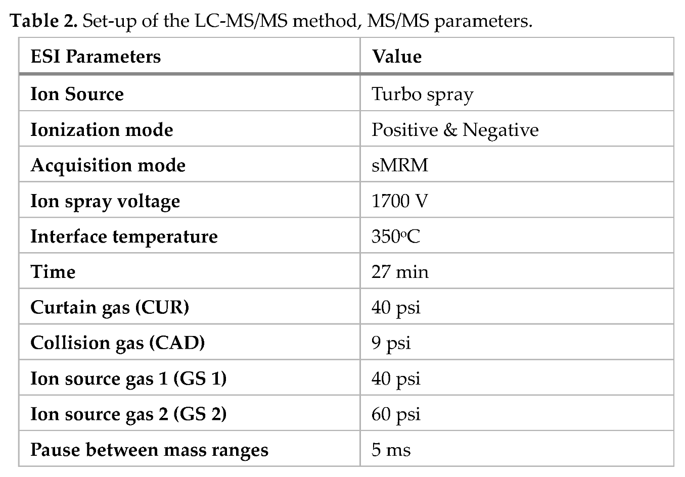
The parameters for MS/MS were as follows: ESI positive with interface temperature 350 °C, ion spray voltage 1700 V, curtain gas 40 psi, collision gas 9 psi, ion source gas 1 set at 40 psi, and gas 2 set at 60 psi. The mass spectrometry worked in the scheduled multiple reaction monitoring (sMRM), and the two most intensive transitions were measured when the relevant substance was eluting to the MS/MS. The dwell times were optimized automatically by Sciex OS Software.
Analyses were performed in scheduled multiple reaction monitoring (sMRM) mode. For each analyte, the two most intense precursor–product ion transitions were monitored within a 1.2 min retention window. Dwell times were automatically optimized by Sciex OS software ([3.0, Sciex]) [108] to ensure adequate data points across each peak. The window of one MSM signal was 1.2 minutes, and the total cycle time was 1.1 s [108].
Analytes required both positive and negative ionization polarities. Complete MS/MS operating settings are provided in Table 2 [67].
2.5.3. Method Validation
Method performance was evaluated in accordance with FDA guidelines. Linearity was assessed using matrix-matched calibration curves prepared in triplicate across a range. Coefficients of determination (R²) exceeded 0.99 for all analytes (See supplemental verification data).
Limits of detection (LOD) and quantification (LOQ) were determined based on signal-to-noise ratios of 3:1 and 10:1, respectively, using spiked blank matrices. Recoveries were assessed by fortifying blank matrices. Mean recoveries ranged from 70% to 120%, with relative standard deviations (RSD) ≤15%. Matrix effects were evaluated by comparing slopes of calibration curves prepared in solvent versus matrix extracts. If mycotoxins were detected, but below the typical LOQ, the data were considered not quantifiable and discarded from the total dataset.
2.6. Statistical Analysis and Comparison to Established Tolerable Intake Levels
For each sample, data were entered into a Microsoft Excel (Version 2601 Build 16.0.19628.20132) spreadsheet and evaluated using descriptive statistics [109]. Data were then converted to micrograms per kilogram (µg/kg) for ease of clinical interpretation and comparison with TDI and current regulatory guidance levels. The mean number of samples in which at least one mycotoxin was detected was also calculated for each tested category. R statistical software (version 4.5.2) running under RStudio [110] and add-on packages were then used to further evaluate the data [111,112,113,114]. A simulated Pearson’s chi-squared test was conducted to determine the distributions of mycotoxins across all groups.
Median and maximum detection levels were computed for mycotoxin contamination levels; mean rates were calculated for the number of positive samples. Maximum and median detection levels were then evaluated against body weight (bw) to assess pediatric risk using pediatric portion sizes (e.g., 57 grams (g) or approximately two ounces), with current TDI recommendations using an algorithm [115]. This equation was developed by the Italian National Institute of Health, the Department of Innovation in Biology at the University of Tuscia, Italy, and an Italian food company to produce processed wheat-based products with child-safe DON levels [115]. This equation provides a method for comparing the median and maximum mycotoxin levels found in food samples with established TDIs.
Exposure ng/kg bw/ day = Mycotoxin Contamination level (µg/kg) × Consumption data (g)/bw (kg)
For consistency, mean toddler weights-for-age for a 24-month-old were obtained from the Centers for Disease Control and Prevention (CDC) growth charts. The 50th percentile for girls and boys combined (12.5 kg/bw) was input into the equation [116].
Because the product was reported in nanograms, the units were converted to micrograms by dividing the product in nanograms by 1000 to provide intake levels in µg/kg bw/day for comparison with the TDI limit for each toxin. TDI limits have not been established for some of the mycotoxins tested for. In such cases, an accurate risk assessment was not possible.
Microsoft Excel was used to evaluate if toddlers would exceed TDI exposure limits using the following equation [109]:
Toddler Exposure =TDI (µg/kg bw/day) /Actual intake (µg/kg bw/day)
This method allowed easy comparison of mycotoxin exposure levels in foods with established TDI limits. All pediatric exposure levels above the TDI limit guidelines were considered a risk to child health.
3. Results
One hundred and eighteen (118) processed foods and ingredients were analyzed. A broad range of foods was sourced for testing, including snack foods, first foods and formulas, packaged cereals, and pastas (n = 72 samples), as well as raw ingredients used in food preparation (n = 46 samples). Contamination rates differed across commodities. The categories of food samples tested are listed in Table 3.
Of 118 samples of food and basic ingredients tested, 88.1 percent (95% CI = (81.1, 92.8)) were contaminated with at least one mycotoxin; the median was 4.0, the mean was 3.8, and the maximum was 13 mold contaminants. In addition, of the 34 mycotoxins tested, 26 were detected at varying levels within the sample set. Roquefortine C, Griseofulvin, Aflatoxin G2, Diacetoxyscirpenol, Fusarenon-X, Neosolaniol, Citrinin, a-Zearalenol, and b-Zearalenol were not detected in any product. Out of 4,012 tests (118 samples × 34 toxins), 445 (11.1 %) were positive for some mycotoxin. A simulated Pearson’s chi-squared test indicates that the proportions of products with at least one mycotoxin detected were approximately equal across food types (P=0.61). However, the types of mycotoxins found differed by commodity. Figure 1 shows the distribution of regulated, unregulated, and emerging mycotoxins detected in foods. Table 4 presents the number of samples contaminated within each mycotoxin category, the level of quantification (LOQ) in parts per million (ppm), the highest and median amounts found in each category, and the type of food in which the highest amount was detected.
Table 5 demonstrates the distribution of mycotoxins by chemical structure across food types. Cyclic hexadepsipeptides (e.g., ENNs and BEA) were most abundant across all food categories, followed by Di-furanocoumarins (e.g., aflatoxins) and then trichothecene mycotoxins (e.g., DON, NIV, HT-2, etc.).
Because the levels and types of mycotoxins detected varied by commodity, the results are broken down below into food category subgroups for ease of interpretation. It is important to note that among all the mycotoxins detected, only aflatoxins, DON, and fumonisins are regulated in the U.S.
3.1. Snack Foods (n=27):
Eighty-five percent of snack foods were contaminated with at least one mycotoxin. The mean number of mycotoxins per sample was 4, with a maximum of 6. Five out of 27 snack samples were contaminated with either type A or type B trichothecene mycotoxins. The trichothecene mycotoxins detected in these samples included 15-Ace, DON, NIV, T-2 toxins, and HT-2 toxin. The highest NIV level recorded in the study was observed in a snack food at 19,070 μg/kg. Six samples were contaminated with AFB1 (22%), 64% were contaminated with at least one ENN mycotoxin, and 67% with BEA. Thirty-three percent of snack samples were also contaminated with AME. Lastly, FB1, FB2, and CPA were detected together in a single corn-based vegetable stick snack.
3.2. Toddler First Foods (n=7):
In the toddler first food category (n = 7), 6 samples contained at least 2 mycotoxins (86%), with the highest number being 8. Apple juice marketed for infants and toddlers contained the highest levels of the emerging mycotoxins EnnA1, EnnA, and EnnB1 recorded in the study (100.7 ppb, 95.6 ppb, and 82.5 ppb, respectively). In addition, 57% contained the emerging mycotoxin AME, and a fruit puree sample contained AOH and AME.
3.3. Processed Cereal Products (n=25)
Eighty-eight percent of processed, packaged cereals contained at least one mycotoxin. Moreover, packaged cereals were often contaminated with multiple mycotoxins from various mold species, with a mean of 4 and a maximum of 7. In this category, 44% were infected with at least one ENN and 68% with BEA. Aflatoxins were also detected in 20% of cereals, but at very low levels well below the FDA limit of 20 μg/kg. CPA was also present in 16% of samples, all of which were made from corn. Twenty-eight percent of the samples were contaminated with trichothecene mycotoxins. HT-2 toxin, a type A trichothecene, was detected at 45.44 μg/kg in one packaged cereal. Additionally, the highest level of the type B trichothecene DON was found in packaged cereal at 2,499 μg/kg, which exceeded FDA pediatric limits. Only two packaged cereals contained either AOH or AME, with AOH at 6.0 ug/kg and AME at 3.6 μg/kg. AOH, AME, ENNs, BEA, and CPA are currently not monitored in the US or Europe.
3.4. Pasta Samples (n = 13):
The pastas tested were all wheat-based, with 92% of the samples containing at least one mycotoxin. OTA was detected in three of the samples (23%). Seventy-seven percent of samples contained at least one of the ENNs, and 38% of the samples contained BEA. HT-2 toxin, DON, and AFB1 were detected in 3 different samples. Lastly, the Alternaria species mold toxin AME was detected in 62% of samples.
3.5. Raw Ingredients (n=46):
Raw ingredients are often considered minimally processed because they typically undergo fewer food processing steps than ultra-processed foods. A total of 46 ingredients were tested. Among the tested ingredients were wheat flour, corn flour, almond flour, coconut flour, arrowroot and tapioca starch, and chocolate powder.
3.5.1. Grain-Based Ingredients
Wheat flour (n = 10) was contaminated with two or more mycotoxins in 90% of samples, with a maximum of 13 mycotoxins detected in one sample. Furthermore, 30% of samples were contaminated with one or more trichothecene mycotoxins, including DON, NIV, or HT-2. Fifty percent of samples contained with low levels of aflatoxins, and 70% contained at least one ENN and one or more Alternaria species mold toxins. BEA was detected in one sample. Additionally, fumonisins were detected at low levels in 40% of samples, and OTA and ZEA were detected in 1 sample each.
Corn flours were found to be more contaminated than wheat flour. Corn flours (n = 21) were found to be 100% contaminated with three or more mycotoxins. In addition, 100% of the samples were contaminated with AFB1, 71% with AFB2, and 19% with AFG1, with 43% exceeding FDA guidance levels, raising concerns about hepatotoxicity. The highest detected concentration was 273.8 μg/kg for combined AFB1, AFB2, and AFG1 in corn flour, while the median concentrations among samples were 1.3, 1.8, and 2.0 μg/kg, respectively.
The precursor to aflatoxin, STC, was also detected in one corn flour sample. CPA, a mycotoxin that frequently co-occurs with aflatoxin, was detected in 24% of samples, with the highest concentration being 3,804 μg/kg. Additionally, 38% of samples contained at least one type A or type B trichothecene mycotoxin, or both. Fumonisins were detected in 57% of samples, EnnA1 in 71%, and AOH and AME were detected in 19% of samples.
Overall, corn-based flours were the most severely contaminated commodities. Corn flours contained the highest levels of AB1, AFB2, T-2, HT2, ZEA, fumonisins, CPA, DOM, and DON-3-Glucoside found during this survey, and at times, AFB1 and AFB2 levels exceeded FDA and European regulatory limits.
3.5.2. Non-Grain Ingredients:
Almond flour samples (n=4) all contained trace amounts of aflatoxins, but at significantly lower levels than those found in corn flour and within FDA regulatory limits. One sample contained all four ENNs, BEA, and AFB1. Coconut flour (n = 3): One sample was free of mycotoxins, and two other samples were contaminated with all four ENNs and BEA.
Arrowroot and tapioca starches (n = 4) were contaminated in 50% of the samples. One tapioca sample was contaminated with all four ENNs, BEA, and AFB1, and arrowroot was contaminated with low levels of 15-ace (i.e., 100 µg/kg). Two samples were found to be free of mold toxins.
Chocolate powder (n = 4) was also tested due to its popularity as an ingredient. Four samples were tested, and all contained trace amounts of various contaminants. All samples contained at least one mycotoxin, typically trace amounts of AFB1 or AFG1, but were within FDA limits. One sample contained six mycotoxins, including all four ENNs, BEA, and AFB1.
3.6. Comparative Mycotoxin Detection Across Heterogeneous Sample Matrices
In general, ENNs were present in only 26% of samples, and BEA in 33% of non-grain samples. Trace amounts of aflatoxins were detected in 60% of non-grain ingredient samples, but all levels were well below the FDA and European safety guidance levels. In addition, non-grain flours and ingredients were not significantly contaminated with trichothecene mycotoxins compared with grain-based foods. Only one sample contained 100 µg/kg of 15-ace, which is below European pediatric guidance levels, and no other trichothecene mycotoxins, fumonisins, or ZEA were detected in non-grain ingredients. In addition, Alternaria spp. molds AOH and AME were not found in any of the non-grain ingredients. They were only detected in processed grain-based ingredients and ultra-processed foods.
In total, corn-based products had the highest contamination rates of trichothecenes, ZEA, fumonisins, aflatoxins, and cyclopiazonic acid. OTA was most frequently detected in wheat-based foods, with only one chocolate sample containing trace amounts. The ENNs and BEA were detected across all categories, but were most prevalent in processed cereals, snack foods, and apple juice. AOH and AME were most frequently found in wheat flour, pasta, and toddler foods (i.e., with prevalences of 70%, 61%, and 57%, respectively), and are currently not regulated in the US or Europe. The extent to which regulated mycotoxins exceeded European TDI limits at the highest and median detected levels is shown in Table 6.
4. Discussion
Based on this survey's findings, pediatric exposure to mycotoxins through processed foods appears to be common rather than incidental. Through the routine consumption of processed foods, toddlers are likely exposed to a broad spectrum of toxins. Importantly, in this survey, both regulated and emerging mycotoxins often occurred together in the same food products, raising concerns that simultaneous exposures could heighten overall toxicity and health risks, especially in young children [13,122,123]. Furthermore, many share biological effects, such as impairing mitochondrial function, inhibiting protein synthesis, dysregulating immune responses, increasing reactive oxygen species (ROS) and lipid peroxidation, depleting glutathione reserves, and activating cellular death pathways like apoptosis and ferroptosis, potentially further compounding risks associated with co-occurrence [36,42,85,93,124]. While little data is available on the combined effects of co-occurring mycotoxins, multi-mycotoxin effects will be discussed where possible. Otherwise, the major mycotoxins detected, along with their health-associated risks, are discussed separately within the context of TDI limits.
4.1. Aflatoxins and Sterigmatocystin (STC)
Aflatoxins and STC are structurally related polyketide-derived mycotoxins produced by Aspergillus species molds. Aflatoxins can contaminate most major crop plants and are well-established Group 1 hepatocellular human carcinogens [7,31,125]. However, interindividual susceptibility to aflatoxin-induced carcinogenesis is believed to be influenced by host factors, including genetic polymorphisms in DNA repair pathways and co-existing hepatitis infections, which are difficult to ascertain in risk assessments [32,33,98]. Aflatoxins may also contribute to growth stunting in children through inhibition of protein synthesis, among other mechanisms [49].
Increasing evidence also indicates that aflatoxins, particularly AFB₁ and aflatoxin M₁ (AFM₁), adversely affect intestinal and immune health. Animal and human cell line studies have demonstrated that aflatoxins disrupt intestinal epithelial integrity by promoting inflammation, compromising tight junctions, and increasing intestinal permeability [100,101,102,126]. When aflatoxins co-occur with other mycotoxins, such as the trichothecenes, deleterious effects on gut barrier function may be amplified, potentially contributing to immune suppression, abdominal pain, vomiting, and GI distress [100,123,127].
Mechanistically, aflatoxins have also been shown to induce ferroptosis, a regulated form of cell death distinct from apoptosis, necrosis, and autophagy [124]. Ferroptosis is driven by iron-dependent lipid peroxidation and requires intracellular ferrous iron (Fe²⁺), which catalyzes the formation of ROS via Fenton chemistry [124]. This process leads to peroxidation of polyunsaturated fatty acids in cell membranes, cellular and mitochondrial injury, and depletion of intracellular glutathione reserves [124]. Dietary exposure to AFB₁ may therefore promote early cellular death, hepatic injury, and disruption of iron homeostasis at sufficiently high exposure levels. Notably, DON, the ENNs, and OTA, among other mycotoxins, have similarly been shown to induce ferroptosis in mammalian cells, suggesting a shared mechanism of toxicity potentially affecting iron homeostasis among these co-occurring contaminants [92,124]. All were well-represented in these samples.
Regulatory limits for aflatoxins have been established in both the U.S. and Europe. In the U.S., the FDA permits up to 20 μg/kg total aflatoxins in foods, and Europe permits 2-12 μg/kg for AFB1 and 4-15 μg/kg total [128]. The Joint FAO/WHO Expert Committee on Food Additives (JECFA) has evaluated aflatoxins on multiple occasions (e.g., 1997, 2001, 2017) and concluded that a TDI or provisional maximum tolerable daily intake (PMTDI) cannot be established for aflatoxins due to their genotoxic and carcinogenic properties [98]. Nonetheless, a proposed intake range of 0.017–0.082 μg/kg bw/day for total aflatoxins has been suggested based on two earlier animal studies in 1987 and 1989 that evaluated immune system effects in mice and observed significant reductions in white blood cell counts at these levels [32,98]. For risk characterization purposes, the lower bound of 0.017 μg/kg bw/day was considered the more conservative and protective estimate for young children and was applied in the present TDI analysis.
Using the maximum observed contamination level of 273.8 μg/kg combined aflatoxins, a toddler consuming just a 57-g sample of the affected product could ingest 1.25 μg/kg bw/day—nearly 73-fold higher than the proposed limit value of 0.017 μg/kg bw/day. Even at median combined contamination levels of AFB1, AFB2, and AFG1 (e.g., 5.1 μg/kg), which are well below FDA regulatory levels, exposure exceeded the lower reference value by approximately 1.4 times for a small child, and such exposures can accumulate throughout the day. These findings underscore the need for improved surveillance, more robust exposure data, and stronger regulatory oversight to better protect young children from potentially harmful aflatoxin exposure.
STC and aflatoxins share a common xanthone–difuran core structure. STC is a biosynthetic precursor of aflatoxin production. STC has been classified by the International Agency for Research on Cancer as a possible human carcinogen (Group 2B) and is recognized for its immunotoxic properties [104]. Experimental studies have shown that STC induces hepatic and renal toxicity and causes chromosomal damage in both in vitro and in vivo models [104,121]. However, due to limited occurrence and exposure data in humans and animals, the European Food Safety Authority (EFSA) CONTAM Panel has been unable to fully characterize the associated human health risk [121]. In this survey, STC was detected in a single corn flour sample at 5.7 μg/kg, underscoring the need for larger, more representative datasets to support meaningful exposure assessment and risk characterization.
4.2. Trichothecene Mycotoxins
Foodborne trichothecenes are primarily produced by Fusarium molds. Trichothecene mycotoxins possess a tricyclic 12,13-epoxytrichothec-9-ene (EPT) nucleus, which is the principal determinant of toxicity [31]. Multiple trichothecene mycotoxins were detected across the samples analyzed. Globally, trichothecenes most commonly infect wheat, corn, barley, rye, oats, millet, triticale, rice, sorghum, grain-derived alcoholic beverages, soy, tea, and dried spices [76,81]
Trichothecene mycotoxins are well known for their adverse effects in animals and humans, including gastroenteritis, nausea, vomiting, anorexia, impaired immune function, fever, headache, and growth retardation [9,11,12,13,14,16,19,20,21,56,57,129]. Chronic exposure to DON, one of the most common mycotoxins in foods, has been shown to compromise epithelial barrier integrity, disrupt intestinal mucosal immunity, alter gut microbiota homeostasis, impair mitochondrial function, and promote inflammation by increasing ROS and oxidative stress [6,124,130]. Due to their harmful effects on intestinal structures and immune regulation, trichothecenes are believed to play a role in IBD and related GI disorders [6,16,31,123,131,132,133]. DON and other trichothecene mycotoxins, such as T-2 and HT-2 toxins, have also been found to induce ferroptosis in intestinal and other organ tissues in animals and in vitro, potentially altering redox reactions and iron metabolism, which could be particularly detrimental to a developing child [124,134].
Although trichothecenes such as DON and its acetylated derivatives were detected less frequently in this study than in some previous surveys, their presence was nonetheless notable and often occurred alongside other mycotoxins [6]. The highest concentration of DON, a type B trichothecene, was 2,499 μg/kg, which is 5 times the FDA limits and 12 times the European limit for toddlers. Several DON-related metabolites and modified forms—including 15-Ace, 3-Ace, DON-3-Glu, and DOM—were detected in varying amounts. Most have been found to have effects similar to those of the parent compound DON, except for DOM, which may be less toxic [135,136].
To better conceptualize risk for toxicity, a toddler consuming a 57-g serving of a cereal product contaminated at 2,499 μg/kg DON would exceed the TDI by 11.4-fold [75,118]. Even at a median DON level of 300 ug/kg, a single meal will exceed a small child's TDI by approximately 1.4-fold, potentially causing adverse effects.
The type B trichothecene NIV was also detected in three samples, with the highest concentration of 19,070 μg/kg in a toddler snack product and a median level of 7,700 μg/kg found in corn flour. NIV’s mode of action is similar to that of DON; however, the FDA has not set limits for this toxin. While NIV is not routinely monitored in the U.S., the European Union has established a TDI of 1.2 μg/kg bw/day [137]. At the highest detected concentration, a toddler consuming a 57-g serving would ingest approximately 72 times the TDI, while at the median concentration, intake would exceed the TDI by approximately 29-fold. These exposure levels are especially concerning due to their effects on the human GI tract and their co-occurrence with other mycotoxins in the same samples.
Type A trichothecenes T-2 and HT-2 toxins were also identified. These compounds are considered more potent than many type B trichothecenes and have been found to target the GI, epidermal, immune, and hematopoietic systems [119]. These toxins are readily absorbed through the GI tract, skin, and lungs [58]. At high enough exposure levels, T-2 and HT-2 toxins can be lethal, as demonstrated during outbreaks of alimentary toxic aleukia (ATA) in the Orenburg region of the former Union of Soviet Socialist Republics during World War II, where mortality rates approached 50% among children under 10 years of age consuming contaminated grains [5].
T-2 toxin is rapidly metabolized to HT-2 toxin following ingestion, and the two compounds are therefore considered together in risk assessments [119]. Considering the toxicity of these compounds, the EFSA has established a TDI of 0.02 μg/kg bw/day for the combined intake of T-2 and HT-2 toxins [119]. HT-2 toxin was detected in eight samples, and T-2 toxin in two samples. The highest T-2 toxin level detected was 49.6 μg/kg in corn flour, and the highest HT-2 toxin level was 71.2 μg/kg, also found in corn flour. The median concentrations of T-2 and HT-2 were 8.9 μg/kg and 29.9 μg/kg, respectively. Based on a 57-g serving size, the estimated exposure for a toddler would exceed the TDI by approximately 11.3-fold and 16.2-fold at the highest detection levels for T-2 and HT-2 toxins, respectively, and by 2-fold and 6.8-fold at the median detected levels. T-2 and HT-2 toxins co-occurred in one corn flour sample, exceeding the TDI by 27.5-fold, and were detected alongside other mycotoxins in wheat flour, snack, and cereal samples, further raising concerns for child safety.
4.3. Fumonisins (FB1, FB2, and FB3)
The fumonisins are a family of water-soluble mycotoxins primarily produced by Fusarium fungi and characterized by a long-chain amino polyol structure (i.e., polyketide). Fumonisins are typically detected alongside other Fusarium-produced mycotoxins in cereal grains, as they were in this study [46].
The International Agency for Research on Cancer has identified FB1 as possibly carcinogenic [9]. Studies have shown that FB1, one of the three fumonisins believed to impact human health, can significantly increase the risk of esophageal cancer in humans [46]. It has also been found to interfere with folic acid and sphingolipid metabolism, potentially increasing the risk of neural tube defects in infants [30].
Eating foods with high concentrations has also been demonstrated to affect intestinal health, leading to abdominal pain, borborygmi, and diarrhea in humans and to increase the number of lymphocytes and monocytes in the ileum and cecum of the intestines [46]. Fumonisins have also been found to increase intestinal permeability (i.e., leaky gut) and the growth of harmful bacteria, such as pathogenic E. coli, in various parts of the intestines (e.g., ileum, cecum, and colon) [47]. Furthermore, fumonisins, along with aflatoxins, AME, and CIT, have also been implicated in disrupting the infant microbiome, potentially increasing the abundance of Klebsiella and Clostridium difficile taxa [138].
Twenty-one samples were positive for FB1, with the highest level of 2000 μg/kg in and a median level of 500 μg/kg in corn flour for FB1, FB2, and FB3 combined. FB1 was the most prevalent of the three. The EFSA established a tolerable TDI for fumonisins in 2018 of 1.0 µg/kg bw/day [117]. Using a 57-g serving to calculate risk showed that a toddler would consume approximately 9-fold the TDI at the maximum and 2.3-fold the median amount, demonstrating a potential risk to child health.
Fumonisins were frequently identified alongside other Fusarium-produced mycotoxins, such as DON, T-2 and HT-2 toxins, and Aspergillus mold toxins, such as aflatoxins and CPA, in this study, raising concerns about synergistic and additive effects of these toxins on multiple organ systems. The Sanitation Hygiene Infant Nutrition Efficacy (SHINE) trial examined the effects of fumonisins, DON, and aflatoxins on enteric dysfunction and growth stunting in infants and children [48]. Thus far, aflatoxins have been evaluated. However, no comprehensive analysis of multi-mycotoxin exposure across all outcomes has been reported.
4.4. Ochratoxin A (OTA)
OTA is a chlorinated, low–molecular–weight mycotoxin with a distinctive isocoumarin–amino acid hybrid structure produced by Aspergillus and Penicillium fungal species [30]. Chronic exposure to OTA has been shown to damage renal tubular cells and impair kidney function, and was historically implicated in Balkan endemic nephropathy, a fatal chronic kidney disease affecting populations in Bulgaria and surrounding regions [9,30]. Subsequent studies have linked OTA exposure to testicular cancer, urinary tract tumors, and potential neurotoxicity, including a proposed contributory role in ASD [8,9,22,23,25,96].
OTA was detected in six samples, with a maximum concentration of 2.5 μg/kg in corn flour and a median concentration of 1.2 μg/kg in wheat-based pasta. The EFSA has established a tolerable weekly intake (TWI) of 0.120 μg/kg bw/week, corresponding to an average daily intake of approximately 0.017 μg/kg bw/day when averaged over a week. Based on this benchmark, the estimated exposure derived from the maximum OTA from a single 57-g serving is approximately 0.0114 μg/kg/bw day, or 67% of the daily-equivalent limit, and 32% of the median level, suggesting that intake from single food items may fall below current safety thresholds [120].
However, these risk estimates do not account for cumulative exposure from multiple foods consumed throughout the day or week. Nor do they consider OTAs’ limited metabolic clearance in humans. OTA is frequently detected in human biomonitoring studies, a finding likely attributable to its prolonged biological half-life and resistance to enzymatic degradation [139]. Compared with the structurally related ochratoxin B, OTA persists longer in the body, in part due to the presence of a chlorine atom at the 5-position of the isocoumarin ring, which increases chemical stability and reduces metabolic breakdown, possibly increasing the risk to child health [139].
Despite its persistence, nephrotoxicity, carcinogenic, and possibly neurotoxic potential, OTA is not regulated in U.S. foods, highlighting a critical gap in food safety oversight for a widely encountered mycotoxin.
4.5. Zearalenone (ZEA)
ZEA is a fungal resorcyclic acid lactone produced by Fusarium fungi whose chemical structure closely mimics endogenous estrogens. ZEA is a potent mycoestrogen with greater estrogenic activity than many other foodborne endocrine disruptors, including genistein, bisphenol A, and phthalates [45]. ZEA has been extensively studied for its adverse reproductive effects in humans and wildlife, and has historically been used in pharmaceutical applications due to its estrogenic properties [2]. Kincade et al. [45] conducted a socioeconomic and dietary predictors US pregnancy cohort study and found that women who ate more processed diets had higher placental levels of hormone-disrupting ZEA.
In addition to its endocrine-disrupting effects, ZEA has been shown to affect GI health. At low doses, it increases intestinal expression of inflammatory cytokines and may suppress tumor suppressor gene activity [43]. In line with this, Abassi et al. [86] found that ZEA appears to promote the growth of human colon cancer cells in vitro. Furthermore, its metabolites, α- and β-zearalenol, have been reported to increase intestinal permeability, potentially enhancing susceptibility to infection, although these metabolites were not detected in the present sample set [44].
Despite its potential adverse effects in humans, ZEA is not routinely monitored or regulated in U.S. foods, highlighting a significant oversight gap for this biologically active mycotoxin that is relevant to pediatric health. However, ZEA is regulated in Europe, and a TDI of 0.25 μg/kg bw/day has been established [45].
ZEA was detected in 9 samples, with the highest concentration observed in corn flour at 126.6 μg/kg. It was also detected in processed cereal products and wheat flour, with a median level of 32.2 μg/kg. Using the TDI as a benchmark, a toddler consuming a 57-g serving of corn flour contaminated at the highest observed level would exceed the TDI by approximately 2.3-fold, while the same serving at the median level in processed cereal would account for approximately 59% of the TDI in a single serving, which could quickly add up due to cumulative exposures throughout the day, again raising concerns about child safety.
4.6. Emerging Mycotoxins
Emerging mycotoxins were among the most frequently detected toxins in this survey. These fungal secondary metabolites are increasingly detected in food and feed but are not yet routinely regulated or comprehensively risk-assessed, despite evidence of biological activity and potential health concerns. As a result, they lack established maximum limits or guidance values in most countries. Furthermore, data on chronic toxicity, particularly in vulnerable populations like children, on metabolites generated, and on the synergistic effects of multiple mycotoxins consumed together, remain sparse. Consequently, TDIs for AOH, AME, BEA, the ENNs, and CPA mycotoxins detected here have not been established.
4.6.1. Alternariol (AOH) and Alternariol Mono-Ethyl Ether (AME)
AOH and AME are structurally related dibenzopyranone mycotoxins—AME being the O-methylated, more lipophilic form of AOH—produced mainly by Alternaria species (spp.) [140]. Among Alternaria spp. mycotoxins, AOH, AME, and tenuazonic acid are the most extensively studied due to their cytotoxic and genotoxic properties, with AOH and AME considered among the most toxic [42,140].
Mechanistic studies show that both toxins exhibit endocrine-disrupting effects, are genotoxic and cytotoxic in vitro, and can induce liver, kidney, intestinal, and splenic toxicity, as well as immune dysfunction, at repeated low µg/kg bw/day exposures [37,41,42,141]. These effects are initiated in part by DNA strand breaks, oxidative stress from increased ROS generation, and inhibition of topoisomerase I and II, enzymes critical for DNA replication and repair [42].
In addition, co-exposure to AOH and AME, frequently observed in this study, has been shown to potentiate cytotoxic effects in human intestinal and hepatic cell lines compared with either compound alone [37]. Both have also been found to increase pro-inflammatory signaling by influencing the NF-κB signaling pathway, and to accumulate in the gut, possibly disrupting gut pH balance and microbiota diversity [42]. Evidence also suggests that these toxins exhibit greater cytotoxic, genotoxic, and estrogenic effects when they occur together or with other mycotoxins, such as DON and ZEA [42].
Occurrence data for AOH and AME in the U.S. are limited. Most analytical surveys have occurred in Europe, Asia, and Africa, but only limited occurrence data are available from North America, primarily from Canada [42,140]. However, based on this survey’s findings, chronic low-level dietary exposure to AOH and AME may be a concern in the U.S., given their potential for toxic effects. Worldwide, AOH and AME have been detected in fruits, vegetables, cereals, oilseeds, and processed products such as juices and tomato-based foods, with the highest prevalence reported in cereal grains and tomato products [42]. In this study, AOH and AME were among the most prevalent contaminants detected in grain-based snacks, pasta, and processed wheat flour, but they were not detected in non-grain flours or chocolate powder.
The EFSA has identified significant data gaps regarding the long-term health effects of AOH and AME and has noted that, despite their frequent co-occurrence with other mycotoxins, these compounds remain unregulated worldwide [140]. Of particular concern is potential exposure among infants and young children, for whom EFSA has proposed a conservative Threshold of Toxicological Concern (TTC) of 0.0025 μg/kg bw/day [140]. Given a 57-g serving at the maximum and median AOH concentrations of 28.4 μg/kg and 6.0 μg/kg, respectively, a toddler would exceed the TTC by 51.8- and 10.9-fold, respectively. Likewise, given the maximum and median AME concentrations of 36.4 μg/kg and 3.4 μg/kg, a toddler would exceed the proposed TTC by 66.4-fold and 6.2-fold, respectively. Collectively, these findings highlight the need for expanded toxicological research, improved exposure surveillance, and enhanced regulatory oversight by U.S. food safety authorities to better characterize risk and protect vulnerable pediatric populations.
4.6.2. Cyclopiazonic Acid (CPA)
CPA is an indole-tetramic acid mycotoxin produced by several Aspergillus and Penicillium species, including Aspergillus flavus, a common foodborne fungus capable of simultaneously producing aflatoxins and CPA [26,27]. Cereal grains, nuts, oil seeds, dried figs, milk, cheese, and processed meats are the foods found to be most contaminated with CPA [26].
Although CPA has been extensively studied in animal models—including rats, pigs, guinea pigs, poultry, and dogs—human toxicological data remain limited. Experimental animal studies indicate that even low-dose exposure can induce inflammation of the GI tract, liver, and kidneys, as well as neurological disturbances [27,28]. Reported toxic effects include severe GI necrosis similar to necrotizing enterocolitis, tremors, and other neurologic manifestations [27]. In addition, CPA has been shown to impair immune function in human cell lines and has been implicated as a possible causative agent in Kodo (Kodua) poisoning outbreaks associated with mold-contaminated millet in northern India [26].
Mechanistically, CPA is of particular concern because it acts as a potent and specific inhibitor of endoplasmic reticulum Ca²⁺-ATPase and exhibits strong metal-chelating activity, thereby disrupting calcium homeostasis and potentially impairing mineral absorption and sufficiency in children [27,142].
Burdock and Flamm [29] and DeWaal [28] discussed establishing a safe CPA TDI limit for humans at the turn of this century. DeWaal proposed that a safe TDI intake limit would be 0.1 μg/kg/bw/day [27,28,29]. However, because of sparse in vitro and animal data on chronic toxicity, a TDI limit was never established. Regardless, using the proposed number may provide general information about the level of toxicity a toddler may experience from its occurrence.
In the present survey, CPA was detected in contaminated corn flour and corn-based products at high and median concentrations of 3804 µg/kg and 134.8 µg/kg, respectively. Using DeWaal’s proposed conservative intake level, a toddler consuming just a 57-g serving would exceed the suggested safety threshold by more than 173-fold at high levels and by 6.2-fold at the median level. Given the documented toxic effects of CPA in animal models, including GI injury and, in some cases, necrotizing enterocolitis-like pathology, this level of exposure is concerning [27,28,29]. These findings underscore the urgent need for additional research to better characterize human health risks associated with CPA exposure, particularly in infants and toddlers, and to inform evidence-based regulatory guidance in the U.S. for this emerging mycotoxin.
4.6.3. Enniatins A, A1, B, B1, (ENNs) and Beauvercin (BEA)
The ENNs and BEA are emerging cyclic hexadepsipeptide mycotoxins produced primarily by Fusarium spp. and Beauveria bassiana [90]. These toxins are more commonly synthesized in milder climates by species such as Fusarium avenaceum and can proliferate during post-harvest transport and under suboptimal storage conditions [143]. ENNs and BEA are lipophilic ionophores that readily integrate into cellular membranes, forming cation-selective pores that disrupt ionic homeostasis [34,36,90,144]. By facilitating the transmembrane movement of mono- and divalent cations, these toxins induce mitochondrial membrane depolarization and dysfunction—effects demonstrated in vitro in human cell lines and in vivo in animal models [34,36,91].
Toxicological studies indicate that ENNs and BEA exhibit a broad range of adverse biological effects, including cytotoxicity, mitochondrial damage, antimicrobial activity, intestinal toxicity, and hepatotoxicity [34,35,65,90,92,93,94,130]. Proteomic analyses of acute EnnB and BEA exposure in rat liver have demonstrated hepatotoxic potential, with altered expression of proteins involved in metabolic regulation, oxidative stress reactions, mitochondrial function, and acetylation pathways, leading to metabolic disturbance [29]. Complementing these findings, Wang et al. [144] reported that exposure to ENNs and BEA in 3D HepaRG human hepatic cell models disrupted glycerophospholipid and sphingolipid metabolism, promoting hepatic lipid accumulation, oxidative stress, and chronic inflammatory signaling. Similar metabolic derangements have been described in the development of liver diseases such as non-alcoholic fatty liver disease (NAFLD), steatohepatitis, and possibly hepatocellular carcinoma [144,145].
Experimental evidence further suggests that ENNs and BEA may induce ferroptosis as a mechanism of liver injury [36,124,144]. Söderdam et al. [92] demonstrated that EnnB and BEA were cytotoxic to salmon hepatocytes at low concentrations and disrupted glutathione metabolism while enriching ferroptosis-associated pathways. Although human exposure data remain limited, these findings warrant further investigation into the potential links between ENNs and BEA exposure, iron dysregulation, liver disease, and anemia, particularly in pediatric populations. In addition, both toxin groups have been associated with microbiome disruption and immunomodulatory effects, and because ENNs and BEA frequently co-occur, combined exposures may produce synergistic toxic effects, potentially increasing health risks in young children [55,90,94,130].
In this study, ENNs and BEA were frequently detected, occurring in 43% and 42% of samples, respectively. Worldwide, ENNs have been reported in grain surveys, with occurrences ranging from 12% to 100% [90]. BEA has been similarly reported in cereal grains, including wheat, rye, oats, barley, and rice, at prevalence rates ranging from 40% to 90% globally [55,90,91].
At present, insufficient toxicological data are available to establish substance-specific TDIs for the ENNs or BEA. Consequently, the EFSA has applied a TTC of 1.5 μg/kg bw/day for ENNs, while cautioning that tolerable exposure levels for BEA may be substantially lower due to its genotoxic potential, and has suggested a TTC of 0.0025 μg/kg bw/day [146]. Notably, the highest levels of ENNs and BEA in this survey were 308.1 μg/kg of combined ENNs in baby apple juice and 190.3 μg/kg of BEA in processed cereal. A 57-g serving of EENs at this level would correspond to approximately 94% of the TTC and a 347-fold exceedance for BEA. Median concentrations across all samples were 41.6 μg/kg for ENNs and 8.3 μg/kg for BEA, corresponding to approximately 13% of the TTC for ENNs and a 15-fold exceedance for BEA. Overall, the concentrations and detection frequencies observed in this study underscore the need for expanded surveillance, refined exposure assessments, and targeted toxicological research in the U.S., particularly to evaluate risks to infants and toddlers [147].
4.7. Public Health and Policy Implications
Mycotoxins have contaminated human food supplies for millennia, and mycotoxin-associated illnesses have been documented across diverse geographic regions [5,9,30,58,59]. In modern globalized food systems characterized by large-scale sourcing, prolonged storage, extensive processing, and long-distance transport, this longstanding problem is increasingly transboundary—particularly in settings with inadequate Hazard Analysis and Critical Control Point (HACCP) controls [42]. Climate change further amplifies these risks, as temperature shifts, drought, and excessive rainfall create conditions that favor fungal infection and mycotoxin production in crops, which may then be distributed across regional and international markets [148,149,150]. Even crops that appear unaffected at harvest may become contaminated during transport, storage, or processing if hygiene and climate controls are insufficient, increasing the likelihood of mycotoxin entry into the food supply [88,148,151].
Many foodborne contaminants—including several emerging mycotoxins such as AOH, AME, CPA, ENNs, and BEA—are not routinely monitored or regulated in many regions (including the U. S.), largely due to limited human toxicological and epidemiological data. As a result, these compounds remain understudied despite growing evidence of their widespread occurrence in commonly consumed foods and their frequent detection in this survey.
Furthermore, contamination may arise from multiple ingredients in a single product, including primary components such as cereal flours and minor additives such as spices or flavorings. This complexity can allow multiple fungal species to contaminate the same food matrix, resulting in co-occurring mycotoxins with potentially additive or synergistic effects. Growing recognition of these cumulative exposures has prompted the Food and Agriculture Organization of the United Nations and the World Health Organization to re-examine global agricultural and food-processing practices related to mycotoxin contamination [11].
Several interrelated agricultural factors are believed to contribute to increasing fungal infections in crops, including climate change, suboptimal crop rotation practices, the heavy use of agrochemicals that upset soil ecosystems, and insect damage, among other agronomic variables [151,152,153,154,155,156,157,158,159]. Additionally, although good agricultural practices remain crucial for prevention, evidence indicates they may become progressively inadequate as climatic conditions evolve. In a 15-year longitudinal study in Croatia and Serbia, Kos et al. [150] demonstrated that shifts in temperature, precipitation, and humidity were associated with increased DON contamination across multiple cereal commodities.
Amidst climate-driven and agronomic pressures that promote fungal growth and mycotoxin formation, chemical control methods have demonstrated limited success. Pesticides and fungicides have not reliably suppressed mycotoxin production and may contribute to human toxicity, promote the emergence of azole-resistant fungal species, and increase CO₂ emissions by disrupting soil ecosystems, thereby further exacerbating climate-related pressures on fungal growth [141,152,160,161,162,163]. Moreover, metal-chelating herbicides such as glyphosate may paradoxically promote the growth of fungal pathogens (e.g., Fusarium spp.) in human food crops by killing off competing microbes and weakening plant immune defenses by making various chelated metals, such as manganese, unavailable for mounting an immune response [155,164,165]. Kremer and Means [157] demonstrated that infection rates for three mycotoxin-producing Fusarium spp. were increased by two- to five-fold in soybeans treated with glyphosate at recommended application rates. Potentially exacerbating this problem, certain fungicides may paradoxically enhance mycotoxin biosynthesis in pathogenic fungi. For instance, strobilurin fungicides have been shown to increase DON concentrations in harvested grain by approximately six to eighteen percent following application [155,157,158,159,164,166,167]. In contrast, biological approaches, including mycotoxin-degrading microorganisms and restoration of soil microbial ecosystems, represent promising alternatives for long-term mitigation [53,127,168,169,170,171]. Adsorbent materials such as clay-based products, activated charcoal, and seaweed-derived biopolymers have also shown some efficacy for protecting animals fed contaminated feed [172].
Beyond crop management, post-harvest handling further influences the risk of mycotoxin contamination. Global trade facilitates the transboundary movement of fungal spores, with the country of origin strongly influencing both fungal species distribution and contamination levels [141,161]. Fungal spores can also persist on agricultural and food-processing equipment and within storage facilities despite routine cleaning, and their germination is strongly influenced by temperature and humidity [88,152]. In large-scale storage systems with limited climate control, maintaining conditions unfavorable to mold growth is often impractical, particularly for fungi that require humidity levels below 20% for suppression [173]. Extended storage periods prior to processing, driven by regional food and food-processing needs, further increase the likelihood that contaminated commodities enter the global food supply [152].
Once present in foods, many mycotoxins are difficult, if not impossible, to eliminate due to their heat-stability and resistance to pH extremes, making prevention the most effective mitigation strategy [76,174,175]. Furthermore, industrial use of preservatives to prevent or mitigate toxigenic fungi and mycotoxins may not be as effective as once thought. While many fungi can be killed by chemical preservatives, their use may induce stress responses that trigger mycotoxin release. Zhelifonova et al. [176] found that a 0.015% sodium nitrate solution did not impair CPA production, and a 0.015% sodium benzoate and 0.02% potassium sorbate solution caused a 1.5-fold increase in CIT, CPA, and mycophenolic acids, and a 1.7-fold and 2.6-fold increase in CPA and mycophenolic acids, respectively. Lee et al. [177] found that the use of preservatives as food additives may pose additional health risks, as they may disrupt the human microbiome and have been linked to the development of IBD in pediatric populations. Accordingly, robust agricultural practices must be complemented by effective post-harvest controls, including storage and transport management and HACCP-based food safety systems that protect the public [42].
While some argue that completely preventing mycotoxin exposure from processed foods might be impossible, especially for vulnerable groups like children, significant reductions in exposure can still be achieved [152,173]. Indeed, promoting the increased consumption of organically farmed, fresh, whole foods harvested locally over processed foods may reduce many chances for children to eat foods contaminated during transit, production, and storage, and could help keep such infections contained within a region [8].
Protecting vulnerable pediatric populations will also likely require more protective regulatory thresholds, inclusion of emerging mycotoxins in monitoring frameworks, and strengthened oversight of food manufacturing and processing practices. Enhanced agricultural surveillance, including oversight of crop rotation, pest management strategies, harvest timing, and on-farm storage, may enable earlier detection and prevent highly contaminated commodities from entering food-processing streams.
Finally, research on foodborne mycotoxins has historically remained largely siloed from clinical and public health practice, limiting translation of scientific findings into prevention and risk-reduction strategies. Consequently, healthcare professionals often receive little formal education regarding dietary mycotoxin exposure or its potential relevance to pediatric health. This gap is increasingly concerning in light of rising rates of chronic pediatric conditions—including IBD, IBS, ASD, and certain liver diseases, among others—whose etiologies remain incompletely understood and may overlap with biological effects observed in chronic mycotoxin exposure. Addressing these issues will therefore require coordinated, cross-sector collaboration among healthcare providers, researchers, agricultural scientists, food producers, and policymakers to develop and implement comprehensive, long-term strategies that reduce exposure and protect child health.
4.8. Limitations and Future Research Implications
This study identified multiple regulated, unregulated, and emerging mycotoxins in foods commonly consumed by U.S. children, suggesting the potential for chronic, low- to high-dose dietary exposure during a sensitive developmental period. The frequent co-occurrence of mycotoxins suggests that toddlers may experience daily combined exposures, with cumulative intake increasing throughout the day. However, interpretation of these findings must account for several limitations within established EFSA risk assessment frameworks [146,178].
First, this cross-sectional survey of commercially available processed and ultra-processed foods is not representative of the broader U.S. food supply. Regional, seasonal, and supply-chain variability—recognized by EFSA as critical determinants of exposure—were not captured, underscoring the need for larger, randomized, nationally representative studies to improve generalizability and the accuracy of risk assessment [178].
Second, although 34 mycotoxins, including masked or modified forms, were quantified by LC-MS/MS, the analytical panel does not cover the full range of fungal secondary metabolites, and additional compounds may contribute to additive exposure.
Third, the toxicological significance of several detected emerging mycotoxins, including AOH, AME, ENNs, BEA, and CPA, remains uncertain because tolerable daily intake values have not been established due to data gaps, which should be addressed in future research [1,140,178,179]. Furthermore, the additive and interactive effects of these and other, more comprehensively studied mycotoxins detected here may be relevant in the context of chronic, possibly synergistic exposures, but evaluating mixture toxicity was beyond the scope of this study, given current limitations.
Fourth, estimated intakes were compared with available TDIs using a mean toddler body weight derived from U.S. growth charts to improve population relevance. However, this approach does not capture inter-individual variability in body weight, dietary patterns, or metabolism, and therefore represents a conservative screening-level assessment rather than a comprehensive probabilistic exposure assessment as defined by EFSA [178].
Future research in the U.S. should focus on examining contamination rates in foods frequently eaten by children, using more nationally representative surveillance, broader analytical coverage, and multistage, stratified random sampling methods to ensure that the results can be generalized to U.S. children at large. Packaged cereals, pasta, pizza, dairy products, and even drinking water should be assessed to gauge the extent of mycotoxin prevalence in these commodities and to provide data to develop a mitigation plan to protect vulnerable youth. Additionally, future research should compare infection rates between sustainably and organically grown foods and industrially farmed foods to better inform mitigation efforts. Emerging interventions, such as the use of food- and human-safe microorganisms capable of degrading mycotoxins in agricultural and processing environments, also warrant further investigation [171].
Finally, because dietary exposure typically involves mixtures, in vitro studies using human cell lines and relevant mycotoxin combinations may help clarify synergistic, additive, or antagonistic effects [36]. Direct exposure studies in children are ethically impermissible; however, mechanistic and dietary intervention studies aimed at reducing mycotoxin intake are feasible. Such studies would monitor clinical, developmental, validated urinary and blood mycotoxin markers, and microbiome-related outcomes. They can also help evaluate individual differences that more accurately reflect real-world conditions and exposures, and clarify their connection to the development of chronic diseases. Collectively, these efforts are essential to inform science-based risk assessment and regulatory decision-making to protect infants and young children from dietary mycotoxin exposure.
5. Conclusions
Foodborne mycotoxins remain underrecognized and understudied in the U. S., despite growing evidence of their widespread presence in the food supply. This survey detected a broad spectrum of regulated and emerging mycotoxins in frequently consumed grains, snack foods, pasta, first foods, and processed ingredients, to which many toddlers may be routinely exposed during early childhood—a critical developmental window—raising concerns about potential adverse health effects. Some children may routinely exceed safe intake levels for multiple mycotoxins simultaneously, yet data on the combined, potentially synergistic effects of these exposures remain limited.
These findings highlight critical gaps in surveillance, toxicological characterization, and risk assessment for pediatric populations. Improved mitigation strategies across agricultural production, storage, and food processing are needed to reduce contamination at its source and throughout the supply chain. Expanded monitoring of foods marketed to infants and young children, refinement of regulatory frameworks, and greater integration of emerging mycotoxins into risk assessment processes are warranted. Addressing these challenges will require coordinated efforts among healthcare providers, scientists, public health professionals, farmers, food manufacturers, and policymakers to develop evidence-based strategies that more effectively protect infants and children from dietary mycotoxin exposure.
Supplementary Materials
The following supporting information can be downloaded at the website of this paper posted on Preprints.org, R Data and https://acrobat.adobe.com/id/urn:aaid:sc:US:a1cc7f4a-9a57-4159-a683-485210156532 .
Author Contributions
Three authors contributed to this study. Below are the specific contributions of each author. The principal investigator and first author was SG. The project was conceptualized, and an experimental design was established by SG and JB. The methodology was developed collaboratively by SG, JB, and ML. The laboratory work was conducted by JB, and SG, and ML selected the statistical methods and conducted all statistical operations. Documents were shared among the authors via Microsoft 365. An iterative review of results and validation was carried out by the three authors, SG, JB, and ML. Investigation, data curation, and analysis of data were carried out by SG, JB, and ML. SG and JB prepared the original draft, with review, editing, and writing performed by SG, JB, and ML. All tables were completed by SG and ML using the journal’s author guidelines. The primary responsibility for supervising the project was fulfilled by SG. No funds were obtained for this research. All authors have read and agreed to the published version of the manuscript.
Funding
This research received no external funding.
Institutional Review Board Statement
Ethical review and approval were waived (i.e., not applicable) because this research did not directly involve human or animal subjects.
Informed Consent Statement
Not applicable.
Data Availability Statement
The majority of data obtained for this study are included in tables within the body of the paper. Raw data and supplemental data supporting the validation of LC-MS/MS methods are available under “Supplementary Materials.”.
Acknowledgments
This project would not have been possible without the technical support of the team at Trilogy: Emilee Easter, Violet Brunkhorst, and Michelle Gerdes, as well as the team at Sciex: Holly Lee, Karl Oetjen, Toyosi Akanji, and Craig Butt. .
Conflicts of Interest
The authors declare no relevant conflicts of interest with the subject matter or materials discussed in the manuscript. This includes consultancies, honoraria, and stock ownership or options, expert testimony, grants or patents received or pending, or royalties.
Abbreviations
The following abbreviations are used in this manuscript:
| AFB1 | Aflatoxin B1 |
| AFB2 | Aflatoxin B2 |
| AFG1 | Aflatoxin G1 |
| AFG2 | Aflatoxin G2 |
| AOH | Alternariol |
| AME | Alternariol monomethyl ether |
| ASD | Autism spectrum disorder |
| BEA | Beauvericin |
| bw | Body weight |
| CDC | Centers for Disease Control and Prevention |
| CPA | Cyclopiazonic acid |
| CIT | Citrinin |
| DAS | Diacetoxyscirpenol |
| DON | Deoxynivalenol |
| DON-3-Glu | Deoxynivalenol-3-glucoside |
| DOM | Deepoxy-deoxynivalenol |
| ENN | Enniatin |
| EnnA | Enniatin A |
| EnnA1 | Enniatin A1 |
| EnnB | Enniatin B |
| EnnB1 | Enniatin B1 |
| ESI | Electrospray ionization |
| FDA | Food and Drug Administration |
| FB1 | Fumonisin B1 |
| FB2 | Fumonisin B2 |
| FB3 | Fumonisin B3 |
| FUS-X | Fusarenon-X |
| GI | Gastrointestinal |
| GRI | Griseofulvin |
| HT-2 | HT-2 toxin |
| IBD | Inflammatory bowel disease |
| LC–MS/MS | Liquid chromatography–tandem mass spectrometry |
| LOD | Limit of detection |
| LOQ | Limit of quantification |
| MRM | Multiple reaction monitoring |
| NEO | Neosolaniol |
| NIV | Nivalenol |
| OTA | Ochratoxin A |
| ppm | Parts per million |
| ROS | Reactive oxygen species |
| RSD | Relative standard deviation |
| sMRM | Scheduled multiple reaction monitoring |
| STC | Sterigmatocystin |
| T2 | T-2 toxin |
| TDI | Tolerable daily intake |
| TWI | Tolerable weekly intake |
| ZEA | Zearalenone |
| α-ZEA | Alpha-zearalenol |
| β-ZEA | Beta-zearalenol |
References
- Eskola, M.; Kos, G.; Elliott, C.T.; Hajslova, J.; Mayar, S.; Krska, R. Worldwide Contamination of Food-Crops with Mycotoxins: Validity of the Widely Cited 'FAO Estimate' of 25. Crit. Rev. Food Sci. Nutr. 2020, 6016, 2773–2789. [Google Scholar] [CrossRef]
- Bennett, J.W.; Klich, M. Mycotoxins. Clin. Microbiol. Rev. 2003, 163, 497–516. [Google Scholar] [CrossRef]
- Maresca, M.; Mahfoud, R.; Garmy, N.; Fantini, J. The Mycotoxin Deoxynivalenol Affects Nutrient Absorption in Human Intestinal Epithelial Cells. J. Nutr. 2002, 1329, 2723–2731. [Google Scholar] [CrossRef] [PubMed]
- Milicevic, D.; Nastasijevic, I.; Petrovic, Z. Mycotoxin in the Food Supply Chain-Implications for Public Health Program. J. Environ. Sci. Health C. Environ. Carcinog. Ecotoxicol. Rev. 2016, 344, 293–319. [Google Scholar] [CrossRef]
- Meggs, W.J. Epidemics of Mold Poisoning Past and Present. Toxicol. Ind. Health 2009, 259-10, 571–576. [Google Scholar] [CrossRef] [PubMed]
- Gonya, S.; Kallmerten, P.; Dinapoli, P. Are Infants and Children at Risk of Adverse Health Effects from Dietary Deoxynivalenol Exposure? An Integrative Review. Int. J. Environ. Res. Public. Health 2024, 216, 808. [Google Scholar] [CrossRef]
- Foerster, C.; Monsalve, L.; Rios-Gajardo, G. Mycotoxin Exposure in Children through Breakfast Cereal Consumption in Chile. Toxins 2022, 145, 324. [Google Scholar] [CrossRef]
- Foerster, C.; Monsalve, L.; Rios-Gajardo, G. Infant Exposure to Ochratoxin A, Zearalenone, and Deoxynivalenol from the Consumption of Milk Formula and Baby Cereal in Chile. Food Res. Int. 2024, 187, 114389. [Google Scholar] [CrossRef]
- Altomare, C.; Logrieco, A.F.; Galio, A. Mycotoxins and mycotoxigenic fungi: Risk Management. A challenge for future global food safety. In Encyclopedia of Mycology; National Research, Council, Ed.; Anonymous; Elsevier Inc.: Bari, Italy, 2021; Volume 1, pp. 64–73. [Google Scholar]
- Zain, M.Z. Impact of Mycotoxins on Humans and Animals. J. Saudi Chem. Soc. 2010, 15, 139. [Google Scholar] [CrossRef]
- Food and Agriculture Organization of the United Nations; World Health Organization. Code of Practice for the Prevention and Reduction of Mycotoxin Contamination in Cereals. 2016, pp. 1–16. Available online: https://www.codexalimentarius.org.
- Flannery, B.M.; Clark, E.S.; Pestka, J.J. Anorexia Induction by the Trichothecene Deoxynivalenol (Vomitoxin) is Mediated by the Release of the Gut Satiety Hormone Peptide YY. Toxicol. Sci. 2012, 1302, 289–297. [Google Scholar] [CrossRef]
- Pestka, J.J. Mechanisms of Deoxynivalenol-Induced Gene Expression and Apoptosis. Food Addit. Contam. Part A. Chem. Anal. Control. Expo. Risk Assess. 2008, 259, 1128–1140. [Google Scholar] [CrossRef]
- Zhang, J.; You, L.; Wu, W.; Wang, X.; Chrienova, Z.; Nepovimova, E.; Wu, Q.; Kuca, K. The Neurotoxicity of Trichothecenes T-2 Toxin and Deoxynivalenol (DON): Current Status and Future Perspectives. Food Chem. Toxicol. 2020, 145, 111676. [Google Scholar] [CrossRef] [PubMed]
- Razafimanjato, H.; Benzaria, A.; Taieb, N.; Guo, X.; Vidal, N.; Di Scala, C.; Varini, K.; Maresca, M. The Ribotoxin Deoxynivalenol Affects the Viability and Functions of Glial Cells. Glia 2011, 5911, 1672–1683. [Google Scholar] [CrossRef]
- Bracarense, A.P.; Lucioli, J.; Grenier, B.; Drociunas Pacheco, G.; Moll, W.D.; Schatzmayr, G.; Oswald, I.P. Chronic Ingestion of Deoxynivalenol and Fumonisin, Alone Or in Interaction, Induces Morphological and Immunological Changes in the Intestine of Piglets. Br. J. Nutr. 2012, 10712, 1776–1786. [Google Scholar] [CrossRef]
- Pestka, J.J. Deoxynivalenol: Mechanisms of Action, Human Exposure, and Toxicological Relevance. Arch. Toxicol. 2010, 849, 663–679. [Google Scholar] [CrossRef]
- Li, F.; Duan, X.; Zhang, L.; Jiang, D.; Zhao, X.; Meng, E.; Yi, R.; Liu, C.; Li, Y.; Wang, J.; et al. Mycotoxin Surveillance on Wheats in Shandong Province, China, Reveals Non-Negligible Probabilistic Health Risk of Chronic Gastrointestinal Diseases Posed by Deoxynivalenol. Environ. Sci. Pollut. Res. Int. 2022, 2947, 71826–71839. [Google Scholar] [CrossRef] [PubMed]
- Cano, P.M.; Seeboth, J.; Meurens, F.; Cognie, J.; Abrami, R.; Oswald, I.P.; Guzylack-Piriou, L. Deoxynivalenol as a New Factor in the Persistence of Intestinal Inflammatory Diseases: An Emerging Hypothesis through Possible Modulation of Th17-Mediated Response. PLoS ONE 2013, 81, e53647. [Google Scholar] [CrossRef]
- Pestka, J.J.; Uzarski, R.L.; Islam, Z. Induction of Apoptosis and Cytokine Production in the Jurkat Human T Cells by Deoxynivalenol: Role of Mitogen-Activated Protein Kinases and Comparison to Other 8-Ketotrichothecenes. Toxicology 2005, 2062, 207–219. [Google Scholar] [CrossRef] [PubMed]
- Yang, H.; Chung, D.H.; Kim, Y.B.; Choi, Y.H.; Moon, Y. Ribotoxic Mycotoxin Deoxynivalenol Induces G2/M Cell Cycle Arrest Via p21Cip/WAF1 mRNA Stabilization in Human Epithelial Cells. Toxicology 2008, 2431-2, 145–154. [Google Scholar] [CrossRef]
- Rissato, D.F.; de Santi Rampazzo, A.P.; Borges, S.C.; Sousa, F.C.; Busso, C.; Buttow, N.C.; Natali, M.R.M. Chronic Ingestion of Deoxynivalenol-Contaminated Diet Dose-Dependently Decreases the Area of Myenteric Neurons and Gliocytes of Rats. Neurogastroenterol. Motil. 2020, 324, e13770. [Google Scholar] [CrossRef]
- De Santis, B.; Raggi, M.E.; Moretti, G.; Facchiano, F.; Mezzelani, A.; Villa, L.; Bonfanti, A.; Campioni, A.; Rossi, S.; Camposeo, S.; et al. Study on the Association among Mycotoxins and Other Variables in Children with Autism. Toxins 2017, 97, 203. [Google Scholar] [CrossRef]
- De Santis, B.; Brera, C.; Mezzelani, A.; Soricelli, S.; Ciceri, F.; Moretti, G.; Debegnach, F.; Bonaglia, M.C.; Villa, L.; Molteni, M.; et al. Role of Mycotoxins in the Pathobiology of Autism: First Evidence. Nutr. Neurosci. 2019, 222, 132–144. [Google Scholar] [CrossRef]
- Park, S.; Lim, W.; You, S.; Song, G. Ochratoxin A Exerts Neurotoxicity in Human Astrocytes through Mitochondria-Dependent Apoptosis and Intracellular Calcium Overload. Toxicol. Lett. 2019, 313, 42–49. [Google Scholar] [CrossRef]
- Ls, M.B.; Kumari, M.P. Potential Risk of Cyclopiazonic Acid Toxicity in Kodua Poisoning. BJSTR 2023, 505. [Google Scholar] [CrossRef]
- Ostry, V.; Toman, J.; Grosse, Y.; Malir, F. Cyclospiazonic Acid: 50th Anniversary of its Discovery. World Mycotoxin J. 2018, 111, 135–148. [Google Scholar] [CrossRef]
- De Waal, E.J. Safety Assessment of Cyclopiazonic Acid. Int. J. Toxicol. 2002, 215, 425–7; discussion 429, 431. [Google Scholar] [CrossRef]
- Burdock, G.A.; Flamm, W.G. Safety Assessment of the Mycotoxin Cyclopiazonic Acid. Int. J. Toxicol. 2000, 19, 195–218. [Google Scholar] [CrossRef]
- Anukul, N.; Vangnai, K.; Mahakarnchanakul, W. Significance of Regulation Limits in Mycotoxin Contamination in Asia and Risk Management Programs at the National Level. J. Food Drug Anal. 2013, 21, 227. [Google Scholar] [CrossRef]
- Keller, N.P.; Turner, G.; Bennett, J.W. Fungal Secondary Metabolism - from Biochemistry to Genomics. Nat. Rev. Microbiol. 2005, 312, 937–947. [Google Scholar] [CrossRef]
- Benkerroum, N. Chronic and Acute Toxicities of Aflatoxins: Mechanisms of Action. Int. J. Environ. Res. Public. Health 2020, 172, 423. [Google Scholar] [CrossRef]
- Azziz-Baumgartner, E.; Lindblade, K.; Gieseker, K.; Rogers, H.S.; Kieszak, S.; Njapau, H.; Schleicher, R.; McCoy, L.F.; Misore, A.; DeCock, K.; et al. Case-Control Study of an Acute Aflatoxicosis Outbreak, Kenya, 2004. Environ. Health Perspect. 2005, 11312, 1779–1783. [Google Scholar] [CrossRef]
- Cimbalo, A.; Frangiamone, M.; Juan, C.; Font, G.; Lozano, M.; Manyes, L. Proteomics Evaluation of Enniatins Acute Toxicity in Rat Liver. Food Chem. Toxicol. 2021, 151, 112130. [Google Scholar] [CrossRef]
- Pietruszka, K.; Panasiuk, L.; Jedziniak, P. Survey of the Enniatins and Beauvericin in Raw and UHT Cow's Milk in Poland. J. Vet. Res. 2023, 672, 259–266. [Google Scholar] [CrossRef]
- Coulet, F.; Hymery, N.; Coton, E.; Coton, M. Enniatin Mycotoxins in Food: A Systematic Review of Global Occurrence, Biosynthesis, and Toxicological Impacts on in Vitro Human Cell Models. Compr. Rev. Food Sci. Food Saf. 2025, 245, e70270–4337.70270. [Google Scholar] [CrossRef] [PubMed]
- den Hollander, D.; Holvoet, C.; Demeyere, K.; De Zutter, N.; Audenaert, K.; Meyer, E.; Croubels, S. Cytotoxic Effects of Alternariol, Alternariol Monomethyl-Ether, and Tenuazonic Acid and their Relevant Combined Mixtures on Human Enterocytes and Hepatocytes. Front. Microbiol. 2022, 13, 849243. [Google Scholar] [CrossRef] [PubMed]
- Lemoinne, S.; Kemgang, A.; Ben Belkacem, K.; Straube, M.; Jegou, S.; Corpechot, C.; Saint-Antoine; IBD Network; Chazouilleres, O.; Housset, C.; Sokol, H. Fungi Participate in the Dysbiosis of Gut Microbiota in Patients with Primary Sclerosing Cholangitis. Gut 2020, 691, 92–102. [Google Scholar] [CrossRef] [PubMed]
- Hidy, P.H.; Baldwin, R.S.; Greasham, R.L.; Keith, C.L.; McMullen, J.R. Zearalenone and some Derivatives: Production and Biological Activities. Adv. Appl. Microbiol. 1977, 22, 59–82. [Google Scholar] [CrossRef]
- Utian, W.H. Comparative Trial of P1496, a New Non-Steroidal Oestrogen Analogue. Br. Med. J. 1973, 15853, 579–581. [Google Scholar] [CrossRef]
- Arcella, D.; Eskola, M.; Gómez Ruiz, J.A. Dietary Exposure Assessment to Alternaria Toxins in the European Population. EFS2 2016, 1412. [Google Scholar] [CrossRef]
- Zhang, Y.; Liu, C.; van der Fels-Klerx, H.J. Occurrence, Toxicity, Dietary Exposure, and Management of Alternaria Mycotoxins in Food and Feed: A Systematic Literature Review. Compr. Rev. Food Sci. Food Saf. 2025, 241, e70085–4337.70085. [Google Scholar] [CrossRef]
- Taranu, I.; Braicu, C.; Marin, D.E.; Pistol, G.C.; Motiu, M.; Balacescu, L.; Beridan Neagoe, I.; Burlacu, R. Exposure to Zearalenone Mycotoxin Alters In vitro Porcine Intestinal Epithelial Cells by Differential Gene Expression. Toxicol. Lett. 2015, 2321, 310–325. [Google Scholar] [CrossRef]
- Marin, D.E.; Motiu, M.; Taranu, I. Food Contaminant Zearalenone and its Metabolites Affect Cytokine Synthesis and Intestinal Epithelial Integrity of Porcine Cells. Toxins 2015, 76, 1979–1988. [Google Scholar] [CrossRef]
- Kinkade, C.W.; Brinker, A.; Buckley, B.; Waysack, O.; Fernandez, I.D.; Kautz, A.; Meng, Y.; Shi, H.; Brunner, J.; Ohman-Strickland, P.; et al. Sociodemographic and Dietary Predictors of Maternal and Placental Mycoestrogen Concentrations in a US Pregnancy Cohort. J. Expo. Sci. Environ. Epidemiol. 2024. [Google Scholar] [CrossRef]
- Kouzi, S.A.; Wright, N.J.; Dirks-Naylor, A.J.; Uddin, M.N. Fumonisins: Effects on Human and Animal Health and Mechanisms of Toxicity. EC. Pharmacol. Toxicol. 2018, 6.4, 187–208. [Google Scholar]
- Bouhet, S.; Oswald, I.P. The Intestine as a Possible Target for Fumonisin Toxicity. Mol. Nutr. Food Res. 2007, 518, 925–931. [Google Scholar] [CrossRef]
- Smith, L.E.; Prendergast, A.J.; Turner, P.C.; Mbuya, M.N.N.; Mutasa, K.; Kembo, G.; Stoltzfus, R.J. Sanitation Hygiene Infant Nutrition Efficacy (SHINE) Trial Team. The Potential Role of Mycotoxins as a Contributor to Stunting in the SHINE Trial. Clin. Infect. Dis. 2015, 61, 733. [Google Scholar] [CrossRef] [PubMed]
- Nejad, B.G.; Mostafaei, Z.; Rezaabad, A.B.; Mehravar, F.; Zarei, M.; Dehghani, A.; Estabragh, M.A.R.; Karami-Mohajeri, S.; Alizadeh, H. A Systematic Review with Meta-Analysis of the Relation of Aflatoxin B1 to Growth Impairment in Infants/Children. BMC Pediatr. 2023, 231, 614–9. [Google Scholar] [CrossRef]
- Saint-Cyr, M.J.; Perrin-Guyomard, A.; Houee, P.; Rolland, J.G.; Laurentie, M. Evaluation of an Oral Subchronic Exposure of Deoxynivalenol on the Composition of Human Gut Microbiota in a Model of Human Microbiota-Associated Rats. PLoS ONE 2013, 811, e80578. [Google Scholar] [CrossRef] [PubMed]
- Becker, H.E.F.; Jamin, C.; Bervoets, L.; Boleij, A.; Xu, P.; Pierik, M.J.; Stassen, F.R.M.; Savelkoul, P.H.M.; Penders, J.; Jonkers, D.M.A.E. Higher Prevalence of Bacteroides Fragilis in Crohn's Disease Exacerbations and Strain-Dependent Increase of Epithelial Resistance. Front. Microbiol. 2021, 12, 598232. [Google Scholar] [CrossRef]
- Carmel, J.; Ghanayem, N.; Mayouf, R.; Saleev, N.; Chaterjee, I.; Getselter, D.; Tikhonov, E.; Turjeman, S.; Shaalan, M.; Khateeb, S.; et al. Bacteroides is Increased in an Autism Cohort and Induces Autism-Relevant Behavioral Changes in Mice in a Sex-Dependent Manner. npj Biofilms Microbiomes 2023, 91, 103–2. [Google Scholar] [CrossRef]
- Liew, W.; Mohd-Redzwan, S. Mycotoxin: Its Impact on Gut Health and Microbiota. Front. Cell. Infect. Microbiol. 2018, 8, 60. [Google Scholar] [CrossRef]
- Wu, J.; Wang, Z.; An, W.; Gao, B.; Li, C.; Han, B.; Tao, H.; Wang, J.; Wang, X.; Li, H. Bacillus Subtilis Simultaneously Detoxified Aflatoxin B1 and Zearalenone. Appl. Sci. 2024, 144. [Google Scholar] [CrossRef]
- Wu, Q.; Patocka, J.; Nepovimova, E.; Kuca, K. A Review on the Synthesis and Bioactivity Aspects of Beauvericin, a Fusarium Mycotoxin. Front. Pharmacol. 2018, 9, 1338. [Google Scholar] [CrossRef] [PubMed]
- Jin, J.; Zhang, C.; Ren, X.; Tai, B.; Xing, F. Metagenome Analysis Identifies Microbial Shifts upon Deoxynivalenol Exposure and Post-Exposure Recovery in the Mouse Gut. Toxins 2023, 154, 243. [Google Scholar] [CrossRef]
- Wang, J.; Zhang, R.; Zhai, Q.; Liu, J.; Li, N.; Liu, W.; Li, L.; Shen, W. Metagenomic Analysis of Gut Microbiota Alteration in a Mouse Model Exposed to Mycotoxin Deoxynivalenol. Toxicol. Appl. Pharmacol. 2019, 372, 47–56. [Google Scholar] [CrossRef]
- Meneely, J.; Greer, B.; Kolawole, O.; Elliott, C. T-2 and HT-2 Toxins: Toxicity, Occurrence and Analysis: A Review. Toxins 2023, 158, 481. [Google Scholar] [CrossRef]
- Yagen, B.; Joffe, A.Z. Screeing of Toxic Isolates of Fusarium Poae and Fusarium Sporotrichiodes Involved in Causing Alimentary Toxic Aleukia. Appl. Environ. Microbiol. 1976, 323, 423–427. [Google Scholar] [CrossRef]
- Gromadzka, K.; Pankiewicz, J.; Beszterda, M.; Paczkowska, M.; Nowakowska, B.; Kocylowski, R. The Presence of Mycotoxins in Human Amniotic Fluid. Toxins 2021, 136, 409. [Google Scholar] [CrossRef] [PubMed]
- Tan, T.; Chen, T.; Zhu, W.; Gong, L.; Yan, Y.; Li, Q.; Chen, L.; Li, Y.; Liu, J.; Li, Y.; et al. Adverse Associations between Maternal Deoxynivalenol Exposure and Birth Outcomes: A Prospective Cohort Study in China. BMC Med. 2023, 211, 328–5. [Google Scholar] [CrossRef]
- Flores-Flores, M.E.; Gonzalez-Penas, E. Short Communication: Analysis of Mycotoxins in Spanish Milk. J. Dairy Sci. 2018, 1011, 113–117. [Google Scholar] [CrossRef]
- Winkler, J.; Kersten, S.; Valenta, H.; Meyer, U.; Engelhardt, U.H.; Danicke, S. Development of a Multi-Toxin Method for Investigating the Carryover of Zearalenone, Deoxynivalenol and their Metabolites into Milk of Dairy Cows. Food Addit. Contam. Part A. Chem. Anal. Control. Expo. Risk Assess. 2015, 323, 371–380. [Google Scholar] [CrossRef] [PubMed]
- Signorini, M.L.; Gaggiotti, M.; Molineri, A.; Chiericatti, C.A.; Zapata de Basilico, M.L.; Basilico, J.C.; Pisani, M. Exposure Assessment of Mycotoxins in Cow's Milk in Argentina. Food Chem. Toxicol. 2012, 502, 250–257. [Google Scholar] [CrossRef] [PubMed]
- Sa, S.V.M.d.; Faria, M.A.; Fernandes, J.O.; Cunha, S.C. In Vitro Digestion and Intestinal Absorption of Mycotoxins due to Exposure from Breakfast Cereals: Implications for Children's Health. Toxins 2024, 165, 205. [Google Scholar] [CrossRef]
- Papageorgiou, M.; Wells, L.; Williams, C.; White, K.; De Santis, B.; Liu, Y.; Debegnach, F.; Miano, B.; Moretti, G.; Greetham, S.; et al. Assessment of Urinary Deoxynivalenol Biomarkers in UK Children and Adolescents. Toxins 2018, 102. [Google Scholar] [CrossRef] [PubMed]
- Kresse, M.; Drinda, H.; Romanotto, A.; Speer, K. Simultaneous Determination of Pesticides, Mycotoxins, and Metabolites as Well as Other Contaminants in Cereals by LC-LC-MS/MS. Chromatogr. B. Anal. Technol. Biomed. Life. Sci. 2019, 1117, 86–102. [Google Scholar] [CrossRef]
- Pestka, J.J. Deoxynivalenol-Induced Proinflammatory Gene Expression: Mechanisms and Pathological Sequelae. Toxins 2010, 26, 1300–1317. [Google Scholar] [CrossRef]
- Maresca, M.; Yahi, N.; Younes-Sakr, L.; Boyron, M.; Caporiccio, B.; Fantini, J. Both Direct and Indirect Effects Account for the Pro-Inflammatory Activity of Enteropathogenic Mycotoxins on the Human Intestinal Epithelium: Stimulation of Interleukin-8 Secretion, Potentiation of Interleukin-1beta Effect and Increase in the Transepithelial Passage of Commensal Bacteria. Toxicol. Appl. Pharmacol. 2008, 2281, 84–92. [Google Scholar] [CrossRef]
- Robert, H.; Payros, D.; Pinton, P.; Theodorou, V.; Mercier-Bonin, M.; Oswald, I.P. Impact of Mycotoxins on the Intestine: Are Mucus and Microbiota New Targets? J. Toxicol. Environ. Health B Crit. Rev. 2017, 205, 249–275. [Google Scholar] [CrossRef]
- Pinton, P.; Tsybulskyy, D.; Lucioli, J.; Laffitte, J.; Callu, P.; Lyazhri, F.; Grosjean, F.; Bracarense, A.P.; Kolf-Clauw, M.; Oswald, I.P. Toxicity of Deoxynivalenol and its Acetylated Derivatives on the Intestine: Differential Effects on Morphology, Barrier Function, Tight Junction Proteins, and Mitogen-Activated Protein Kinases. Toxicol. Sci. 2012, 1301, 180–190. [Google Scholar] [CrossRef]
- Pinton, P.; Graziani, F.; Pujol, A.; Nicoletti, C.; Paris, O.; Ernouf, P.; Di Pasquale, E.; Perrier, J.; Oswald, I.P.; Maresca, M. Deoxynivalenol Inhibits the Expression by Goblet Cells of Intestinal Mucins through a PKR and MAP Kinase Dependent Repression of the Resistin-Like Molecule Beta. Mol. Nutr. Food Res. 2015, 596, 1076–1087. [Google Scholar] [CrossRef]
- Garcia, G.R.; Payros, D.; Pinton, P.; Dogi, C.A.; Laffitte, J.; Neves, M.; Gonzalez Pereyra, M.L.; Cavaglieri, L.R.; Oswald, I.P. Intestinal Toxicity of Deoxynivalenol is Limited by Lactobacillus Rhamnosus RC007 in Pig Jejunum Explants. Arch. Toxicol. 2018, 922, 983–993. [Google Scholar] [CrossRef] [PubMed]
- Pierron, A.; Bracarense, A.P.F.L.; Cossalter, A.M.; Laffitte, J.; Schwartz-Zimmermann, H.E.; Schatzmayr, G.; Pinton, P.; Moll, W.D.; Oswald, I.P. Deepoxy-Deoxynivalenol Retains Some Immune-Modulatory Properties of the Parent Molecule Deoxynivalenol in Piglets. Arch. Toxicol. 2018, 9211, 3381–3389. [Google Scholar] [CrossRef] [PubMed]
- Food and Drug Administration. Guidance for Industry and FDA: Advisory Levels for Deoxynivalenol (DON) in Finished Wheat Products for Human Consumption and Grains and Grain by-Products used for Animal Feed. 2010, Rockville, MD, FDA-2013-S-0610, 4 January, 2023. Available online: https://www.fda.gov/regulatory-information/search-fda-guidance-documents/guidance-industry-and-fda-advisory-levels-deoxynivalenol-don-finished-wheat-products-human.
- Mishra, S.; Srivastava, S.; Dewangan, J.; Divakar, A.; Kumar Rath, S. Global Occurrence of Deoxynivalenol in Food Commodities and Exposure Risk Assessment in Humans in the Last Decade: A Survey. Crit. Rev. Food Sci. Nutr. 2019, 1–29. [Google Scholar] [CrossRef]
- Adesso, S.; Autore, G.; Quaroni, A.; Popolo, A.; Severino, L.; Marzocco, S. The Food Contaminants Nivalenol and Deoxynivalenol Induce Inflammation in Intestinal Epithelial Cells by Regulating Reactive Oxygen Species Release. Nutrients 2017, 912, 1343. [Google Scholar] [CrossRef]
- Bhat, R.V.; Beedu, S.R.; Ramakrishna, Y.; Munshi, K.L. Outbreak of Trichothecene Mycotoxicosis Associated with Consumption of Mould-Damaged Wheat Production in Kashmir Valley, India. Lancet 1989, 18628, 35–37. [Google Scholar] [CrossRef]
- Khatibi, P.A.; McMaster, N.J.; Musser, R.; Schmale, D.G., III. Survey of Mycotoxins in Corn Distillers' Dried Grains with Solubles from Seventy-Eight Ethanol Plants in Twelve States in the U.S. in 2011. Toxins 2014, 64, 1155–1168. [Google Scholar] [CrossRef]
- Lee, T.; Lee, S.H.; Lee, S.H.; Shin, J.Y.; Yun, J.C.; Lee, Y.W.; Ryu, J.G. Occurrence of Fusarium Mycotoxins in Rice and its Milling by-Products in Korea. J. Food Prot. 2011, 747, 1169–1174. [Google Scholar] [CrossRef] [PubMed]
- Janik, E.; Niemcewicz, M.; Podogrocki, M.; Ceremuga, M.; Stela, M.; Bijak, M. T-2 Toxin-the most Toxic Trichothecene Mycotoxin: Metabolism, Toxicity, and Decontamination Strategies. Molecules 2021, 2622, 6868. [Google Scholar] [CrossRef]
- Watson, S.A.; Mirocha, C.J.; Hayes, A.W. Analysis for Trichothecenes in Samples from Southeast Asia Associated with "Yellow Rain. Fundam. Appl. Toxicol. 1984, 45, 700–717. [Google Scholar] [CrossRef]
- Kankkunen, P.; Rintahaka, J.; Aalto, A.; Leino, M.; Majuri, M.; Alenius, H.; Wolff, H.; Matikainen, S. Trichothecene Mycotoxins Activate Inflammatory Response in Human Macrophages. J. Immunol. 2009, 18210, 6418–6425. [Google Scholar] [CrossRef]
- Food and Drug Administration. Updated Pages Summarizing FDA Focus & Guidance., Bethesda MD. Available online: http://www.fda.gov/food/natural-toxins-food/mycotoxins.
- Wu, Q.; Dohnal, V.; Huang, L.; Kuca, K.; Yuan, Z. Metabolic Pathways of Trichothecenes. Drug Metab. Rev. 2010, 422, 250–267. [Google Scholar] [CrossRef]
- Abassi, H.; Ayed-Boussema, I.; Shirley, S.; Abid, S.; Bacha, H.; Micheau, O. The Mycotoxin Zearalenone Enhances Cell Proliferation, Colony Formation and Promotes Cell Migration in the Human Colon Carcinoma Cell Line HCT116. Toxicol. Lett. 2016, 254, 1–7. [Google Scholar] [CrossRef]
- Hsu, T.; Yi, P.; Lee, T.; Liu, J. Probiotic Characteristics and Zearalenone-Removal Ability of a Bacillus Licheniformis Strain. PLoS ONE 2018, 134, e0194866. [Google Scholar] [CrossRef]
- Alshannaq, A.; Yu, J.H. Occurrence, Toxicity, and Analysis of Major Mycotoxins in Food. Int. J. Environ. Res. Public. Health 2017, 146. [Google Scholar] [CrossRef] [PubMed]
- European Commission. Appropriateness to Set a Group Health-based Guidance Value for Fumonisins and their Modified Forms. 30 October 2018. Available online: https://www.efsa.europa.eu/en/efsajournal/pub/5172. [CrossRef]
- Hasuda, A.L.; Bracarense, A.P.F.R.L. Toxicity of the Emerging Mycotoxins Beauvericin and Enniatins: A Mini-Review. Toxicon 2024, 239, 107534. [Google Scholar] [CrossRef]
- Escriva, L.; Alonso-Garrido, M.; Font, G.; Manyes, L. Transcriptional Study After Beauvericin and Enniatin B Combined Exposure in Jurkat T Cells. Food Chem. Toxicol. 2019, 130, 122–129. [Google Scholar] [CrossRef]
- Soderstrom, S.; Lie, K.K.; Lundebye, A.; Softeland, L. Beauvericin (BEA) and Enniatin B (ENNB)-Induced Impairment of Mitochondria and Lysosomes - Potential Sources of Intracellular Reactive Iron Triggering Ferroptosis in Atlantic Salmon Primary Hepatocytes. Food Chem. Toxicol. 2022, 161, 112819. [Google Scholar] [CrossRef]
- Lazaro, A.; Vila-Donat, P.; Manyes, L. Emerging Mycotoxins and Preventive Strategies Related to Gut Microbiota Changes: Probiotics, Prebiotics, and Postbiotics - a Systematic Review. Food Funct. 2024, 1518, 8998–9023. [Google Scholar] [CrossRef]
- Roig, M.; Meca, G.; Marin, R.; Ferrer, E.; Manes, J. Antibacterial Activity of the Emerging Fusarium Mycotoxins Enniatins A, A(1), A(2), B, B(1), and B(4) on Probiotic Microorganisms. Toxicon 2014, 85, 1–4. [Google Scholar] [CrossRef] [PubMed]
- Bhat, P.V.; Md, P.; Khanum, F.; Tamatam, A. Cytotoxic Effects of Ochratoxin A in Neuro-2a Cells: Role of Oxidative Stress Evidenced by N-Acetylcysteine. Front. Microbiol. 2016, 7, 1142. [Google Scholar] [CrossRef]
- Razafimanjato, H.; Garmy, N.; Guo, X.; Varini, K.; Di Scala, C.; Di Pasquale, E.; Taieb, N.; Maresca, M. The Food-Associated Fungal Neurotoxin Ochratoxin A Inhibits the Absorption of Glutamate by Astrocytes through a Decrease in Cell Surface Expression of the Excitatory Amino-Acid Transporters GLAST and GLT-1. Neurotoxicology 2010, 315, 475–484. [Google Scholar] [CrossRef]
- Zargar, S.; Wani, T.A. Food Toxicity of Mycotoxin Citrinin and Molecular Mechanisms of its Potential Toxicity Effects through the Implicated Targets Predicted by Computer-Aided Multidimensional Data Analysis. Life. 2023, 134, 880. [Google Scholar] [CrossRef]
- Saha Turna, N.; Wu, F. Estimation of Tolerable Daily Intake (TDI) for Immunological Effects of Aflatoxin. Risk Anal. 2022, 423, 431–438. [Google Scholar] [CrossRef] [PubMed]
- Gao, Y.; Bao, X.; Meng, L.; Liu, H.; Wang, J.; Zheng, N. Aflatoxin B1 and Aflatoxin M1 Induce Compromised Intestinal Integrity through Clathrin-Mediated Endocytosis. Toxins 2021, 133, 184. [Google Scholar] [CrossRef]
- Gao, Y.; Li, S.; Bao, X.; Luo, C.; Yang, H.; Wang, J.; Zhao, S.; Zheng, N. Transcriptional and Proteomic Analysis Revealed a Synergistic Effect of Aflatoxin M1 and Ochratoxin A Mycotoxins on the Intestinal Epithelial Integrity of Differentiated Human Caco-2 Cells. J. Proteome Res. 2018, 179, 3128–3142. [Google Scholar] [CrossRef]
- Zhang, J.; Zheng, N.; Liu, J.; Li, F.D.; Li, S.L.; Wang, J.Q. Aflatoxin B1 and Aflatoxin M1 Induced Cytotoxicity and DNA Damage in Differentiated and Undifferentiated Caco-2 Cells. Food Chem. Toxicol. 2015, 83, 54–60. [Google Scholar] [CrossRef] [PubMed]
- Akinrinmade, F.J.; Akinrinde, A.S.; Amid, A. Changes in Serum Cytokine Levels, Hepatic and Intestinal Morphology in Aflatoxin B1-Induced Injury: Modulatory Roles of Melatonin and Flavonoid-Rich Fractions from Chromolena Odorata. Mycotoxin Res. 2016, 322, 53–60. [Google Scholar] [CrossRef]
- Food and Drug Administration. Compliance Policy Guide (CPG) Sec. 555.4000 - Aflatoxins in Human Food. Available online: http://www.fda.gov/media/149666/download.
- Viegas, C.; Nurme, J.; Pieckova, E.; Viegas, S. Sterigmatocystin in Foodstuffs and Feed: Aspects to Consider. Mycology 2018, 112, 91–104. [Google Scholar] [CrossRef]
- Malekinejad, H.; Aghazadeh-Attari, J.; Rezabakhsh, A.; Sattari, M.; Ghasemsoltani-Momtaz, B. Neurotoxicity of Mycotoxins Produced in Vitro by Penicillium Roqueforti Isolated from Maize and Grass Silage. Hum. Exp. Toxicol. 2015, 3410, 997–1005. [Google Scholar] [CrossRef]
- Aris, P.; Wei, Y.; Mohamadzadeh, M.; Xia, X. Griseofulvin: An Updated Overview of Old and Current Knowledge. Molecules 2022, 2720, 7034. [Google Scholar] [CrossRef] [PubMed]
- Yu, N.; Fu, Y.; Fan, Q.; Lin, L.; Ning, Z.; Leng, D.; Hu, M.; She, T. Antitumor Properties of Griseofulvin and its Toxicity. Front. Pharmacol. 2024, 15, 1459539. [Google Scholar] [CrossRef]
- SCIEX. Quantitative Data Processing using SCIEX OS Software; Marlborough, MA, 2026. [Google Scholar]
- Microsoft®. Microsoft® Excel® for Microsoft 365 MSO 64-Bit. One Microsoft Way, Redmond, WA 98052, January 19, 2026.
- Posit team. RStudio: Integrated Development Environment for R. Posit Software. 2026, PBC, Boston, MA http://www.posit.co/.
- R Core Team. R: A Language and Environment for Statistical Computing; R Foundation for Statistical Computing: Vienna, Austria, 2026; Available online: https://www.R-project.org.
- Wickham, H.; Averick, M.; Bryan, J.; Winston, C.; McGowan, L.D.; Francois, R.; Grolemund, G.; Hayes, A.; Henry, L.; Hester, J.; et al. Welcome to the Tidyverse. J. Open Source Softw. 2019, 443, 1686. [Google Scholar] [CrossRef]
- Aragon, T. epitools: Epidemiology Tools. 2020. [Google Scholar] [CrossRef]
- Gohel, D.; Skintzos, P. Flextable: Functions for Tabular Reporting; 2026. [Google Scholar]
- Brera, C.; De Santis, C.; Marzona, S.; Gregori, E.; Prisco, S.S.; Monti, M.; Chilosi, G.; Pantanali, A. Exposure Assessment to Deoxynivalenol of Children Over 3 Years Deriving from the Consumption of Processed Wheat-Based Products Produced from a Dedicated Flour. Toxins 2023, 1510, 615. [Google Scholar] [CrossRef]
- Centers for Disease Control and Prevention. CDC Growth Charts: United States. 15 March 2009. Available online: https://www.cdc.gov/growthcharts/background.htm.
- Knutsen, H.; Barregård, L.; Bignami, M.; Brüschweiler, B.; Ceccatelli, S.; Cottrill, B.; Dinovi, M.; Edler, L.; Grasl-kraupp, B.; Hogstrand, C.; et al. Appropriateness to Set a Group Health-based Guidance Value for Fumonisins and their Modified Forms. EFS2 2018, 162. [Google Scholar] [CrossRef]
- EFSA Panel on Contaminants in the Food Chain (CONTAM). Risks to Human and Animal Health Related to the Presence of Deoxynivalenol and its Acetylated and Modified Forms in Food and Feed. EFSA Journal , 2017, 1–345. https://efsa.onlinelibrary.wiley.com/doi/10.2903/j.efsa.2017.4718#:~:text=Abstract,and%20DON%2D3%2Dglucoside. [CrossRef]
- EFSA Panel on Contaminants in the Food Chain (CONTAM). Appropriateness to Set a Group Health-Based Guidance Value for T2 and HT2 Toxin and its Modified Forms. , 2017, 1–53. https://efsa.onlinelibrary.wiley.com/doi/epdf/10.2903/j.efsa.2017.4655. [CrossRef]
- Schrenk, D.; Bodin, L.; Chipman, J.K.; Del Mazo, J.; Grasl-kraupp, B.; Hogstrand, C.; Hoogenboom, L.; Leblanc, J.; Nebbia, C.S.; Nielsen, E.; et al. Risk Assessment of Ochratoxin A in Food. EFS2 2020, 185. [Google Scholar] [CrossRef]
- Benford, D.; Ceccatelli, S.; Cottrill, B.; Dinovi, M.; Dogliotti, E.; Farmer, P.; Fürst, P.; Hoogenboom, R.; Knutsen, K.; Lundebye Haldorsen, A.; et al. Scientific Opinion on the Risk for Public and Animal Health Related to the Presence of Sterigmatocystin in Food and Feed. EFSA J. 2013, 116. [Google Scholar] [CrossRef]
- Maresca, M.; Fantini, J. Some Food-Associated Mycotoxins as Potential Risk Factors in Humans Predisposed to Chronic Intestinal Inflammatory Diseases. Toxicon 2010, 563, 282–294. [Google Scholar] [CrossRef]
- Grenier, B.; Applegate, T.J. Modulation of Intestinal Functions Following Mycotoxin Ingestion: Meta-Analysis of Published Experiments in Animals. Toxins 2013, 52, 396–430. [Google Scholar] [CrossRef] [PubMed]
- Li, K.; Zhou, R.; Yin, Z.; Ren, D.; Fan, S. Role of Ferroptosis in the Redox Biology of Mycotoxins. FASEB J. 2025, 3915, e70916. [Google Scholar] [CrossRef]
- Acar, Y.; Akbulut, G. Evaluation of Aflatoxins Occurrence and Exposure in Cereal-Based Baby Foods: An Update Review. Curr. Nutr. Rep. 2024, 131, 59–68. [Google Scholar] [CrossRef]
- Gao, Y.; Bao, X.; Meng, L.; Liu, H.; Wang, J.; Zheng, N. Aflatoxin B1 and Aflatoxin M1 Induce Compromised Intestinal Integrity through Clathrin-Mediated Endocytosis. Toxins 2021, 133, 184. [Google Scholar] [CrossRef]
- Liu, L.; Xie, M.; Wei, D. Biological Detoxification of Mycotoxins: Current Status and Future Advances. Int. J. Mol. Sci. 2022, 233, 1064. [Google Scholar] [CrossRef]
- Food and Drug Administration. Guidance for Industry: Action Levels for Poisonous Or Deleterious Substances in Human Food and Animal Feed. https://www.fda.gov/. 2000, Rockville, MD 208521/11 https://www.fda.gov/regulatory-information/search-fda-guidance-documents/guidance-industry-action-levels-poisonous-or-deleterious-substances-human-food-and-animal-feed?utm_source=chatgpt.com#afla.
- Saint-Cyr, M.J.; Perrin-Guyomard, A.; Houee, P.; Rolland, J.; Laurentie, M. Evaluation of an Oral Subchronic Exposure of Deoxynivalenol on the Composition of Human Gut Microbiota in a Model of Human Microbiota-Associated Rats. PLoS ONE 2013, 811, e80578. [Google Scholar] [CrossRef] [PubMed]
- Dabrowski, M.; Olleik, H.; Di Maio, A.; Kadri, A.; Camps, V.; Perrier, J.; Ajandouz, E.H.; Pinton, P.; Santos, R.R.; Oswald, I.P.; et al. Impact of Regulated and Non-Regulated Food-Associated Mycotoxins on the Viability and Proliferation of Enteric Glial Cells. Toxins 2025, 1712, 587. [Google Scholar] [CrossRef]
- Mukhopadhya, I.; Hansen, R.; Meharg, C.; Thomson, J.M.; Russell, R.K.; Berry, S.H.; El-Omar, E.M.; Hold, G.L. The Fungal Microbiota of De-Novo Paediatric Inflammatory Bowel Disease. Microbes Infect. 2015, 174, 304–310. [Google Scholar] [CrossRef] [PubMed]
- Food and Agriculture Organization of the United Nations, World Health Organization. Code of Practice for the Prevention and Reduction of Mycotoxin Contamination in Cereals (CXC 51-2003). 2016, Rome, Italy, CXC 51-2003, 4 January, 2023.http://www.fao.org/fao-who-codexalimentarius/codex-texts/codes-of-practice/en.
- Gonya, S. Do we really Know What's in our Food: The Relationship between Pediatric Crohn's Disease and Mycotoxin Exposure. 2023, Durham, New Hampshirehttps://www.proquest.com/openview/58fed7091af08de16c40c28da74d0b23/1?pq-origsite=gscholar&cbl=18750&diss=y.
- Dixon, S.J.; Lemberg, K.M.; Lamprecht, M.R.; Skouta, R.; Zaitsev, E.M.; Gleason, C.E.; Patel, D.N.; Bauer, A.J.; Cantley, A.M.; Yang, W.S.; et al. Ferroptosis: An Iron-Dependent Form of Nonapoptotic Cell Death. Cell 2012, 1495, 1060–1072. [Google Scholar] [CrossRef]
- Turner, P.C.; Taylor, E.F.; White, K.L.; Cade, J.E.; Wild, C.P. A Comparison of 24 H Urinary Deoxynivalenol with Recent V. Average Cereal Consumption for UK Adults. Br. J. Nutr. 2009, 1029, 1276–1279. [Google Scholar] [CrossRef]
- Alizadeh, A.; Braber, S.; Akbari, P.; Kraneveld, A.; Garssen, J.; Fink-Gremmels, J. Deoxynivalenol and its Modified Forms: Are there Major Differences? Toxins 2016, 811, DOI:E334 [pii]. [Google Scholar] [CrossRef]
- EFSA Panel on Contaminant in the food Chain (CONTAM). Scientific Opinion on Risks for Animal and Public Health Related to the Presence of Nivalenol in Food and Feed. EFSA J. 2013, 11(6), 3262. Available online: https://www.efsa.europa.eu/efsajournal. [CrossRef]
- Ayeni, K.I.; Seki, D.; Pjevac, P.; Hausmann, B.; Krausova, M.; Braun, D.; Wisgrill, L.; Berry, D.; Warth, B.; Ezekiel, C.N. Biomonitoring of Dietary Mycotoxin Exposure and Associated Impact on the Gut Microbiome in Nigerian Infants. Environ. Sci. Technol. 2024, 585, 2236–2246. [Google Scholar] [CrossRef] [PubMed]
- Kőszegi, T.; Poór, M. Ochratoxin A: Molecular Interactions, Mechanisms of Toxicity and Prevention at the Molecular Level. Toxins 2016, 84. [Google Scholar] [CrossRef] [PubMed]
- EFSA Panel on Contaminants in the Food Chain (CONTAM). Scientific Opinion on the Risks for Animal and Public Health Related to the Presence of Alternaria Toxins in Feed and Food. EFSA J. 20119, 11, 2407, 1–98. Available online: https://www.efsa.europa.eu/efsajournal.
- Saleh, I.; Goktepe, I. The Characteristics, Occurrence, and Toxicological Effects of Patulin. Food Chem. Toxicol. 2019, 129, 301–311. [Google Scholar] [CrossRef]
- Norred, W.P.; Plattner, R.D.; Dombrink-Kurtzman, M.A.; Meredith, F.I.; Riley, R.T. Mycotoxin-Induced Elevation of Free Sphingoid Bases in Precision-Cut Rat Liver Slices: Specificity of the Response and Structure-Activity Relationships. Toxicol. Appl. Pharmacol. 1997, 1471, 63–70. [Google Scholar] [CrossRef]
- Valenti, I.; Tini, F.; Sevarika, M.; Agazzi, A.; Beccari, G.; Bellezza, I.; Ederli, L.; Grottelli, S.; Pasquali, M.; Romani, R.; et al. Impact of Enniatin and Deoxynivalenol Co-Occurrence on Plant, Microbial, Insect, Animal and Human Systems: Current Knowledge and Future Perspectives. Toxins 2023, 154, 271. [Google Scholar] [CrossRef] [PubMed]
- Wang, Q.; Li, Y.; Hu, P.; Zhang, Y.; Liu, Y.; Yang, Q.; Xu, L.; Gong, Z.; Yang, J.; Sun, W.; et al. Impact of Enniatins and Beauvericin on Lipid Metabolism: Insights from a 3D HepaRG Spheroid Model. Environ. Int. 2024, 191, 108969. [Google Scholar] [CrossRef] [PubMed]
- Loomba, R.; Friedman, S.L.; Shulman, G.I. Mechanisms and Disease Consequences of Nonalcoholic Fatty Liver Disease. Cell 2021, 18410, 2537–2564. [Google Scholar] [CrossRef] [PubMed]
- European Food Safety Authority. EFSA CONTAM Panel (EFSA Panel on Contaminants in the Food Chain). Eur. Food Saf. Auth. 2014, 12(8), 3802. [Google Scholar] [CrossRef]
- Annunziata, L.; Campana, G.; De Massis, M.R.; Colagrande, M.N.; Scortichini, G.; Visciano, P. Occurrence of Mycotoxins of the Beauvericin and Enniatin Groups in Infant Foods and Risk Assessment Study. Food Control 2024, 168. [Google Scholar] [CrossRef]
- Miraglia, M.; Marvin, H.J.P.; Kleter, G.A.; Battilani, P.; Brera, C.; Coni, E.; Cubadda, F.; Croci, L.; De Santis, B.; Dekkers, S.; et al. Climate Change and Food Safety: An Emerging Issue with Special Focus on Europe. Food Chem. Toxicol. 2009, 475, 1009–1021. [Google Scholar] [CrossRef]
- Milicevic, D.; Petronijevic, R.; Petrovic, Z.; Djinovic-Stojanovic, J.; Jovanovic, J.; Baltic, T.; Jankovic, S. Impact of Climate Change on Aflatoxin M1 Contamination of Raw Milk with Special Focus on Climate Conditions in Serbia. J. Sci. Food Agric. 2019, 9911, 5202–5210. [Google Scholar] [CrossRef]
- Kos, J.; Radic, B.; Lesic, T.; Anic, M.; Jovanov, P.; Saric, B.; Pleadin, J. Climate Change and Mycotoxins Trends in Serbia and Croatia: A 15-Year Review. Foods 2024, 139, 1391. [Google Scholar] [CrossRef] [PubMed]
- Food and Agricultural Organization of the United Nations; World Health Organization. Codex Alimentarius International Food Standards CXS 193-1995; Rome, Italy, 1995; pp. 43–47. Available online: https://www.fao.org/fao-who-codexalimentarius/sh-proxy/en/?lnk=1&url=https%253A%252F%252Fworkspace.fao.org%252Fsites%252Fcodex%252FStandards%252FCXS%2B193-1995%252FCXS_193e.pdf.
- Food and Agricultural Organization of the United Nations; World Health Organization. Code of Practice for the Prevention and Reduction of Mycotoxin Contamination in Cereals (CXC 51-2003). In Codex Alimentarius International Food Standards. 2016; Rome, Italy, 4 January 2023; pp. 1–16. [Google Scholar]
- Castro, J.V.J.; Peralba, M.C.R.; Ayub, M.A.Z. Biodegradation of the Herbicide Glyphosate by Filamentous Fungi in Platform Shaker and Batch Bioreactor. J. Environ. Sci. Health B. 2007, 428, 883–886. [Google Scholar] [CrossRef]
- Krzysko-Lupicka, T.; Sudol, T. Interactions between Glyphosate and Autochthonous Soil Fungi Surviving in Aqueous Solution of Glyphosate. Chemosphere 2008, 717, 1386–1391. [Google Scholar] [CrossRef]
- Mertens, M.; Hoss, S.; Neumann, G.; Afzal, J.; Reichenbecher, W. Glyphosate, a Chelating Agent-Relevant for Ecological Risk Assessment? Environ. Sci. Pollut. Res. Int. 2018, 256, 5298–5317. [Google Scholar] [CrossRef]
- Carranza, C.S.; Barberis, C.L.; Chiacchiera, S.M.; Magnoli, C.E. Assessment of Growth of Aspergillus Spp. from Agricultural Soils in the Presence of Glyphosate. Rev. Argent. Microbiol. 2017, 494, 384–393. [Google Scholar] [CrossRef]
- Kremer, R.J.; Means, N.E. Glyphosate and Glyphosate-Resistant Crop Interactions with Rhizosphere Microorganisms. Eur. J. Agron. 2009, 313, 153–161. [Google Scholar] [CrossRef]
- Carranza, C.S.; Aluffi, M.E.; Benito, N.; Magnoli, K.; Barberis, C.L.; Magnoli, C.E. Effect of in Vitro Glyphosate on Fusarium Spp. Growth and Disease Severity in Maize. J. Sci. Food Agric. 2019, 9911, 5064–5072. [Google Scholar] [CrossRef]
- Bernhoft, A.; Wang, J.; Leifert, C. Effect of Organic and Conventional Cereal Production Methods on Fusarium Head Blight and Mycotoxin Contamination Levels. Agronomy 2022, 124. [Google Scholar] [CrossRef]
- Batista, B.G.; Chaves, M.A.d.; Reginatto, P.; Saraiva, O.J.; Fuentefria, A.M. Human Fusariosis: An Emerging Infection that is Difficult to Treat. Rev. Soc. Bras. Med. Trop. 2020, 53, e20200013–2020. [Google Scholar] [CrossRef]
- Saleh, I.; Zeidan, R.; Abu-Dieyeh, M. The Characteristics, Occurrence, and Toxicological Effects of Alternariol: A Mycotoxin. Arch. Toxicol. 2024, 986, 1659–1683. [Google Scholar] [CrossRef]
- Asha Sharma; Margaret Reeves; Calista Washburn. Pesticides and Climate Change: A Vicious Cycle. 2023, 2029 University Ave #200, Berkeley, CA 94704, 1–16, January 10, 2026.https://www.panna.org/wp-content/uploads/2025/05/202308ClimateChangeEng-Aug23-.pdf?utm_source=chatgpt.com.
- Cornish, C.M.; Johnson, O.F.; Bansal, S.; Meier, J.A.; Harris, T.D.; Sweetman, J.N. Common use Herbicides Increase Wetland Greenhouse Gas Emissions. Sci. Total Environ. 2024, 933, 172881. [Google Scholar] [CrossRef] [PubMed]
- Martínez, D.A.; Loening, U.E.; Graham, M.C. Impacts of Glyphosate-Based Herbicides on Plant Disease Resistance and Soil Microbial Ecology. Environ. Sci. Eur. 2018, 302, 1–14. Available online: https://pmc.ncbi.nlm.nih.gov/articles/PMC5770481/pdf/12302_2018_Article_131.pdf.
- Samsel, A.; Seneff, S. Glyphosate, Pathways to Modern Diseases II: Celiac Sprue and Gluten Intolerance. Interdiscip. Toxicol. 2013, 64, 159–184. [Google Scholar] [CrossRef]
- Furlong, E.B.; Buffon, J.G.; Cerqueira, M.B.; Kupski, L. Mitigation of Mycotoxins in Food-is it Possible? Foods 2024, 137, 1112. [Google Scholar] [CrossRef]
- Sydenham, S.; De Villiers, C. Fusarium Head Blight Incidence is on the Rise, Globally. 2016 4 January https://www.grainsa.co.za/fusarium-head-blight-incidence-is-on-the-rise,-globally.
- Prates Junior, P.; Moreira, B.C.; da Silva, M.d.C.S.; Veloso, T.G.R.; Sturmer, S.L.; Fernandes, R.B.A.; Mendonca, E.d.S.; Kasuya, M.C.M. Agroecological Coffee Management Increases Arbuscular Mycorrhizal Fungi Diversity. PLoS ONE 2019, 141, e0209093. [Google Scholar] [CrossRef] [PubMed]
- Baffoni, L.; Gaggia, F.; Dalanaj, N.; Prodi, A.; Nipoti, P.; Pisi, A.; Biavati, B.; Di Gioia, D. Microbial Inoculants for the Biocontrol of Fusarium Spp. in Durum Wheat. BMC Microbiol. 2015, 15, 242–7. [Google Scholar] [CrossRef] [PubMed]
- Maidana, L.; de Souza, M.; Bracarense, A.P.F.R.L. Lactobacillus Plantarum and Deoxynivalenol Detoxification: A Concise Review. J. Food Prot. 2022, 8512, 1815–1823. [Google Scholar] [CrossRef]
- Mischler, S.; Andre, A.; Chetschik, I.; Miescher Schwenninger, S. Potential for the Bio-Detoxification of the Mycotoxins Enniatin B and Deoxynivalenol by Lactic Acid Bacteria and Bacillus Spp. Microorganisms 2024, 129, 1892. [Google Scholar] [CrossRef] [PubMed]
- Bhatti, S.A.; Khan, M.Z.; Hassan, Z.U.; Saleemi, M.K.; Saqib, M.; Khatoon, A.; Akhter, M. Comparative Efficacy of Bentonite Clay, Activated Charcoal and Trichosporon Mycotoxinivorans in Regulating the Feed-to-Tissue Transfer of Mycotoxins. J. Sci. Food Agric. 2018, 983, 884–890. [Google Scholar] [CrossRef]
- United States Department of Agriculture. Grain Fungal Diseases and Mycotoxin Reference. 2006, STOP 3630, 1, 3 March, 2020.https://www.gipsa.usda.gov/fgis/publication/ref/mycobook.pdf.
- Karlovsky, P.; Suman, M.; Berthiller, F.; De Meester, J.; Eisenbrand, G.; Perrin, I.; Oswald, I.P.; Speijers, G.; Chiodini, A.; Recker, T.; et al. Impact of Food Processing and Detoxification Treatments on Mycotoxin Contamination. Mycotoxin Res. 2016, 324, 179–205. [Google Scholar] [CrossRef] [PubMed]
- Zhang, K.; Wong, J.W.; Jia, Z.; Vaclavikova, M.; Trucksess, M.W.; Begley, T.H. Screening Multimycotoxins in Food-Grade Gums by Stable Isotope Dilution and Liquid Chromatography/Tandem Mass Spectrometry. J. AOAC Int. 2014, 973, 889–895. [Google Scholar] [CrossRef] [PubMed]
- Zhelifonova, V.P.; Antipova, T.V.; Kozlovskii, A.G. Effect of Potassium Sorbate, Sodium Benzoate, and Sodium Nitrate on Biosynthesis of Cyclopiazonic and Mycophenolic Acids and Citrinin by Fungi of the Penicillium Genus. Appl. Biochem. Microbiol. 2017, 536, 711–714. [Google Scholar] [CrossRef]
- Lee, D.; Swan, C.K.; Suskind, D.; Wahbeh, G.; Vanamala, J.; Baldassano, R.N.; Leonard, M.B.; Lampe, J.W. Children with Crohn's Disease Frequently Consume Select Food Additives. Dig. Dis. Sci. 2018, 6310, 2722–2728. [Google Scholar] [CrossRef]
- Cattaneo, I.; Kalian, A.D.; Di Nicola, M.R.; Dujardin, B.; Levorato, S.; Mohimont, L.; Nathanail, A.V.; Carnessechi, E.; Astuto, M.C.; Tarazona, J.V.; et al. Risk Assessment of Combined Exposure to Multiple Chemicals at the European Food Safety Authority: Principles, Guidance Documents, Applications and Future Challenges. Toxins 2023, 151, 40. [Google Scholar] [CrossRef]
- Scientific Opinion on the Risks to Human and Animal Health Related to the Presence of Beauvericin and Enniatins in Food and Feed EFS2. 2014, 128. [CrossRef]
Figure 1.
Number of Positive Samples by Mycotoxin Category. Cyclic hexadepsipeptides include Beauvericin (BEA), Enniatin A (EnnA), Enniatin A1 (EnnA1), Enniatin B (EnnB), and Enniatin B1 (EnnB1). Dibenzopyrones include Alternariol (AOH) and Alternariol monomethyl ether (AME). Difuranocoumarins include Aflatoxin B1 (AFB1), Aflatoxin B2 (AFB2), Aflatoxin G1 (AFG1), and Aflatoxin G2 (AFG2). Polyketides include Citrinin (CIT), Fumonisin B1 (FB1), Fumonisin B2 (FB2), Fumonisin B3 (FB3), and Griseofulvin (GRI). Trichothecenes include: Type A: HT-2 Toxin (HT-2), T-2 Toxin (T2), Diacetoxyscirpenol (DAS), and Neosolaniol (NEO) Type B: Deoxynivalenol (DON), 3-Acetyl Deoxynivalenol (Ace-3), 15-Acetyl Deoxynivalenol (Ace-15), Deoxynivalenol-3-Glucoside (DON-3-Glu), Deepoxy-Deoxynivalenol (DOM), Nivalenol (NIV), and Fusarenon-x (FUS-X) Other includes Zearalenone (ZEA); α-Zearalenone (a-ZEA) and ß-Zearalenone (β-ZEA); Sterigmatocystin (STC); Rocquefortine C (Rocq); Cyclopiazonic Acid (CPA); and Ochratoxin A (OTA).
Figure 1.
Number of Positive Samples by Mycotoxin Category. Cyclic hexadepsipeptides include Beauvericin (BEA), Enniatin A (EnnA), Enniatin A1 (EnnA1), Enniatin B (EnnB), and Enniatin B1 (EnnB1). Dibenzopyrones include Alternariol (AOH) and Alternariol monomethyl ether (AME). Difuranocoumarins include Aflatoxin B1 (AFB1), Aflatoxin B2 (AFB2), Aflatoxin G1 (AFG1), and Aflatoxin G2 (AFG2). Polyketides include Citrinin (CIT), Fumonisin B1 (FB1), Fumonisin B2 (FB2), Fumonisin B3 (FB3), and Griseofulvin (GRI). Trichothecenes include: Type A: HT-2 Toxin (HT-2), T-2 Toxin (T2), Diacetoxyscirpenol (DAS), and Neosolaniol (NEO) Type B: Deoxynivalenol (DON), 3-Acetyl Deoxynivalenol (Ace-3), 15-Acetyl Deoxynivalenol (Ace-15), Deoxynivalenol-3-Glucoside (DON-3-Glu), Deepoxy-Deoxynivalenol (DOM), Nivalenol (NIV), and Fusarenon-x (FUS-X) Other includes Zearalenone (ZEA); α-Zearalenone (a-ZEA) and ß-Zearalenone (β-ZEA); Sterigmatocystin (STC); Rocquefortine C (Rocq); Cyclopiazonic Acid (CPA); and Ochratoxin A (OTA).
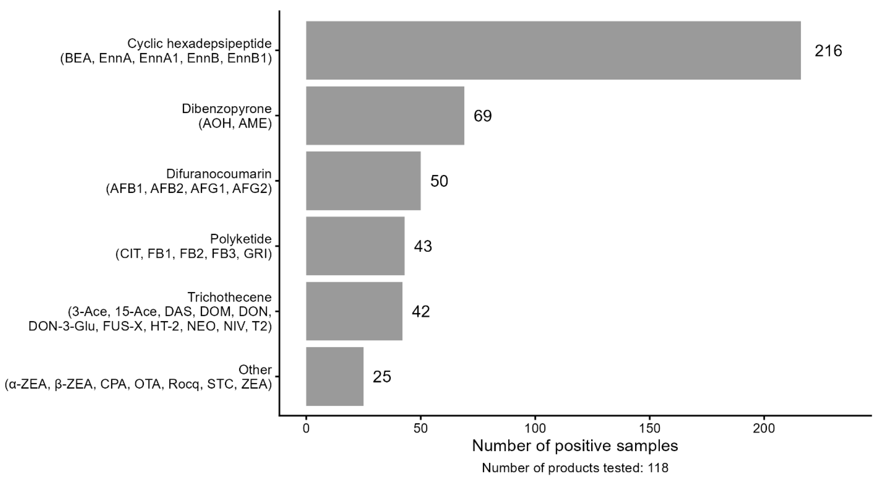

Table 1.
Foodborne Mycotoxins Tested.
| Mycotoxin | Chemical Class/Structure | Foods Detected In & Producing Fungi | Known Human Effects and US FDA Monitoring in ug/kg |
References |
|---|---|---|---|---|
| 15-Acetyl Deoxynivalenol | Type B trichothecene (acetylated DON) | Cereals (wheat, barley, maize, oats); Fusarium spp. | GI upset and toxicity, nausea, vomiting, immunotoxicity, microbiome shifts, and possibly neurotoxicity. No US guidance or direct monitoring. |
[13,15,17,68,69,70,71,72,73] |
| 3-Acetyl Deoxynivalenol | Type B trichothecene | Cereals; Fusarium spp. | Similar to DON but lower toxicity; GI toxicity and gut barrier effects, microbiome shifts, and possibly neurotoxicity. No US guidance or direct monitoring. |
[13,15,17,68,69,70,71,72,73] |
| Deoxynivalenol-3-Glucoside | Masked trichothecene (DON conjugate) | Processed cereals; Fusarium spp. | Hydrolyzed in gut → DON exposure; GI toxicity, microbiome shifts. No US guidance or direct monitoring. |
[13,15,17,68,69,70,71,72,73] |
| Deepoxy-Deoxynivalenol | Reduced trichothecene metabolite | Formed in ruminants and microbiota; cereals with Fusarium spp. |
Less toxic metabolite of DON; lower GI toxicity. No US guidance or direct monitoring. |
[74] |
| Deoxynivalenol (Vomitoxin) | Type B trichothecene | Wheat, maize, barley, oats; Fusarium spp. | Targets actively dividing cells, such as those lining the GI tract, skin, lymphoid, and erythroid cells. Causes immune system dysregulation. Categorized as a “ribotoxin” and protein synthesis inhibitor. Symptoms include anorexia, vomiting, abdominal pain, intestinal bleeding, fever, headache, immune modulation, microbiome shifts, and possibly neurotoxicity. Monitored in the US:1000 ug/kg in finished wheat products like flour, bran, and germ for adults, 500 ug/kg in cereal-based foods for infants and children. | [13,15,17,68,69,70,71,72,73,75,76] |
| Nivalenol | Type B trichothecene | Barley, maize, wheat, rice; Fusarium spp. | GI toxicity, hematotoxicity, immunosuppression. No US guidance or direct monitoring. |
[20,77,78,79,80] |
| T-2 Toxin | Type A trichothecene | Cereals, oats, maize; Fusarium spp. | Severe cytotoxicity. Targets actively dividing cells, such as those lining the GI tract, skin, lymphoid, and erythroid cells. Causes immune system dysregulation. Categorized as a “ribotoxin” and protein synthesis inhibitor. Symptoms include anorexia, vomiting, abdominal pain, intestinal bleeding, fever, headache, oral ulcers, petechia, hepatotoxicity, hematotoxicity, and bleeding. No US guidance or direct monitoring. |
[14,20,58,78,79,80,81,82,83,84] |
| HT-2 Toxin | Type A trichothecene (T-2 metabolite) | Oats, wheat, maize; Fusarium spp. | Similar to T-2, GI toxicity, immunotoxicity, and hematotoxicity. No US guidance or direct monitoring. |
[14,20,58,78,79,80,81,82,83,84] |
| Diacetoxyscirpenol | Type A trichothecene | Maize, barley; Fusarium spp. | GI toxicity, leukopenia, immunosuppression. No US guidance or direct monitoring. |
[85] |
| Fusarenon-X | Type B trichothecene | Wheat, barley, maize; Fusarium spp. |
GI effects, immunotoxicity, cytotoxicity. No US guidance or direct monitoring. |
[85] |
| Neosolaniol | Type A trichothecene | Wheat, maize, barley; Fusarium spp. | Less toxic than T-2, GI toxicity, and immunotoxicity. No US guidance or direct monitoring. |
[85] |
| Zearalenone | Resorcyclic acid lactone | Maize, wheat, barley; Fusarium spp. | An estrogenic hormone disruptor in some animals and possibly humans. Linked to hyperestrogenism, reproductive disorders, infertility, and perhaps early puberty in animals. No US guidance or direct monitoring. |
[7,8,43,84,86,87,88] |
| α-Zearalenol | Reduced metabolite of ZEA | Formed in animals; cereals with Fusarium spp. | Similar to zearalenone, but with stronger estrogenic activity. No US guidance or direct monitoring. |
[7,8,43,86,87] |
| β-Zearalenol | Reduced metabolite of ZEA | Same as above | Weaker estrogenic activity than the α-isomer. No US guidance or direct monitoring. |
[7,8,43,86,87] |
| Fumonisin B1 | Polyketide | Maize and other cereals, sorghum; Fusarium spp. | Disrupts sphingolipid metabolism in cell membranes and may be a causal factor in esophageal cancer and neural tube defects. Monitored in the US: 2000-4000 for FB1, FB2, and FB3 combined in foods consumed by humans. | [2,9,30,46,47,84,89] |
| Fumonisin B2 | Polyketide | Maize and other cereals, sorghum; Fusarium spp. | Similar to FB1, possibly hepatotoxic. Monitored in the US: 2000-4000 for FB1, FB2, and FB3 combined in foods consumed by humans. |
[2,9,30,46,47,84,89] |
| Fumonisin B3 | Polyketide | Maize and other cereals; Fusarium spp. | Less potent than FB1, but with the same toxic profile. Monitored in the US: 2000-4000 for FB1, FB2, and FB3 combined in foods consumed by humans. |
[2,9,30,46,47,84,89] |
| Enniatin A | Cyclic hexadepsipeptide | Grains, stored commodities, and dairy products; Fusarium spp. | Ionophoric, cytotoxic, adverse mitochondrial effects, and possibly hepatotoxic. No US guidance or direct monitoring. |
[34,35,55,90,91,92] |
| Enniatin A1 | Cyclic hexadepsipeptide | Grains, stored commodities, and dairy products; Fusarium spp. |
Similar to Enniatin A and Enniatin B, it induces apoptosis, has mitochondrial effects, and may also exhibit hepatotoxicity. No US guidance or direct monitoring. |
[34,35,65,90,92,93,94] |
| Enniatin B | Cyclic hexadepsipeptide | Grains, stored commodities, and dairy products; Fusarium spp. |
Most abundant; cytotoxic, antimicrobial, mitochondrial effects, possibly hepatotoxic. No US guidance or direct monitoring. |
[34,35,65,90,92,93,94] |
| Enniatin B1 | Cyclic hexadepsipeptide | Grains, stored commodities, and dairy products; Fusarium spp. |
Similar to Enniatin B, cytotoxic, antimicrobial, mitochondrial effects, possibly hepatotoxic. No US guidance or direct monitoring. |
[34,35,65,90,92,93,94] |
| Beauvericin | Cyclic hexadepsipeptide | Cereals, maize, rice, stored commodities; Fusarium spp., Bassiana Beauveria |
Ionophoric, induces apoptosis, cardiotoxic in vitro, antimicrobial, and has mitochondrial effects. No US guidance or direct monitoring. |
[35,65,90,91,92,93,94] |
| Ochratoxin A | Isocoumarin + phenylalanine | Cereals, coffee, dried fruit, cacao, wine; Aspergillus spp., Penicillium spp. | Nephrotoxic, immunotoxic, neurotoxic, and possibly carcinogenic. No US guidance or direct monitoring. |
[9,25,30,84,95,96] |
| Citrinin | Polyketide | Cereals, rice, cheese, cacao; Penicillium spp., Aspergillus spp. | Nephrotoxic, mitochondrial dysfunction. No US guidance or direct monitoring. |
[2,97] |
| Aflatoxin B1 | Difuranocoumarin | Cereals, maize, peanuts, tree nuts; Aspergillus spp. | Hepatotoxic and hepatocarcinogenic (Group 1), immunosuppressive, and GI toxicity. Monitored in the US: 20 for total (AB1, AB2, AG1, and AG2 combined). |
[2,31,32,33,88,98,99,100,101,102,103] |
| Aflatoxin B2 | Difuranocoumarin | Same foods; Aspergillus spp. |
Similar to B1, less potent. Monitored in the US: 20 for total (AB1, AB2, AG1, and AG2 combined). |
[2,31,32,33,88,98,99,100,101,102,103] |
| Aflatoxin G1 | Difuranocoumarin | Maize, nuts; Aspergillus spp. |
Carcinogenic and hepatotoxic. Monitored in the US: 20 for total (AB1, AB2, AG1, and AG2 combined). | [2,31,32,33,88,98,99,100,101,102,103] |
| Aflatoxin G2 | Difuranocoumarin | Same foods; Aspergillus spp. |
Lower toxicity than G1, carcinogenic, and hepatotoxic. Monitored in the US: 20 for total (AB1, AB2, AG1, and AG2 combined). |
[2,31,32,33,88,98,99,100,101,102,103] |
| Sterigmatocystin | Xanthone precursor of aflatoxin | Cereals, cheese; Aspergillus spp. | Hepatotoxic, probable carcinogen 2B. No US guidance or direct monitoring. |
[104] |
| Cyclopiazonic Acid | Indole-tetramic acid | Maize, peanuts, milk, cheese, meat products, and eggs; Aspergillus spp., Penicillium spp. |
Muscle tremors, neurotoxin, and GI toxicity. Possibly responsible for Kodua poisoning in humans. No US guidance or direct monitoring. |
[26,27] |
| Alternariol | Dibenzopyrone | Tomatoes, citrus, cereals; Alternaria spp. |
Carcinogenic, nephrotoxic, hepatotoxic, and immunotoxic in animals at low µg/kg/day in repeated doses. Has demonstrated antibiotic/antibacterial activity (e.g., against Staphylococcus aureus and Candida albicans), cholinesterase inhibition, and some antioxidant activity in specific assays. No US guidance or direct monitoring. |
[37,41,42] |
| Alternariol-Monomethyl Ether | Dibenzopyrone | Tomatoes, cereals; Alternaria spp. |
Genotoxic in vitro and shows toxicity to liver, kidney, spleen, and immune function in animals at low µg/kg/day in repeated doses. No US guidance or direct monitoring. |
[37,41,42] |
| Roquefortine C | Indole alkaloid | Cheese, cereals; Penicillium spp. | Neurotoxic, with increased generation of reactive oxygen species (ROS), convulsant activity in animals. No US guidance or direct monitoring. |
[105] |
| Griseofulvin | Polyketide secondary metabolite | Cereals, chocolate; Penicillium spp. | Hepatocarcinogen, GI disturbances, and allergic reactions. Used in medicine as an antifungal medication. No US guidance or direct monitoring. | [106,107] |
Table 3.
Categories of Foods Sampled (n=118).
| Foods Tested | Number of samples |
|---|---|
| Snack puffs, crackers, and biscuits | 27 |
| Processed cereal products (wheat, corn, oats, rice, and barley) | 25 |
| Pasta (wheat-based) | 13 |
| Misc. foods (plant-based formulas, beans, juice, fruit puree) | 7 |
| Wheat flour | 10 |
| Non-grain ingredients (almond flour, tapioca starch, arrowroot, coconut flour, chocolate powder) | 15 |
| Corn flour | 21 |
Table 4.
Number of Samples Contaminated with Each Mycotoxin.
| Chemical Class/Structure | Analyte and Abbreviation |
LOQ (ppm) |
Number of positive samples |
Highest amount (µg/kg) |
Food type with the highest amount | Median amount (µg/kg) |
|---|---|---|---|---|---|---|
| Cyclic hexadepsipeptide |
Beauvericin (BEA) | 0.002 | 49 | 190.3 | Breakfast cereals | 8.3 |
| Enniatin A (EnnA) | 0.002 | 25 | 95.6 | Baby apple juice | 14.3 | |
| Enniatin A1 (EnnA1) | 0.002 | 46 | 100.7 | Baby apple juice | 7.8 | |
| Enniatin B (EnnB) | 0.002 | 51 | 85.6 | Pasta | 9.8 | |
| Enniatin B1 (EnnB1) | 0.002 | 45 | 82.5 | Baby apple juice | 9.5 | |
| Dibenzopyrone | Alternariol (AOH) | 0.002 | 11 | 28.4 | Wheat flour | 6.0 |
| Alternariol-Monomethyl Ether (AME) | 0.002 | 31 | 36.4 | Corn flour | 3.6 | |
| Difuranocoumarin | Aflatoxin B1 (AFB1) | 0.0005 | 47 | 246.0 | Corn flour | 1.3 |
| Aflatoxin B2 (AFB2) | 0.0005 | 16 | 25.2 | Corn flour | 1.8 | |
| Aflatoxin G1 (AFG1) | 0.0005 | 5 | 2.6 | Corn flour | 2.0 | |
| Aflatoxin G2 (AFG2) | 0.0005 | 0 | N/A | N/A | N/A | |
| Difuranocoumarin xanthone precursor to aflatoxin |
Sterigmatocystin (STC) | 0.005 | 1 | 5.7 | Corn flour | 5.7 |
| Indole alkaloid | Rocquefortine C (Rocq) | 0.002 | 0 | N/A | N/A | N/A |
| Indole-tetramic acid | Cyclopiazonic Acid (CPA) | 0.002 | 10 | 3804.0 | Corn flour | 134.8 |
| Isocoumarin + phenylalanine |
Ochratoxin A (OTA) | 0.001 | 6 | 2.5 | Corn flour | 1.2 |
| Polyketide | Citrinin (CIT) | 0.05 | 0 | N/A | N/A | N/A |
| Fumonisin B1 (FB1) | 0.1 | 21 | 1299.0 | Corn flour | 300.0 | |
| Fumonisin B2 (FB2) | 0.1 | 15 | 467.0 | Corn flour | 100.0 | |
| Fumonisin B3 (FB3) | 0.1 | 5 | 200.0 | Corn flour | 100.0 | |
| Griseofulvin (GRI) | 0.002 | 0 | N/A | Non-grain | N/A | |
| Resorcyclic acid | Zearalenone (ZEA) | 0.0125 | 9 | 126.6 | Corn flour | 32.2 |
| Resorcyclic acid reduced |
Alpha-Zearalenone (a-ZEA) |
0.05 | 0 | N/A | N/A | N/A |
| Beta-Zearalenone (b-ZEA) | 0.05 | 0 | N/A | N/A | N/A | |
| Trichothecene Type A |
Diacetoxyscirpenol (DAS) | 0.1 | 0 | N/A | N/A | N/A |
| HT-2 Toxin (HT-2) | 0.005 | 7 | 71.2 | Corn flour | 8.9 | |
| Neosolaniol (NEO) | 0.02 | 0 | N/A | N/A | N/A | |
| T-2 Toxin (T2) | 0.005 | 2 | 49.6 | Corn flour | 29.9 | |
| Trichothecene Type B |
15-Acetyl Deoxynivalenol (Ace-15) | 0.1 | 7 | 700.0 | Breakfast cereals | 100.0 |
| 3-Acetyl Deoxynivalenol (Ace-3) | 0.1 | 2 | 100.0 | Breakfast cereals | 100.0 | |
| Deepoxy-Deoxynivalenol (DOM) | 0.02 | 9 | 93.4 | Corn flour | 48.8 | |
| Deoxynivalenol (DON) | 0.1 | 17 | 2499.0 | Breakfast cereals | 300.0 | |
| Deoxynivalenol-3-Glucoside (DON-3-Glu) |
0.02 | 3 | 585.3 | Corn flour | 24.6 | |
| Fusarenon-x (FUS-X) | 0.5 | 0 | N/A | N/A | N/A | |
| Nivalenol (NIV) | 0.5 | 3 | 19070.0 | Snack foods | 7700.0 |
Table 5.
Number of Mycotoxins per Food Type by Chemical Structure.
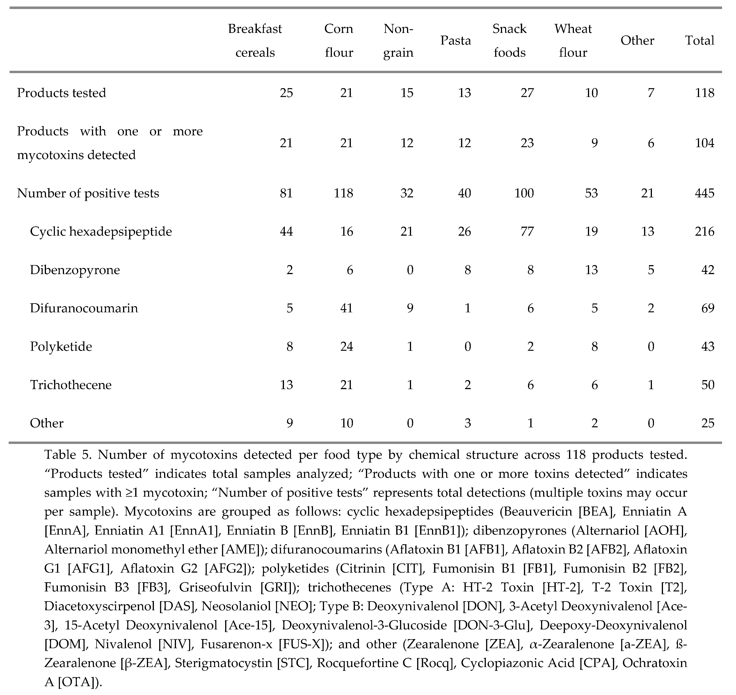 |
Table 6.
Tolerable Daily Intake (TDI) Toddler Assessment by Mycotoxin Detected.
| Mycotoxin | TDI | Highest Level and Fold Exceedance of TDI |
Median Level and Fold Exceedance of TDI |
|---|---|---|---|
| *Aflatoxins (total AFB1, AFB2, AFG1, AFG2) | 0.017 μg/kg bw/day | 273.8 μg/kg = 73-fold higher |
5.1 μg/kg= 1.4-fold higher |
| Deoxynivalenol plus acetylated derivatives and masked forms | 1 μg/kg bw/day | 2,499 μg/kg = 11.4-fold higher |
300 μg/kg = 1.4-fold higher |
| Nivalenol | 1.2 μg/kg bw/day | 19,070 μg/kg = 72-fold higher |
7,700 μg/kg = 29-fold higher |
| Fumonisins (total FB1, FB2, and FB3) | 1 μg/kg bw/day | 1,966 μg/kg = 9-fold higher |
500 μg/kg = 2.3-fold higher |
| Ochratoxin A | 0.120 μg/kg bw/week or 0.017/day | 2.5 μg/kg= 0.67 of the TDI |
1.2 μg/kg = 0.32 of the TDI |
|
**T-2 Toxin and HT-2 Toxin combined |
0.02 μg/kg bw/day | 120.8 μg/kg= 27.5-fold higher |
NA*** |
| T-2 Toxin | 0.02 μg/kg bw/day | 49.6 μg/kg = 11.3-fold higher |
8.9 μg/kg= 2-fold higher |
| HT-2 Toxin | 0.02 μg/kg bw/day | 71.2 μg/kg= 16.2-fold higher |
29.9 μg/kg= 6.8-fold higher |
| Zearalenone | 0.25 μg/kg bw/day | 126.6 μg/kg= 2.3-fold higher |
32.2 μg/kg= 0.59 of the TDI |
Note: Emerging mycotoxins without regulatory guidance and mycotoxins not detected are not included in the table. Calculations were based on using a 57-gram serving size and the 12.5 kg bw for toddler assessment using the following equations: Exposure (ng/kg/bw day) = Mycotoxin Contamination level (µg/kg) × Consumption data (g)/bw (kg) divided by 1,000 for μg/kg bw/day, and Toddler Exposure =TDI (µg/kg bw day) /Actual intake (µg/kg bw day). *There is no established TDI for Aflatoxins, only a proposed value. **T-2 and HT-2 Toxins are usually considered together in risk assessments, but are separated here because they did not always occur together in samples. ***There was no median data available for T-2 and HT-2 toxins combined. Citations: [32,45,98,117,118,119,120,121].
Disclaimer/Publisher’s Note: The statements, opinions and data contained in all publications are solely those of the individual author(s) and contributor(s) and not of MDPI and/or the editor(s). MDPI and/or the editor(s) disclaim responsibility for any injury to people or property resulting from any ideas, methods, instructions or products referred to in the content. |
© 2026 by the authors. Licensee MDPI, Basel, Switzerland. This article is an open access article distributed under the terms and conditions of the Creative Commons Attribution (CC BY) license (http://creativecommons.org/licenses/by/4.0/).
Copyright: This open access article is published under a Creative Commons CC BY 4.0 license, which permit the free download, distribution, and reuse, provided that the author and preprint are cited in any reuse.