Submitted:
29 April 2026
Posted:
02 May 2026
You are already at the latest version
Abstract
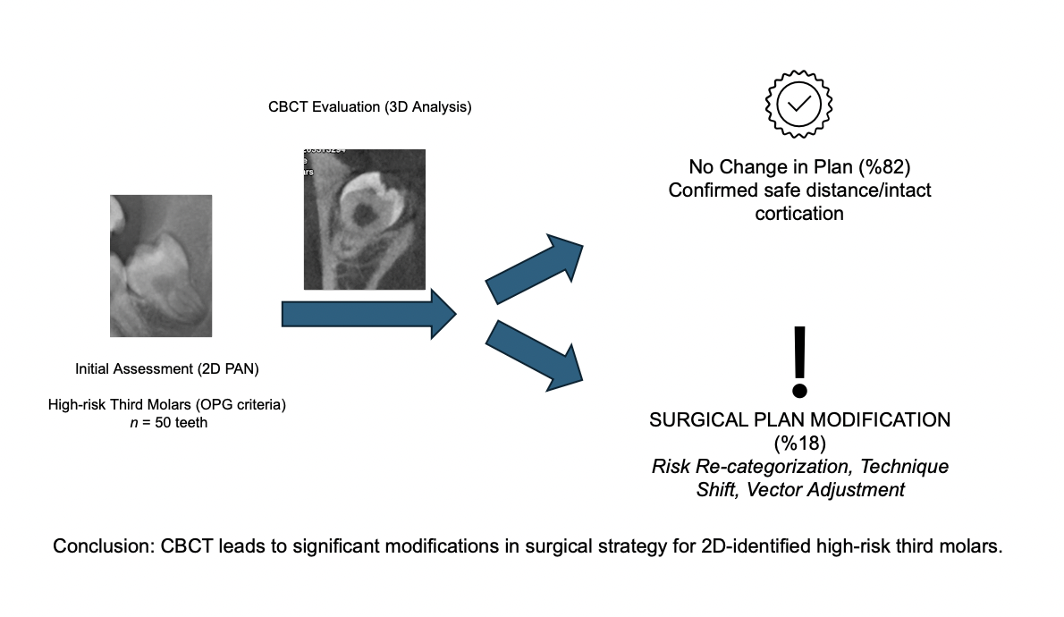
Aim. This study aimed to evaluate the impact of cone-beam computed tomography (CBCT) on preoperative surgical decision-making and risk assessment for mandibular third molar (MM3) extractions in cases identified as high-risk by orthopantomograph (OPG).
Materials and Methods. This prospective clinical study utilized the purposive sampling method, recruting50 MM3s from 33 patients (mean age 24.24 ± 6.77 years). Samples were categorized into five distinct radiographic groups based on the proximity of roots to the inferior alveolar canal (IAC) on OPG. The methodology involved a comparative 3D analysis to determine neurovascular contact, spatial orientation, and the presence of cortical border. Surgical strategies, specifically the necessity for coronectomy or the lingual split technique, were reassessed following 3D evaluation. Postoperative neurosensory outcomes were recorded. Statistical analysis was performed using the Fisher-Freeman-Halton and Kruskal-Wallis tests.
Results. CBCT identified direct IAC contact in 74% of the cases. In 18% of the cases initially deemed high-risk by OPG, CBCT revealed a safe distance, thereby altering the surgical approach. Tooth angulation (P = 0.012) and Pell and Gregory classification (P = 0.0024) were significant predictors of contact. Temporary neurosensory disturbances occured in 4% (n = 2) of the sample, specifically in cases where CBCT had confirmed loss of canal cortication.
Conclusion. In accordance with the study aim, CBCT provides essential 3D data that refines surgical planning in nearly one-fifth of high-risk cases. The findings justify selective CBCT use, guided by the ALADA principle, to minimize iatrogenic injury.
Keywords:
cone-beam computed tomography
; mandibular nerve
; molar
; third
; oral surgicalprocedures
; prospective studies
Copyright: This open access article is published under a Creative Commons CC BY 4.0 license, which permit the free download, distribution, and reuse, provided that the author and preprint are cited in any reuse.


