

Submitted:
29 April 2026
Posted:
01 May 2026
You are already at the latest version
Abstract
Parkinson’s disease (PD) encompasses marked heterogeneity across motor, cognitive, and non-motor domains, reflecting variable balances of neurodegeneration and compensation across distributed brain circuits. Diffusion MRI tractography enables pathway-specific characterization of white matter alterations and offers a framework for linking clinical subtypes to patterns of degeneration and compensation along individual tracts that are often obscured by skeleton-based or connectomic averaging. Although several tract-specific correlational diffusion studies have linked individual pathways to clinical features and progression, much of the literature has relied on group-level skeleton or network representations, limiting generalizability and reproducibility across subtypes. Here, we synthesize tractography-based evidence across PD subtypes—including tremor-dominant, postural instability/gait difficulty, freezing of gait, and cognitive phenotypes—while situating these findings within a complementary multimodal imaging context. We review diffusion models ranging from diffusion tensor imaging to advanced free-water, neurite and fixel-based frameworks, highlighting how these approaches constrain and interpret tract-level findings and help distinguish degenerative processes from adaptive neuroplasticity. Emerging analytical approaches, including harmonization pipelines, radiomic tractometry (the extraction of along-tract microstructural and radiomic features), and machine learning classifiers, further enhance tract-level sensitivity and reproducibility. Cognitive subtypes illustrate how degeneration of posterior association and limbic tracts, in interaction with non-dopaminergic systems such as cholinergic and noradrenergic pathways, shapes clinical progression. Integrating tractography with molecular, genetic, and functional markers enables subtype-specific biomarkers for risk stratification, prognosis, and targeted therapeutic intervention. We propose a conceptual and methodological roadmap for leveraging tractography to refine PD subtype definitions and inform precision neuromodulation and rehabilitation strategies.
Keywords:
diffusion MRI
; disease subtypes
; neuroplasticity
; Parkinson’s disease
; tractography
; white matter
1. Introduction
Parkinson’s disease (PD) is the second most common neurodegenerative disorder, affecting over 10 million people worldwide [1]. It is increasingly recognized as a clinically and biologically heterogeneous disorder, encompassing variability in motor and non-motor symptoms, disease progression, and treatment response. This heterogeneity has motivated efforts to classify PD into subtypes such as tremor-dominant (TD), postural instability/gait difficulty (PIGD), akinetic-rigid (AR), cognitive variants, and genetic forms, with the goal of improving prognostication and guiding personalized interventions [2,3]. Neuroimaging has played a central role in these efforts by characterizing the neural substrates of subtype differences, revealing both degenerative and compensatory mechanisms that shape clinical trajectories.
Neuroplasticity provides a framework for understanding PD heterogeneity, reflecting both dopaminergic loss and compensatory network adaptations [4,5]. Functional MRI shows early compensatory activity in premotor and parietal areas [6,7,8], with the white matter (WM) substrates increasingly explored with diffusion MRI (dMRI) and tractography. Conventional tensor metrics such as fractional anisotropy (FA) and mean diffusivity (MD) show changes in corpus callosum, corticospinal, and frontostriatal pathways [9], while advanced models (free-water imaging [FWI], neurite orientation dispersion and density imaging [NODDI], and fixel-based analysis [FBA]) offer detailed microstructural insights [10,11]. Large harmonized studies confirm widespread WM alterations, supporting PD as a network disorder [12]. Subtype-specific differences are evident: PIGD shows extensive degeneration, while TD exhibits limited changes and potential compensatory circuitry [13].
Despite substantial progress in understanding clinical and imaging heterogeneity in PD, how these dimensions intersect with tractography findings has rarely been examined. Prior reviews have largely synthesized structural (e.g., cortical thickness, volumetry, diffusion tensor imaging (DTI)-based connectomics) and functional neuroimaging evidence (e.g., fMRI, PET/SPECT) related to PD subtypes [14,15,16,17], focusing on circuit- and network-level abnormalities within basal ganglia–thalamo–cortical (BGTC) and cerebello–thalamo–cortical (CTC) pathways. While these approaches are statistically tractable and widely adopted, their reliance on skeleton- or network-level abstractions may obscure tract-level heterogeneity. For example, alterations in the corticospinal tract (CST) may characterize AR subtypes, whereas cingulum bundle changes may underlie cognitive-predominant subtypes. Tract-based spatial statistics (TBSS) or connectomic analyses may average across these differences, masking tract-specific patterns that could be clinically relevant in tracking disease progression, designing disease and subtype-specific neuroinformed interventions, diagnosis, and personalized treatment regimen.
Bridging this gap requires analytical approaches that retain pathway-specific resolution while remaining interpretable within broader circuit and network frameworks. Tractography, in this context, provides spatially precise information along individual WM pathways, e.g. application in PD to reconstruct clinically relevant projections, including brainstem locomotor pathways and cortico-subthalamic connections used to guide deep brain stimulation (DBS) targeting [18]; but its implementation requires higher-quality data and robust modeling to overcome challenges of crossing fibers, partial volume effects, and reproducibility [19,20]. Consequently, while tract-level diffusion measures are increasingly available in large cohorts, relatively few studies have leveraged deterministic or probabilistic reconstruction of individual pathways to interrogate subtype-specific neuroplasticity in a mechanistic manner. Our understanding of whether diffusion alterations vary systematically along particular tracts and how such differences align with clinical heterogeneity is, thus, limited [12,21]. Moreover, although tractography is central to characterizing tract-specific alterations in PD, its interpretation cannot occur in isolation. DTI, TBSS, FWI, NODDI, FBA, and functional imaging each capture complementary aspects of WM organization, microstructural change, and network-level adaptation. Accordingly, this review situates tractography within a broader multimodal neuroimaging context, using these approaches to constrain, interpret, and validate tract-level findings. This integrated framework is essential for distinguishing degenerative processes from compensatory neuroplasticity and for understanding how tract-specific alterations scale to circuit- and network-level dysfunction across PD subtypes.
Recent diffusion work has begun to reveal tract- and region-specific signatures across motor and cognitive networks in PD. For example, cerebellar peduncles and associated pathways show differential FA and MD changes between PIGD and TD phenotypes, implicating CTC circuits in subtype-specific motor manifestations [22]. Moreover, tract alterations within executive networks (e.g., superior longitudinal fasciculus [SLF]/frontotemporal association pathways) differ across phenotypes and parallel executive deficits, and diffusion-network measures predict longitudinal worsening toward diffuse-malignant subtypes [23,24]. Importantly, most of these tract-level inferences are derived from voxelwise, skeleton-based, or connectomic analyses rather than from explicit reconstruction of individual pathways. Emerging subtyping frameworks increasingly integrate tract-level neuroimaging with clinical and biomarker data, moving beyond broad circuit-level models to more granular structural phenotyping [25]. These findings emphasize the potential of tract-level metrics as early prognostic markers that complement traditional network-level analyses. Collectively, these distinct tract-level patterns across PD subtypes and disease trajectories underscore the need for a dedicated synthesis of tractography studies to clarify both degenerative and compensatory processes [12,26,27].
The aim of this review is therefore theoretical and integrative: to synthesize current evidence on WM tractography in PD as the primary analytical framework, while situating tract-level findings within the broader neuroimaging context, with particular emphasis on how subtype-specific differences in neuroplasticity are reflected in tract-based findings and analytical approaches. We first outline the conceptual framework of neuroplasticity in PD, then provide a methodological overview of diffusion models and tractography strategies. Building on this foundation, we highlight consistent WM alterations across PD, evaluate subtype-specific findings, and examine longitudinal evidence of plasticity and degeneration. Special attention is given to how tract-level alterations relate not only to motor subtypes but also to cognitive subtypes and associated impairments, particularly within executive and limbic networks. Finally, we discuss the translational relevance of these insights — from refining patient stratification to informing DBS targeting, prognosis, and rehabilitative interventions.
2. Conceptualizing Neuroplasticity in PD
Neuroplasticity in PD has a dual role: enabling compensatory mechanisms that sustain motor and cognitive function despite dopaminergic loss, while also fostering maladaptive changes that exacerbate symptoms such as dyskinesias, freezing of gait (FOG), or fatigue.
Functional imaging shows increased frontal and parietal activity in early PD, reflecting cortical recruitment to offset basal ganglia dysfunction. In a landmark fMRI study of 350 early PD patients, Johansson et al. (2024) found that variability in motor severity was more strongly linked to loss of parieto-premotor cortical compensation than to striatal dysfunction, accentuating cortical adaptive networks in symptom expression [5]. Yet not all plasticity is beneficial: pathological resonance in BGTC loops contributes to tremor [28]; maladaptive limbic connectivity involving orbitofrontal, caudate, and insular circuits underlies impulse control disorders [29]; and dopaminergic therapy can induce pathological frontal oscillations tied to levodopa-induced dyskinesias [30].
Neuroplasticity also involves CTC reorganization, preserved in TD but disrupted in PIGD [31,32]. Diffusion studies highlight the SN as a structural hub linking motor, limbic, and hippocampal networks, explaining cognitive and affective symptoms [20]. Genetic background further biases these neuroplastic processes e.g., GBA-associated PD exhibits more widespread WM degeneration across limbic, association, and projection pathways, whereas idiopathic PD (iPD) more often shows relative preservation or regionally increased structural connectivity in motor-related tracts—patterns consistent with reduced versus preserved compensatory capacity, respectively [33,34]. Thus, tractography provides not only a marker of burden but a window into adaptive capacity.
3. Methodological Considerations in dMRI and Tractography
DMRI has transformed PD research by enabling in vivo assessment of WM microstructure and connectivity. Early DTI studies revealed decreased FA and increased MD in corticospinal and frontostriatal tracts, but the non-specificity of these metrics limited interpretation [35]. Advances in multi-compartment models, tractography, and connectomics now provide richer frameworks to link WM changes with neuroplastic processes and subtype-specific trajectories. Methodological precision is critical, since acquisition and analysis choices directly shape reproducibility and clinical interpretability.
3.1. Acquisition and Preprocessing, and the Limits of Cross-Site Reproducibility
High-quality acquisition underpins reliable dMRI, but reproducibility across scanners, protocols, and cohorts remains a central challenge for tract-specific inference in PD. Multi-shell high angular resolution diffusion imaging (HARDI) with sufficient angular coverage improves sensitivity to complex WM organization [36]. Wei et al. (2024) applied a clinically feasible multi-shell protocol (b=1000/2000 s/mm²; 64 directions; 1.8 mm voxels) in PD patients, showing neurite density changes linked to gait dysfunction [37]. Key acquisition considerations include ≤2 mm isotropic voxels to reduce partial volume effects, high signal-to-noise ratios (SNR), and dual phase-encoding for susceptibility distortion correction. Scan durations of ~15–20 minutes balance spatial resolution with patient tolerance.
Preprocessing pipelines typically include denoising, Gibbs ringing removal, eddy-current and motion correction, susceptibility distortion correction, bias field correction, and brain masking [38]. Multisite studies additionally require harmonization, but this remains a nontrivial source of variability. ComBat and its variants are widely used to adjust for scanner-related additive and multiplicative biases while attempting to preserve biological signal [39,40]. However, recent methodological reassessments demonstrate that ComBat relies on strong assumptions, including comparable covariate slopes, balanced demographic distributions, and sufficient sample sizes across sites—that are frequently violated in neurodegenerative cohorts. When these assumptions are unmet, harmonization can attenuate or distort biologically meaningful effects rather than clarify them, particularly in pathological populations [41].
Accordingly, harmonization should be treated as a modelling step rather than a corrective “black box,” and tract-level findings, especially subtle subtype differences, should be interpreted in the context of acquisition parameters, cohort composition, and residual site effects [42]. Despite these limitations, applications in PD demonstrate that harmonization can reduce site bias and improve classification of motor subtypes when carefully applied [43,44,45]. Large-scale initiatives such as ENIGMA-DTI [12] and PANDA [46] further extend these efforts with through standardized training, quality control, and centralized pipelines, providing an essential foundation for reproducible multisite diffusion studies.
3.2. Tractography Approaches: Deterministic vs. Probabilistic
Tractography is a computational method that reconstructs WM pathways from dMRI data, allowing in vivo mapping of structural connectivity. All tractography methods infer the likely trajectory of fiber bundles by following local estimates of diffusion orientation across voxels, but they differ in how the uncertainty is handled. Deterministic tractography follows the principal diffusion direction voxel by voxel, producing a single, streamline-based reconstruction. It is fast, intuitive, and widely used for clinical applications. Probabilistic tractography, by contrast, samples from a distribution of possible fiber orientations, generating a connectivity map that incorporates uncertainty, particularly useful in regions with crossing, kissing, or branching fibers [47,48]. Together, these complementary approaches have become foundational tools in structural connectomics.
Deterministic tractography has been used to map key basal ganglia and motor pathways, including nigrostriatal and pallidal tracts, revealing reduced FA and streamline counts that correlate with disease severity [49]. However, deterministic tractography can underestimate connectivity in regions of complex fiber architecture [50]. Probabilistic approaches improve robustness by estimating orientation distributions, enabling more reliable mapping in challenging regions. For example, preserved cerebellar–thalamic connectivity in TD relative to other subtypes has been revealed using probabilistic tractography [51], and hyperdirect pathway reconstructions for DBS planning have shown greater accuracy compared to deterministic methods [52]. Finally, filtering techniques such as Spherical-deconvolution Informed Filtering of Tractograms (SIFT2) and Convex Optimization Modeling for Microstructure Informed Tractography (COMMIT) reduce false positives and improve biological plausibility [53,54,55]. Together, these approaches form the backbone of connectome reconstruction and enable increasingly precise mapping of PD-related network alterations.
3.3. Connectomics and Network-Level Analyses
Applying graph theory to tractography-derived connectomes allows network-level characterization of PD. Global efficiency is consistently reduced in PIGD, reflecting widespread disruption of long-range connectivity, while TD patients may preserve local efficiency and modularity, suggestive of compensatory mechanisms [56,57]. Hub vulnerability is another key concept: highly connected nodes such as supplementary motor area (SMA), thalamus, and basal ganglia show preferential WM disconnection correlating with progression and severity [58]. By capturing distributed rather than isolated changes, connectomics reframes PD as a disorder of network integrity, offering potential imaging biomarkers for disease progression and treatment response. Beyond degeneration, connectomic dMRI studies also provide evidence for compensatory reorganization, with preserved or enhanced network efficiency and hub stability reported in early and TD-PD, underscoring that network-level adaptation can be captured structurally as well as functionally.
3.4. Diffusion Models: From DTI to Advanced Frameworks
DTI remains widely used due to its short scan times and interpretability, and it has consistently revealed WM abnormalities in callosal and corticospinal pathways [59]. But because DTI uses a single-tensor model, it oversimplifies regions with fiber crossings and conflates underlying microstructural changes, thereby limiting sensitivity, especially in circuits critical to PD (e.g. crossing cerebellar, association, and basal ganglia fibers) [60]. Advanced multi-compartment models help disambiguate whether observed diffusion changes reflect extracellular water expansion, axonal degeneration, or tract-level remodeling (Figure 1). Recent methodological syntheses emphasize that these advances are most informative when coupled with tractography, which enables pathway-specific microstructural inference beyond voxelwise or skeleton-based analyses [20].
These models provide complementary rather than convergent information. FWI, NODDI, and FBA capture distinct microstructural phenomena and should not be interpreted as interchangeable markers of the same biological mechanism, but instead, as orthogonal perspectives on WM architecture.
Schematic overview of three advanced dMRI models—Free-Water Imaging (FWI), Neurite Orientation Dispersion and Density Imaging (NODDI), and Fixel-Based Analysis (FBA). Importantly, these models do not provide convergent estimates of the same biological process. Rather, each offers a complementary—and fundamentally non-equivalent—microstructural sensitivity: FWI isolates extracellular fluid, sensitive to neuroinflammation and predictive of progression. NODDI quantifies neurite density and dispersion, capturing axonal degeneration and tract disorganization. FBA resolves crossing fibers and detects pathway-specific degeneration or remodeling through fiber-specific metrics (FD, FC). Divergences across their metrics often reflect different underlying microstructural processes. Together, these models help disentangle FA changes, support subtype stratification (e.g., PIGD vs. TD), and offer clinically actionable endpoints for trials.
Diffusion microstructural imaging (DMI) partitions diffusion into intra-, extra-axonal, and isotropic compartments, offering mechanistic clarity. It shows promise as an early biomarker, especially in prodromal or genetic PD, yet its adoption remains limited by longer acquisition times [61]. Validation in large, longitudinal PD cohorts also remains limited, so translational uptake is still at an early stage. FWI separates extracellular water from tissue signal [62], improving specificity in CSF-adjacent regions. Longitudinal work indicates that nigral FW increases predict progression and cognitive decline [10,63], and that FW fluctuations occur in non-nigral regions such as putamen and cerebellar pathways, particularly in PIGD phenotypes [64].
NODDI [65] captures microstructural changes in both WM and gray matter. Reduced NDI in CST correlates with gait impairment, and levodopa exposure appears to modulate these metrics [37,66]. Constrained spherical deconvolution (CSD) resolves multiple fiber orientations per voxel, improving delineation of complex pathways [67]. For example, CSD enhances reconstruction of corticospinal and cerebellar tracts implicated in tremor, enabling better microstructural assessment [68].
FBA builds on these models by providing fiber-specific indices (fiber density [FD], fiber-bundle cross-section [FC], and their combined measure [FDC]). In PD, FBA has revealed subtype-specific signatures: TD patients often show preserved or increased corticospinal integrity [56,69,70], whereas PIGD and cognitive subtypes demonstrate greater tract-specific degeneration [11,56,71,72,73,74]. Supporting these, one recent study [75] identified distinct FBA-derived signatures that successfully discriminated TD from PIGD patients, underscoring FBA’s potential to reveal nuanced, subtype-specific network differences that remain imperceptible to traditional diffusion metrics. This sensitivity has made FBA particularly valuable for subtype stratification in PD. Beyond subtype discrimination, FBA has also revealed clinically meaningful subregional alterations that standard DTI fails to detect. A recent study demonstrated that corpus callosum subregions exhibit distinct fixel-based signatures associated with motor (AR), autonomic, and cognitive symptoms in early PD, highlighting the enhanced sensitivity of FBA for uncovering tract- and subregion-specific pathology [76].
Conventional DTI has provided important early evidence that motor subtypes in PD are associated with differential patterns of WM involvement, particularly within cortico–basal ganglia and CTC systems. A recent systematic synthesis of dMRI studies across PD motor subtypes demonstrated that TD and PIGD phenotypes show partially distinct DTI signatures, consistent with differential engagement of striato-thalamo-cortical (STC) and CTC circuits [77]. However, this work also highlighted substantial heterogeneity across studies, with subtype effects varying by region, analytic approach, and disease stage, underscoring the limited specificity of tensor-based metrics for resolving overlapping motor networks. These limitations motivate the adoption of advanced diffusion models and tractography-based analyses, which better account for crossing fibers, extracellular FW, and fiber-specific degeneration, and are therefore more suited to enhance biological specificity for earlier tract-based evidence and disentangling subtype-specific circuit pathology. Table 1 summarizes the strengths, limitations, and representative PD applications of each diffusion model and how they pair with tractography strategies.
3.5. Analytical Limitations and Model-Specific Biases in dMRI
Several inherent limitations constrain the interpretability and reproducibility of dMRI–based tract-specific findings in PD. These limitations arise at multiple levels, including signal modeling, tract reconstruction, metric interpretation, and cross-site harmonization, and are particularly salient in neurodegenerative cohorts characterized by low SNR, motion, and microstructural heterogeneity. Conventional DTI metrics such as FA and MD are sensitive but biologically nonspecific, conflating axonal density, myelination, fiber coherence, and extracellular water content. As a result, similar FA changes can reflect distinct—and sometimes opposing—microstructural processes, limiting mechanistic inference at both voxel and tract levels [78]. These ambiguities are exacerbated in regions with complex fiber architecture, where tensor-based models fundamentally fail to represent crossing, kissing, or branching fibers [79].
Both deterministic and probabilistic approaches are prone to false positives and false negatives, and streamline counts or densities cannot be interpreted as direct measures of axonal number or connectivity strength [80]. These limitations are particularly relevant in PD, where degeneration, free-water expansion, and iron-related susceptibility effects disproportionately affect the basal ganglia, brainstem, and cerebellar pathways that are central to disease mechanisms.
Advanced approaches such as CSD and FBA improve sensitivity to crossing fibers but introduce additional dependencies on signal quality and model assumptions. Fiber orientation distribution (FOD) estimation relies critically on accurate response function calibration; even modest miscalibration can inflate apparent fiber density, generate spurious peaks, or obscure genuine tract degeneration - effects that are amplified at low SNR and in pathological tissue where assumptions derived from healthy WM may not hold [11,81].
These limitations that are particularly relevant in PD. where subcortical, brainstem, and cerebellar regions (regions central to PD mechanisms) are disproportionately affected by susceptibility artifacts, partial-volume effects, and motion, reducing the reliability of fixel estimation in precisely these circuits [82,83]. As a result, FBA-derived subtype differences may preferentially reflect supratentorial association and projection pathways, while changes in deep gray nuclei and brainstem tracts may be underestimated or inconsistently detected across studies.
These constraints necessitate cautious interpretation of negative findings in subcortical regions and support the integration of complementary diffusion models and region-specific quality control when applying FBA to PD cohorts [84].
Graph-theoretical analyses further abstract diffusion data, amplifying sensitivity to preprocessing choices, parcellation schemes, and thresholding strategies. Network metrics such as efficiency or hubness are not uniquely defined and may vary substantially across pipelines, limiting cross-study comparability and mechanistic specificity [85]. These issues are further amplified in multicenter PD studies, where variability in SNR and susceptibility correction can differentially affect fixel estimation in subcortical and brainstem regions, underscoring the importance of harmonized acquisition and region-aware validation.
These analytical constraints motivate the development of harmonized pipelines, multiquantitative integration strategies, and machine learning (ML)–based frameworks designed to capture distributed network effects rather than relying on isolated tract-level markers (Table 2).
3.6. Emerging Analytical Tools and Reproducibility Challenges
Building on the methodological distinctions outlined in Section 3.2, reproducibility in tractography-based analyses is further influenced by algorithmic choices, particularly the use of deterministic versus probabilistic tracking. Deterministic tractography offers higher anatomical specificity and interpretability but is sensitive to noise and may prematurely terminate in regions of low anisotropy or complex fiber geometry, such as the brainstem, cerebellar peduncles, and basal ganglia projections that are central to PD pathology. Probabilistic approaches better capture uncertainty and crossing-fiber architecture but at the cost of increased false-positive streamlines and reduced tract specificity, complicating biological interpretation. In PD, where degeneration, partial-volume effects, and extracellular FW alter diffusion profiles in precisely these complex regions, pathway-specific findings can vary substantially depending on tracking strategy. These dependencies underscore the need for standardized reporting of tractography parameters, cross-algorithm validation, and harmonized reconstruction pipelines in multicenter PD studies before tractography-derived features are deployed in ML frameworks. Even under harmonized acquisition, differences in tracking algorithms, stopping criteria, and filtering strategies can yield non-overlapping tract reconstructions, contributing to inconsistent tract-specific findings across studies.
ML enhances diffusion data quality, harmonization, and classification. Deep learning models improve FOD estimation and inter-site comparability [86,87]. ML classifiers trained on diffusion features distinguish PD from controls and predict progression [88]. Large-scale initiatives like ENIGMA-PD are incorporating AI pipelines for harmonization [12]. Radiomic tractometry (RadTract) integrates diffusion metrics with radiomic features (e.g., texture, shape) along tracts, outperforming classical tractometry in classifying PD subtypes and predicting clinical outcomes [89]. By capturing nuanced structural variation, RadTract strengthens modelling of neuroplastic adaptation. Beyond subtype prediction, large multi-protocol studies have leveraged whole-brain parcellated diffusion features with ML classifiers to improve differential diagnosis among PD, multiple system atrophy (MSA), and progressive supranuclear palsy (PSP). A three-step classifier (patients vs. controls; PD vs. Parkinson-plus; PSP vs. MSA) achieved F1 scores of ≈87% for patients vs. controls and ≈82.5% for PD. Discriminative features localized to the cingulum, frontal gyri, insula, and superior parietal cortex, with cerebellar MD increases prominent in MSA and frontal–subcortical involvement in PSP—highlighting the added value of tract-/region-level specificity that network abstractions may average out [90].
Methodological advances in dMRI—from acquisition protocols to diffusion modeling and network analysis—shape our understanding of PD neuroplasticity. While DTI remains clinically practical, advanced models such as FWI, NODDI, and FBA enhance specificity, revealing adaptive vs degenerative processes across subtypes. Tractography, combined with harmonization and emerging ML-based methods, positions dMRI as a cornerstone for subtype-specific biomarker development and precision medicine.
However, the application of ML to dMRI in PD also faces substantial methodical challenges that temper its translational impact. A major limitation is overfitting, which arises from models trained on small, imbalanced, or convenience samples typical of PD neuroimaging cohorts; even models demonstrating high internal performance often fail to generalize when tested on independent data, reflecting fundamental issues in training on limited samples and high-dimensional feature spaces [91]. Domain shift (systematic variation across scanners, acquisition parameters, and preprocessing pipelines) can degrade classifier performance more than biological variation itself, with recent multi-site benchmarking studies showing classifier accuracy drops of 20–40% when applied out-of-distribution (OOD) [92]. At the feature level, instability of imaging biomarkers (including tract-derived metrics) can propagate noise into ML models, complicating interpretation and reducing reproducibility across cohorts; this is a recognized challenge for MRI-based classifiers more broadly [93]. Cross-site generalization remains difficult even after harmonization, as site effects, cohort imbalance, and algorithmic variability interact in ways that are not fully correctable post hoc, reinforcing the need for conservative interpretation of tract-level biomarkers and independent replication across cohorts [41,92]. Moreover, integrating multimodal features (e.g., combining dMRI with PET, CSF, clinical time series, or genotype) offers conceptual advantages but increases model complexity and risk of overfitting unless supported by large, harmonized datasets [91]. Emerging solutions such as domain-adaptation networks, transfer learning, and invariant feature learning show promise for improving generalization and scanner robustness but require systematic validation across independent PD cohorts before clinical deployment.
These methodological considerations suggest that tractography is most informative when embedded within multivariate ML frameworks that integrate distributed connectivity patterns rather than relying on single-tract predictors.
4. Structural Substrates of Neuroplasticity in PD
This section outlines core structural motifs of WM degeneration and plasticity that recur across PD, providing a conceptual and empirical framework for the subtype-specific analyses developed in the following section. Rather than focusing on individual phenotypes, we summarize common patterns of vulnerability, progression, and multimodal coupling that shape how neuroplastic processes are expressed across motor and cognitive domains.
4.1. Widespread WM Alterations as a Core Signature
Across diverse cohorts, a consistent finding in PD is widespread WM microstructural alteration beyond the classic nigrostriatal system. Large, harmonized dMRI efforts, most notably the 2024 Global PD Diffusion Study (>1,600 patients), show stage-dependent effects: early PD often exhibits paradoxically higher FA and lower MD. While this pattern has been interpreted as compensatory reorganization, FA increases are not specific markers of compensation, as they may arise from reduced fiber complexity, altered axonal packing, or selective loss of crossing fibers [94]. Consequently, FA-based findings require cautious interpretation and benefit from contextualization with multi-compartment models (e.g., NODDI, FWI, FBA) that better disambiguate degenerative from adaptive processes.
In later disease stages, widespread degeneration emerges across major WM tracts, characterized by lower FA and higher MD [12]. Involvement spans association pathways (e.g., SLF, ILF, uncinate), projection fibers (internal capsule, corona radiata, CST), and commissural connections (corpus callosum) [95]. Meta-analyses reinforce these observations: Atkinson-Clément et al. (2017) reported robust FA reductions in the SN, corpus callosum, and cingulum [35], while Wei et al. (2021) identified convergent association and projection pathway alterations using TBSS [96].
Importantly, WM changes are detectable in the prodromal phase, particularly isolated REM sleep behavior disorder (iRBD), and longitudinal imaging indicates that higher SN FW and reduced callosal or corticospinal integrity predict conversion to PD or dementia with Lewy bodies [97]. Contemporary single- and multimodal studies align: Mohsen et al. 2024 [98] demonstrated FA reductions in SN and cingulum with MD increases in the corpus callosum; while Wang et al. 2025 [32] combined automated fiber quantification with FDG-PET to link corticospinal and thalamic microstructural compromise to metabolic reorganization in motor and striatal networks.
Taken together, diffusion studies reveal a partially ordered pattern of WM involvement across disease stages. Early and prodromal phases preferentially involve the SN, posterior callosal fibers, and selected association pathways (cingulum, posterior SLF/ILF), whereas disease progression is marked by increasingly widespread degeneration of long-range association tracts, commissural fibers, and motor projection pathways. Limbic and associative tracts show earlier vulnerability than primary motor pathways, while hippocampal and medial temporal connections are relatively preserved until later stages [97]. This staged organization supports a network-first model in which WM disintegration precedes and shapes the emergence of both motor and non-motor symptoms (Figure 2).
Evidence synthesized from large harmonized dMRI cohorts, meta-analyses, and longitudinal studies [12,32,35,73,95,96,97,98,170], summarizes reproducible patterns of WM tract involvement across prodromal, early, and advanced stages of PD, showing preferential early involvement of substantia nigra, posterior callosal, and association tracts, with widespread degeneration of long-range association, commissural, and projection fibers, supporting a network-first model of PD progression. (Color encoding reflects the relative role and consistency of tract involvement at each stage, not absolute severity: Blue — Primary early vulnerability: tracts that show consistent, reproducible involvement in prodromal or very early PD and are thought to participate in disease initiation or early propagation. Yellow — Intermediate or emerging involvement: tracts that are frequently affected but are not universally involved nor sufficient alone to define disease stage. Red — Widespread, outcome-relevant involvement: tracts that show robust degeneration in advanced disease and strongly relate to motor, cognitive, or functional disability. Importantly, colors encode functional role across stages, not a simple severity gradient. A tract may therefore appear in different colors across stages, reflecting its evolving contribution to disease mechanisms.)
Collectively, these findings position WM disintegration as a core feature of PD that emerges during prodromal stages and progresses with disease burden, highlighting tract-level vulnerability as a promising substrate for early stratification and targeted intervention.
4.2. Longitudinal Progression and FW Increases
One of the most reproducible longitudinal diffusion signatures PD is progressive extracellular FW increase, particularly within the posterior SN. FW reflects isotropic diffusion in extracellular space and is thought to index microstructural disintegration related to dopaminergic neuronal loss, gliosis and neuroinflammatory processes. Early studies demonstrated elevated SN FW relative to controls with one-year increases predicting worsening bradykinesia and global cognitive decline [10]. These findings have been replicated across multi-year follow-up, with FW increases tracking disease progression and remaining absent in healthy controls [99].
Yet FW is not pathologically specific: elevations may reflect iron accumulation [100], CSF contamination [101], or microvascular pathology [102], or inflammatory processes, limiting its interpretation as a direct proxy for nigral neurodegeneration [103]. Individual FW trajectories vary considerably [104], with prognostic value modulated by genotype, clinical phenotype and anatomical subregion [105,106]. Biphasic and region-dependent patterns have been reported [107,108], mirroring clinical variability; hence FW alone is insufficient for individualized monitoring despite its robustness at the group level. Notably, these biphasic FW trajectories should not be assumed to generalize uniformly across PD biology: genotype can shift both baseline FW burden and the apparent timing or shape of longitudinal change, e.g. cognitive-vulnerable genotypes such as GBA-PD may show earlier extra-nigral microstructural involvement and faster network-level decline, whereas LRRK2-PD often exhibits relative preservation of diffusion profiles early in the disease course, consistent with slower progression and different compensatory capacity [34,109,110,111,112]. In this context, ‘biphasic’ FW patterns may reflect an interaction between stage (e.g., early compensation vs later degeneration), treatment status, and genotype-linked vulnerability rather than a single canonical progression curve [107].
Importantly, FW imaging in the SN is constrained by methodological limitations that affect reproducibility. At conventional clinical diffusion resolutions (2–2.5 mm isotropic), the SN contains only ~45–62 voxels, with even fewer representing the posterior subregion most sensitive to progression. This makes FW estimates vulnerable to partial-volume contamination from adjacent structures such as the cerebral peduncles and red nucleus, as well as susceptibility-induced distortions that disproportionately affect midbrain regions [82]. These challenges are exacerbated by the use of spin-echo EPI sequences and iron-related susceptibility effects, which degrade diffusion contrast and boundary delineation. Longitudinal reliability is further compromised by slice-prescription variability, B0 inhomogeneity, and low SNR. Accordingly, several studies recommend cross-modal validation using neuromelanin-sensitive MRI, quantitative susceptibility mapping, or high-resolution sub-millimeter 7T diffusion imaging to ensure that observed FW changes reflect biological processes rather than acquisition-related artifacts [113,114,115].
Advanced diffusion models extend interpretation beyond FW alone. Multi-shell FW-DTI and NODDI reveal FW increases accompanied by reduced neurite density and orientation coherence in the SN and adjacent tracts [66], suggesting combined extracellular expansion and axonal degeneration. Complementary FBA reveals longitudinal reductions in fiber density and cross-section—particularly in the corpus callosum and CST—implicating network-wide processes beyond nigrostriatal pathways [11]. Experts emphasize the need for harmonized acquisition and processing pipelines before reliable multi-center translation [74,92,116].
Clinically, FW changes correlate with faster motor decline, gait impairment, and executive dysfunction [10], and elevated FW in prodromal iRBD identifies individuals at highest phenoconversion risk [97], motivating FW as a non-invasive surrogate in disease-modifying trials. Overall, FW imaging bridges cellular pathology and network-level decline and offers sensitive, reproducible group-level markers and contribute to probabilistic modeling—best embedded within multimodal frameworks given its anatomical constraints, limited pathological specificity, and heterogeneous trajectories.
4.3. Integration with Multimodal Biomarkers
Diffusion metrics gain translational power when integrated with fluid, molecular, and advanced imaging biomarkers. FW changes in the SN and connected tracts correlate with CSF and plasma neurodegeneration markers such as neurofilament light-chain (NfL) and α-synuclein, with higher FW associated with elevated NfL and worse clinical status; inflammatory cytokines similarly track with FW, suggesting a neuroinflammatory component [103,117]. Connectomic measures (global efficiency, hub disruption) align with Braak-stage pathways and predict faster motor and cognitive decline [73,118], indicating that network-level signatures may stratify progression risk more effectively than regional indices alone.
Multimodal imaging further enhances mechanistic specificity: neuromelanin-sensitive MRI indexes dopaminergic neuronal loss, while QSM quantifies iron accumulation, which may both confound and interact with FW measures [100,119]. Joint application of NODDI or FBA with QSM and neuromelanin contrasts enables within-subject differentiation between extracellular expansion, axonal degeneration, and iron-driven effects [66]. Dopaminergic PET/SPECT provides functional discrimination between PD and atypical parkinsonian syndromes [120], while molecular assays add further stratification: α-synuclein seed amplification positivity is associated with greater FW elevations and tract disruption [121], and plasma exosomal α-synuclein and tau levels track WM integrity [73].
Genetic factors further modulate WM vulnerability and neuroplastic responses in PD, contributing to heterogeneity in the spatial distribution and progression of tract-level alterations.
While these structural and multimodal patterns establish a common substrate of neuroplasticity and degeneration in PD, their clinical relevance becomes most apparent when interpreted in relation to motor and cognitive phenotypes. The following section therefore builds on this framework to examine how these shared substrates are differentially expressed across clinical subtypes.
5. Subtype-Specific Neuroplastic Changes
A growing body of deterministic and probabilistic tractography work has begun to delineate subtype-specific vulnerability of individual WM pathways, particularly those linking cortical, basal ganglia, cerebellar, and brainstem systems [122]. Importantly, subtype-relevant differences are increasingly being reported using probabilistic tractography-based structural connectivity, including motor-network comparisons in TD vs non-tremor phenotypes [51] and tractography analyses embedded in TD vs PIGD subtype work [68]. Against this background, it becomes essential to consider how the aforementioned WM vulnerabilities manifest differently across clinical, cognitive, and genetic subtypes, thereby situating WM alterations within the broader context of PD heterogeneity. Indeed, PD may be best understood as a spectrum of circuit-specific disorders (and not as a single entity), each with unique balances of degeneration and compensation that dMRI and tractography can begin to disentangle.
5.1. TD vs PIGD Motor Subtypes
Motor subtyping remains a clinically meaningful distinction in PD. TD and PIGD phenotypes differ not only in symptom profile but also in underlying circuitry and disease trajectory. TD patients show relatively preserved postural control and slower progression, whereas PIGD patients develop early axial and gait deficits with higher risks of cognitive decline and disability [108,123]. This subsection focuses on broad motor subtype differences in WM integrity and neuroplastic trajectories, establishing a general motor-control framework within which specific phenotypes are later examined.
Importantly, these motor subtype–specific network patterns are thought to originate upstream at the level of nigral neuropathology and its downstream circuit engagement. Neuropathological and imaging evidence suggests that nontremor-dominant phenotypes (including PIGD and AR) exhibit more extensive degeneration of ventrolateral nigral territories, which preferentially project to STC circuits supporting axial motor control, posture, and executive integration. In contrast, TD-PD shows relatively greater involvement of medial nigral regions and CTC pathways, consistent with preserved axial stability and slower progression [77].
A recent systematic review synthesizing dMRI findings across PD motor subtypes confirmed convergent evidence for preferential STC network vulnerability in PIGD, alongside relative preservation or compensatory engagement of cerebellar pathways in TD phenotypes [77]. Together, this framework provides a mechanistic bridge between nigral pathology and the tract- and network-level diffusion findings described below.
Motor impairment in PD is tightly coupled to degeneration of projection pathways that support voluntary movement, postural control, and locomotion. Across cohorts, alterations in corticospinal and cortico–brainstem tracts—reflected by lower FA, fiber density, and higher MD or FW—scale with UPDRS-III scores, gait impairment, and postural instability. These relationships provide a clinical anchor for interpreting diffusion findings, linking tract-level degeneration to measurable motor severity and gait impairment across PD phenotypes.
5.1.1. Conventional Diffusion Findings
Early dMRI consistently revealed distinct WM patterns. TD patients often demonstrate preserved or enhanced FA in association fibers (e.g., SLF) and commissural tracts (e.g., corpus callosum), suggesting compensatory neuroplastic mechanisms supporting slower progression and milder non-motor burden [124,125]. PIGD patients show widespread FA reductions and MD/RD increases in internal capsule, CST, anterior corona radiata, and callosal genu, consistent with demyelination/axonal loss [96,126]. These align with histopathology showing more diffuse cholinergic and dopaminergic degeneration in PIGD [127]. However, in regions with complex fiber geometry, apparent FA increases may arise from selective degeneration of crossing fibers or altered fiber composition, reflecting microstructural simplification rather than adaptive plasticity.
Connectomic studies complement this, revealing reduced global efficiency and altered hub topology in prefrontal, cerebellar, and visual cortices in PIGD, consistent with impaired network integration [128,129]. WM changes in frontotemporal and insular tracts correlate with executive dysfunction and reduced fluency [23]. Degeneration of nucleus basalis of Meynert (NBM)–WM projections to the frontal cortex is directly linked to gait and balance impairment in PIGD, supporting the contribution of cholinergic dysfunction to this subtype [130].
In addition to TBSS and connectomics, probabilistic tractography has been used to reconstruct subtype-relevant pathways directly. For example, Vervoort et al. combined TBSS with probabilistic tractography between regions previously showing altered functional connectivity, demonstrating that PIGD exhibits broader tract-level disruption, whereas TD showed more focal alterations in specific cortico-parietal–premotor connections [68]. This type of targeted pathway reconstruction helps bridge skeleton-based findings with tract-resolved circuitry that may better capture motor-subtype heterogeneity.
5.1.2. Advanced Diffusion Models and Longitudinal Evidence
Longitudinal studies confirm accelerated WM disruption in PIGD, especially in frontostriatal and parietal pathways, paralleling clinical decline [131]. Complementing these microstructural models, probabilistic tractography-based motor-network analyses have also reported phenotype-linked differences in structural connectivity architecture between TD and non-tremor motor phenotypes, supporting the view that motor subtypes reflect distinct configurations of cortico–basal ganglia and CTC coupling [51]. Fiber-specific analyses highlight divergent circuit involvement: TD shows increased CST FC/FDC (trend-level SLF increases), consistent with compensatory remodeling, whereas PIGD lacks such plasticity [56]. FWI corroborates this: over two years, PIGD patients exhibited greater FW increases in putamen, globus pallidus, and cerebellar lobule V compared with TD, indicating accelerated extra-nigral degeneration [108]. In contrast, TD patients maintain relatively stable WM microstructure.
NODDI demonstrates reduced NDI in corticospinal/association pathways correlating with gait imbalance, especially in PIGD [37]. Thus, motor impairment in PIGD reflects not only dopaminergic loss but distributed WM degradation across sensorimotor and cognitive circuits.
5.1.3. Clinical Implications
TD and PIGD reflect fundamentally different neuroplastic trajectories. TD patients may recruit compensatory CTC mechanisms preserving mobility and cognition. PIGD shows diffuse degeneration spanning motor, cholinergic, and cognitive networks, leading to faster decline, dementia risk, and poorer treatment response (Figure 3).
Left panel: TD patients typically show preserved cerebello-thalamo-cortical loops, balanced global network efficiency, and compensatory increases in FA and network integrity, consistent with slower disease progression and better rehabilitation response.
Right panel: PIGD patients exhibit early gait/postural deficits, faster clinical progression, and higher dementia risk. Microstructural changes include reduced fiber density/cross-section on FBA, increased free water in subcortical and cerebellar regions, and reduced NODDI-derived neurite density in motor tracts, indicating axonal loss and demyelination. These alterations highlight the distinct neurobiological substrates underlying the two motor subtypes, with implications for prognosis, therapy, and rehabilitation strategies.
Patients with marked cholinergic denervation often present with PIGD, rapid progression, and cognitive deterioration [132]. Subtype-aware imaging markers may thus stratify trials, inform risk stratification, and tailor therapies—prioritizing tremor suppression in TD, versus axial motor stabilization, balance support, and cholinergic interventions in PIGD.
Within this spectrum of motor subtypes, FOG represents a severe locomotor phenotype associated with breakdown of cortico–brainstem control, discussed separately below.
5.2. FOG as a Circuit-Based Subtype
FOG is clinically defined as a transient episodic inability to initiate or maintain stepping, substantially increasing fall risk, loss of independence, and reduced quality of life [133,134,135]. Although commonly arising within the PIGD spectrum, FOG can emerge across phenotypes, reflecting convergent breakdown of locomotor networks under cognitive load or environmental challenge [135,136,137,138]. Clinically, freezing severity is closely linked to degeneration of cortico–brainstem and frontal–subcortical pathways supporting gait initiation, motor sequencing, and postural adjustment, anchoring FOG as a circuit-level disorder rather than a simple extension of disease severity.
5.2.1. Structural and Network-Level Alterations
DTI highlights impaired communication among prefrontal, basal ganglia, cerebellar, and brainstem locomotor centers, including the PPN and mesencephalic locomotor region (MLR) [139]. PD-FOG patients show reduced FA and increased MD in corpus callosum, internal capsule, and SLF, indicating disrupted interhemispheric transfer and sensorimotor integration [140]. Degeneration of prefrontal–basal ganglia–brainstem tracts correlates with freezing severity, implicating cognitive–motor gating deficits [141,142].
Beyond voxelwise diffusion and network-level analyses, multiple tractography studies provide direct evidence of pathway-specific disruption in PD-FOG. Tractography work targeting the pedunculopontine region demonstrates that PPN-centered connectivity patterns relate to gait and balance heterogeneity, providing an anatomical substrate for cholinergic and brainstem contributions to freezing [18]. Complementary studies similarly emphasize disrupted PPN network organization and locomotor circuit dysconnectivity in PD-FOG, consistent with freezing as a failure of brainstem–cortical integration rather than a nonspecific consequence of advanced PIGD pathology [143,144,145]
Graph-theoretical analyses further frame FOG as a “disconnection syndrome,” with reduced efficiency, weakened hubs, and disrupted modularity across executive, visuospatial, and cerebellar circuits [68,146]. Ren et al. 2024 [147] identified thalamo-limbic–premotor disconnection patterns that correlated with gait disturbance. Functional MRI complements these findings, revealing hyperconnectivity between the SMA and subcortical regions during freezing episodes—likely reflecting maladaptive compensatory recruitment [148,149,150]. In contrast to TD (relative network preservation) and PIGD (diffuse degeneration), FOG shows selective vulnerability of frontoparietal–cerebellar circuits critical for adaptive locomotor control.
5.2.2. Advanced Diffusion Models and Longitudinal Evidence
Advanced diffusion models further delineate the microstructural substrates of freezing. FBA reveals reduced fiber density in corticospinal and cortico-brainstem pathways, while FW imaging shows progressive increases in putamen, globus pallidus, brainstem, and cerebellum that track worsening freezing severity over two years [108,151,152]. NODDI indicates reduced NDI in corticospinal and association fibers, correlating with freezing severity, gait imbalance, and impaired dual-task performance [37]. Collectively, these findings suggest that FOG emerges when degeneration overwhelms residual compensatory scaffolds, precipitating network-level collapse.
5.2.3. Clinical Implications
FOG shows limited responsiveness to dopaminergic therapy, consistent with contributions from non-dopaminergic systems, including cholinergic (PPN–cortical) and noradrenergic circuits [153,154,155]. This supports circuit-targeted approaches: rhythmic auditory stimulation (RAS) and visual cueing may bypass impaired gating by engaging cerebellar and premotor pathways [156,157]. Neuromodulation strategies—including PPN stimulation, directional STN-DBS, and non-invasive prefrontal stimulation—show promise [158,159]. Integrating tractography-informed diffusion biomarkers (FW, NODDI, FBA) with such therapies interventions may enable precision targeting of dysfunctional locomotor networks, improving individualized care for PD-FOG.
5.3. Cognitive Subtypes
Cognitive heterogeneity in PD provides a critical lens on subtype-specific neuroplasticity, reflecting how distributed non-motor networks adapt (or fail to adapt) to neurodegeneration. Beyond motor and gait-based classifications, cognitive subtypes capture differences in network resilience that shape prognosis, independence, and quality of life.
Mirroring the motor domain and locomotor circuit collapse, cognitive subtypes reveal parallel vulnerability within long-range associative and limbic circuits supporting executive and memory functions. Across PD-MCI and PDD cohorts, reduced FA, fiber density, and cross-section in the cingulum, SLF/ILF, uncinate fasciculus, and corpus callosum correlate with executive dysfunction, visuospatial deficits, and memory decline [71,73,160]. Multimodal metabolic coupling studies further link tract injury to network dysfunction by showing microstructural compromise alongside altered glucose utilization [27].
5.3.1. Early Cognitive Impairment, Subtype Divergence, and Structural Correlates
Up to 30–40% of patients present with PD-MCI at diagnosis [161], escalating to 75% over 10 years. Consensus criteria [162] identify executive/dysexecutive, memory-predominant, visuospatial, attention/processing speed, and language-based subtypes. Prognostically, PIGD and FOG patients exhibit disproportionately high rates of early impairment and greater risk for PDD compared to TD [163,164], and FOG independently predicts cognitive decline [165]. These trajectories parallel findings of thalamo-limbic–premotor disconnection, reduced global efficiency, and cholinergic basal forebrain vulnerability, pointing to shared non-dopaminergic substrates for gait and cognition [166,167].
Early involvement of limbic–associative networks is further supported by olfactory connectomics: a recent systematic review integrating structural and functional imaging found reduced FA in the olfactory tract alongside more consistent functional dysconnectivity across primary and secondary olfactory regions, suggesting early structural–functional dissociation within non-motor networks [168]. Beyond clinical PD, prodromal synucleinopathy cohorts such as iRBD already show structural connectivity alterations and reduced network efficiency on dMRI, particularly within posterior and limbic networks [169,170]. These early signatures foreshadow cognitive vulnerability before clinical onset.
Visuospatial and posterior-cortical variants also have a tract-level signature that is increasingly well characterized. Studies linking visual dysfunction to cognitive decline highlight vulnerability of posterior association pathways, including inferior longitudinal and inferior fronto-occipital systems that support visuoperceptual integration; visual dysfunction predicts later cognitive impairment and relates to WM involvement in posterior tracts in PD [72]. Consistent with this posterior-network vulnerability, disruption of the ILF has also been associated with visual hallucinations in PD, an important marker of posterior cortical dysfunction and dementia risk [171]. Together, these findings strengthen the “posterior cortical” cognitive subtype framing by linking visuospatial symptoms to tract-resolved degradation of ventral visual–temporal association pathways.
Cognitive subtypes themselves are not homogeneous. Amnestic PD-MCI typically involves degeneration of temporal–limbic tracts such as the ILF, uncinate fasciculus, and cingulum, consistent with memory and associative impairments. By contrast, non-amnestic PD-MCI is more strongly linked to fronto-striatal disconnection, reflected in microstructural alterations of the corpus callosum and SLF, underlying executive and attentional deficits [71,97]. Importantly, these domain-linked patterns are also supported by not only by voxelwise TBSS but also tract-specific and probabilistic tractography studies. Tract-based diffusion analyses have reported cingulum abnormalities that differentiate PD with dementia from non-demented PD, linking pathway-level degeneration to cognitive status [172]. Whole-brain probabilistic tractography approaches likewise detect reduced structural connectivity in PD-MCI relative to cognitively normal PD, reinforcing that cognitive decline is accompanied by tract-resolved dysconnectivity rather than diffuse global change [173].
Within this framework, language phenotypes in PD, often expressed as reduced semantic fluency, word retrieval difficulty, and discourse inefficiency - map onto distinct tract-level vulnerabilities. Verbal fluency decline has been linked to disruption of frontal and frontostriatal pathways along DBS-related trajectories, underscoring the relevance of tract anatomy to language performance [174]. More broadly, language and semantic dysfunction have been associated with alterations in interhemispheric and cerebellar systems [175], supporting a network-level view in which semantic fluency depends on distributed connectivity rather than a single cortical locus. Together, these findings suggest that non-amnestic cognitive subtypes may reflect distinct configurations of dorsal (frontoparietal) and ventral (frontotemporal) pathway disruption.
As cognitive decline advances toward PDD, tract involvement becomes more widespread, with pronounced changes in posterior association tracts and interhemispheric fibers, supporting the posterior cortical atrophy hypothesis [73]. Longitudinal FBA studies have shown posterior-to-anterior progression of WM degeneration across cognitive trajectories, aligning tract changes with clinical evolution [73,176]. Cholinergic basal forebrain integrity is another critical substrate: lower volume and elevated FW within the NBM predict cognitive decline even in cognitively intact PD, highlighting a key non-dopaminergic driver of trajectories toward dementia [177,178].
5.3.2. Cross-Subtype and Longitudinal Evidence
Large multicenter and meta-analytic datasets reveal both tract-specific and generalized WM changes in PD cognitive subtypes (Table 3). In parallel, tractography-based structural connectivity frameworks increasingly complement these datasets by resolving which long-range pathways lose integrative capacity in PD-MCI [173].
Converging evidence suggests that WM alterations can precede gray-matter changes and track cognitive worsening—especially within the corpus callosum and posterior association pathways—providing a structural scaffold for later cortical dysfunction [74]. Genetic factors further modulate these trajectories: early-stage GBA-PD shows fiber-specific differences on FBA relative to iPD, consistent with the accelerated cognitive decline reported in GBA carriers and supporting the importance of genotype-aware stratification [34,179].
Some PD-MCI subgroups show preserved or even paradoxically increased FA in posterior tracts early in the course, consistent with transient adaptive responses during early cognitive decline. However, with progression, widespread FA reductions and increased diffusivity consistently emerge across commissural and association tracts [12,96]. Longitudinal studies demonstrate that early WM alterations predict cognitive trajectories: posterior callosal and parietal vulnerability on FBA serves as an early marker of transition from PD-MCI to PDD [176], with reductions in fiber cross-section and FA preceding clinical conversion [71,73]. Increased FW in posterior association pathways similarly predicts faster cognitive decline [84]. Notably, some early cognitive subtypes exhibit increased FA or enhanced fronto-parietal efficiency, interpreted as adaptive plasticity that temporarily preserves function despite accumulating pathology [5,6]. This pattern may mirror compensatory mechanisms described in motor subtypes, underscoring that cognitive heterogeneity reflects not only degeneration but also variable plastic potential.
Methodologically, ML models trained on intra- and inter-voxel diffusion features can accurately identify PD-MCI, suggesting that multi-feature diffusion signatures capture latent network compromise beyond single-metric thresholds [180]. Finally, umbrella reviews emphasize WMH burden from small-vessel disease as a determinant of cognitive outcomes, acting as both a confound and moderator of diffusion biomarkers, with the integration of vascular measures with diffusion metrics significantly improving the specificity of subtype classification and prognostication [74].
Because WM hyperintensities (WMH) related to small-vessel disease are common in aging and PD, diffusion-based markers of cognitive vulnerability must be interpreted in the context of vascular burden. Across cohorts, greater WMH load tracks with worse executive and memory performance and accelerates progression from PD-MCI to dementia, acting both as a confound and a modifier of tract-level degeneration [181,182]. Emerging work demonstrates that WMH distribution and microstructural tract damage interact nonlinearly, emphasizing the need for multimodal frameworks that integrate diffusion metrics with vascular imaging and metabolic measures [183]. In this context, degeneration of callosal and frontotemporal association pathways remains most strongly linked to executive and memory decline, but the strength and timing of these associations vary by subtype, disease stage, and analytic approach [160]. These findings motivate large, harmonized datasets and multimodal models combining dMRI, vascular burden, and metabolism to improve individualized cognitive prognostication [181].
5.3.3. Neurotransmitter Contributions and Non-Dopaminergic Involvement
Cognitive subtypes highlight the limits of a dopaminergic model. Degeneration of the NBM and locus coeruleus (LC) (as captured by FWI and neuromelanin MRI, respectively) maps onto posterior association and attentional networks, providing a mechanistic bridge between neurotransmitter system failure and the tract-level alterations observed in executive and visuospatial cognitive subtypes [177,178,184].
While executive dysfunction relates partly to frontostriatal dopamine loss, progression toward dementia is strongly tied to cholinergic denervation, particularly NBM–cortical projections [127,185]. Noradrenergic and serotonergic dysfunction further modulate attentional and affective circuits, paralleling non-dopaminergic mechanisms in PIGD and FOG [132]. These neurotransmitter system alterations interact with structural degeneration: for example, cholinergic loss aligns with posterior tract vulnerability and predicts cognitive decline [166]. Thus, cognitive heterogeneity in PD reflects a multi-neurotransmitter, multi-network model in which both tract degeneration and plastic capacity shape subtype-specific trajectories.
5.3.4. Clinical Implications
Cognitive subtype identification has direct prognostic and therapeutic relevance. Dysexecutive patients may benefit from dopaminergic optimization and cognitive rehabilitation, while posterior cortical involvement supports the use of cholinesterase inhibitors or experimental neuromodulation targeting the NBM. Advanced dMRI and connectomic markers enable early stratification, extending subtype-aware imaging beyond motor and gait phenotypes into cognitive domains.
Multimodal integration (combining tractography with CSF, PET, and genetic markers) further enhances classification and prognostication. For example, posterior WM alterations in amnestic subtypes align with α-synuclein seeding status and tau-PET findings, illustrating how molecular and structural markers can converge to define risk profiles [97,121]. A pragmatic pathway is genotype- and network-aware stratification: combine tractography or FBA of posterior callosal–parietal pathways with NBM FW metrics, LC neuromelanin imaging, and vascular load assessments to flag patients at high risk of progression from PD-MCI to PDD [74,178,184]. Such genotype- and network-aware approaches may enable earlier intervention through cholinergic or noradrenergic therapies tailored to individual cognitive phenotypes. Precision medicine in PD will therefore depend on jointly modelling cognitive, motor and gait subtypes, integrating structural, molecular, and functional biomarkers to guide targeted interventions.
5.4. Genetic Subtypes
Genetic forms of PD extend the principle that clinical heterogeneity reflects differential network vulnerabilities. By linking molecular mechanisms to tract-level alterations, genetic subtyping highlights how predisposition shapes both degeneration and neuroplastic potential.
Among these, GBA-PD is the most established cognitively vulnerable subtype. Advanced dMRI, particularly FBA, reveals early and pronounced fiber-specific degeneration within long association and projection pathways, including the corpus callosum, cingulum, and internal capsule, consistent with its elevated dementia risk [34,109,186]. In contrast, LRRK2-PD is generally associated with TD or indeterminate phenotypes, slower progression, and relative cognitive preservation. Early-stage G2019S LRRK2 carriers often exhibit minimal diffusion abnormalities on conventional DTI, suggesting delayed degeneration or compensatory resilience within key WM networks, despite later vulnerability emerging with disease duration [109]. Longitudinal studies indicate that while iPD and GBA-PD decline across multiple cognitive domains, LRRK2-PD declines are often confined to processing speed, suggesting preserved basal forebrain reserve [110,111,112]. Importantly, longitudinal diffusion studies suggest that apparent biphasic FW trajectories observed at the group level may reflect genotype-specific differences in network vulnerability, showing earlier, more monotonic FW increases in cognitively malignant genotypes such as GBA-PD, versus delayed or attenuated FW expansion in LRRK2-PD, rather than a uniform progression pattern across PD. However, LRRK2 dysfunction is also implicated in sporadic PD through kinase hyperactivation, autophagy disruption, and immune dysregulation [187]. Therapeutically, this makes LRRK2 one of the most tractable molecular targets, with pipelines ranging from small-molecule inhibitors to PROTACs and antisense oligonucleotides. Recent dMRI evidence in dual LRRK2/GBA carriers demonstrates early WM vulnerability in corticospinal and associative tracts, suggesting distinct trajectories requiring genotype-specific biomarkers [188].
Recessive forms, including PRKN, PINK1, and DJ-1, form a “motor-pure” cluster characterized by early onset, slow progression, and preserved cognition [189,190,191]. Imaging in PRKN-PD shows tract-based abnormalities associated with disease duration and oxidative stress [192,193]. Although human dMRI data remain sparse for PINK1- and DJ-1-PD, preclinical PINK1 models demonstrate early basal ganglia, hippocampal, and cerebellar alterations [194], suggesting diffuse presymptomatic network changes despite cognitive resilience. Such work is important in integrating animal dMRI evidence when extrapolating to under-characterized human genotypes like PINK1-PD. At the malignant end, SNCA mutations and multiplications produce aggressive disease with early dementia and widespread WM disintegration. PET-MRI, organoid models, and cellular studies converge on diffuse cortical vulnerability driven by α-synuclein misfolding and propagation through large-scale networks [195,196,197,198,199].
By juxtaposing gene function, imaging findings, and clinical implications, Table 4 provides a concise reference that complements the detailed descriptions above.
Across genotypes, converging evidence highlights two integrating points: (1) callosal and long association tract changes scale with cognitive decline, and (2) WMH burden from small-vessel disease moderates genotype–cognition associations. Together, this supports precision medicine strategies that integrate genotype with tract-level imaging to improve prognosis, risk stratification, and therapeutic targeting—whether through lysosomal interventions in GBA1-PD or kinase inhibition in LRRK2-PD [12,86,127,181,182,200,201,202,203,204].
5.5. Data-Driven Progression-Based Models
While genetic and clinical phenotypes reflect static categories, progression- and trajectory-based models capture the temporal and spatial evolution of PD. These include data-driven subtypes based on disease pace, as well as mechanistic propagation models such as brain-first vs body-first, which describe distinct initiation sites and spread patterns.
Data-driven subtyping in PD has progressed through several methodological phases. Early approaches used cluster analyses based on clinical features alone, such as Graham & Sagar’s initial three-cluster model and subsequent refinements that highlighted age of onset and progression rate as key discriminators [14]. A systematic cluster analysis of large, well-characterized clinical datasets by van Rooden and colleagues (2010) identified clinically meaningful subgroups with distinct prognoses [205]. Lawton et al. [206,207] expanded the clinical clustering framework by applying latent class mixed models to longitudinal data, delineating four distinct progression trajectories across motor and cognitive domains. These subtypes, ranging from slow motor and cognitive progression to rapid diffuse progression, were validated across two independent cohorts, providing evidence that clinical heterogeneity reflects different temporal trajectories rather than simply severity differences. More recently, biomarker-informed approaches such as Subtype and Stage Inference (SuStaIn) [208] have added a probabilistic dimension, jointly inferring subtype and disease stage from multimodal data to disentangle spatial and temporal heterogeneity. SuStaIn has been adapted to PD to uncover divergent progression trajectories of clinical and neurodegeneration events. In a multimodal study, SuStaIn identified two distinct progression sequences that differ in the ordering of olfactory, autonomic, sleep, cognitive, and imaging abnormalities, empirically supporting the notion that PD follows multiple spatiotemporal paths rather than a single cascade [209]. Shakya et al. 2022 [210] introduced a standardized k-means clustering pipeline using comprehensive motor, non-motor, SPECT-DaT, and CSF biomarker data from the Parkinson's Progression Markers Initiative (PPMI) cohort. They identified two stable subtypes—mild motor–non-motor (MMNS) and severe motor–non-motor (SMNS)—that differed in striatal binding ratios, p-tau/α-synuclein CSF levels, and three-year progression rates. Their methodological rigor (feature standardization, reproducibility checks, longitudinal validation) addresses key challenges that also apply to tractography-based subtyping, particularly regarding harmonization and stability across cohorts.
Building on these, PACE integrates subtype–stage inference with an additional “pace” axis, distinguishing fast- and slow-progressing variants within otherwise shared biomarker trajectories [17]. PACE classifies patients by rate of longitudinal worsening across motor, cognitive, and non-motor domains, yielding three reproducible subtypes—PD-I (Inching), PD-M (Moderate), and PD-R (Rapid). Notably, it associates faster progression with CSF p-tau/α-synuclein ratios and specific patterns of regional atrophy, and proposed progression-targeted treatment repurposing candidates (e.g., metformin), with validation in an independent cohort. Recent graph-based molecular approaches extend PACE subtyping beyond clinical or imaging data. For example, Zhang et al. 2025 [211] used dual-view graph neural networks to identify PACE-defined progression subtypes from whole-blood transcriptomic data, thereby not only integrating temporal and topological gene network features to enhance classification accuracy, but also complementing tractography-based models, enabling cross-scale integration of progression phenotypes. Together, PACE and SuStaIn provide orthogonal structure to PD heterogeneity: PACE captures how fast patients worsen, while SuStaIn captures the order and topology of progression. Integrating these models with multivariate tract-informed diffusion markers (e.g., FBA, FW) may bridge progression speed, propagation topology (brain-first or body-first), and microstructural substrates, enabling subtype-aware prognosis and targeted intervention design.
Complementing these biomarker-informed models, an image-only SuStaIn applied to large volumetric datasets revealed deep-grey–first and cortex-first trajectories, with cortex-first subtypes progressing more rapidly (year/stage, levodopa equivalent daily dose [LEDD] escalation) and enriched for RBD/levodopa-induced dyskinesia (LID), while also identifying a non-progressor subgroup [212]. Independent multi-cohort modeling further revealed fast- vs slow-progressing subtypes that were stable across datasets and aligned with brain-first vs body-first concepts, suggesting that data-driven sequence models and mechanistic propagation phenotypes are complementary views of the same heterogeneity. Importantly, enriching trials with predicted fast progressors can substantially reduce required sample sizes, highlighting practical value for stratified therapeutics [166]. Methodological choices strongly shape these subtyping results: cross-cohort MRI clustering shows that global-atrophy adjustment critically determines subtype solutions, yielding stable three-cluster solutions without adjustment, but unstable eight-cluster solutions with it—highlighting the need for standardized harmonization across studies [213]. This reflects a broader trend in the field: moving from simple, symptom-based categories toward multimodal, data-driven frameworks that incorporate clinical, biomarker, and longitudinal information to capture PD’s biological and clinical heterogeneity.
5.6. Etiological Axes Shaping Subtype-Specific Neuroplasticity: Brain-First vs Body-First PD
Rather than constituting independent clinical subtypes, brain-first and body-first PD represent etiological axes that bias the spatial pattern, symmetry, and temporal evolution of WM degeneration and compensation. These initiation patterns intersect with the motor, gait, cognitive, and genetic subtypes described above, shaping whether neuroplastic responses are preserved, asymmetric, or rapidly exhausted. Framed in this way, the brain-first and body-first distinction provides a mechanistic lens through which subtype-specific tractographic signatures and neuroplastic trajectories can be interpreted across disease stages.
5.6.1. Clinical and Phenotypic Features
An emerging framework proposes that PD encompasses two dominant pathophysiological initiation patterns: brain-first and body-first subtypes. This concept, first articulated in the Braak staging model [214] and later refined by Horsager et al. (2020, 2024), posits that α-synuclein pathology can originate either in central olfactory or limbic regions (brain-first) or in the peripheral autonomic and enteric nervous system (body-first), before propagating along neural pathways to other regions [215,216]. These distinct initiation sites are hypothesized to underlie differences in spatiotemporal spread, clinical trajectories, and imaging signatures.
Brain-first PD is characterized by early involvement of olfactory and limbic networks, typically presenting with asymmetrical dopaminergic degeneration, unilateral motor onset, and often isolated hyposmia as a prodromal feature. In contrast, body-first PD involves early autonomic and brainstem pathology, commonly manifesting with RBD, autonomic dysfunction, and symmetrical dopaminergic loss at diagnosis. This dichotomy provides a mechanistic explanation for the diversity of prodromal phenotypes and clinical heterogeneity observed across patients, including differences in progression rates and treatment responses.
Importantly, these initiation patterns may predispose patients toward different subtype trajectories, such that brain-first phenotypes more often align with TD or cognitively preserved profiles, whereas body-first phenotypes more frequently converge on PIGD, FOG, and malignant cognitive trajectories.
5.6.2. dMRI Perspectives on Etiological Bias in Subtype-Specific Network Degeneration
Recent imaging evidence, including dMRI and tractography, has begun to delineate how these etiological axes bias tract-level vulnerability and plasticity across PD subtypes. Brain-first PD is associated with earlier and more asymmetrical degeneration in rostral limbic–olfactory circuits, a pattern consistent with preserved lateralized motor onset and delayed axial involvement observed in TD and cognitively preserved phenotypes. High-resolution diffusion studies demonstrate limbic microstructural abnormalities (e.g., amygdala degeneration) in brain-first relative to body-first PD [217]. Similarly, glymphatic DTI-ALPS (Analysis Along the Perivascular Space) reveal asymmetric preservation patterns in brain-first phenotypes, reflecting rostral-to-caudal propagation [218]. Olfactory pathway tractography at ultra–high field has also shown early FA reductions and functional–structural dissociation, providing a feasible imaging handle on prodromal brain-first changes [168].
Complementing these findings, Heijmans et al. 2022 used ultra–high-field 7T DTI to examine olfactory tract microstructure in early PD, demonstrating the feasibility of tract reconstruction but finding no significant group-level differences in FA, MD, RD, or axial diffusivity (AD) between PD (including hyposmic and normosmic subgroups) and controls [219]. However, within the hyposmic subgroup, MD and AD showed a positive correlation with age, which was absent in normosmic PD and controls, suggesting accelerated age-related degeneration selectively within olfactory pathways. FA also correlated with motor severity, though likely reflecting tractography limitations in regions with complex fiber geometry [219]. These results temper earlier 3T reports of robust FA reductions, indicating that standard tensor metrics may lack sensitivity to early brain-first olfactory involvement and underscoring the need for advanced diffusion models to detect subtle prodromal changes that may differentiate brain-first trajectories before overt subtype divergence.
By contrast, body-first PD shows earlier and more symmetrical involvement of brainstem and autonomic tracts, aligning with a peripheral origin. Passaretti et al. 2025 identified symmetric striatal degeneration and earlier caudal LC involvement in body-first phenotypes, while neuromelanin-sensitive MRI and diffusion imaging highlight early LC–attentional network alterations [218]. Patients with probable RBD (representative of body-first prodromes) demonstrate accelerated and spatially widespread WM degeneration longitudinally, particularly in posterior and limbic association pathways, even before clinical onset [217]. Structural MRI and volumetric imaging show mixed support [220], but advanced diffusion and FBA increasingly reveal distinct spatial, temporal, and asymmetry patterns across phenotypes. Recognizing these patterns may improve early stratification, clarify why certain patients rapidly transition toward PIGD, FOG, or cognitive subtypes, and link molecular mechanisms with tract-level imaging biomarkers in the preclinical phase of PD.
These etiological axes are most visible during prodromal stages, when compensatory capacity is highest and tract-specific vulnerability first emerges, motivating a focused discussion of prodromal neuroplasticity in the following section.
5.7. Temporal Windows of Neuroplastic Divergence
5.7.1. Idiopathic REM Sleep Behavior Disorder (iRBD) as Prodromal PD
While clinical subtypes are typically defined after motor onset, accumulating evidence indicates that subtype-specific neuroplastic trajectories are already biased during prodromal and early asymmetric stages of PD. Prodromal syndromes and symptom laterality therefore represent temporal windows during which tract-level vulnerability and compensatory capacity diverge, shaping whether patients later express TD, PIGD, FOG, or cognitive-predominant phenotypes; iRBD is one of the most robust prodromal markers of synucleinopathies (>80% conversion to PD or related disorders within 10–15 years) and represents a critical early window during which these trajectories become detectably divergent, particularly along body-first pathways [221,222]. DMRI shows early microstructural alterations in association and commissural tracts, including the ATR, IFOF, corpus callosum, and temporal WM [223,224,225]. FW imaging reveals elevated extracellular FW in the posterior SN, progressing over ~19 months in prodromal cohorts [226], though trajectories appear biphasic, plateauing in later medicated stages. Putaminal FW increases provide additional markers [227]. Morphometric and diffusion studies extend vulnerability to temporal and parietal WM [224,228], supporting iRBD as an early multisystem disorder with predictive value for trajectories toward TD, PIGD, or cognitive-vulnerable phenotypes [221,229].
Consistent with a body-first trajectory, iRBD already shows neuromelanin loss in the middle LC despite largely preserved group-level SN neuromelanin and NODDI metrics. Importantly, SN neuromelanin correlates with local NODDI measures and putaminal DAT binding, and several iRBD participants with abnormal DAT subsequently phenoconverted on follow-up. RBD severity relates to middle-LC neuromelanin loss, reinforcing LC degeneration as an early signature [230]. Positioning iRBD within a body-first framework also aligns with broader imaging and autonomic markers, i.e. more symmetric striatal dopaminergic loss, reduced LC neuromelanin, impaired cardiac MIBG (metaiodobenzylguanidine scintigraphy), and objective colonic dysfunction, while highlighting the importance of polysomnography-confirmed RBD and careful onset timing in classification [231]. These body-first signatures are mirrored by early brainstem-restricted DTI changes, supporting the integration of diffusion and neuromelanin-sensitive MRI to stage prodromal subtypes.
Beyond diffusion, multimodal and ultra-high-field imaging refine prodromal staging. Using 7T MRI, Madelung et al. 2025 resolved nigrosome-level pathology, showing neuromelanin loss and iron accumulation in N1/N2 and N4, with R2* and QSM correlating with residual motor severity, even ON medication [232]. Takahashi et al. 2022 combined neuromelanin MRI, DTI, and DAT-SPECT, finding microstructural SNpc abnormalities in both iRBD and early PD; longitudinal data indicated DAT-SPECT as the most sensitive conversion marker, with neuromelanin and diffusion changes providing complementary vulnerability signatures [233]. Reviews [234] emphasize that iRBD reflects a multisystem prodromal network disorder involving dopaminergic, cholinergic, and associative pathways. Emerging evidence suggests posterior callosal and cingulate tract degeneration may accompany phenoconversion [170,224].
5.7.2. Symptom Laterality as a Modifier of Phenotype
The unilateral onset of motor symptoms provides a unique window into PD heterogeneity. dMRI shows contralateral FA reductions in corticospinal and basal ganglia–connected tracts early in disease [33,235,236]. Zhu et al. 2023 confirmed side-specific asymmetries across multicenter cohorts, identifying cingulate, SFOF, uncinate, and tapetum alterations, with right-onset patients showing greater asymmetry [237]. Graph-theoretical studies demonstrate lateralized disruptions in frontoparietal and basal ganglia networks [238,239]. For example, Zhang et al. 2022 [240] reported side-dependent differences in global and local efficiency, with nodal metrics correlating with motor and cognitive severity. Such network-level changes suggest that laterality shapes not only motor dominance but also the functional integration of cognitive–motor circuits.
Clinically, laterality modifies non-motor outcomes. Earlier work linked left-onset PD (right hemisphere) to cognitive/psychiatric burden, and right-onset PD (left hemisphere) to motor predominance. A systematic review of 80 studies refined this: right-sided onset (left hemisphere pathology) predicts higher risk of global cognitive decline and dementia, whereas left-sided onset is more associated with psychiatric disturbances and visuospatial deficits [241]. These patterns align with hemispheric specialization, i.e. language and executive functions left-lateralized, affective/visuospatial right-lateralized.
Altogether, laterality acts as a circuit-level modifier of phenotype. Hemispheric asymmetries in diffusion, network topology, and clinical course [235] argue for integrating side of onset into subtyping frameworks and biomarker development. By biasing hemispheric involvement of motor, executive, visuospatial, and language networks, symptom laterality influences whether early compensatory mechanisms are preserved or prematurely exhausted, thereby modulating the likelihood of subsequent TD, PIGD, or cognitive-predominant subtype expression.
6. Longitudinal Neuroplasticity and Treatment States in PD
Understanding PD requires moving beyond static cross-sectional imaging toward longitudinal models that capture how networks evolve with disease progression and treatment. Structural and dMRI studies increasingly reveal that neuroplasticity is not uniformly lost but instead follows dynamic, stage- and treatment-dependent trajectories.
6.1. Neuroplasticity (Adaptive and Maladaptive) Markers
Neuroplasticity in PD reflects an interplay between compensatory reorganization and degenerative collapse, shifting with disease stage and treatment exposure. Early in PD, convergent evidence from diffusion, functional, and electrophysiological imaging highlights compensatory remodeling within motor and associative pathways. Longitudinal diffusion studies provide some of the clearest structural evidence for this adaptive phase. Tract-specific and FBA demonstrate relative preservation—or even localized increases—in FA, FD and FDC within callosal, CST and frontoparietal tracts, particularly in TD phenotypes. These findings are interpreted as strengthening of CTC loops that buffer motor and cognitive decline [12,95,131]. Increased frontoparietal efficiency and delayed degeneration of commissural fibers further suggest that early adaptive remodeling involves integrative network recruitment rather than purely local compensatory effects.
Complementary fMRI reveals early hyperconnectivity in frontoparietal and cerebellar networks compensating for basal ganglia dysfunction [242,243,244]. Importantly, converging evidence for compensatory reorganization also comes from network-based dMRI analyses. Structural connectomics studies using graph-theoretical metrics have reported preserved or increased local efficiency, modular segregation, and hub resilience in early PD and TD phenotypes, despite emerging microstructural degeneration. These findings suggest that compensation is not purely functional but is accompanied by reconfiguration of structural connectivity, potentially reflecting reweighting of alternative pathways and redistribution of network load rather than uniform tract preservation. For example, diffusion-based network analyses have demonstrated preserved or enhanced efficiency within motor and associative subnetworks in early or TD-PD, contrasting with the diffuse network breakdown observed in PIGD and malignant phenotypes [245,246]. Electrophysiology supports this adaptive remodeling, with beta-band oscillations showing state-dependent modulation of cortical–basal ganglia interactions [247,248,249]. As disease progresses, compensatory responses fail. PIGD and FOG patients demonstrate accelerated loss of corticospinal, cingulate, and parietal tracts, paralleling clinical decline and dopaminergic unresponsiveness [12,37,250]. DTI confirms longitudinal reductions in FA and increased diffusivity within corticospinal, corpus callosum, and frontostriatal pathways [251,252].
Recent longitudinal diffusion studies have also begun to differentiate brain-first and body-first propagation subtypes, revealing distinct temporal trajectories of WM change. Brain-first patients show earlier and more asymmetric degeneration of limbic, olfactory, and fronto-parietal tracts, often accompanied by preserved brainstem integrity in early disease; over time, degeneration progresses rostral-to-caudal, with fixel-based reductions emerging first in olfactory–limbic pathways [217]. In contrast, body-first phenotypes exhibit earlier and more symmetric alterations in brainstem and autonomic tracts, with subsequent widespread posterior association involvement, consistent with a caudo–rostral propagation pattern [216,217].
Data-driven modeling further supports these differential trajectories: the imaging-defined phenotypes in Zhou et al. 2023 were also associated with differential genetic profiles (e.g., GBA carrier status) and clinical progression rates, reinforcing their prognostic significance [209]. Clinically, body-first phenotypes are closely linked to prodromal RBD and autonomic dysfunction, while brain-first trajectories align with early hyposmia and asymmetric onset [231]. These differences are mirrored by diffusion-derived markers of plasticity: brain-first patients tend to show early frontoparietal upregulation and delayed commissural degeneration, whereas body-first patients display less pronounced compensatory remodeling and earlier widespread network disruption. Integrating propagation subtype into longitudinal diffusion analyses may therefore help explain heterogeneity in both structural trajectories.
Dopaminergic PET shows declining presynaptic binding, though early recruitment of striatal and cortical circuits can temporarily buffer performance [253,254,255]. FW imaging and NODDI refine these trajectories, identifying extracellular expansion and neurite density loss as markers of collapsing adaptive scaffolds [10,106]. Importantly, as detailed in Section 4.2, longitudinal FW trajectories are not uniform across PD biology: apparent biphasic patterns likely reflect interactions between disease stage, treatment state, and genotype (e.g., GBA vs LRRK2), rather than a single canonical progression curve. Interpretation of longitudinal FW changes, however, also requires consideration of potential vascular and interstitial confounders. FW is sensitive to extracellular water accumulation arising not only from neurodegeneration and neuroinflammation, but also from vascular and interstitial processes that increase extracellular fluid content in WM e.g., age-related small-vessel disease, blood–brain barrier dysfunction, and perivascular fluid shifts. FW increases have been observed in PD WM and grey matter, consistent with extracellular expansion that may include non-degenerative components [256]. These vascular contributions are particularly relevant in posterior WM, periventricular regions, and older PD cohorts, where WMH frequently coexist with PD pathology [257]. FW elevations within or adjacent to WMH and vascular burden correlate with cognitive and motor impairments in aging populations, indicating that vascular load can confound diffusion-derived markers [258]. Importantly, several studies indicate that FW retains prognostic value even after accounting for WMH burden, but effect sizes and regional specificity may be moderated by vascular load. Consequently, the clinical utility of FW as a progression marker is strongest when interpreted alongside vascular imaging markers (e.g., WMH volume) and regional context, rather than as a stand-alone surrogate of neurodegeneration [259].
Large longitudinal cohorts confirm posterior SN FW increases over 1–4 years, predicting Hoehn & Yahr progression and inversely correlating with putaminal DAT binding, validating FW as a pragmatic progression marker [60]. Emerging longitudinal studies also suggest that patterns of plasticity and degeneration may differ across clinical subtypes, with TD patients showing more sustained callosal and CTC preservation, while PIGD, cognitive, and body-first subtypes display earlier and more diffuse degeneration of commissural–parietal networks [12,73,217].
Thus, neuroplasticity in PD represents a dynamic balance: an early adaptive buffer sustaining function, and a later liability as cumulative burden overwhelms networks. Task-fMRI further shows that parieto-premotor upregulation during action selection is critical—preserved in mild phenotypes, diminished in diffuse-malignant PD—and that its collapse, more than basal-ganglia loss, predicts clinical worsening [5].
6.2. Impact of Treatment States
Therapeutic interventions modulate neuroplastic trajectories in complex, state- and phenotype-dependent ways. Dopaminergic therapy remains the strongest driver. Acute levodopa restores cortico-striatal connectivity, normalizes abnormal beta oscillations, and transiently enhances network efficiency [260,261]. Yet chronic exposure induces maladaptive plasticity, manifesting as LIDs through aberrant striatal plasticity, D1 receptor sensitization, and cortical excitability shifts [262,263]. Dopamine agonists exert subtler network modulation. However, dopaminergic therapy does not address degeneration in non-dopaminergic circuits. Cholinergic tract loss, including basal forebrain and PPN pathways, contributes to gait and non-motor decline resistant to dopaminergic rescue [155,264,265]. Noradrenergic degeneration is also implicated but less fully characterized.
DBS offers a potent neuromodulatory intervention. STN- and globus pallidus internus (GPi)-DBS reduce pathological beta synchrony while enhancing frontostriatal and thalamocortical communication, promoting long-term reorganization [266,267,268,269]. Longitudinal imaging shows WM and network connectivity improvements in frontostriatal motor circuits post-DBS [270,271]. Yet DBS can also induce maladaptive hyperconnectivity in associative networks, underlining its dual potential [272,273]. Importantly, dopaminergic therapy may not restore cortical compensation; Johansson et al. 2024 found that motor improvements coexisted with limited recovery of parieto-premotor activation [5].
Advanced diffusion techniques like FW and NODDI reveal hemisphere- and subtype-dependent responses to levodopa and DBS, serving as biomarkers of therapeutic responsiveness [209,235]. Bergamino et al. 2025 demonstrated that acute levodopa increases neurite density and FW fraction in motor pathways, distinguishing PD from controls with high accuracy (AUC ≈ 0.96), while correlations with symptoms weakened in the ON state—emphasizing that advanced dMRI captures both degeneration and treatment-induced plasticity [66]. Together, treatment in PD does not merely restore baseline function but dynamically reshapes brain networks. Identifying when these interventions tip from adaptive to maladaptive plasticity will be critical for optimizing therapy and personalizing treatment strategies.
6.3. Rehabilitation and Non-Pharmacological Interventions
Non-pharmacological interventions are increasingly recognized as drivers of adaptive plasticity. Aerobic exercise enhances neurotrophic factor release (e.g., BDNF), promotes WM integrity, and strengthens motor network connectivity [274,275,276]. Physiotherapy and task-specific training remodel cortical maps, with TMS evidence of sustained corticospinal excitability gains. In aging cohorts, structured exercise correlates with preserved WM integrity [277,278], and early PD trials of music therapy report functional and clinical gains [279]. Neurologic music therapy (NMT), particularly RAS, entrains motor networks, improving gait and timing while enhancing auditory–motor connectivity [280,281,282,283]. These interventions may reinforce the parieto-premotor compensation linked to resilience in imaging studies [5]. Multimodal synergies with pharmacological therapy help sustain adaptive plasticity and counteract progressive disconnection [284]. However, long-term imaging comparisons between intervention participants and non-participants remain scarce.
7. Structural Connectomics to the Clinic: Translational Implications
7.1. DBS Targeting and Symptom-Specific Tracts
DBS in PD has historically focused on gray matter nuclei (STN, GPi, Vim), yet growing evidence shows that clinical benefit stems from engaging distributed WM pathways rather than isolated nuclei [273]. This has shifted practice from “sweet spots” to “sweet networks,” where outcomes depend on tract-level connectivity.
DMRI and tractography have delineated symptom-specific pathways. Tremor suppression consistently maps to overlap between the volume of tissue activated (VTA; the functional “footprint” of stimulation) and the dentato-rubro-thalamic tract (DRTT), regardless of electrode placement in Vim, STN, or posterior subthalamic area [285,286,287]. By contrast, bradykinesia and rigidity improvements depend on hyperdirect SMA/M1–STN projections, with stronger connectivity predicting greater response [266,288]. Pallidal DBS efficacy relates to stimulation of pallidothalamic efferents, whereas side effects such as dysarthria spread into the internal capsule [289,290]. These findings highlight the utility of tractography in refining both surgical targeting and postoperative contact selection, enabling prioritization of pathways most relevant to the patient’s symptom profile while minimizing off-target activation.
Beyond single tracts, connectomic fingerprinting shows that connectivity from VTAs to sensorimotor, premotor, and cerebellar cortices distinguishes responders from non-responders [267,291]. Symptom-specific associations have emerged: DRTT with tremor, hyperdirect SMA/M1–STN projections with bradykinesia, and brainstem locomotor projections (MLR/PPN) with gait improvement [267,286,287,288,292,293,294,295,296].
Clinically, targeting is moving toward integration of patient-specific WM profiles. TD patients may benefit from contacts engaging DRTT, whereas PIGD patients may require SMA–brainstem trajectories. Advanced diffusion methods offer individualized tract integrity assessment, guiding contact prioritization and directional programming [272,297,298]. Some centers already incorporate tractography into surgical planning, marking a paradigm shift from nucleus-based to network-informed DBS.
Challenges remain: acquisition variability, limitations of DTI in resolving crossing fibers, and differences between patient-specific and normative connectomes. Nonetheless, converging evidence supports DBS as a network-based therapy, enabling symptom-specific and subtype-tailored programming.
7.2. Risk Stratification & Prognosis
Structural connectomics also provides prognostic insight. Graph-theoretical metrics—global efficiency, modularity, hub reorganization—correlate with and sometimes predict motor, gait, and cognitive decline [12,129,299,300,301]. Reduced frontoparietal and DMN efficiency accelerates PD-MCI to PDD conversion, while posterior callosal vulnerability predicts visuospatial decline [72,302].
Recent multimodal reviews emphasize that connectomic markers gain power when integrated with fluid biomarkers (CSF α-synuclein, plasma NfL) and clinical profiles [74,303]. Such composite signatures enable early identification of patients at high dementia or gait-risk trajectories, advancing precision prognostics where imaging complements traditional scales and biochemical measures.
8. Roadmap for Tractography-Driven Subtyping and Clinical Translation in Parkinson’s Disease
Building on the empirical evidence reviewed above, we now synthesize these findings into a practical roadmap for leveraging dMRI and tractography in PD. This roadmap defines an end-to-end framework—from harmonized acquisition and preprocessing, through multiquantitative tractography and network analysis, to subtype-aware clinical interpretation—that is explicitly designed to refine subtype definitions, improve prognostication, and inform precision therapies. Rather than proposing a single analytic technique, the roadmap emphasizes convergent evidence across models, longitudinal validation, and clinical anchoring as prerequisites for translation.
The need for such a roadmap arises from persistent methodological variability across dMRI studies, where differences in acquisition, preprocessing, modelling, and tractography can obscure biological signal and limit reproducibility. Large initiatives such as ENIGMA-DTI and PPMI emphasize that harmonized pipelines are a prerequisite for biomarker discovery rather than a downstream convenience [12,40]. Accordingly, the following sections operationalizes the roadmap outlined above by consolidating best-practice recommendations into an end-to-end dMRI pipeline for PD (Figure 4), detailing harmonization strategies for multi-site studies, identifying common analytical pitfalls, and concluding with an integrative perspective on how methodological rigor constrains (or enables) clinical translation.
The core stages of the dMRI pipeline—from acquisition through preprocessing, harmonization, modeling, tractography, and connectome construction—emphasizing methodological best practices at each step. Recommendations include multi-shell HARDI acquisition, rigorous preprocessing and harmonization to mitigate site effects, probabilistic tractography with filtering to reduce false positives, and transparent reporting of model parameters. Caveats highlight interpretative boundaries, cautioning against equating tractography-derived connections with ground-truth anatomy or interpreting diffusion metrics as direct markers of plasticity without contextual evidence. Reproducibility practices such as code sharing, pre-registration, and reporting of negative results are also underscored to enhance methodological rigor and translational validity.
8.1. Harmonization in Multi-Center Tractography in PD
The challenge in multi-site PD dMRI is to remove scanner/protocol/site effects while preserving biological variance. Harmonization spans acquisition-level standardization, preprocessing consistency, and feature/signal adjustment prior to analyses.
8.1.1. Acquisition
Key parameters—b-values, gradient directions, voxel size, echo time, phase-encoding, and field-map strategy—should be locked across sites where feasible, since no post-hoc method can fully undo large deviations. PPMI provides exemplars [108]. When identical sequences are not possible, deviations must be documented in Brain Imaging Data Structure (BIDS), including scanner software versions. Methods such as those of Cetin-Karayumak et al. 2024 [304] demonstrate cross-site adjustment for b-values, spatial resolution, and gradient direction. The PANDA study [46] shows how prespecified protocols (fixed voxel size, multi-band acceleration, standardized TR/TE) can be successfully implemented across multiple sites, proving feasibility in PD cohorts.
8.1.2. Preprocessing
Single, transparent pipelines such as QSIPrep should be applied consistently: MP-PCA denoising, Gibbs ringing removal, eddy/susceptibility correction, motion correction with gradient-table rotation, co-registration to T1w, and standardized normalization [305]. The MICCAI-CDMRI QuantConn challenge (2024) showed how harmonized preprocessing reduces metric bias [306].
8.1.3. Feature/Signal Harmonization
ComBat-family methods remove additive and multiplicative site effects while preserving biological variance [39,41]. Signal-space methods such as RISH [307] align the dMRI signal directly, improving comparability without reliance on tract parcellations. Newer strategies extend harmonization to tract profiles and connectome features [308].
8.1.4. Validation
“Travelling-heads” datasets, in which the same volunteers are scanned across sites, quantify residual site bias and now represent the gold standard. The ON-Harmony dataset (2025) spans six scanners across GE/Philips/Siemens vendors for exactly this purpose [309]. Normative reference curves from harmonized datasets (N≈40,900) allow age-adjusted z-scores, directly applicable to PD progression modelling [116].
8.2. Standardization and Ethical Considerations in Multi-Site Harmonization
For the roadmap to be applicable to future large-scale and multi-center studies, harmonization must be grounded in standardized acquisition and quality-control protocols rather than relying solely on post-hoc statistical correction. Wherever feasible, multi-site PD studies should adopt prospectively harmonized sequences, prespecified preprocessing pipelines, and centralized quality metrics, as exemplified by PPMI, ENIGMA, and PANDA. Importantly, harmonization is not ethically neutral: methods such as ComBat assume balanced demographic and clinical distributions across sites, and violations of these assumptions can disproportionately attenuate disease effects in underrepresented populations or introduce bias when cohorts differ in age, sex, ancestry, or disease severity.
Consequently, harmonization strategies should be accompanied by transparent reporting of cohort composition, stratified validation, and sensitivity analyses that explicitly test whether biological signals are preserved across demographic subgroups. Incorporating these safeguards into the methodological roadmap is essential to ensure that tractography-derived biomarkers advance equitable, generalizable, and ethically responsible precision medicine in PD.
8.3. Common Pitfalls
Harmonization itself carries risks. Feature-level methods may inadvertently remove disease signals if group proportions differ by site or if covariates are omitted; ComBat “best practices” stress stratified modelling and site-balanced folds [41]. No single method fits all datasets: e.g., single-shell versus multi-shell data may require different tiers of harmonization [307]. Moreover, under-reporting of pipelines undermines reproducibility. Without BIDS-level provenance and pinned workflow versions, results cannot be reliably replicated.
Tractography also suffers from false positives, producing plausible but invalid streamlines, and from gyral bias, where streamlines terminate preferentially in sulcal crowns [310]. These biases can confound subtle group differences in PD. Over-interpretation of DTI metrics in crossing-fiber regions is another pitfall: FA decreases may reflect degeneration, but equally fiber complexity. Without complementary measures such as FW, NODDI, or FBA, conclusions risk circular reasoning.
Graph-theoretical measures are especially sensitive. Small changes in thresholding or weighting can alter conclusions about network topology. Sensitivity analyses and transparent reporting are therefore essential [311,312]. Theis et al. 2023 introduced an objective function–based thresholding approach that preserves community structure, reinforcing the need for robust threshold evaluation in network studies [313].
Other sources of variability include parcellation schemes (anatomical vs functional vs multimodal), which influence network topology and reproducibility, and tractography algorithms, which vary in fiber modelling. Outputs may diverge across MRtrix, FSL, and DSI Studio, making transparency in algorithm choice and version control indispensable [314]. These biases propagate upward: structural connectomes serve as inputs to whole-brain computational models, meaning tractography and parcellation choices directly constrain biological validity. Pathak et al. 2022 highlight future opportunities, including integration of myelin-sensitive measures (e.g., g-ratio mapping, R1-weighted connectomes) and receptor-informed connectomics, which may eventually provide richer, multimodal structural scaffolds for PD research [314].
8.4. Toward Trial-Ready Biomarkers
Large multimodal initiatives such as PANDA [46] provide a blueprint for trial-ready biomarkers. By combining harmonized acquisition with standardized preprocessing, digital phenotyping, and fluid biomarker integration, PANDA establishes the infrastructure to validate tractography-derived metrics as progression markers. Its emphasis on subtype stratification aligns with precision neurology in PD, where imaging biomarkers must contextualize WM degeneration within broader biological and behavioural signatures.
8.5. Integrative Perspective
Methodological rigor defines the interpretive bandwidth of PD dMRI research. Acquisition and preprocessing set the floor, modelling and tractography shape sensitivity to biological signal, and transparent reporting ensures reproducibility. For PD specifically, rigor determines whether observed differences reflect genuine subtype-specific neuroplasticity or methodological noise. When harmonization is executed with acquisition standardization, transparent preprocessing, careful feature adjustment, and validation, diffusion-derived metrics can advance from research tools to qualified biomarkers. Embedding dMRI within longitudinal, multimodal frameworks ensures that structural connectomics will support clinical translation—from DBS targeting to stratified rehabilitation trials—by linking neuroplastic signatures with outcomes in a reproducible, biologically valid manner.
9. Future Directions
Despite growing interest in pathway-specific analyses, relatively few PD studies have employed deterministic or probabilistic reconstruction of individual WM tracts, with much of the diffusion literature still dominated by voxelwise TBSS, ROI-based metrics, or connectomic abstractions. A recent systematic review [97] integrating DTI and tractography across the Parkinsonian spectrum highlighted this imbalance, noting that although tractography is increasingly applied, it remains underrepresented relative to group-level and network-based approaches [20]. These limitations largely reflect historical constraints in acquisition quality, scan duration, and modeling robustness rather than lack of biological relevance, underscoring the need for coordinated, large-scale tractography efforts to advance subtype-specific phenotyping.
An important limitation of tract-specific approaches is that PD progression unfolds along multiple, partially overlapping trajectories rather than a single hierarchical pathway [166,209]. As a result, focusing on isolated tracts or small tract clusters to predict individual-level clinical progression is likely to be overly optimistic. While tractography provides mechanistic insight into vulnerable circuits, individual outcomes are shaped by distributed network interactions, compensatory capacity, comorbid pathology, and genetic background. Consequently, future prognostic efforts will require multiquantitative, whole-network representations that integrate tractography-derived features with complementary diffusion metrics, connectomic measures, and clinical variables [315]. ML and data-driven models may be particularly well equipped to support these high-dimensional, non-linear relationships, enabling probabilistic rather than deterministic prediction of individual disease trajectories [316,317].
As dMRI and tractography mature, the next decade of PD research must move beyond descriptive group-level analyses toward clinically actionable biomarkers and trial enrichment strategies. Four interconnected priorities are particularly salient: subtype-first trials, genotype–phenotype integration, network-targeted therapies, and reproducibility standards (Figure 5).
A schematic overview of key priorities for advancing diffusion MRI (dMRI) and tractography from descriptive research toward clinical translation. Four interconnected trajectories are highlighted: (1) Subtype-first clinical trials leveraging dMRI-derived biomarkers (e.g., FWI, NODDI, FBA) for cohort enrichment; (2) Genotype–phenotype integration, combining imaging with genetic and fluid markers to capture disease mechanisms and risk profiles; (3) Network-targeted therapies, including DBS, rTMS, and neurofeedback, guided by individualized structural connectomes; and (4) Data standards and open science, emphasizing harmonized pipelines, normative databases, and reproducibility frameworks (e.g., ENIGMA-PD, PPMI 2.0). Together, these strategies chart a path toward precision medicine in PD, where structural connectivity biomarkers inform individualized prognostics and therapeutic targeting.
Collectively, these priorities signal a shift from exploratory group comparisons to mechanistically grounded, patient-specific biomarkers. By embedding harmonization, integrating genetics and phenotyping, and using connectomics to guide therapy, dMRI can move from research applications toward precision medicine in PD.
10. Conclusion
This review highlights how WM tractography, integrated with multimodal biomarkers and clinical anchors, offers novel insights into subtype-specific mechanisms in PD. Across motor and cognitive subtypes, distinct tract-level signatures emerge: TD subtypes often show relatively preserved or compensatory changes in cerebellothalamic, callosal, and association fibers, whereas PIGD subtypes display widespread degeneration affecting corticospinal, brainstem, and frontostriatal circuits, aligning with their faster clinical progression. Cognitive variants exhibit selective disruption of associative and limbic tracts, reflecting differential vulnerability of networks underpinning executive and memory functions. Together, these findings move beyond a uniform model toward a tract- and subtype-specific framework that accommodates both degenerative and adaptive neuroplastic processes.
These mechanistic distinctions provide a critical foundation for empirical research on cognitive and motor mechanisms, enabling more precise testing of hypotheses about compensatory network recruitment, tract-specific degeneration, and their interactions over time. Tractography offers spatial precision for localizing structural adaptations that underlie functional reorganization observed in fMRI, thereby bridging microstructure with cognitive mechanisms. Longitudinal studies integrating diffusion, molecular, and clinical measures can illuminate how plasticity and degeneration unfold differently across subtypes, informing both prognosis and mechanistic models of disease progression.
Importantly, this subtype-specific tractographic framework has translational implications. Identifying preserved and adaptive tracts in TD subtypes may guide targeted rehabilitation and noninvasive stimulation strategies that enhance compensatory pathways. Recognizing early degeneration in PIGD-associated tracts can help stratify patients for clinical trials, target interventions toward vulnerable networks, and refine DBS targeting by focusing on fiber pathways mediating clinical benefit. Moreover, multimodal integration with molecular and vascular biomarkers strengthens the ability to inform trajectories, enabling more subtype-aware therapeutic strategies.
In sum, focusing on subtype-specific tractographic profiles provides a powerful lens for understanding PD heterogeneity. By linking structural alterations to cognitive mechanisms, neuroplasticity, and clinical outcomes, this approach lays the groundwork for more mechanistically informed empirical studies and precision therapeutic interventions in PD. As tractography-based biomarkers move toward large-scale deployment, ethical considerations surrounding harmonization, particularly the risk of bias in diverse and imbalanced cohorts, must be addressed through standardized protocols, transparent reporting, and subgroup-aware validation.
Author Contributions
CRediT Taxonomy. Conceptualization: Poulami Kar, Bhoomika R. Kar; Literature Search: Dipayan Roy, Poulami Kar; Writing - original draft preparation: Dipayan Roy, Poulami Kar; Writing - review and editing: Bhoomika R. Kar, Dipayan Roy; Writing - illustrations: Dipayan Roy; Supervision: Bhoomika R. Kar. All authors read and approved the final version of the manuscript.
Funding and/or Competing Interests
No funding was received for this study. The authors have no relevant financial or non-financial interests to disclose.
Ethics approval/Institutional Review Board Statement
As this is a review article, an approval by an ethics committee/IRB was not applicable.
Data Availability Statement
Not applicable
Conflict of Interest
On behalf of all authors, the corresponding author states that there is no conflict of interest.
Clinical trial number
not applicable.
Consent to Participate declaration
not applicable.
Consent to Publish declaration
not applicable.
Abbreviations
The following abbreviations are used in this manuscript:
ACT: Anatomically-constrained tractography, AD: Axial Diffusivity, ALIC: anterior limb of the internal capsule, ATR: anterior thalamic radiation, BG: Basal Ganglia, CC: Corpus Callosum, COMMIT: Convex Optimization Modeling for Microstructure Informed Tractography, CSD: Constrained Spherical Deconvolution, CSF: cerebrospinal fluid, CST: corticospinal tract, CTC: cerebellothalamocortical, DBS: deep brain stimulation, DJ-1/PARK7: Parkinsonism-associated deglycase 1, DTI: Diffusion Tensor Imaging, FA: Fractional Anisotropy, FBA: Fixel-Based Analysis, FD: fiber density, FDC: fiber cross-section, FDC: Combined fiber density and cross-section, fISO: Isotropic volume fraction, FOD: fiber orientation distribution, FOG: Freezing of Gait, FW: free water, FWI: free water imaging, GBA1: Glucocerebrosidase 1, GM: Gray Matter, HY: Hoehn and Yahr, IFOF: inferior fronto-occipital fasciculus, ILF: inferior longitudinal fasciculi, LRRK2: Leucine-rich Repeat Kinase 2, MD: Mean Diffusivity, NDI: Neurite Density Index, NODDI: Neurite Orientation Dispersion and Density Imaging, ODI: Orientation Dispersion Index, PD: Parkinson’s disease, PDD: Parkinson’s disease dementia, PD-MCI: Parkinson’s disease-mild cognitive impairment, PET: Positron Emission Tomography, PIGD: postural instability/gait difficulty, PINK1: PTEN induced kinase 1, PINK1-KO: PINK1 knockout, PRKN: Parkin, RD: Radial Diffusivity, SIFT2: Spherical-deconvolution Informed Filtering of Tractograms, version 2, SLF: superior longitudinal fasciculus, SNCA: Synuclein Alpha, SNR: signal-to-noise ratio, STN: sub-thalamic nucleus, TBSS: Tract-based spatial statistics, TD: tremor-dominant, TE: time of echo, UF: uncinate fasciculus, WM: white matter, WMH: White Matter Hyperintensities
References
- GBD 2016 Neurology Collaborators (2019). Global, regional, and national burden of neurological disorders, 1990-2016: a systematic analysis for the Global Burden of Disease Study 2016. The Lancet. Neurology, 18(5), 459–480. [CrossRef]
- Deng, X., Mehta, A., Xiao, B., Ray Chaudhuri, K., Tan, E. K., & Tan, L. C. (2025). Parkinson's disease subtypes: Approaches and clinical implications. Parkinsonism & related disorders, 130, 107208. [CrossRef]
- Kong, D., Li, C., Ma, L., Du, L., Jiang, N., Zhao, X., Zhang, S., Zhao, Z., Fang, L., & Du, G. (2024). Identifying genetic targets in clinical subtypes of Parkinson's disease for optimizing pharmacological treatment strategies. Signal transduction and targeted therapy, 9(1), 320. [CrossRef]
- Deschwanden, P. F., Hotz, I., Mérillat, S., & Jäncke, L. (2025). Functional connectivity-based compensation in the brains of non-demented older adults and the influence of lifestyle: A longitudinal 7-year study. NeuroImage, 308, 121075. [CrossRef]
- Johansson, M. E., Toni, I., Kessels, R. P. C., Bloem, B. R., & Helmich, R. C. (2024). Clinical severity in Parkinson's disease is determined by decline in cortical compensation. Brain : a journal of neurology, 147(3), 871–886. [CrossRef]
- Chung, J. W., Bower, A. E., Malik, I., Martello, J. P., Knight, C. A., Jeka, J. J., & Burciu, R. G. (2023). fMRI changes during multi-limb movements in Parkinson's disease. Frontiers in human neuroscience, 17, 1248636. [CrossRef]
- Pan, P., Zhang, Y., Liu, Y., Zhang, H., Guan, D., & Xu, Y. (2017). Abnormalities of regional brain function in Parkinson's disease: a meta-analysis of resting state functional magnetic resonance imaging studies. Scientific reports, 7, 40469. [CrossRef]
- Tessitore, A., Giordano, A., De Micco, R., Russo, A., & Tedeschi, G. (2014). Sensorimotor connectivity in Parkinson's disease: the role of functional neuroimaging. Frontiers in neurology, 5, 180. [CrossRef]
- Monchi, O., Pinilla-Monsalve, G. D., Almgren, H., Ghahremani, M., Kibreab, M., Maarouf, N., Kathol, I., Boré, A., Rheault, F., Descoteaux, M., & Ismail, Z. (2024). White Matter Microstructural Underpinnings of Mild Behavioral Impairment in Parkinson's Disease. Movement disorders : official journal of the Movement Disorder Society, 39(6), 1026–1036. [CrossRef]
- Ofori, E., Pasternak, O., Planetta, P. J., Burciu, R., Snyder, A., Febo, M., Golde, T. E., Okun, M. S., & Vaillancourt, D. E. (2015). Increased free water in the substantia nigra of Parkinson's disease: a single-site and multi-site study. Neurobiology of aging, 36(2), 1097–1104. [CrossRef]
- Dhollander, T., Clemente, A., Singh, M., Boonstra, F., Civier, O., Duque, J. D., Egorova, N., Enticott, P., Fuelscher, I., Gajamange, S., Genc, S., Gottlieb, E., Hyde, C., Imms, P., Kelly, C., Kirkovski, M., Kolbe, S., Liang, X., Malhotra, A., Mito, R., … Caeyenberghs, K. (2021). Fixel-based Analysis of Diffusion MRI: Methods, Applications, Challenges and Opportunities. NeuroImage, 241, 118417. [CrossRef]
- Owens-Walton, C., Nir, T. M., Al-Bachari, S., Ambrogi, S., Anderson, T. J., Aventurato, Í. K., Cendes, F., Chen, Y. L., Ciullo, V., Cook, P., Dalrymple-Alford, J. C., Dirkx, M. F., Druzgal, J., Emsley, H. C. A., Guimarães, R., Haroon, H. A., Helmich, R. C., Hu, M. T., Johansson, M. E., Kim, H. B., … van der Werf, Y. (2024). A worldwide study of white matter microstructural alterations in people living with Parkinson's disease. NPJ Parkinson's disease, 10(1), 151. [CrossRef]
- Prasad, S., Nagaraj, T., Bhat, S. G., Purushotham, S. S., Taneja, R., Bhardwaj, M., Samartha, D. K., Aramugham, S. S., Kumar, K. J., Mailankody, P., Mahale, R. R., Holla, V. V., Kamble, N., Yadav, R., Saini, J., & Pal, P. K. (2025). White matter alterations in tremor-dominant and postural instability and gait disturbance subtypes of Parkinson's disease. Journal of neural transmission (Vienna, Austria : 1996), 10.1007/s00702-025-03001-2. Advance online publication. [CrossRef]
- Fereshtehnejad, S. M., & Postuma, R. B. (2017). Subtypes of Parkinson's Disease: What Do They Tell Us About Disease Progression?. Current neurology and neuroscience reports, 17(4), 34. [CrossRef]
- Kim, M., Won, J. H., Hong, J., Kwon, J., Park, H., & Shen, L. (2020). DEEP NETWORK-BASED FEATURE SELECTION FOR IMAGING GENETICS: APPLICATION TO IDENTIFYING BIOMARKERS FOR PARKINSON'S DISEASE. Proceedings. IEEE International Symposium on Biomedical Imaging, 2020, 10.1109/isbi45749.2020.9098471. [CrossRef]
- Marras, C., Chaudhuri, K. R., Titova, N., & Mestre, T. A. (2020). Therapy of Parkinson's Disease Subtypes. Neurotherapeutics : the journal of the American Society for Experimental NeuroTherapeutics, 17(4), 1366–1377. [CrossRef]
- Su, C., Hou, Y., Xu, J., Xu, Z., Zhou, M., Ke, A., Li, H., Xu, J., Brendel, M., Maasch, J. R. M. A., Bai, Z., Zhang, H., Zhu, Y., Cincotta, M. C., Shi, X., Henchcliffe, C., Leverenz, J. B., Cummings, J., Okun, M. S., Bian, J., … Wang, F. (2024). Identification of Parkinson's disease PACE subtypes and repurposing treatments through integrative analyses of multimodal data. NPJ digital medicine, 7(1), 184. [CrossRef]
- Raghu, A. L. B., Parker, T., Zand, A. P. D., Payne, S., Andersson, J., Stein, J., Aziz, T. Z., & Green, A. L. (2021). Tractography patterns of pedunculopontine nucleus deep brain stimulation. Journal of neural transmission (Vienna, Austria : 1996), 128(5), 659–670. [CrossRef]
- Wang, J., Zhang, F., Zhao, C., Zeng, Q., He, J., O'Donnell, L. J., & Feng, Y. (2020). Investigation of local white matter abnormality in Parkinson's disease by using an automatic fiber tract parcellation. Behavioural brain research, 394, 112805. [CrossRef]
- Shih, Y. C., Tseng, W. I., & Montaser-Kouhsari, L. (2023). Recent advances in using diffusion tensor imaging to study white matter alterations in Parkinson's disease: A mini review. Frontiers in aging neuroscience, 14, 1018017. [CrossRef]
- Patriat, R., Pisharady, P. K., Amundsen-Huffmaster, S., Linn-Evans, M., Howell, M., Chung, J. W., Petrucci, M. N., Videnovic, A., Holker, E., De Kam, J., Tuite, P., Lenglet, C., Harel, N., & MacKinnon, C. D. (2022). White matter microstructure in Parkinson's disease with and without elevated rapid eye movement sleep muscle tone. Brain communications, 4(2), fcac027. [CrossRef]
- Haghshomar, M., Shobeiri, P., Seyedi, S. A., Abbasi-Feijani, F., Poopak, A., Sotoudeh, H., Kamali, A., & Aarabi, M. H. (2022). Cerebellar Microstructural Abnormalities in Parkinson's Disease: a Systematic Review of Diffusion Tensor Imaging Studies. Cerebellum (London, England), 21(4), 545–571. [CrossRef]
- Abbasi, N., Fereshtehnejad, S. M., Zeighami, Y., Larcher, K. M., Postuma, R. B., & Dagher, A. (2020). Predicting severity and prognosis in Parkinson's disease from brain microstructure and connectivity. NeuroImage. Clinical, 25, 102111. [CrossRef]
- Yang, Q., Nanivadekar, S., Taylor, P. A., Dou, Z., Lungu, C. I., & Horovitz, S. G. (2021). Executive function network's white matter alterations relate to Parkinson's disease motor phenotype. Neuroscience letters, 741, 135486. [CrossRef]
- Lee, S. H., Park, S. M., Yeo, S. S., Kwon, O., Lee, M. K., Yoo, H., Ahn, E. K., Jang, J. Y., & Jang, J. H. (2022). Parkinson's Disease Subtyping Using Clinical Features and Biomarkers: Literature Review and Preliminary Study of Subtype Clustering. Diagnostics (Basel, Switzerland), 12(1), 112. [CrossRef]
- Vijayakumari, A. A., Sakaie, K. E., Fernandez, H. H., & Walter, B. L. (2025). Parkinson's disease subtypes and their association with probable rapid eye movement sleep behavior disorder severity: a brainstem tractography and machine learning investigation. Brain imaging and behavior, 19(1), 189–194. [CrossRef]
- Wang, M., Liu, Y., Han, W., Yang, Y., Jiao, Y., Hu, Y., Yang, Y., Zhang, L., Wang, Y., Fu, P., & Zhao, C. (2025). White matter microstructural alterations and brain metabolism distributions in Parkinson's disease. Brain imaging and behavior, 19(4), 938–948. [CrossRef]
- Müller, E. J., van Albada, S. J., Kim, J. W., & Robinson, P. A. (2017). Unified neural field theory of brain dynamics underlying oscillations in Parkinson's disease and generalized epilepsies. Journal of theoretical biology, 428, 132–146. [CrossRef]
- Zhu, X., Liu, L., Xiao, Y., Li, F., Huang, Y., Han, D., Yang, C., & Pan, S. (2021). Abnormal Topological Network in Parkinson's Disease With Impulse Control Disorders: A Resting-State Functional Magnetic Resonance Imaging Study. Frontiers in neuroscience, 15, 651710. [CrossRef]
- Chen Thomsen, B. L., Vinding, M. C., Meder, D., Marner, L., Løkkegaard, A., & Siebner, H. R. (2024). Functional motor network abnormalities associated with levodopa-induced dyskinesia in Parkinson's disease: A systematic review. NeuroImage. Clinical, 44, 103705. [CrossRef]
- Qiu, T., Liu, M., Qiu, X., Li, T., & Le, W. (2024). Cerebellar involvement in Parkinson's disease: Pathophysiology and neuroimaging. Chinese medical journal, 137(20), 2395–2403. [CrossRef]
- Wang, Z., Jakowec, M. W., Petzinger, G. M., & Holschneider, D. P. (2025). Functional remapping in networks of the Parkinsonian brain: A preclinical neuroimaging perspective with clinical correlates. Translational neuroscience, 16(1), 20250374. [CrossRef]
- Xiao, Y., Peters, T. M., & Khan, A. R. (2021). Characterizing white matter alterations subject to clinical laterality in drug-naïve de novo Parkinson's disease. Human brain mapping, 42(14), 4465–4477. [CrossRef]
- Andica, C., Kamagata, K., Uchida, W., Saito, Y., Takabayashi, K., Hagiwara, A., Takeshige-Amano, H., Hatano, T., Hattori, N., & Aoki, S. (2023). Fiber-Specific White Matter Alterations in Parkinson's Disease Patients with GBA Gene Mutations. Movement disorders : official journal of the Movement Disorder Society, 38(11), 2019–2030. [CrossRef]
- Atkinson-Clement, C., Pinto, S., Eusebio, A., & Coulon, O. (2017). Diffusion tensor imaging in Parkinson's disease: Review and meta-analysis. NeuroImage. Clinical, 16, 98–110. [CrossRef]
- Tournier, J. D., Smith, R., Raffelt, D., Tabbara, R., Dhollander, T., Pietsch, M., Christiaens, D., Jeurissen, B., Yeh, C. H., & Connelly, A. (2019). MRtrix3: A fast, flexible and open software framework for medical image processing and visualisation. NeuroImage, 202, 116137. [CrossRef]
- Wei, X., Wang, S., Zhang, M., Yan, Y., Wang, Z., Wei, W., Tuo, H., & Wang, Z. (2024). Gait impairment-related axonal degeneration in Parkinson's disease by neurite orientation dispersion and density imaging. NPJ Parkinson's disease, 10(1), 45. [CrossRef]
- Veraart, J., Fieremans, E., & Novikov, D. S. (2016). Diffusion MRI noise mapping using random matrix theory. Magnetic resonance in medicine, 76(5), 1582–1593. [CrossRef]
- Fortin, J. P., Parker, D., Tunç, B., Watanabe, T., Elliott, M. A., Ruparel, K., Roalf, D. R., Satterthwaite, T. D., Gur, R. C., Gur, R. E., Schultz, R. T., Verma, R., & Shinohara, R. T. (2017). Harmonization of multi-site diffusion tensor imaging data. NeuroImage, 161, 149–170. [CrossRef]
- Tafuri, B., Lombardi, A., Nigro, S., Urso, D., Monaco, A., Pantaleo, E., Diacono, D., De Blasi, R., Bellotti, R., Tangaro, S., & Logroscino, G. (2022). The impact of harmonization on radiomic features in Parkinson's disease and healthy controls: A multicenter study. Frontiers in neuroscience, 16, 1012287. [CrossRef]
- Jodoin, P. M., Edde, M., Girard, G., Dumais, F., Theaud, G., Dumont, M., Houde, J. C., David, Y., Descoteaux, M., & Alzheimer’s Disease Neuroimaging Initiative (2025). Challenges and best practices when using ComBAT to harmonize diffusion MRI data. Scientific reports, 15(1), 41508. [CrossRef]
- Yeh, C. H., Jones, D. K., Liang, X., Descoteaux, M., & Connelly, A. (2021). Mapping Structural Connectivity Using Diffusion MRI: Challenges and Opportunities. Journal of magnetic resonance imaging : JMRI, 53(6), 1666–1682. [CrossRef]
- Marzi, C., Giannelli, M., Barucci, A., Tessa, C., Mascalchi, M., & Diciotti, S. (2024). Efficacy of MRI data harmonization in the age of machine learning: a multicenter study across 36 datasets. Scientific data, 11(1), 115. [CrossRef]
- Onicas, A. I., Ware, A. L., Harris, A. D., Beauchamp, M. H., Beaulieu, C., Craig, W., Doan, Q., Freedman, S. B., Goodyear, B. G., Zemek, R., Yeates, K. O., & Lebel, C. (2022). Multisite Harmonization of Structural DTI Networks in Children: An A-CAP Study. Frontiers in neurology, 13, 850642. [CrossRef]
- Panahi, M., & Hosseini, M. S. (2024). Multi-modality radiomics of conventional T1 weighted and diffusion tensor imaging for differentiating Parkinson's disease motor subtypes in early-stages. Scientific reports, 14(1), 20708. [CrossRef]
- Yang, R., He, C., Rong, S., Gao, Z., Li, Z., Luo, Q., Cui, X., Li, Y., Zhang, P., Cai, M., & Zhang, Y. (2025). PANDA study: subtyping of Parkinson's disease - cohort study protocol. BMJ open, 15(7), e102417. [CrossRef]
- Behrens, T. E., Berg, H. J., Jbabdi, S., Rushworth, M. F., & Woolrich, M. W. (2007). Probabilistic diffusion tractography with multiple fibre orientations: What can we gain?. NeuroImage, 34(1), 144–155. [CrossRef]
- Jbabdi, S., & Johansen-Berg, H. (2011). Tractography: where do we go from here?. Brain connectivity, 1(3), 169–183. [CrossRef]
- Tan, W. Q., Yeoh, C. S., Rumpel, H., Nadkarni, N., Lye, W. K., Tan, E. K., & Chan, L. L. (2015). Deterministic Tractography of the Nigrostriatal-Nigropallidal Pathway in Parkinson's Disease. Scientific reports, 5, 17283. [CrossRef]
- Vedaei, F., Fayed, I., Alizadeh, M., Miller, C., Zhang, A. B., Koa, V., Khan, S., Mohamed, F. B., & Wu, C. (2023). Effect of Enlarged Perivascular Spaces in Reliable Distinction of Prospective Targeting During Deep Brain Stimulation in Patients With Advanced Parkinson's Disease: A Study of Deterministic and Probabilistic Tractography. Neurosurgery, 93(3), 691–698. [CrossRef]
- Barbagallo, G., Caligiuri, M. E., Arabia, G., Cherubini, A., Lupo, A., Nisticò, R., Salsone, M., Novellino, F., Morelli, M., Cascini, G. L., Galea, D., & Quattrone, A. (2017). Structural connectivity differences in motor network between tremor-dominant and nontremor Parkinson's disease. Human brain mapping, 38(9), 4716–4729. [CrossRef]
- Petersen, M. V., Lund, T. E., Sunde, N., Frandsen, J., Rosendal, F., Juul, N., & Østergaard, K. (2017). Probabilistic versus deterministic tractography for delineation of the cortico-subthalamic hyperdirect pathway in patients with Parkinson disease selected for deep brain stimulation. Journal of neurosurgery, 126(5), 1657–1668. [CrossRef]
- Daducci, A., Dal Palù, A., Lemkaddem, A., & Thiran, J. P. (2015). COMMIT: Convex optimization modeling for microstructure informed tractography. IEEE transactions on medical imaging, 34(1), 246–257. [CrossRef]
- Koch, P. J., Girard, G., Brügger, J., Cadic-Melchior, A. G., Beanato, E., Park, C. H., Morishita, T., Wessel, M. J., Pizzolato, M., Canales-Rodríguez, E. J., Fischi-Gomez, E., Schiavi, S., Daducci, A., Piredda, G. F., Hilbert, T., Kober, T., Thiran, J. P., & Hummel, F. C. (2022). Evaluating reproducibility and subject-specificity of microstructure-informed connectivity. NeuroImage, 258, 119356. [CrossRef]
- Smith, R. E., Tournier, J. D., Calamante, F., & Connelly, A. (2015). SIFT2: Enabling dense quantitative assessment of brain white matter connectivity using streamlines tractography. NeuroImage, 119, 338–351. [CrossRef]
- Andica, C., Kamagata, K., Saito, Y., Uchida, W., Fujita, S., Hagiwara, A., Akashi, T., Wada, A., Ogawa, T., Hatano, T., Hattori, N., & Aoki, S. (2021). Fiber-specific white matter alterations in early-stage tremor-dominant Parkinson's disease. NPJ Parkinson's disease, 7(1), 51. [CrossRef]
- Baggio, H. C., Segura, B., Sala-Llonch, R., Marti, M. J., Valldeoriola, F., Compta, Y., Tolosa, E., & Junqué, C. (2015). Cognitive impairment and resting-state network connectivity in Parkinson's disease. Human brain mapping, 36(1), 199–212. [CrossRef]
- Olde Dubbelink, K. T., Schoonheim, M. M., Deijen, J. B., Twisk, J. W., Barkhof, F., & Berendse, H. W. (2014). Functional connectivity and cognitive decline over 3 years in Parkinson disease. Neurology, 83(22), 2046–2053. [CrossRef]
- Bergamino, M., Keeling, E. G., Mishra, V. R., Stokes, A. M., & Walsh, R. R. (2020). Assessing White Matter Pathology in Early-Stage Parkinson Disease Using Diffusion MRI: A Systematic Review. Frontiers in neurology, 11, 314. [CrossRef]
- Georgiopoulos, C., Werlin, A., Lasic, S., Hall, S., van Westen, D., Spotorno, N., Hansson, O., & Nilsson, M. (2024). Diffusion tensor imaging along the perivascular space: the bias from crossing fibres. Brain communications, 6(6), fcae421. [CrossRef]
- Ghaderi, S., Mohammadi, S., & Fatehi, F. (2025). A systematic review of diffusion microstructure imaging (DMI): Current and future applications in neurology research. Brain Disorders, 19, 100238. [CrossRef]
- Pasternak, O., Sochen, N., Gur, Y., Intrator, N., & Assaf, Y. (2009). Free water elimination and mapping from diffusion MRI. Magnetic resonance in medicine, 62(3), 717–730. [CrossRef]
- Burciu, R. G., Ofori, E., Archer, D. B., Wu, S. S., Pasternak, O., McFarland, N. R., Okun, M. S., & Vaillancourt, D. E. (2017). Progression marker of Parkinson's disease: a 4-year multi-site imaging study. Brain : a journal of neurology, 140(8), 2183–2192. [CrossRef]
- Ogawa, T., Hatano, T., Kamagata, K., Andica, C., Takeshige-Amano, H., Uchida, W., Saito, Y., Shimo, Y., Oyama, G., Umemura, A., Iwamuro, H., Ito, M., Hori, M., Aoki, S., & Hattori, N. (2021). White matter alterations in Parkinson's disease with levodopa-induced dyskinesia. Parkinsonism & related disorders, 90, 8–14. [CrossRef]
- Zhang, H., Schneider, T., Wheeler-Kingshott, C. A., & Alexander, D. C. (2012). NODDI: practical in vivo neurite orientation dispersion and density imaging of the human brain. NeuroImage, 61(4), 1000–1016. [CrossRef]
- Bergamino, M., Zhu, S., Shill, H. A., & Stokes, A. M. (2025). Investigating white matter alterations in Parkinson's disease using multi-shell free-water DTI and NODDI: insights into neurodegeneration and levodopa effects. Frontiers in neurology, 16, 1605753. [CrossRef]
- Tournier, J. D., Calamante, F., & Connelly, A. (2007). Robust determination of the fibre orientation distribution in diffusion MRI: non-negativity constrained super-resolved spherical deconvolution. NeuroImage, 35(4), 1459–1472. [CrossRef]
- Vervoort, G., Leunissen, I., Firbank, M., Heremans, E., Nackaerts, E., Vandenberghe, W., & Nieuwboer, A. (2016). Structural Brain Alterations in Motor Subtypes of Parkinson's Disease: Evidence from Probabilistic Tractography and Shape Analysis. PloS one, 11(6), e0157743. [CrossRef]
- Andica, C., & Kamagata, K. (2024). Neural plasticity in Parkinson's disease: a neuroimaging perspective. Neural regeneration research, 19(6), 1203–1205. [CrossRef]
- Li, Y., Guo, T., Guan, X., Gao, T., Sheng, W., Zhou, C., Wu, J., Xuan, M., Gu, Q., Zhang, M., Yang, Y., & Huang, P. (2020). Fixel-based analysis reveals fiber-specific alterations during the progression of Parkinson's disease. NeuroImage. Clinical, 27, 102355. [CrossRef]
- Liao, T. W., Wang, J. J., Tsai, C. C., Wang, P. N., Chen, Y. L., Wu, Y. M., & Wu, Y. R. (2024). A fixel-based analysis of white matter reductions early detects Parkinson disease with mild cognitive impairment. Biomedical journal, 47(5), 100678. [CrossRef]
- Zarkali, A., McColgan, P., Leyland, L. A., Lees, A. J., & Weil, R. S. (2021). Visual Dysfunction Predicts Cognitive Impairment and White Matter Degeneration in Parkinson's Disease. Movement disorders : official journal of the Movement Disorder Society, 36(5), 1191–120. [CrossRef]
- Zarkali, A., Hannaway, N., McColgan, P., Heslegrave, A. J., Veleva, E., Laban, R., Zetterberg, H., Lees, A. J., Fox, N. C., & Weil, R. S. (2024). Neuroimaging and plasma evidence of early white matter loss in Parkinson's disease with poor outcomes. Brain communications, 6(3), fcae130. [CrossRef]
- Zarkali, A., Thomas, G. E. C., Zetterberg, H., & Weil, R. S. (2024). Neuroimaging and fluid biomarkers in Parkinson's disease in an era of targeted interventions. Nature communications, 15(1), 5661. [CrossRef]
- Nguyen, T. T., Cheng, J. S., Chen, Y. L., Lin, Y. C., Tsai, C. C., Lu, C. S., Weng, Y. H., Wu, Y. M., Hoang, N. T., & Wang, J. J. (2021). Fixel-Based Analysis of White Matter Degeneration in Patients With Progressive Supranuclear Palsy or Multiple System Atrophy, as Compared to Parkinson's Disease. Frontiers in aging neuroscience, 13, 625874. [CrossRef]
- Wei, J., Liu, Z., Su, H., She, Y., Wang, J., Liu, Y., Zhong, Y., Mo, L., Tan, C., Liu, X., & Chen, L. (2025). Subregional alterations in corpus callosum is associated with different symptoms in early-stage parkinson's disease. Neurological sciences : official journal of the Italian Neurological Society and of the Italian Society of Clinical Neurophysiology, 46(5), 2115–2127. [CrossRef]
- Boonstra, J. T., Michielse, S., Temel, Y., Hoogland, G., & Jahanshahi, A. (2020). Neuroimaging Detectable Differences between Parkinson's Disease Motor Subtypes: A Systematic Review. Movement disorders clinical practice, 8(2), 175–192. [CrossRef]
- Mishra, V., Guo, X., Delgado, M. R., & Huang, H. (2015). Toward tract-specific fractional anisotropy (TSFA) at crossing-fiber regions with clinical diffusion MRI. Magnetic resonance in medicine, 74(6), 1768–1779. [CrossRef]
- Golub, M., Neto Henriques, R., & Gouveia Nunes, R. (2021). Free-water DTI estimates from single b-value data might seem plausible but must be interpreted with care. Magnetic resonance in medicine, 85(5), 2537–2551. [CrossRef]
- Schilling, K. G., Nath, V., Hansen, C., Parvathaneni, P., Blaber, J., Gao, Y., Neher, P., Aydogan, D. B., Shi, Y., Ocampo-Pineda, M., Schiavi, S., Daducci, A., Girard, G., Barakovic, M., Rafael-Patino, J., Romascano, D., Rensonnet, G., Pizzolato, M., Bates, A., Fischi, E., … Landman, B. A. (2019). Limits to anatomical accuracy of diffusion tractography using modern approaches. NeuroImage, 185, 1–11. [CrossRef]
- Guo, F., Tax, C. M. W., De Luca, A., Viergever, M. A., Heemskerk, A., & Leemans, A. (2021). Fiber orientation distribution from diffusion MRI: Effects of inaccurate response function calibration. Journal of neuroimaging : official journal of the American Society of Neuroimaging, 31(6), 1082–1098. [CrossRef]
- Du, G., Lewis, M. M., Sica, C., He, L., Connor, J. R., Kong, L., Mailman, R. B., & Huang, X. (2018). Distinct progression pattern of susceptibility MRI in the substantia nigra of Parkinson's patients. Movement disorders : official journal of the Movement Disorder Society, 33(9), 1423–1431. [CrossRef]
- Langley, J., Huddleston, D. E., Chen, X., Sedlacik, J., Zachariah, N., & Hu, X. (2015). A multicontrast approach for comprehensive imaging of substantia nigra. NeuroImage, 112, 7–13. [CrossRef]
- Planetta, P. J., Ofori, E., Pasternak, O., Burciu, R. G., Shukla, P., DeSimone, J. C., Okun, M. S., McFarland, N. R., & Vaillancourt, D. E. (2016). Free-water imaging in Parkinson's disease and atypical parkinsonism. Brain : a journal of neurology, 139(Pt 2), 495–508. [CrossRef]
- Mishra, V. R., Sreenivasan, K. R., Zhuang, X., Yang, Z., Cordes, D., & Walsh, R. R. (2019). Influence of analytic techniques on comparing DTI-derived measurements in early stage Parkinson's disease. Heliyon, 5(4), e01481. [CrossRef]
- Golkov, V., Dosovitskiy, A., Sperl, J. I., Menzel, M. I., Czisch, M., Samann, P., Brox, T., & Cremers, D. (2016). q-Space Deep Learning: Twelve-Fold Shorter and Model-Free Diffusion MRI Scans. IEEE transactions on medical imaging, 35(5), 1344–1351. [CrossRef]
- Nath, V., Schilling, K. G., Parvathaneni, P., Hansen, C. B., Hainline, A. E., Huo, Y., Blaber, J. A., Lyu, I., Janve, V., Gao, Y., Stepniewska, I., Anderson, A. W., & Landman, B. A. (2019). Deep learning reveals untapped information for local white-matter fiber reconstruction in diffusion-weighted MRI. Magnetic resonance imaging, 62, 220–227. [CrossRef]
- Gu, Q., Zhang, H., Xuan, M., Luo, W., Huang, P., Xia, S., & Zhang, M. (2016). Automatic Classification on Multi-Modal MRI Data for Diagnosis of the Postural Instability and Gait Difficulty Subtype of Parkinson's Disease. Journal of Parkinson's disease, 6(3), 545–556. [CrossRef]
- Neher, P., Hirjak, D., & Maier-Hein, K. (2024). Radiomic tractometry reveals tract-specific imaging biomarkers in white matter. Nature communications, 15(1), 303. [CrossRef]
- Tsai, C. C., Chen, Y. L., Lu, C. S., Cheng, J. S., Weng, Y. H., Lin, S. H., Wu, Y. M., & Wang, J. J. (2023). Diffusion tensor imaging for the differential diagnosis of Parkinsonism by machine learning. Biomedical journal, 46(3), 100541. [CrossRef]
- Garcia Santa Cruz, B., Husch, A., & Hertel, F. (2023). Machine learning models for diagnosis and prognosis of Parkinson's disease using brain imaging: general overview, main challenges, and future directions. Frontiers in aging neuroscience, 15, 1216163. [CrossRef]
- Saqib, M., & Horovitz, S. G. (2024). Harmonization for Parkinson's Disease Multi-Dataset T1 MRI Morphometry Classification. NeuroSci, 5(4), 600–613. [CrossRef]
- Wen, G., Shim, V., Holdsworth, S. J., Fernandez, J., Qiao, M., Kasabov, N., & Wang, A. (2023). Machine Learning for Brain MRI Data Harmonisation: A Systematic Review. Bioengineering (Basel, Switzerland), 10(4), 397. [CrossRef]
- Hoeft, F., Barnea-Goraly, N., Haas, B. W., Golarai, G., Ng, D., Mills, D., Korenberg, J., Bellugi, U., Galaburda, A., & Reiss, A. L. (2007). More is not always better: increased fractional anisotropy of superior longitudinal fasciculus associated with poor visuospatial abilities in Williams syndrome. The Journal of neuroscience : the official journal of the Society for Neuroscience, 27(44), 11960–11965. [CrossRef]
- Yang, K., Wu, Z., Long, J., Li, W., Wang, X., Hu, N., Zhao, X., & Sun, T. (2023). White matter changes in Parkinson's disease. NPJ Parkinson's disease, 9(1), 150. [CrossRef]
- Wei, X., Luo, C., Li, Q., Hu, N., Xiao, Y., Liu, N., Lui, S., & Gong, Q. (2021). White Matter Abnormalities in Patients With Parkinson's Disease: A Meta-Analysis of Diffusion Tensor Imaging Using Tract-Based Spatial Statistics. Frontiers in aging neuroscience, 12, 610962. [CrossRef]
- Laurell, A. A. S., Mak, E., & O'Brien, J. T. (2025). A systematic review of diffusion tensor imaging and tractography in dementia with Lewy bodies and Parkinson's disease dementia. Neuroscience and biobehavioral reviews, 169, 106007. [CrossRef]
- Mohsen, M., Mohamed, N. A. E., Bedir, A. ET. M., Razek A. A. K. A., & Saied, A. E. A. M. (2024). Advanced MRI-based evaluation of gray and white matter changes in Parkinson’s disease. Egyptian Journal of Radiology and Nuclear Medicine, 55, 165. [CrossRef]
- Guttuso, T., Jr, Bergsland, N., Hagemeier, J., Lichter, D. G., Pasternak, O., & Zivadinov, R. (2018). Substantia Nigra Free Water Increases Longitudinally in Parkinson Disease. AJNR. American journal of neuroradiology, 39(3), 479–484. [CrossRef]
- Lee, S., & Kovacs, G. G. (2024). The Irony of Iron: The Element with Diverse Influence on Neurodegenerative Diseases. International journal of molecular sciences, 25(8), 4269. [CrossRef]
- Correia, M. M., Henriques, R. N., Golub, M., Winzeck, S., & Nunes, R. G. (2024). The trouble with free-water elimination using single-shell diffusion MRI data: A case study in ageing. Imaging neuroscience (Cambridge, Mass.), 2, imag-2-00252. [CrossRef]
- Li, H., Jacob, M. A., Cai, M., Kessels, R. P. C., Norris, D. G., Duering, M., De Leeuw, F. E., & Tuladhar, A. M. (2024). Perivascular Spaces, Diffusivity Along Perivascular Spaces, and Free Water in Cerebral Small Vessel Disease. Neurology, 102(9), e209306. [CrossRef]
- Shah, A., Prasad, S., Indoria, A., Pal, P. K., Saini, J., & Ingalhalikar, M. (2024). Free water imaging in Parkinson's disease and atypical parkinsonian disorders. Journal of neurology, 271(5), 2521–2528. [CrossRef]
- Wüllner, U., Borghammer, P., Choe, C. U., Csoti, I., Falkenburger, B., Gasser, T., Lingor, P., & Riederer, P. (2023). The heterogeneity of Parkinson's disease. Journal of neural transmission (Vienna, Austria : 1996), 130(6), 827–838. [CrossRef]
- Crowley, S. J., Amin, M., Tanner, J. J., Ding, M., Mareci, T. A., & Price, C. C. (2022). Free Water Fraction Predicts Cognitive Decline for Individuals with Idiopathic Parkinson's disease. Parkinsonism & related disorders, 104, 72–77. [CrossRef]
- Zhang, D., Shi, Y., Yao, J., Zhou, L., Wei, H., Liu, J., Tong, Q., Ma, L., He, H., & Wu, T. (2023). Free-Water Imaging of the Substantia Nigra in GBA Pathogenic Variant Carriers. Movement disorders : official journal of the Movement Disorder Society, 38(5), 764–773. [CrossRef]
- Zhou, G., Ren, J., Rong, D., Zhou, H., Ning, H., Wang, H., Pan, C., Wang, Y., Zhang, R., Guo, Z., Huang, P., & Liu, W. (2023). Monitoring Substantia Nigra Degeneration Using Free Water Imaging across Prodromal and Clinical Parkinson's Disease. Movement disorders : official journal of the Movement Disorder Society, 38(5), 774–782. [CrossRef]
- Bower, A. E., Crisomia, S. J., Chung, J. W., Martello, J. P., & Burciu, R. G. (2023). Free water imaging unravels unique patterns of longitudinal structural brain changes in Parkinson's disease subtypes. Frontiers in neurology, 14, 1278065. [CrossRef]
- Thaler, A., Artzi, M., Mirelman, A., Jacob, Y., Helmich, R. C., van Nuenen, B. F., Gurevich, T., Orr-Urtreger, A., Marder, K., Bressman, S., Bloem, B. R., Hendler, T., Giladi, N., Ben Bashat, D., & LRRK2 Ashkenazi Jewish Consortium (2014). A voxel-based morphometry and diffusion tensor imaging analysis of asymptomatic Parkinson's disease-related G2019S LRRK2 mutation carriers. Movement disorders : official journal of the Movement Disorder Society, 29(6), 823–827. [CrossRef]
- Batzu, L., Urso, D., Grothe, M. J., Veréb, D., Chaudhuri, K. R., & Pereira, J. B. (2023). Increased basal forebrain volumes could prevent cognitive decline in LRRK2 Parkinson's disease. Neurobiology of disease, 183, 106182. [CrossRef]
- Droby, A., Thaler, A., & Mirelman, A. (2023). Imaging Markers in Genetic Forms of Parkinson's Disease. Brain sciences, 13(8), 1212. [CrossRef]
- Schumacher, J., Ray, N., Teipel, S., & Storch, A. (2024). Associations of cholinergic system integrity with cognitive decline in GBA1 and LRRK2 mutation carriers. NPJ Parkinson's disease, 10(1), 127. [CrossRef]
- Andersson Forsman, O., Sjöström, H., Svenningsson, P., & Granberg, T. (2024). Combined MR quantitative susceptibility mapping and multi-shell diffusion in Parkinson's disease. Journal of neuroimaging : official journal of the American Society of Neuroimaging, 34(5), 603–611. [CrossRef]
- Lakhani, D. A., Zhou, X., Tao, S., Patel, V., Wen, S., Okromelidze, L., Greco, E., Lin, C., Westerhold, E. M., Straub, S., Wszolek, Z. K., Tipton, P. W., Uitti, R. J., Grewal, S. S., & Middlebrooks, E. H. (2024). Diagnostic utility of 7T neuromelanin imaging of the substantia nigra in Parkinson's disease. NPJ Parkinson's disease, 10(1), 13.
- Guan, X., Lancione, M., Ayton, S., Dusek, P., Langkammer, C., & Zhang, M. (2024). Neuroimaging of Parkinson's disease by quantitative susceptibility mapping. NeuroImage, 289, 120547. [CrossRef]
- Zhu, A. H., Nir, T. M., Javid, S., Villalón-Reina, J. E., Rodrigue, A. L., Strike, L. T., de Zubicaray, G. I., McMahon, K. L., Wright, M. J., Medland, S. E., Blangero, J., Glahn, D. C., Kochunov, P., Williamson, D. E., Håberg, A. K., Thompson, P. M., & Jahanshad, N. (2025). Lifespan reference curves for harmonizing multi-site regional brain white matter metrics from diffusion MRI. Scientific data, 12(1), 748. [CrossRef]
- Meeker, K. L., Butt, O. H., Gordon, B. A., Fagan, A. M., Schindler, S. E., Morris, J. C., Benzinger, T. L. S., & Ances, B. M. (2022). Cerebrospinal fluid neurofilament light chain is a marker of aging and white matter damage. Neurobiology of disease, 166, 105662. [CrossRef]
- Vriend, C., van den Heuvel, O. A., Berendse, H. W., van der Werf, Y. D., & Douw, L. (2018). Global and Subnetwork Changes of the Structural Connectome in de novo Parkinson's Disease. Neuroscience, 386, 295–308. [CrossRef]
- Li, X., Bu, S., Pang, H., Yu, H., Zhao, M., Wang, J., Jiang, Y., Nittka, M., Liu, Y., & Fan, G. (2025). Association of Myelin Disruption and Iron Accumulation on MRI With Parkinson's Disease Severity. Journal of magnetic resonance imaging : JMRI, 10.1002/jmri.29832. Advance online publication. [CrossRef]
- Bidesi, N. S. R., Vang Andersen, I., Windhorst, A. D., Shalgunov, V., & Herth, M. M. (2021). The role of neuroimaging in Parkinson's disease. Journal of neurochemistry, 159(4), 660–689. [CrossRef]
- Brockmann, K., Lerche, S., Baiardi, S., Rossi, M., Wurster, I., Quadalti, C., Roeben, B., Mammana, A., Zimmermann, M., Hauser, A. K., Deuschle, C., Schulte, C., Liepelt-Scarfone, I., Gasser, T., & Parchi, P. (2024). CSF α-synuclein seed amplification kinetic profiles are associated with cognitive decline in Parkinson's disease. NPJ Parkinson's disease, 10(1), 24. [CrossRef]
- Zhang, Y., & Burock, M. A. (2020). Diffusion Tensor Imaging in Parkinson's Disease and Parkinsonian Syndrome: A Systematic Review. Frontiers in neurology, 11, 531993. [CrossRef]
- Stebbins, G. T., & Goetz, C. G. (1998). Factor structure of the Unified Parkinson's Disease Rating Scale: Motor Examination section. Movement disorders : official journal of the Movement Disorder Society, 13(4), 633–636. [CrossRef]
- Wen, M. C., Heng, H. S. E., Lu, Z., Xu, Z., Chan, L. L., Tan, E. K., & Tan, L. C. S. (2018). Differential White Matter Regional Alterations in Motor Subtypes of Early Drug-Naive Parkinson's Disease Patients. Neurorehabilitation and neural repair, 32(2), 129–141. [CrossRef]
- Zhang, Y., Wu, I. W., Tosun, D., Foster, E., Schuff, N., & Parkinson’s Progression Markers Initiative (2016). Progression of Regional Microstructural Degeneration in Parkinson's Disease: A Multicenter Diffusion Tensor Imaging Study. PloS one, 11(10), e0165540. [CrossRef]
- Tan, S. Y. Z., Keong, N. C. H., Selvan, R. M. P., Li, H., Ooi, L. Q. R., Tan, E. K., & Chan, L. L. (2019). Periventricular White Matter Abnormalities on Diffusion Tensor Imaging of Postural Instability Gait Disorder Parkinsonism. AJNR. American journal of neuroradiology, 40(4), 609–613. [CrossRef]
- Bohnen, N. I., & Albin, R. L. (2011). The cholinergic system and Parkinson disease. Behavioural brain research, 221(2), 564–573. [CrossRef]
- Kim, J., Criaud, M., Cho, S. S., Díez-Cirarda, M., Mihaescu, A., Coakeley, S., Ghadery, C., Valli, M., Jacobs, M. F., Houle, S., & Strafella, A. P. (2017). Abnormal intrinsic brain functional network dynamics in Parkinson's disease. Brain : a journal of neurology, 140(11), 2955–2967. [CrossRef]
- Li, Z., Liu, J., Miao, X., Ge, S., Shen, J., Jin, S., Gu, Z., Jia, Y., Zhang, K., Wang, J., & Wang, M. (2024). Reorganization of structural brain networks in Parkinson's disease with postural instability/gait difficulty. Neuroscience letters, 827, 137736. [CrossRef]
- Nazmuddin, M., van Dalen, J. W., Borra, R. J. H., Stormezand, G. N., van der Horn, H. J., van der Zee, S., Boertien, J., & van Laar, T. (2021). Postural and gait symptoms in de novo Parkinson's disease patients correlate with cholinergic white matter pathology. Parkinsonism & related disorders, 93, 43–49. [CrossRef]
- Gao, Y., Nie, K., Huang, B., Mei, M., Guo, M., Xie, S., Huang, Z., Wang, L., Zhao, J., Zhang, Y., & Wang, L. (2017). Changes of brain structure in Parkinson's disease patients with mild cognitive impairment analyzed via VBM technology. Neuroscience letters, 658, 121–132. [CrossRef]
- Bohnen, N. I., Roytman, S., van der Zee, S., Carli, G., Michalakis, F., Luker, A., Slingerland, S., Frey, K. A., Scott, P. J. H., Koeppe, R. A., van Laar, T., Albin, R. L., & Kanel, P. (2025). A multicenter longitudinal study of cholinergic subgroups in Parkinson disease. Nature communications, 16(1), 5655. [CrossRef]
- Cui, C. K., & Lewis, S. J. G. (2021). Future Therapeutic Strategies for Freezing of Gait in Parkinson's Disease. Frontiers in human neuroscience, 15, 741918. [CrossRef]
- Nieuwboer, A., & Giladi, N. (2013). Characterizing freezing of gait in Parkinson's disease: models of an episodic phenomenon. Movement disorders : official journal of the Movement Disorder Society, 28(11), 1509–1519. [CrossRef]
- Weiss, D., Schoellmann, A., Fox, M. D., Bohnen, N. I., Factor, S. A., Nieuwboer, A., Hallett, M., & Lewis, S. J. G. (2020). Freezing of gait: understanding the complexity of an enigmatic phenomenon. Brain : a journal of neurology, 143(1), 14–30. [CrossRef]
- Lichter, D. G., Benedict, R. H. B., & Hershey, L. A. (2021). Freezing of Gait in Parkinson's Disease: Risk Factors, Their Interactions, and Associated Nonmotor Symptoms. Parkinson's disease, 2021, 8857204. [CrossRef]
- Noga, B. R., & Whelan, P. J. (2022). The Mesencephalic Locomotor Region: Beyond Locomotor Control. Frontiers in neural circuits, 16, 884785. [CrossRef]
- Nonnekes, J., Snijders, A. H., Nutt, J. G., Deuschl, G., Giladi, N., & Bloem, B. R. (2015). Freezing of gait: a practical approach to management. The Lancet. Neurology, 14(7), 768–778. [CrossRef]
- Lin, C., Ridder, M. C., & Sah, P. (2023). The PPN and motor control: Preclinical studies to deep brain stimulation for Parkinson's disease. Frontiers in neural circuits, 17, 1095441. [CrossRef]
- Lenka, A., Naduthota, R. M., Jha, M., Panda, R., Prajapati, A., Jhunjhunwala, K., Saini, J., Yadav, R., Bharath, R. D., & Pal, P. K. (2016). Freezing of gait in Parkinson's disease is associated with altered functional brain connectivity. Parkinsonism & related disorders, 24, 100–106. [CrossRef]
- Shine, J. M., Matar, E., Ward, P. B., Bolitho, S. J., Gilat, M., Pearson, M., Naismith, S. L., & Lewis, S. J. (2013). Exploring the cortical and subcortical functional magnetic resonance imaging changes associated with freezing in Parkinson's disease. Brain : a journal of neurology, 136(Pt 4), 1204–1215. [CrossRef]
- Vercruysse, S., Leunissen, I., Vervoort, G., Vandenberghe, W., Swinnen, S., & Nieuwboer, A. (2015). Microstructural changes in white matter associated with freezing of gait in Parkinson's disease. Movement disorders : official journal of the Movement Disorder Society, 30(4), 567–576. [CrossRef]
- Craig, C. E., Jenkinson, N. J., Brittain, J. S., Grothe, M. J., Rochester, L., Silverdale, M., Alho, A. T. D. L., Alho, E. J. L., Holmes, P. S., & Ray, N. J. (2020). Pedunculopontine Nucleus Microstructure Predicts Postural and Gait Symptoms in Parkinson's Disease. Movement disorders : official journal of the Movement Disorder Society, 35(7), 1199–1207. [CrossRef]
- Joza, S., Camicioli, R., Martin, W. R. W., Wieler, M., Gee, M., & Ba, F. (2022). Pedunculopontine Nucleus Dysconnectivity Correlates With Gait Impairment in Parkinson's Disease: An Exploratory Study. Frontiers in aging neuroscience, 14, 874692. [CrossRef]
- Jha, A., Litvak, V., Taulu, S., Thevathasan, W., Hyam, J. A., Foltynie, T., Limousin, P., Bogdanovic, M., Zrinzo, L., Green, A. L., Aziz, T. Z., Friston, K., & Brown, P. (2017). Functional Connectivity of the Pedunculopontine Nucleus and Surrounding Region in Parkinson's Disease. Cerebral cortex (New York, N.Y. : 1991), 27(1), 54–67. [CrossRef]
- Tessitore, A., Amboni, M., Cirillo, G., Corbo, D., Picillo, M., Russo, A., Vitale, C., Santangelo, G., Erro, R., Cirillo, M., Esposito, F., Barone, P., & Tedeschi, G. (2012). Regional gray matter atrophy in patients with Parkinson disease and freezing of gait. AJNR. American journal of neuroradiology, 33(9), 1804–1809. [CrossRef]
- Ren, Q., Zhao, S., Yu, R., Xu, Z., Liu, S., Zhang, B., Sun, Q., Jiang, Q., Zhao, C., & Meng, X. (2024). Thalamic-limbic circuit dysfunction and white matter topological alteration in Parkinson's disease are correlated with gait disturbance. Frontiers in aging neuroscience, 16, 1426754. [CrossRef]
- Ehgoetz Martens, K. A., Shine, J. M., Walton, C. C., Georgiades, M. J., Gilat, M., Hall, J. M., Muller, A. J., Szeto, J. Y. Y., & Lewis, S. J. G. (2018). Evidence for subtypes of freezing of gait in Parkinson's disease. Movement disorders : official journal of the Movement Disorder Society, 33(7), 1174–1178. [CrossRef]
- Fling, B. W., Cohen, R. G., Mancini, M., Carpenter, S. D., Fair, D. A., Nutt, J. G., & Horak, F. B. (2014). Functional reorganization of the locomotor network in Parkinson patients with freezing of gait. PloS one, 9(6), e100291. [CrossRef]
- Wróbel, P. P., Peter, A., Kirsten, M., Gulberti, A., Bester, M., Goebell, E., Cheng, B., Rathi, Y., Pasternak, O., Magnus, T., Thomalla, G., Quandt, F., Schulz, R., Higgen, F. L., & Pötter-Nerger, M. (2025). Supplementary motor area microstructure defines the extent of gait impairment in Parkinson's disease. NPJ Parkinson's disease, 11(1), 260. [CrossRef]
- Kou, W., Wang, X., Zheng, Y., Zhao, J., Cai, H., Chen, H., Sui, B., & Feng, T. (2022). Freezing of gait in Parkinson's disease is associated with the microstructural and functional changes of globus pallidus internus. Frontiers in aging neuroscience, 14, 975068. [CrossRef]
- Canu, E., Agosta, F., Sarasso, E., Volontè, M. A., Basaia, S., Stojkovic, T., Stefanova, E., Comi, G., Falini, A., Kostic, V. S., Gatti, R., & Filippi, M. (2015). Brain structural and functional connectivity in Parkinson's disease with freezing of gait. Human brain mapping, 36(12), 5064–5078. [CrossRef]
- Chou, K. L., Kanel, P., van Emde Boas, M., Roytman, S., Carli, G., Albin, R. L., & Bohnen, N. I. (2025). Cholinergic System Changes in Dopa-Unresponsive Freezing of Gait in Parkinson's Disease. Movement disorders : official journal of the Movement Disorder Society, 40(8), 1584–1594. [CrossRef]
- Karachi, C., Grabli, D., Bernard, F. A., Tandé, D., Wattiez, N., Belaid, H., Bardinet, E., Prigent, A., Nothacker, H. P., Hunot, S., Hartmann, A., Lehéricy, S., Hirsch, E. C., & François, C. (2010). Cholinergic mesencephalic neurons are involved in gait and postural disorders in Parkinson disease. The Journal of clinical investigation, 120(8), 2745–2754. [CrossRef]
- Zhang, X., Wang, M., Lee, S. Y., Yue, Y., Chen, Z., Zhang, Y., Wang, L., Guan, Q., Fan, W., & Shen, T. (2024). Cholinergic nucleus degeneration and its association with gait impairment in Parkinson's disease. Journal of neuroengineering and rehabilitation, 21(1), 120. [CrossRef]
- Nieuwboer, A., Rochester, L., Müncks, L., & Swinnen, S. P. (2009). Motor learning in Parkinson's disease: limitations and potential for rehabilitation. Parkinsonism & related disorders, 15 Suppl 3, S53–S58. [CrossRef]
- Nombela, C., Hughes, L. E., Owen, A. M., & Grahn, J. A. (2013). Into the groove: can rhythm influence Parkinson's disease?. Neuroscience and biobehavioral reviews, 37(10 Pt 2), 2564–2570. [CrossRef]
- Rahimpour, S., Gaztanaga, W., Yadav, A. P., Chang, S. J., Krucoff, M. O., Cajigas, I., Turner, D. A., & Wang, D. D. (2021). Freezing of Gait in Parkinson's Disease: Invasive and Noninvasive Neuromodulation. Neuromodulation : journal of the International Neuromodulation Society, 24(5), 829–842. [CrossRef]
- Snijders, A. H., Takakusaki, K., Debu, B., Lozano, A. M., Krishna, V., Fasano, A., Aziz, T. Z., Papa, S. M., Factor, S. A., & Hallett, M. (2016). Physiology of freezing of gait. Annals of neurology, 80(5), 644–659. [CrossRef]
- Yu, Z., Pang, H., Yu, H., Wu, Z., Ding, Z., & Fan, G. (2023). Segmental disturbance of white matter microstructure in predicting mild cognitive impairment in idiopathic Parkinson's disease: An individualized study based on automated fiber quantification tractography. Parkinsonism & related disorders, 115, 105802. [CrossRef]
- Jellinger, K.A. (2024). Mild cognitive impairment in Parkinson's disease: current view. Frontiers in Cognition, 3, 1369538. [CrossRef]
- Litvan, I., Goldman, J. G., Tröster, A. I., Schmand, B. A., Weintraub, D., Petersen, R. C., Mollenhauer, B., Adler, C. H., Marder, K., Williams-Gray, C. H., Aarsland, D., Kulisevsky, J., Rodriguez-Oroz, M. C., Burn, D. J., Barker, R. A., & Emre, M. (2012). Diagnostic criteria for mild cognitive impairment in Parkinson's disease: Movement Disorder Society Task Force guidelines. Movement disorders : official journal of the Movement Disorder Society, 27(3), 349–356. [CrossRef]
- Aarsland, D., Creese, B., Politis, M., Chaudhuri, K. R., Ffytche, D. H., Weintraub, D., & Ballard, C. (2017). Cognitive decline in Parkinson disease. Nature reviews. Neurology, 13(4), 217–231. [CrossRef]
- Michels, J., van der Wurp, H., Kalbe, E., Rehberg, S., Storch, A., Linse, K., Schneider, C., Gräber, S., Berg, D., Dams, J., Balzer-Geldsetzer, M., Hilker-Roggendorf, R., Oberschmidt, C., Baudrexel, S., Witt, K., Schmidt, N., Deuschl, G., Mollenhauer, B., Trenkwalder, C., Liepelt-Scarfone, I., … Reetz, K. (2022). Long-Term Cognitive Decline Related to the Motor Phenotype in Parkinson's Disease. Journal of Parkinson's disease, 12(3), 905–916. [CrossRef]
- Qu, Y., Li, J., Chen, Y., Li, J., Qin, Q., Wang, D., Zhao, J., Yang, Q., Mao, Z., Xiong, Y., Min, Z., & Xue, Z. (2023). Freezing of gait is a risk factor for cognitive decline in Parkinson's disease. Journal of neurology, 270(1), 466–476. [CrossRef]
- Hähnel, T., Raschka, T., Sapienza, S., Klucken, J., Glaab, E., Corvol, J. C., Falkenburger, B. H., & Fröhlich, H. (2024). Progression subtypes in Parkinson's disease identified by a data-driven multi cohort analysis. NPJ Parkinson's disease, 10(1), 95. [CrossRef]
- Ray, N. J., Bradburn, S., Murgatroyd, C., Toseeb, U., Mir, P., Kountouriotis, G. K., Teipel, S. J., & Grothe, M. J. (2018). In vivo cholinergic basal forebrain atrophy predicts cognitive decline in de novo Parkinson's disease. Brain : a journal of neurology, 141(1), 165–176. [CrossRef]
- Ielo, A., Bonanno, L., Brunati, C., Cannuli, A., Basile, G. A., Dattola, S., Migliorato, A., Trimarchi, F., Cascio, F., Milardi, D., Cerasa, A., Quartarone, A., & Cacciola, A. (2025). Structural and functional connectomics of the olfactory system in Parkinson's disease: a systematic review. Parkinsonism & related disorders, 132, 107230. [CrossRef]
- Pardo, J., Roura, I., Montal, V., Martín-Barceló, C., Oltra, J., Campabadal, A., Bargalló, N., Serradell, M., Mayà, G., Montini, A., Gaig, C., Pont-Sunyer, C., Fortea, J., Iranzo, A., Junqué, C., & Segura, B. (2025). Cortical macro- and microstructural changes in isolated rapid eye movement sleep behavior disorder. NPJ Parkinson's disease, 11(1), 199. [CrossRef]
- Tremblay, C., Pastor-Bernier, A., Rheault, F., Daneault, V., Ayral, V., Filiatrault, M., Desaulniers, L., Vo, A., Gagnon, J. F., Postuma, R. B., Dusek, P., Marecek, S., Varga, Z., Klein, J. C., Hu, M. T., Lehéricy, S., Arnulf, I., Dodet, P., Vidailhet, M., Corvol, J. C., … Rahayel, S. (2025). Altered network efficiency in isolated REM sleep behavior disorder: A multicentric study. Alzheimer's & dementia : the journal of the Alzheimer's Association, 21(8), e70574. [CrossRef]
- Yuki, N., Yoshioka, A., Mizuhara, R., & Kimura, T. (2020). Visual hallucinations and inferior longitudinal fasciculus in Parkinson's disease. Brain and behavior, 10(12), e01883. [CrossRef]
- Kamagata, K., Motoi, Y., Abe, O., Shimoji, K., Hori, M., Nakanishi, A., Sano, T., Kuwatsuru, R., Aoki, S., & Hattori, N. (2012). White matter alteration of the cingulum in Parkinson disease with and without dementia: evaluation by diffusion tensor tract-specific analysis. AJNR. American journal of neuroradiology, 33(5), 890–895. [CrossRef]
- Inguanzo, A., Segura, B., Sala-Llonch, R., Monte-Rubio, G., Abos, A., Campabadal, A., Uribe, C., Baggio, H. C., Marti, M. J., Valldeoriola, F., Compta, Y., Bargallo, N., & Junque, C. (2021). Impaired Structural Connectivity in Parkinson's Disease Patients with Mild Cognitive Impairment: A Study Based on Probabilistic Tractography. Brain connectivity, 11(5), 380–392. [CrossRef]
- Costentin, G., Derrey, S., Gérardin, E., Cruypeninck, Y., Pressat-Laffouilhere, T., Anouar, Y., Wallon, D., Le Goff, F., Welter, M. L., & Maltête, D. (2019). White matter tracts lesions and decline of verbal fluency after deep brain stimulation in Parkinson's disease. Human brain mapping, 40(9), 2561–2570. [CrossRef]
- Liu, H., Zhong, Y., Liu, G., Su, H., Liu, Z., Wei, J., Mo, L., Tan, C., Liu, X., & Chen, L. (2024). Corpus callosum and cerebellum participate in semantic dysfunction of Parkinson's disease: a diffusion tensor imaging-based cross-sectional study. Neuroreport, 35(6), 366–373. [CrossRef]
- Rau, Y. A., Wang, S. M., Tournier, J. D., Lin, S. H., Lu, C. S., Weng, Y. H., Chen, Y. L., Ng, S. H., Yu, S. W., Wu, Y. M., Tsai, C. C., & Wang, J. J. (2019). A longitudinal fixel-based analysis of white matter alterations in patients with Parkinson's disease. NeuroImage. Clinical, 24, 102098. [CrossRef]
- Schulz, J., Pagano, G., Fernández Bonfante, J. A., Wilson, H., & Politis, M. (2018). Nucleus basalis of Meynert degeneration precedes and predicts cognitive impairment in Parkinson's disease. Brain : a journal of neurology, 141(5), 1501–1516. [CrossRef]
- Zhang, D., Zhou, L., Lu, C., Feng, T., Liu, J., & Wu, T. (2024). Free-Water Imaging of the Nucleus Basalis of Meynert in Patients With Idiopathic REM Sleep Behavior Disorder and Parkinson Disease. Neurology, 102(7), e209220. [CrossRef]
- Longo, C., Liccari, M., Bacchin, R., Papagno, C., Ottaviani, D., Giacopo, R. D., Catalan, M., Menichelli, A., Marano, M., Fonzo, A. D., Duro, G., Zizzo, C., Manganotti, P., Giometto, B., & Malaguti, M. C. (2025). Distinctive cognitive phenotypes in Parkinson's disease patients with GBA mutations and without dementia: a multicentre cross-sectional retrospective study. Clinical parkinsonism & related disorders, 13, 100365. [CrossRef]
- Chen, B., Xu, M., Yu, H., He, J., Li, Y., Song, D., & Fan, G. G. (2023). Detection of mild cognitive impairment in Parkinson's disease using gradient boosting decision tree models based on multilevel DTI indices. Journal of translational medicine, 21(1), 310. [CrossRef]
- Carvalho de Abreu, D. C., Pieruccini-Faria, F., Son, S., Montero-Odasso, M., & Camicioli, R. (2024). Is white matter hyperintensity burden associated with cognitive and motor impairment in patients with parkinson's disease? A systematic review and meta-analysis. Neuroscience and biobehavioral reviews, 161, 105677. [CrossRef]
- Zhao, W., Cheng, B., Zhu, T., Cui, Y., Shen, Y., Fu, X., Li, M., Feng, Y., & Zhang, S. (2023). Effects of white matter hyperintensity on cognitive function in PD patients: a meta-analysis. Frontiers in neurology, 14, 1203311. [CrossRef]
- Reheman, M., Buch, S., He, N., Huang, P., Yu, Q., Wang, X., Liu, Y., Zhang, Y., Jin, Z., Li, Y., Liu, P., Chen, S., Haacke, E. M., & Yan, F. (2025). White matter hyperintensity tissue property spatial variations as a function of cognitive status in Parkinson's disease. NeuroImage, 312, 121236. [CrossRef]
- Pasquini, J., Sigurdsson, H. P., Firbank, M., Best, L., Foster, V., Galley, D., Maxwell, R., Silani, V., Ceravolo, R., Petrides, G., Brooks, D. J., & Pavese, N. (2025). Locus coeruleus neuromelanin, cognitive dysfunction, and brain metabolism in multiple system atrophy. Journal of neurology, 272(3), 195. [CrossRef]
- Gratwicke, J., Jahanshahi, M., & Foltynie, T. (2015). Parkinson's disease dementia: a neural networks perspective. Brain : a journal of neurology, 138(Pt 6), 1454–1476. [CrossRef]
- Rashidi, F., Khanmirzaei, M. H., Hosseinzadeh, F., Kolahchi, Z., Jafarimehrabady, N., Moghisseh, B., & Aarabi, M. H. (2023). Cingulum and Uncinate Fasciculus Microstructural Abnormalities in Parkinson's Disease: A Systematic Review of Diffusion Tensor Imaging Studies. Biology, 12(3), 475. [CrossRef]
- Karami, M., Sanaye, P. M., Ghorbani, A., Amirian, R., Goleij, P., Babamohamadi, M., & Izadi, Z. (2025). Recent advances in targeting LRRK2 for Parkinson's disease treatment. Journal of translational medicine, 23(1), 754. [CrossRef]
- Lin, F., Ruan, X., Zou, X., Weng, H., Zeng, Y., Zheng, J., Ye, Q., Meng, F., Chen, X., & Cai, G. (2024). Left corticospinal tract could be a biomarker to identify the dual prodromal LRRK2/GBA mutated Parkinson's disease. CNS neuroscience & therapeutics, 30(6), e14728. [CrossRef]
- Aasly J. O. (2020). Long-Term Outcomes of Genetic Parkinson's Disease. Journal of movement disorders, 13(2), 81–96. [CrossRef]
- Lind-Holm Mogensen, F., Scafidi, A., Poli, A., & Michelucci, A. (2023). PARK7/DJ-1 in microglia: implications in Parkinson's disease and relevance as a therapeutic target. Journal of neuroinflammation, 20(1), 95. [CrossRef]
- Skou, L. D., Johansen, S. K., Okarmus, J., & Meyer, M. (2024). Pathogenesis of DJ-1/PARK7-Mediated Parkinson's Disease. Cells, 13(4), 296. [CrossRef]
- Koinuma, T., Hatano, T., Kamagata, K., Andica, C., Mori, A., Ogawa, T., Takeshige-Amano, H., Uchida, W., Saiki, S., Okuzumi, A., Ueno, S. I., Oji, Y., Saito, Y., Hori, M., Aoki, S., & Hattori, N. (2021). Diffusion MRI Captures White Matter Microstructure Alterations in PRKN Disease. Journal of Parkinson's disease, 11(3), 1221–1235. [CrossRef]
- Yu, J., Chen, L., Cai, G., Wang, Y., Chen, X., Hong, W., & Ye, Q. (2022). Evaluating white matter alterations in Parkinson's disease-related parkin S/N167 mutation carriers using tract-based spatial statistics. Quantitative imaging in medicine and surgery, 12(8), 4272–4285. [CrossRef]
- Cai, X., Qiao, J., Knox, T., Iriah, S., Kulkarni, P., Madularu, D., Morrison, T., Waszczak, B., Hartner, J. C., & Ferris, C. F. (2019). In search of early neuroradiological biomarkers for Parkinson's Disease: Alterations in resting state functional connectivity and gray matter microarchitecture in PINK1 -/- rats. Brain research, 1706, 58–67. [CrossRef]
- Caramiello, A. M., & Pirota, V. (2024). Novel Therapeutic Horizons: SNCA Targeting in Parkinson's Disease. Biomolecules, 14(8), 949. [CrossRef]
- Duan, R. N., Liu, G. Y., Han, Y. L., Li, P. Z., Zhang, B. H., & Liu, Y. M. (2023). Characterization of SNCA Multiplication in Parkinson's Disease: 2 New Cases and Evaluation of the Literature. Movement disorders clinical practice, 10(10), 1536–1541. [CrossRef]
- Jin, Y., Li, F., Li, Z., Ikezu, T. C., O'Leary, J., Selvaraj, M., Zhu, Y., Martens, Y. A., Koga, S., Santhakumar, H., Li, Y., Lu, W., You, Y., Lolo, K., DeTure, M., Beasley, A. I., Davis, M. D., McLean, P. J., Ross, O. A., Kanekiyo, T., … Zhao, N. (2024). Modeling Lewy body disease with SNCA triplication iPSC-derived cortical organoids and identifying therapeutic drugs. Science advances, 10(37), eadk3700. [CrossRef]
- Wurster, I., Quadalti, C., Rossi, M., Hauser, A. K., Deuschle, C., Schulte, C., Waniek, K., Lachmann, I., la Fougere, C., Doppler, K., Gasser, T., Bender, B., Parchi, P., & Brockmann, K. (2022). Linking the phenotype of SNCA Triplication with PET-MRI imaging pattern and alpha-synuclein CSF seeding. NPJ Parkinson's disease, 8(1), 117. [CrossRef]
- Yang, Y., & Zhang, Z. (2024). α-Synuclein pathology from the body to the brain: so many seeds so close to the central soil. Neural regeneration research, 19(7), 1463–1472. [CrossRef]
- Botz, J., Lohner, V., & Schirmer, M. D. (2023). Spatial patterns of white matter hyperintensities: a systematic review. Frontiers in aging neuroscience, 15, 1165324. [CrossRef]
- Dayan, R., Bick, A. S., Weill, C., Bauer, M., Erdman, H. B., Israel, Z., Bergman, H., Levin, N., & Arkadir, D. (2024). Atrophy-related corticospinal changes in advanced Parkinson's disease are associated with the genetic etiology of the disease. Journal of Parkinson's disease, 14(8), 1584–1593. [CrossRef]
- Goldman, J. G., Bledsoe, I. O., Merkitch, D., Dinh, V., Bernard, B., & Stebbins, G. T. (2017). Corpus callosal atrophy and associations with cognitive impairment in Parkinson disease. Neurology, 88(13), 1265–1272. [CrossRef]
- Liu, L., Shen, Q., Zhang, D., Bao, Y., Xu, F., Huang, H., & Xu, Y. (2024). Mendelian Randomization Analysis to Assess Whether Magnetic Resonance Imaging Signs of Cerebral Small Vessel Disease Can Cause Cognitive Decline and Dementia. The journal of prevention of Alzheimer's disease, 11(5), 1390–1396. [CrossRef]
- Wu, J., Guo, T., Zhou, C., Guan, X., Gao, T., Xuan, M., Gu, Q., Huang, P., Song, Z., Pu, J., Yan, Y., Tian, J., Zhang, B., Xu, X., & Zhang, M. (2020). Longitudinal Macro/Microstructural Alterations of Different Callosal Subsections in Parkinson's Disease Using Connectivity-Based Parcellation. Frontiers in aging neuroscience, 12, 572086. [CrossRef]
- van Rooden, S. M., Heiser, W. J., Kok, J. N., Verbaan, D., van Hilten, J. J., & Marinus, J. (2010). The identification of Parkinson's disease subtypes using cluster analysis: a systematic review. Movement disorders : official journal of the Movement Disorder Society, 25(8), 969–978. [CrossRef]
- Lawton, M., Baig, F., Rolinski, M., Ruffman, C., Nithi, K., May, M. T., Ben-Shlomo, Y., & Hu, M. T. (2015). Parkinson's Disease Subtypes in the Oxford Parkinson Disease Centre (OPDC) Discovery Cohort. Journal of Parkinson's disease, 5(2), 269–279. [CrossRef]
- Lawton, M., Ben-Shlomo, Y., May, M. T., Baig, F., Barber, T. R., Klein, J. C., Swallow, D. M. A., Malek, N., Grosset, K. A., Bajaj, N., Barker, R. A., Williams, N., Burn, D. J., Foltynie, T., Morris, H. R., Wood, N. W., Grosset, D. G., & Hu, M. T. M. (2018). Developing and validating Parkinson's disease subtypes and their motor and cognitive progression. Journal of neurology, neurosurgery, and psychiatry, 89(12), 1279–1287. [CrossRef]
- Young, A. L., Marinescu, R. V., Oxtoby, N. P., Bocchetta, M., Yong, K., Firth, N. C., Cash, D. M., Thomas, D. L., Dick, K. M., Cardoso, J., van Swieten, J., Borroni, B., Galimberti, D., Masellis, M., Tartaglia, M. C., Rowe, J. B., Graff, C., Tagliavini, F., Frisoni, G. B., Laforce, R., Jr, … Alzheimer’s Disease Neuroimaging Initiative (ADNI) (2018). Uncovering the heterogeneity and temporal complexity of neurodegenerative diseases with Subtype and Stage Inference. Nature communications, 9(1), 4273. [CrossRef]
- Zhou, C., Wang, L., Cheng, W., Lv, J., Guan, X., Guo, T., Wu, J., Zhang, W., Gao, T., Liu, X., Bai, X., Wu, H., Cao, Z., Gu, L., Chen, J., Wen, J., Huang, P., Xu, X., Zhang, B., Feng, J., … Zhang, M. (2023). Two distinct trajectories of clinical and neurodegeneration events in Parkinson's disease. NPJ Parkinson's disease, 9(1), 111. [CrossRef]
- Shakya, S., Prevett, J., Hu, X., & Xiao, R. (2022). Characterization of Parkinson's Disease Subtypes and Related Attributes. Frontiers in neurology, 13, 810038. [CrossRef]
- Zhang, W., Xu, Z., Yu, R., Jiang, M., & Dai, Q. (2025). DualGCN-GE: integration of spatiotemporal representations from whole-blood expression data with dual-view graph convolution network to identify Parkinson's disease subtypes. BMC bioinformatics, 26(1), 208. [CrossRef]
- Park, G., Ha, J., Lee, J. S., Ahn, J. H., Cho, J. W., Seo, S. W., Youn, J., & Kim, H. (2025). Data-driven, cross-sectional image-based subtyping and staging of brain volumetric changes in Parkinson's disease. Neurobiology of disease, 212, 106970. [CrossRef]
- Inguanzo, A., Mohanty, R., Poulakis, K., Ferreira, D., Segura, B., Albrecht, F., Muehlboeck, J. S., Granberg, T., Sjöström, H., Svenningsson, P., Franzén, E., Junqué, C., & Westman, E. (2024). MRI subtypes in Parkinson's disease across diverse populations and clustering approaches. NPJ Parkinson's disease, 10(1), 159. [CrossRef]
- Braak, H., Del Tredici, K., Rüb, U., de Vos, R. A., Jansen Steur, E. N., & Braak, E. (2003). Staging of brain pathology related to sporadic Parkinson's disease. Neurobiology of aging, 24(2), 197–211. [CrossRef]
- Horsager, J., Andersen, K. B., Knudsen, K., Skjærbæk, C., Fedorova, T. D., Okkels, N., Schaeffer, E., Bonkat, S. K., Geday, J., Otto, M., Sommerauer, M., Danielsen, E. H., Bech, E., Kraft, J., Munk, O. L., Hansen, S. D., Pavese, N., Göder, R., Brooks, D. J., Berg, D., … Borghammer, P. (2020). Brain-first versus body-first Parkinson's disease: a multimodal imaging case-control study. Brain : a journal of neurology, 143(10), 3077–3088. [CrossRef]
- Horsager, J., & Borghammer, P. (2024). Brain-first vs. body-first Parkinson's disease: An update on recent evidence. Parkinsonism & related disorders, 122, 106101. [CrossRef]
- Schröter, N., Matinpalo, L., Hosp, J. A., Reisert, M., Philipsen, L., Jost, W. H., Wiendl, H., Urbach, H., Rijntjes, M., & Rau, A. (2025). Amygdala neurodegeneration differentiates brain-first and body-first Parkinson's disease: An MRI study. Parkinsonism & related disorders, 135, 107827. [CrossRef]
- Passaretti, M., Veréb, D., Mijalkov, M., Chang, Y. W., Zhao, H., Zufiria-Gerbolés, B., Sun, J., Volpe, G., Rivera, N., Bologna, M., & Pereira, J. B. (2025). Clinical progression and genetic pathways in body-first and brain-first Parkinson's disease. Molecular neurodegeneration, 20(1), 74. [CrossRef]
- Heijmans, M., Wolters, A. F., Temel, Y., Kuijf, M. L., & Michielse, S. (2022). Comparison of Olfactory Tract Diffusion Measures Between Early Stage Parkinson's Disease Patients and Healthy Controls Using Ultra-High Field MRI. Journal of Parkinson's disease, 12(7), 2161–2170. [CrossRef]
- Banwinkler, M., Dzialas, V., Parkinson's Progression Markers Initiative, Hoenig, M. C., & van Eimeren, T. (2022). Gray Matter Volume Loss in Proposed Brain-First and Body-First Parkinson's Disease Subtypes. Movement disorders : official journal of the Movement Disorder Society, 37(10), 2066–2074. [CrossRef]
- Campabadal, A., Segura, B., Junque, C., & Iranzo, A. (2021). Structural and functional magnetic resonance imaging in isolated REM sleep behavior disorder: A systematic review of studies using neuroimaging software. Sleep medicine reviews, 59, 101495. [CrossRef]
- Najafzadeh, M., Mohammadian, F., Mirabian, S., Ganji, Z., Akbari, H., Rezaie, M., Ranjbar, E., Zare, H., Nasseri, S., & Ferini-Strambi, L. (2024). Rapid eye movement sleep behavior disorder and its relation to Parkinson's disease: The potential of graph measures as brain biomarkers to identify the underlying physiopathology of the disorder. Brain and behavior, 14(3), e3460. [CrossRef]
- Byun, J. I., Oh, S., Sunwoo, J. S., Shin, J. W., Kim, T. J., Jun, J. S., Kim, H. J., Shin, W. C., Seong, J. K., & Jung, K. Y. (2022). White matter tract-specific microstructural disruption is associated with depressive symptoms in isolated RBD. NeuroImage. Clinical, 36, 103186. [CrossRef]
- Grimaldi, S., Guye, M., Bianciardi, M., & Eusebio, A. (2023). Brain MRI Biomarkers in Isolated Rapid Eye Movement Sleep Behavior Disorder: Where Are We? A Systematic Review. Brain sciences, 13(10), 1398. [CrossRef]
- Unger, M. M., Belke, M., Menzler, K., Heverhagen, J. T., Keil, B., Stiasny-Kolster, K., Rosenow, F., Diederich, N. J., Mayer, G., Möller, J. C., Oertel, W. H., & Knake, S. (2010). Diffusion tensor imaging in idiopathic REM sleep behavior disorder reveals microstructural changes in the brainstem, substantia nigra, olfactory region, and other brain regions. Sleep, 33(6), 767–773. [CrossRef]
- Zhou, L., Li, G., Zhang, Y., Zhang, M., Chen, Z., Zhang, L., Wang, X., Zhang, M., Ye, G., Li, Y., Chen, S., Li, B., Wei, H., & Liu, J. (2021). Increased free water in the substantia nigra in idiopathic REM sleep behaviour disorder. Brain : a journal of neurology, 144(5), 1488–1497. [CrossRef]
- Zhang, D., Zhou, L., Yao, J., Shi, Y., He, H., Wei, H., Tong, Q., Liu, J., & Wu, T. (2023). Increased Free Water in the Putamen in Idiopathic REM Sleep Behavior Disorder. Movement disorders : official journal of the Movement Disorder Society, 38(9), 1645–1654. [CrossRef]
- Park, G., Jo, H., Chai, Y., Park, H. R., Lee, H., Joo, E. Y., & Kim, H. (2024). Static and dynamic brain morphological changes in isolated REM sleep behavior disorder compared to normal aging. Frontiers in neuroscience, 18, 1365307. [CrossRef]
- Holtbernd, F., Hohenfeld, C., Oertel, W. H., Knake, S., Sittig, E., Romanzetti, S., Heidbreder, A., Michels, J., Dogan, I., Schulz, J. B., Schiefer, J., Janzen, A., & Reetz, K. (2024). The functional brain connectome in isolated rapid eye movement sleep behavior disorder and Parkinson's disease. Sleep medicine, 117, 184–191. [CrossRef]
- Pasquini, J., Firbank, M. J., Best, L., Foster, V., Stewart, C., Silani, V., Durcan, R., Roberts, G., Petrides, G., Ceravolo, R., Brooks, D. J., Anderson, K. N., & Pavese, N. (2025). Substantia nigra and locus coeruleus microstructural abnormalities in isolated rapid eye movement sleep behaviour disorder and Parkinson's disease. Brain communications, 7(1), fcaf023. [CrossRef]
- Horsager, J., Knudsen, K., & Sommerauer, M. (2022). Clinical and imaging evidence of brain-first and body-first Parkinson's disease. Neurobiology of disease, 164, 105626. [CrossRef]
- Madelung, C. F., Løkkegaard, A., Fuglsang, S. A., Marques, M. M., Boer, V. O., Madsen, K. H., Hejl, A. M., Meder, D., & Siebner, H. R. (2025). High-resolution mapping of substantia nigra in Parkinson's disease using 7 tesla magnetic resonance imaging. NPJ Parkinson's disease, 11(1), 113. [CrossRef]
- Takahashi, H., Kashiwagi, N., Arisawa, A., Matsuo, C., Kato, H., Adachi, H., Kajiyama, Y., Mochizuki, H., & Tomiyama, N. (2022). Imaging of the nigrostriatal system for evaluating the preclinical phase of Parkinson's disease development: the utility of neuromelanin, diffusion MRI, and DAT-SPECT. The British journal of radiology, 95(1130), 20210837. [CrossRef]
- Valli, M., Uribe, C., Mihaescu, A., & Strafella, A. P. (2022). Neuroimaging of rapid eye movement sleep behavior disorder and its relation to Parkinson's disease. Journal of neuroscience research, 100(10), 1815–183. [CrossRef]
- Li, Z., Miao, X., Zhang, Q., Shen, J., Jia, Y., Ge, S., Ji, P., Wang, J., Zhang, K., & Wang, M. (2025). Diffusion-tensor MRI study of the relationship between glymphatic system asymmetry and onset lateralization in Parkinson's disease. NPJ Parkinson's disease, 11(1), 218. [CrossRef]
- Pelizzari, L., Di Tella, S., Laganà, M. M., Bergsland, N., Rossetto, F., Nemni, R., & Baglio, F. (2020). White matter alterations in early Parkinson's disease: role of motor symptom lateralization. Neurological sciences : official journal of the Italian Neurological Society and of the Italian Society of Clinical Neurophysiology, 41(2), 357–364. [CrossRef]
- Zhu, Y., Li, S., Da, X., Lai, H., Tan, C., Liu, X., Deng, F., & Chen, L. (2023). Study of the relationship between onset lateralization and hemispheric white matter asymmetry in Parkinson's disease. Journal of neurology, 270(10), 5004–5016. [CrossRef]
- Akbari, S., Deevband, M. R., Alvar, A. A., Fatemi Zadeh, E., Tabar, H. R., Kelley, P., & Tavakoli, M. (2024). Brain network analysis in Parkinson’s disease patients based on graph theory. Neuroscience Informatics, 4(4), 100173. [CrossRef]
- Cascone, A. D., Langella, S., Sklerov, M., & Dayan, E. (2021). Frontoparietal network resilience is associated with protection against cognitive decline in Parkinson's disease. Communications biology, 4(1), 1021. [CrossRef]
- Zhang, X., Li, R., Xia, Y., Zhao, H., Cai, L., Sha, J., Xiao, Q., Xiang, J., Zhang, C., & Xu, K. (2022). Topological patterns of motor networks in Parkinson's disease with different sides of onset: A resting-state-informed structural connectome study. Frontiers in aging neuroscience, 14, 1041744. [CrossRef]
- Voruz, P., Guérin, D., & Péron, J. A. (2025). Impact of motor symptom asymmetry on non-motor outcomes in Parkinson's disease: a systematic review. NPJ Parkinson's disease, 11(1), 188. [CrossRef]
- Helmich, R. C., Toni, I., Deuschl, G., & Bloem, B. R. (2013). The pathophysiology of essential tremor and Parkinson's tremor. Current neurology and neuroscience reports, 13(9), 378. [CrossRef]
- Jiang, L., Zhuo, J., Furman, A., Fishman, P. S., & Gullapalli, R. (2023). Cerebellar functional connectivity change is associated with motor and neuropsychological function in early stage drug-naïve patients with Parkinson's disease. Frontiers in neuroscience, 17, 1113889. [CrossRef]
- Pietracupa, S., Ojha, A., Belvisi, D., Piervincenzi, C., Tommasin, S., Petsas, N., De Bartolo, M. I., Costanzo, M., Fabbrini, A., Conte, A., Berardelli, A., & Pantano, P. (2024). Understanding the role of cerebellum in early Parkinson's disease: a structural and functional MRI study. NPJ Parkinson's disease, 10(1), 119. [CrossRef]
- Mishra, V. R., Sreenivasan, K. R., Yang, Z., Zhuang, X., Cordes, D., Mari, Z., Litvan, I., Fernandez, H. H., Eidelberg, D., Ritter, A., Cummings, J. L., & Walsh, R. R. (2020). Unique white matter structural connectivity in early-stage drug-naive Parkinson disease. Neurology, 94(8), e774–e784. [CrossRef]
- Yun, J. J., Gailly de Taurines, A., Tai, Y. F., & Haar, S. (2025). Anatomical abnormalities suggest a compensatory role of the cerebellum in early Parkinson's disease. NeuroImage, 310, 121121. [CrossRef]
- Cagnan, H., Mallet, N., Moll, C. K. E., Gulberti, A., Holt, A. B., Westphal, M., Gerloff, C., Engel, A. K., Hamel, W., Magill, P. J., Brown, P., & Sharott, A. (2019). Temporal evolution of beta bursts in the parkinsonian cortical and basal ganglia network. Proceedings of the National Academy of Sciences of the United States of America, 116(32), 16095–16104. [CrossRef]
- Holt, A. B., Kormann, E., Gulberti, A., Pötter-Nerger, M., McNamara, C. G., Cagnan, H., Baaske, M. K., Little, S., Köppen, J. A., Buhmann, C., Westphal, M., Gerloff, C., Engel, A. K., Brown, P., Hamel, W., Moll, C. K. E., & Sharott, A. (2019). Phase-Dependent Suppression of Beta Oscillations in Parkinson's Disease Patients. The Journal of neuroscience : the official journal of the Society for Neuroscience, 39(6), 1119–1134. [CrossRef]
- Little, S., & Brown, P. (2014). The functional role of beta oscillations in Parkinson's disease. Parkinsonism & related disorders, 20 Suppl 1, S44–S48. [CrossRef]
- Du, J., Zhou, X., Liang, Y., Zhao, L., Dai, C., Zhong, Y., Liu, H., Liu, G., Mo, L., Tan, C., Liu, X., & Chen, L. (2022). Levodopa responsiveness and white matter alterations in Parkinson's disease: A DTI-based study and brain network analysis: A cross-sectional study. Brain and behavior, 12(12), e2825. [CrossRef]
- Kamagata, K., Andica, C., Kato, A., Saito, Y., Uchida, W., Hatano, T., Lukies, M., Ogawa, T., Takeshige-Amano, H., Akashi, T., Hagiwara, A., Fujita, S., & Aoki, S. (2021). Diffusion Magnetic Resonance Imaging-Based Biomarkers for Neurodegenerative Diseases. International journal of molecular sciences, 22(10), 5216. [CrossRef]
- Zhang, Y., Wu, I. W., Buckley, S., Coffey, C. S., Foster, E., Mendick, S., Seibyl, J., & Schuff, N. (2015). Diffusion tensor imaging of the nigrostriatal fibers in Parkinson's disease. Movement disorders : official journal of the Movement Disorder Society, 30(9), 1229–1236. [CrossRef]
- Leung, I. H. K., & Strudwick, M. W. (2024). A systematic review of the challenges, emerging solutions and applications, and future directions of PET/MRI in Parkinson's disease. EJNMMI reports, 8(1), 3. [CrossRef]
- Xian, W. B., Shi, X. C., Luo, G. H., Yi, C., Zhang, X. S., & Pei, Z. (2021). Co-registration Analysis of Fluorodopa and Fluorodeoxyglucose Positron Emission Tomography for Differentiating Multiple System Atrophy Parkinsonism Type From Parkinson's Disease. Frontiers in aging neuroscience, 13, 648531. [CrossRef]
- Yang, Y., Li, X., Lu, J., Ge, J., Chen, M., Yao, R., Tian, M., Wang, J., Liu, F., & Zuo, C. (2025). Recent progress in the applications of presynaptic dopaminergic positron emission tomography imaging in parkinsonism. Neural regeneration research, 20(1), 93–106. [CrossRef]
- Andica, C., Kamagata, K., Hatano, T., Saito, A., Uchida, W., Ogawa, T., Takeshige-Amano, H., Zalesky, A., Wada, A., Suzuki, M., Hagiwara, A., Irie, R., Hori, M., Kumamaru, K. K., Oyama, G., Shimo, Y., Umemura, A., Pantelis, C., Hattori, N., & Aoki, S. (2019). Free-Water Imaging in White and Gray Matter in Parkinson's Disease. Cells, 8(8), 839. [CrossRef]
- Huang, H., Song, W., Wang, P., Zhu, Y., Zheng, L., Shen, C., Xu, H. and Qiu, J. (2025), White Matter Hyperintensities: Cerebral Small-Vessel Diseases and White Matter Microstructural Impairments. iRADIOLOGY, 3: 5-25. [CrossRef]
- Berger, M., Pirpamer, L., Hofer, E., Ropele, S., Duering, M., Gesierich, B., Pasternak, O., Enzinger, C., Schmidt, R., & Koini, M. (2022). Free water diffusion MRI and executive function with a speed component in healthy aging. NeuroImage, 257, 119303. [CrossRef]
- Drenthen, G. S., Elschot, E. P., van der Knaap, N., Uher, D., Voorter, P. H. M., Backes, W. H., Jansen, J. F. A., & van der Thiel, M. M. (2024). Imaging Interstitial Fluid With MRI: A Narrative Review on the Associations of Altered Interstitial Fluid With Vascular and Neurodegenerative Abnormalities. Journal of magnetic resonance imaging : JMRI, 60(1), 40–53. [CrossRef]
- Herz, D. M., Eickhoff, S. B., Løkkegaard, A., & Siebner, H. R. (2014). Functional neuroimaging of motor control in Parkinson's disease: a meta-analysis. Human brain mapping, 35(7), 3227–3237. [CrossRef]
- Tahmasian, M., Bettray, L. M., van Eimeren, T., Drzezga, A., Timmermann, L., Eickhoff, C. R., Eickhoff, S. B., & Eggers, C. (2015). A systematic review on the applications of resting-state fMRI in Parkinson's disease: Does dopamine replacement therapy play a role?. Cortex; a journal devoted to the study of the nervous system and behavior, 73, 80–105. [CrossRef]
- Cenci, M. A., & Konradi, C. (2010). Maladaptive striatal plasticity in L-DOPA-induced dyskinesia. Progress in brain research, 183, 209–233. [CrossRef]
- Picconi, B., Hernández, L. F., Obeso, J. A., & Calabresi, P. (2018). Motor complications in Parkinson's disease: Striatal molecular and electrophysiological mechanisms of dyskinesias. Movement disorders : official journal of the Movement Disorder Society, 33(6), 867–876. [CrossRef]
- Bohnen, N. I., Frey, K. A., Studenski, S., Kotagal, V., Koeppe, R. A., Scott, P. J., Albin, R. L., & Müller, M. L. (2013). Gait speed in Parkinson disease correlates with cholinergic degeneration. Neurology, 81(18), 1611–1616. [CrossRef]
- Radlicka-Borysewska, A., Jabłońska, J., Lenarczyk, M., Szumiec, Ł., Harda, Z., Bagińska, M., Barut, J., Pera, J., Kreiner, G., Wójcik, D. K., & Rodriguez Parkitna, J. (2024). Non-motor symptoms associated with progressive loss of dopaminergic neurons in a mouse model of Parkinson's disease. Frontiers in neuroscience, 18, 1375265. [CrossRef]
- Horn, A., Neumann, W. J., Degen, K., Schneider, G. H., & Kühn, A. A. (2017). Toward an electrophysiological "sweet spot" for deep brain stimulation in the subthalamic nucleus. Human brain mapping, 38(7), 3377–3390. [CrossRef]
- Horn, A., Reich, M., Vorwerk, J., Li, N., Wenzel, G., Fang, Q., Schmitz-Hübsch, T., Nickl, R., Kupsch, A., Volkmann, J., Kühn, A. A., & Fox, M. D. (2017). Connectivity Predicts deep brain stimulation outcome in Parkinson disease. Annals of neurology, 82(1), 67–78. [CrossRef]
- Neumann, W. J., Turner, R. S., Blankertz, B., Mitchell, T., Kühn, A. A., & Richardson, R. M. (2019). Toward Electrophysiology-Based Intelligent Adaptive Deep Brain Stimulation for Movement Disorders. Neurotherapeutics : the journal of the American Society for Experimental NeuroTherapeutics, 16(1), 105–118. [CrossRef]
- Weingarten, C. P., Sundman, M. H., Hickey, P., & Chen, N. K. (2015). Neuroimaging of Parkinson's disease: Expanding views. Neuroscience and biobehavioral reviews, 59, 16–52. [CrossRef]
- Huang, L. C., Chen, L. G., Wu, P. A., Pang, C. Y., Lin, S. Z., Tsai, S. T., & Chen, S. Y. (2022). Effect of deep brain stimulation on brain network and white matter integrity in Parkinson's disease. CNS neuroscience & therapeutics, 28(1), 92–104. [CrossRef]
- Li, Y., He, N., Zhang, C., Liu, Y., Li, J., Sun, B., Lai, Y., Li, H., Wang, C., Haacke, E. M., Yan, F., & Li, D. (2022). Mapping Motor Pathways in Parkinson's Disease Patients with Subthalamic Deep Brain Stimulator: A Diffusion MRI Tractography Study. Neurology and therapy, 11(2), 659–677. [CrossRef]
- Akram, H., Dayal, V., Mahlknecht, P., Georgiev, D., Hyam, J., Foltynie, T., Limousin, P., De Vita, E., Jahanshahi, M., Ashburner, J., Behrens, T., Hariz, M., & Zrinzo, L. (2018). Connectivity derived thalamic segmentation in deep brain stimulation for tremor. NeuroImage. Clinical, 18, 130–142. [CrossRef]
- Horn, A., & Fox, M. D. (2020). Opportunities of connectomic neuromodulation. NeuroImage, 221, 117180. [CrossRef]
- Mitchell, A. K., Bliss, R. R., & Church, F. C. (2024). Exercise, Neuroprotective Exerkines, and Parkinson's Disease: A Narrative Review. Biomolecules, 14(10), 1241. [CrossRef]
- Paterno, A., Polsinelli, G., & Federico, B. (2024). Changes of brain-derived neurotrophic factor (BDNF) levels after different exercise protocols: a systematic review of clinical studies in Parkinson's disease. Frontiers in physiology, 15, 1352305. [CrossRef]
- Petzinger, G. M., Fisher, B. E., McEwen, S., Beeler, J. A., Walsh, J. P., & Jakowec, M. W. (2013). Exercise-enhanced neuroplasticity targeting motor and cognitive circuitry in Parkinson's disease. The Lancet. Neurology, 12(7), 716–726. [CrossRef]
- Bashir, S., Al-Sultan, F., Jamea, A. A., Almousa, A., Alnafisah, M., Alzahrani, M., Abualait, T., & Yoo, W. K. (2021). Physical exercise keeps the brain connected by increasing white matter integrity in healthy controls. Medicine, 100(35), e27015. [CrossRef]
- Lien, R., Furlano, J. A., Witt, S. T., Xian, C., & Nagamatsu, L. S. (2024). The effects of a six-month exercise intervention on white matter microstructure in older adults at risk for diabetes. Cerebral circulation - cognition and behavior, 7, 100369. [CrossRef]
- Fan, L., Hu, E. Y., Hey, G. E., & Hu, W. (2023). Music Therapy for Gait and Speech Deficits in Parkinson's Disease: A Mini-Review. Brain sciences, 13(7), 993. [CrossRef]
- Altenmüller, E., & Schlaug, G. (2015). Apollo's gift: new aspects of neurologic music therapy. Progress in brain research, 217, 237–252. [CrossRef]
- Huang, L. Z., & Qi, Z. (2025). Neurobiological mechanism of music improving gait disorder in patients with Parkinson's disease: a mini review. Frontiers in neurology, 15, 1502561. [CrossRef]
- Porciuncula, F., Cavanaugh, J. T., Zajac, J., Wendel, N., Baker, T., Arumukhom Revi, D., Eklund, N., Holmes, M. B., Awad, L. N., & Ellis, T. D. (2025). Amplifying walking activity in Parkinson's disease through autonomous music-based rhythmic auditory stimulation: randomized controlled trial. NPJ Parkinson's disease, 11(1), 100. [CrossRef]
- Scataglini, S., Van Dyck, Z., Declercq, V., Van Cleemput, G., Struyf, N., & Truijen, S. (2023). Effect of Music Based Therapy Rhythmic Auditory Stimulation (RAS) Using Wearable Device in Rehabilitation of Neurological Patients: A Systematic Review. Sensors (Basel, Switzerland), 23(13), 5933. [CrossRef]
- Diaz-Galvan, P., Franco-Rosado, P., Silva-Rodriguez, J., Castro-Labrador, S., Labrador-Espinosa, M. A., Muñoz-Delgado, L., Grothe, M. J., & Mir, P. (2025). Association of Physical Exercise With Structural Brain Changes and Cognitive Decline in Patients With Early Parkinson Disease. Neurology, 105(5), e213932. [CrossRef]
- Al-Fatly, B., Ewert, S., Kübler, D., Kroneberg, D., Horn, A., & Kühn, A. A. (2019). Connectivity profile of thalamic deep brain stimulation to effectively treat essential tremor. Brain : a journal of neurology, 142(10), 3086–3098. [CrossRef]
- Coenen, V. A., Allert, N., & Mädler, B. (2011). A role of diffusion tensor imaging fiber tracking in deep brain stimulation surgery: DBS of the dentato-rubro-thalamic tract (drt) for the treatment of therapy-refractory tremor. Acta neurochirurgica, 153(8), 1579–1585. [CrossRef]
- Sammartino, F., Krishna, V., King, N. K., Lozano, A. M., Schwartz, M. L., Huang, Y., & Hodaie, M. (2016). Tractography-Based Ventral Intermediate Nucleus Targeting: Novel Methodology and Intraoperative Validation. Movement disorders : official journal of the Movement Disorder Society, 31(8), 1217–1225. [CrossRef]
- Akram, H., Sotiropoulos, S. N., Jbabdi, S., Georgiev, D., Mahlknecht, P., Hyam, J., Foltynie, T., Limousin, P., De Vita, E., Jahanshahi, M., Hariz, M., Ashburner, J., Behrens, T., & Zrinzo, L. (2017). Subthalamic deep brain stimulation sweet spots and hyperdirect cortical connectivity in Parkinson's disease. NeuroImage, 158, 332–345. [CrossRef]
- Lecy, E., Linn-Evans, M. E., Amundsen-Huffmaster, S. L., Palnitkar, T., Patriat, R., Chung, J. W., Noecker, A. M., Park, M. C., McIntyre, C. C., Vitek, J. L., Cooper, S. E., Harel, N., Johnson, M. D., & MacKinnon, C. D. (2024). Neural pathways associated with reduced rigidity during pallidal deep brain stimulation for Parkinson's disease. Journal of neurophysiology, 132(3), 953–967. [CrossRef]
- Middlebrooks, E. H., Domingo, R. A., Vivas-Buitrago, T., Okromelidze, L., Tsuboi, T., Wong, J. K., Eisinger, R. S., Almeida, L., Burns, M. R., Horn, A., Uitti, R. J., Wharen, R. E., Jr, Holanda, V. M., & Grewal, S. S. (2020). Neuroimaging Advances in Deep Brain Stimulation: Review of Indications, Anatomy, and Brain Connectomics. AJNR. American journal of neuroradiology, 41(9), 1558–1568. [CrossRef]
- Baldermann, J. C., Melzer, C., Zapf, A., Kohl, S., Timmermann, L., Tittgemeyer, M., Huys, D., Visser-Vandewalle, V., Kühn, A. A., Horn, A., & Kuhn, J. (2019). Connectivity Profile Predictive of Effective Deep Brain Stimulation in Obsessive-Compulsive Disorder. Biological psychiatry, 85(9), 735–743. [CrossRef]
- Cherif, S., Tempier, N., Yeche, M., Temiz, G., Perrière, J., Romanato, M., Ziri, D., Fernandez-Vidal, S., Hainque, E., Maltête, D., Derrey, S., Bardinet, E., Lau, B., Karachi, C., & Welter, M. L. (2024). Directional Subthalamic Deep Brain Stimulation Better Improves Gait and Balance Disorders in Parkinson's Disease Patients: A Randomized Controlled Study. Annals of neurology, 97(1), 149–162. [CrossRef]
- Nickl, R. C., Eldebakey, H., Grossmann, J. A., Roothans, J., Navrátil, P., Isaias, I. U., Lange, F., Fricke, P., Capetian, P., Matthies, C., Volkmann, J., & Reich, M. M. (2025). Probabilistic mapping of gait changes after STN-DBS for Parkinson's disease. Parkinsonism & related disorders, 136, 107871. [CrossRef]
- Rajamani, N., Friedrich, H., Butenko, K., Dembek, T., Lange, F., Navrátil, P., Zvarova, P., Hollunder, B., de Bie, R. M. A., Odekerken, V. J. J., Volkmann, J., Xu, X., Ling, Z., Yao, C., Ritter, P., Neumann, W. J., Skandalakis, G. P., Komaitis, S., Kalyvas, A., Koutsarnakis, C., … Horn, A. (2024). Deep brain stimulation of symptom-specific networks in Parkinson's disease. Nature communications, 15(1), 4662. [CrossRef]
- Wiśniewski, K., Gajos, A., Zaczkowski, K., Szulia, A., Grzegorczyk, M., Dąbkowska, A., Wójcik, R., Bobeff, E. J., Kwiecień, K., Brandel, M. G., Fahlström, A., Bogucki, A., Ciszek, B., & Jaskólski, D. J. (2024). Overlapping stimulation of subthalamic nucleus and dentato-rubro-thalamic tract in Parkinson's disease after deep brain stimulation. Acta neurochirurgica, 166(1), 106. [CrossRef]
- Yu, K., Ren, Z., Guo, S., Li, J., & Li, Y. (2020). Effects of pedunculopontine nucleus deep brain stimulation on gait disorders in Parkinson's Disease: A meta-analysis of the literature. Clinical neurology and neurosurgery, 198, 106108. [CrossRef]
- Neumann, W. J., Schroll, H., de Almeida Marcelino, A. L., Horn, A., Ewert, S., Irmen, F., Krause, P., Schneider, G. H., Hamker, F., & Kühn, A. A. (2018). Functional segregation of basal ganglia pathways in Parkinson's disease. Brain : a journal of neurology, 141(9), 2655–2669. [CrossRef]
- Joutsa, J., Horn, A., Hsu, J., & Fox, M. D. (2018). Localizing parkinsonism based on focal brain lesions. Brain : a journal of neurology, 141(8), 2445–2456. [CrossRef]
- Liu, R. P., Lin, G. L., Ma, L. L., Huang, S. S., Yuan, C. X., Zhu, S. G., Sheng, M. L., Zou, M., Zhu, J. H., Zhang, X., & Wang, J. Y. (2024). Changes of brain structure and structural covariance networks in Parkinson's disease associated cognitive impairment. Frontiers in aging neuroscience, 16, 1449276. [CrossRef]
- Ysbæk-Nielsen A. T. (2024). Connectome-based predictive modelling estimates individual cognitive status in Parkinson's disease. Parkinsonism & related disorders, 123, 106020. [CrossRef]
- Zhong, Y., Liu, Y., Su, H., Liu, H., Liu, G., Liu, Z., Wei, J., Wang, J., She, Y., Tan, C., Mo, L., Han, L., Deng, F., Liu, X., & Chen, L. (2025). Structural changes in early-stage Parkinson's disease with resting tremor at node, edge and network level. Brain research bulletin, 220, 111169. [CrossRef]
- Amandola, M., Sinha, A., Amandola, M. J., & Leung, H. C. (2022). Longitudinal corpus callosum microstructural decline in early-stage Parkinson's disease in association with akinetic-rigid symptom severity. NPJ Parkinson's disease, 8(1), 108. [CrossRef]
- Sharma, P., & Dhamija, R. K. (2025). The quest for Parkinson's disease biomarkers: traditional and emerging multi-omics approaches. Molecular biology reports, 52(1), 83. [CrossRef]
- Cetin-Karayumak, S., Zhang, F., Zurrin, R., Billah, T., Zekelman, L., Makris, N., Pieper, S., O'Donnell, L. J., & Rathi, Y. (2024). Harmonized diffusion MRI data and white matter measures from the Adolescent Brain Cognitive Development Study. Scientific data, 11(1), 249. [CrossRef]
- Cieslak, M., Cook, P. A., He, X., Yeh, F. C., Dhollander, T., Adebimpe, A., Aguirre, G. K., Bassett, D. S., Betzel, R. F., Bourque, J., Cabral, L. M., Davatzikos, C., Detre, J. A., Earl, E., Elliott, M. A., Fadnavis, S., Fair, D. A., Foran, W., Fotiadis, P., Garyfallidis, E., … Satterthwaite, T. D. (2021). QSIPrep: an integrative platform for preprocessing and reconstructing diffusion MRI data. Nature methods, 18(7), 775–778. [CrossRef]
- Newlin, N. R., Schilling, K., Koudoro, S., Chandio, B. Q., Kanakaraj, P., Moyer, D., Kelly, C. E., Genc, S., Chen, J., Yang, J. Y., Wu, Y., He, Y., Zhang, J., Zeng, Q., Zhang, F., Adluru, N., Nath, V., Pathak, S., Schneider, W., Gade, A., … Landman, B. A. (2024). MICCAI-CDMRI 2023 QuantConn Challenge Findings on Achieving Robust Quantitative Connectivity through Harmonized Preprocessing of Diffusion MRI. ArXiv, arXiv:2411.09618v1.
- Xia, Y., & Shi, Y. (2024). Diffusion MRI harmonization via personalized template mapping. Human brain mapping, 45(5), e26661. [CrossRef]
- Zhou, Z., Fischl, B., Aganj, I., & Alzheimer's Disease Neuroimaging Initiative (2025). Harmonization of Structural Brain Connectivity Through Distribution Matching. Human brain mapping, 46(9), e70257. [CrossRef]
- Warrington, S., Torchi, A., Mougin, O., Campbell, J., Ntata, A., Craig, M., Assimopoulos, S., Alfaro-Almagro, F., Miller, K. L., Jenkinson, M., Morgan, P. S., & Sotiropoulos, S. N. (2025). A multi-site, multi-modal travelling-heads resource for brain MRI harmonisation. Scientific data, 12(1), 609. [CrossRef]
- Maier-Hein, K. H., Neher, P. F., Houde, J. C., Côté, M. A., Garyfallidis, E., Zhong, J., Chamberland, M., Yeh, F. C., Lin, Y. C., Ji, Q., Reddick, W. E., Glass, J. O., Chen, D. Q., Feng, Y., Gao, C., Wu, Y., Ma, J., He, R., Li, Q., Westin, C. F., … Descoteaux, M. (2017). The challenge of mapping the human connectome based on diffusion tractography. Nature communications, 8(1), 1349. [CrossRef]
- Drakesmith, M., Caeyenberghs, K., Dutt, A., Lewis, G., David, A. S., & Jones, D. K. (2015). Overcoming the effects of false positives and threshold bias in graph theoretical analyses of neuroimaging data. NeuroImage, 118, 313–333. [CrossRef]
- Xu, F., Garai, S., Duong-Tran, D., Saykin, A. J., Zhao, Y., Shen, L., & ADNI (2022). Consistency of Graph Theoretical Measurements of Alzheimer's Disease Fiber Density Connectomes Across Multiple Parcellation Scales. Proceedings. IEEE International Conference on Bioinformatics and Biomedicine, 2022, 1323–1328. [CrossRef]
- Theis, N., Rubin, J., Cape, J., Iyengar, S., & Prasad, K. M. (2023). Threshold Selection for Brain Connectomes. Brain connectivity, 13(7), 383–393. [CrossRef]
- Pathak, A., Roy, D., & Banerjee, A. (2022). Whole-Brain Network Models: From Physics to Bedside. Frontiers in computational neuroscience, 16, 866517. [CrossRef]
- Mellema, C. J., Nguyen, K. P., Treacher, A., Andrade, A. X., Pouratian, N., Sharma, V. D., O'Suileabhain, P., & Montillo, A. A. (2024). Longitudinal prognosis of Parkinson's outcomes using causal connectivity. NeuroImage. Clinical, 42, 103571. [CrossRef]
- Dennis, A. P., & Strafella, A. P. (2024). The role of AI and machine learning in the diagnosis of Parkinson's disease and atypical parkinsonisms. Parkinsonism & related disorders, 126, 106986. [CrossRef]
- Balßuweit, K., Bublak, P., Finke, K., Ruiz-Rizzo, A. L., Wagner, F., Klingner, C., & Brodoehl, S. (2025). A multimodal MRI framework employing machine learning for detecting beginning cognitive impairment in Parkinson's disease. Frontiers in neuroscience, 19, 1689302. [CrossRef]
- Edwards, L. J., Pine, K. J., Ellerbrock, I., Weiskopf, N., & Mohammadi, S. (2017). NODDI-DTI: Estimating Neurite Orientation and Dispersion Parameters from a Diffusion Tensor in Healthy White Matter. Frontiers in neuroscience, 11, 720. [CrossRef]
- Farrher, E., Cho, K. H., Chiang, C. W., Chen, M. J., Huang, S. M., Kuo, L. W., Choi, C. H., & Shah, N. J. (2025). On the use of multi-echo NODDI MRI with released intrinsic diffusivity for the assessment of tissue diffusion and relaxation properties in experimental ischaemic stroke. NeuroImage, 318, 121390. [CrossRef]
- Hoy, A. R., Koay, C. G., Kecskemeti, S. R., & Alexander, A. L. (2014). Optimization of a free water elimination two-compartment model for diffusion tensor imaging. NeuroImage, 103, 323–333. [CrossRef]
- Dell'Acqua, F., & Tournier, J. D. (2019). Modelling white matter with spherical deconvolution: How and why?. NMR in biomedicine, 32(4), e3945. [CrossRef]
- Muller, J., Alizadeh, M., Matias, C. M., Thalheimer, S., Romo, V., Martello, J., Liang, T. W., Mohamed, F. B., & Wu, C. (2021). Use of probabilistic tractography to provide reliable distinction of the motor and sensory thalamus for prospective targeting during asleep deep brain stimulation. Journal of neurosurgery, 136(5), 1371–1380. [CrossRef]
- Schlaier, J. R., Beer, A. L., Faltermeier, R., Fellner, C., Steib, K., Lange, M., Greenlee, M. W., Brawanski, A. T., & Anthofer, J. M. (2017). Probabilistic vs. deterministic fiber tracking and the influence of different seed regions to delineate cerebellar-thalamic fibers in deep brain stimulation. The European journal of neuroscience, 45(12), 1623–1633. [CrossRef]
- Descoteaux, M., Deriche, R., Knösche, T. R., & Anwander, A. (2009). Deterministic and probabilistic tractography based on complex fibre orientation distributions. IEEE transactions on medical imaging, 28(2), 269–286. [CrossRef]
- Smith, R. E., Tournier, J. D., Calamante, F., & Connelly, A. (2012). Anatomically-constrained tractography: improved diffusion MRI streamlines tractography through effective use of anatomical information. NeuroImage, 62(3), 1924–1938. [CrossRef]
- Smith, R. E., Tournier, J. D., Calamante, F., & Connelly, A. (2013). SIFT: Spherical-deconvolution informed filtering of tractograms. NeuroImage, 67, 298–312. [CrossRef]
- Jeurissen, B., Tournier, J. D., Dhollander, T., Connelly, A., & Sijbers, J. (2014). Multi-tissue constrained spherical deconvolution for improved analysis of multi-shell diffusion MRI data. NeuroImage, 103, 411–426. [CrossRef]
- Ofori, E., Pasternak, O., Planetta, P. J., Li, H., Burciu, R. G., Snyder, A. F., Lai, S., Okun, M. S., & Vaillancourt, D. E. (2015). Longitudinal changes in free-water within the substantia nigra of Parkinson's disease. Brain : a journal of neurology, 138(Pt 8), 2322–2331. [CrossRef]
- Schiavi, S., Petracca, M., Battocchio, M., El Mendili, M. M., Paduri, S., Fleysher, L., Inglese, M., & Daducci, A. (2020). Sensory-motor network topology in multiple sclerosis: Structural connectivity analysis accounting for intrinsic density discrepancy. Human brain mapping, 41(11), 2951–2963. [CrossRef]
- Sarwar, T., Ramamohanarao, K., Daducci, A., Schiavi, S., Smith, R. E., & Zalesky, A. (2023). Evaluation of tractogram filtering methods using human-like connectome phantoms. NeuroImage, 281, 120376. [CrossRef]
- Sotiropoulos, S. N., & Zalesky, A. (2019). Building connectomes using diffusion MRI: why, how and but. NMR in biomedicine, 32(4), e3752. [CrossRef]
- Albi, A., Pasternak, O., Minati, L., Marizzoni, M., Bartrés-Faz, D., Bargalló, N., Bosch, B., Rossini, P. M., Marra, C., Müller, B., Fiedler, U., Wiltfang, J., Roccatagliata, L., Picco, A., Nobili, F. M., Blin, O., Sein, J., Ranjeva, J. P., Didic, M., Bombois, S., … PharmaCog Consortium (2017). Free water elimination improves test-retest reproducibility of diffusion tensor imaging indices in the brain: A longitudinal multisite study of healthy elderly subjects. Human brain mapping, 38(1), 12–26. [CrossRef]
- Kamagata, K., Hatano, T., Okuzumi, A., Motoi, Y., Abe, O., Shimoji, K., Kamiya, K., Suzuki, M., Hori, M., Kumamaru, K. K., Hattori, N., & Aoki, S. (2016). Neurite orientation dispersion and density imaging in the substantia nigra in idiopathic Parkinson disease. European radiology, 26(8), 2567–2577. [CrossRef]
- Mitchell, T., Archer, D. B., Chu, W. T., Coombes, S. A., Lai, S., Wilkes, B. J., McFarland, N. R., Okun, M. S., Black, M. L., Herschel, E., Simuni, T., Comella, C., Xie, T., Li, H., Parrish, T. B., Kurani, A. S., Corcos, D. M., & Vaillancourt, D. E. (2019). Neurite orientation dispersion and density imaging (NODDI) and free-water imaging in Parkinsonism. Human brain mapping, 40(17), 5094–5107. [CrossRef]
- Figley, C. R., Uddin, M. N., Wong, K., Kornelsen, J., Puig, J., & Figley, T. D. (2022). Potential Pitfalls of Using Fractional Anisotropy, Axial Diffusivity, and Radial Diffusivity as Biomarkers of Cerebral White Matter Microstructure. Frontiers in neuroscience, 15, 799576. [CrossRef]
- Jbabdi, S., Sotiropoulos, S. N., Savio, A. M., Graña, M., & Behrens, T. E. (2012). Model-based analysis of multishell diffusion MR data for tractography: how to get over fitting problems. Magnetic resonance in medicine, 68(6), 1846–1855. [CrossRef]
- Jones, D. K., Knösche, T. R., & Turner, R. (2013). White matter integrity, fiber count, and other fallacies: the do's and don'ts of diffusion MRI. NeuroImage, 73, 239–254. [CrossRef]
- Álvarez-Avellón, T., Solares, C., Álvarez-Carriles, J., & Menéndez-González, M. (2025). Subtyping Early Parkinson's Disease by Mapping Cognitive Profiles to Brain Atrophy with Visual MRI Ratings. Brain sciences, 15(7), 751. [CrossRef]
- Jellinger K. A. (2023). Pathobiology of Cognitive Impairment in Parkinson Disease: Challenges and Outlooks. International journal of molecular sciences, 25(1), 498. [CrossRef]
- Tae, W. S., Ham, B. J., Pyun, S. B., Kang, S. H., & Kim, B. J. (2018). Current Clinical Applications of Diffusion-Tensor Imaging in Neurological Disorders. Journal of clinical neurology (Seoul, Korea), 14(2), 129–140. [CrossRef]
- Tong, S., Wang, R., Li, H., Tong, Z., Geng, D., Zhang, X., & Ren, C. (2025). Executive dysfunction in Parkinson's disease: From neurochemistry to circuits, genetics and neuroimaging. Progress in neuro-psychopharmacology & biological psychiatry, 137, 111272. [CrossRef]
- Zheng, Z., Shemmassian, S., Wijekoon, C., Kim, W., Bookheimer, S. Y., & Pouratian, N. (2014). DTI correlates of distinct cognitive impairments in Parkinson's disease. Human brain mapping, 35(4), 1325–1333. [CrossRef]
- Chen, C. L., Cheng, S. Y., Montaser-Kouhsari, L., Wu, W. C., Hsu, Y. C., Tai, C. H., Tseng, W. I., Kuo, M. C., & Wu, R. M. (2024). Advanced brain aging in Parkinson's disease with cognitive impairment. NPJ Parkinson's disease, 10(1), 62. [CrossRef]
- Agosta, F., Canu, E., Stefanova, E., Sarro, L., Tomić, A., Špica, V., Comi, G., Kostić, V. S., & Filippi, M. (2014). Mild cognitive impairment in Parkinson's disease is associated with a distributed pattern of brain white matter damage. Human brain mapping, 35(5), 1921–1929. [CrossRef]
- Labrador-Espinosa, M. A., Silva-Rodríguez, J., Mir, P., & Grothe, M. J. (2025). Data-driven characterization of distinct cognitive subtypes in Parkinson's disease dementia. NPJ Parkinson's disease, 11(1), 119. [CrossRef]
- Zarkali, A., McColgan, P., Leyland, L. A., Lees, A. J., & Weil, R. S. (2022). Longitudinal thalamic white and grey matter changes associated with visual hallucinations in Parkinson's disease. Journal of neurology, neurosurgery, and psychiatry, 93(2), 169–179. [CrossRef]
- Price, C. C., Tanner, J., Nguyen, P. T., Schwab, N. A., Mitchell, S., Slonena, E., Brumback, B., Okun, M. S., Mareci, T. H., & Bowers, D. (2016). Gray and White Matter Contributions to Cognitive Frontostriatal Deficits in Non-Demented Parkinson's Disease. PloS one, 11(1), e0147332. [CrossRef]
- Vriend, C., van Balkom, T. D., Berendse, H. W., van der Werf, Y. D., & van den Heuvel, O. A. (2021). Cognitive Training in Parkinson's Disease Induces Local, Not Global, Changes in White Matter Microstructure. Neurotherapeutics : the journal of the American Society for Experimental NeuroTherapeutics, 18(4), 2518–2528. [CrossRef]
- Broberg, D. N., Haddad, S. M. H., Aveni, K., Havens, A., McLaughlin, P. M., Binns, M. A., Orange, J. B., Arnott, S. R., Berezuk, C., Casaubon, L. K., Dowlatshahi, D., Hassan, A., Nanayakkara, N. D., Peltsch, A. J., Ramirez, J., Saposnik, G., Scott, C. J. M., Swartz, R. H., Symons, S., Troyer, A. K., … Bartha, R. (2025). White matter tract correlations with spoken language in cerebrovascular disease. Brain communications, 7(3), fcaf145. [CrossRef]
- Teipel, S. J., Hoffmann, H., Storch, A., Hermann, A., Dyrba, M., & Schumacher, J. (2024). Brain age in genetic and idiopathic Parkinson's disease. Brain communications, 6(6), fcae382. [CrossRef]
- Callegari, S., Kirk, N. S., Gan, Z. Y., Dite, T., Cobbold, S. A., Leis, A., Dagley, L. F., Glukhova, A., & Komander, D. (2025). Structure of human PINK1 at a mitochondrial TOM-VDAC array. Science (New York, N.Y.), 388(6744), 303–310. [CrossRef]
- Wang, Z., Chan, S. W., Zhao, H., Miu, K. K., & Chan, W. Y. (2023). Outlook of PINK1/Parkin signaling in molecular etiology of Parkinson's disease, with insights into Pink1 knockout models. Zoological research, 44(3), 559–576. [CrossRef]
- Fujita, M., Nishijima, H., Katagai, A., Suzuki, C., Hattori, N., & Tomiyama, M. (2024). Case report: Early-onset Parkinson's disease with lower limb spasticity in a new DJ-1/PARK7 patient. Frontiers in neuroscience, 18, 1400001. [CrossRef]
Figure 1.
Complementary diffusion MRI models for disentangling white matter changes in Parkinson’s disease.
Figure 1.
Complementary diffusion MRI models for disentangling white matter changes in Parkinson’s disease.
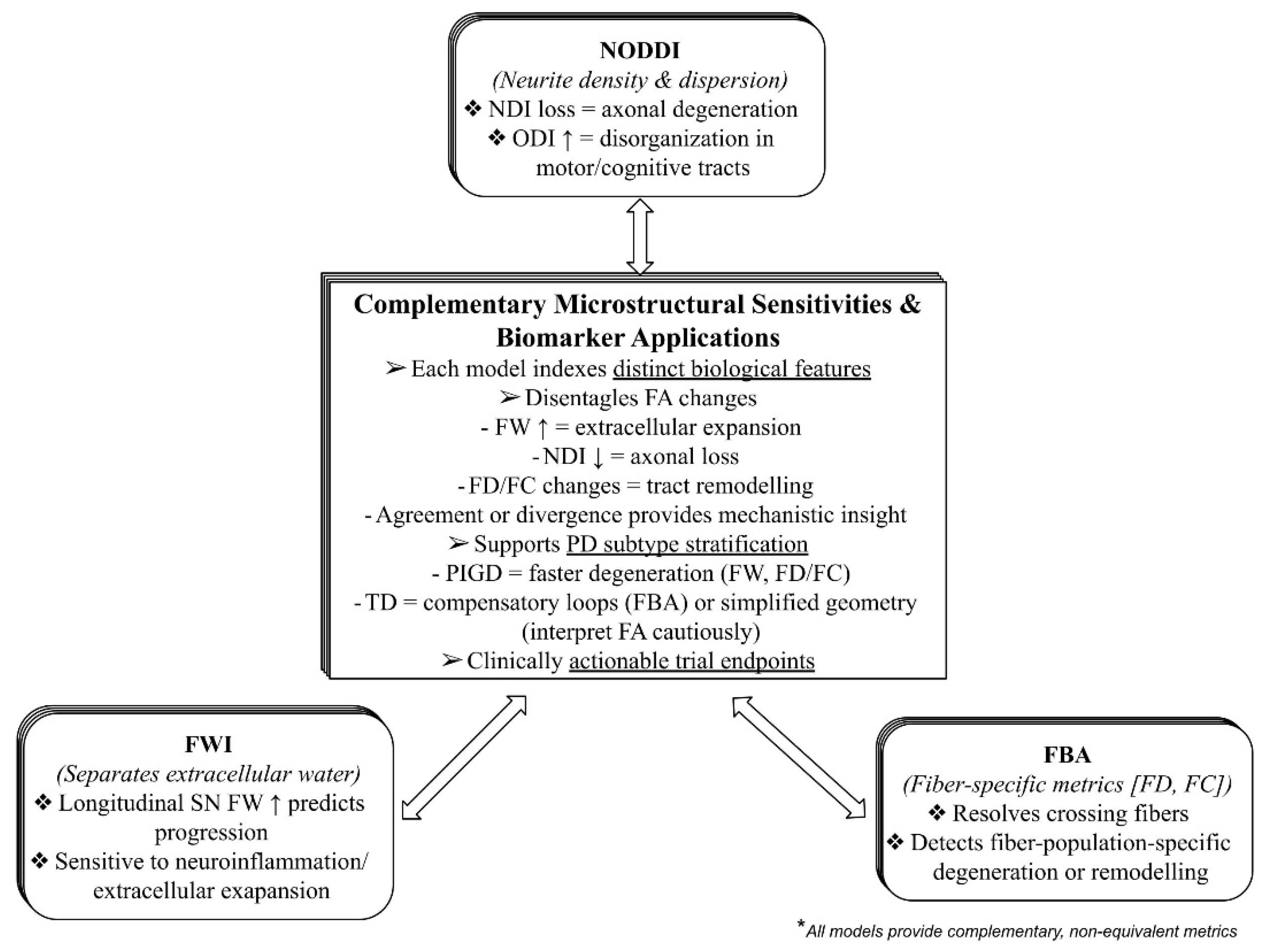
Figure 2.
Stage-dependent patterns of white-matter tract involvement in Parkinson’s disease.
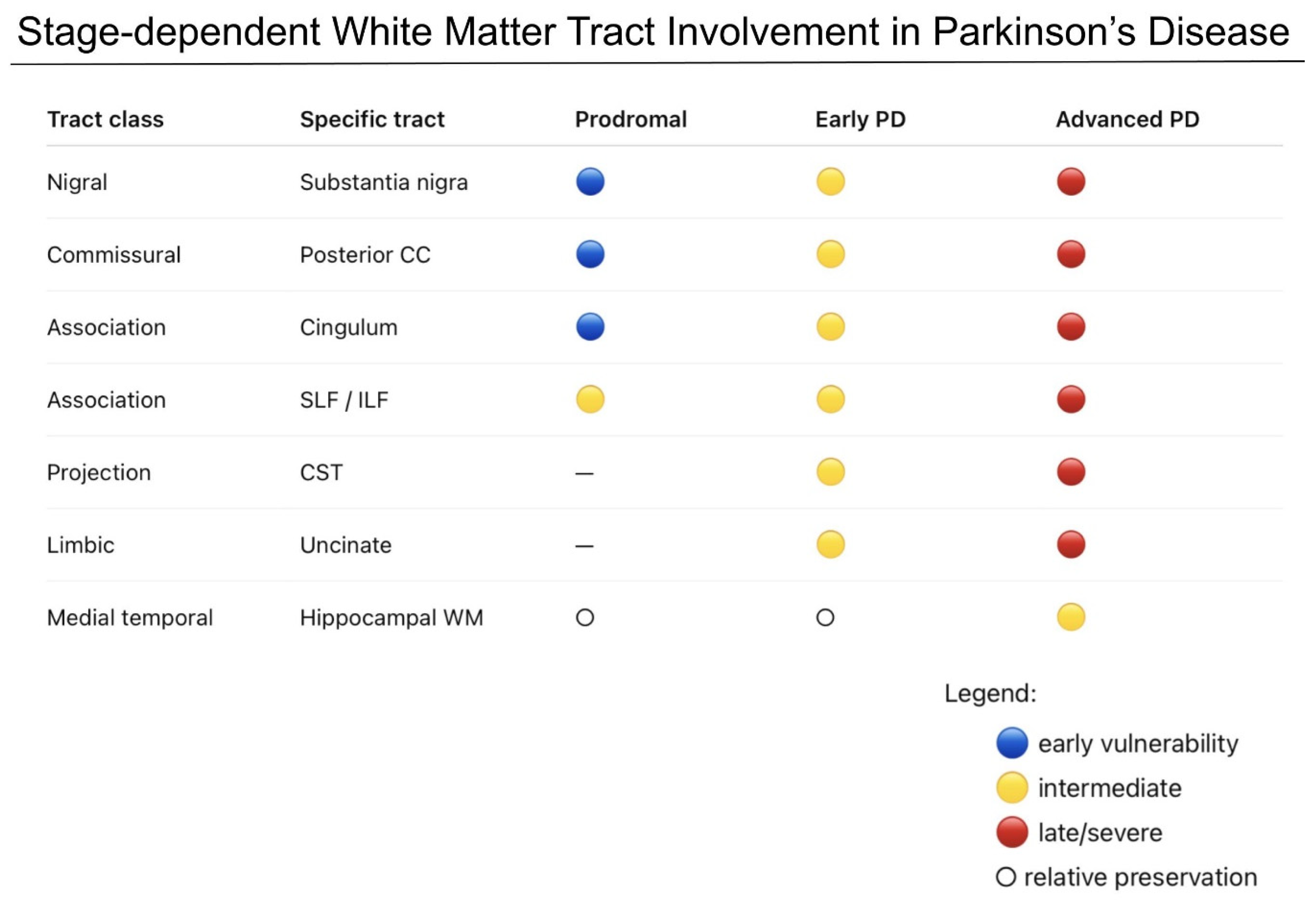
Figure 3.
Subtype-specific white-matter alterations in tremor-dominant (TD) versus postural instability/gait difficulty (PIGD) Parkinson’s disease.
Figure 3.
Subtype-specific white-matter alterations in tremor-dominant (TD) versus postural instability/gait difficulty (PIGD) Parkinson’s disease.
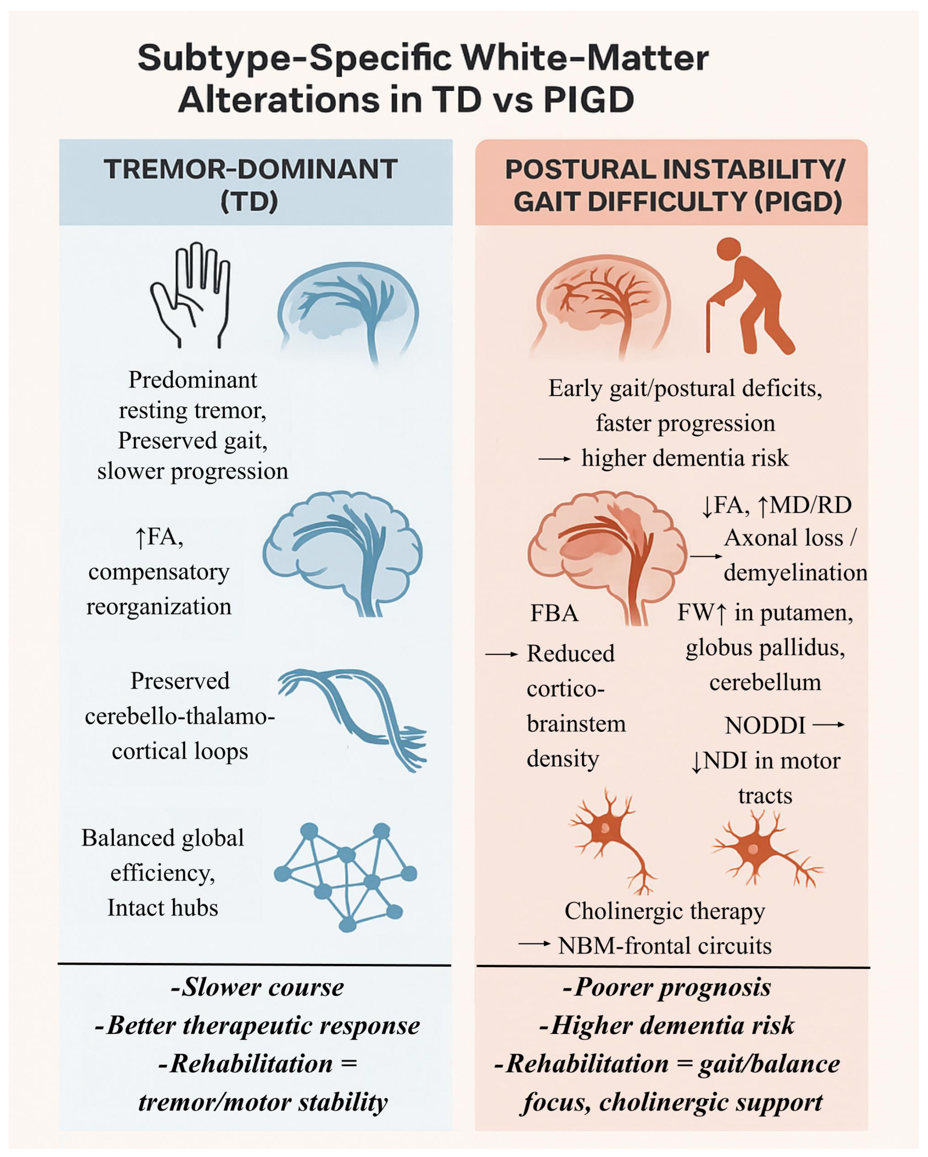
Figure 4.
An end-to-end roadmap for tractography-based subtype stratification and clinical translation in Parkinson’s disease.
Figure 4.
An end-to-end roadmap for tractography-based subtype stratification and clinical translation in Parkinson’s disease.
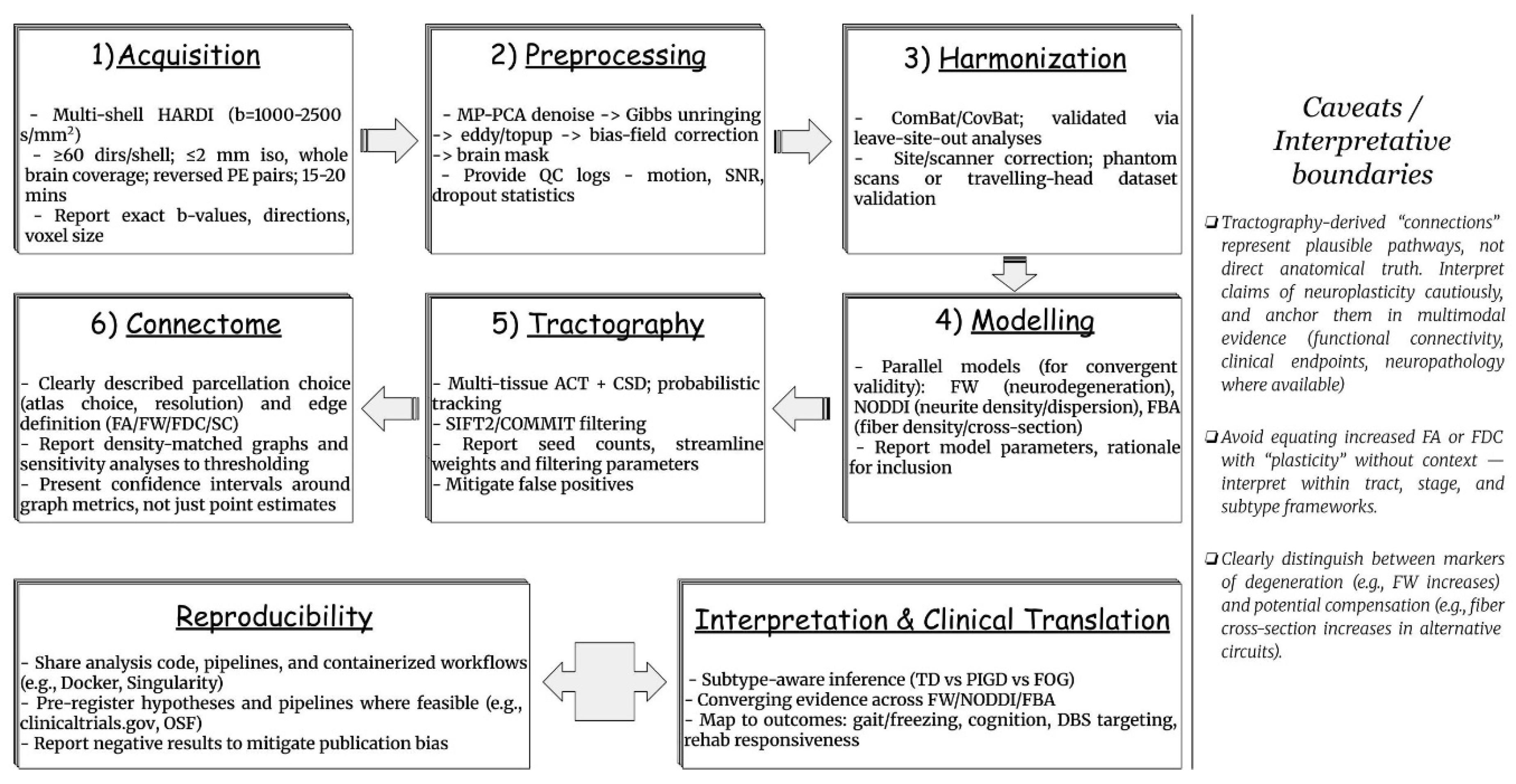
Figure 5.
Future Directions in Structural Connectomics for Parkinson’s Disease.
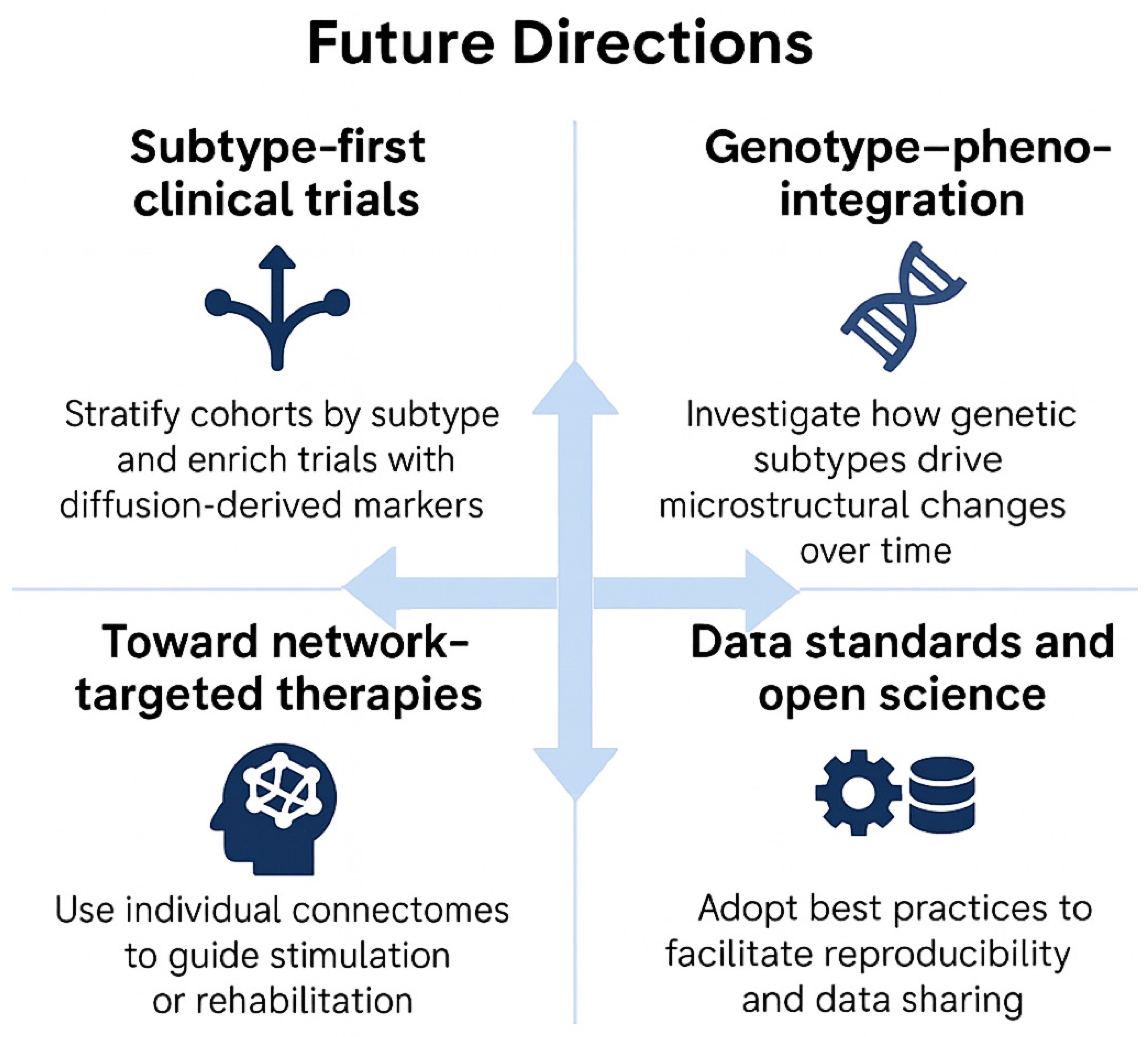

Table 1.
Summarized methodological overview of diffusion MRI models and tractography approaches in Parkinson’s disease.
Table 1.
Summarized methodological overview of diffusion MRI models and tractography approaches in Parkinson’s disease.
| Method / Type of Modelling | Key Metrics / Outputs | Strengths | Limitations | Applications in PD | References |
|---|---|---|---|---|---|
| DTI; Tensor model of Gaussian diffusion, assumes a single principal direction | FA, MD, RD, AD | - Simple, robust, widely validated - Short scan time - Sensitive to WM changes and easy to interpret |
- Single-tensor assumption - Poor fidelity in microstructure or crossing fibers (~90% of WM voxels) - Low biological specificity |
Macrostructural degeneration markers (CC, CST, BG projections); correlates with motor and cognitive decline | [20,59] |
| NODDI; Multi-compartment biophysical model (intra-neurite, extra-neurite, isotropic) | NDI, ODI, fISO | - Separates intra-/extra-neurite & CSF - Robust in GM and complex regions - Biologically interpretable |
- Requires multi-shell data - parameter bias if SNR/TE not optimized (can be resolved by multi-echo NODDI) - Limited clinical standardization |
Cortical pathology (frontal, hippocampal, limbic); links WM/GM changes to cognition and neuropsychiatric symptoms; potential subtype characterization when paired with clinical phenotypes | [37,65,66,113,318,319] |
| FWI; Bi-tensor model that separates a tissue compartment from an isotropic “free-water” compartment | FW fraction, tissue FA | - Corrects CSF contamination - Improves specificity - Reliable longitudinal tracking |
- Simplified bi-tensor model - Requires multi-b diffusion and robust fitting - Limited to FW correction (not full microstructure) |
Nigral FW as progression marker; subtype differences (PIGD > TD); distinguishing PD from atypical parkinsonism; neuroinflammation and degeneration monitoring | [10,62,63,64,84,320] |
| CSD; Deconvolves diffusion signal into FODs—handles crossing fibers (single- or multi-tissue variants) | FOD peaks; apparent fiber density per lobe (from FOD integrals) | - Resolves multiple fiber orientation - Backbone of advanced tractography |
- Needs high angular resolution and response-function estimation - Susceptible to bias/noise if acquisition is limited |
CST, cerebellar, brainstem delineation relevant to PD circuitry; Building blocks for connectomics in PD; pre-surgical pathway mapping (e.g., hyperdirect pathway to STN) | [52,67,68,72,321] |
| FBA; Fiber-population–specific analysis using CSD FODs at the “fixel” (fiber element) level | FD, FC, FDC | - Detects fiber-specific degeneration patterns - Network-level insights |
- Needs quality multi-shell data and CSD preprocessing - Less clinically routine |
Subtype-specific WM degeneration; identification of fiber-specific neuroplastic changes in PD | [11,70,71,73,74,75] |
| Deterministic Tractography; starts from a seed voxel and follows the single most likely fiber orientation step by step (e.g., principal eigenvector in DTI, or largest FOD peak in CSD) | Streamlines along dominant direction | - Clear, interpretable, efficient - Useful for clinical & longitudinal PD |
Fails in crossing fibers; underestimates connectivity | Nigrostriatal–nigropallidal tract delineation; monitoring motor severity; longitudinal monitoring in clinical cohorts | [49,50,322,323] |
| Probabilistic Tractography; Samples repeatedly from the FOD and builds up a probability distribution of possible pathways from each seed | Probability distributions of connectivity | - Robust to noise - Resolves complex fiber architecture - Produces connectivity likelihoods instead of binary paths |
- Computationally intensive - Spurious streamlines if priors/constraints are weak (more false positives) |
Preserved CTC in TD PD; hyperdirect pathway mapping for DBS; subtype-specific network mapping | [51,52,173,271,323,324] |
Table 2.
Methodological limitations and sources of bias across dMRI models, tractography, and connectome analyses in Parkinson’s disease.
Table 2.
Methodological limitations and sources of bias across dMRI models, tractography, and connectome analyses in Parkinson’s disease.
| Methodological issue | Common pitfall | Best practice recommendation | Key references |
|---|---|---|---|
| Anatomical plausibility | Streamlines enter CSF or terminate prematurely | Multi-tissue ACT with WM–GM constraints | [325,326] |
| Resolving crossing fibers | Single-tensor DTI fails in regions of complex fibre architecture, missing key tracts, resulting in false negatives |
|
[327] |
| Reconstructing subcortical pathways |
|
|
[84,328] |
| Quantitative accuracy of connectomes | Streamline count bias: Streamline counts and even weighted connectomes remain sensitive to reconstruction and modeling assumptions and cannot be interpreted as direct measures of axonal connectivity | Streamline weighting and filtering (e.g., SIFT2/COMMIT) improve quantitative plausibility but do not eliminate bias; connectome findings require cross-validation and conservative interpretation. | [42,55,329,330,331] |
| Diffusion metrics interpretation | FA/MD conflated with fiber density, orientation dispersion, and extracellular changes, leading to ambiguous biological interpretations of plasticity |
|
[62,332,333,334,335] |
| High b-value and SNR trade-off | High b-values increase microstructural contrast but reduce SNR, leading to noisy FOD estimates and bias in CSD models |
|
[336,337] |
Table 3.
Cognitive Subtypes in Parkinson’s disease and Associated White Matter Correlates.
| Subtype / Characterization | Key WM alterations | Neuroplasticity / Network Evidence | Prognostic implications | References |
|---|---|---|---|---|
| Executive / Dysexecutive; Set-shifting, planning, inhibitory control deficits |
- ↓FA / ↑MD in frontostriatal & frontoparietal tracts: anterior corona radiata, ALIC, SLF, IFOF, genu/body of CC - ATR deterioration correlates with executive impairment. |
- Disruption of thalamo-cortical executive loops shows early compensatory hyperconnectivity followed by decline - Microstructural changes in association fibers relate to slowing in processing, suggesting network inefficiency |
- Most common PD-MCI subtype - Predicts faster functional decline and higher risk of conversion from PD-MCI to PDD - WM integrity in the frontal tracts and ATR at baseline correlates with longitudinal executive decline |
[35,96,338,339,340,341,342] |
| Memory-predominant; Impairments mainly in episodic memory (immediate/delayed recall), with relatively preserved visuospatial and executive domains | - Microstructural degeneration in limbic association fibers: cingulum bundle and UF show ↓FA, ↑RD correlating with memory decline - Hippocampal output tracts (fornix) show reduced integrity in PD-MCI progressing to PDD |
- Marked limbic pathway involvement relevant to memory decline - Large fornix effect in advanced PD: HY 4/5 showed FA difference d ≈ −1.01 (PD < controls) - WMH in periventricular/temporal regions exacerbate memory decline, hinting at vascular contributions interacting with PD pathology |
- More likely to progress to PDD than other subtypes with preserved memory - Baseline integrity of UF/cingulum/fornix predicts rate of memory decline over follow-up intervals |
[12,73,182,186,343,344,345] |
| Visuospatial; Predominant deficits in visuospatial perception/constructive abilities (figure copy, cube analysis, navigation) with relative early preservation of language and simple attention | - Posterior network disconnection involving ILF, IFOF, posterior thalamic radiation, and callosal splenium on FBA - Longitudinal vulnerability of the splenium is a typical PD alteration and relates to posterior cognitive decline |
- Low visual function marks posterior WM degeneration and predicts cognitive decline; hallucinators show progressive posterior WM loss and thalamic changes - Visual dysfunction → worsening cognition with fiber-specific posterior tract reductions - In a longitudinal cohort with baseline visual testing, poor visual performance predicted faster global cognitive decline and greater posterior WM degeneration over ~18–24 months |
- Visuospatial-predominant PD-MCI has elevated risk of progression toward PDD - Posterior callosal/occipito-temporal tract integrity tracks worsening visuospatial performance |
[72,76,176,302,346] |
| Attention/processing speed; Slowed information processing and attentional control | - Widespread frontoparietal disconnection involving ATR, frontoparietal association tracts, and callosal fibers - ↓FA in prefrontal WM and caudate nucleus linked to slower processing speed |
- Disrupted integration across prefrontal–striatal–thalamic loops correlates with attentional slowing and cognitive control deficits - Reduced FA in prefrontal WM and caudate nucleus shows significant associations with processing speed (r ≈ 0.30–0.40) in non-demented PD cohorts - Longitudinal data show ATR diffusivity metrics predict rate of decline in attention/processing speed across follow-up periods (impaired compensatory recruitment of attentional circuits) |
- Sensitive early biomarker: ATR/connectivity disruptions track progression from PD-MCI to dementia - Attention/processing-speed decline predicts functional slowing and everyday task inefficiency, making it clinically actionable |
[35,340,347,348] |
| Language-predominant; Prominent deficits in semantic fluency, naming, or word retrieval, and language functions more impaired relative to other cognitive domains | - ↓FA / ↑ diffusivity in UF, IFOF, ILF associated with poorer semantic fluency (r ≈ −0.45 to −0.50) - Higher volumes of WMH and ↓FA in language-relevant tracts (e.g. IFOF, UF) correlate with worse performance on naming/word retrieval |
- Disruption of ventral language stream (IFOF, UF) reflects loss of semantic hub connectivity; dorsal stream may be relatively preserved in early language subtypes - FA in the UF and cingulum shows group-level differences (PD with language problems vs PD without) with Cohen’s d ~ 0.6–0.8 - Early microstructural damage before overt cortical atrophy, suggestive of emerging compensatory recruitment |
- Associated with earlier decline in semantic fluency and higher risk for PDD - Baseline FA/WMH burden in language tracts may predict later language decline and communication difficulties in daily life |
[175,186,349] |
Table 4.
Genetic subtypes of Parkinson’s disease: diffusion MRI findings and clinical implications.
| Gene | Clinical profile | Recent dMRI / imaging findings | Clinical correlations | Key references |
|---|---|---|---|---|
| GBA1 | Earlier cognitive decline, PIGD/FOG, rapid progression to dementia. | - Fiber-specific WM alterations in cingulum, uncinate, posterior callosum (FBA, dMRI) - Prodromal carriers show CST differences. |
Variants in GBA1 increase PD risk 5–7 fold; Cognitive-vulnerable subtype; strong association with dementia risk. | [34,73,186,188] |
| LRRK2 | Autosomal-dominant; often TD or indeterminate; slower motor progression; preserved cognition | - Subtle WM alterations - Genotype-specific differences in processing speed - Some prodromal carriers show CST microstructural changes. |
Lower brain-age gap in LRRK2-PD compared with idiopathic and GBA-PD; relatively benign trajectory, slower progression and relative resilience to widespread neurodegeneration; Narrower cognitive profile than GBA | [161,188,350] |
| PRKN | Recessive, early-onset; motor-pure; slow progression; preserved cognition. | WM abnormalities linked to disease duration and oxidative stress markers (TBSS, microstructure). | Imaging suggests nigrostriatal-dominant pathology with limited diffuse WM involvement. | [96,192,193] |
| PINK1 | Recessive, early-onset; resembles PRKN with slow progression. | Human dMRI data sparse; preclinical and mechanistic studies suggest WM/circuit alterations (e.g., PINK1-KO dMRI shows WM sensitivity) | Likely motor-pure (PINK1-KO rats possess non-motor and non-dopaminergic symptoms); cognitively resilient subtype. | [111,194,351,352] |
| DJ-1 (PARK7) | Recessive, very early-onset; motor-predominant; preserved cognition. | dMRI studies lacking; mechanistic reviews highlight oxidative stress/mitochondrial vulnerability. | Grouped with PRKN/PINK1 as motor-pure cluster. | [190,191,353] |
| SNCA | Rare, aggressive; early dementia; psychiatric symptoms. | PET-MRI in triplication shows widespread network involvement; case series confirm malignant trajectory. dMRI reports limited but consistent with diffuse WM degeneration. | Malignant subtype with high dementia penetrance. | [195,196,197,198,199] |
Disclaimer/Publisher’s Note: The statements, opinions and data contained in all publications are solely those of the individual author(s) and contributor(s) and not of MDPI and/or the editor(s). MDPI and/or the editor(s) disclaim responsibility for any injury to people or property resulting from any ideas, methods, instructions or products referred to in the content. |
© 2026 by the authors. Licensee MDPI, Basel, Switzerland. This article is an open access article distributed under the terms and conditions of the Creative Commons Attribution (CC BY) license (http://creativecommons.org/licenses/by/4.0/).
Copyright: This open access article is published under a Creative Commons CC BY 4.0 license, which permit the free download, distribution, and reuse, provided that the author and preprint are cited in any reuse.