

Submitted:
28 April 2026
Posted:
30 April 2026
You are already at the latest version
Abstract
The microbiota-gut-brain axis (MGBA) represents a bidirectional neuroendocrine system essential for maintaining metabolic and neurological homeostasis. While dietary macronutrients are known modulators of this axis, the cumulative impact of modern industrial xenobiotics remains insufficiently characterized. This review synthesizes contemporary, multidisciplinary evidence to elucidate how four ubiquitous environmental stressors Particulate Matter (PM2.5), Microplastics (MPs), Inorganic Nanoparticles (NPs), and Non-Nutritive Sweeteners (NNS) synergistically perturb this delicate enteric ecosystem. We integrate independent lines of research to propose a unifying pathological framework: these agents induce profound dysbiosis, significantly depleting beneficial, short chain fatty acid (SCFA) producing taxa (e.g., Lachnospiraceae, Faecalibacterium) and sharply diminishing the bioavailability of critical neuroactive mediators, including butyrate, GABA, serotonin, and indole derivatives. Concurrently, NNS-driven bacteriostatic shifts, the MP “plastisphere” phenomenon, and NP-induced oxidative mucosal abrasion critically compromise the intestinal barrier. This “leaky gut” facilitates the unrestricted systemic translocation of lipopolysaccharides (LPS) and trimethylamine-N-oxide (TMAO), driving a peripheral Treg/Th17 immune imbalance that propagates via the gut-liver and gut-heart axes directly to the central nervous system (CNS). Crucially, the synthesis of this data points toward a potential “Dual-Hit” mechanism, suggesting that these xenobiotics aggravate neurological pathology through simultaneous mechanisms: acting as direct neurotoxicants via CNS translocation (e.g., NPs crossing the blood-brain barrier to trigger epigenetic reprogramming and amyloid aggregation) while concurrently driving “bottom-up” systemic neuroinflammation. By linking these disruptions to classic neurodegeneration (Alzheimer’s, Parkinson’s) as well as underexplored pathologies (migraine, epilepsy, restless leg syndrome, and substance use disorders), this review underscores the urgent need for a paradigm shift in environmental neurotoxicology and the development of targeted microbiome-based interventions.
Keywords:
microbiota-gut-brain axis
; xenobiotics
; PM2.5
; microplastics
; nanoparticles
; neurological disorders
1. Introduction
The human gastrointestinal tract harbours a vast and complex community of microorganisms, collectively known as the gut microbiota, which plays a fundamental role in maintaining host physiology. Functioning as a “virtual organ,” this ecosystem is integral to nutrient metabolism, immunomodulation, and the maintenance of intestinal barrier integrity. The composition of this microbial community is highly dynamic and susceptible to modulation by a myriad of internal and external factors. Traditionally, host genetics, mode of delivery at birth, antibiotic usage, and most significantly, dietary patterns have been identified as the primary determinants of microbial diversity [1] their functional capacity to influence susceptibility to chronic diseases [2].
Crucially, the physiological influence of this microbial community extends far beyond the local enteric environment. Through the circulation of metabolites and immunomodulatory signals, the microbiota engages in intricate crosstalk with distant organs, establishing functional axes such as the gut-liver, gut-lung, and gut-heart axes that are vital for systemic homeostasis. Among these inter-organ networks, the MGBA stands out as a paramount regulator of neurological function. It represents a dynamic, bidirectional communication network linking the enteric ecosystem to the CNS through complex neural, immunological, and endocrine pathways. In a homeostatic state, this axis functions as a critical guardian of neurological health, regulating everything from blood-brain barrier (BBB) integrity to neurogenesis and mood regulation. However, while the impact of established factors like diet is well-documented, the modern industrial era has introduced a novel set of challenges that threaten this delicate equilibrium.
1.1. Physiological Mediators of the Axis: The “Good”
To understand how environmental stressors disrupt neurological function, it is essential to first characterize the mechanisms by which a healthy microbiome supports the brain. The gut microbiota synthesizes a diverse repertoire of metabolites that serve as critical signalling molecules within the MGBA. Among these, bioactive compounds such as SCFAs, aromatic amino acids, and TMAO exert significant regulatory influence over neurological function.
1.1.1. Short-Chain Fatty Acids (SCFAs)
In the large intestine, the primary metabolic output consists of SCFAs (specifically butyrate, propionate, and acetate), derived from the anaerobic fermentation of non-digestible polysaccharides, including resistant starch and dietary fibers. Key taxonomic groups, such as Lactobacillus, Ruminococcus, Bifidobacterium, Akkermansia, Faecalibacterium, and members of the Lachnospiraceae family, are principal drivers of SCFA biosynthesis from complex carbohydrates [3]. Following their biosynthesis, SCFAs undergo transmembrane translocation across the colonic epithelium. This process is primarily facilitated by specialized monocarboxylate transporters (MCTs), including both H+-dependent and sodium-coupled monocarboxylate transporters (SMCTs), which mediate the efficient absorption of these metabolites into the host circulation [4].
SCFAs modulate gastrointestinal mucosal immunity, barrier function, and structural integrity through two primary molecular mechanisms: the activation of G protein-coupled receptors (GPCRs) and the inhibition of histone deacetylases (HDACs). Specifically, SCFAs serve as ligands for free fatty acid receptors (FFAR2 and FFAR3), GPR109a/HCAR2, and GPR164. Upon binding to receptors on enteroendocrine cells, SCFAs stimulate the release of key gut peptides, including peptide YY (PYY) and glucagon-like peptide-1 (GLP-1), alongside neurotransmitters such as gamma-aminobutyric acid (GABA) and serotonin (5-HT). These molecules facilitate neuroendocrine signalling to the central nervous system via both the vagus nerve and systemic circulation [5].
Upon entering the systemic circulation, colon-derived SCFAs exert pleiotropic effects on peripheral tissues. These metabolites augment the thermogenic activity of brown adipose tissue, modulate hepatic mitochondrial biogenesis, and potentiate insulin secretion from pancreatic beta-cells, thereby maintaining global energy homeostasis. Systemically, SCFAs function as potent immunomodulators, regulating the pro-inflammatory cytokine profile by controlling interleukin production and promoting the peripheral differentiation of regulatory T cells (Tregs) [6].
Crucially, SCFAs facilitate neuro-metabolic crosstalk by traversing the blood-brain barrier (BBB) via monocarboxylate transporters expressed in brain endothelial cells. Upon entry, SCFAs bolster BBB structural integrity by upregulating the expression of tight junction proteins, such as occludin and claudin-5. Within the CNS, these metabolites mitigate neuroinflammation by modulating neurotrophic factor expression, fostering neurogenesis, and catalysing serotonin biosynthesis. Furthermore, SCFAs maintain neuronal homeostasis and influence glial cell morphology and activation states. Collectively, these multifaceted interactions within the MGBA serve as critical determinants of cognitive function, emotional regulation, and the underlying pathophysiology of neurological disorders [7].
1.1.2. Neurotransmitters and Biogenic Amines
The gut microbiota functions as a significant neuroendocrine organ, synthesizing a wide array of neurotransmitters through both direct and indirect biosynthetic pathways. Several commensal and environmental species most notably Corynebacterium glutamicum, Lactobacillus plantarum, Lactococcus lactis, and Bacillus subtilis possess the enzymatic machinery required to catalyse the conversion of L-glutamate into its enantiomer, D-glutamate. This microbial transformation is facilitated by glutamate racemases, representing a critical intersection between bacterial metabolism and host neuro-signalling [8,9]. Subsequently, the biotransformation of D-glutamate into GABA is mediated by glutamate decarboxylase (GAD; EC 4.1.1.15). The metabolic flux of D-amino acids within the central nervous system is significantly modulated by gut-derived microbial activity. Clinically, a notable depletion in plasma D-glutamate concentrations has been identified as a potential biomarker for cognitive decline, particularly in the pathogenesis of Alzheimer’s disease [10].
Several studies have highlighted the robust production of GABA by gut microbiota, with notable contributions from Lactobacillus, Bifidobacterium, and Bacteroides, particularly B. fragilis [11,12]. Notably, Escherichia coli K12 demonstrates metabolic versatility by utilizing GABA as a primary substrate for both carbon and nitrogen assimilation. Furthermore, recent taxonomic investigations have identified Evtepia gabavorous, a novel species within the Ruminococcaceae family characterized by its specialized GABA-consuming phenotype. The identification of such ‘GABA-vorous’ taxa suggests an intricate competition for neurotransmitter availability within the enteric ecosystem [13]. The metabolic niche of E. gabavorous is characterized by an obligate dependency on GABA, with its proliferation strictly contingent upon the presence of GABA-synthesizing commensals such as Bacteroides fragilis. While the number of microorganisms capable of interfacing with the GABAergic system remains limited, specific strains exhibit profound neuro-modulatory potential. For instance, Lactobacillus rhamnosus JB-1 has been shown to modulate the transcriptional expression of GABA receptors (GABARs) within the CNS, an effect correlated with attenuated anxiety and depression-like behaviours [14].
Serotonin, or 5-hydroxytryptamine (5-HT), is a pleiotropic neurotransmitter and hormone that exerts a profound regulatory influence over diverse physiological processes, including gastrointestinal (GI) motility, appetite homeostasis, mood stabilization, and sleep-wake cycles [15]. Notably, the vast majority of systemic 5-HT approximately 90% is biosynthesized within the enteric environment, primarily by specialized enterochromaffin (EC) cells residing in the intestinal mucosa [16]. Specific microbial taxa, including members of the Enterococcus, Streptococcus, and Escherichia genera, synthesize bioactive metabolites that stimulate EC cells to upregulate serotonin biosynthesis [17]. Contemporary research underscores the pivotal role of the gastrointestinal microbiota in 5-HT homeostasis, suggesting that a significant proportion of enteric serotonin is modulated by a diverse consortium of bacteria. This includes E. coli, Hafnia, Bacteroides, Streptococcus, Bifidobacterium, Lactococcus, Lactobacillus, Morganella, Klebsiella, Propionibacterium, Eubacterium, Roseburia, and Prevotella, which collectively influence the serotonergic landscape of the host [18].
Dietary tryptophan, an essential amino acid, serves as the primary substrate for the biosynthesis of 5-HT, a process facilitated by the enzymatic activity of specific microbial taxa, most notably Candida and Escherichia species [19]. The delicate balance of this axis is critical; perturbations are closely implicated in the pathogenesis of IBD. Histopathological analyses of tissues from patients with Crohn’s disease and ulcerative colitis reveal a marked proliferation of serotonin-secreting cells, and this elevation in serotonin-immunoreactivity underscores the potential for neuro-endocrine dysregulation to drive chronic intestinal inflammation [20]. Conversely, in healthy individuals, Clostridium perfringens optimally modulates serotonin synthesis through its interaction with host-derived tryptophan hydroxylase (TPH), the rate-limiting enzyme in the 5-HT biosynthetic pathway [21]. Beyond its canonical gastrointestinal functions, serotonin serves as a potent immunomodulator and a regulator of glial cell dynamics within both the enteric and central nervous systems.
Furthermore, the gut microbiota catabolizes tryptophan into bioactive indole derivatives via enzymatic routes involving tryptophan monooxygenase and indole-3-acetamide hydrolase. Notably, indole-3-aldehyde, synthesized by Lactobacillus species, functions as a ligand for aryl hydrocarbon receptors (AhR) on mucosal and immune cells. This activation precipitates the secretion of interleukin-22 (IL-22) and the subsequent release of antimicrobial peptides, thereby fortifying intestinal barrier integrity [22]. Specific commensal species, including Lactobacillus reuteri, Lactobacillus johnsonii, and Lactobacillus murinus, facilitate the catabolism of tryptophan into indole derivatives. These metabolites serve as critical signalling cues for the lineage commitment and differentiation of T cell subsets [23].
In parallel, the biogenic amine histamine exerts a multifaceted regulatory influence over host homeostasis, most notably in the promotion of wakefulness and the modulation of appetitive and motivational behaviours. The biosynthesis of histamine within the enteric environment is attributed to a phylogenetically diverse consortium of bacteria, comprising Lactobacillus spp., Lactococcus lactis, Oenococcus oeni, Pediococcus parvulus, Streptococcus thermophilus, Enterobacter spp., Citrobacter freundii, and Hafnia alvei, alongside opportunistic pathogens such as Morganella morganii and Klebsiella pneumoniae [12]. A comprehensive summary of these key gut microbiota-derived metabolites, their primary biosynthetic taxa, and their specific neuroprotective roles within the MGBA is detailed in Table 1.
1.2. The Modern Industrial Shift: The “Bad”
While the MGBA evolved to process natural dietary components, the modern industrial environment has introduced a novel class of persistent stressors that challenge this delicate equilibrium. Specifically, the pervasive infiltration of microplastics (MPs), the ubiquity of inorganic nanoparticles (NPs) (both food-grade and environmental), the massive consumption of artificial sweeteners, and the rising burden of air pollution (PM2.5) represent a “tetrad” of modern disruptors.
1.2.1. The Xenobiotic Paradigm
This review classifies these agents as xenobiotics: foreign compounds that the human gut has not evolved to process. Despite their differing origins whether inhaled as particulate matter or ingested as a food additive these stressors converge on a common pathological pathway: the disruption of mucosal homeostasis. This leads to a state of chronic dysbiosis and barrier dysfunction, facilitating the systemic translocation of inflammatory mediators.
1.2.2. Scope and Synthesis
To provide a more comprehensive overview, this synthesis extension beyond classical neurodegenerative diseases (Alzheimer’s, Parkinson’s) to include epilepsy, migraine, restless leg syndrome, and substance use disorders conditions where the intersection of environmental toxicology and the gut-brain axis is significantly less explored. It is important to note that while substantial evidence exists for xenobiotic impact on the gut, and separate evidence links gut dysbiosis to these neurological disorders, research findings that directly establish the role of these specific stressors (MPs, PM2.5, NPs, NNSs) in causing neurological diseases by altering gut microbiome homeostasis are lacking. Therefore, this review synthesizes the role of modern industrial byproducts in developing and aggravating neurological disorders by bridging these published independent studies to propose a cohesive mechanistic pathway.
2. Effect of Modern Food Additives: Non-Nutritive Sweeteners
The modern diet is characterized by the excessive consumption of industrial sweeteners, particularly non-nutritive sweeteners (NNS). NNS offer a distinct metabolic advantage over conventional caloric sweeteners due to their negligible caloric density and lack of a direct glycaemic response. However, given their ubiquity as modern food additives, substantial research has emerged investigating the capacity of these compounds to act as potent modulators of the composition and function of the gut microbiota [37,38.
2.1. Saccharin and Metabolic Dysregulation
Synthetic NNS, such as saccharin, have been shown to directly modulate the composition and metabolic function of the gut microbiota. This induction of gut dysbiosis appears to drive the downstream glucose intolerance phenotype observed in mammalian hosts. Clinically, NNS consumption exhibits a significant positive correlation with several biomarkers of metabolic syndrome, including markers of obesity (increased weight and waist-to-hip ratio) and impaired glycaemic control (elevated fasting blood glucose, glycosylated haemoglobin (HbA1c), and diminished glucose tolerance test (GTT) performance. Furthermore, elevated serum alanine aminotransferase (ALT) levels suggest secondary hepatic involvement, potentially linked to non-alcoholic fatty liver disease (NAFLD) a condition intricately tied to systemic inflammation and gut barrier permeability.
In a controlled intervention, participants consuming the FDA’s maximal acceptable daily intake (ADI) of commercial saccharin (5 mg/kg body weight) for just seven days exhibited profound microbial shifts. These changes were characterized by a 20-fold relative increase in Bacteroides fragilis (order Bacteroidales) and Weissella cibaria (order Lactobacillales), contrasted by an approximately tenfold reduction in Candidatus arthromitus (order Clostridiales) [39].
2.2. Comparative Impacts of Modern Sweeteners
Recent comparative studies highlight the divergent effects of various sweeteners on the enteric landscape. Alex et al. (2025) utilized mini-bioreactor arrays to evaluate the differential impacts of five prevalent non-nutritive sweeteners including Acesulfame K, Rebaudioside A, Saccharin, Sucralose, and Xylitol on gut microbial diversity. Family-level taxonomic analysis revealed that while all compounds perturbed the microbial equilibrium, the synthetic sweetener sucralose induced the most profound dysbiosis. Specifically, sucralose fostered the enrichment of the Enterobacteriaceae family, including pathogenic genera such as Escherichia and Citrobacter, which are frequently implicated in driving intestinal inflammation.
Conversely, the naturally derived sweeteners Rebaudioside A (found in the Stevia plant) and Xylitol promoted the proliferation of Lachnospiraceae and Ruminococcaceae. These taxa are critical for the biosynthesis of SCFAs, such as butyrate, which are essential for maintaining gut barrier integrity, modulating systemic inflammation, and preserving metabolic homeostasis along the gut-brain axis [40]. To provide a comprehensive overview of these dietary impacts, Table 2 summarizes recent experimental and clinical findings detailing the specific taxonomic perturbations induced by various non-nutritive sweeteners and food additives.
Note on Processed Caloric Sweeteners It is worth noting that metabolic disruption is not exclusive to synthetic xenobiotics. High Fructose Corn Syrup (HFCS), while derived from natural sources, represents a highly processed industrial additive that similarly disrupts gut homeostasis [41]. Excessive intake of free fructose monomers found in HFCS has been shown to overwhelm intestinal transporters, reaching the colon to degrade the mucus layer and increase intestinal permeability, mirroring the endotoxemia observed with synthetic stressors [42,43].
3. Effect of Microplastics
The gut microbiota, a central regulator of metabolic homeostasis, nutrient assimilation, and systemic immune modulation, is increasingly recognized as a primary target for microplastic (MP) toxicity. Growing evidence suggests that MP exposure precipitates intestinal dysbiosis, characterized by a deleterious shift in the ratio of commensal to pathogenic taxa. Such microbial perturbations can compromise intestinal barrier integrity, attenuate immune responsiveness, and heighten susceptibility to diverse gastrointestinal and systemic disorders [55,56,57].
3.1. Exposure Pathways and Dysbiosis
Dietary ingestion via seafood, salt, and water represents a primary exposure pathway. Experimental evidence suggests that polyethylene ingestion increases the relative abundance of Firmicutes and Bacteroidetes [58,59]. In Indonesian coastal and highland populations, MP contamination correlated with the prevalence of Roseburia, Clostridium, and Prevotella, alongside an enrichment of genes encoding plastic-degrading enzymes within the gut metagenome [60].
Recent investigations into chronic polyethylene (PE) microplastic exposure have revealed significant disruptions to infant gut microbiota and intestinal barrier function. Prolonged PE ingestion facilitates the proliferation of opportunistic pathobionts, most notably members of the Dethiosulfovibrionaceae and Enterobacteriaceae families, concurrently resulting in a marked reduction in enteric butyrate concentrations [61]. Furthermore, the biotransformation of microplastics, specifically polyethylene terephthalate (PET), evaluated using dynamic gastrointestinal simulators, demonstrated significant modulation of the human colonic microbiota and evidence of microbial colonization on plastic surfaces, suggesting potent biofilm formation (the “plastisphere”) within the enteric environment [62]. The dual physical and microbiological threat posed by MPs at the intestinal mucosal interface is comprehensively illustrated in Figure 1.
3.2. Systemic Inflammation and Immune Dysregulation
The translocation of microbial-derived products, such as lipopolysaccharides (LPS), into the systemic circulation secondary to MP-induced mucosal damage triggers a robust pro-inflammatory cascade. This chronic systemic inflammation serves as a primary driver of immune dysregulation, significantly elevating the risk for autoimmune pathologies, including rheumatoid arthritis, systemic lupus erythematosus (SLE), and inflammatory bowel disease (IBD) [63]. Intestinal dysbiosis compromises the induction and differentiation of regulatory T cells (Tregs), which are indispensable for maintaining peripheral immune tolerance. A disruption in the homeostatic equilibrium between immunosuppressive Tregs and pro-inflammatory Th17 cells often termed the Treg/Th17 balance is a critical factor in the exacerbation of neuroinflammatory conditions such as multiple sclerosis (MS) [64]. Furthermore, the clinical progression of these conditions is often exacerbated by deleterious mutations in FOXP3 the master transcription factor governing Treg lineage commitment precipitating a profound loss of immunological self-tolerance [65].
3.3. The Gut-Liver and Gut-Heart Axes: Systemic Conduits to Neuroinflammation
The ‘gut-liver axis’ provides a critical framework for evaluating the systemic toxicity of MPs. As the primary site for the metabolism of xenobiotics, the liver is uniquely susceptible to MP-induced injury. In vitro studies utilizing human liver organoids demonstrate that exposure to polystyrene MPs precipitates significant hepatotoxicity, characterized by lipo-toxicity, oxidative stress, and a robust inflammatory response [66]. Crucially, this hepatic impairment diminishes the liver’s capacity to detoxify circulating neurotoxins (e.g., ammonia) and amplifies the systemic release of pro-inflammatory cytokines. These peripheral inflammatory signals subsequently compromise the integrity of the BBB, initiating neuro-inflammatory cascades within the CNS.
Similarly, the ‘gut-heart axis’ is compromised by MP-induced endotoxemia. Increased intestinal permeability facilitates the systemic translocation of deleterious metabolites, such as TMAO, into the portal circulation. Upon reaching the liver, these compounds exacerbate chronic systemic inflammation, subsequently compromising cardiovascular integrity [67,68]. Critically, recent clinical observations reveal that MP accumulation in carotid artery plaques is associated with a significantly elevated risk of major adverse cardiovascular events (MACE), demonstrating a hazard ratio of 4.53 [69]. This cardiovascular deterioration is inextricably linked to neurological decline; endothelial dysfunction and compromised vascular health inevitably extend to the cerebral microvasculature. Furthermore, microbiota-derived metabolites like TMAO not only drive atherosclerosis but are also emerging as potent promoters of brain aging and neuroinflammation, actively disrupting BBB tight junctions and paving the way for neurological disorders.
3.4. The Gut-Brain Axis and Neurotoxicity
Beyond indirect systemic inflammation, emerging evidence suggests that MPs may also infiltrate the CNS directly via the olfactory pathway. A post-mortem analysis in Brazil identified synthetic polymers in the olfactory bulb tissues of 53% of the cohort, providing a plausible mechanism for MP translocation to the brain, bypassing the BBB and raising critical concerns regarding localized neurotoxicity [70]. To illustrate the breadth of these xenobiotic impacts, Table 3 summarizes recent human cohorts and murine models demonstrating the profound gut microbial architectural shifts induced by varying forms of microplastic exposure.
4. Effect of Nanoparticles
The rapid expansion of nanotechnology has introduced a paradigm shift in active and intelligent food packaging through enhanced barrier functions and antimicrobial surface modifications. However, the propensity for nanomaterials (NMs) to migrate from polymeric matrices into food simulants is well-documented, facilitating a direct pathway for human internal exposure [78]. Consequently, the gastrointestinal tract is increasingly exposed to both intentional food-grade additives and unintentional anthropogenic nanoparticles. These particles possess unique physicochemical properties—specifically their high surface-area-to-volume ratio—that allow them to interact directly with the enteric microbiota and bypass physiological barriers [79].
4.1. Food-Grade Nanoparticles and Packaging Migrants
Food-grade nanomaterials and packaging migrants -such as Titanium Dioxide (TiO2/E171), Silver Nanoparticles (AgNPs), and Zinc Oxide (ZnO) exert a profound influence on the intestinal microbial landscape. Zinc oxide nanoparticles, frequently utilized for their potent antimicrobial efficacy within food packaging systems, induce significant taxonomic perturbations [80]. Following a 28-day administration of ZnO NPs (1000 mg/kg) in murine models, responses exhibited clear sexual dimorphism: male rats displayed an expansion of the Firmicutes phylum and contraction of Bacteroidetes, whereas female rats displayed a contraction in Firmicutes alongside a significant proliferation of Verrucomicrobiota and Lactobacillus probiotics [81].
Similarly, silver nanoparticles (AgNPs) extensively utilized against multi-drug-resistant pathogens demonstrate persistent modulatory effects. Chronic, escalating exposure (0.1, 2, 40 µg) induces a characteristic shift in the Firmicutes-to-Bacteroidetes (F/B) ratio, driven primarily by an enrichment of Firmicutes [82]. Shorter-term interventions (0.5–2.5 mg/kg) corroborate this trend, precipitating a notable expansion of the Lachnospiraceae family alongside a depletion of Bacteroidetes [83]. Finally, TiO2 NPs significantly reshape the enteric environment; experimental exposures (2, 10, 50 mg/kg) resulted in the selective enrichment of Levilactobacillus and Allobaculum, contrasted by a marked reduction in the Clostridiaceae family [84].
Beyond the gut level, systemic bioaccumulation drives further pathology. For example, longitudinal assessments of silver nanoparticles (AgNPs) indicate that chronic systemic exposure facilitates significant bioaccumulation within the hepatic and splenic parenchyma, precipitating marked inflammation and deleterious histopathological alterations that compound the body’s overall systemic inflammatory burden [85].
4.2. Unintentional Anthropogenic Nanoparticles
Beyond intentional additives, the gut is increasingly exposed to unintentional nanoparticles derived from environmental pollution and industrial processes. Investigations in zebrafish models reveal that exposure to Carbon Black nanoparticles (CBNPs) precipitates a marked reduction in intestinal flora diversity and profoundly alters core microbial populations. This dysbiosis is coupled with increased permeability of the intestinal mucosal barrier—driven by the dysregulation of tight junction-related genes and concurrent hepatic insult, characterized by vascular degeneration and lipid accumulation [86] Similarly, ingested silica nanoparticles have been observed to potentiate intestinal inflammation [87] by activating the NLRP3 inflammasome, and exacerbate secondary white matter injury by disrupting the blood–brain barrier, inducing neuroinflammation, and interfering with nerve regeneration [88].
4.3. Mechanisms of Nanoparticle-Induced MGBA Disruption
Metallic and metal-oxide nanoparticles act as primary drivers of cellular homeostatic disruption. Locally within the gastrointestinal tract, NPs (particularly ZnO NPs) catalyze the overproduction of reactive oxygen species (ROS) across diverse cellular lineages. This oxidative surge, coupled with subsequent DNA fragmentation, presents a genotoxic and pro-inflammatory profile that damages the structural and functional integrity of the intestinal epithelial barrier [89]. Furthermore, contemporary research demonstrates that titanium dioxide nanoparticles exhibit the capacity for transepithelial translocation from the gastrointestinal lumen to distal organs, including the liver, kidneys, and CNS, following oral ingestion [90].
Once inside the central nervous system, these translocated nanoparticles (including titanium dioxide, nanosilica, and nanosilver) function as potent catalysts for neurodegenerative pathologies. Contemporary research reveals a convergent mechanism of neurotoxicity driven by epigenetic reprogramming. Specifically, NP exposure precipitates aberrant DNA methylation and dysregulates ryanodine receptor-Ca2+ signalling within the murine brain. These molecular shifts culminate in lysosomal dysfunction and the subsequent attenuation of autophagic flux in neurons. Critically, this proteostatic failure compromises the clearance of neurotoxic aggregates, specifically β-amyloid and hyperphosphorylated tau (p-Tau). The resultant accumulation of these aggregates ultimately drives the clinical manifestation of spatial cognition and memory deficits [91]. The complex sequence of events, from initial enteric translocation to subsequent neuronal epigenetic and proteostatic failure, is visually summarized in Figure 2. The diverse taxonomic perturbations induced by various food-grade and environmental nanoparticles across different in vivo models are systematically summarized in Table 4.
5. Effect of Air Pollution: Particulate Matter (PM2.5)
Fine particulate matter (PM2.5) atmospheric particulates with a diameter of 2.5 µm or less derived primarily from combustion activities, industrial processes, and natural sources like wildfires has emerged as a significant contributor to global health risks [101]. The infiltration of these particulates across the pulmonary-capillary barrier into the systemic circulation is a well-established driver of obstructive respiratory illnesses [102,103], cardiovascular dysfunction [104], and oncogenic transformations [105]. This episodic and chronic exposure is particularly devastating at the extremes of the age spectrum, affecting the immature respiratory systems of children and the diminished compensatory mechanisms of the elderly [106]. However, its specific capacity to perturb the gastrointestinal tract and the gut-brain axis represents a newly recognized dimension of its systemic toxicity.
5.1. The Lung-Gut Exposure Route and Barrier Dysfunction
Inhaled PM2.5 undergoes gastrointestinal translocation primarily via the mucociliary escalator; particulates trapped within the tracheobronchial mucosa are cephalad-propelled to the oropharynx and subsequently ingested. Compelling experimental and epidemiological evidence indicates that this direct enteric exposure severely compromises intestinal barrier integrity [107].
Xie et al. demonstrated that chronic exposure to elevated PM2.5 concentrations in murine models induces a dose- and duration-dependent upregulation of pro-inflammatory cytokines (IL-6, IL-10, IL-1β, and TNF-α). This inflammatory surge was associated with significant structural insult to the colonic mucosa. Mechanistically, this pathogenesis is driven by a linear, dose-dependent downregulation in the expression of critical transmembrane tight junction proteins, specifically Occludin and Zonula Occludens-1 (ZO-1), underscoring a systematic compromise of the intestinal epithelial barrier [108].
5.2. Profound Dysbiosis and Metabolic Perturbation
In vivo investigations utilizing various murine models indicate that PM2.5 exposure induces significant perturbations in the intestinal microbiota and its associated metabolome. In longitudinal studies, male C57BL/6 and BALB/c models subjected to atmospheric PM2.5 inhalation trigger a systemic restructuring of the microbial architecture. At the phylum level, this is characterized by a significant contraction in the relative abundance of Bacteroidetes, contrasted by a marked compensatory expansion of the Firmicutes and Cyanobacteria phyla [109].
At the genus level, particulate insult precipitates a marked depletion of commensal SCFA producers, most notably Prevotella, Bacteroides, and Tremihum, alongside the enrichment of opportunistic genera such as Lactobacillus, Parabacteroides, Oscillospira, Coprococcus, and Desulfovibrio [110]. Furthermore, PM2.5 exposure significantly reduces vitamin B12-associated genera, including Faecalibacterium, Citrobacter, and Collinsella. The suppression of these specific taxa disrupts the vitamin B12 biosynthetic pathway, potentially driving the aberrant secretion of neurotransmitters and contributing directly to neuro-metabolic dysfunction [111].
5.3. Neuroinflammatory and Neurodegenerative Consequences
Sustained pro-inflammatory signalling, driven by the vascular translocation of fine particulates and gut-derived endotoxins, serves as a catalyst for systemic homeostatic disruption that ultimately reaches the CNS. Substantial evidence identifies PM2.5 as a critical environmental determinant in the aetiology of neurodegenerative pathologies.
Notably, cognitive impairment a primary prodromal manifestation of Alzheimer’s disease (AD) is strongly associated with chronic PM2.5 exposure, with geriatric populations in high-pollution locales exhibiting accelerated cognitive decline [112]. These clinical observations are corroborated by a landmark cohort study of 602 autopsy-confirmed cases, which demonstrated that elevated PM2.5 exposure significantly correlates with increased severity of Alzheimer’s Disease Neuropathologic Change (ADNC) and progressed clinical markers of dementia [113].
Similarly, a longitudinal cohort study in Ontario demonstrated that even low-level PM2.5 exposure (3.8 μg/m3) significantly correlates with an elevated risk of Parkinson’s disease (PD) and increased PD-related hospitalization rates [114]. Furthermore, research in New York indicates that chronic PM2.5 exposure precipitates clinical deterioration in existing PD patients. Emerging data suggest that these deleterious neurodegenerative effects are intrinsically linked to the dysbiosis-induced breakdown of the gut-brain axis and modulated by the specific physicochemical composition of the translocated particulates [115]. A synthesis of recent epidemiological and experimental studies detailing the specific dysbiotic profiles and taxonomic shifts induced by PM2.5 exposure is provided in Table 5.
6. Effect on Neurological Diseases
6.1. Neurodegenerative Disorders
Neurodegenerative diseases (NDDs) are defined by the progressive attrition of neuronal populations, culminating in debilitating cognitive or motor impairments. Central to the pathogenesis of these disorders is the MGBA, a bidirectional communication network that integrates the resident enteric microbiota with the CNS.
6.1.1. Alzheimer’s Disease (AD)
Alzheimer’s disease (AD), the primary cause of dementia globally, is defined by the extracellular accumulation of amyloid-β (Aβ) plaques, intracellular neurofibrillary tau tangles, and a progressive decline in cognitive function. Recent metagenomic analyses have consistently demonstrated that the AD gut microbiome is significantly distinct from that of age-matched, cognitively unimpaired cohorts. A hallmark of this dysbiosis is the marked depletion of anti-inflammatory genera, such as Faecalibacterium and Eubacterium rectale, coupled with the enrichment of pro-inflammatory or opportunistic taxa, specifically Escherichia/Shigella [124]. Clinical investigations have revealed that cognitively impaired individuals with confirmed brain amyloidosis exhibit an overabundance of Escherichia/Shigella and a deficit in E. rectale. Crucially, these taxonomic perturbations correlate with elevated levels of peripheral pro-inflammatory cytokines, suggesting a systemic link between enteric dysbiosis and neuro-inflammatory progression [125,126].
6.1.2. Parkinson’s Disease (PD)
Parkinson’s disease (PD) is a progressive neurodegenerative movement disorder characterized by the selective attrition of dopaminergic neurons in the substantia nigra and the systemic accumulation of α-synuclein aggregates. Gastrointestinal (GI) dysfunction represents a hallmark prodromal feature of PD, with up to 80% of patients manifesting chronic constipation and enteric motility issues years prior to the onset of motor deficits. Recent metagenomic characterizations have consistently identified significant dysbiosis in PD cohorts relative to neurologically normative controls. Specifically, the Prevotellaceae family notably the Prevotella genus, which is instrumental in butyrate biosynthesis and mucosal homeostasis is markedly depleted in PD patients [127]. Prevotella also contribute in mucin synthesis in gut which in turn maintain barrier integrity and immune regulation. The mucin-degrading bacterium Akkermansia muciniphila is frequently enriched in PD faecal profiles [128]. While Akkermansia is typically associated with metabolic health, its overabundance in PD may indicate a maladaptive shift toward excessive mucin degradation, potentially compromising the structural integrity of the intestinal barrier [129].
6.2. Neuroinflammation and Autoimmune Disorders
Neuroinflammation is characterized by an orchestrated inflammatory response arising from the dysregulated synthesis and secretion of pro- and anti-inflammatory cytokines within the CNS. Recent evidence identifies intestinal dysbiosis as a primary driver of neuroinflammation through the modulation of systemic inflammatory mediators. Specifically, a reduction in microbially-derived SCFAs attenuates their native anti-inflammatory signalling. This microbiome dysbiosis compromises the intestinal barrier, facilitating the translocation of pathogen-associated molecular patterns (PAMPs), such as LPS.
6.2.1. Multiple Sclerosis (MS)
Multiple sclerosis (MS) is an immune-mediated neuroinflammatory disorder defined by autoreactive inflammation, the demyelination of CNS axons, and subsequent neurodegeneration. Mounting evidence identifies significant enteric dysbiosis in MS patients, characterized by a marked reduction in butyrate-producing taxa such as Faecalibacterium prausnitzii and Butyricicoccus. These microbial deficits are typically accompanied by the enrichment of pro-inflammatory or mucin-degrading species, including Akkermansia muciniphila, Prevotella spp., Methanobrevibacter, and Eggerthella [130]. Under physiological conditions, butyrate serves as a critical signalling molecule for the induction and maintenance of regulatory T cells (Tregs). However, the overabundance of Akkermansia and Ruminococcus in MS cohorts which facilitates the erosion of the protective mucosal barrier—correlates with the expansion of pro-inflammatory Th17 cells within the intestinal mucosa, thereby driving systemic autoimmune activity [131].
6.2.2. Amyotrophic Lateral Sclerosis (ALS)
Amyotrophic Lateral Sclerosis (ALS) is a rapidly progressive and fatal neurodegenerative disorder characterized by the selective attrition of motor neurons, culminating in muscular atrophy and paralysis. Clinically, ALS is frequently associated with a hypermetabolic state and gastrointestinal (GI) dysfunction, such as significant weight loss, which may enter into a bidirectional relationship with the enteric microbiota. In a pilot clinical trial, ALS patients adhering to a hypercaloric dietary regimen exhibited a significantly attenuated rate of functional decline compared to those on a normative caloric intake. This dietary shift is often associated with the enrichment of Akkermansia muciniphila, a taxa that thrives on mucin substrates under conditions of high lipid and low fibre intake. Notably, Akkermansia may exert neuroprotective effects by synthesizing bioactive metabolites such as nicotinamide [132]. These findings suggest that the gut microbiota can modulate the trajectory of neurodegeneration in ALS through the enhancement of specific microbial metabolic functions, including the biosynthesis of essential vitamins and cofactors.
6.3. Neuropsychiatric Disorders
6.3.1. Major Depressive Disorder (MDD)
Major Depressive Disorder (MDD) is a multifaceted psychiatric condition primarily characterized by persistent anhedonia, psychomotor retardation, and cognitive impairment. Metagenomic characterizations of the MDD gut microbiome reveal significant taxonomic perturbations, most notably an enrichment of Eggerthella (Actinobacteria), Subdoligranulum, and Coprococcus (Lachnospiraceae), alongside a concomitant depletion of SCFA producing taxa within the Ruminococcaceae family. This dysbiotic profile is particularly acute in the inflammatory MDD subtype, which is characterized by an expansion of the Bacteroidetes phylum and a reduction in the Clostridiales order. Notably, the Sellimonas genus exhibits a dose-dependent positive correlation with clinical severity, potentially exerting its pro-inflammatory effects through the activation of the canonical TLR4/NF-κB pathway [133].
6.3.2. Schizophrenia
Schizophrenia is a multifaceted neuropsychiatric syndrome characterized by a complex constellation of cognitive, emotional, and occupational impairments. Emerging evidence suggests that intestinal barrier dysfunction, systemic bacterial translocation, and gastrointestinal comorbidities are significantly more prevalent in schizophrenic cohorts than in the general population. The pathophysiology of the disorder is increasingly linked to enteric dysbiosis and the aberrant shunting of the tryptophan-kynurenine metabolic pathway [134]. Specifically, taxonomic enrichment of the genera Lactobacillus and Prevotella has been identified in ultra-high-risk (UHR) patients. Enteric dysbiosis appears to exacerbate systemic inflammation by compromising intestinal barrier integrity—a process intricately linked to the dysregulation of the kynurenine pathway.
6.3.3. Autism Spectrum Disorder (ASD)
Autism spectrum disorder (ASD) is a complex neurodevelopmental condition characterized by repetitive behavioural patterns and significant deficits in social and linguistic communication. Clinically, ASD is frequently comorbid with diverse pathologies, including mood disorders, epilepsy, and gastrointestinal (GI) dysbiosis. The aetiology of ASD is increasingly linked to a homeostatic imbalance between pro-inflammatory Clostridium species and anti-inflammatory Bifidobacterium spp. [135]. Notably, the dietary intake of propionic acid or its metabolic precursors is correlated with exacerbated autistic behaviours and GI symptomatology.
6.3.4. Bipolar Disorder (BD)
Bipolar disorder (BD) is a chronic, recurrent affective disorder that shares significant symptomatic overlap with schizophrenia. It is clinically defined by pathological oscillations between depressive episodes and manic phases. Metagenomic characterizations indicate that individuals with BD exhibit a four-fold reduction in the Clostridiaceae family compared to healthy controls [136]. Furthermore, taxonomic stratification reveals that the class Coriobacteriia is significantly enriched in BD cohorts, whereas butyrate-producing taxa such as Ruminococcaceae and Faecalibacterium are markedly more abundant in normative controls, suggesting a loss of anti-inflammatory microbial support in the disorder [137].
6.4. Other Neurological Disorders
6.4.1. Epilepsy
Epilepsy is a neurological disorder defined by a predisposition to unprovoked, spontaneous seizures, primarily arising from a homeostatic imbalance between neuronal excitation and inhibition. Comparative metagenomic analysis of patients with drug-resistant epilepsy versus those responsive to therapy has revealed distinct microbial signatures. Notably, the gut microbiota composition of drug-responsive individuals appears comparable to that of healthy controls, suggesting a preservation of microbial homeostasis. Furthermore, a higher prevalence of Bifidobacterium and Lactobacillus has been correlated with lower seizure frequency (four or fewer annual episodes). Conversely, patients with drug-resistant epilepsy exhibit significantly elevated alpha-diversity indices, indicating that increased microbial richness in this context may be associated with refractory clinical outcomes [138].
6.4.2. Migraine
Migraine is a debilitating neurovascular disorder characterized by recurrent paroxysms of severe cephalalgia, frequently accompanied by autonomic symptoms such as nausea, emesis, and heightened sensitivity to multimodal sensory stimuli. The precise mechanisms governing the microbiota-gut-brain axis in migraineurs remain to be fully elucidated; however, current evidence suggests a multifactorial interplay involving pro-inflammatory cytokines (IL-1β, IL-6, IL-8, and TNF-α), distinct microbial profiles, and the dysregulation of neuropeptide and serotonergic signalling [139]. Glutamate, the primary excitatory neurotransmitter, is fundamentally implicated in migraine pathogenesis by facilitating cortical spreading depression (CSD), mediating central sensitization, and activating the trigeminovascular system.
6.4.3. Restless Leg Syndrome (RLS)
Restless Legs Syndrome (RLS), also clinically recognized as Willis-Ekbom Disease, is a sensorimotor neurological disorder characterized by an imperative urge to move the lower extremities. Differential abundance analysis has identified a statistically significant depletion of the Lachnoclostridium and Flavonifractor genera in RLS cohorts compared to both healthy controls (HC) and insomnia (INS) patient groups. Furthermore, a marked decrease in the relative abundance of four additional genera—Bacteroides, Eisenbergiella, Blautia, and Dorea—was observed in RLS patients relative to healthy controls, suggesting a distinct microbial deficit specific to the RLS enteric landscape [140].
6.4.4. Substance Use Disorder
Substance use disorders (SUDs) are characterized by persistent, maladaptive dependence and a profound inability to regulate consumption despite escalating biopsychosocial consequences. These disorders are underpinned by chronic neuroplastic alterations within central circuits governing executive function, stress reactivity, and reward processing. Vulnerability to such addiction-related phenotypes is a multifactorial byproduct of genetic predisposition, developmental stage, and adverse social determinants of health [141]. The gut-brain axis serves as a bidirectional biochemical conduit, where metabolic signals from the enteric microbiota influence CNS homeostasis and behaviour. Gut commensals are pivotal in the biosynthesis of neuroactive metabolites, including SCFAs and primary neurotransmitters like serotonin and dopamine. Addictive substances (e.g., alcohol, opioids, and/or other drugs) exploit the brain’s dopaminergic reward pathway, precipitating a supraphysiological release of dopamine that drives repetitive consumption. Emerging evidence suggests that gut microbes modulate reward perception for both natural and pharmacological stimuli. Pathologically, SUDs are frequently associated with intestinal barrier dysfunction (“leaky gut”), which facilitates the systemic translocation of microbial products. This triggers a peripheral pro-inflammatory cascade; subsequent cytokine infiltration across the BBB induces neuroinflammation, thereby altering neuronal excitability within the reward circuitry and modulating substance tolerance and cravings [142,143].
Alcohol consumption exerts a direct, deleterious impact on intestinal micro-ecology. Both murine and clinical investigations demonstrate that alcohol induces significant shifts in microbial diversity and taxonomic composition. Patients with Alcohol Use Disorder (AUD) exhibit a distinct microbial signature characterized by a contraction of Akkermansia and an expansion of Bacteroides which serves as a diagnostic biomarker with 93.4% accuracy. This dysbiosis is associated with a systemic pro-inflammatory state, evidenced by significantly elevated serum lipopolysaccharide (LPS) and cytokines (TNF-α, IL-1, IL-6) [144]. The relationship between AUD and psychiatric comorbidities, such as depression and anxiety, is inherently bidirectional: pre-existing affective disorders increase the risk of alcohol abuse, while chronic alcohol exposure exacerbates neuropsychiatric symptoms [145]. The mechanistic link is further elucidated by faecal microbiota transplantation (FMT) studies. In a landmark study, mice receiving FMT from AUD donors who presented with high depression scores and depleted F. prausnitzii and A. muciniphila displayed impaired social behaviour, depressive-like endophenotypes, and hyper-cortisolemia. Intriguingly, the significant post-FMT enrichment of A. muciniphila in these mice suggests a complex, non-linear interaction between microbial consortia and the neurobehavioral axis, potentially involving species-specific synergies or host-specific metabolic responses [146].
Investigations into the interplay between opioid use disorders and the intestinal microbiome are rapidly expanding. In a comparative study involving 45 polysubstance abusers characterized by daily alcohol and nicotine consumption alongside heroin, methamphetamine (MA), or ephedrine, significant taxonomic shifts were observed relative to 48 healthy controls. Notably, the patient cohort exhibited a paradoxical increase in microbial diversity indices. Genus-level stratification revealed a distinct enrichment of Prevotella, Paracoccus, and Thauera, contrasted by a marked contraction of the Bacteroides phylum [147].
7. Conclusion and Future Directions
The evidence reviewed herein systematically challenges the traditional assumption that modern industrial xenobiotics function as isolated, organ-specific hazards. Instead, Particulate Matter (PM2.5), Microplastics (MPs), Inorganic Nanoparticles (NPs), and Non-Nutritive Sweeteners (NNS) converge to act as potent, synergistic disruptors of the MGBA. While each stressor initiates toxicity via distinct mechanisms—whether it be the epigenetic reprogramming induced by NPs [91], the mucosal abrasion and “plastisphere” pathogenic vectoring of MPs [62], the bacteriostatic shifts driven by NNS [40], or the mucociliary-to-gut translocation of inflammatory PM2.5 [107] they ultimately share a highly conserved, cascading pathological trajectory:
- Profound Dysbiosis: A consistent depletion of neuroprotective, SCFA-producing taxa (e.g., Faecalibacterium, Lachnospiraceae) and the aberrant proliferation of opportunistic pathobionts (e.g., Enterobacteriaceae, Proteobacteria). This sharply diminishes the bioavailability of essential neuroactive mediators, including butyrate, GABA, serotonin, and indole derivatives.
- Barrier Disruption & Systemic Translocation: The physical, chemical, and oxidative (ROS) erosion of critical tight junction proteins (ZO-1, Occludin) precipitates a “leaky gut” phenotype [89]. This facilitates the unrestricted systemic translocation of neurotoxic microbial products, such as lipopolysaccharides (LPS).
- Neurodegeneration: The culmination of systemic inflammation and, in the case of nanoparticles and microplastics, direct xenobiotic translocation across the blood-brain barrier [70], triggers microglial activation, attenuates autophagic flux, and accelerates the aggregation of neurotoxic proteins (e.g., amyloid-β and p-Tau).
7.1. A Potential “Dual-Hit” Mechanism
Beyond the synergistic ‘cocktail effect’ of multiple xenobiotics, the reviewed evidence suggests a potential “dual-hit” scenario: modern industrial xenobiotics likely aggravate neurological pathology through simultaneous, compounding mechanisms. The first hit (Hit-1) is direct neurotoxicity: xenobiotics (such as nanoparticles and microplastics) physically translocate into the CNS via systemic circulation or olfactory routes, acting as immediate neurotoxicants that induce localized cellular damage. The second hit (Hit-2) is indirect: these same agents concurrently perturb the gut-brain axis from the “bottom up” through mucosal damage, dysbiosis, and chronic endotoxemia. The resulting gut-derived systemic inflammatory storm reaches the brain and severely exacerbates the direct toxic effects of Hit-1. This bidirectional assault—where the brain is targeted from both the systemic circulation and the enteric nervous system—may explain the accelerated trajectory of cognitive decline and affective disorders observed in highly industrialized populations.
7.2. The Therapeutic Paradox and Environmental Complexity
As nanotechnology rapidly advances, NPs are increasingly utilized as antimicrobial agents and targeted drug delivery systems. However, this review urges extreme caution: the specific physicochemical properties that facilitate their therapeutic translocation into the CNS may inadvertently induce off-target enteric dysbiosis and long-term neurotoxicity [148]. Furthermore, a critical disconnect exists between pristine laboratory toxicology and environmental reality. From a prospective vantage point, the industrial and biomedical utilization of NPs is projected to increase exponentially, leading to inevitable environmental bioaccumulation. A sobering historical parallel must be drawn with plastics: macro-plastics discarded half a century ago have insidiously degraded into the pervasive, bio-reactive microplastics currently disrupting environment and human health [149]. Similarly, the continuous accumulation and environmental weathering of today’s nanoparticles may precipitate unforeseen, multigenerational ecological and physiological crises. Current safety assessments must evolve to account for the “eco-corona” the complex layer of biomolecules and concurrent pollutants that adsorb onto xenobiotics in real-world environments, dramatically altering their bioavailability and toxicity [150,151].
7.3. Future Directions:
To effectively mitigate the neurological burden of the modern industrial exposome, future research must prioritize four strategic domains:
- Addressing the “Cocktail Effect”: Real-world exposure is cumulative. Longitudinal human studies and advanced in vivo modelling must evaluate the synergistic toxicity of simultaneous exposure to microplastics, nanoparticles, air pollution, and non-nutritive sweeteners on neuro-metabolic markers.
- Broadening the Neurological Scope: There is a critical need to expand investigations beyond classical neurodegeneration (AD/PD) to elucidate the causal correlations between modern xenobiotics and underexplored pathologies, including migraine, epilepsy, restless leg syndrome, and the alarming escalation of substance use disorders.
- Safe-by-Design Nanomedicine: The development of future oral therapeutics and food packaging must prioritize rigorous preclinical screening to filter out nanomaterials that inadvertently trigger mucosal oxidative stress or downregulate tight junction proteins.
- Targeted Microbiome Interventions: Therapeutic strategies must pivot towards rescuing the MGBA through “psychobiotics” and targeted postbiotics. Exploring the therapeutic administration of specific neuroprotective metabolites (e.g., SCFAs, Indole Propionic Acid) or bio-engineering probiotic consortia capable of degrading the MP “plastisphere” offers a promising frontier for preserving long-term neurological resilience against environmental disruptors.
Author Contributions
CRediT SR: Investigation, Writing—original draft. PS: Conceptualization, Methodology, Supervision, Writing—original draft, Writing—review & editing.
Acknowledgments
The author(s) declared that financial support was received for this work and/or its publication. We are grateful to the Indian Knowledge System (IKS) Division, Ministry of Education (MoE), Government of India, for the financial support provided for this research work and fellowship for SR through project [F.2-71/IKS-RFP-6/2025-26].
Competing interests: All authors declare no financial or non-financial competing interests.
References
- Rinninella, E.; et al. What is the Healthy Gut Microbiota Composition? A Changing Ecosystem across Age, Environment, Diet, and Diseases. Microorganisms 2019, 7, 14. [Google Scholar] [CrossRef]
- Ray, S.; Shankaran, P. Nutrition and the gut microbiome: a symbiotic dialogue influencing health and disease. Front. Nutr. 2026, 13. [Google Scholar] [CrossRef] [PubMed]
- Agus, A.; Clément, K.; Sokol, H. Gut microbiota-derived metabolites as central regulators in metabolic disorders. Gut 2021, 70, 1174–1182. [Google Scholar] [CrossRef]
- Silva, Y. P.; Bernardi, A.; Frozza, R. L. The Role of Short-Chain Fatty Acids From Gut Microbiota in Gut-Brain Communication. Front. Endocrinol. . 2020, 11. [Google Scholar] [CrossRef]
- Priyadarshini, M.; Kotlo, K. U.; Dudeja, P. K.; Layden, B. T. Role of Short Chain Fatty Acid Receptors in Intestinal Physiology and Pathophysiology. In in Comprehensive Physiology; Wiley, 2018; pp. 1091–1115. [Google Scholar] [CrossRef]
- Parada Venegas, D.; et al. Short Chain Fatty Acids (SCFAs)-Mediated Gut Epithelial and Immune Regulation and Its Relevance for Inflammatory Bowel Diseases. Front. Immunol. 2019, 10. [Google Scholar]
- Parker, A.; Fonseca, S.; Carding, S. R. Gut microbes and metabolites as modulators of blood-brain barrier integrity and brain health. Gut Microbes 2020, 11, 135–157. [Google Scholar] [CrossRef]
- Sano, C. History of glutamate production. Am. J. Clin. Nutr. 2009, 90, 728S–732S. [Google Scholar] [CrossRef]
- Tanous, C.; Chambellon, E.; Sepulchre, A.-M.; Yvon, M. The Gene Encoding the Glutamate Dehydrogenase in Lactococcus lactis Is Part of a Remnant Tn 3 Transposon Carried by a Large Plasmid. J. Bacteriol. 2005, 187, 5019–5022. [Google Scholar] [CrossRef] [PubMed]
- Chang, C.-H.; Lin, C.-H.; Lane, H.-Y. d-glutamate and Gut Microbiota in Alzheimer’s Disease. Int. J. Mol. Sci. 2020, 21, 2676. [Google Scholar] [CrossRef] [PubMed]
- Strandwitz, P.; et al. GABA-modulating bacteria of the human gut microbiota. Nat. Microbiol. 2018, 4, 396–403. [Google Scholar] [CrossRef]
- Strandwitz, P. Neurotransmitter modulation by the gut microbiota. Brain Res. 2018, 1693, 128–133. [Google Scholar] [CrossRef]
- Quillin, S. J.; Tran, P.; Prindle, A. Potential Roles for Gamma-Aminobutyric Acid Signaling in Bacterial Communities. Bioelectricity 2021, 3, 120–125. [Google Scholar] [CrossRef]
- Jiang, H.; et al. Altered fecal microbiota composition in patients with major depressive disorder. Brain Behav. Immun. 2015, 48, 186–194. [Google Scholar] [CrossRef] [PubMed]
- Jenkins, T.; Nguyen, J.; Polglaze, K.; Bertrand, P. Influence of Tryptophan and Serotonin on Mood and Cognition with a Possible Role of the Gut-Brain Axis. Nutrients 2016, 8, 56. [Google Scholar] [CrossRef]
- Guzel, T.; Mirowska-Guzel, D. The Role of Serotonin Neurotransmission in Gastrointestinal Tract and Pharmacotherapy. Molecules 2022, 27, 1680. [Google Scholar] [CrossRef] [PubMed]
- Hwang, Y. K.; Oh, J. S. Interaction of the Vagus Nerve and Serotonin in the Gut–Brain Axis. Int. J. Mol. Sci. 2025, 26, 1160. [Google Scholar] [CrossRef] [PubMed]
- Dicks, L. M. T.; Hurn, D.; Hermanus, D. Gut Bacteria and Neuropsychiatric Disorders. Microorganisms 2021, 9, 2583. [Google Scholar] [CrossRef]
- Sun, L.-J.; Li, J.-N.; Nie, Y.-Z. Gut hormones in microbiota-gut-brain cross-talk. Chin. Med. J. (Engl) . 2020, 133, 826–833. [Google Scholar] [CrossRef]
- El-Salhy, M.; Solomon, T.; Hausken, T.; Gilja, O. H.; Hatlebakk, J. G. Gastrointestinal neuroendocrine peptides/amines in inflammatory bowel disease. World J. Gastroenterol. 2017, 23, 5068. [Google Scholar] [CrossRef]
- Dicks, L. M. T. Gut Bacteria and Neurotransmitters. Microorganisms 2022, 10, 1838. [Google Scholar] [CrossRef]
- Li, S. Modulation of immunity by tryptophan microbial metabolites. Front. Nutr. 2023, 10. [Google Scholar] [CrossRef]
- Mu, Q.; Tavella, V. J.; Luo, X. M. Role of Lactobacillus reuteri in Human Health and Diseases. Front. Microbiol. 2018, 9. [Google Scholar] [CrossRef]
- Wang, X.; et al. Sodium butyrate facilitates CRHR2 expression to alleviate HPA axis hyperactivity in autism-like rats induced by prenatal lipopolysaccharides through histone deacetylase inhibition. mSystems 2023. [Google Scholar] [CrossRef] [PubMed]
- Fernando, W. M. A. D. B.; et al. Sodium Butyrate Reduces Brain Amyloid-β Levels and Improves Cognitive Memory Performance in an Alzheimer’s Disease Transgenic Mouse Model at an Early Disease Stage. J. Alzheimer’s Dis. 2020, 74, 91–99. [Google Scholar] [CrossRef]
- Tu, J.; Zhang, J.; Chen, G. Higher dietary butyrate intake is associated with better cognitive function in older adults: evidence from a cross-sectional study. Front. Aging Neurosci. 2025, 17. [Google Scholar] [CrossRef] [PubMed]
- Huang, T.; Shi, H.; Xu, Y.; Ji, L. The gut microbiota metabolite propionate ameliorates intestinal epithelial barrier dysfunction-mediated Parkinson’s disease via the AKT signaling pathway. Neuroreport 2021, 32, 244–251. [Google Scholar] [CrossRef]
- Almutairi, S.; Sivadas, A.; Kwakowsky, A. The Effect of Oral GABA on the Nervous System: Potential for Therapeutic Intervention. Nutraceuticals 2024, 4, 241–259. [Google Scholar] [CrossRef]
- Zhao, H.; et al. GABAergic System Dysfunction in Autism Spectrum Disorders. Front. Cell Dev. Biol. 2022, 9. [Google Scholar] [CrossRef]
- Alharbi, B.; et al. Role of GABA pathway in motor and non-motor symptoms in Parkinson’s disease: a bidirectional circuit. Eur. J. Med. Res. 2024, 29, 205. [Google Scholar] [CrossRef]
- Monti, J. M. Serotonin control of sleep-wake behavior. Sleep Med. Rev. 2011, 15, 269–281. [Google Scholar] [CrossRef]
- Kuuskmäe, C.; et al. Negr1 deficiency alters glutamate signalling and kynurenine pathway in a mouse model of psychiatric disorders. Sci. Rep. 2026, 16, 5317. [Google Scholar] [CrossRef] [PubMed]
- Ciaglia, T.; et al. Neuroprotective Potential of Indole-Based Compounds: A Biochemical Study on Antioxidant Properties and Amyloid Disaggregation in Neuroblastoma Cells. Antioxidants 2024, 13, 1585. [Google Scholar] [CrossRef]
- Zhao, Q.; et al. Indole-3-propionic Acid Attenuates HI-Related Blood–Brain Barrier Injury in Neonatal Rats by Modulating the PXR Signaling Pathway. ACS Chem. Neurosci. 2022, 13, 2897–2912. [Google Scholar] [CrossRef]
- Ji, Y.; et al. Anti-Inflammatory and Anti-Oxidative Activity of Indole-3-Acetic Acid Involves Induction of HO-1 and Neutralization of Free Radicals in RAW264.7 Cells. Int. J. Mol. Sci. 2020, 21, 1579. [Google Scholar] [CrossRef] [PubMed]
- Sun, J.; et al. Microbiota-derived metabolite Indoles induced aryl hydrocarbon receptor activation and inhibited neuroinflammation in APP/PS1 mice. Brain Behav. Immun. 2022, 106, 76–88. [Google Scholar] [CrossRef]
- Richardson, I. L.; Frese, S. A. Non-nutritive sweeteners and their impacts on the gut microbiome and host physiology. Front. Nutr. 2022, 9. [Google Scholar] [CrossRef]
- Coccurello, R. Disrupting the Gut–Brain Axis: How Artificial Sweeteners Rewire Microbiota and Reward Pathways. Int. J. Mol. Sci. 2025, 26, 10220. [Google Scholar] [CrossRef]
- Suez, J.; et al. Artificial sweeteners induce glucose intolerance by altering the gut microbiota. Nature 2014, 514, 181–186. [Google Scholar] [CrossRef]
- Kidangathazhe, A.; et al. Synthetic vs. non-synthetic sweeteners: their differential effects on gut microbiome diversity and function. Front. Microbiol. 2025, 16. [Google Scholar] [CrossRef] [PubMed]
- Li, Z.; et al. Fructose metabolism and its roles in metabolic diseases, inflammatory diseases, and cancer. Mol. Biomed. 2025, 6, 43. [Google Scholar] [CrossRef]
- Westerbeke, F. H. M.; Rios-Morales, M.; Attaye, I.; Nieuwdorp, M. Fructose catabolism and its metabolic effects: Exploring host–microbiota interactions and the impact of ethnicity. J. Physiol. 2025, 603, 7661–7681. [Google Scholar] [CrossRef] [PubMed]
- Spruss, A.; Bergheim, I. Dietary fructose and intestinal barrier: potential risk factor in the pathogenesis of nonalcoholic fatty liver disease. J. Nutr. Biochem. 2009, 20, 657–662. [Google Scholar] [CrossRef]
- Nettleton, J. E.; et al. Maternal low-dose aspartame and stevia consumption with an obesogenic diet alters metabolism, gut microbiota and mesolimbic reward system in rat dams and their offspring. Gut 2020, 69, 1807–1817. [Google Scholar] [CrossRef] [PubMed]
- Suez, J.; et al. Personalized microbiome-driven effects of non-nutritive sweeteners on human glucose tolerance. Cell 2022, 185, 3307–3328.e19. [Google Scholar] [CrossRef] [PubMed]
- Olivier-Van Stichelen, S.; Rother, K. I.; Hanover, J. A. Maternal Exposure to Non-nutritive Sweeteners Impacts Progeny’s Metabolism and Microbiome. Front. Microbiol. 2019, 10. [Google Scholar] [CrossRef]
- Hanawa, Y.; et al. Acesulfame potassium induces dysbiosis and intestinal injury with enhanced lymphocyte migration to intestinal mucosa. J. Gastroenterol. Hepatol. 2021, 36, 3140–3148. [Google Scholar] [CrossRef]
- Méndez-García, L. A.; et al. Ten-Week Sucralose Consumption Induces Gut Dysbiosis and Altered Glucose and Insulin Levels in Healthy Young Adults. Microorganisms 2022, 10, 434. [Google Scholar] [CrossRef]
- Zheng, Z.; et al. Low Dose of Sucralose Alter Gut Microbiome in Mice. Front. Nutr. 2022, 9. [Google Scholar] [CrossRef]
- Dai, X.; et al. Maternal sucralose intake alters gut microbiota of offspring and exacerbates hepatic steatosis in adulthood. Gut Microbes 2020, 11, 1043–1063. [Google Scholar] [CrossRef]
- Wang, Q.-P.; Browman, D.; Herzog, H.; Neely, G. G. Non-nutritive sweeteners possess a bacteriostatic effect and alter gut microbiota in mice. PLoS ONE 2018, 13, e0199080. [Google Scholar] [CrossRef]
- Uebanso, T.; et al. Effects of Low-Dose Non-Caloric Sweetener Consumption on Gut Microbiota in Mice. Nutrients 2017, 9, 560. [Google Scholar] [CrossRef]
- Bian, X.; et al. Gut Microbiome Response to Sucralose and Its Potential Role in Inducing Liver Inflammation in Mice. Front. Physiol. 2017, 8. [Google Scholar] [CrossRef]
- Bian, X.; et al. Saccharin induced liver inflammation in mice by altering the gut microbiota and its metabolic functions. Food Chem. Toxicol. 2017, 107, 530–539. [Google Scholar] [CrossRef] [PubMed]
- Bora, S. S.; et al. Microplastics and human health: unveiling the gut microbiome disruption and chronic disease risks. Front. Cell. Infect. Microbiol. 2024, 14. [Google Scholar] [CrossRef]
- Ravindra, K.; Kaur, M.; Mor, S. Impacts of microplastics on gut health: Current status and future directions. Indian J. Gastroenterol. 2026, 45, 20–39. [Google Scholar] [CrossRef]
- Kalra, A.; Dominoni, D. M.; Boonekamp, J. The impact of microplastics on the mice gut microbiome: a meta-analysis. 2025. [Google Scholar] [CrossRef]
- Campanale, C.; Massarelli, C.; Savino, I.; Locaputo, V.; Uricchio, V. F. A Detailed Review Study on Potential Effects of Microplastics and Additives of Concern on Human Health. Int. J. Environ. Res. Public Health 2020, 17, 1212. [Google Scholar] [CrossRef] [PubMed]
- Emenike, E. C.; et al. From oceans to dinner plates: The impact of microplastics on human health. Heliyon 2023, 9, e20440. [Google Scholar] [CrossRef]
- Nugrahapraja, H.; et al. Effects of Microplastic on Human Gut Microbiome: Detection of Plastic-Degrading Genes in Human Gut Exposed to Microplastics—Preliminary Study. Environments 2022, 9, 140. [Google Scholar] [CrossRef]
- Fournier, E.; et al. Exposure to polyethylene microplastics alters immature gut microbiome in an infant in vitro gut model. J. Hazard. Mater. 2023, 443, 130383. [Google Scholar] [CrossRef] [PubMed]
- Tamargo, A.; et al. PET microplastics affect human gut microbiota communities during simulated gastrointestinal digestion, first evidence of plausible polymer biodegradation during human digestion. Sci. Rep. 2022, 12, 528. [Google Scholar] [CrossRef] [PubMed]
- Schwarzfischer, M.; Rogler, G. The Intestinal Barrier—Shielding the Body from Nano- and Microparticles in Our Diet. Metabolites 2022, 12, 223. [Google Scholar] [CrossRef] [PubMed]
- Shim, J. A.; Ryu, J. H.; Jo, Y.; Hong, C. The role of gut microbiota in T cell immunity and immune mediated disorders. Int. J. Biol. Sci. 2023, 19, 1178–1191. [Google Scholar] [CrossRef]
- Oparaugo, N. C.; Ouyang, K.; Nguyen, N. P. N.; Nelson, A. M.; Agak, G. W. Human Regulatory T Cells: Understanding the Role of Tregs in Select Autoimmune Skin Diseases and Post-Transplant Nonmelanoma Skin Cancers. Int. J. Mol. Sci. 2023, 24, 1527. [Google Scholar] [CrossRef]
- Cheng, W.; et al. Polystyrene microplastics induce hepatotoxicity and disrupt lipid metabolism in the liver organoids. Sci. Total Environ. 2022, 806, 150328. [Google Scholar] [CrossRef]
- Candelli, M.; et al. Interaction between Lipopolysaccharide and Gut Microbiota in Inflammatory Bowel Diseases. Int. J. Mol. Sci. 2021, 22, 6242. [Google Scholar] [CrossRef]
- Canyelles, M.; et al. Gut Microbiota-Derived TMAO: A Causal Factor Promoting Atherosclerotic Cardiovascular Disease? Int. J. Mol. Sci. 2023, 24, 1940. [Google Scholar] [CrossRef]
- Marfella, R.; et al. Microplastics and Nanoplastics in Atheromas and Cardiovascular Events. N. Engl. J. Med. 2024, 390, 900–910. [Google Scholar] [CrossRef]
- Amato-Lourenço, L. F.; et al. Microplastics in the Olfactory Bulb of the Human Brain. JAMA Netw. Open 2024, 7, e2440018. [Google Scholar] [CrossRef] [PubMed]
- Peng, Y.; Lu, J.; Fan, L.; Dong, W.; Jiang, M. Simulated gastrointestinal digestion of two different sources of biodegradable microplastics and the influence on gut microbiota. Food Chem. Toxicol. 2024, 185, 114474. [Google Scholar] [CrossRef]
- Zhang, X.; et al. Effects of thermal exposure to disposable plastic tableware on human gut microbiota and metabolites: A quasi-experimental study. J. Hazard. Mater. 2024, 462, 132800. [Google Scholar] [CrossRef]
- Zhang, X.; et al. Effect of microplastics on nasal and intestinal microbiota of the high-exposure population. Front. Public Health 2022, 10. [Google Scholar] [CrossRef]
- Ke, D.; et al. Occurrence of microplastics and disturbance of gut microbiota: a pilot study of preschool children in Xiamen, China. EBioMedicine 2023, 97, 104828. [Google Scholar] [CrossRef]
- Gao, B.; et al. Association between microplastics and the functionalities of human gut microbiome. Ecotoxicol. Environ. Saf. 2025, 290, 117497. [Google Scholar] [CrossRef] [PubMed]
- Gao, B.; et al. Mixture Effects of Polystyrene Microplastics on the Gut Microbiota in C57BL/6 Mice. ACS Omega 2025, 10, 7597–7608. [Google Scholar] [CrossRef]
- Hong, Y.; et al. Take-out food enhances the risk of MPs ingestion and obesity, altering the gut microbiome in young adults. J. Hazard. Mater. 2024, 476, 135125. [Google Scholar] [CrossRef] [PubMed]
- Gupta, R. K.; Guha, P.; Srivastav, P. P. Investigating the toxicological effects of nanomaterials in food packaging associated with human health and the environment. J. Hazard. Mater. Lett. 2024, 5, 100125. [Google Scholar] [CrossRef]
- Mc Carthy, D. J.; Malhotra, M.; O’Mahony, A. M.; Cryan, J. F.; O’Driscoll, C. M. Nanoparticles and the Blood-Brain Barrier: Advancing from In-Vitro Models Towards Therapeutic Significance. Pharm. Res. 2015, 32, 1161–1185. [Google Scholar] [CrossRef]
- Chen, C.; et al. Zinc Oxide Nanoparticle-Induced Neurotoxicity: Underlying Molecular Mechanisms and Future Perspectives. Toxics 2025, 14, 11. [Google Scholar] [CrossRef]
- Zhu, X.; et al. Evaluation of the gut microbiome alterations in healthy rats after dietary exposure to different synthetic ZnO nanoparticles. Life Sci. 2023, 312, 121250. [Google Scholar] [CrossRef]
- Wang, X.-L.; et al. Changes in Gut Microbiota Structure: A Potential Pathway for Silver Nanoparticles to Affect the Host Metabolism. ACS Nano 2022, 16, 19002–19012. [Google Scholar] [CrossRef]
- Wang, X.; Cui, X.; Wu, J.; Bao, L.; Chen, C. Oral administration of silver nanomaterials affects the gut microbiota and metabolic profile altering the secretion of 5-HT in mice. J. Mater. Chem. B 2023, 11, 1904–1915. [Google Scholar] [CrossRef]
- Pinget, G.; et al. Impact of the Food Additive Titanium Dioxide (E171) on Gut Microbiota-Host Interaction. Front. Nutr. 2019, 6. [Google Scholar] [CrossRef]
- Ferdous, Z.; Nemmar, A. Health Impact of Silver Nanoparticles: A Review of the Biodistribution and Toxicity Following Various Routes of Exposure. Int. J. Mol. Sci. 2020, 21, 2375. [Google Scholar] [CrossRef] [PubMed]
- Liu, Q.; Wang, B.; Wang, S.; Jing, H.; Xu, S. Carbon Black Nanoparticles Exposure Induces Intestinal Flora Dysbiosis and Consequent Activation of Gut-liver Axis Leading to Liver Lipid Accumulation in Zebrafish. 2021. [Google Scholar] [CrossRef] [PubMed]
- Ogawa, T.; et al. Oral intake of silica nanoparticles exacerbates intestinal inflammation. Biochem. Biophys. Res. Commun. 2021, 534, 540–546. [Google Scholar] [CrossRef]
- Cai, X.; et al. NLRP3 inflammasome and gut microbiota–brain axis: A new perspective on white matter injury after intracerebral hemorrhage. Neural Regen. Res. 2026, 21, 62–80. [Google Scholar] [CrossRef]
- Krishnamoorthy, R.; et al. Antibacterial Mechanisms of Zinc Oxide Nanoparticle against Bacterial Food Pathogens Resistant to Beta-Lactam Antibiotics. Molecules 2022, 27, 2489. [Google Scholar] [CrossRef] [PubMed]
- Li, C.; Tang, M. The toxicological effects of nano titanium dioxide on target organs and mechanisms of toxicity. J. Appl. Toxicol. 2024, 44, 152–164. [Google Scholar] [CrossRef]
- Shang, J.; et al. Oral Exposure to Food-Grade Nanoparticles Poses a Risk of Alzheimer’s Disease-Like Symptoms by Triggering Autophagy Defects in Neurons. Adv. Sci. 2026, 13. [Google Scholar] [CrossRef]
- Mao, Z.; et al. Exposure to Titanium Dioxide Nanoparticles During Pregnancy Changed Maternal Gut Microbiota and Increased Blood Glucose of Rat. Nanoscale Res. Lett. 2019, 14, 26. [Google Scholar] [CrossRef] [PubMed]
- Chen, Z.; Zhou, D.; Han, S.; Zhou, S.; Jia, G. Hepatotoxicity and the role of the gut-liver axis in rats after oral administration of titanium dioxide nanoparticles. Part. Fibre Toxicol. 2019, 16, 48. [Google Scholar] [CrossRef]
- Li, J.; et al. Oral administration of rutile and anatase TiO 2 nanoparticles shifts mouse gut microbiota structure. Nanoscale 2018, 10, 7736–7745. [Google Scholar] [CrossRef]
- Javurek, A. B.; et al. Gut Dysbiosis and Neurobehavioral Alterations in Rats Exposed to Silver Nanoparticles. Sci. Rep. 2017, 7, 2822. [Google Scholar] [CrossRef]
- Chen, H.; et al. The effects of orally administered Ag, TiO 2 and SiO 2 nanoparticles on gut microbiota composition and colitis induction in mice. NanoImpact 2017, 8, 80–88. [Google Scholar] [CrossRef]
- Xia, T.; et al. Dietary ZnO nanoparticles alters intestinal microbiota and inflammation response in weaned piglets. Oncotarget 2017, 8, 64878–64891. [Google Scholar] [CrossRef]
- Yausheva, E.; Miroshnikov, S.; Sizova, E. Intestinal microbiome of broiler chickens after use of nanoparticles and metal salts. Environ. Sci. Pollut. Res. 2018, 25, 18109–18120. [Google Scholar] [CrossRef]
- Chen, Z.; Han, S.; Zhou, D.; Zhou, S.; Jia, G. Effects of oral exposure to titanium dioxide nanoparticles on gut microbiota and gut-associated metabolism in vivo. Nanoscale 2019, 11, 22398–22412. [Google Scholar] [CrossRef]
- Landsiedel, R.; et al. Gut microbiome and plasma metabolome changes in rats after oral gavage of nanoparticles: sensitive indicators of possible adverse health effects. Part. Fibre Toxicol. 2022, 19, 21. [Google Scholar] [CrossRef] [PubMed]
- Delbari, S. H.; Zare Shahne, M.; Hosseini, V. An Analysis of Primary Contributing Sources to the PM2.5 Composition in a Port City in Canada Influenced by Traffic, Marine, and Wildfire Emissions. Atmos. Environ. 2024, 334, 120712. [Google Scholar] [CrossRef]
- Yang, L.; Li, C.; Tang, X. The Impact of PM2.5 on the Host Defense of Respiratory System. Front. Cell Dev. Biol. 2020, 8. [Google Scholar] [CrossRef]
- Pang, X.; Huang, P.; Huang, S.; Liu, X. The gut–lung axis: a new perspective on the impact of atmospheric particulate matter exposure on chronic obstructive pulmonary disease. Front. Immunol. 2025, 16. [Google Scholar] [CrossRef]
- Basith, S.; et al. The Impact of Fine Particulate Matter 2.5 on the Cardiovascular System: A Review of the Invisible Killer. Nanomaterials 2022, 12, 2656. [Google Scholar] [CrossRef] [PubMed]
- Wang, M.; et al. Particulate matter air pollution as a cause of lung cancer: epidemiological and experimental evidence. Br. J. Cancer 2025, 132, 986–996. [Google Scholar] [CrossRef] [PubMed]
- Amnuaylojaroen, T.; Parasin, N. Pathogenesis of PM2.5-Related Disorders in Different Age Groups: Children, Adults, and the Elderly. Epigenomes 2024, 8, 13. [Google Scholar] [CrossRef] [PubMed]
- Mutlu, E. A.; et al. Inhalational exposure to particulate matter air pollution alters the composition of the gut microbiome. Environ. Pollut. 2018, 240, 817–830. [Google Scholar] [CrossRef]
- Xie, S.; Zhang, C.; Zhao, J.; Li, D.; Chen, J. Exposure to concentrated ambient PM2.5 (CAPM) induces intestinal disturbance via inflammation and alternation of gut microbiome. Environ. Int. 2022, 161, 107138. [Google Scholar] [CrossRef] [PubMed]
- Dai, S.; Wang, Z.; Yang, Y.; Du, P.; Li, X. PM2.5 induced weight loss of mice through altering the intestinal microenvironment: Mucus barrier, gut microbiota, and metabolic profiling. J. Hazard. Mater. 2022, 431, 128653. [Google Scholar] [CrossRef]
- Yu, X.; et al. Influences of PM2.5 on gut physiology, microbiota and metabolites. Ecotoxicol. Environ. Saf. 2025, 307, 119423. [Google Scholar] [CrossRef]
- Zhu, X.; et al. PM 2.5 induced neurotoxicity through unbalancing vitamin B12 metabolism by gut microbiota disturbance. Gut Microbes 2023, 15. [Google Scholar] [CrossRef]
- Olloquequi, J.; et al. From Inhalation to Neurodegeneration: Air Pollution as a Modifiable Risk Factor for Alzheimer’s Disease. Int. J. Mol. Sci. 2024, 25, 6928. [Google Scholar] [CrossRef]
- Kim, B.; et al. Ambient Air Pollution and the Severity of Alzheimer Disease Neuropathology. JAMA Neurol. 2025, 82, 1153. [Google Scholar] [CrossRef]
- Shin, S.; et al. Effects of ambient air pollution on incident Parkinson’s disease in Ontario, 2001 to 2013: a population-based cohort study. Int. J. Epidemiol. 2018, 47, 2038–2048. [Google Scholar] [CrossRef] [PubMed]
- Nunez, Y.; et al. Parkinson’s disease aggravation in association with fine particle components in New York State. Environ. Res. 2021, 201, 111554. [Google Scholar] [CrossRef]
- Qiu, T.; et al. Short-term exposures to PM2.5, PM2.5 chemical components, and antenatal depression: Exploring the mediating roles of gut microbiota and fecal short-chain fatty acids. Ecotoxicol. Environ. Saf. 2024, 277, 116398. [Google Scholar] [CrossRef]
- Wang, J.; et al. Mapping multi-omics characteristics related to short-term PM2.5 trajectory and their impact on type 2 diabetes in middle-aged and elderly adults in Southern China. J. Hazard. Mater. 2024, 468, 133784. [Google Scholar] [CrossRef]
- Liu, Y.; et al. Prenatal PM 2.5 Exposure Associated with Neonatal Gut Bacterial Colonization and Early Children’s Cognitive Development. Environ. Health 2024, 2, 802–815. [Google Scholar] [CrossRef] [PubMed]
- Ding, E. M.; et al. A panel study on the effect of atmospheric PM2.5 exposure on the gut microbiome in healthy elderly people aged 60-69 years old. Zhonghua Yu Fang. Yi Xue Za Zhi 2023, 57, 1018–1025. [Google Scholar] [PubMed]
- Bailey, M. J.; et al. Postnatal exposure to ambient air pollutants is associated with the composition of the infant gut microbiota at 6-months of age. Gut Microbes 2022, 14. [Google Scholar] [CrossRef] [PubMed]
- Wang, J.; et al. The effect of real-ambient PM2.5 exposure on the lung and gut microbiomes and the regulation of Nrf2. Ecotoxicol. Environ. Saf. 2023, 254, 114702. [Google Scholar] [CrossRef]
- Zhang, Y.; Li, M.; Pu, Z.; Chi, X.; Yang, J. Multi-omics data reveals the disturbance of glycerophospholipid metabolism and linoleic acid metabolism caused by disordered gut microbiota in PM2.5 gastrointestinal exposed rats. Ecotoxicol. Environ. Saf. 2023, 262, 115182. [Google Scholar] [CrossRef] [PubMed]
- Dong, X.; et al. Alterations in the gut microbiota and its metabolic profile of PM2.5 exposure-induced thyroid dysfunction rats. Sci. Total Environ. 2022, 838, 156402. [Google Scholar] [CrossRef]
- Zhao, H.; et al. Multi-omics analyses identify gut microbiota-fecal metabolites-brain-cognition pathways in the Alzheimer’s disease continuum. Alzheimers Res. Ther. 2025, 17, 36. [Google Scholar] [CrossRef]
- Marizzoni, M.; et al. A peripheral signature of Alzheimer’s disease featuring microbiota-gut-brain axis markers. Alzheimers Res. Ther. 2023, 15, 101. [Google Scholar] [CrossRef]
- Cattaneo, A.; et al. Association of brain amyloidosis with pro-inflammatory gut bacterial taxa and peripheral inflammation markers in cognitively impaired elderly. Neurobiol. Aging 2017, 49, 60–68. [Google Scholar] [CrossRef]
- Wallen, Z. D.; et al. Metagenomics of Parkinson’s disease implicates the gut microbiome in multiple disease mechanisms. Nat. Commun. 2022, 13, 6958. [Google Scholar] [CrossRef]
- Ma, X.; Liu, Q.; Yang, G. The multifaceted roles of Akkermansia muciniphila in neurological disorders. Trends Neurosci. 2025, 48, 403–415. [Google Scholar] [CrossRef] [PubMed]
- Sollid, L. M.; Iversen, R. Tango of B cells with T cells in the making of secretory antibodies to gut bacteria. Nat. Rev. Gastroenterol. Hepatol. 2023, 20, 120–128. [Google Scholar] [CrossRef]
- Jangi, S.; et al. Alterations of the human gut microbiome in multiple sclerosis. Nat. Commun. 2016, 7, 12015. [Google Scholar] [CrossRef] [PubMed]
- Cosorich, I.; et al. High frequency of intestinal T H 17 cells correlates with microbiota alterations and disease activity in multiple sclerosis. Sci. Adv. 3 2017. [Google Scholar] [CrossRef] [PubMed]
- Wills, A.-M.; et al. Hypercaloric enteral nutrition in patients with amyotrophic lateral sclerosis: a randomised, double-blind, placebo-controlled phase 2 trial. The Lancet 2014, 383, 2065–2072. [Google Scholar] [CrossRef]
- Park, K. J.; Gao, Y. Gut-brain axis and neurodegeneration: mechanisms and therapeutic potentials. Front. Neurosci. 2024, 18. [Google Scholar] [CrossRef]
- Zhu, F.; et al. Transplantation of microbiota from drug-free patients with schizophrenia causes schizophrenia-like abnormal behaviors and dysregulated kynurenine metabolism in mice. Mol. Psychiatry 2020, 25, 2905–2918. [Google Scholar] [CrossRef] [PubMed]
- Weston, B.; Fogal, B.; Cook, D.; Dhurjati, P. An agent-based modeling framework for evaluating hypotheses on risks for developing autism: Effects of the gut microbial environment. Med. Hypotheses 2015, 84, 395–401. [Google Scholar] [CrossRef]
- McIntyre, R. S.; et al. Characterizing the gut microbiota in adults with bipolar disorder: a pilot study. Nutr. Neurosci. 2021, 24, 173–180. [Google Scholar] [CrossRef]
- Painold, A.; et al. A step ahead: Exploring the gut microbiota in inpatients with bipolar disorder during a depressive episode. Bipolar Disord. 2019, 21, 40–49. [Google Scholar] [CrossRef]
- Peng, A.; et al. Altered composition of the gut microbiome in patients with drug-resistant epilepsy. Epilepsy Res. 2018, 147, 102–107. [Google Scholar] [CrossRef]
- Arzani, M.; et al. Gut-brain Axis and migraine headache: a comprehensive review. J. Headache Pain 2020, 21, 15. [Google Scholar] [CrossRef] [PubMed]
- Montini, A.; et al. Analysis of gut microbiota in Restless Legs Syndrome: searching for a metagenomic signature. SLEEPJ 2025. [Google Scholar] [CrossRef] [PubMed]
- Volkow, N. D.; Blanco, C. Substance use disorders: a comprehensive update of classification, epidemiology, neurobiology, clinical aspects, treatment and prevention. World Psychiatry 2023, 22, 203–229. [Google Scholar] [CrossRef]
- Gao, Y.; et al. Intestinal barrier damage caused by addictive substance use disorder. Eur. J. Med. Res. 2025, 30, 226. [Google Scholar] [CrossRef]
- Fu, X.; et al. The Microbiome–Gut–Brain Axis, a Potential Therapeutic Target for Substance-Related Disorders. Front. Microbiol. 2021, 12. [Google Scholar] [CrossRef]
- Addolorato, G.; et al. Gut microbiota compositional and functional fingerprint in patients with alcohol use disorder and alcohol-associated liver disease. Liver Int. 2020, 40, 878–888. [Google Scholar] [CrossRef]
- Carbia, C.; et al. A biological framework for emotional dysregulation in alcohol misuse: from gut to brain. Mol. Psychiatry 2021, 26, 1098–1118. [Google Scholar] [CrossRef]
- Leclercq, S.; et al. Gut Microbiota-Induced Changes in β-Hydroxybutyrate Metabolism Are Linked to Altered Sociability and Depression in Alcohol Use Disorder. Cell Rep. 2020, 33, 108238. [Google Scholar] [CrossRef]
- Xu, Y.; et al. Bacterial Diversity of Intestinal Microbiota in Patients with Substance Use Disorders Revealed by 16S rRNA Gene Deep Sequencing. Sci. Rep. 2017, 7, 3628. [Google Scholar] [CrossRef]
- Fortune, A.; Aime, A.; Raymond, D.; Kumar, S. Nanotechnology in medicine: a double-edged sword for health outcomes. Health Nanotechnol. 2025, 1, 9. [Google Scholar] [CrossRef]
- Thompson, R. C.; et al. Twenty years of microplastic pollution research—what have we learned? Science (1979) . 2024, 386. [Google Scholar] [CrossRef] [PubMed]
- Liu, S.; et al. Eco-corona formation and associated ecotoxicological impacts of nanoplastics in the environment. Sci. Total Environ. 2022, 836, 155703. [Google Scholar] [CrossRef] [PubMed]
- Yang, H.; et al. A Review of Eco-Corona Formation on Micro/Nanoplastics and Its Effects on Stability, Bioavailability, and Toxicity. Water . 2025, 17, 1124. [Google Scholar] [CrossRef]
Figure 1.
The Microplastic-Gut-Pathogen Axis. Ingested microplastics act as physical vectors (the “plastisphere”) for opportunistic pathobionts, facilitating their colonization in the enteric ecosystem. The jagged plastic fragments mechanically abrade the protective intestinal mucus layer and disrupt epithelial tight junctions, creating a “leaky gut.” This structural compromise allows pathogen-derived lipopolysaccharides (LPS) and pro-inflammatory cytokines to translocate into the systemic circulation, ultimately breaching the blood-brain barrier and triggering profound neuroinflammation.
Figure 1.
The Microplastic-Gut-Pathogen Axis. Ingested microplastics act as physical vectors (the “plastisphere”) for opportunistic pathobionts, facilitating their colonization in the enteric ecosystem. The jagged plastic fragments mechanically abrade the protective intestinal mucus layer and disrupt epithelial tight junctions, creating a “leaky gut.” This structural compromise allows pathogen-derived lipopolysaccharides (LPS) and pro-inflammatory cytokines to translocate into the systemic circulation, ultimately breaching the blood-brain barrier and triggering profound neuroinflammation.
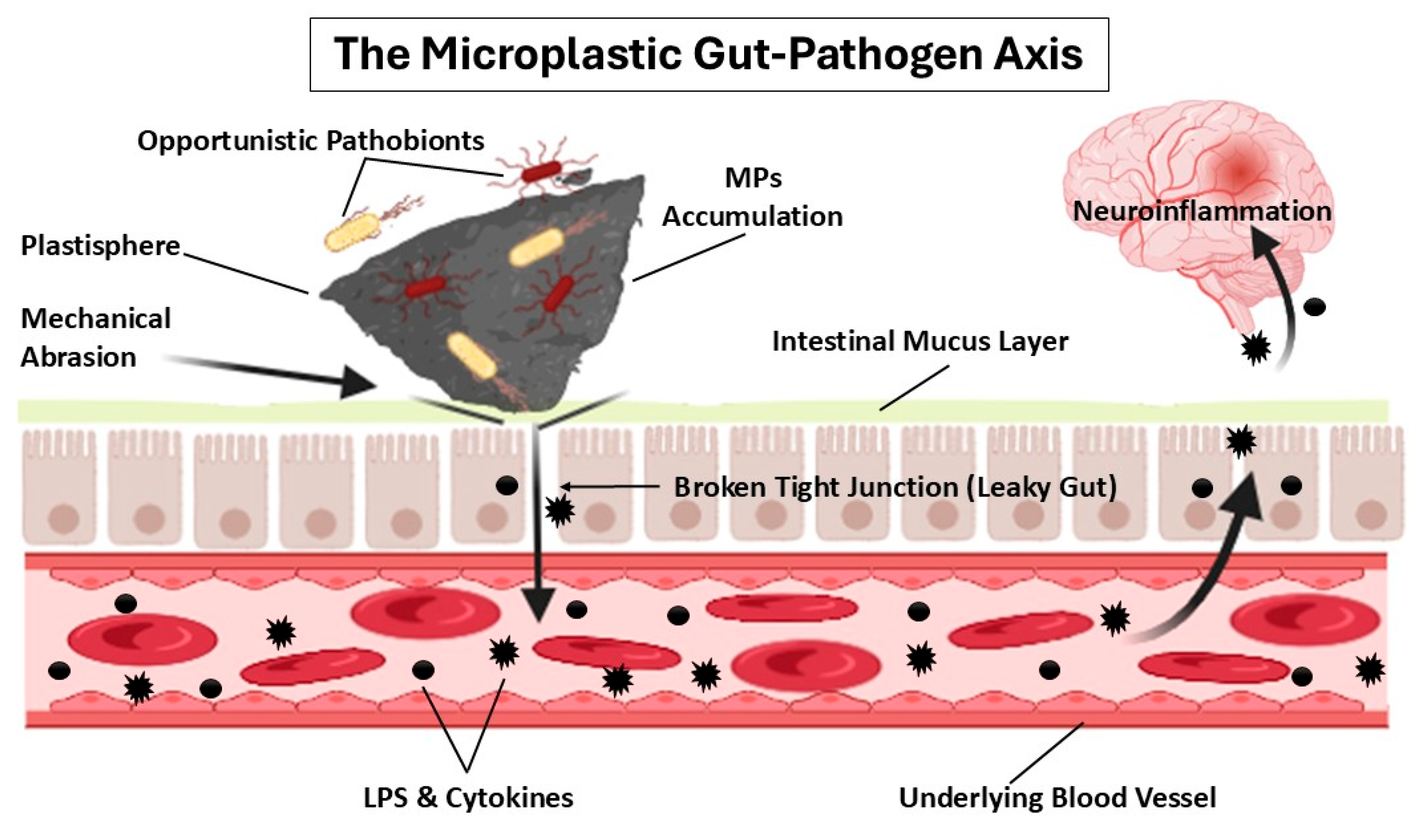
Figure 2.
The Nanoparticle Dual-Hit Hypothesis. This diagram illustrates the two-pronged mechanism by which inorganic nanoparticles (NPs) drive neurodegeneration. The first “hit” (Hit-1) is direct: NPs physically translocate into the central nervous system through systemic circulation (breaching the blood-brain barrier) or olfactory pathways, acting as immediate neurotoxicants that induce epigenetic reprogramming and proteostatic failure. The second “hit” (Hit-2) is indirect: NPs profoundly alter the gut microbiome and enteric nervous system, compromising the mucosal barrier to unleash a systemic wave of inflammatory mediators. This gut-derived neuroinflammatory storm traverses the gut-brain axis, severely exacerbating the direct toxic effects of the accumulated particles in the brain.
Figure 2.
The Nanoparticle Dual-Hit Hypothesis. This diagram illustrates the two-pronged mechanism by which inorganic nanoparticles (NPs) drive neurodegeneration. The first “hit” (Hit-1) is direct: NPs physically translocate into the central nervous system through systemic circulation (breaching the blood-brain barrier) or olfactory pathways, acting as immediate neurotoxicants that induce epigenetic reprogramming and proteostatic failure. The second “hit” (Hit-2) is indirect: NPs profoundly alter the gut microbiome and enteric nervous system, compromising the mucosal barrier to unleash a systemic wave of inflammatory mediators. This gut-derived neuroinflammatory storm traverses the gut-brain axis, severely exacerbating the direct toxic effects of the accumulated particles in the brain.
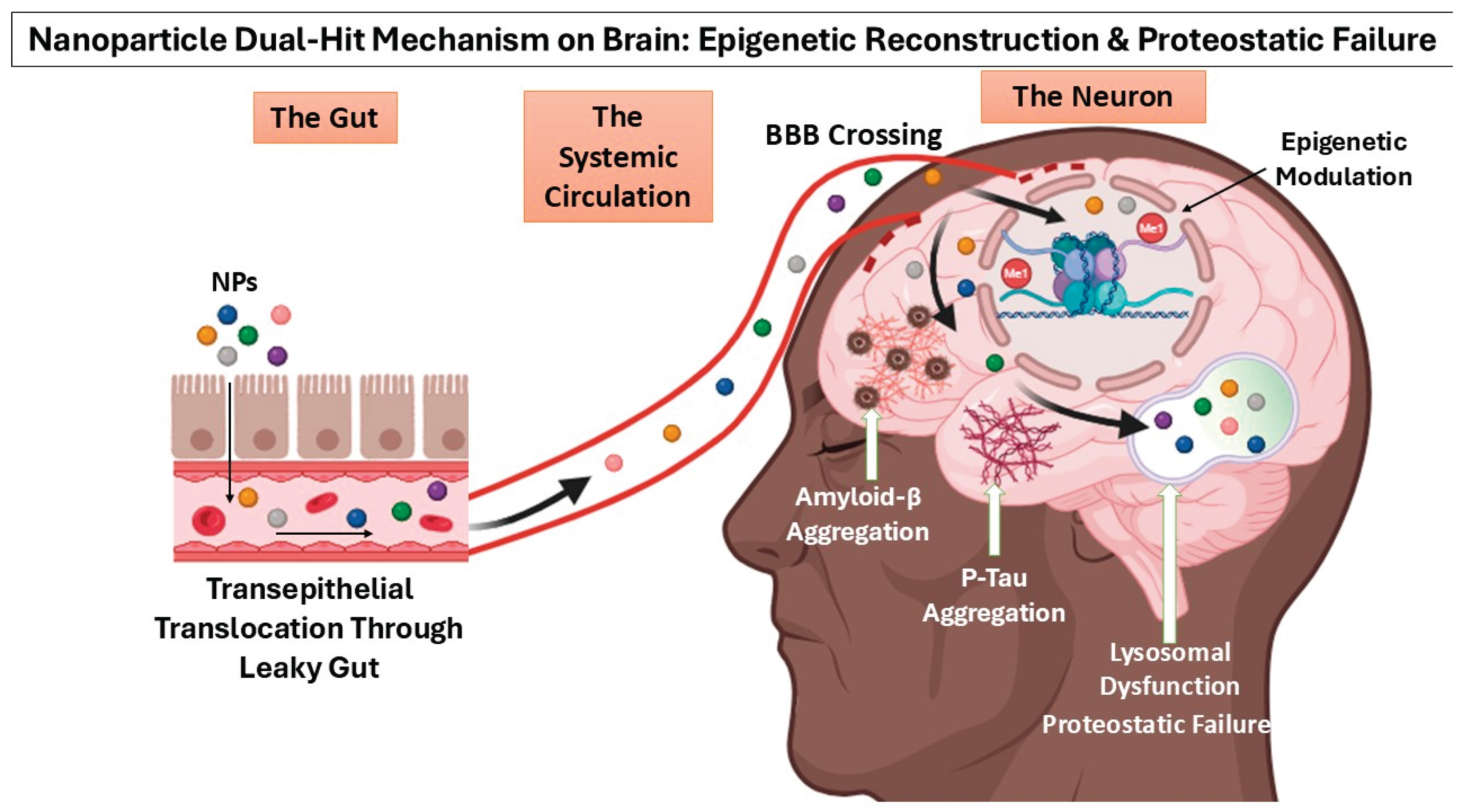

Table 1.
Key Gut Microbiota-Derived Metabolites and Their Neuroprotective Functions within the MGBA.
Table 1.
Key Gut Microbiota-Derived Metabolites and Their Neuroprotective Functions within the MGBA.
| Metabolite | Primary Biosynthetic Taxa | Neuroprotective & Homeostatic Functions | References |
| Butyrate | Faecalibacterium prausnitzii, Roseburia spp., Eubacterium rectale, Clostridium butyricum, Coprococcus, Butyricicoccus pullicaecorum | Attenuates ASD-like endophenotypes; enhances memory in advanced Alzheimer’s disease stages; promotes neuroprotection, learning, and preserves cognitive function. | [24,25,26] |
| Propionate | Bacteroides fragilis, B. vulgatus, B. thetaiotaomicron, Prevotella copri, Veillonella parvula, V. alcalescens, Akkermansia muciniphila | Ameliorates motor and non-motor abnormalities in Parkinson’s disease models. | [27] |
| γ-Aminobutyric Acid (GABA) | Lactobacillus brevis, L. plantarum, L. buchneri, L. paracasei, Bifidobacterium adolescentis, B. dentium, B. infantis | Prevents neurodegeneration; mitigates oxidative stress and mitochondrial dysfunction (deficits are linked to PD neuropathology); attenuates ASD severity. | [28,29,30] |
| Serotonin (5-HT) | Ligilactobacillus ruminis, Limosilactobacillus mucosae, Escherichia spp., Enterococcus spp., Klebsiella pneumoniae, Streptococcus spp. | Regulates sleep-wake cycles, mood stabilization, and cognitive processes. | [31] |
| Kynurenine | Actinobacteria, Bacteroides, Firmicutes, Fusobacteria, Proteobacteria | Functions as an NMDA receptor antagonist, conferring neuroprotection by blocking excessive glutamate excitotoxicity. | [32] |
| Indole Propionic Acid (IPA) | Escherichia coli, Clostridium sporogenes, Bacteroides ovatus, B. thetaiotaomicron | Exerts potent antioxidant effects; scavenges reactive oxygen species (ROS) to protect neuronal integrity and attenuates central neuroinflammation. | [33,34] |
| Indole Acetic Acid (IAA) | Intestinibacter bartlettii, Blautia hydrogenotrophica, Lactobacillus reuteri, L. acidophilus, Bifidobacterium spp., Faecalibacterium | Attenuates neuroinflammatory responses in microglia and suppresses macrophage production of pro-inflammatory cytokines. | [35,36] |
Table 2.
Impact of Non-Nutritive Sweeteners (NNS) and Dietary Additives on Gut Microbial Architecture Across Human and Murine Models.
Table 2.
Impact of Non-Nutritive Sweeteners (NNS) and Dietary Additives on Gut Microbial Architecture Across Human and Murine Models.
| Study Model & Exposure Paradigm | Taxonomic Perturbations & Dysbiosis Profile | References |
| Female SD rats (8 weeks old, n=150); exposed to high-fat diet + aspartame (5-7 mg/kg) | ↓ Enterococcaceae, Enterococcus, Parasutterella; ↑ Clostridium cluster IV | [44] |
| Human cohort (n=120, divided into 4 groups); aspartame, saccharin, sucralose, or stevia | ↓ Abundance of Porphyromonas, Prevotella nanceiensis | [45] |
| Pregnant C57BL/6 mice (8 weeks old); fed sucralose (0.1 mg) + acesulfame-K (0.25 mg) | In offspring microbiome: ↑ Firmicutes; ↓ Akkermansia muciniphila | [46] |
| Male C57BL/6J mice (8 weeks old); acesulfame-K (150 mg/kg body weight/day for 8 weeks) | ↓ Clostridiaceae, Lachnospiraceae, Ruminococcaceae | [47] |
| Human volunteers (n=47, ages 18-35); administered sucralose (48 mg/day for 10 weeks) | ↑ Abundance of Blautia coccoides | [48] |
| Male C57BL/6J mice (SPF, age 28 days); sucralose (0.0003-0.3 mg/mL for 16 weeks) | Jejunum/Ileum/Colon: ↑ Tenacibaculum, Ruegeria, Staphylococcus, Allobaculum Cecum: ↓ Lachnoclostridium, Lachnospiraceae (in high dose group) |
[49] |
| Pregnant C57BL/6 mice; high-fat diet (4 weeks) then sucralose (0.1 mg/mL for 6 weeks) | Maternal: ↑ Firmicutes, Proteobacteria; ↓ Bacteroidetes Post-HFD: enhanced reduction in Proteobacteria compared to control |
[50] |
| Mice (5 weeks old); standard diet + sucralose vs. high-fat diet (HFD) + sucralose | ↑ Firmicutes; ↓ Bacteroidetes (Note: Firmicutes also increased in the standard HFD group) | [51] |
| Male C57BL/6J mice (8 weeks old); sucralose (15 mg/kg body weight/day for 8 weeks) | Faecal microbiota: ↓ Clostridium cluster XIVa | [52] |
| Male C57BL/6 mice; sucralose in drinking water (5 mg/kg body weight/day) | 3 months: ↑ Ruminococcus; ↓ Lachnospiraceae, Staphylococcus, Bacillus 6 months: ↑ Akkermansia, Roseburia; ↓ Streptococcus, Lachnospiraceae |
[53] |
| Male C57BL/6J mice (8 weeks old); saccharin in drinking water (0.3 mg/mL) | 3 months: ↑ Akkermansia, Oscillospira, Corynebacterium; ↓ Anaerostipes, Ruminococcus 6 months: ↑ Corynebacterium, Roseburia, Turicibacter; ↓ Ruminococcus, Dorea |
[54] |
Table 3.
Impact of Microplastic (MP) Exposure on Gut Microbial Architecture in Human Cohorts and Murine Models.
Table 3.
Impact of Microplastic (MP) Exposure on Gut Microbial Architecture in Human Cohorts and Murine Models.
| Study Model & Exposure Paradigm | Taxonomic Perturbations & Dysbiosis Profile | References |
| In vitro human GI digestion (n=6 volunteers, ages 20-27); PCL (150 nm) & PLA (75 nm) MPs | ↓ Richness and α-diversity; ↓ Bifidobacterium, Faecalibacterium; ↑ Pathogenic taxa (e.g., Prevotella) | [71] |
| Student cohort (n=60); exposed via disposable plastic tableware (3 hot meals/day) | ↓ Bacteroidota, ↑ Actinobacteriota; ↓ Faecalibacterium, ↑ Blautia | [72] |
| Occupational cohort (n=40; plastic factory workers vs. low exposure); Polyurethane predominantly detected | ↑ Bifidobacterium, Streptococcus, Sphingomonas; ↓ Ruminococcus, Dorea, Fusobacterium, Coprococcus | [73] |
| Preschool children cohort (n=69); Stool MPs (PVC, PET, PE, PA6) at 425.0 µg/kg/day | ↓ α-diversity; Altered probiotic taxa including ↓ Parabacteroides, Alistipes | [74] |
| Adult cohort (n=39, ages 25-69); Stool MPs (PE, PVC, PS, PP, PA6) | ↑ Enterobacteriaceae, Escherichia coli; ↓ Faecalibacterium prausnitzii | [75] |
| Male C57BL/6 mice; Polystyrene (PS) MPs (0.05–0.1 μm, 9–10 μm, or mixture at 100 ppb) | Mixed PS: ↑ Campylobacterota; Individual PS: ↓ Proteobacteria, Cyanobacteria; Small PS (0.1 µm): ↑ Actinobacteria | [76] |
| College student cohort (n=125, ages 18-30); Take-out food consumers (9 MP types detected) | Dose-dependent altered abundances of Firmicutes, Bacteroidota, Blautia, Lachnospiraceae, Faecalibacterium, Bacteroides, and Bifidobacterium | [77] |
Table 4.
Impact of Inorganic Nanoparticle (NP) Exposure on Gut Microbial Architecture Across In Vivo Models.
Table 4.
Impact of Inorganic Nanoparticle (NP) Exposure on Gut Microbial Architecture Across In Vivo Models.
| Study Model & Exposure Paradigm | Taxonomic Perturbations & Dysbiosis Profile | References |
| Female rats (prenatal exposure); TiO2 NPs (5 mg/kg, GD 5-18) | ↑ Clostridiales (GD 10); ↓ Dehalobacteriaceae (GD 17) | [92] |
| Rats; oral TiO2 NPs (29nm, 0-50 mg/kg/day for 90 days) | ↑ Lactobacillus reuteri; ↓ Romboutsia | [93] |
| Mice; oral TiO2 NPs (100 mg/kg/day for 28 days) | ↑ Actinobacteria, Proteobacteria; ↓ Firmicutes, Bacteroidetes | [94] |
| Rats and Mice; oral AgNPs (2.5 or 3.6 mg/kg for 7-14 days) | Shifted Firmicutes/Bacteroidetes (F/B) ratio | [95,96] |
| Piglets; oral ZnO NPs (600 mg/kg for 14 days) | Ileum: ↑ Streptococcus, ↓ Lactobacillus; Colon: ↑ Lactobacillus, ↓ Oscillospira, Prevotella | [97] |
| Broiler chickens; oral ZnO NPs (5 mg/kg for 42 days) | ↑ Ruminococcaceae, Bacteroidaceae; ↓ Lachnospiraceae, Lactobacillaceae, Rikenellaceae | [98] |
| Rats; oral TiO2 NPs (2-50 mg/kg/day for 30 days) | ↑ Lactobacillus gasseri, Turicibacter; ↓ Veillonella | [99] |
| Male C57BL/6JAusb mice (5-6 weeks old); oral TiO2 NPs (drinking water, 0-50 mg/kg for 3 weeks) | ↑ Lactobacillus, Allobaculum; ↓ Adlercreutzia, unclassified Clostridiaceae | [84] |
| C57BL/6 mice; oral AgNPs (0.1, 2, 40 µg for 120 days) | ↑ Firmicutes; ↓ Bacteroidetes | [82] |
| Wistar rats; oral AgNPs (7nm, 100 mg/kg for 28 days) | ↑ Bacteroidota; ↓ Verrucomicrobia, Proteobacteria, Lactobacillaceae | [100] |
| Wistar albino rats; oral ZnO NPs (1000 mg/kg for 28 days) | Males: ↑ Firmicutes, ↓ Bacteroidetes; Females: ↓ Firmicutes, ↑ Verrucomicrobia | [81] |
Table 5.
Impact of Particulate Matter (PM2.5) Exposure on Gut Microbial Architecture in Human Cohorts and Murine Models.
Table 5.
Impact of Particulate Matter (PM2.5) Exposure on Gut Microbial Architecture in Human Cohorts and Murine Models.
| Study Model & Exposure Paradigm | Taxonomic Perturbations & Dysbiosis Profile | References |
| Southwest China cohort (n=1583); long-term exposure to PM2.5 | ↓ α-diversity; ↓ Bacteroidetes, ↑ Proteobacteria | [116] |
| Guangdong Province cohort (n=3267, ages 40–75); exposure to PM2.5 | ↓ Bacteroidetes; ↑ Actinobacteria, Firmicutes, Ruminococcus | [117] |
| Shanghai maternal–child pairs (n=361); prenatal PM2.5 exposure | Validation of seven PM2.5-correlated genera: Ruminococcus gnavus group, Romboutsia, Burkholderiaceae, Blautia, Alistipes, Parabacteroides, and Bacteroides | [118] |
| Healthy seniors (n=76, ages 60–69); exposure to PM2.5 | ↓ α-diversity; ↑ Lachnoclostridium, Streptococcus, Veillonella; ↓ Megamonas, Erysipelatoclostridium, Dialister, Subdoligranulum, Holdemanella, Blautia | [119] |
| Latino breastfed infants (n=103, Southern California, 6 months old); exposure to PM2.5 | ↓ Alistipes, Proteobacteria, Rikenellaceae; ↑ Actinomyces | [120] |
| Nrf2+/- rat (8-week-old); filtered vs. outdoor air (16h/day, 6 & 12 weeks) | ↓ Bacteroides, Firmicutes, Allobaculum, Prevotella, Sutterella; ↑ Alphaproteobacteria, Pseudomonadaceae, Desulfovibrionaceae | [121] |
| Male C57BL/6 WT mice; exposed to PM2.5 for 5 months | ↓ α-diversity; ↑ Proteobacteria; ↓ Lactobacillus, Parabacteroides, Prevotella, Alloprevotella, Ruminococcus, Faecalibacterium, Bifidobacterium | [111] |
| SD rats (6-8 weeks); oral gavage of PM2.5 (10 mg/kg/day for 28 days) | ↓ Firmicutes, Ruminococcaceae; ↑ Lactobacillaceae, Clostridium, Ruminae, Atopobiaceae, Coriobacteriaceae_UCG_002 | [122] |
| Male C57BL/6J mice (3-week-old); exposed to PM2.5 (8, 16, 24 weeks) | ↓ Bacteroidetes, ↑ Proteobacteria; ↑ Clostridium, Akkermansia, Acetatifactor | [108] |
| Male BALB/c mice (8-week-old); exposed to PM2.5 (8h/day, 6 days/week for 6 weeks) | ↓ Bacteroidetes/Firmicutes ratio; ↑ Lactobacillus, Clostridium; ↓ Bacteroides, Parabacteroides. (Post-recovery dominance of Oscillospira) | [109] |
| Male SD rats (SPF grade); passive lung inhalation of PM2.5 (10-20 mg/kg) | ↓ Cyanobacteria, Bacteroidetes, Proteobacteria; ↑ Verrucomicrobia, Elusimicrobiota, Desulfobacterota, Patescibacteria, Firmicutes | [123] |
Disclaimer/Publisher’s Note: The statements, opinions and data contained in all publications are solely those of the individual author(s) and contributor(s) and not of MDPI and/or the editor(s). MDPI and/or the editor(s) disclaim responsibility for any injury to people or property resulting from any ideas, methods, instructions or products referred to in the content. |
© 2026 by the authors. Licensee MDPI, Basel, Switzerland. This article is an open access article distributed under the terms and conditions of the Creative Commons Attribution (CC BY) license (http://creativecommons.org/licenses/by/4.0/).
Copyright: This open access article is published under a Creative Commons CC BY 4.0 license, which permit the free download, distribution, and reuse, provided that the author and preprint are cited in any reuse.