

Submitted:
22 July 2026
Posted:
23 July 2026
You are already at the latest version
Abstract
Non-alcoholic fatty liver disease (NAFLD) specifically includes the stage of simple fatty liver and the stage of hepatitis. Recent research found mesenchymal stem cell therapy for NAFLD. Mesenchymal stem cells regulate metabolic pathways and energy transport pathways in disease models, reducing the synthesis and utilisation of glucose and fatty acids and restoring the homeostasis of the internal microenvironment. For areas with more severe damage, umbilical cord mesenchymal stem cells (UCMSC) are used for regenerative repair with classic Wnt pathway. The text explores the therapeutic effects of UCMSCs on hepatitis and the surrounding environment from a multifunctional perspective. Mesenchymal stem cells release exosomes in a paracrine manner into the damaged areas of the liver, promoting the differentiation and development of hepatic progenitor cells into hepatocytes, reducing differentiation into cholangiocytes, while assisting hepatic progenitor cells in resisting the progression of inflammation and fibrosis, and promoting the normal function of adaptive immune responses.
Keywords:
umbilical cord mesenchymal stem cells
; chronic liver disease
; non-alcoholic fatty liver disease
; stem cell differentiation and development
1. Introduction
In February 2020, over 30 experts from 15 countries officially renamed NAFLD (non-alcoholic fatty liver) to MAFLD (metabolic dysfunction-associated liver disease). The definition of MAFLD has a wide scope, and the diagnostic methods include type 2 diabetes, obesity, or the presence of other cardiovascular diseases [1,9,58]. Triglyceride accumulation in hepatocytes is a fundamental factor leading to the development of MAFLD. Obesity is an important factor causing liver damage; it can lead to the formation of MAFLD, resulting in excessive enlargement of the liver and even compression of other organs. The disease itself, as an early-stage hepatitis, can progress toward irreversible liver cirrhosis and liver cancer. Kupffer cells secrete inflammatory factors and anti-tumor suppressive factors, and with an increase in free fatty acids (FFAs), they can lead to upregulation of vimentin and α-smooth muscle actin expression, promoting fibrosis in hepatitis disease. Non-alcoholic steatohepatitis (NASH) involves complex pathological conditions and is associated with fibrosis accompanied by inflammation. When multiple injuries coexist, researches also needs to consider the patient’s own nutritional issues, hepatocyte mitochondrial dysfunction, endoplasmic reticulum oxidative stress, changes in gut microbiota, and epigenetic issues in cellular mechanisms [3,4,5]. And the CRP-triglyceride-glucose index, as a key measure of recent metabolism, has shown a strong link between changes in plasma and metabolic diseases [105].
Stem cells show advantages beyond traditional medicine in treating MAFLD patients,as they can promote liver regeneration [7,8]. They are suitable for a variety of patient groups and provide treatment by reducing fibrosis, inflammation, and lipid deposition. With the development of stem cell exosome technology, small active molecules can be delivered directly to the lesion site, bypassing anatomical barriers. Research on related stem cell-derived exosomes shows that they play an important role in liver diseases by inhibiting inflammation, reducing oxidative stress, and regulating immune responses, highlighting their significant potential in MAFLD treatment [10,12]. Severe liver damage can lead to reduced liver regeneration ability or even structural changes, while umbilical cord-derived mesenchymal stem cells can better improve the liver microenvironment through the classic Wnt pathway [13,22,23]. Umbilical cord mesenchymal stem cells efficiently act on liver parenchymal cells through the vascular walls of the portal vein and central vein in a paracrine manner, utilizing stem cell exosomes. Umbilical cord mesenchymal stem cells can prevent excessive fibrosis from leading to steaosis of liver microvessels, which in turn can cause a coagulant state. Therefore, stem cells have an advantage over other chemical reagents in improving systemic coagulation and the liver microenvironment. Mesenchymal stem cells primarily migrate to target sites via the bloodstream. Due to their cellular plasticity, they can be used for in vitro organoid construction or drug screening through 3D printing technology. These cells inherently exhibit low immunogenicity, lack MHC class II expression, and possess anti-inflammatory properties, making them particularly suitable for allogeneic cell therapy [11].
However, the liver not only has a role in detoxification, it also plays an important role in maintaining stable blood glucose levels and regulating lipid homeostasis. The liver mainly comprises six types of cells: hepatocytes, large polygonal epithelial cells, cholangiocytes, hepatic stellate cells, Kupffer cells, liver sinusoidal endothelial cells, and portal fibroblasts. Numerous experiments indicate that immune cells are widely present in MAFLD and NASH. When excessive free fatty acids exist in hepatocytes, they can alter the homeostasis of the immune microenvironment around the hepatocytes.We reviews the molecular pathology of non-alcoholic fatty liver disease, Wnt signaling biology, cell type-specific Wnt effects in the liver (LSECs, HSCs, cholangiocytes, immune cells), and the UCMSC/MSC-EV mediated mechanisms (miRNA, circRNA, mitochondrial transfer) [31,75]. Finally, we discuss the classic Wnt pathway play dominant effect in microenvironment surrounded liver organ.
2. Molecular-Level Investigation of Non-Alcoholic Fatty Liver Disease
The existing MAFLD models are too simple and need to combine metabolic dysfunction or obesity traits for modeling. MAFLD often co-occurs with obesity, and individuals who are obese under normal conditions are more prone to developing NAFLD. Proprotein Convertase Subtilisin/Kexin Type 9 (PCSK9), a serine protease encoded by the PCSK9 gene, is primarily produced in the liver. Additionally, PCSK9 is also expressed in other organs such as the intestine, heart, and pancreas [95]. Human carbonic anhydrase VA (CA-VA) shows increased expression in the liver and brain, where it influences gluconeogenesis and lipid synthesis through catalyzing HCO3−, thereby affecting obesity [8,15]. Under obese conditions, AlkB homolog 5 (ALKBH5), an RNA demethylase, becomes phosphorylated in the nucleus via PKA-mediated signaling, translocates into the cytoplasm, undergoes self-methylation, and subsequently activates the GCGR-cAMP pathway, reducing blood glucose levels and improving glucose tolerance [6]. Notably, in mice with liver-specific knockout of the RNA demethylase ALKBH5, both the GCGR and mTORC1 pathways are activated, leading to decreased blood glucose levels [9,10]. Glucose levels undergo significant changes in the livers of NAFLD mice, accompanied by a marked increase in the expression of the E3 ubiquitin ligase TRIM7, which was initially thought to be involved in glycogen metabolism [14,17]. Mechanistic studies reveal that TRIM7 interacts with DUSP10 and catalyzes its ubiquitination and proteasomal degradation, leading to excessive activation of the IKKβ-NF-κB and JNK/p38 MAPK signaling pathways. TRIM7 is a key regulatory factor in the pathogenesis of MAFLD/NASH, and targeting the TRIM7-DUSP10 axis offers a promising therapeutic strategy for MAFLD [11,12,13,14]. Additionally, GCN5 functions as an acetyltransferase and plays a crucial role in regulating hepatic glycogen synthesis through histone modification [15]. MARCHF6 has a steroid-sensing structure and mediates protein degradation by recognizing acetylation signals at the N-terminus of substrates. When MARCHF6 is missing, SREBP1 gets activated, leading to cell-intrinsic liver fat metabolism problems [16].
Histone propionylation has been identified as a key modification in de novo lipogenesis, and RPN11 acts as a novel regulator of MAFLD/NASH [62]. Hepatocyte-specific knockout of RPN11 shows potential therapeutic effects by inhibiting the deubiquitinase RPN11 to treat MAFLD and NASH [18,29]. Mice with hepatocyte-specific RPN11 gene deletion are protected against diet-induced hepatic steatosis, insulin resistance, and non-alcoholic steatohepatitis. The products from histidine metabolism and mitochondrial beta-oxidation eventually end up entering the TCA cycle. Histidine is deaminated by histidase (HAL) to form trans-urocanic acid, which is then converted by urocanase (UROC1) into 4-imidazolone-5-propionic acid, and eventually turned into glutamate that enters the TCA cycle [96]. The beta-oxidation process keeps releasing acetyl-CoA and producing NADH and FADH2.Solute Carrier Family 25 Member 47 (SLC25A47) transports NAD+ into mitochondria, providing substrate for histone deacetylase SIRT3. Activated SIRT3 further initiates the AMPK signaling pathway, thereby inhibiting energy supply to the liver and preventing hepatic steatosis [97,98]. Loss of this gene leads to hepatic steatosis, development of fatty liver, and spontaneous formation of hepatocellular carcinoma [58,60]. The biological hormones secreted by the human body itself can also prevent the development of diseases by altering protein function.Androgen receptor transcriptionally activates CAND1 protein which exert the regulatory role of ubiquitin ligase, thereby blocking the degradation of ACAA2, impairing fatty acid β-oxidation, and leading to excessive fat accumulation in the liver [61]. In the absence of CAND1, CULLIN1, FBXO42, and ACAA2 assemble more readily into an active complex, thus suppressing ACAA2 expression levels [61]. Meanwhile, ALKBH5 binds to the intronic enhancer of one member of the epidermal growth factor receptor family (EGFR) via its demethylase activity, activating the PI3K-AKT-mTORC1 signaling pathway and promoting lipid synthesis [6].
When endoplasmic reticulum oxidative stress occurs, plasma cells, as high-intensity protein-secreting cells, tend to activate XBP1 protein. IRE1α is a key regulatory molecule in the activation of the XBP1 protein signaling pathway. After XBP1 enters the nucleus, activated IRE1α performs unconventional splicing of XBP1 mRNA, removing a 26-nucleotide intron, changing the reading frame to translate a protein with strong transcriptional activity [99]. Studies have shown that myeloid-derived dendritic cells in the tumor microenvironment assemble MHC-I, inhibit the proliferation and activation of CD8 T cells, and promote tumor cell proliferation and immune evasion [11,59].
3. Hepatic Microenvironment
For liver regeneration after injury, it mainly involves rebuilding damaged cells, tissue repair that doesn’t rely on buds, and repairing the broken parts.
3.1. Extracellular Matrix
Cross-linking of the extracellular matrix can worsen fibrosis, and in patients with cirrhosis, it can lead to higher glucose levels. Advanced glycation end products also contribute to the cross-linking of the extracellular matrix. Glucose levels are mainly used as an indicator to check the extent of late-stage fibrosis and to further assess how strongly the cellular matrix can affect collagen fibers and attract macrophages [100]. ECM provides the substrates needed for regeneration,it provides the basic framework and firmness of the liver and serves as a place for mast cells and macrophages, with the collagen produced by mast cells often appearing together with HSP47 [93,94]. The extracellular matrix mainly consists of structural proteins, adhesive proteins, proteoglycans, and cytokines. Keratin, as an important structural protein, is mainly secreted by liver cells and serves as a key part of the cell’s cytoskeleton. CK18 is continuously cleaved by caspases to form the M30 fragment, as well as the M65 fragment that comes from apoptotic and necrotic cells. CK19 is mainly expressed in benign liver lesions and is an important marker for determining tissue origin. Both of these are often used as strong indicators to check the regeneration of a damaged liver [101,102]. Whether it’s the rise of glucose levels in the plasma or the excessive buildup of advanced glycation end products in the extracellular matrix, both can lead to the production of ATP and the activation of the AMPK energy pathway. AMPK, as a key gene in the energy pathway, can activate the downstream fatty acid synthase pathway, which in turn affects the host’s diet and insulin levels.When glucose energy is released and too much ATP is produced, the SMC1/3 coiled coil of the cohesin protein bends, looping the DNA inside the SMC chamber and the SMC-NIPBL chamber.Whether it’s the rise of glucose levels in the plasma or the excessive buildup of advanced glycation end products in the extracellular matrix, both can lead to the production of ATP and the activation of the AMPK energy pathway [22]. For example, infiltrating Seplplg macrophages are significantly expanded in MASH, and after glycosylation modifications, they specifically bind to the C-type lectin domain of Sell on the surface of neutrophils, inducing hepatic stellate cells to transdifferentiate into myofibroblasts.
3.2. Immune Cell
Immune cells mainly regulate pro-inflammatory ATM and insulin resistance through recruiting peripheral blood immune cells and resident T cells in the fat tissue of obese people. The OX40L ligand is the molecule that’s most significantly upregulated under obesity. Although macrophages lacking OX40L don’t show noticeable changes in phenotype, they can reduce Th1 cell expression [71]. Immune cells recruit through macrophages, mediate through biological chemokines, and undergo self metabolic reprogramming to help reduce phenotype in damaged liver regions and promote the occurrence of liver dysfunction. When treating damaged liver, unlike relying solely on anti-inflammatory and anti fibrotic chemical agents, it is necessary to change the type and content of genes. As a chemotactic factor, IL-27 not only attracts immune cells to the target site, but also performs functions similar to those of immune cells. Mast cells release heparin through degranulation, which binds to lipoprotein lipase on blood vessel walls, helping break down fat into fatty acids and lowering blood lipid levels. The MASH model found that CD8 T cell expression levels were reduced, but after UCMSC treatment, they increased significantly. This is mainly because Tim3, as an immune checkpoint, promotes DC lipid peroxidation and ferroptosis in a high-fat environment, leading to CD8 T cell exhaustion, which then contributes to fatty liver-induced liver cancer [30]. The role of metabolites being transported and carried out through the hepatic vein hasn’t really been noticed by current researchers; they mainly focus on the liver parenchymal cells.The liver receives a dual blood supply from the portal vein and hepatic artery, bearing a large number of antigens while having good tolerance to them. LPS entering the liver via the portal vein can downregulate the antigen-presenting ability of liver sinusoidal endothelial cells and Kupffer cells [2]; Only high concentrations of antigens are presented by liver sinusoidal endothelial cells, leading to the clonal deletion and functional impairment of antigen-specific T cells, and inducing the activation of regulatory T cells. Studies have shown that Tregs tend to undergo oxidative phosphorylation and fatty acid β-oxidation processes, which are crucial for maintaining immunosuppression [32,47].
3.3. Microenvironment Changes and Metabolic Reprogramming
The process of metabolic reprogramming mainly includes sugar metabolism, fat metabolism, and amino acid metabolism represented by glutamine [81]. The formation of lipid droplets mainly happens in the endoplasmic reticulum, so the normal structure and function of the ER are really important. PLIN5 can bind to both lipid droplets and mitochondria, and there are some mutation experiments as evidence showing that after PLIN5 gets phosphorylated, it strengthens the coupling between lipid droplets and the ER, which then leads to an increase in liver fat content [31,36,65]. CLCC1 is a molecule that senses phospholipid imbalance in the endoplasmic reticulum membrane [35]. It works together with TMEM41B to regulate phospholipid flipping across layers, and when the metabolic pathways they jointly control are impaired, it can lead to fat buildup in the liver [24]. Experiments have shown that increasing CLCC1 can improve liver damage caused by obesity [32]. In current beet plant models, the upregulation of genes like pyruvate decarboxylase 1 (PDC1), aldehyde dehydrogenase 7 (ALDH7), and alcohol dehydrogenase 7 (ADH7) is predicted to help detoxify toxic aldehydes, which often show up under low oxygen and energy-compromised conditions. Also, these two enzymes are present in the human body as well [33].
4. Origin and Biological Characteristics of the Wnt Pathway
Dysfunction of the Wnt/β-catenin pathway plays an important role in the occurrence and development of various diseases, as well as in cell differentiation and development. The Wnt pathway is divided into the canonical Wnt pathway and the tumor Wnt pathway, while the non-canonical Wnt pathway has only two branches: the non-planar cell polarity Wnt-PCP pathway and the Wnt/Ca2+ pathway, which play regulatory roles in tissue microenvironment, immune system response to damaged tissues, and ion channel changes [33,34,37,38].
Under normal circumstances, the β-catenin/APC/AXIN pathway is maintained at a relatively low level due to degradation. In colorectal cancer, the Wnt pathway is overly activated, leading to excessive differentiation and maturation of tissue stem cells, altering the basic structure of organs [35]. APC regulates phosphorylated β-catenin in association with the cytoplasm-mediated ubiquitin-proteasome degradation pathway, thereby negatively regulating this pathway. AXIN is an important protein that supports the binding of GSK3β and APC [36]. Wnt downregulates GSK3β activity through the LRP5 signal transduction and LRP6 co-receptor, increasing nuclear β-catenin levels [37]. In organs such as the immune system, breast, adipose tissue, bone, and skin, Wnt10b is involved in the signaling network that controls stemness, pluripotency, and cell fate determination [38,39]. Recruited co-activators, including CBP, P300, PYGO, and BCL9, bridge the C-terminal of β-catenin with TCF/LEF and other transcription elements to stimulate downstream gene expression and cell proliferation. Dishevelled (DSH/Dvl), a cytoplasmic protein widely present in tissues, mainly functions in the aggregation of β-catenin, thereby regulating the gene expression that enters the cell nucleus [40,46].
Wnt5a and Wnt11 molecules can specifically bind to FZD, thereby activating the Wnt/Ca2+ pathway. Wnt5a can interact with the ROR family of orphan receptor tyrosine kinases to activate JNK and RHOA [41,42]. By inhibiting the β-catenin and TCF complex, it antagonizes the classical Wnt pathway [43]. In addition, PI3K/AKT can regulate this pathway by inhibiting Wnt11 feedback [44]. Porc, as an upstream regulatory molecule of the Wnt pathway, can covalently link monounsaturated fatty acids to serine residues. Experiments have shown that if Porc function is lost, Wnt proteins (such as the Wg protein in Drosophila) are retained inside the cell and cannot be secreted outside the cell to exert their function [45,46]. However, the activity of β-catenin in the canonical Wnt pathway can also be regulated in a manner independent of GSK3β and β-TrCP [18,47]. The non-canonical Wnt pathway can promote the proteasomal degradation of β-catenin through an alternative E3 ubiquitin ligase complex containing APC, EBI, and SIAH1 or SIAH2, thereby inhibiting the classical Wnt signaling pathway. The non-canonical Wnt pathway can antagonize the classical Wnt pathway in multiple ways [48,49]. Different Wnt pathway mutations may be associated with different pathogenic mechanisms. The membrane-bound protein E3 ligase, RING finger protein (RNF43), and/or zinc finger protein ZNF34 trigger the status of the Wnt pathway, disrupting a negative feedback loop that drives Wnt receptor endocytosis in adult stem cells [22,80].
5. The Role of the Wnt/β-Catenin Pathway in the Liver
The Wnt signaling pathway is an important regulator of mammalian liver metabolism. The Wnt pathway mainly functions around the central vein, and Wnt2, as a key regulatory signal, shows significant changes in levels during chronic liver injury [42,43,44] (Figure 1A). As a signal pathway sensitive to oxygen gradients and spatial constraints, the Wnt pathway promotes the formation of metabolic zonation, and within different metabolic zones, proteins with biological functions and varying enzyme activities are spatially arranged around the hepatic lobule. Various chronic liver diseases, while affecting hepatocytes, also involve damage to other organs [50].
As key protein in PORCN plays a regulatory role in the Wnt pathway, and its biological function is mainly reflected in the palmitoylation of Wnt proteins, the secretion and activity of Wnt proteins, and the maintenance of Wnt signaling. PORCN is located upstream of the Wnt pathway, and small-molecule inhibitors can specifically bind to PORCN [103]. When PORCN is inhibited, it can block the secretion of Wnt1/2/3/6/7a/7b/8a/8b/9a/9b/10a/10b proteins, making it one of the important targets for cancer therapy [42] (Figure 1B).
Key regulatory factors include GSK3β, which is activated upon de-repression after AKT inactivation, phosphorylating and activating the acetyltransferase TIP60. TIP60 acetylates the autophagy core kinase ULK1, activating ULK1 and initiating autophagy [18,78]. Cells clear damaged components dominated by lipid droplets through autophagy, while effective acetylation can efficiently recruit the HOPS complex [46,54]. In a MAFLD mouse model, overexpression of PACER can effectively alleviate hepatic steatosis and injury. Overexpression of SIRT1 inhibits adipogenesis in mesenchymal stem cells (MSCs). SIRT1 activates the Wnt/β-catenin pathway by deacetylating histones of sFRPs and deacetylating β-catenin, thereby inhibiting adipogenesis and ultimately reducing fat deposition [54,62]. In a 2020 study on hepatic ischemia-reperfusion injury, it was found that GSK3α (rather than GSK3β) exerts a protective effect on hepatocytes by activating the TIP60-mediated autophagy pathway [54] (Figure 1C).
The loss of the APC gene and activation of β-catenin promote nuclear transcription of c-Myc. Around the hepatic veins, APC and β-catenin are expressed oppositely. Hepatocyte nuclear factor-4α (HNF-4α) is a transcription factor in the liver responsible for the expression of periportal genes in hepatocytes and inhibits β-catenin and TCF-dependent gene expression [55,59]. Conversely, β-catenin and TCF-dependent genes can inhibit the expression of HNF4α. This demonstrates that HNF4α is one of the main indicators of liver functional zonation. As a key gene in this pathway, β-catenin can regulate increased metabolic activity of ammonia and glutamine. During ammonia detoxification and metabolism, glutamine synthetase increases with β-catenin activation, and when β-catenin is knocked out, the content of glutamine synthetase decreases, and plasma ammonia concentration increases, which may also be one of the causes of hepatic encephalopathy [56]. CUL1 can regulated RPN11 and promote β-catenin degraded to inhibit cell excess proferation. EVs play an important role in the pathogenesis of MAFLD. EVs released by damaged hepatocytes promote the progression of liver injury by activating non-parenchymal liver cells, such as LSECs and HSCs. Kupffer cells are an important source of Wnt proteins, which stimulate β-catenin in hepatocytes in a paracrine manner after partial hepatectomy. Deletion of the SPTLC2 gene leads to degradation of cadherin and disrupts the distribution of β-catenin [57].
Through the collection and analysis of data from public datasets related to MAFLD, the experimental results obtained after sorting and analysis revealed that MAFLD primarily disrupts the cell proliferation cycle, increases inflammation and fibrosis phenotypes in the early stages of damage, and additionally affects glucose and insulin levels in the later stages. This alters the microenvironment of the liver, thereby affecting its autonomous regenerative capacity (Figure 1D). Through the dbGAP database, mainly including 50 patient samples, which are primarily divided into four disease phenotypes: inflammation, fibrosis, endoplasmic reticulum oxidative stress, and mitochondrial damage. After analyzing differential gene expression using the R package pheatmap with the GSE296996 public dataset, it was found that in the control group, HSPA5, IGFBP1, HSP90B1, NUCB2, and CCPG1 were upregulated, while DGAT2, DB1, SQLE, HELLS, and DHCR24 were downregulated (Figure 1E). Enrichment analysis of cellular components, biological processes, and molecular functions revealed that the top negatively regulated GO-BP term was DNA replication (p=3.53e-28), and the top negatively regulated KEGG pathway was DNA replication (p=6.76e-17). The expression of the β-catenin complex in the Wnt pathway was downregulated. The top positively regulated GO-BP enrichment was the endoplasmic reticulum oxidative stress process, and the top positively regulated KEGG enrichment was the response to endoplasmic reticulum oxidative stress. GSK3β not only regulates c-Myc activity, but c-Myc in turn can regulate the expression of genes GADD45 and GADD153, thereby regulating the DNA damage process and controlling the cell cycle. Additionally, GSK3 can control the activity of transcription factors such as nuclear factor-κB (NF-κB). The study used Wnt5a as a gene to detect the activation of the Wnt pathway and CREBBP as a gene to amplify the Wnt pathway signal.
5.1. Liver Sinusoidal Cells
The health status and differentiation phenotype of LSECs depend on Wnt signaling. The fenestrated structure of liver endothelial cells allows for contact with microbes and nutrients brought by the portal vein. Studies have found that the transcription factor Gata4 maintains the normal differentiation state of LSECs by regulating the expression of Wnt2 signaling, allowing them to retain their characteristic fenestrations [58]. Once this signaling axis is disrupted, LSECs ‘dedifferentiate’ and lose their fenestrations, which is an early event in liver sinusoid injury. Changes in the phenotype of liver sinusoids prevent triglycerides and cholesterol from entering sinusoids from hepatocytes, leading to fat accumulation. Hepatic stellate cells activate and envelop liver sinusoidal endothelial cells, increasing sinusoidal cell coverage and promoting structural changes in the sinusoids. Metabolic products derived from sphingolipids promote liver regeneration by driving pro-regenerative vascular remodeling and producing inflammatory mediators in an LSEC-dependent manner [51,52,53]. Transferring neutral sphingomyelinase and sphingosine kinase 2 (SK2) to hepatocyte-derived exosomes has been shown to enhance S1P synthesis in target hepatocytes [59]. Japanese researchers have demonstrated that direct contact between LSECs and platelets triggers platelet secretion of S1P, which subsequently induces LSECs to produce IL-6 and VEGF, thereby regulating DNA synthesis in hepatocytes [25].
5.2. Hepatic Stellate Cells
Liver fibrosis is the intrinsic core pathological feature of chronic liver diseases, including MAFLD and MASLD. A pro-fibrotic liver microenvironment activates hepatic stellate cells (HSCs) and promotes collagen deposition. Oleamide (OA) promotes HSC proliferation and activation by activating the transcription factor ATF3, thereby driving collagen deposition, whereas activation of LONP1 or inhibition of DHODH can improve fibrosis severity in MASH [63]. Abnormal activation of HSCs mediated by Wnt1/Wnt5a is a key factor in fibrosis progression [60]. Netrin-1, as an autocrine factor, has shown in functional studies that AAV-mediated overexpression of Netrin-1 in the liver significantly worsens liver fibrosis, while HSC-mediated knockdown of Netrin reduces the degree of liver fibrosis, including improvements in the fibrotic phenotype of mice via lipid nanoparticle-mediated Netrin-1 siRNA knockdown. Netrin-1 promotes HSC activation through autocrine signaling mediated by its receptor UNC5B, a process that triggers rapid intracellular Ca2+ mobilization, further inducing SMAD2 phosphorylation and promoting fibrosis gene expression. The expression and protein levels of the ZNF469 gene increase; the gene family itself has a zinc finger motif that facilitates the interaction between proteins and DNA [61]. The Wnt pathway in HSCs is abnormally activated, leading to increased secretion of CXCL12, which attracts a large number of macrophages to the inflammatory sites, forming a vicious cycle. The expression of Frizzled-4 (Fzd4) and Frizzled-7 (Fzd7) is also closely related to the activation status of HSCs. DCDC2 is an important endogenous inhibitory molecule that blocks classical Wnt signaling by inhibiting β-catenin activation and its translocation to the nucleus, thereby suppressing HSC activation, proliferation, and epithelial-mesenchymal transition (EMT)-like processes. In fibrotic liver tissue, DCDC2 expression is significantly downregulated. RSPO, through secreted factors, alters liver microenvironmental zoning and promotes remodeling and regeneration after liver injury [57,62].
5.3. Bile Duct Cells
β-catenin also plays an important role in cholangiocyte morphological differentiation. In β-catenin knockout mice, the bile ducts are dilated and tortuous, and the microvilli on the surface of normal hepatocyte bile ducts are sparse. Overexpression of β-catenin in mouse hepatocytes leads to increased nuclear translocation of FoxO1. Through insulin-sensitized stimulation of the Akt pathway, the phosphorylation regulatory network of FoxO1 is also influenced by β-catenin. Mice lacking β-catenin show increased insulin-dependent phosphorylation of Akt and GSK-3β, resulting in insulin resistance. Conversely, overexpression of β-catenin hinders insulin signaling and reduces glycogen synthesis.
5.4. Immune Cells in the Liver
Transgenic mice overexpressing IL-22 exhibit accelerated liver regeneration after partial hepatectomy. Lymphotoxin produced by T cells increases IL-6 production by stimulating lymphotoxin β receptors on hepatocytes [67,68]. CD8 T cells can also induce the proliferation of liver progenitor cells during liver repair in chronic liver injury models. Studies have shown that NK cells play an important role in promoting liver regeneration; NK cells can clear ATP through P2 receptors, and immunodeficient mice lacking NK cells have reduced proliferative capacity compared to wild-type mice. Plasma IL-22 levels in patients undergoing major hepatectomy remain elevated. In addition, studies have shown that environmentally induced mild stress can increase the number of ILC1 cells by activating the sympathetic β-adrenergic signaling pathway, ultimately promoting the regeneration of the remaining liver [67,91].
6. Mechanism Study of UCMSC in Improving MAFLD
Among hundreds of hepatocytes, only one can become a liver organoid. The proliferation and formation rate of liver organoids is lower than that of bile duct organoids; in fetal liver, the doubling time is 5-7 days, while if the hepatic progenitor cells are derived from adults, they passage once every 50-75 days. Human-derived hepatocytes are more mature than mouse-derived hepatocytes in organoid culture because organoids derived from mice have lower cytochrome enzyme and ALB activity [79]. Liver diseases caused by single-gene defects or mutations are more suitable for organoid-based disease modeling. Formation of the circFoxo3-p21-CDK2 ternary complex inhibits CDK2 function, which is usually necessary for the cell cycle. CircPan3 enhances the stability of mRNA encoding the IL-13 receptor subunit α1, thereby ultimately activating the Wnt signaling pathway, which is essential for maintaining intestinal stem cells [68,69].
6.1. Umbilical Cord Blood/UC Blood Mesenchymal Stem Cells
Immunophenotypic analysis showed that the cultured UCMSCs were positive for CD73, CD105, and CD90 expression (>90%) and negative for CD45, CD34, and CD14 expression (<2%). In addition, HepG2 cells were cultured to simulate the disease, and the cellular-level phenotypes of inflammation and steatosis were observed in vitro for mechanistic studies. UCMSCs are associated with the upregulation of the HNF4α-CES2 pathway, mainly by inhibiting genes involved in lipid synthesis and upregulating genes involved in fatty acid oxidation. HNF4α acts as a core transcription factor, and after binding with CES2, it promotes hepatic lipid hydrolysis, leading to steatosis [69,70].
6.2. MSC and Related EVs
Small molecules between cells act as communication signals and are also very important; they often use MSCs as carriers [86]. Indeed, MSC can exert multiple treating role by secreting miRNA.
MIR192 is abnormally upregulated in MAFLD, directly targeting the negative regulator HNF-1, which promotes SREBP-1 to enter the nucleus and increases fat accumulation in hepatocytes. By targeting and inhibiting epithelial cells such as ZEB2, it promotes epithelial-mesenchymal transition of hepatic stellate cells, activating them into collagen-producing myofibroblasts [52]. After MSC-ex carrying CAMMKK1 is transfected into hepatocytes, it inhibits SREBP-1 from entering the nucleus and reduces the synthesis of endogenous fatty acids. It also enhances fatty acid oxidation through the regulation of the PPARα signaling pathway [72].
MIR122 is expressed almost exclusively in the liver. The serum levels of MIR122 are increased in MAFLD patients, but its association with HCC is weak. When MIR122 levels decrease, the Wnt pathway is also inhibited. miR-122-modified AMSCs express high levels of miR-122 while still maintaining the same phenotype and differentiation potential as the original AMSCs, effectively inhibiting collagen deposition and fibrosis in hepatic stellate cells [66].
MIR124 is expressed in macrophages and can promote an increase in anti-inflammatory phenotypes. At the same time, it targets and inhibits CDK6, suppressing the G1/S transition of the cell cycle [83]. Studies have found that MIR124 secreted by human umbilical cord blood mesenchymal stem cells (hUC-MSCs) promotes liver regeneration and inhibits Foxg1 to enhance hepatocyte proliferation after partial hepatectomy in rats [74]. MIR155 is widely present in macrophages, hepatic parenchymal cells, and hepatocytes, especially in the blood of patients with alcoholic hepatitis or fatty liver disease, playing a malignant promoting role. It can inhibit the expression of SOCS1 and SHIP-1 genes, leading to overactivation of the immune system and resulting in damage [80]. Notably, when macrophages undergo apoptosis, a large amount of apoV enriched with MIR155 is produced, promoting MSC osteogenic and adipogenic differentiation [79,80].
MIR34a, as a key regulatory effector downstream of TP53, regulates the upregulation of MIR34a expression in response to DNA damage. This microRNA can simultaneously inhibit multiple tumor growth factors. HK1, as a target of MIR34a, directly acts on repeatedly proliferated and senescent MSCs to regulate them and enhance the glycolytic capacity of MSCs themselves [75]. lncRNA-MUF, as a competitive molecule of MIR34a, leads to snail upregulation and EMT activation which influenced Wnt pathway [26,76,84,85].
MIR1290 has a bidirectional effect; it can promote cancer cell proliferation, invasion, metastasis, drug resistance, and maintenance of stemness. ICAT, FOXA1, and NKD1, as key downstream molecules, mainly regulate the state of the Wnt/β-catenin pathway and can therefore serve as liquid biopsy markers for colorectal cancer and liver cancer. It promotes epithelial-mesenchymal transition by targeting FOXA1. Experiments have shown that co-culturing UCMSCs with hepatocytes can induce the release of this molecule [77].
MIR27b is a key negative regulator of adipogenesis and directly targets two core transcription factors of adipocyte differentiation—PPARγ and C/EBPα—thereby inhibiting the differentiation of preadipocytes into mature adipocytes [66]. It also targets and suppresses fatty acid synthase and GPAM, reducing triglyceride synthesis. In obesity and fatty liver, liver miR-27b is often compensatorily elevated to attempt to inhibit excessive lipid accumulation. The levels of circulating miR-27b are associated with obesity, fatty liver, and lipid abnormalities, showing potential as an indicator of metabolic health. Systemic administration of MSC-EV leads to MIR27b enrichment, significantly reducing LOXL2 expression and collagen cross-linking, treating hepatitis induced by CCL4 modeling [87].
MIR375 finely regulates the glucose-stimulated insulin secretion pathway by targeting genes such as PDK1. By inhibiting TXNIP (a pro-apoptotic protein) and others, it protects β-cells from stress-induced death. However, overexpression of MIR375 leads to downregulation of PDK1 expression; MIR375 significantly inhibits Akt activation by downregulating phosphorylation at T308 and S473, affecting HGF-induced MSC filtration migration and migration rate issues [28,76,88].
In a fatty liver disease model, MIR146a expression is increased, promoting the expression of the APC gene, leading to increased accumulation of the β-catenin gene and decreased levels of VEGF, thereby reducing the likelihood of hepatitis progressing to tumors. When hepatitis is accompanied by an inflammatory phenotype, MIR146a is activated and inhibits key signaling proteins such as IRAK1 and TRAF6, suppressing pro-inflammatory signaling pathways like NF-κB to prevent excessive and uncontrolled inflammatory responses [81,90]. In vitro results show that MSC-sEV can reduce senescence markers, decrease senescence-associated secretory phenotype (SASP), and restore angiogenesis, migration, and other functional impairments in ECs caused by oxidative stress-induced senescence. MIR-146a inhibitors abolished the pro-senescence effects of MSC-sEV [81].
MIR155 can target the lungs, causing inflammation, increased neutrophils, and more collagen matrix. But tissues containing SHIP1 can reverse the damage caused by MIR155. In the liver, MIR155 is mainly linked to virus-induced hepatitis, so it’s thought to have little effect on MASH patients. MIR155 is closely related to genes expressed after liver injury and affects the immune system by influencing dendritic cells [89,104].
Mesenchymal stem cells undergo cell proliferation and division under the action of the Wnt pathway. In studying the role of the Wnt pathway in the proliferation of mesenchymal stem cells, it was found that Wnt5a activates the non-canonical Wnt signaling pathway, antagonizes the canonical Wnt pathway, downregulates the expression of cyclin D1, and inhibits the proliferation of mesenchymal stem cells [82].
6.3. UCMSC Inhibition of Inflammation and Fibrosis NF-KB Treatment of MAFLD
Excessive accumulation of fatty acids is toxic to hepatocytes, and therefore induces the release of hepatocyte EVs (hepatocyte-derived EVs) through lipotoxicity, which may mediate the progression of fibrosis by activating nearby macrophages and hepatic stellate cells (Figure 2A). Hepatic stellate cells are important sensors for sensing tissue changes and initiating the innate immune system [82]. Current studies have found that sTRAIL levels are positively correlated with triglyceride concentrations in patients with MAFLD. Toll-like receptor (TLR) signaling that promotes Treg cell proliferation can increase PI3K-Akt-mTORC1 signaling, glycolysis, and Glut1 expression. Treg cells can effectively maintain intrahepatic homeostasis while alleviating inflammation and fibrotic phenotypes [81]. The TLR1/2/5 family mainly recognizes the type and content of lipids and proteins on the cell membrane surface. In models of MAFLD and NASH, TLR2/TLR4/TLR9 expression is upregulated. In diet-induced NASH models, TLR2 deficiency reduces hepatic steatosis, primarily reflected in reduced inflammasome activation [70]. TLR2 and palmitic acid synergistically activate inflammasomes in KCs. Mice deficient in TLR2 show decreased inflammasome activation and reduced liver inflammation. TLR2 stimulation leads to the release of IL12 inhibitory p40 homodimers, which favors Th2 development, as well as chemokines CXCL8 and IL23/p19, CXCL10, and CXCL3 [56]. TLR4 can recognize lipopolysaccharides, and TLR4/MyD88 signaling in hepatic parenchymal cells plays a key role in the initial development of MAFLD [72]. TLR4 stimulation promotes the expression of Th1-inducing cytokine IL12p70 heterodimers. TLR9 in bone marrow-derived or non-bone marrow-derived cells participates in inflammasome activation via the TLR9/MyD88-dependent pathway in steatohepatitis models [73]. TLR9 activation leads to IL-1β release from KCs, which is associated with hepatocyte lipid accumulation, fibrosis, and cell death. Microparticles, including mitochondrial DNA, from steatotic hepatocytes stimulate pro-inflammatory responses in KCs/macrophages in a TLR9-dependent manner. MSC-EVs can fundamentally suppress overactivation and inflammatory responses by directly delivering functional mitochondrial proteins to reprogram the energy metabolism patterns of CD4 T cells [83,84] (Figure 2B).
Changes in the gut microbiota and liver fibrosis are influenced by bile acid recirculation. The use of antibiotics can significantly improve the activity of the gut microbiota, reduce portal vein bile acid secretion, and thereby improve the degree of fibrosis [74]. Damaged hepatocytes recruit macrophages and upregulate Metrnl via paracrine mechanisms, inhibiting the transcription factor EGR1 and blocking the PDGFB/PDGFRβ pathway to activate hepatic stellate cells. This inhibits the activation of hepatic stellate cells from the source. Metrnl can upregulate the expression of the E3 ubiquitin ligase (C2 and WW domain containing E3 ubiquitin protein ligase 2, HECW2), inhibit K48-linked ubiquitination of FN, and prevent its proteasomal degradation. The study collected RNA-seq datasets from the GEO database (GSE281797) to determine RNA-seq data in non-pathological states or in MAFLD patients with obesity. The results showed that KEGG enrichment was associated with inflammation and lipid deposition. In high-fat patients, the inflammatory phenotype appears first at the early stage, and later worsens the degree of fibrosis (Figure 2E) [74,75]. RNA-seq shows that CYP7B1 is a key target gene regulated by IL-22/IL22RA1 signaling [26,64]. These findings indicate that IL22RA1 plays a crucial role in maintaining liver lipid homeostasis, and its mechanism depends on activating transcription factor 3/oxysterol 7α-hydroxylase (CYP7B1), establishing a link between 3β HCA and liver lipid homeostasis. TGR5 regulates NF-κB via negative feedback in response to liver inflammation, mainly exerting anti-inflammatory effects by inhibiting IκBα phosphorylation, P65 nuclear translocation, NF-κB DNA binding activity, and transcriptional activity [26,76] (Figure 2D).
6.4. UCMSC Improve FFAs Treatment for MAFLD
In disease models of patients and mice, significant expression of FABP4 appears in the liver. Studies have found that ApoA5 downregulates FABP4 levels through CIDEC. The interaction between ApoA5 and CIDEC promotes hepatic fat deposition, inflammation, and fibrosis. As early as a 2020 study, it was found that during the differentiation of human adipose-derived mesenchymal stem cells into adipocytes, increasing ApoA5 could directly downregulate FABP4 expression and reduce the accumulation of intracellular lipid droplets [77]. When cells undergo autophagy, RAB2 in the Golgi apparatus is relocalized to the autophagic membrane structures through vesicular transport along microtubules [19]. RAB2 mediates the activation of ULK1 to promote the formation of autophagosome precursors required for autophagy initiation. RAB2 also interacts and colocalizes with RUBCNL and STX17. RUBCNL is often upregulated in liver cancer patients, it inhibits anticancer autophagy, promotes tumor inflammation, and suppresses immune surveillance, helping tumor cells survive and grow, and is therefore considered an oncogene. TLCD1 and TLCD2 are mainly localized on the endoplasmic reticulum and Golgi cell membranes, upstream of lysophosphatidyl acyltransferases (LPLATs), regulating the addition of MUFA to the sn1 position of PE, a function conserved in human cells. The main function of mammalian TLCD1 and TLCD2 is to limit the formation of phospholipids containing ω-3 long-chain polyunsaturated fatty acids (LCPUFA) [21]. UCMSCs-EVs deliver circular RNA Circ-Tulp4 to hepatocytes, thereby inhibiting the HNRNPC/ABHD6 signaling axis, ultimately reducing pyroptosis and improving the condition of diabetes complicated with nonalcoholic fatty liver disease. Supplementation with CIRC-TULP4 can effectively inhibit the NLRP3/cleaved Caspase-1/GSDMD-N pathway closely associated with pyroptosis and alleviate hepatic steatosis [61,86].
7. UCMSC-Mediated Multidimensional Culture Improves MAFLD
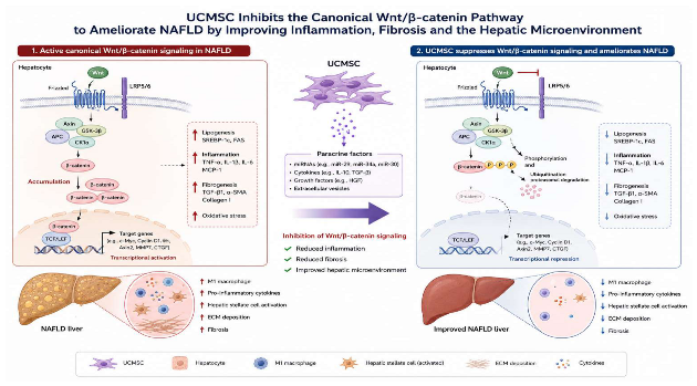
In the pathological model of MAFLD, regenerative hepatic parenchyma leads to impaired liver function, exacerbates nodule formation, and easily progresses to secondary liver cancer [69]. In healthy postpartum mice, β-catenin gene knockout experiments can detect reduced liver weight and body weight, confirming weakened regenerative capacity, indicating that β-catenin has significant regulatory significance in directing liver oval cells and bipotential stem cells. The Wnt pathway plays a bidirectional regulatory role by either activating or inhibiting during UCMSC influence on the liver, with its direction depending on the specific context and cell state. During the differentiation stage of stem cells into hepatocytes, early studies found that when inducing UCMSCs to differentiate into hepatocytes, it is necessary to inhibit Wnt/β-catenin signaling. At this time, β-catenin protein transfers from the nucleus to the cell membrane and cytoplasm, and its activity reduction is key to initiating stem cell specialization into hepatic lineages. In repairing already damaged liver structures, for livers that have already developed fibrosis, UCMSCs function by activating Wnt/β-catenin signaling. They regulate downstream signals of the Wnt/β-catenin cascade that induce senescence. A three-dimensional spheroid model constructed from primary human hepatocytes shows that Wnt/β-catenin signaling induces major hepatocyte proliferation by inhibiting the p53-PAI1 signaling axis. The introduction of UCMSCs can significantly upregulate the expression levels of β-catenin and its downstream target genes in the liver (such as Cyclin D1 and c-Myc), thereby initiating the liver regenerative repair program. UCMSCs, by activating the Wnt pathway, significantly reduce collagen fiber deposition in the liver and decrease the fibrosis area [85]. Restore the normal structure of hepatic lobules, thereby restoring the normal function of the liver. Studies have shown that after receiving UCMSC treatment, serum liver function indicators (such as ALT and AST) in liver-injured mice significantly decreased, and the inflammatory response in the liver was also alleviated. Current research directions also include mitochondrial transfer technology, which commonly uses the co-culture model of isolated free mitochondria from MAFLD cell models with UCMSC for in vitro pretreatment, followed by reinfusion of UCMSC into the diseased mice, alleviating the problem that damaged mitochondria in hepatocytes cannot perform fatty acid β-oxidation. Free mitochondria can enhance the therapeutic effect of UCMSC [65].
8. Summary
MAFLD leads to apoptosis and necrosis of liver parenchymal cells, which in turn causes excessive activation of hepatic stellate cells, fibrosis, the emergence of inflammatory phenotypes, and activation of the liver’s innate immune response, further triggering adaptive immune responses [92]. In addition, after liver injury occurs, mesenchymal stem cells can induce the directional differentiation of hepatic progenitor cells by activating three classical pathways and assist in treating hepatitis through the activation of immune pathways, thereby preventing the further progression of chronic hepatitis. Studies on treating chronic endocrine diseases and aging-related diseases with a large number of stem cells have found that mesenchymal stem cells mainly function to assist adult stem cells in native tissues with differentiation, replace damaged cells, and regulate the overall cell cycle, differentiation, and apoptosis of stem cells. Umbilical cord blood mesenchymal stem cells have lower immunogenicity and stronger immunomodulatory effects compared to other cells. Umbilical cord mesenchymal stem cells secrete cytokines at levels much higher than bone marrow mesenchymal stem cells, while inter-individual differences are smaller, and there are no differences in proliferation and chemotactic abilities compared to adipose-derived stem cells.
Funding
Enterprise Joint Fund Project of Hunan Provincial Natural Science Foundation, 2024JJ9097.
Data Availability Statement
Data will be made available on request. Experimental data was processed with Python, with the code provided by DeepSeek and the figure images sourced from Nanobanana2.0 (https://gemini-d-google-d-com-s-gmn.mid.tuanai.me/app/a7b5b11b9eed5c46).
Conflicts of Interest
The authors declare no conflict of interest.
References
- Alami, F.; Alizadeh, M.; Shateri, K. The effect of a fruit-rich diet on liver biomarkers, insulin resistance, and lipid profile in patients with non-alcoholic fatty liver disease: a randomized clinical trial. Scand. J. Gastroenterol. 2022, 57, 1238–1249. [Google Scholar] [CrossRef] [PubMed]
- Zhu, Y.-L.; Meng, L.-L.; Ma, J.-H.; Yuan, X.; Chen, S.-W.; Yi, X.-R.; Li, X.-Y.; Wang, Y.; Tang, Y.-S.; Xue, M.; et al. Loss of LBP triggers lipid metabolic disorder through H3K27 acetylation-mediated C/EBPβ-SCD activation in non-alcoholic fatty liver disease. Zool. Res. 2024, 45, 79–94. [Google Scholar] [CrossRef] [PubMed]
- Taslim, N.A.; Yusuf, M.; Ambari, A.M.; Puling, I.M.D.R.; Ibrahim, F.Z.; Hardinsyah, H.; Kurniawan, R.; Ben Gunawan, W.; Mayulu, N.; Joseph, V.F.F.; et al. Anti-Inflammatory, Antioxidant, Metabolic and Gut Microbiota Modulation Activities of Probiotic in Cardiac Remodeling Condition: Evidence from Systematic Study and Meta-Analysis of Randomized Controlled Trials. Probiotics Antimicrob. Proteins 2023, 15, 1049–1061. [Google Scholar] [CrossRef] [PubMed]
- Smith, G.I.; Shankaran, M.; Yoshino, M.; Schweitzer, G.G.; Chondronikola, M.; Beals, J.W.; Okunade, A.L.; Patterson, B.W.; Nyangau, E.; Field, T.; et al. Insulin resistance drives hepatic de novo lipogenesis in nonalcoholic fatty liver disease. J. Clin. Investig. 2020, 130, 1453–1460. [Google Scholar] [CrossRef] [PubMed]
- Scutiero, G.; Iannone, P.; Bernardi, G.; Bonaccorsi, G.; Spadaro, S.; Volta, C.A.; Greco, P.; Nappi, L. Oxidative Stress and Endometriosis: A Systematic Review of the Literature. Oxidative Med. Cell. Longev. 2017, 2017, 7265238. [Google Scholar] [CrossRef] [PubMed]
- Ding, K.; Zhang, Z.; Han, Z.; Shi, L.; Li, X.; Liu, Y.; Li, Z.; Zhao, C.; Cui, Y.; Zhou, L.; et al. Liver ALKBH5 regulates glucose and lipid homeostasis independently through GCGR and mTORC1 signaling. Science 2025, 387, eadp4120. [Google Scholar] [CrossRef] [PubMed]
- Nuciforo, S.; Heim, M. H. Organoids to model liver disease. JHEP Rep. 2020, 3, 100198. [Google Scholar] [PubMed]
- Yang, F.; Wu, Y.; Chen, Y.; Xi, J.; Chu, Y.; Jin, J.; Yan, Y. Human umbilical cord mesenchymal stem cell-derived exosomes ameliorate liver steatosis by promoting fatty acid oxidation and reducing fatty acid synthesis. JHEP Rep. 2023, 5, 100746. [Google Scholar] [CrossRef] [PubMed]
- Baranova, A.; Maltseva, D.; Tonevitsky, A. Adipose may actively delay progression of NAFLD by releasing tumor-suppressing, anti-fibrotic miR-122 into circulation. Obes. Rev. 2018, 20, 108–118. [Google Scholar] [CrossRef] [PubMed]
- Zhang, B.; Zhang, B.; Lai, R. C.; et al. MSC-sEV treatment polarizes pro-fibrotic M2 macrophages without exacerbating liver fibrosis in NASH. Int. J. Mol. Sci. 2023, 24, 8092. [Google Scholar] [CrossRef] [PubMed]
- Moayedfard, Z.; Sani, F.; Alizadeh, A.; Lankarani, K.B.; Zarei, M.; Azarpira, N. The role of the immune system in the pathogenesis of NAFLD and potential therapeutic impacts of mesenchymal stem cell-derived extracellular vesicles. Stem Cell Res. Ther. 2022, 13, 1–16. [Google Scholar] [CrossRef] [PubMed]
- Kholodenko, I.V.; Yarygin, K.N. Hepatic Macrophages as Targets for the MSC-Based Cell Therapy in Non-Alcoholic Steatohepatitis. Biomedicines 2023, 11, 3056. [Google Scholar] [CrossRef] [PubMed]
- Dorairaj, V.; Sulaiman, S.A.; Abu, N.; Murad, N.A.A. Extracellular Vesicles in the Development of the Non-Alcoholic Fatty Liver Disease: An Update. Biomolecules 2020, 10, 1494. [Google Scholar] [CrossRef] [PubMed]
- Yan, F.-J.; Ding, H.; Zhang, N.; Yan, S.-R.; Huang, M.-X.; Lu, J.-W.; Wang, Y.-J.; Yan, Y.-J.; Li, R.-P.; Wang, Q. The E3 ligase tripartite motif 7 drives the progression of non-alcoholic fatty liver disease by targeting DUSP10 degradation in male mice. Nat. Commun. 2025, 16, 10437. [Google Scholar] [CrossRef] [PubMed]
- Eslam, M.; George, J. MAFLD: from a disease framework to patient care. Hepatol. Int. 2024, 18, 823–826. [Google Scholar] [CrossRef] [PubMed]
- Xu, Y.; Yue, T.; Batbold, U.; Magassa, A.; Liu, K.; Jiang, L.; Liu, D.; Do, L.N.H.; Liu, X.; Yang, L.; et al. MARCHF6 orchestrates hepatic lipid homeostasis by targeting SREBP1 for ER-associated degradation. J. Hepatol. 2026. [Google Scholar] [CrossRef] [PubMed]
- Yan, F.; Zhang, X.; Wang, W.; Ji, Y.; Wang, P.; Yang, Y.; Gong, J.; Shen, L.; Zhu, X.; Huang, Z.; et al. The E3 ligase tripartite motif 8 targets TAK1 to promote insulin resistance and steatohepatitis. Hepatology 2016, 65, 1492–1511. [Google Scholar] [CrossRef] [PubMed]
- Cheng, X.; Ma, X.; Zhu, Q.; Song, D.; Ding, X.; Li, L.; Jiang, X.; Wang, X.; Tian, R.; Su, H.; et al. Pacer Is a Mediator of mTORC1 and GSK3-TIP60 Signaling in Regulation of Autophagosome Maturation and Lipid Metabolism. Mol. Cell 2019, 73, 788–802.e7. [Google Scholar] [CrossRef] [PubMed]
- Ding, X.; Jiang, X.; Tian, R.; Zhao, P.; Li, L.; Wang, X.; Chen, S.; Zhu, Y.; Mei, M.; Bao, S.; et al. RAB2 regulates the formation of autophagosome and autolysosome in mammalian cells. Autophagy 2019, 15, 1774–1786. [Google Scholar] [CrossRef] [PubMed]
- Huang, Y.; Yu, F.; Ding, Y.; Zhang, H.; Li, X.; Wang, X.; Wu, X.; Xu, J.; Wang, L.; Tian, C.; et al. Hepatic IL22RA1 deficiency promotes hepatic steatosis by modulating oxysterol in the liver. Hepatology 2024, 81, 1564–1582. [Google Scholar] [CrossRef] [PubMed]
- Petkevicius, K.; Palmgren, H.; Glover, M.S.; Ahnmark, A.; Andréasson, A.-C.; Madeyski-Bengtson, K.; Kawana, H.; Allman, E.L.; Kaper, D.; Uhrbom, M.; et al. TLCD1 and TLCD2 regulate cellular phosphatidylethanolamine composition and promote the progression of non-alcoholic steatohepatitis. Nat. Commun. 2022, 13, 1–14. [Google Scholar] [CrossRef] [PubMed]
- Li, B.; Cheng, Y.; Yu, S.; Zang, L.; Yin, Y.; Liu, J.; Zhang, L.; Mu, Y. Human Umbilical Cord-Derived Mesenchymal Stem Cell Therapy Ameliorates Nonalcoholic Fatty Liver Disease in Obese Type 2 Diabetic Mice. Stem Cells Int. 2019, 2019, 1–12. [Google Scholar] [CrossRef] [PubMed]
- Alison, M.R. Cholangiocytes: No Longer Cinderellas to the Hepatic Regenerative Response. Cell Stem Cell 2017, 21, 159–160. [Google Scholar] [CrossRef] [PubMed]
- Hou, J.; Zhang, J.; Cui, P.; Zhou, Y.; Liu, C.; Wu, X.; Ji, Y.; Wang, S.; Cheng, B.; Ye, H.; et al. TREM2 sustains macrophage-hepatocyte metabolic coordination in nonalcoholic fatty liver disease and sepsis. J. Clin. Investig. 2021, 131. [Google Scholar] [CrossRef] [PubMed]
- Hou, X.; Yin, S.; Ren, R.; Liu, S.; Yong, L.; Liu, Y.; Li, Y.; Zheng, M.; Kunos, G.; Gao, B.; et al. Myeloid-Cell–Specific IL-6 Signaling Promotes MicroRNA-223-Enriched Exosome Production to Attenuate NAFLD-Associated Fibrosis. Hepatology 2021, 74, 116–132. [Google Scholar] [CrossRef] [PubMed]
- Shen, Y.; Cheng, L.; Xu, M.; Wang, W.; Wan, Z.; Xiong, H.; Guo, W.; Cai, M.; Xu, F. SGLT2 inhibitor empagliflozin downregulates miRNA-34a-5p and targets GREM2 to inactivate hepatic stellate cells and ameliorate non-alcoholic fatty liver disease-associated fibrosis. Metabolism 2023, 146, 155657. [Google Scholar] [CrossRef] [PubMed]
- Shi, Z.; et al. Cryo-EM structure of the human cohesin-NIPBL-DNA complex. Science 2020, 368, 1454–1459. [Google Scholar] [CrossRef] [PubMed]
- Nian, F.; Wu, C.; Wu, D.; Ma, Y.; Chen, Y.; Xu, L.; Li, Y.; Dong, L.; Xiong, W. Macrophage–neutrophil crosstalk via the Selplg–Sell–YAP axis drives NETosis and MASH-associated liver fibrosis. Hepatology 2026. [Google Scholar] [CrossRef] [PubMed]
- Zhou, B.; Luo, Y.; Bi, H.; Zhang, N.; Ma, M.; Dong, Z.; Ji, N.; Zhang, S.; Wang, X.; Liu, Y.; et al. Amelioration of nonalcoholic fatty liver disease by inhibiting the deubiquitylating enzyme RPN11. Cell Metab. 2024, 36, 2228–2244.e7. [Google Scholar] [CrossRef] [PubMed]
- Li, N.; Song, X.; Peng, X.; Li, M.; Zhu, M.; Xiao, R.; Wang, L.; Ling, L.; Zhao, Y.; Wang, T.; et al. Tim-3 facilitates dendritic cell ferroptosis and impairs antitumor immunity in steatohepatitis-related HCC. J. Hepatol. 2026, 85, 331–344. [Google Scholar] [CrossRef] [PubMed]
- Xi, Z.-H.; Sun, W.-J.; Li, M.-H.; Dong, Q.-Q.; Dong, X.; Qiao, Z.-L.; Tao, H.; Yang, J.-J.; Lu, C. Extracellular matrix stiffness in chronic liver disease: From mechanisms to treatments. Biochem. Pharmacol. 2026, 252. [Google Scholar] [CrossRef] [PubMed]
- Ahmed, H.; Umar, M.I.; Imran, S.; Javaid, F.; Syed, S.K.; Riaz, R.; Hassan, W. TGF-β1 signaling can worsen NAFLD with liver fibrosis backdrop. Exp. Mol. Pathol. 2022, 124, 104733. [Google Scholar] [CrossRef] [PubMed]
- Zhang, L.; Wang, H. Hypoxia-induced transcriptional reprogramming in sugar beet seedlings: Linking energy conservation and metabolic maintenance. J. Plant Physiol. 2023, 45, 112–125. [Google Scholar]
- Martini, T.; Naef, F.; Tchorz, J.S. Spatiotemporal Metabolic Liver Zonation and Consequences on Pathophysiology. Annu. Rev. Pathol. Mech. Dis. 2023, 18, 439–466. [Google Scholar] [CrossRef] [PubMed]
- Wu, L.; Wang, J.; Wang, Y.; Yang, J.; Yao, Y.; Wang, Y.; Huang, D.; Hu, Y.; Xu, X.; Wang, R.; et al. CLCC1 governs ER bilayer equilibration to maintain lipid homeostasis. Nature 2026, 652, 471–480. [Google Scholar] [CrossRef] [PubMed]
- Kang, S.W.S.; Brown, L.A.; Miller, C.B.; Barrows, K.M.; Golino, J.L.; Liu, H.; Cultraro, C.M.; Feliciano, D.; Cornelius-Muwanuzi, M.B.; Remmert, K.; et al. PLIN5 phosphorylation orchestrates mitochondria lipid-droplet coupling to control hepatic lipid flux and steatosis. Nat. Metab. 2026, 8, 587–603. [Google Scholar] [CrossRef] [PubMed]
- Zhou, Y.; Zhao, Y.; Carbonaro, M.; Chen, H.; Germino, M.; Adler, C.; Ni, M.; Zhu, Y.O.; Kim, S.Y.; Altarejos, J.; et al. Perturbed liver gene zonation in a mouse model of non-alcoholic steatohepatitis. Metabolism 2024, 154, 155830. [Google Scholar] [CrossRef] [PubMed]
- van Breugel, M.; de Jong, E.; Buikema, H.J.; Petoukhov, I.; Nawijn, M.C.; Burgess, J.K.; Timens, W. A deep learning framework for histopathological analysis of pixel-level extracellular matrix variation in standard H&E-stained images. Sci. Rep. 2026. [Google Scholar] [CrossRef] [PubMed]
- Lee, D. S.; An, T. H.; Kim, H.; et al. Tcf7l2 in hepatocytes regulates de novo lipogenesis in diet-induced non-alcoholic fatty liver disease in mice. Diabetologia 2023, 66, 931–954. [Google Scholar] [CrossRef] [PubMed]
- Koriem, K.M.M. Calculus bovis in hepatocellular carcinoma: Tumor molecular basis, Wnt/β-catenin pathway role, and protective mechanism. World J. Gastroenterol. 2024, 30, 3959–3964. [Google Scholar] [CrossRef] [PubMed]
- Nian, F.; Chen, Y.; Xia, Q.; Zhu, C.; Wu, L.; Lu, X. Gut microbiota metabolite trimethylamine N-oxide promoted NAFLD progression by exacerbating intestinal barrier disruption and intrahepatic cellular imbalance. Int. Immunopharmacol. 2024, 142, 113173. [Google Scholar] [CrossRef] [PubMed]
- Harini, K.S.; Ezhilarasan, D. Wnt/beta-catenin signaling and its modulators in nonalcoholic fatty liver diseases. Hepatobiliary Pancreat. Dis. Int. 2022, 22, 333–345. [Google Scholar] [CrossRef] [PubMed]
- Oh, K.-K.; Gupta, H.; Min, B.H.; Ganesan, R.; Sharma, S.P.; Won, S.M.; Jeong, J.J.; Lee, S.B.; Cha, M.G.; Kwon, G.H.; et al. The identification of metabolites from gut microbiota in NAFLD via network pharmacology. Sci. Rep. 2023, 13, 1–13. [Google Scholar] [CrossRef] [PubMed]
- Zhao, H.; Ming, T.; Tang, S.; Ren, S.; Yang, H.; Liu, M.; Tao, Q.; Xu, H. Wnt signaling in colorectal cancer: pathogenic role and therapeutic target. Mol. Cancer 2022, 21, 1–34. [Google Scholar] [CrossRef] [PubMed]
- Xu, X.; Zhang, M.; Xu, F.; Jiang, S. Wnt signaling in breast cancer: biological mechanisms, challenges and opportunities. Mol. Cancer 2020, 19, 1–35. [Google Scholar] [CrossRef] [PubMed]
- Steinhart, Z.; Angers, S. Wnt signaling in development and tissue homeostasis. Development 2018, 145, dev146589. [Google Scholar] [CrossRef] [PubMed]
- Li, C.; Yang, D.; Wang, X.; Zhang, S.; Dai, W.; Zhao, S.; Zhao, W. ApoA5-CIDEC axis regulates hepatic lipid metabolism, inflammation, and fibrosis in MASLD. J. Mol. Med. 2025, 104, 3. [Google Scholar] [CrossRef] [PubMed]
- He, Q.; He, W.; Dong, H.; Guo, Y.; Yuan, G.; Shi, X.; Wang, D.; Lu, F. Role of liver sinusoidal endothelial cell in metabolic dysfunction-associated fatty liver disease. Cell Commun. Signal. 2024, 22, 1–18. [Google Scholar] [CrossRef] [PubMed]
- Li, Z.-W.; Ruan, B.; Yang, P.-J.; Liu, J.-J.; Song, P.; Duan, J.-L.; Wang, L. Oit3, a promising hallmark gene for targeting liver sinusoidal endothelial cells. Signal Transduct. Target. Ther. 2023, 8, 1–10. [Google Scholar] [CrossRef] [PubMed]
- Pabst, O.; Hornef, M.W.; Schaap, F.G.; Cerovic, V.; Clavel, T.; Bruns, T. Gut–liver axis: barriers and functional circuits. Nat. Rev. Gastroenterol. Hepatol. 2023, 20, 447–461. [Google Scholar] [CrossRef] [PubMed]
- Casey, L.M.; Hughes, K.R.; Saunders, M.N.; Miller, S.D.; Pearson, R.M.; Shea, L.D. Mechanistic contributions of Kupffer cells and liver sinusoidal endothelial cells in nanoparticle-induced antigen-specific immune tolerance. Biomaterials 2022, 283, 121457–121457. [Google Scholar] [CrossRef] [PubMed]
- de Haan, W.; Dheedene, W.; Apelt, K.; Décombas-Deschamps, S.; Vinckier, S.; Verhulst, S.; Conidi, A.; Deffieux, T.; Staring, M.W.; Vandervoort, P.; et al. Endothelial Zeb2 preserves the hepatic angioarchitecture and protects against liver fibrosis. Cardiovasc. Res. 2021, 118, 1262–1275. [Google Scholar] [CrossRef] [PubMed]
- Kawashita, E.; Ozaki, T.; Ishihara, K.; Kashiwada, C.; Akiba, S. Endothelial group IVA phospholipase A2 promotes hepatic fibrosis with sinusoidal capillarization in the early stage of non-alcoholic steatohepatitis in mice. Life Sci. 2022, 294, 120355. [Google Scholar] [CrossRef] [PubMed]
- Furuta, K.; Tang, X.; Islam, S.; Tapia, A.; Chen, Z.B.; Ibrahim, S.H. Endotheliopathy in the metabolic syndrome: Mechanisms and clinical implications. Pharmacol. Ther. 2023, 244, 108372–108372. [Google Scholar] [CrossRef] [PubMed]
- Fang, Z.; Ruan, B.; Liu, J.; Duan, J.; Yue, Z.; Song, P.; Xu, H.; Ding, J.; Xu, C.; Dou, G.; et al. Notch-triggered maladaptation of liver sinusoidal endothelium aggravates nonalcoholic steatohepatitis through endothelial nitric oxide synthase. Hepatology 2022, 76, 742–758. [Google Scholar] [CrossRef] [PubMed]
- Greuter, T.; Yaqoob, U.; Gan, C.; et al. Mechanotransduction-induced glycolysis epigenetically regulates a CXCL1-dominant angiocrine signaling program in liver sinusoidal endothelial cells in vitro and in vivo. J. Hepatol. 2022, *77*(3), 723–734. [Google Scholar] [CrossRef] [PubMed]
- Zhang, L.-F.; Wang, X.-H.; Zhang, C.-L.; Lee, J.; Duan, B.-W.; Xing, L.; Li, L.; Oh, Y.-K.; Jiang, H.-L. Sequential Nano-Penetrators of Capillarized Liver Sinusoids and Extracellular Matrix Barriers for Liver Fibrosis Therapy. ACS Nano 2022, 16, 14029–14042. [Google Scholar] [CrossRef] [PubMed]
- Kotsiliti, E. NAFLD prevalence in older patients with T2DM. Nat. Rev. Gastroenterol. Hepatol. 2022, 20, 65–65. [Google Scholar] [CrossRef] [PubMed]
- Gougelet, A.; Torre, C.; Veber, P.; Sartor, C.; Bachelot, L.; Denechaud, P.-D.; Godard, C.; Moldes, M.; Burnol, A.-F.; Dubuquoy, C.; et al. T-cell factor 4 and β-catenin chromatin occupancies pattern zonal liver metabolism in mice. Hepatology 2013, 59, 2344–2357. [Google Scholar] [CrossRef] [PubMed]
- Isfahani, M.M.; Sepanlou, S.G.; Joukar, F.; Mansour-Ghanaei, F.; Taheri, E. Comparison of prevalence of fatty liver disease according to the different nomenclatures of NAFLD, MAFLD, and MASLD in a large Iranian Population-based study. Sci. Rep. 2025, 16. [Google Scholar] [CrossRef] [PubMed]
- Huang, X.; Liu, X.; Li, X.; Zhang, Y.; Gao, J.; Yang, Y.; Jiang, Y.; Gao, H.; Sun, C.; Xuan, L.; et al. Cullin-associated and neddylation-dissociated protein 1 (CAND1) alleviates NAFLD by reducing ubiquitinated degradation of ACAA2. Nat. Commun. 2023, 14, 1–16. [Google Scholar] [CrossRef] [PubMed]
- Jiao, Q.; Ren, Y.; Teng, X.; Feng, M.; Liu, X.; Cai, Y.; Hu, T.; Wang, M.; Wang, Y. Positive feedback between histone H4K16 lactylation and glycolysis promotes MAFLD progression. Hepatol. Int. 2025, 20, 646–663. [Google Scholar] [CrossRef] [PubMed]
- Hayakawa, S.; Tamura, A.; Nikiforov, N.; Koike, H.; Kudo, F.; Cheng, Y.; Miyazaki, T.; Kubekina, M.; Kirichenko, T.V.; Orekhov, A.N.; et al. Activated cholesterol metabolism is integral for innate macrophage responses by amplifying Myd88 signaling. J. Clin. Investig. 2022, 7. [Google Scholar] [CrossRef] [PubMed]
- Rodríguez-Ortigosa, C.M.; Celay, J.; Olivas, I.; Juanarena, N.; Arcelus, S.; Uriarte, I.; Marín, J.J.G.; Avila, M.A.; Medina, J.F.; Prieto, J. A GAPDH-Mediated Trans-Nitrosylation Pathway Is Required for Feedback Inhibition of Bile Salt Synthesis in Rat Liver. Gastroenterology 2014, 147, 1084–1093. [Google Scholar] [CrossRef] [PubMed]
- Li, Z.; Kabir, I.; Jiang, H.; Zhou, H.; Libien, J.; Zeng, J.; Stanek, A.; Ou, P.; Li, K.R.; Zhang, S.; et al. Liver serine palmitoyltransferase activity deficiency in early life impairs adherens junctions and promotes tumorigenesis. Hepatology 2016, 64, 2089–2102. [Google Scholar] [CrossRef] [PubMed]
- Wang, Y.; Nakajima, T.; Gonzalez, F.J.; Tanaka, N. PPARs as Metabolic Regulators in the Liver: Lessons from Liver-Specific PPAR-Null Mice. Int. J. Mol. Sci. 2020, 21, 2061. [Google Scholar] [CrossRef] [PubMed]
- Clemens, M.M.; Kennon-McGill, S.; Vazquez, J.H.; Stephens, O.W.; Peterson, E.A.; Johann, D.J.; Allard, F.D.; Yee, E.U.; McCullough, S.S.; James, L.P.; et al. Exogenous phosphatidic acid reduces acetaminophen-induced liver injury in mice by activating hepatic interleukin-6 signaling through inter-organ crosstalk. Acta Pharm. Sin. B 2021, 11, 3836–3846. [Google Scholar] [CrossRef] [PubMed]
- Masubuchi, Y.; Bourdi, M.; Reilly, T.P.; Graf, M.L.M.; George, J.W.; Pohl, L.R. Role of interleukin-6 in hepatic heat shock protein expression and protection against acetaminophen-induced liver disease. Biochem. Biophys. Res. Commun. 2003, 304, 207–212. [Google Scholar] [CrossRef] [PubMed]
- Devarbhavi, H.; Asrani, S.K.; Arab, J.P.; Nartey, Y.A.; Pose, E.; Kamath, P.S. Global burden of liver disease: 2023 update. J. Hepatol. 2023, 79, 516–537. [Google Scholar] [CrossRef] [PubMed]
- Han, Y.; Ling, Q.; Wu, L.; Wang, X.; Wang, Z.; Chen, J.; Zheng, Z.; Zhou, Z.; Jia, L.; Li, L.; et al. Akkermansia muciniphila inhibits nonalcoholic steatohepatitis by orchestrating TLR2-activated γδT17 cell and macrophage polarization. Gut Microbes 2023, 15, 2221485. [Google Scholar] [CrossRef] [PubMed]
- Song, J.; Zeng, Q.; Jiao, Y.; Sun, X.; Xie, L.; Qiu, J.; Ding, Y.; Hu, W.; Wang, F.; Huang, B.; et al. Adipocyte OX40L promotes adipose T cell activation and insulin resistance in obesity. Exp. Mol. Med. 2026. [Google Scholar] [CrossRef] [PubMed]
- Yong, Q.; Huang, C.; Chen, B.; An, J.; Zheng, Y.; Zhao, L.; Peng, C.; Liu, F. Gentiopicroside improves NASH and liver fibrosis by suppressing TLR4 and NLRP3 signaling pathways. Biomed. Pharmacother. 2024, 177, 116952. [Google Scholar] [CrossRef] [PubMed]
- Handa, P.; Vemulakonda, A.; Kowdley, K.V.; Uribe, M.; Méndez-Sánchez, N. Mitochondrial DNA from hepatocytes as a ligand for TLR9: Drivers of nonalcoholic steatohepatitis? World J. Gastroenterol. 2016, 22, 6965–71. [Google Scholar] [CrossRef] [PubMed]
- Hu, R.; Zhao, J.; Cheng, Y.; Su, W.; Ren, R.; Zhang, H.; Zhang, Y.; Wang, A.; Mu, Y.; Yu, S. Exogenous Mitochondrial Pretreatment Enhances the Therapeutic Effect of UC-MSCs on NAFLD in Type 2 Diabetic Mice by Mediating Mitochondrial Transfer. Stem Cells Int. 2025, 2025, 4639115. [Google Scholar] [CrossRef] [PubMed]
- Han, J.-J.; Li, J.; Huang, D.-H. Mesenchymal Stem Cell-Derived Extracellular Vesicles Carrying Circ-Tulp4 Attenuate Diabetes Mellitus with Nonalcoholic Fatty Liver Disease by Inhibiting Cell Pyroptosis through the HNRNPC/ABHD6 Axis. Tissue Eng. Regen. Med. 2024, 22, 23–41. [Google Scholar] [CrossRef] [PubMed]
- Xiao, K.; Liu, C.; Wang, H.; Hou, F.; Shi, Y.; Qian, Z.R.; Zhang, H.; Deng, D.Y.; Xie, L. Umbilical cord mesenchymal stem cells overexpressing CXCR7 facilitate treatment of ARDS-associated pulmonary fibrosis via inhibition of Notch/Jag1 mediated by the Wnt/β-catenin pathway. Biomed. Pharmacother. 2023, 165, 115124. [Google Scholar] [CrossRef] [PubMed]
- Feng, X.; Liu, J.; Xu, Y.; Zhu, J.; Chen, W.; Feng, B.; Pan, Q.; Yu, J.; Shi, X.; Yang, J.; et al. Molecular mechanism underlying the difference in proliferation between placenta-derived and umbilical cord-derived mesenchymal stem cells. J. Cell. Physiol. 2020, 235, 6779–6793. [Google Scholar] [CrossRef] [PubMed]
- Zhang, H.; Liu, P.; Deng, Y.; Wu, L.; Fan, O.; Cui, Y.; Zhang, C.; Zhu, W.; Sun, Y.E.; Lin, C.; et al. Human umbilical cord-derived mesenchymal stem cells ameliorate non-alcoholic fatty liver disease via activating TFEB-mediated autophagy in male mice. Stem Cell Res. Ther. 2025, 17. [Google Scholar] [CrossRef] [PubMed]
- Tzouanas, C.N.; Shay, J.E.; Sherman, M.S.; Rubin, A.J.; Mead, B.E.; Dao, T.T.; Tao, J.; Lehrich, B.M.; Eng, G.; Patterson-Fortin, J.; et al. Hepatic adaptation to chronic metabolic stress primes tumorigenesis. Cell 2025, 189, 435–460.e28. [Google Scholar] [CrossRef] [PubMed]
- Sugimoto, A.; Saito, Y.; Wang, G. Hepatic stellate cells control liver zonation, size and functions via R-spondin 3. Nature 2025, 640, 752–761. [Google Scholar] [CrossRef] [PubMed]
- Kim, D. H.; Kim, M. K.; Kim, D.; et al. Liver cancer chronically exposed to palmitate acquires ferroptosis resistance via the downregulation of glutamine-driven hepcidin expression. Metabolism 2026, 176, 156469. [Google Scholar] [CrossRef] [PubMed]
- Lou, G.; Yang, Y.; Liu, F.; Ye, B.; Chen, Z.; Zheng, M.; Liu, Y. MiR-122 modification enhances the therapeutic efficacy of adipose tissue-derived mesenchymal stem cells against liver fibrosis. J. Cell. Mol. Med. 2017, 21, 2963–2973. [Google Scholar] [CrossRef] [PubMed]
- Song, X.-J.; Zhang, L.; Li, Q.; Li, Y.; Ding, F.-H.; Li, X. hUCB-MSC derived exosomal miR-124 promotes rat liver regeneration after partial hepatectomy via downregulating Foxg1. Life Sci. 2021, 265, 118821. [Google Scholar] [CrossRef] [PubMed]
- Sun, Y.; Zhang, C.; Ma, Q.; Yu, X.; Gao, X.; Zhang, H.; Shi, Y.; Li, Y.; He, X. MiR-34a-HK1 signal axis retards bone marrow mesenchymal stem cell senescence via ameliorating glycolytic metabolism. Stem Cell Res. Ther. 2024, 15, 1–15. [Google Scholar] [CrossRef] [PubMed]
- Yan, X.; Zhang, D.; Wu, W.; et al. Mesenchymal stem cells promote hepatocarcinogenesis via lncRNA-MUF interaction with ANXA2 and miR-34a. Cancer Res. 2017, 77, 6704–6716. [Google Scholar] [CrossRef] [PubMed]
- Zhou, X.; Cui, L.; Zhou, X.; et al. Induction of hepatocyte-like cells from human umbilical cord-derived mesenchymal stem cells by defined microRNAs. J. Cell. Mol. Med. 2017, 21, 881–893. [Google Scholar] [PubMed]
- Cheng, F.; Yang, F.; Wang, Y.; Zhou, J.; Qian, H.; Yan, Y. Mesenchymal stem cell-derived exosomal miR-27b-3p alleviates liver fibrosis via downregulating YAP/LOXL2 pathway. J. Nanobiotechnology 2023, 21, 1–19. [Google Scholar] [CrossRef] [PubMed]
- He, L.; Wang, X.; Kang, N.; Xu, J.; Dai, N.; Xu, X.; Zhang, H. MiR-375 inhibits the hepatocyte growth factor-elicited migration of mesenchymal stem cells by downregulating Akt signaling. Cell Tissue Res. 2018, 372, 99–114. [Google Scholar] [CrossRef] [PubMed]
- Zhu, Y.; Zhang, X.; Yang, K.; Shao, Y.; Gu, R.; Liu, X.; Liu, H.; Liu, Y.; Zhou, Y. Macrophage-derived apoptotic vesicles regulate fate commitment of mesenchymal stem cells via miR155. Stem Cell Res. Ther. 2022, 13, 1–17. [Google Scholar] [CrossRef] [PubMed]
- Xiao, X.; Xu, M.; Yu, H.; et al. Mesenchymal stem cell-derived small extracellular vesicles mitigate oxidative stress-induced senescence in endothelial cells via regulation of miR-146a/Src. Signal Transduct. Target. Ther. 2021, 6, 354. [Google Scholar] [CrossRef] [PubMed]
- Weiskirchen, R.; Tacke, F. Cellular and molecular functions of hepatic stellate cells in inflammatory responses and liver immunology. 2014, 3, 344–363. [Google Scholar] [CrossRef] [PubMed]
- Filliol, A.; Saito, Y.; Nair, A.; Dapito, D.H.; Yu, L.-X.; Ravichandra, A.; Bhattacharjee, S.; Affo, S.; Fujiwara, N.; Su, H.; et al. Opposing roles of hepatic stellate cell subpopulations in hepatocarcinogenesis. Nature 2022, 610, 356–365. [Google Scholar] [CrossRef] [PubMed]
- Zhang, D.; Abulitipu, A.; Pang, P.; Bai, L.; Liu, L.; Tuerxunmaimaiti, S.; Chen, W.; Chen, S.; Lv, H.; Fu, Y.; et al. ECM1 protects against liver steatosis through PCBP1-mediated iron homeostasis. Hepatology 2025, 83, 603–622. [Google Scholar] [CrossRef] [PubMed]
- Xi, Y.; Zeng, W.; Luo, J.; Zhou, J.; Wang, L.; Sun, J.; He, Z.; Li, W.; Zhu, S.; Qi, W.; et al. RYK is a GPNMB receptor that drives MASH. Nature 2026, 652, 703–711. [Google Scholar] [CrossRef] [PubMed]
- Vafai, S.B.; Täubel, J.; Ashdown, T.; Patel, R.S.; Diamondali, S.; Cegla, J.; Soran, H.; Bashir, B.; Abitbol, A.; Gaudet, D.; et al. In Vivo Base Editing of PCSK9 with VERVE-102 for Hypercholesterolemia. New Engl. J. Med. 2026. [Google Scholar] [CrossRef] [PubMed]
- Holeček, M. Influence of Histidine Administration on Ammonia and Amino Acid Metabolism: A Review. Physiol. Res. 2020, 69, 555–564. [Google Scholar] [CrossRef] [PubMed]
- Cheng, L.; Deepak, R.K.; Wang, G.; Meng, Z.; Tao, L.; Xie, M.; Chi, W.; Zhang, Y.; Yang, M.; Liao, Y.; et al. Hepatic mitochondrial NAD+ transporter SLC25A47 activates AMPKα mediating lipid metabolism and tumorigenesis. Hepatology 2023, 78, 1828–1842. [Google Scholar] [CrossRef] [PubMed]
- Wei, X.; Wang, T.; Xing, Z.; Shi, Q.; Gu, J.; Fan, Q.; Wang, H.; Chen, B.; Cheng, J.; Cai, R. Sirtuin 3 Protects Lung Adenocarcinoma from Ferroptosis by Deacetylating and Stabilizing Mitochondrial Glutamate Transporter Solute Carrier Family 25 Member A22. Antioxidants 2025, 14, 403. [Google Scholar] [CrossRef] [PubMed]
- Tang, Y.; Yao, T.; Tian, X.; Xia, X.; Huang, X.; Qin, Z.; Shen, Z.; Zhao, L.; Zhao, Y.; Diao, B.; et al. Hepatic IRE1α-XBP1 signaling promotes GDF15-mediated anorexia and body weight loss in chemotherapy. J. Exp. Med. 2024, 221. [Google Scholar] [CrossRef] [PubMed]
- Vasse, G.F.; Russo, S.; Barcaru, A.; Oun, A.A.A.; Dolga, A.M.; van Rijn, P.; Kwiatkowski, M.; Govorukhina, N.; Bischoff, R.; Melgert, B.N. Collagen type I alters the proteomic signature of macrophages in a collagen morphology-dependent manner. Sci. Rep. 2023, 13, 1–14. [Google Scholar] [CrossRef] [PubMed]
- Ku, N.; Strnad, P.; Bantel, H.; Omary, M.B. Keratins: Biomarkers and modulators of apoptotic and necrotic cell death in the liver. Hepatology 2016, 64, 966–976. [Google Scholar] [CrossRef] [PubMed]
- Herzer, K.; Kneiseler, G.; Bechmann, L.P.; Post, F.; Schlattjan, M.; Sowa, J.-P.; Neumann, T.; Marggraf, G.; Erbel, R.; Gerken, G.; et al. Onset of heart failure determines the hepatic cell death pattern. Ann. Hepatol. 2011, 10, 174–179. [Google Scholar] [CrossRef]
- Shi, J.-J.; Liu, Y.-J.; Yu, J.; Wang, R.; Chen, R.-Y.; Yang, G.-J.; Chen, J. Porcupine (PORCN): structural insights, functional mechanisms, and therapeutic potential in Wnt-driven diseases. Bioorganic Chem. 2025, 165, 108955. [Google Scholar] [CrossRef] [PubMed]
- Feng, X.; Bao, J.; Song, C.; Xie, L.; Tan, X.; Li, J.; Jia, H.; Tian, M.; Qi, J.; Qin, C.; et al. Functional role of miR-155 in physiological and pathological processes of liver injury (Review). Mol. Med. Rep. 2021, 24, 1–8. [Google Scholar] [CrossRef] [PubMed]
- Luwen, H.; Lijun, M.; Linlin, L.; Ming, Y. The Role of the C-Reactive Protein–Triglyceride Glucose Index in Predicting New-Onset Chronic Diseases: Evidence From a Longitudinal Cohort Study. Brain Behav. 2026, 16, e71299. [Google Scholar] [CrossRef] [PubMed]
Figure 1.
(A) Wnt signaling and metabolic zonation in the liver lobule structure;(B) Wnt pathway components and signaling cascades in two different circumstance, one in wnt secretion and regulation, other in β-catenin and target genes;(C) Crosstalk in liver autophagy and regeneration;(D) Impact of chronic liver diseases and MAFLD progression with virual formal pictures;(E) Bioinformation analyse about GEO data files.Heatmap of GSE281797 data which serevely reated to WNT pathway.Red represented up reguration, blue represented the adversed effection.GSE296996 Statistical validity of the top 5 highly expressed genes in the full dataset for volcano plot drawing.
Figure 1.
(A) Wnt signaling and metabolic zonation in the liver lobule structure;(B) Wnt pathway components and signaling cascades in two different circumstance, one in wnt secretion and regulation, other in β-catenin and target genes;(C) Crosstalk in liver autophagy and regeneration;(D) Impact of chronic liver diseases and MAFLD progression with virual formal pictures;(E) Bioinformation analyse about GEO data files.Heatmap of GSE281797 data which serevely reated to WNT pathway.Red represented up reguration, blue represented the adversed effection.GSE296996 Statistical validity of the top 5 highly expressed genes in the full dataset for volcano plot drawing.
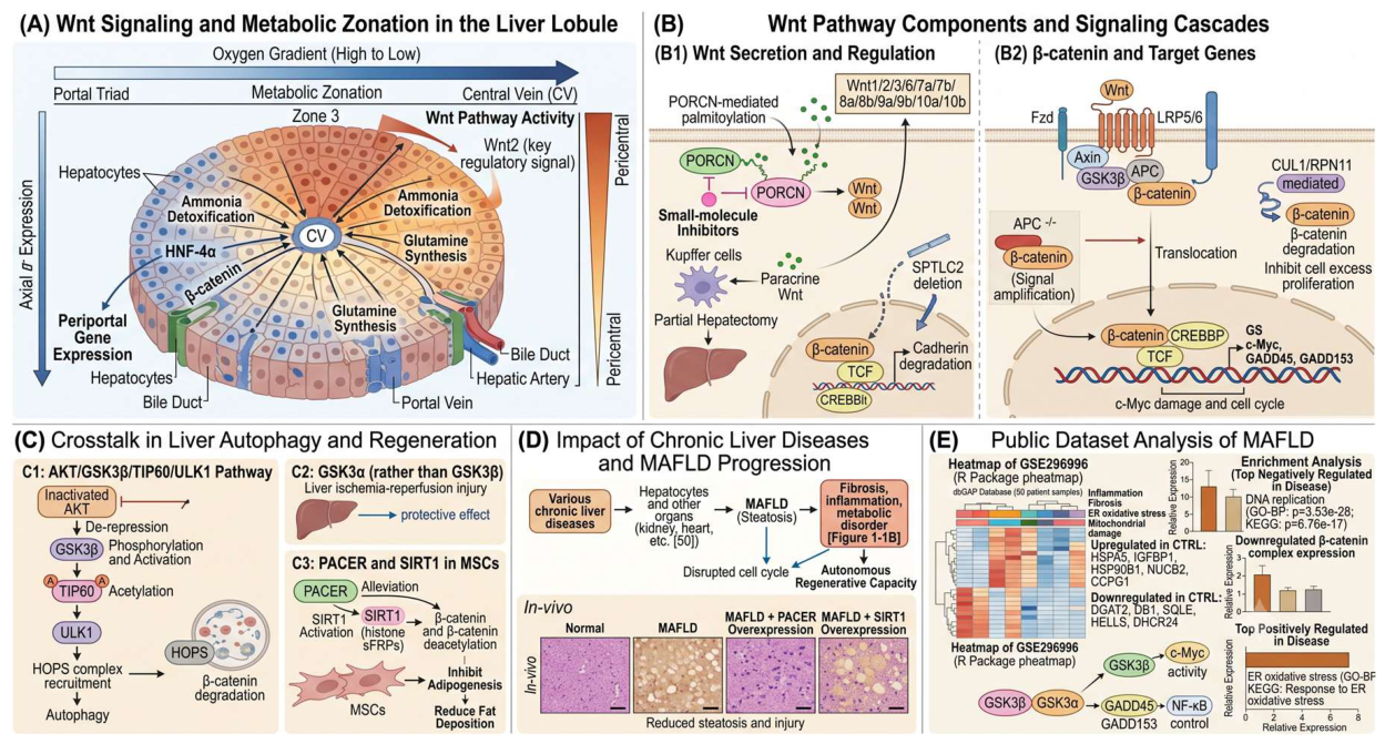
Figure 2.
(A) Hepatocyte liver injure,ev-mediated cell cross-talk,and fibrosis in flow chart;(B) Toll-like receptors regulation of inflammasome and immune phenotypes,especially in TLR2,TLR4,TLR9 pathway;(C) Bile acid recircullation,gut microbiota, and liver fibrosis;(D) Metrnl and IL-22 signaling in HSC activation and lipid homeostasis;(E) Bioinformation analyse focus on biological function in human samples with KEGG and GO enrichment.
Figure 2.
(A) Hepatocyte liver injure,ev-mediated cell cross-talk,and fibrosis in flow chart;(B) Toll-like receptors regulation of inflammasome and immune phenotypes,especially in TLR2,TLR4,TLR9 pathway;(C) Bile acid recircullation,gut microbiota, and liver fibrosis;(D) Metrnl and IL-22 signaling in HSC activation and lipid homeostasis;(E) Bioinformation analyse focus on biological function in human samples with KEGG and GO enrichment.
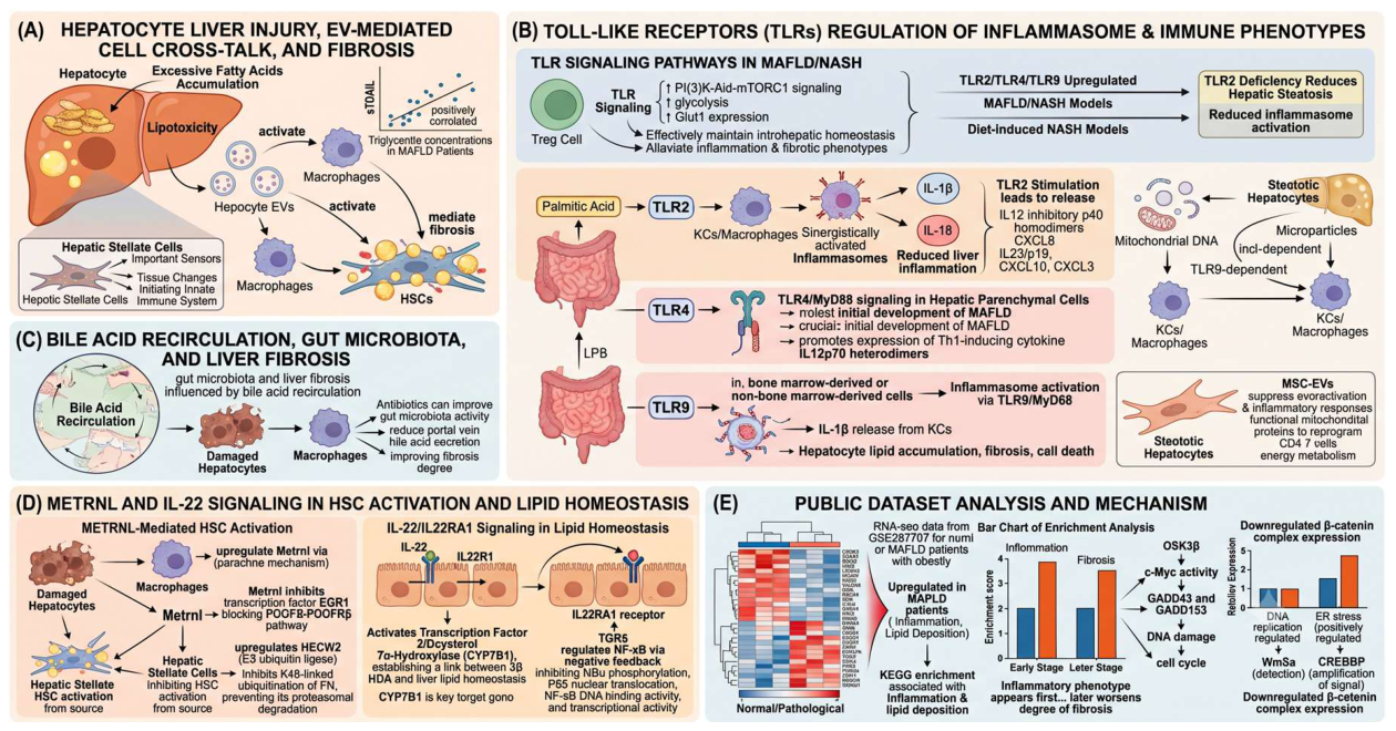
Disclaimer/Publisher’s Note: The statements, opinions and data contained in all publications are solely those of the individual author(s) and contributor(s) and not of MDPI and/or the editor(s). MDPI and/or the editor(s) disclaim responsibility for any injury to people or property resulting from any ideas, methods, instructions or products referred to in the content. |
© 2026 by the authors. Licensee MDPI, Basel, Switzerland. This article is an open access article distributed under the terms and conditions of the Creative Commons Attribution (CC BY) license (http://creativecommons.org/licenses/by/4.0/).
Copyright: This open access article is published under a Creative Commons CC BY 4.0 license, which permit the free download, distribution, and reuse, provided that the author and preprint are cited in any reuse.


