

Submitted:
23 April 2026
Posted:
24 April 2026
You are already at the latest version
Abstract
The valorization of agricultural plant waste as a sustainable source of natural fibers has gained increasing attention due to environmental and economic concerns. This study investigates the feasibility of extracting bast fibers from Egyptian Corchorus olitorius L. (Molokhia) plant residues and evaluates the influence of different extraction methods on fiber properties. Fibers were extracted using biological retting, cold al-kaline chemical treatment (4% NaOH), and manual scraping, followed by comprehensive characterization of their morphological, chemical, crystalline, mechanical, thermal, and environmental properties. The results showed that the extraction method significantly affected fiber performance. Chemically extracted fibers exhibited the smallest average diameter (13.76 ± 0.44 μm), the highest cellulose content (72.23%), and the lowest lignin content (3.20%), indicating effective removal of amorphous components. XRD analysis revealed the highest crystallinity index for chemically extracted fibers (70.0%), compared to bi-ological (60.0%) and manual extraction (64.0%). These structural improvements resulted in superior mechanical properties, with tensile strength and Young’s modulus reaching 600.67 ± 11.73 MPa and 38.96 ± 0.64 GPa, respectively, compared to lower values for biologically and manually extracted fibers. Weight loss analysis indicated optimal extraction durations of 21 days for biological retting and 9 days for chemical treatment. ICP-MS analysis confirmed that heavy metal contents were well below Oeko-Tex® Standard 100 limits. Overall, the findings demonstrate that Molokhia plant waste is a promising and environmentally safe source of natural fibers, with cold chemical extraction offering the most effective route for producing high-quality fibers suitable for bio-based composite applications.
Keywords:
Corchorus olitorius
; Molokhia fibers
; bast fibers
; extraction methods
; crystallinity
; mechanical properties
; agricultural waste
; sustainable materials
; bio-based composites
1. Introduction
The global textile industry plays a vital role in modern society; however, it is also associated with significant environmental challenges, including high energy consumption, excessive water usage, and the generation of pollutants. In response to these concerns, increasing attention has been directed toward sustainable and eco-friendly alternatives. Natural lignocellulosic fibers have emerged as promising candidates due to their biodegradability, renewability, low density, and reduced environmental impact during processing. Recent studies have demonstrated the effectiveness of natural fibers as reinforcement materials in polymeric composites, offering advantages such as cost-efficiency, recyclability, and improved sustainability [1,2,3,4,5].
Among natural fibers, lignocellulosic fibers such as jute, flax, hemp, kenaf, coir, and sisal have been extensively investigated and widely applied in composite materials across various industries, including automotive, construction, and packaging [6]. In parallel, recent research efforts have increasingly focused on identifying and characterizing novel and underutilized plant fibers. Several studies have reported the successful extraction and application of fibers from unconventional plant sources such as Phytolacca americana [7], reddish shell bean [8], Sambucus ebulus L. [9], beetroot [10], Alcea rosea [11], Grewia flavescens [12], Ficus benjamina [13], and Martynia annua [14]. These studies highlight the growing global interest in diversifying natural fiber resources and promoting the valorization of agricultural residues within a circular economy framework.
Plant fibers are primarily composed of cellulose, hemicellulose, lignin, and pectin, and their properties are strongly influenced by the relative composition and structural organization of these components [15,16]. The hierarchical structure of plant fibers, particularly the secondary cell wall (S2 layer), plays a critical role in determining their mechanical performance, which depends on factors such as cellulose content, degree of polymerization, and microfibrillar angle [17,18].
Despite the significant progress achieved in the field of natural fibers, several research gaps remain. First, most previous studies have focused on a single extraction method, with limited comparative investigations evaluating multiple extraction techniques under controlled conditions. Second, although analytical techniques such as scanning electron microscopy (SEM), Fourier transform infrared spectroscopy (FTIR), and X-ray diffraction (XRD) are widely used, there is a lack of integrated studies that establish clear structure–property relationships linking fiber composition, crystallinity, and mechanical behavior. Third, the relationship between the maximum XRD peak intensity and the crystallinity index—an important indicator of cellulose ordering—has not been sufficiently explored in natural fiber research.
Furthermore, Corchorus olitorius L. (Molokhia), although botanically related to jute, remains largely underexplored as a fiber source, particularly in Arab regions such as Egypt, where it is widely cultivated as a food crop. In these regions, Molokhia stems are typically treated as agricultural waste and are often burned or discarded, leading to environmental concerns. Despite their potential as a lignocellulosic resource, there is a lack of systematic studies addressing the extraction and comprehensive characterization of Molokhia fibers, especially considering the influence of local environmental conditions such as soil composition and climate.
Therefore, this study aims to address these gaps by investigating the sustainable extraction of bast fibers from Egyptian Molokhia (Corchorus olitorius) stems using three different methods: biological retting, cold alkaline chemical treatment, and manual extraction. A comprehensive characterization of the extracted fibers is conducted, including morphological (SEM), structural (XRD), chemical (FTIR), and mechanical analyses. In addition, this work explores the relationship between crystallinity index and XRD peak intensity, providing new insights into the structural behavior of Molokhia fibers. The findings contribute to the development of sustainable textile materials and support the valorization of agricultural waste into high-value bio-based products.
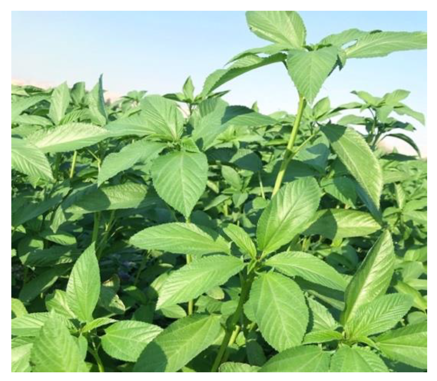
Figure 1.
Molokhia plant (Corchorus olitorius L) grown in Egypt.
2. Materials and Methods
To comprehensively evaluate how extraction techniques influence the structural and functional properties of natural fibers, four primary methods are commonly considered: biological retting, chemical extraction, enzymatic retting, and manual scraping. In this study, three methods—biological retting, cold alkaline chemical extraction, and manual scraping—were employed to extract bast fibers from Corchorus olitorius (Molokhia) stems. Each method offers distinct mechanisms and advantages. Biological retting involves the natural action of microbial communities in water to degrade pectin and separate fibers. It is widely used in traditional agricultural settings due to its simplicity and low cost, despite its long processing time and limited control over microbial activity and fiber uniformity (Paridah et al., 2011) [19].
Cold chemical extraction using 4% NaOH at ambient temperature facilitates more efficient removal of non-cellulosic materials, particularly lignin and hemicellulose, leading to improved fiber purity and higher crystallinity (Xue Li et al., 2007) [20]. Manual scraping, on the other hand, provides a mechanical means of fiber separation with minimal alteration to the fiber’s native chemical structure, making it useful for preserving original plant characteristics.
Although enzymatic retting—employing specific enzymes such as pectinase or xylanase—has gained attention for its rapid action, high selectivity, and ability to produce cleaner fibers with consistent quality (Angulu & Gusovius, 2024) [21] its relatively high cost, sensitivity to pH and temperature, and the need for strict process control make it less practical for small-scale or rural applications. Therefore, in this study, biological retting was selected over enzymatic methods as a more accessible, sustainable, and economically feasible option that better reflects conditions applicable to local fiber valorization initiatives in Egypt.
2.1. Plant Material Preparation
Fibers were extracted from Molokhia (Corchorus olitorius) plant waste cultivated in Upper Egypt. Following leaf removal, the stems were subjected to natural sun-drying for a period of 10–15 days, as shown in Figure 2a (during drying) and 2b (after drying), with the drying duration adjusted in accordance with prevailing ambient temperature conditions. All procedures involving plant materials were performed in strict compliance with applicable institutional, national, and international guidelines and regulatory frameworks.
2.2. Fiber Extraction Methods
2.2.1.. Biological Retting Process
Stems with an average length of 2 m (±15 cm) were halved and subjected to natural water retting to facilitate fiber separation. The retting process was conducted in water with an initial pH of 7.52 and total dissolved solids (TDS) of 135 ppm. The stems remained submerged for 15–23 days at a controlled room temperature of 27 ± 3 °C, allowing microbial activity to degrade pectin within the stem bundles, thereby liberating individual fibers as clearly demonstrated in Figure 3. Upon completion of retting, the stems were removed, rinsed with tap water to remove residual debris, and the isolated fibers were air-dried. Post-retting water analysis indicated an increase in pH (8.25) and TDS (820 ppm), suggesting microbial proliferation and organic matter dissolution. The slightly alkaline pH and moderate temperature (25–30 °C) were conducive to bacterial and fungal growth, which played a critical role in pectin degradation. This method was done according to (Tahir et al., 2011; Angulu et al., 2024( [21].
2.2.2. Chemical Extraction Method (Cold Alkaline Treatment)
The chemical extraction of fibers was performed using a mild alkaline treatment at ambient temperature to minimize energy consumption and preserve fiber integrity. Dried Molokhia stems were cut into 20 cm segments and immersed in a 4% (w/v) sodium hydroxide (NaOH) solution at a solid-to-liquid ratio of 1:15 fibers The mixture was kept at room temperature (27 ± 3 °C) for 9 days, as clearly demonstrated in Figure 4 with periodic agitation to ensure uniform chemical penetration and enhance delignification. A stable alkaline pH of ~13 was maintained throughout. After the treatment period, the stems were thoroughly rinsed with distilled water to remove residual alkali and solubilized organic compounds. The fibers were then neutralized using a 1% (v/v) acetic acid solution to reach pH ≈ 7.0, eliminate remaining NaOH and prevent fiber damage. Finally, the extracted fibers were washed repeatedly with distilled water until a neutral pH was achieved and subsequently air-dried at ambient conditions for 48 hours. Similar mild alkaline retting methods have been reported for bast fibers such as Kenaf, using low-temperature NaOH to preserve fiber integrity (Paridah et al., 2011) [19].
2.2.3. Manual Extraction (Scraping Method)
The manual extraction of fibers was carried out using a traditional scraping technique to mechanically separate fibers from the dried Molokhia stems. The stems, with an average length of 2 m (±15 cm), were first soaked in water for 24 hours to soften the outer bark and facilitate fiber separation. After soaking, the stems were manually beaten using a wooden mallet to loosen the fibrous bundles from the woody core. Subsequently, the fibers were carefully scraped off using a blunt knife or a specialized scraping tool, ensuring minimal damage to the fiber structure as clearly demonstrated in Figure 5. The extracted fibers were then washed thoroughly with clean water to remove any residual plant debris and dried under shade at ambient temperature (27 ± 3 °C) for 48 hours to prevent excessive brittleness. (ScienceDirect Topics, 2025) [22]. Figure 6 provide a flow chart and images, respectively, illustrating different steps during the extraction process.
2.3. Characterization
2.3.1. Scanning Electron Microscopy (SEM)
SEM analysis was conducted using a high-resolution scanning electron microscope, specifically the EDAX AMETEK Quanta FEG 250 equipped with a field emission gun and an accelerating voltage of 30 kV. This analysis was carried out at the Desert Research Centre of the Egyptian Ministry of Agriculture and Land Reclamation. The Molokhia fiber samples, cut for examination, were coated with a gold film prior to analysis. The equipment used is from the FEI company in the Netherlands.
2.3.2. Fourier Transform Infrared Spectroscopy (FTIR)
Fourier Transform Infrared Spectroscopy (FTIR), specifically Attenuated Total Reflectance Fourier Transform Infrared Spectroscopy (ATR-FTIR), was conducted using a Thermo Scientific Spectrometer equipped with a Smart Orbit ATR accessory. The reflectance element utilized was a diamond crystal. Spectra were gathered in the range of 4,000–650 cm-1, employing 16 scans and a resolution of 16.
2.3.3. X-Ray Diffraction (XRD)
X-ray diffraction (XRD) was utilized to study the crystallinity and microstructure of the Egyptian Molokhia fiber samples. The analysis was performed using a Bruker D2 Phaser 2nd Gen X-ray diffractometer with Cu/Kα radiation at 30 kV and 10 mA, over a 2θ range of 5–80°, with a step size of 0.02° and 0.2 seconds per step.
2.3.4. The Measurement of Chemical Composition.
The chemical composition of Molokhia fibers was evaluated in accordance with the Technical Association of Pulp and Paper Industry (TAPPI) standards. The determination of cellulose and hemicellulose content followed the procedure outlined in TAPPI T19 m-54, as detailed by de Morais (Teixeira et al. 2010) [23] and (Li et al. 2009) [24]. The analysis of lignin content involved a reaction with sulfuric acid, following the standard method recommended in ASTM D 1106-56 [25] and ash content was determined using ASTM D1102-84 [26]. The percentages of cellulose, hemicellulose, lignin, and ash were calculated, and the average and standard deviation values were reported based on five samples.
2.3.5. Tensile Tests
The mechanical properties of the Molokhia fibers were determined using a tensile test. Specimen preparation followed the ASTM C1557 standard [27]. Prior to testing, the fibers were adhered to 90 g/m² paper using adhesive. The tensile tests were conducted with an EMIC DL 10,000 universal testing machine (Instron, São José dos Pinhais, Paraná). Tests were performed on specimens with a 40-mm gauge length at a displacement rate of 0.2 mm/min. and the fibers were tested in their received condition [28]. The cross-sections of the fibers were assumed to be circular, and these areas were used to calculate the tensile strength.
2.3.6. Density Determination
To determine the volume density of the fiber, a method involving linear density and diameter calculation was employed according to (Truong et al., 2009) [29]. This approach uses the average linear density (fiber mass and length) along with the fiber diameter to calculate the volume density using the equation (1)

where "M" represents the mass of the fiber, "d" is the fiber diameter, and "l" is the fiber length.
2.3.7. Moisture Content
Desiccators, or humidity chambers, were established with 100% humidity using distilled water. Bundles of individual fibers bound together, specifically Molokhia fibers, were prepared as specimens. These specimens underwent a drying process in an air oven at 60°C for 24 hours, followed by precise weighing on a balance accurate to four decimal places (±0.1 mg). Subsequently, they were placed in the humidity chamber maintained at room temperature. According to the DIN standard 53495 [31] the weight difference was measured at various time intervals, and the total water uptake was calculated using equation (2).
where MC = moisture content, mi = mass of the sample after exposing it in humidity and mo = mass of the dried sample. three samples were utilized for the analysis of fibers.
2.3.8. Weight Loss Rate
Equation 3, as per (Zheng Fan Li, et al 2020) [31] was employed to determine the weight loss.
2.3.9. Inductive Coupled Plasma Emission Spectrometer (ICP-MS) Detection
The (Agilent 7700 ICP-MS) instrument from Japan was utilized to analyze mineral levels in Egyptian Molokhia fibers. The goal was to quantify the concentrations of various elements, including Al, B, Ba, Cd, Co, Cr, Fe, Mn, Mo, Ni, Pb, Si, Sr, V, P, Na, K, Cu, and Zn. To achieve this, samples underwent digestion using the (Speed Wave Entry Multi wave system from Berghof, Germany). About 0.1 grams of the Molokhia fiber samples were mixed with a solution containing 65% HNO3 (5 mL) and 30% H2O2 (1 mL), and the digestion process occurred at 200°C. After digestion, the resulting solutions were analyzed using an ICP-MS for metal content verification. Oeko-Tex Standard 100 is a global testing and certification framework for textiles, imposing restrictions on specific chemical usage. Textiles bearing this designation are verified to stay within prescribed threshold values for particular hazardous substances. The limit values for these substances under Oeko-Tex Standard 100 are detailed in Table 1 [32].
2.3.10. Thermal Analysis
The thermal analysis was conducted using a TA Instruments Trios V4.1 system, designed for precise thermogravimetric measurements across a temperature range of 0–800°C. The instrument offers high sensitivity, capable of detecting weight changes as small as 0.1%, with an adjustable heating rate to meet specific experimental requirements, ensuring precise thermal control. It operates under various atmospheric conditions, including inert (e.g., nitrogen, argon) and oxidative (e.g., air), allowing for tailored experimental environments. Equipped with a high-precision thermobalance, the system ensures accurate monitoring of weight changes throughout the thermal process. The analysis was managed using TA Instruments’ Trios V4.1 software, which facilitates parameter configuration, real-time data monitoring, and comprehensive analysis. Data outputs include weight loss curves (%Weight) and their derivatives {d (Weight)/d (Temperature)} as a function of temperature, enabling an in-depth understanding of thermal degradation behavior. This advanced instrumentation ensures accurate and reliable characterization of the thermal properties of Molokhia fibers.
2.3.11. Statistical Analysis
We conducted statistical analysis using Microsoft Excel 365 from Microsoft, USA. The data were analyzed using trend lines, their equations and values of the correlation coefficient (R2) determination coefficient values, and one-way analysis of variance (ONEWAY-ANOVA).
3. Results and Discussion
3.1. Morphology Studies
The provided SEM (scanning electron microscope) images illustrate the surface morphology of Molokhia fibers extracted through three different methods: biological extraction, chemical extraction, and manual extraction. Figure 7(a) and 7(b) with magnifications of 1000×, and 2000× respectively. depict the surface characteristics of Molokhia fibers obtained via biological extraction. In Figure 7(a) with magnifications of 1000×, the longitudinal view reveals microfibers partially covered by non-cellulosic materials, resulting in an uneven surface. Small white particles, possibly wax, lignin, or impurities, are observed [33], A closer examination in Figure 7(b) with magnifications of 2000× identifies elementary fibers or fibrils bound lengthwise by pectin, lignin, and other non-cellulosic compounds, forming fiber bundles [34,35]. The rugged surface of these bundles, characterized by micro-voids, grooves, fibril cracks, and impurities (e.g., wax, pectin, lignin, and oil), enhances the mechanical properties of composite materials by promoting strong adhesion to the polymer matrix. Additionally, the presence of wax and oils forms a protective layer on the fiber surface 36. Furthermore, as shown in Figure 7(a) with magnifications of 1000×, the thin Molokhia fiber strands display separated microfibrils and longitudinal splitting, with openings between fibrils indicating a high surface area suitable for composite reinforcement.
Figure 7(c) with magnifications of 70× shows the surface morphology of fibers extracted using chemical methods. The surface exhibits pronounced fibrillation, with separated microfibrils due to the removal of hemicellulose and lignin by NaOH treatment. This process, driven by NaOH’s ability to break hydrogen bonds and dissolve non-cellulosic components, leads to significant fibril separation. The selective degradation of amorphous components enhances the surface area, improving the fibers’ compatibility with composite materials by facilitating adhesion.
Figure 7(d) with magnifications of 1000× illustrates the surface morphology of fibers obtained through manual extraction. The surface shows irregular tearing and fraying, which are characteristic of physical scraping. Unlike fibers extracted through chemical or biological methods, manually extracted fibers exhibit uneven surface disruptions due to the mechanical force applied during scraping. Exposed microfibrils are visible but appear less aligned compared to chemically processed fibers, indicating incomplete separation of the fiber bundles. Residual lignin and hemicellulose patches remain adhered to the surface, as manual scraping does not fully remove these non-cellulosic components. Additionally, waxy deposits commonly found in raw Molokhia are still present, reducing the uniformity of the fiber surface. The fibers display non-uniform thickness, with thinner regions resulting from scraping and thicker areas where fiber bundles remain intact. The edges are jagged and split, reflecting the abrasive nature of the manual extraction process.
Figure 7(e) with magnifications of 1000×, 7(f) with magnifications of 1000×, and 7(g) with magnifications of 200× present the fiber diameters obtained through different extraction methods. Specifically, Figure 7(e) with magnifications of 1000× depicts the diameters of fibers extracted via biological extraction, with an average diameter of approximately 64.8 µm. Figure 7(f) with magnifications of 1000×, illustrates the diameters of fibers obtained through chemical extraction, which exhibit a significantly smaller average diameter of around 13.7 µm. In contrast, Figure 7(g) with magnifications of 200× demonstrates the diameters of fibers extracted manually, with an average diameter of approximately 69.99µm. These findings indicate that chemical extraction yields finer, and thinner fibers compared to biological and manual methods. This is attributed to the action of chemicals in breaking down non-cellulosic components, such as lignin and pectin, thereby exposing the finer cellulose fibers.
3.2. Fourier Transform Infrared Spectroscopy (FTIR) Analysis of the Molokhia Fibers
A comparison of the absorption bands in the FTIR spectra of Egyptian raw Molokhia fibers extracted biologically (Figure 8a), chemically (Figure 8b), and manually (Figure 8c) revealed no changes or breakage in chemical bonds. This suggests that cold chemical extraction operates at slower reaction rates due to the low temperature, ensuring better process control and reducing the risk of over-processing or fiber damage. Similarly, the absence of bond breakage in biological extraction confirms that the soaking time was optimally chosen. Furthermore, when examining the FTIR spectra of Chinese raw jute fibers and Bangladeshi raw jute fibers Figure 9, it was observed that the absorption bands of Egyptian Molokhia fibers closely resemble those of both Bangladeshi and Chinese jute fibers. This similarity can be attributed to the common composition of these fibers, which includes three main components: cellulose, hemicellulose, and lignin [37]. The structures of these constituents are illustrated in Figure 10. In the case of cellulose and hemicellulose (characterized by the repetition of cellulose with a -C-O-C- group), there exist C-C, C-O-C, and C=C bonds, which are relatively weaker compared to the lignin (containing phenol groups) due to their shorter chain length. Consequently, cellulose and then hemicellulose are responsive to FTIR at higher wavelengths (lower frequencies), while lignin is responsive at lower wavelengths. This observation is elucidated by the FTIR transmittance peak. The results of the transmittance spectra, presented in Figures (8 a, b, c) over the range of 4000–650 cm−1, depict the relationship between transmittance (%) and wave number.
Table 2 presents the assignment of absorption bands in the FTIR spectrum of raw Molokhia fibers. In Figure 6 (a), the FTIR spectra of these raw Molokhia fibers clearly depict absorption bands at 3339.69470 cm-1, 1736.93943 cm-1, and 2914.77819 cm-1, corresponding to O-H stretching vibration, C=O stretching vibration, and C-H stretching vibration, respectively. These bands are attributed to the hydroxyl group in cellulose, the carbonyl group of acetyl ester in hemicellulose, and the carbonyl aldehyde in lignin [41,42]. Peaks at 1423.84305 and 1595.30059 cm-1 arise from various lignin components. The presence of peaks at 1028.74524 cm-1 is linked to Glycogen absorption, specifically associated with C-O and C-C stretching, as well as C-O-H deformation motions [43]. Other peaks correspond to distinctive groups, as detailed in Table 2.
3.3. X-Ray Diffraction (XRD) Analysis
Cellulose exhibits four crystallographic polymorphisms: I, II, III, and IV. Cellulose I is the naturally occurring crystalline form produced by a variety of species, including trees, plants, tunicates, algae, and bacteria. This form is thermodynamically metastable and can be transformed into cellulose II or III, with all cellulose strands arranged in a highly organized parallel pattern. [50,51] Cellulose I can be converted to cellulose II through regeneration (solubilization and recrystallization) or mercerization (alkaline treatment), resulting in a monoclinic structure. Cellulose III is formed by combining liquid ammonia and thermal treatments with cellulose I and II, while cellulose IV is derived from cellulose III. [50,52].
The X-ray diffraction (XRD) pattern of Egyptian Molokhia fibers is shown in Figure 11. The crystallinity index (CrI) of Corchorus olitorius (Molokhia) fibers extracted via three different techniques—biological retting, cold chemical treatment, and manual scraping—was evaluated using X-ray diffraction analysis. The CrI was calculated using the Segal method according to Equation (4):
where represents the maximum intensity of the (002) diffraction peak at 2θ ≈ 22.5°, corresponding to the crystalline region of cellulose Iβ, and represents the intensity of the amorphous region at 2θ ≈ 18° [53,54].
The biologically extracted fibers exhibited a crystallinity index of 60%, while chemically extracted fibers showed the highest crystallinity (70.0%), followed by manually extracted fibers (64.0%). These results indicate that chemical treatment effectively removes amorphous components such as hemicellulose and lignin, leading to an increase in the relative crystalline content of cellulose.
It should be noted that the Segal method provides an approximate estimation of crystallinity, as it is based on peak intensity rather than a full deconvolution of crystalline and amorphous phases. Therefore, the reported values should be considered comparative rather than absolute.
The enhanced crystallinity in chemically treated fibers is attributed to the more efficient removal of hemicellulose and lignin—amorphous components that interfere with the regular packing of cellulose microfibrils—thereby facilitating better alignment and ordering of cellulose chains [55,56]. Conversely, biological retting preserves more non-cellulosic content due to its milder degradation process, resulting in broader and less intense XRD peaks and hence a lower CrI. Manual extraction, which involves minimal chemical or biological disruption, retains the natural cell wall matrix and yields intermediate crystallinity values. The XRD pattern is presented in Figure 12, with Figure (12a) displaying the XRD peaks of raw and treated jute fibers, Figure (12b) showing the XRD peaks of hemp fibers, and Figure (12c) illustrating the XRD peaks of raw pineapple fibers. Notably, the same two distinct peaks at 2θ = 16.5 and 22.5, corresponding to the 110 and 200 planes respectively, are observed. These peaks are characteristic of natural fibers.
When benchmarked against traditional bast fibers such as ramie (74.5%), jute (71.5%), flax (65.2%), kenaf (59.3%), and hemp (70.0%), Molokhia fibers exhibited a moderate degree of crystallinity that strongly depended on the extraction method [57,58,59]. Specifically, biologically retted Molokhia fibers showed a crystallinity index of approximately 60%, while manually extracted fibers exhibited a slightly higher value of about 64%, and chemically extracted fibers reached up to 70%, approaching the crystallinity levels of well-established bast fibers such as hemp and jute. Despite this improvement, Molokhia fibers generally maintained a lower or comparable crystallinity relative to highly crystalline fibers like ramie and jute, reflecting a partially amorphous structure. This semi-amorphous nature may result in reduced stiffness and tensile strength compared to highly crystalline bast fibers; however, it can be advantageous in enhancing flexibility, moisture absorption, and interfacial compatibility with hydrophilic polymer matrices in green composite applications 60. hese findings confirm the significant influence of extraction technique on the crystalline structure of Molokhia fibers and underscore their potential as sustainable reinforcements in biocomposites tailored to specific performance requirements.
The maximum XRD peak intensity provides insights into the crystalline order and molecular packing within natural fibers, especially at the characteristic cellulose Iβ plane (2θ ≈ 22°). In this study, the biologically extracted Molokhia fiber exhibited a peak intensity of approximately 2200 counts, while the chemically extracted and manually extracted fibers showed higher intensities of about 2500 and 2700 counts, respectively.
This trend suggests that manual extraction, which minimally disrupts the native cellulose structure, preserves a higher level of molecular order compared to the more disruptive chemical and biological treatments. However, cold chemical treatment, while effective at removing amorphous components like lignin and hemicellulose, may also lead to slight disruption of some crystalline regions depending on reagent concentration and exposure time [53,55].
In comparison to other well-known natural fibers, the maximum XRD peak intensities are significantly higher: Ramie: ~5200 counts, Jute: ~5000 counts, Flax: ~4700 counts, and Hemp: ~4800 counts, Kenaf: ~4300 counts. These results reflect the naturally higher crystallinity and better-organized cellulose microfibrils in conventional bast fibers compared to Molokhia. The lower XRD intensities in Molokhia fibers reinforce the finding that their cellulose matrix is more amorphous, yet this can be advantageous in applications requiring higher surface reactivity or improved interfacial adhesion with polymer matrices [56,57,58].
Figure 12 presents the XRD patterns of selected natural fibers. Specifically, Figure 12a displays the diffraction peaks of raw and treated jute fibers, Figure 12b illustrates the profile of hemp fibers, and Figure 12c shows the diffraction patterns of raw pineapple leaf fibers. In all cases, two prominent peaks are consistently observed at 2θ ≈ 16.5° and 22.5°, corresponding to the (110) and (200) crystallographic planes of cellulose I. These peaks are typical of lignocellulosic natural fibers and indicate the presence of semicrystalline cellulose I structures. Table 3 present a comparative analysis of the crystallinity percentage and the maximum XRD peak intensity of Egyptian Molokhia fibers extracted using different methods, alongside selected conventional natural fibers.
3.4. Chemical Composition
A thorough understanding of the chemical composition and surface adhesive properties of natural fibers is crucial for developing effective natural fiber-reinforced composites. These fibers consist of cellulose, hemicellulose, lignin, pectin, waxes, and water-soluble substances. Table 4 presents the composition of Egyptian Molokhia fibers and and their comparison with traditional natural fibers and some new fibers. It's important to note that the chemical makeup of natural fibers can vary based on factors such as growing conditions and testing methods, even for the same fiber type. Cellulose is a semicrystalline polysaccharide made up of D-glucopyranose units linked by β-(1-4)-glucosidic bonds 64. The large number of hydroxyl groups in cellulose gives natural fibers hydrophilic properties, which can result in poor adhesion and moisture resistance when used in hydrophobic matrices [64]. Hemicellulose, which is linked to cellulose fibrils via hydrogen bonds, is an amorphous and branched polymer with a much lower molecular weight compared to cellulose. Its structure, which contains many hydroxyl and acetyl groups, makes hemicellulose somewhat soluble in water and hygroscopic [65]. Lignin, on the other hand, is an amorphous, highly complex, and primarily aromatic polymer made up of phenylpropane units 66, and it has the lowest water absorption among the components of natural fibers [67].
The data presented in Table 4 regarding the composition of Corchorus olitorius (Molokhia) fibers highlight significant variations based on the extraction methods employed. Chemical extraction resulted in the highest cellulose content (72.23%), aligning with prior research demonstrating that alkaline and acid treatments effectively remove non-cellulosic components (Reddy & Yang, 2005) [68]. In contrast, manual extraction yielded a lower cellulose content. (64.16%), indicative of incomplete delignification, a characteristic commonly associated with non-chemical methods (Sun, R., et al, 2004) [69].
Hemicellulose retention was notably higher in manual extraction (26%) compared to chemical methods (17%), consistent with findings that intensive chemical treatments hydrolyze hemicellulose (Faruk et al., 2012) [70]. The lignin content was significantly reduced in chemically treated fibers (3.2%), corroborating reports that sodium hydroxide effectively breaks β-aryl ether bonds (Siqueira, G., Bras, J., & Dufresne., 2010) [71]. Conversely, biological retting yielded a higher lignin content (6.5%), reflecting partial lignin degradation via microbial enzymatic activity (Li et al., 2015) [72].
The ash content (0.5–1.98%) was within the typical range for bast fibers (1–5%) as reported by (Thyavihalli Girijappa et al 2019) [73]. The moisture content (3.21–4.13%) exhibited minimal variation, likely due to consistent drying conditions across extraction methods, which are crucial for ensuring fiber stability (Bledzki & Gassan, 1999) [57]. Table 5 shows one-way Analysis of Variance (ANOVA) was employed to evaluate the effect of extraction methods (Biological, Chemical, Manual) on the chemical composition of Molokhia fibers. The results revealed significant differences in cellulose content (F(2,12) = 16.08, p = 0.001), hemicellulose (F(2,12) = 15.67, p = 0.0011), lignin (F(2,12) = 15.26, p = 0.0012), pectin (F(2,12) = 5.00, p = 0.0397), ash content (F(2,12) = 13.54, p = 0.0016), and moisture content (F(2,12) = 9.86, p = 0.0044) among the different extraction methods.
These findings indicate that the biological extraction method significantly enhanced cellulose content while reducing lignin and ash levels compared to chemical and manual methods, reflecting its positive impact on fiber purity. The chemical method demonstrated a notable reduction in hemicellulose and ash content compared to manual extraction, although lignin content remained higher in manual extraction. Post-hoc analysis using Tukey’s HSD test confirmed significant differences in chemical composition between the extraction methods. Chemically extracted fibers showed significantly higher cellulose content and lower hemicellulose and lignin contents compared to biological and manual methods (p < 0.05). These results confirm the effectiveness of chemical treatment in removing amorphous components and enhancing fiber purity. The bar charts further in Figure 13 confirmed these differences, displaying distinct mean variations with limited dispersion (±SD) among groups.
3.5. Physical and Mechanical Properties
The tensile properties determine a material's ability to withstand tensile loads before failure. Tensile tests of fibers are conducted to measure their breaking force, tenacity, modulus, and elongation. These properties are crucial in determining the suitability of fibers for specific applications. Table 6 presents the physical and mechanical properties of Egyptian Molokhia (Corchorus olitorius) fibers and their comparison with traditional natural fibers and some new fibers. Chemically extracted fibers exhibited the highest density (1.4 5± 0.01g/cm³), followed by biologically extracted fibers (1.23 ±0.01g/cm³) and hand-extracted fibers (1.12 ± 0.01 g/cm³). The elevated density of chemically treated fibers reflects their high purity and cellulose content (72.23%) due to the effective removal of lignin and hemicellulose through chemical treatments, such as alkaline processing (Thygesen et al., 2005) [95]. In contrast, the lower density of hand-extracted fibers is likely attributed to residual impurities and a higher proportion of non-fibrous materials, including pectin and lignin (Faruk et al., 2012) [96].
The elongation at break (EAB) was comparable across all fiber types (1.6–1.8%), with chemically extracted fibers showing a slight advantage (1.8 ±0.10 %). These low elongation at break )EAB( values (<2%) are characteristic of cellulose-rich natural fibers, where strong hydrogen bonding between cellulose chains limits elasticity (John & Thomas, 2008) [97] The marginally higher elongation observed in chemically extracted fibers may result from reduced lignin content, which decreases fiber stiffness (Kabir et al., 2012) [96].
Tensile strength was highest in chemically extracted fibers (600.67± 11.73MPa), followed by biologically extracted fibers (520.87 ± 9.58MPa) and hand-extracted fibers (450.89 ±7.34MPa). The superior tensile strength of chemically treated fibers can be attributed to increased cellulose crystallinity achieved through the removal of amorphous components, such as lignin and hemicellulose (Li et al., 2007) [98]. Chemical treatments also enhance inter-fiber bonding, contributing to improved tensile properties (Sgriccia et al., 2008) [64]. Conversely, the lower tensile strength of hand-extracted fibers is a result of incomplete lignin removal, which compromises fiber cohesion (Reddy & Yang, 2005) [68].
Young’s modulus was highest in chemically extracted fibers (38.955 ± 0.64 GPa) and lowest in hand-extracted fibers (20.955 ± 0.34 GPa). A higher Young’s modulus indicates greater stiffness, which is closely related to the cellulose content and crystallinity of the fibers (Nishino et al., 2004) [99]. Chemical treatments are known to enhance fiber stiffness by increasing cellulose crystallinity and eliminating structural defects, leading to improved load transfer along the fiber axis (Siqueira et al., 2010) [100].
The relatively high stiffness observed in chemically extracted Molokhia fibers can therefore be attributed to the effective removal of amorphous components such as hemicellulose and lignin, resulting in a more ordered cellulose structure and improved microfibrillar alignment. These structural modifications contribute to enhanced mechanical performance compared to biologically and manually extracted fibers, where the presence of residual non-cellulosic materials and structural irregularities may limit stiffness.
However, it is important to note that the calculation of Young’s modulus in this study assumed of a circular fiber cross-section derived from average diameter measurements. Natural fibers typically exhibit irregular and non-circular cross-sectional geometries, which may introduce uncertainty in the estimation of cross-sectional area and potentially lead to an overestimation of the modulus values.
Therefore, the reported Young’s modulus values should be interpreted as approximate and comparative rather than absolute. Further studies using more accurate cross-sectional characterization techniques, such as image-based area analysis, are recommended to obtain more precise mechanical measurements.
A detailed analysis was conducted to investigate the relationship between the mechanical properties of Molokhia fibers and their crystallinity index (CI) across three extraction methods: biological retting, cold chemical extraction, and manual extraction. The cold chemical extraction method yielded the highest CI (70.0%) and XRD peak intensity (~2500 counts), corresponding to the highest tensile strength (mean = 600.67 MPa), Young’s modulus (mean = 38.96 GPa), and smallest fiber diameter (mean = 13.76 µm ± 0.68). This indicates a strong molecular alignment and superior crystalline structure that enhances mechanical behavior. Conversely, the biological method, with the lowest CI (60.0%) and largest diameter (64.80 µm ± 19.52), showed intermediate strength (520.87 MPa) and stiffness (29.96 GPa). The manual method, though with a moderate CI (64.0%), exhibited the weakest mechanical properties (tensile strength = 450.89 MPa), likely due to large fiber diameter (69.99 µm ± 10.29) and remaining impurities. The stress-strain behavior of Molokhia fibers extracted using different methods demonstrates clear variation in mechanical response: The fiber obtained through chemical extraction exhibits the highest stress capacity and elongation, indicating enhanced ductility and strength due to its highly crystalline and compact structure. Biologically extracted fibers present a moderate stress-strain profile, reflecting balanced mechanical performance. Manual fibers, with the lowest elongation and tensile strength, show a more brittle response, likely due to the presence of non-cellulosic residues and weaker fiber packing. The Figure 14 shows the representative stress-strain curves each curve highlights the comparative performance and deformation behavior of fibers under load.
A one-way analysis of variance (ANOVA) was conducted using five replicates for each group to evaluate the effect of extraction method on the mechanical and morphological properties of Molokhia fibers. The results, summarized in Table 7, indicate statistically significant differences among the extraction methods for all measured properties.
For tensile strength, the ANOVA yielded an F-value of 297.43 (p < 0.0001), confirming that the extraction method has a significant effect on fiber strength. Similarly, Young’s modulus exhibited a highly significant difference with an F-value of 1799.28 (p < 0.0001), indicating substantial variation in fiber stiffness. The fiber diameter also showed a significant difference, with an F-value of 839.38 (p < 0.0001), reflecting clear morphological variations between the extraction methods.
Post-hoc analysis using Tukey’s HSD test revealed significant differences between the extraction methods. Chemically extracted fibers were significantly different from both biological and manual methods in terms of tensile strength and Young’s modulus. In contrast, no significant difference was observed between biological and manual methods for fiber diameter.
These statistical outcomes demonstrate that the extraction method plays a decisive role in determining both the structural and mechanical performance of Molokhia fibers. The observed trends align with the crystallinity index (CI) and XRD peak intensity results, suggesting that higher crystallinity correlates with improved tensile properties and reduced fiber diameter.
The visual representations in Figure 15 comprise both boxplots, which illustrate data distribution and variability, and bar charts, which emphasize mean values alongside their corresponding standard deviations. Collectively, these graphical analyses clearly demonstrate the significant influence of the extraction method on both the morphological characteristics and mechanical performance of the fibers.
When Comparison of Mechanical and Physical Properties between Molokhia and Pineapple Leaf Fibers we notice that: Molokhia fibers extracted exhibit tensile strength values ranging from approximately 450 to 600 MPa, Young’s modulus between 20 and 39 GPa, and fiber diameters in the range of 13–70 µm, depending on the extraction technique. These fibers typically possess a crystallinity index (CI) between 25.9% and 33.0%, with higher CI values correlating with improved tensile performance and reduced fiber diameter.
Pineapple leaf fibers (PALF) were selected for comparison due to their well-documented use as a high-performance natural fiber in composite applications and their status as an agricultural by-product like Molokhia stems. PALF exhibits tensile strengths typically ranging from 170 to 1627 MPa, Young’s modulus between 34.5 and 82.5 GPa, and fiber diameters of 20–80 µm, as reported in previous studies 104,105,106. Its superior stiffness is largely attributed to its higher cellulose content (70–82%) and greater crystallinity index (60–82%).
While PALF generally demonstrates higher stiffness and crystallinity than Molokhia fibers, the latter shows competitive tensile properties when processed via cold chemical extraction, despite having lower crystallinity and cellulose content. This comparison highlights the potential of Molokhia fibers as an alternative, sustainable reinforcement in polymer composites, especially in contexts where agricultural waste valorization and eco-friendly processing are prioritized.
3.6. Effect of Fiber Extraction Methods on Weight Loss Rate
Table 8 depict the weight loss percentages observed across various fiber extraction methods—biological retting, chemical extraction, and manual extraction.
- Biological Retting Method: The weight loss of Molokhia fibers subjected to biological retting increased progressively from 59.70% to 71.40% as the retting duration extended from 15 to 21 days, followed by a slight decrease to 70.50% at 23 days. This trend reflects the microbial degradation of pectin, hemicellulose, and other non-cellulosic materials, leading to fiber separation (Di Candilo et al., 2010)[107]. The observed plateau and minor decline after 21 days suggest that most soluble binding compounds were already decomposed, consistent with previous findings highlighting diminishing returns with prolonged retting (Akin, 2010) [108]. The optimal extraction point, therefore, was identified at 21 days, indicating maximum removal of unwanted components such as pectin, waxes, and lignin, beyond which fiber deterioration may occur due to overexposure to microbial activity.
- Chemical Extraction Method: In the chemical extraction process, weight loss showed a sharp increase from 50.72% (Day 3) to 80.70% (Day 9), with marginal change afterward (80.53% at Day 11), indicating a rapid and efficient dissolution of non-cellulosic constituents by chemical agents like NaOH or H₂SO₄ (Reddy & Yang, 2019) [109]. This stabilization implies that by Day 9, most of the amorphous and matrix-binding materials had been removed, in agreement with literature suggesting that extending chemical treatments beyond optimal duration offers no significant improvement in yield (Reddy & Yang, 2005) [68]. Although efficient, prolonged chemical exposure may adversely affect fiber integrity, including surface morphology, tensile strength, and stiffness.
- Manual Extraction Method: Manual extraction resulted in a fixed weight loss of 64.60%, placing it between biological and chemical methods. This mechanical separation technique, while less efficient in removing residual non-cellulosic matter, helps maintain fiber structure and reduces the risk of chemical or microbial degradation. These observations are aligned with previous studies (Akhtar et al., 2018) [116], which confirmed that manual methods preserve tensile strength but leave behind more impurities due to limited removal capacity.
The weight loss percentage was analyzed across different extraction methods and durations to evaluate the removal efficiency of non-cellulosic components such as hemicellulose, lignin, waxes, and pectin. For biological retting, the mean weight loss increased from 59.70 ± 0.46% at 15 days to 71.40 ± 0.48% at 21 days, followed by a slight decrease to 70.50 ± 0.50% at 23 days. One-way ANOVA revealed a statistically significant difference between the retting durations (F = 1281.08, p < 0.001), confirming that the 21-day period yielded the maximum removal of non-cellulosic materials, after which extended microbial exposure may have caused minor fiber degradation (Di Candilo et al., 2010; Akin, 2010) [107,108].
For chemical extraction, weight loss increased sharply from 50.72 ± 0.40% at 3 days to 80.70 ± 0.42% at 9 days, with no significant change at 11 days (80.53 ± 0.46%). ANOVA indicated significant variation among durations (F = 13314.54, p < 0.001), confirming that 9 days is the optimal chemical treatment period, beyond which additional processing does not significantly improve yield (Reddy & Yang, 2019; Reddy & Yang, 2005) [68,109].
Manual extraction produced a constant weight loss of 64.60%, representing an intermediate value between biological and chemical methods. While less effective in removing residual non-cellulosic matter, this method minimizes the risk of over-processing and preserves fiber strength (Akhtar et al., 2018) [110]. Table 9 shows the ANOVA analysis for weight loss percentage was analyzed across different extraction methods.
A comparative reference with pineapple leaf fibers shows that they exhibit weight loss values ranging between 72–82% depending on the chemical treatment used (NaOH, steam explosion, etc.) and duration (George et al., 2021; Arjmandi et al., 2015) [111,112]. This closely matches the chemically extracted Molokhia fibers, reinforcing the efficiency of chemical methods in maximizing yield, while biological retting remains a more environmentally sustainable approach.
The statistical results and standard deviations are illustrated in Figure 16, which highlights the clear efficiency gap between chemical and biological treatments, and the stability of manual extraction outcomes.
These findings highlight the distinct mechanisms and efficiencies of each extraction technique:
- Chemical extraction provides the highest yield in the shortest time,
- Biological retting ensures gradual and eco-friendly separation with optimal results at 21 days,
- Manual methods offer structural preservation but moderate extraction efficiency.
Further research is recommended to explore how extended extraction durations influence not only yield but also the mechanical and morphological quality of the extracted fibers.
3.7. Analysis of Heavy Metals
Fibers obtained after extraction may contain various chemical residues, some of which can volatilize into the air or be absorbed through the skin. Certain chemicals are known to be carcinogenic or may pose risks to human health, including developmental effects in children, allergic reactions, and chronic diseases such as kidney failure and cancer. Therefore, assessing the metal content in textile materials is of critical importance. The Oeko-Tex Standard 100 is an internationally recognized testing and certification system that restricts the use of harmful substances in textiles. Materials complying with this standard are verified to remain below specified limit values for hazardous compounds, as presented in Table 1.
Egyptian Molokhia fibers were analyzed using Inductively Coupled Plasma Mass Spectrometry (ICP-MS), a highly sensitive technique widely used for the analysis of environmental and textile samples. In recent years, ICP-MS has gained increasing importance due to its ability to detect trace concentrations of metals in raw materials and finished textile products, particularly in the context of eco-certification standards [113]. Additionally, ICP-MS has proven effective in identifying mordants and dyes in historical textiles, including metal-based compounds such as copper, iron, tin, aluminum, and uranium, as well as brominated organic dyes [114].
In the present study, ICP-MS analysis was conducted on biologically extracted Molokhia fibers, and the heavy metal concentrations after wet digestion are summarized in Table 10. The highest concentrations were recorded for strontium (Sr) (11.16 ± 0.73 mg kg⁻¹), sodium (Na) (8.12 ± 0.52 mg kg⁻¹), iron (Fe) (5.12 ± 0.30 mg kg⁻¹), and potassium (K) (4.11 ± 0.28 mg kg⁻¹). Other metals, including Ba, Cu, Ni, Cr, and Pb, were found within the permissible limits defined by Oeko-Tex Standard 100. Boron, cadmium, cobalt, and phosphorus were not detected in the analyzed fibers. It is worth noting that Oeko-Tex does not specify limit values for certain elements such as Sr, Na, Fe, K, Al, Mn, Mo, Si, V, and Zn. A comparison with literature-reported values for textile materials is provided in Table 11.
The relatively low concentrations of hazardous metals can be attributed to the biological retting process, which relies on microbial activity without introducing additional chemical reagents. In contrast, previous studies have indicated that chemical extraction processes, particularly alkaline treatments, may influence the metal content of natural fibers depending on processing conditions, potentially contributing to either impurity removal or trace element introduction.
Therefore, while the present results confirm that biologically extracted Molokhia fibers comply with Oeko-Tex Standard 100 and are suitable for safe textile applications, a comparative investigation of heavy metal content across different extraction methods would provide further insight and is recommended for future work.
Based on these findings, Molokhia bast fibers can be considered suitable for a wide range of applications, including apparel (daily wear and sportswear), protective clothing, and home textiles such as curtains, upholstery, and bed coverings. Moreover, their low heavy metal content enhances their potential for technical textile applications, including automotive interiors, filtration media, and fiber-reinforced composites. Combined with their favorable mechanical properties, fiber morphology, and environmentally friendly extraction process, the compliance with international safety standards positions Molokhia fibers as a promising sustainable alternative in both consumer and industrial textile sectors. Figure 17 shows a comparison between the measured heavy metal concentrations in Molokhia fibers and the permissible limits defined by Oeko-Tex Standard 100.
3.8. Thermal Degradation Analysis of Molokhia Fibers (TGA)
The thermogravimetric (TGA) and derivative thermogravimetric (DTG) curves of biologically extracted Molokhia fibers are presented in Figure 18. As shown, the fibers exhibit a typical multi-stage degradation pattern characteristic of lignocellulosic materials.
The initial weight loss below 150°C corresponds to the evaporation of absorbed moisture, with an estimated mass loss of approximately 5–7%. (Brown, M.E. (2001) [120].
The main thermal degradation occurs in the range of 250–400°C, where a significant weight loss is observed due to the decomposition of hemicellulose and cellulose. The DTG curve reveals a pronounced peak at approximately 360–370°C, indicating the maximum degradation rate of the cellulose phase. This well-defined peak suggests a relatively ordered cellulose structure within the biologically extracted fibers. (Yang et al., 2007) [121].
At higher temperatures (>400°C), a gradual degradation stage is observed, corresponding to the breakdown of lignin and the formation of char residue. The extended degradation tail reflects the presence of thermally stable aromatic structures typical of lignin. (Esin et al., 2023) [122].
Overall, the thermal behavior confirms that biologically extracted Molokhia fibers retain a moderate amount of non-cellulosic components, resulting in a broad degradation profile while maintaining a distinct cellulose decomposition peak.
The observed DTG peak at approximately 360–370°C for the biologically extracted Molokhia fibers is consistent with the thermal degradation behavior reported for other lignocellulosic fibers such as jute, hemp, and kenaf, where the main cellulose decomposition typically occurs within the range of 330–380°C. This similarity indicates that Molokhia fibers exhibit a comparable cellulose stability to conventional bast fibers despite being derived from agricultural residues. [123]
However, the relatively broad degradation profile observed in the present study suggests the presence of residual hemicellulose and lignin, which is characteristic of biologically retted fibers. In contrast, chemically treated fibers reported in the literature often show sharper DTG peaks and higher thermal stability due to the more effective removal of amorphous components such as hemicellulose and lignin.[124]
These findings highlight that biological extraction provides a balanced approach, preserving fiber integrity while maintaining acceptable thermal stability. This behavior is particularly advantageous for sustainable textile and bio-composite applications where environmentally friendly processing is prioritized over aggressive chemical treatments.
Recommended Continuous-Use Temperatures (with safety margins).
- Home & apparel textiles (woven/knits, household fabrics):
≤120–140 °C continuous (washing/drying/ironing “medium”). Short peaks up to 150 °C are acceptable if exposure is brief (seconds–minutes) (Bledzki & Gassan, 1999) [57].
- 2.
- Technical/home textiles (upholstery, curtains, interior panels):≤120 °C continuous; ≤140–150 °C short-term (Pickering et al., 2016) [125].
- 3.
- Automotive/interior trim & semi-structural panels (non-engine bay):
≤90–110 °C continuous; ≤120–130 °C short-term cabin peaks (Pickering et al., 2016) [125].
- 4.
- 4- Protective clothing (not for high-heat/thermal exposure):
Suitable for low-to-moderate heat environments only; avoid applications requiring service >150 °C or flame/arc resistance (Esin et al., 2023) [122].
- 5.
- Polymer Composite Processing Windows
- -
- Thermoplastics:
Pair with matrices processed <200 °C (e.g., PP ~160–180 °C, PE ~130–140 °C). Keep residence time minimal and ensure pre-drying to avoid foaming/voids. Avoid matrices needing ≥200–220 °C (PA6, PET) unless stabilized (Pickering et al., 2016) [125].
- -
- Thermosets (epoxy/polyester/vinyl ester):
Prefer cures ≤120–150 °C. Post-cures up to 160–180 °C only if time is short and fiber is pre-dried; prolonged holds near 180 °C are not recommended (Gregorio & Oliva, 2023) [123].
- -
- Practical Notes
Operate at least 30–50 °C below the onset of major degradation (~200 °C) for continuous service (Yang et al., 2007) [129]. The residual mass >500 °C represents char/inorganics and does not imply usable service at those temperatures (Poletto et al., 2012) [133]. Surface or chemical stabilization (e.g., alkali/silane) can slightly improve thermal tolerance but does not shift the fundamental ~200 °C degradation onset (Bledzki & Gassan, 1999; Pickering et al., 2016) [57,125].
Bottom line: Molokhia bast fibers are thermally suitable for apparel, home, and many technical textiles, and for composites processed and used below ~150–180 °C (short-term) and ≤120–140 °C (continuous). Avoid high-temperature or flame-exposed applications unless fibers are specially treated and certified (Esin et al., 2023; Gregorio & Oliva, 2023) [122,123].
3.9. Linking Molokhia Fiber Extraction Method to Potential End-Uses:
The comparative analysis of Molokhia fibers obtained via biological retting, cold chemical extraction, and manual extraction highlights distinct performance profiles suited for different end-use applications.
Biological retting fibers exhibited a balanced combination of tensile strength, moderate Young’s modulus, and relatively fine diameters, making them ideal for reinforcement in lightweight polymer composites where strong interfacial bonding and good mechanical properties are critical (Bledzki & Gassan, 1999; Pickering et al., 2016) [57,125].
Cold chemical extraction produced fibers with the highest Young’s modulus and crystallinity, enhancing stiffness and dimensional stability. These attributes favor applications in technical textiles, protective fabrics, and structural composite reinforcements where rigidity is advantageous (Faruk et al., 2012; Mohanty et al., 2005) [70,126].
Manual extraction yielded fibers with larger diameters and lower stiffness, but good flexibility, making them better suited for home textiles, ropes, matting, and decorative fabrics where softness and workability are prioritized over maximum strength (Bledzki & Gassan, 1999; Pickering et al., 2016) [57,125].
This mapping of extraction method to final use ensures that fiber processing is optimized for the intended performance, reducing waste and improving application-specific efficiency. Figure 19. Shows Recommended applications for Molokhia fibers based on extraction method.
4. Conclusions
This study successfully demonstrated the feasibility of valorizing Egyptian Corchorus olitorius L. (Molokhia) plant waste as a sustainable source of high-performance bast fibers through biological retting, cold alkaline chemical extraction, and manual scraping. The extraction method was found to play a decisive role in governing the morphological, chemical, crystalline, mechanical, and environmental characteristics of the resulting fibers.
Among the investigated techniques, cold alkaline chemical extraction proved to be the most effective. Chemically extracted fibers exhibited the smallest average diameter (13.76 ± 0.44 μm), the highest cellulose content (72.23%), and the lowest lignin content (3.20%), indicating efficient removal of amorphous non-cellulosic components. XRD analysis confirmed that this method yielded the highest crystallinity index (70.0%), compared to biological retting (60.0%) and manual extraction (64.0%), with corresponding maximum peak intensities of approximately 2500, 2200, and 2700 counts, respectively.
The enhanced structural order achieved through chemical extraction translated directly into superior mechanical performance. Chemically extracted fibers exhibited the highest tensile strength (600.67 ± 11.73 MPa) and Young’s modulus (38.96 ± 0.64 GPa), outperforming biologically extracted fibers (520.87 ± 9.58 MPa; 29.96 ± 0.39 GPa) and manually extracted fibers (450.89 ± 7.34 MPa; 20.96 ± 0.34 GPa). All fibers showed low elongation at break (1.6–1.8%), characteristic of cellulose-rich bast fibers.
Weight loss analysis indicated optimal extraction durations of 21 days for biological retting (≈71.4%) and 9 days for chemical treatment (≈80.7%), while manual extraction resulted in a moderate and constant weight loss (≈64.6%). Importantly, ICP-MS analysis confirmed that all detected heavy metal concentrations were well below the Oeko-Tex® Standard 100 permissible limits, highlighting the environmental safety of Molokhia fibers.
Overall, the findings confirm that Molokhia plant waste represents a promising, safe, and sustainable reinforcement material for bio-based and biodegradable composites, with cold chemical extraction offering the most favorable balance between fiber purity, crystallinity, and mechanical performance. These findings highlight the potential of Molokhia fibers as a sustainable alternative; however, further work is required to validate their performance in real-world composite applications and to assess their full environmental impact.
Future research should focus on the fabrication and performance evaluation of bio-composites reinforced with Molokhia fibers to validate their practical applicability in structural and semi-structural applications. Investigating the interfacial bonding between Molokhia fibers and various polymer matrices (thermosetting and thermoplastic) would provide deeper insight into their reinforcement efficiency.
Additionally, a comprehensive life-cycle assessment (LCA) is recommended to quantitatively evaluate the environmental impact of Molokhia fiber production compared to conventional synthetic and natural fibers. This would further support the sustainability claims of the present study by assessing parameters such as energy consumption, carbon footprint, and end-of-life scenarios.
Future studies may also explore surface modification techniques and hybridization with other natural or synthetic fibers to optimize mechanical performance, durability, and moisture resistance for advanced textile and composite applications.
Funding
The author(s) received no financial support for the research, authorship, and/or publication of this article.
Conflicts of Interest
The author(s) declared no potential conflicts of interest with respect to the research, authorship, and/ or publication of this article.
References
- Ali, A.; Shaker, K.; Nawab, Y.; Ashraf, M.; Basit, A.; Shahid, S.; Umair, M. Impact of hydrophobic treatment of jute on moisture regain and mechanical properties of composite material. J. Reinf. Plast. Compos. 2015, 34, 2059–2068. [Google Scholar] [CrossRef]
- Ameer, M. H.; Shaker, K.; Ashraf, M.; Karahan, M.; Nawab, Y.; Ahmad, S.; Ali Nasir, M. Interdependence of moisture, mechanical properties, and hydrophobic treat- ment of jute fibre-reinforced composite materials. J.Text. Inst. 2017, 108, 1768–1776. [Google Scholar] [CrossRef]
- Koyuncu, M.; Karahan, N.; Shaker, K.; Nawab, Y. Static and dynamic mechanical properties of cotton/epoxy green composites. Fibres Text. East. Eur. 2016, 24, 105–111. [Google Scholar] [CrossRef]
- Nawab, Y.; Kashif, M.; Asghar, M. A.; Asghar, A.; Umair, M.; Shaker, K.; Zeeshan, M. Development characterization of green composites using novel 3D woven preforms. Appl. Compos. Mater. 2018, 25, 747–759. Available online: https://link.springer.com/article/10.1007/s10443-018-9720-2. [CrossRef]
- d’Almeida, J. R. M.; Aquino, R.C.M.P.; Monteiro, S. N. Tensile mechanical proper- ties, morphological aspects and chemical characterization of piassava (Attalea funi- fera) fibers. Compos. Part A Appl. Sci. Manuf. 2006, 37, 1473–1479. [Google Scholar] [CrossRef]
- Huber, T.; Müssig, J.; Curnow, O.; Pang, S.; Bickerton, S.; Staiger, M.P. A critical review of allcellulose composites. J. Mater. Sci. 2011, 47, 1171–1186. [Google Scholar] [CrossRef]
- Eyupoglu, S.; Eyupoglu, C.; Merdan, N. Extraction and characterization of novel cellulosic fiber from Phytolacca americana plant stem. Biomass Convers. Biorefnery 2025, 4259–4268. [Google Scholar] [CrossRef]
- Eyupoglu, S.; Eyupoglu, C.; Merdan, N. Characterization of a novel natural plant-based fber from reddish shell bean as a potential reinforcement in bio-composites. Biomass Convers. Biorefnery 2024, 15, 4259–4268. [Google Scholar] [CrossRef]
- Eyupoglu, S.; Eyupoglu, C.; Merdan, N. Investigation of the effect of enzymatic and alkali treatments on the physico-chemical properties of Sambucus ebulus L. plant fiber. Int. J. Biol. Macromol. 2024, 266(2), 130968. [Google Scholar] [CrossRef]
- Eyupoglu, S.; Eyupoglu, C. Investigation of a New Natural Cellulosic Fiber Extracted from Beetroot Plant. J. Nat. Fibers 2022, 19(16), 13852–13863. [Google Scholar] [CrossRef]
- Eyupoglu, S.; Merdan, N. Investigation of the Characteristic and Sound Absorption Properties of a New Cellulose-based Fiber from Alcea rose L. Plant. J. Nat. Fibers 2022, 19(15), 10082–10093. [Google Scholar] [CrossRef]
- Tiwari, Y. M.; Sarangi, S. K.; Singh, A. K.; Senthamaraikannan, P.; Suyambulingam, I.; Kumar, R. Mechanical, Morphological and Hygroscopic Characterization of Bio-Composites Reinforced with Untreated and Alkali-Treated Grewia flavescens Fibers. J. Nat. Fibers 2025, 22(1), 2502649. [Google Scholar] [CrossRef]
- Joe, M. S.; Sudherson, D. P. S.; Suyambulingam, I.; Senthamaraikannan, P.; Kumar, R. Characterization of Ficus benjamina L. Aerial Root Fiber Reinforced Polyester Composite. J. Nat. Fibers 2025, 22, 1, 2475158. [Google Scholar] [CrossRef]
- Velmurugan, T.; Priyadharshini, G. S.; Senthamaraikannan, P.; Suyambulingam, I. Extraction and characterization of Martynia annua stem fiber as reinforcement for prospective application. Biomass Conversion and Biorefnery. 2025. Available online: https://link.springer.com/article/10.1007/s13399-025-06719-x.
- Loumerem, M.; Alercia, A. Descriptors for jute (Corchorus olitorius L.) Genet Resour. Crop Evol. 2016, 63, 1103–1111. Available online: https://link.springer.com/article/10.1007/s10722-016-0415-y. [CrossRef]
- Kundu, A.; Sarkar, D.; Mandal, N.A.; Sinha, M.K.; Mahapatra, B.S. A secondary phloic (bast) fibre-shy (bfs) mutant of dark jute (Corchorus olitorius L.) develops lignified fibre cells but is defective in cambial activity. Plant Growth Regul. 2012, 67, 45–55. [Google Scholar] [CrossRef]
- Tulio, A.Z.; Ose, K.; Chachin, K.; Ueda, Y. Effects of storage temperatures on the postharvest quality of jute leaves(Corchorus olitorius L.). Postharvest Biol. Technol. 2002, 26, 329–38. [Google Scholar] [CrossRef]
- Abdelmouleh, M.; Boufi, S.; Belgacem, M. N.; Dufresne, A. Short natural-fibre reinforced polyethylene and natural rubber composites: effect of silane coupling agents and ibres loading. Compos. Sci. Technol. 2007, 67, 1627–1639. [Google Scholar] [CrossRef]
- Paridah, M. T.; Ahmed, A. B.; SaifulAzry, S. O. A.; Ahmed, Z. Retting process of some bast plant fibres and its effect on fibre quality: A review. BioResources 2011, 6(4), 5260–5281. [Google Scholar] [CrossRef]
- Xue Li, Tabil, L.G., Panigrahi, S. Chemical Treatments of Natural Fiber for Use in Natural Fiber-Reinforced Composites: A Review. J. Polym. Environ. 2007, 15, 25–33. [CrossRef]
- Angulu, M.; Gusovius, H.-J. Retting of Bast Fiber Crops Like Hemp and Flax—A Review for Classification of Procedures. Fibers 2024, 12(3), 28. [Google Scholar] [CrossRef]
- Topics, ScienceDirect. Fibre Extraction - an overview. 2025. Available online: https://www.sciencedirect.com/topics/engineering/fibre-extraction.
- de Morais Teixeira, E.; Correˆa, A.; Manzoli, A.; de Lima Leite, F.; de Oliveira, C.; Mattoso, L. Cellulose nanofibers from white and naturally colored cotton fibers. Cellulose 2010, 17)3, 595–606. Available online: https://link.springer.com/article/10.1007/s10570-010-9403-0. [CrossRef]
- Li, R.; Fei, J.; Cai, Y.; Li, Y.; Feng, J.; Yao, J. Cellulose whiskers extracted from mulberry: a novel biomass production. Carbohydr. Polym. 2009, 76(1), 94–99. [Google Scholar] [CrossRef]
- ASTM D 1106-96; Standard test method for lignin in wood. American Society for Testing and Materials: Philadelphia PA, 1974.
- ASTM D1102-84; Standard test method for ash in wood. American Society forTesting and Materials: Philadelphia PA, 2007.
- ASTM C1557-20; Standard Test Method for Tensile Strength and Young’s Modulus of Fibers. ASTM International: West Conshohocken, PA, USA, 2017.
- Fidelis, M. E. A.; Pereira, T. V. C.; Gomes, O. F. M.; Silva, F. A.; Filho, R. D. T. The effect of fiber morphology on the tensile strength of natural fibers. J. Mater. Res. Technol. 2013, 2, 149–157. [Google Scholar] [CrossRef]
- Truong, M.; Zhong, W.; Boyko, S.; Alcock, M. A comparative study on natural fibre density measurement. J. Text. Inst. 2009, 100, 525–529. [Google Scholar] [CrossRef]
- Testing of plastics; determination of water absorption. 04 1984. Available online: https://www.dinmedia.de/en/standard/din-53495/1105684.
- Li, Zhengfan; Zou, Yanling; Li, Siyu; Guo, Yuwei; Zeng, Xinyi; Zhu, Jingguo; Zhang, Shangyong. Direct extraction of fibre from a ramie bark. J. Eng. Fibers Fabr. 2020, 15, 1–7. [Google Scholar] [CrossRef]
- Oeko-Tex Standard 100, Product classes specific limit values according to Annex 4. Available online: https://www.hohenstein.us/fileadmin/user_upload/Downloads/Test_Standards/OEKO-TEX/OEKO-TEX_STANDARD_100_Limit_Values_Appendices_4_5_EN.pdf.
- Zimniewska, M.; Kicińska-Jakubowska, A. Vegetable Fibers Sheet. Institute of Natural Fibres & Medicinal Plants. Available online: https://dnfi.org/wp-content/uploads/2012/01/fact-sheet-plant-fibers.pdf.
- Senthamaraikannan, P.; Kathiresan, M. Characterization of raw and alkali treated new natural cellulosic fiber from Coc- cinia grandis. L. Carbohydrate Polym. 2018, 186, 332–343. [Google Scholar] [CrossRef] [PubMed]
- De Rosa, I.M.; Kenny, J.M.; Puglia, D.; et al. Morphological, thermal and mechanical characterization of okra (Abelmoschus escu- lentus) fibres as potential reinforcement in polymer composites. Compos. Sci. Technol. 2010, 70, 116–122. [Google Scholar] [CrossRef]
- Bekraoui, N.; El Qoubaa, Z.; Chouiyakh, H.; et al. Banana fiber extraction and surface characterization of hybrid banana re- inforced composite. J. Nat. Fibers 2022, 19, 12982–12995. [Google Scholar] [CrossRef]
- Mwaikambo, L. Y.; Ansell, M. P. Chemical modification of hemp, sisal, jute and kapok fibres by alkalization. J. Appl. Polym. Sci. 2002, 84, 2222–2234. [Google Scholar] [CrossRef]
- Rong M Z, Zhang M Q, Liu Y, Yang G C, Z.H. The effect of fibre treatment on the mechanical properties of unidirectional sisal-reinforced epoxy composites. Compos. Sci. Technol. 2001, 61, 1437–1447. [CrossRef]
- Rout, A. K.; Kar, J.; Jesthi, D. K.; Sutar, A. K. Effect of surface treatment on the physical, chemical, and mechanical properties of palm tree leaf stalk fibers. BioRes 2016, 11(2), 4432–4445. Available online: https://bioresources.cnr.ncsu.edu/BioRes_11/BioRes_11_2_4432_Rout_Surface_Leaf_Stalk_Palm_Tree_8664.pdf. [CrossRef]
- Shahinur, S.; Hasan, M.; Ahsan, Q.; Haider, J. Effect of Chemical Treatment on Thermal Properties of Jute Fiber Used in Polymer Composites. J. Compos. Sci. 2020, 4, 132. [Google Scholar] [CrossRef]
- Siti Syazwani a, N.; Ervina Efzan a, M. N.; Kok a, C. K.; Nurhidayatullaili, M. J. Analysis on extracted jute cellulose nanofibers by Fourier transform infrared and X-Ray diffraction. J. Build. Eng. 2022, 48, 103744. [Google Scholar] [CrossRef]
- Ahmed1, Abu Saleh; Islam, Md. Saiful; Hassan, Azman; Mohamad Haafiz, M. K.; Islam, Kh Nurul; Arjmandi, Reza. Impact of Succinic Anhydride on the Properties of Jute Fiber/Polypropylene Biocomposites. Fibers Polym. 2014, 15(2), 307–314. Available online: https://link.springer.com/article/10.1007/s12221-014-0307-8. [CrossRef]
- Kalia, S.; Kaith, B. S.; Kaur, I. Cellulose Fibers: Bio- and Nano-Polymer Composites. In Green Chemistry and Technology; Springer : Berlin; Available online: https://link.springer.com/book/10.1007/978-3-642-17370-7.
- RAY, D.I.P.A.; SARKAR, B. K. Characterization of Alkali-Treated Jute Fibers for Physical and Mechanical Properties. J. Appl. Polym. Sci. 2001, 80(7), 1013–1020. [Google Scholar] [CrossRef]
- Shahinur, Sweety; Hasan, Mahbub; Ahsan, Qumrul; Sultana, Nayer; Ahmed, Zakaria; Haider, Julfikar. Effect of Rot-, Fire-, and Water-Retardant Treatments on Jute Fiber and Their Associated Thermoplastic Composites: A Study by FTIR. Polymers 2021, 13(15), 2571. [Google Scholar] [CrossRef]
- Huleihel, M.; Salman, A.; Erukhimovich, V.; Ramesh, J.; Hammody, Z.; Mordechai, S. Novel optical method for study of viral carcinogenesis in vitro. J. Biochem. Biophys. Methods 2002, 50, 111–121. [Google Scholar] [CrossRef]
- Movasaghi, Zanyar; Rehman, Shazza; Rehman, Ihtesham ur. Fourier Transform Infrared (FTIR) Spectroscopy of Biological Tissues. Appl. Spectrosc. Rev. 2008, 43(2), 134–179. [Google Scholar] [CrossRef]
- Dayan, A. R.; Habib, M.; Kaysar, M. A.; Uddin, M. Study on the Physico-Mechanical Properties of Okra Fibre at Different Harvesting Time. Saudi J. Eng. Technol. 2020, 5, 304–309. Available online: https://saudijournals.com/media/articles/SJEAT_58_304-309.pdf. [CrossRef]
- Roy, K.; Debnath, S. C.; Tzounis, L.; Pongwisuthiruchte, A.; Potiyaraj, P. Effect of Various Surface Treatments on the Performance of Jute Fibers Filled Natural Rubber (NR) Composites. Polymers 2020, 12, 369. [Google Scholar] [CrossRef]
- Abouzaid, Hanaa; Abouzaid, Khalil. Production and investigation of bio-textile films produced from bacterial cellulose biosynthesis from black tea and ginger, and cultivation on sugar cane media. J. Ind. Text. 2024, 54, 1–35. [Google Scholar] [CrossRef]
- Yamamoto, H.; Horn, F. In Situ crystallization of bacterial cellulose I. Influences of polymeric additives, stirring and temperature on the formation celluloses I α and I β as revealed by cross polarization/magic angle spinning (CP/MAS)13C NMR spectroscopy. Cellulose 1994, 1, 57–66. Available online: https://link.springer.com/article/10.1007/BF00818798. [CrossRef]
- Revol, J.F.; Dietrich, A.; Goring, D.A.I. Effect of mercerization on the crystallite size and crystallinity index in cellulose from different sources. Can. J. Chem. 1987, 65, 1724–1725. [Google Scholar] [CrossRef]
- Oh, Sang Youn; Yoo, Dong Il; Shin, Younsook; Kim, Hwan Chul; Kim, Hak Yong; Chung, Yong Sik; Parkd, Won Ho; Youke, Ji Ho. Crystalline structure analysis of cellulose treated with sodium hydroxide and carbon dioxide by means of X-ray diffraction and FTIR spectroscopy. Carbohydr. Res. 2005, 340, 2376–2391. [Google Scholar] [CrossRef] [PubMed]
- French, A.D. Idealized powder diffraction patterns for cellulose polymorphs. Cellulose 2014, 21(2), 885–896. [Google Scholar] [CrossRef]
- Segal, L.; Creely, J.J.; Martin, A.E.; Conrad, C.M. An empirical method for estimating the degree of crystallinity of native cellulose using the X-ray diffractometer. Text. Res. J. 1959, 29(10), 786–794. [Google Scholar] [CrossRef]
- Kalia, S.; Kaith, B.S.; Kaur, I. Pretreatments of natural fibers and their application as reinforcing material in polymer composites—a review. Polym. Eng. Sci. 2011, 51(8), 1231–1249. [Google Scholar] [CrossRef]
- Bledzki, A. K.; Gassan, J. Composites reinforced with cellulose based fibres. Prog. Polym. Sci. 1999, 24(2), 221–274. [Google Scholar] [CrossRef]
- Satyanarayana, K.G.; Arizaga, G.G.C.; Wypych, F. Biodegradable composites based on lignocellulosic fibers—An overview. Prog. Polym. Sci. 2009, 34(9), 982–1021. [Google Scholar] [CrossRef]
- John, M.J.; Thomas, S. Biofibres and biocomposites. Carbohydr. Polym. 2008, 71(3), 343–364. [Google Scholar] [CrossRef]
- Joseph, P.V.; Rabello, M.S.; Mattoso, L.H.C.; et al. Environmental effects on the degradation behaviour of sisal fibre reinforced polypropylene composites. Compos. Sci. Technol. 2002, 62(10–11), 1357–1372. [Google Scholar] [CrossRef]
- RAY, D.I.P.A.; SARKAR, B. K. Characterization of Alkali-Treated Jute Fibers for Physical and Mechanical Properties. J. Appl. Polym. Sci. 2001, 80((7) 16), 1013–1020. [Google Scholar] [CrossRef]
- Dai, Dasong; Fan, Mizi. Characteristic and Performance of Elementary Hemp Fibre. Mater. Sci. Appl. 2010, 1, 336–342. [Google Scholar] [CrossRef]
- Wan Nadirah, W. O.; Jawaid, M.; Al Masri, Abeer A; Abdul Khalil, H. P. S.; Suhaily, S. S.; Mohamed, A. R. Cell Wall Morphology, Chemical and Thermal Analysis of Cultivated Pineapple Leaf Fibres for Industrial Applications. J. Polym. Env. 2012, 20, 404–411. [Google Scholar] [CrossRef]
- Sgriccia, N.; Hawley, M. C.; Misra, M. Characterization of natural fiber surfaces and natural fiber composites. Compos. Part A Appl. Sci. Manuf. 2008, 39(10), 1632–1637. [Google Scholar] [CrossRef]
- Paper and Composites from Agro-based Resources. In Carbohydrate Polymers; Rowell, R. M., Young, R. A., Rowell, J. K., Eds.; CRC Press: Boca Raton, 2000; Volume 41, 1, p. 69. [Google Scholar] [CrossRef]
- Alvarez, V.A.; Ruscekaite, R.A.; Vazquez, A. Mechanical Properties and Water Absorption Behavior of Composites Made from a Biodegradable Matrix and Alkaline-Treated Sisal Fibers. J. Compos. Mater. 2003, 37(17), 1575. [Google Scholar] [CrossRef]
- Frederick, T.W.; Norman, W. Natural fibers plastics and composites. In Kluwer Academic Publishers. Springer; New York, 2004; Available online: https://link.springer.com/book/10.1007/978-1-4419-9050-1.
- Reddy, N.; Yang, Y. Biofibers from agricultural byproducts for industrial applications. Trends Biotechnol. 2005, 23(1), 2227. [Google Scholar] [CrossRef] [PubMed]
- Sun, R.; et al. Fractional isolation and structural characterization of lignins from oil palm trunk and empty fruit bunch fibers. J. Agric. Food Chem. 2004, 52(9), 2563–2568. [Google Scholar] [CrossRef]
- Faruk, O.; Bledzki, A. K.; Fink, H.-P.; Sain, M. Biocomposites reinforced with natural fibers: 2000-2010. Prog. Polym. Sci. 2012, 37(11), 1552–1596. [Google Scholar] [CrossRef]
- Siqueira, G.; Bras, J.; Dufresne, A. Cellulosic bionanocomposites: A review of preparation, properties and applications. Polymers 2010, 2(4), 728–765. [Google Scholar] [CrossRef]
- Li, Y.; Pickering, K. L.; Farrell, R. L. Analysis of green hemp fibre reinforced composites using bag retting and white rot fungal treatments. Ind. Crop. Prod. 2015, 76, 355–363. [Google Scholar] [CrossRef]
- Thyavihalli Girijappa, Y. G.; Mavinkere Rangappa, S.; Parameswaranpillai, J.; Siengchin, S. Natural fibers as sustainable and renewable resource for development of eco-friendly composites: A comprehensive review. Front. Mater. 2019, 6, 226. [Google Scholar] [CrossRef]
- Satyanarayana, K. G.; Guimara ̃es, J. L.; Wypych, F. Studies on lignocellulosic fibers of Brazil. Part I: Source, production, morphology, properties and applications. Compos. Part A Appl. Sci. Manuf. 2007, 38, 1694–1709. [Google Scholar] [CrossRef]
- Xue Li, Lope G, Tabil, Satyanarayan Panigrahi. Chemical Treatments of Natural Fiber for Use in Natural Fiber-Reinforced Composites: A Review. J. Polym. Environmen 2007, 15, 25–33. Available online: https://link.springer.com/article/10.1007/S10924-006-0042-3. [CrossRef]
- Composites based on natural fibre fabrics, Woven Fabric Engineering. In InTech; Cristaldi, G., Latteri, A., Recca, G., Cicala, G., Dobnik, Dubrovski Polona, Eds.; 2010; pp. 317–42. Available online: http://www.intechopen.com/books/woven-fabric-engineering/composites-based-on-natural-fibre-fabricsISBN 978-953-307-194-7.
- Lee, C. H.; Khalina, A.; Lee, S. H.; Liu, M. A Comprehensive Review on Bast Fibre Retting Process for Optimal Performance in Fibre-Reinforced Polymer Composites. Adv. Mater. Sci. Eng. 2020, 6074063. [Google Scholar] [CrossRef]
- Jankauskiene, Zofija; Butkute, Bronislava; Gruzdeviene, Elvyra; Cesevicien, Jurgita; Fernando, Ana Luisa. Chemical composition and physical properties of dew- and water-retted hemp fibers. Ind. Crop. Prod. 2015, 75, 206–211. [Google Scholar] [CrossRef]
- Bacci, L.; Lonardo, S. D.; Albanese, L.; Mastromei, G.; Perito, B. Effect of different extraction methods on fiber quality of nettle (Urtica dioica L.). Text. Res. J. 2011, 81(8), 827–837. [Google Scholar] [CrossRef]
- Rowell, R.M.; Han, J.S.; Rowell, J.S. Characterization and factors affecting fiber properties. In Natural polymers and agrofibers composites; Frollini, E., Lea ̃o, A., Mattoso, L.H.C., Eds.; USP/UNESP and Embrapa: Sao Carlos, Brasil, 2000; pp. 115–34. Available online: https://www.researchgate.net/publication/237255433_Characterization_and_Factors_Effecting_Fiber_Properties.
- Sydenstricker, T.H.D.; Monachnacz, S.; Amico, S.C. Pull-out and other evaluations in sasal reinforced polyester composites. Polym. Test. 2003, 22, 375–80. [Google Scholar] [CrossRef]
- Nam, Sunghyun; Netravali, Anil N. Green composites. Physical properties of ramie fibers for environment – friendly green composites. Fibers Polym. 2006, 7, 372–379. [Google Scholar] [CrossRef]
- Faulstich de Paiva, Jane Maria; Frollini, Elisabete. Unmodified and Modified Surface Sisal Fibers as Reinforcement of Phenolic and Lignophenolic Matrices Composites: Thermal Analyses of Fibers and Composites. Macromol. Mater. Eng. A Wiley Polym. J. 2006, 291(4), 293–437. [Google Scholar] [CrossRef]
- MITRA, B. C.; BASAK, R. K.; SARKAR, M. Studies on Jute-Reinforced Composites, Its Limitations, and Some Solutions Through Chemical Modifications of Fibers. J. Appl. Polym. Sci. 1998, 67(6), 1093–1100. [Google Scholar] [CrossRef]
- Wakchaure, M. R.; Kute, S. Y. Effect of Moisture Content on Physical and Mechanical Properties of Bamboo. Asian J. Civ. Eng. 2012, 13(6), 753–763. Available online: https://www.sid.ir/paper/298947/en#downloadbottom.
- Karthikeyan, M.K.V.; Balaji, A.N.; Vignesh, V. Effect of rope mat and random orientation on mechanical and thermal properties of banana ribbon-reinforced polyester composites and its application. Int. J. Polym. Anal. Charact. 2016, 21(4), 296e304. [Google Scholar] [CrossRef]
- El-Kassas, A. M.; Elsheikh, A. H. A new eco-friendly mechanical technique for production of rice straw fbers for medium density fberboards manufacturing. Int. J. Environ. Sci. Technol. 2021, 18, 979–988. [Google Scholar] [CrossRef]
- Sethupathi, Murugan; Khumalo, Mandla Vincent; Skosana, Sifiso John; Muniyasamy, Sudhakar. Recent Developments of Pineapple Leaf Fiber (PALF) Utilization in the Polymer Composites—A Review. MDPI J. 2024, 11(8), 245. [Google Scholar] [CrossRef]
- Mangal, Ravindra; Saxena, N.S.; Sreekala, M.S.; Thomas, S.; Singh, Kedar. Thermal properties of pineapple leaf fiber reinforced composites. Mater. Sci. Eng. A 2003, 339(1-2), 281–285. [Google Scholar] [CrossRef]
- Mishra, Supriya; Mohanty, Amar K.; Drzal, Lawrence T; Misra, Manjusri; Hinrichsen, Georg. A Review on Pineapple Leaf Fibers, Sisal Fibers and Their Biocomposites. Macromol. Mater. Eng. 2004, 289, 955–974. [Google Scholar] [CrossRef]
- Vignesh, V.; Balaji, A. N.; Nagaprasad, N.; Sanjay, Mavinkere Rangappa. Indian mallow fiber reinforced polyester composites: mechanical and thermal properties. J. Mater. Res. Technol. 2021, 11, 274–284. [Google Scholar] [CrossRef]
- Filho, P.A.; Bahr, O. Biomass resources for energy in North-Eastern Brazil. Appl. Energy 2004, 77(1), 51–67. [Google Scholar] [CrossRef]
- SachinYadav, Gourav Gupta, Ravi Bhatnagar. A Review on Composition and Properties of Bagasse Fibers. Int. J. Sci. Eng. Res. 2015, 6(5), 2229–5518. Available online: https://www.researchgate.net/profile/Ravi-Kumar-Bhatnagar/publication/367023260_A_Review_on_Composition_and_Properties_of_Bagasse_Fibers/links/63be940756d41566df599e1a/A-Review-on-Composition-and-Properties-of-Bagasse-Fibers.pdf.
- Gurunathan, T.; Mohanty, S.; Nayak, S.K. A review of the recent developments in biocomposites based on natural fibres and their application perspectives. Compos. Part A Appl. Sci. Manuf. 2015, 77, 1–25. [Google Scholar] [CrossRef]
- Thygesen, Anders; Oddershede, Jette; Lilholt, Hans; Thomsen, Anne Belinda; Stahl, Kenny. On the determination of crystallinity and cellulose content in plant fibres. Cellulose 2005, 12, 563–576. [Google Scholar] [CrossRef]
- Kabir, M. M.; Wang, H.; Lau, K. T.; Cardona, F. Chemical treatments on plant-based natural fiber reinforced polymer composites: An overview. Compos. Part B Eng. 2012, 36, 570–576. [Google Scholar] [CrossRef]
- John, M. J.; Thomas, S. Biofibres and biocomposites. Carbohydr. Polym. 2008, 71(3), 343–364. [Google Scholar] [CrossRef]
- Li, X.; Tabil, L. G.; Panigrahi, S. Chemical treatments of natural fiber for use in natural fiber-reinforced composites: A review. J. Polym. Environ. 2007, 15(7–8), 25–33. Available online: https://link.springer.com/article/10.1007/s10924-006-0042-3. [CrossRef]
- Nishino, T.; Matsuda, I.; Hirao, K. All-cellulose composite. Biomacromolecules 2004, 37(20), 7683–7687. [Google Scholar] [CrossRef]
- Siqueira, G.; Bras, J.; Dufresne, A. Cellulosic bionanocomposites: A review of preparation, properties and applications. Polymers 2010, 2(4), 728–765. [Google Scholar] [CrossRef]
- Shahzad, A. Hemp fiber and its composites – A review. J. Compos. Mater. 2011, 46(8), 973–986. [Google Scholar] [CrossRef]
- Pickering, K.L.; Aruan Efendy, M.G.; Le, T.M. A review of recent developments in natural fibre compositesand their mechanical performance. Compos. Part A Appl. Sci. Manuf. 2016, 83, 98–112. [Google Scholar] [CrossRef]
- Arya, Raj Kumar; et al. Natural Fibers Composites: Origin, Importance, Consumption Pattern, and Challenges. J. Compos. Sci. 2023, 7(12), 506. [Google Scholar] [CrossRef]
- Chaves, Diego M.; et al. Extraction, characterization and properties evaluation of pineapple leaf fibers from Azores pineapple. Heliyon 2024, 10(4), e26698. [Google Scholar] [CrossRef]
- Joshi, Sarang; Patel, Shivdayal. Review on Mechanical and Thermal Properties of Pineapple Leaf Fiber (PALF) Reinforced Composite. J. Nat. Fibers 2022, 19(15), 10157–10178. [Google Scholar] [CrossRef]
- Abesho, Kinisa Wareso; Jiru, Moera Gutu; Lemu, Hirpa G.; Alfeki, Mohammed Abdulkedir. Study of mechanical and physical properties of pineapple leaf fiber and coffee husk filler reinforced polymer composite using response surface method. Polym. Test. 2025, 150, 108915. [Google Scholar] [CrossRef]
- Thygesen, Anders; et al. Effect of harvest time and field retting duration on the chemical composition, morphology and mechanical properties of hemp fibers. Ind. Crop. Prod. 2015, 69, 29–39. [Google Scholar] [CrossRef]
- Akin, D.E. Flax retting and the effect of extended time on fiber properties. J. Nat. Fibers 2010, 7(3), 205–218. [Google Scholar] [CrossRef]
- Reddy, N.; Yang, Y. Rapid chemical retting of hemp fibers using sulfuric acid-alkali sequential treatment. ACS Sustain. Chem. Eng. 2019, 7(2), 2400–2407. [Google Scholar] [CrossRef]
- Akhtar, N.; et al. Extraction and characterization of natural fibers from plant sources for textile applications. J. Nat. Fibers 2018, 15(4), 545–556. [Google Scholar] [CrossRef]
- Thygesen, Anders; et al. A Comprehensive Physical, Chemical and Morphological Characterization of Novel Cellulosic Fiber Extracted from the Stem of Elettaria Cardamomum Plant. J. Nat. Fibers 2019, 18(10), 1460–1471. [Google Scholar] [CrossRef]
- Saha, Abir; Kumar, Santosh; Kumar, Avinash. Influence of pineapple leaf particulate on mechanical, thermal and biodegradation characteristics of pineapple leaf fiber reinforced polymer composite. J. Polym. Res. 2021, 28, 66. [Google Scholar] [CrossRef]
- M Zeiner, Reziæ, and I Steffan. Analytical Methods for the Determination of Heavy Metals in the Textile Industry. Kem. Ind. 2007, 56(11), 587–595. Available online: https://hrcak.srce.hr/file/26987.
- Saracoglu, S.; Divrikli, U.; Soylak, M.; Elci, L.; Dogan, M. Determination of trace elements of some textiles by atomic absorption spectrometry. J. Trace Microprobe Tech. 2003, 21(2), 389–396. [Google Scholar] [CrossRef]
- Angelova, V.; R Ivanova, b; V Delibaltova, b; Ivanov a., K. Bio-accumulation and distribution of heavy metals in fibre crops (flax, cotton and hemp). Ind. Crop. Prod. 2004, 19(3), 197–205. [Google Scholar] [CrossRef]
- Tuzen, M.; Onal, A.; Soylak, M. Determination of trace heavy metals in some textile products produced in Turkey. Bull. Chem. Soc. Ethiop. 2008, 22(3), 379–384. [Google Scholar] [CrossRef]
- Rezic ́, I.; Steffan, I. ICP-OES determination of metals present in textile materials. Microchem. J. 2007, 85(1), 46–51. [Google Scholar] [CrossRef]
- Sungur, Fana; Gülmez, Fatih. Determination of Metal Contents of Various Fibers Used in Textile Industry by MP-AES. J. Spectrosc. 2015, 640271, 5. [Google Scholar] [CrossRef]
- Dog ̆an, M.; Soylak, M.; Elc ̧i, L.; Von Bohlen, A. Application of total reflection X-ray fluorescence spectrometry in the textile industry. Mikrochim. Acta 2002, 138(1-2), 77–82. Available online: https://link.springer.com/article/10.1007/s006040200012.
- Brown, M.E. Introduction to Thermal Analysis: Techniques and Applications, 2nd Ed 2001 ed; Springer; Available online: https://link.springer.com/book/10.1007/0-306-48404-8.
- Yang, H.; et al. Characteristics of hemicellulose, cellulose, and lignin pyrolysis. Fuel 2007, 86(12-13), 1781–1788. [Google Scholar] [CrossRef]
- Varol, Esin Apaydın; Mutlu, Ülker. TGA-FTIR Analysis of Biomass Samples Based on the Thermal Decomposition Behavior of Hemicellulose, Cellulose, and Lignin. Energies 2023, 16(9), 3674. [Google Scholar] [CrossRef]
- Antolin., Gregorio; Oliva, & D. Characterization of sugarcane bagasse by thermal analysis. Inf. Tecnol. 2023, 14(4), 91–96. Available online: https://www.researchgate.net/publication/289576568_Characterization_of_sugar_cane_bagasse_through_thermal_analysis.
- Poletto, M.; et al. Thermal decomposition of wood: Kinetics and degradation mechanisms. Bioresour. Technol. 2012, 126, 7. [Google Scholar] [CrossRef]
- Pickering, K.L.; Efendy, M.G.A.; Le, T.M. A review of recent developments in natural fibre composites and their mechanical performance. Compos. Part A Appl. Sci. Manuf. 2016, 83, 98–112. [Google Scholar] [CrossRef]
- Mohanty, A.K.; Misra, M.; Drzal, L.T. Natural fibers, biopolymers, and biocomposites. In CRC Press; 2005. [Google Scholar] [CrossRef]
Figure 2.
(a) during drying, and (b) after drying.
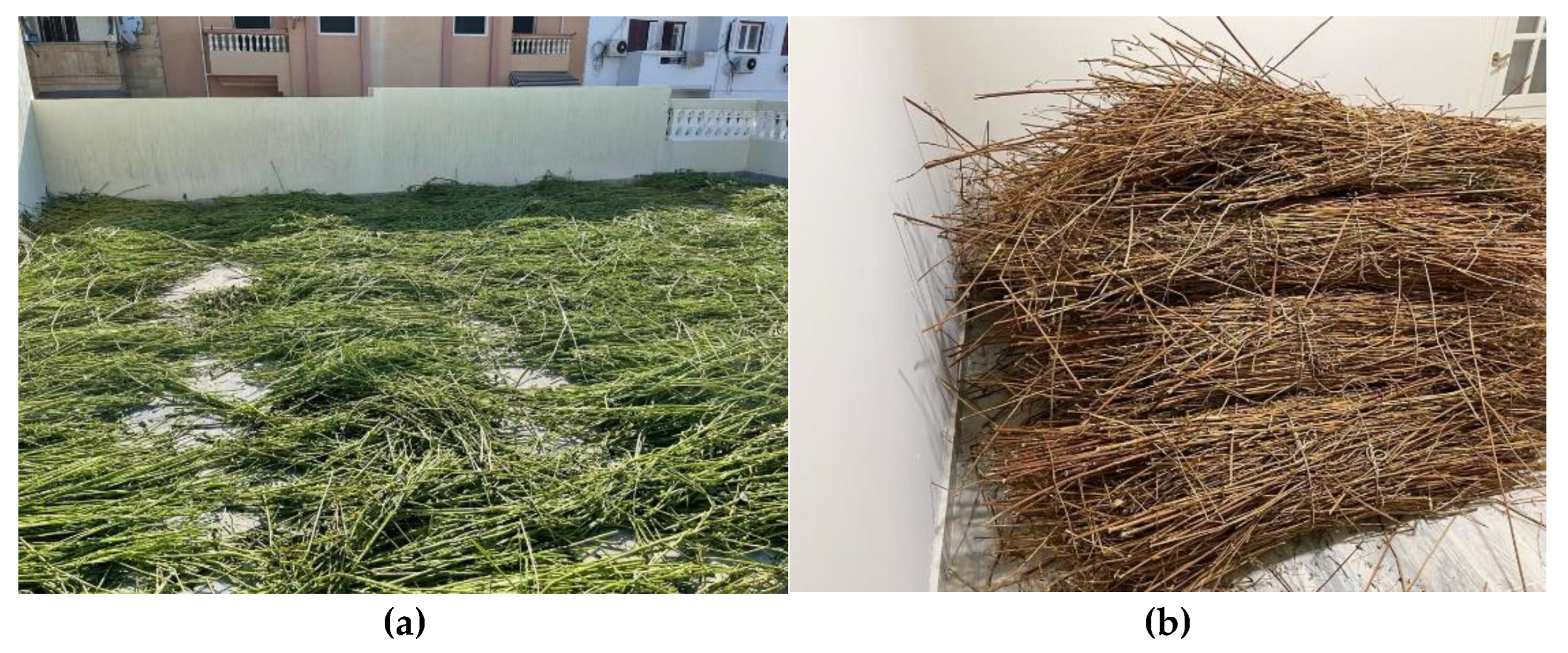
Figure 3.
Biological Retting Process degrade pectin within the stem bundles.
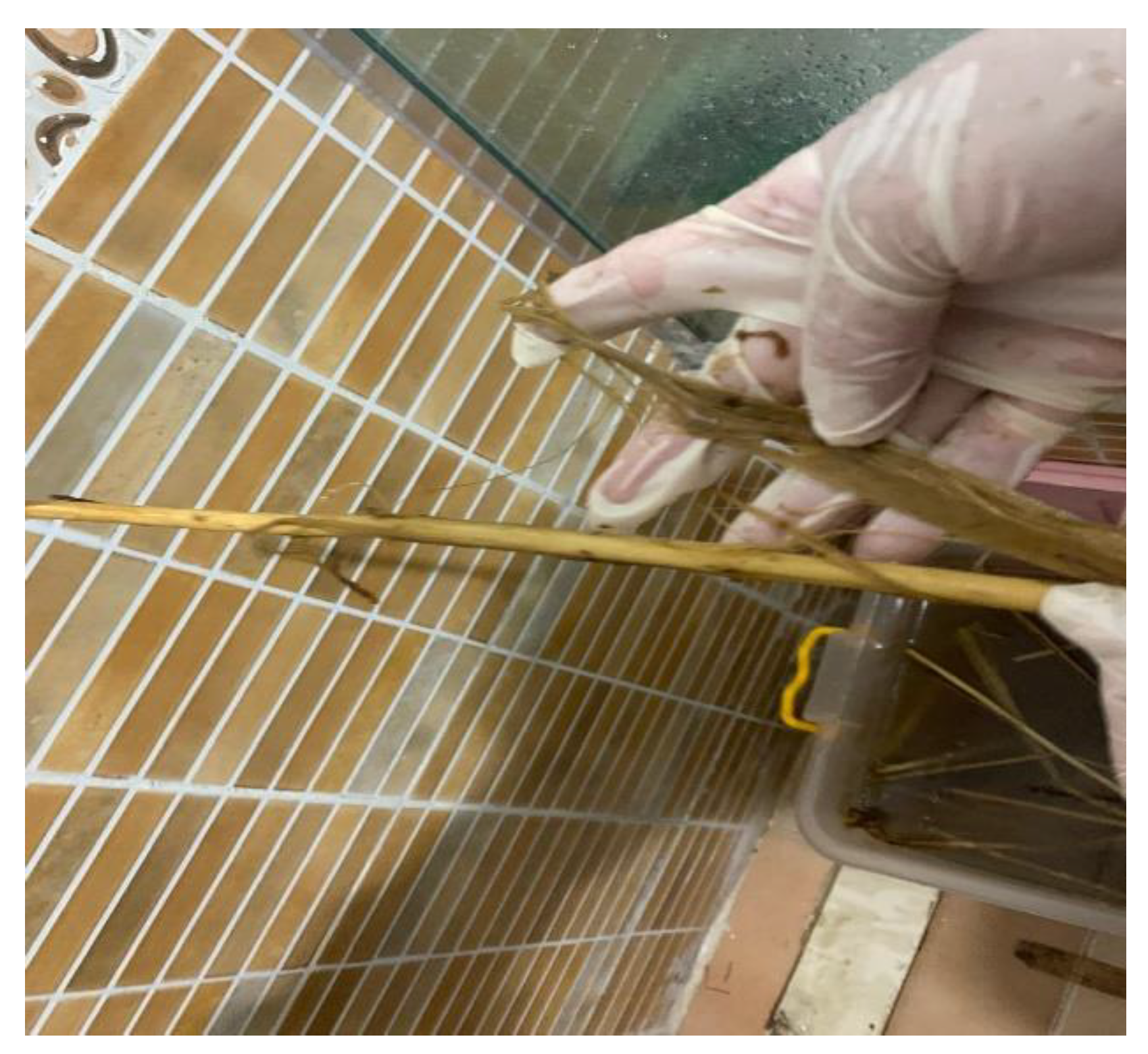
Figure 4.
Chemical Extraction Method (Cold Alkaline Treatment).
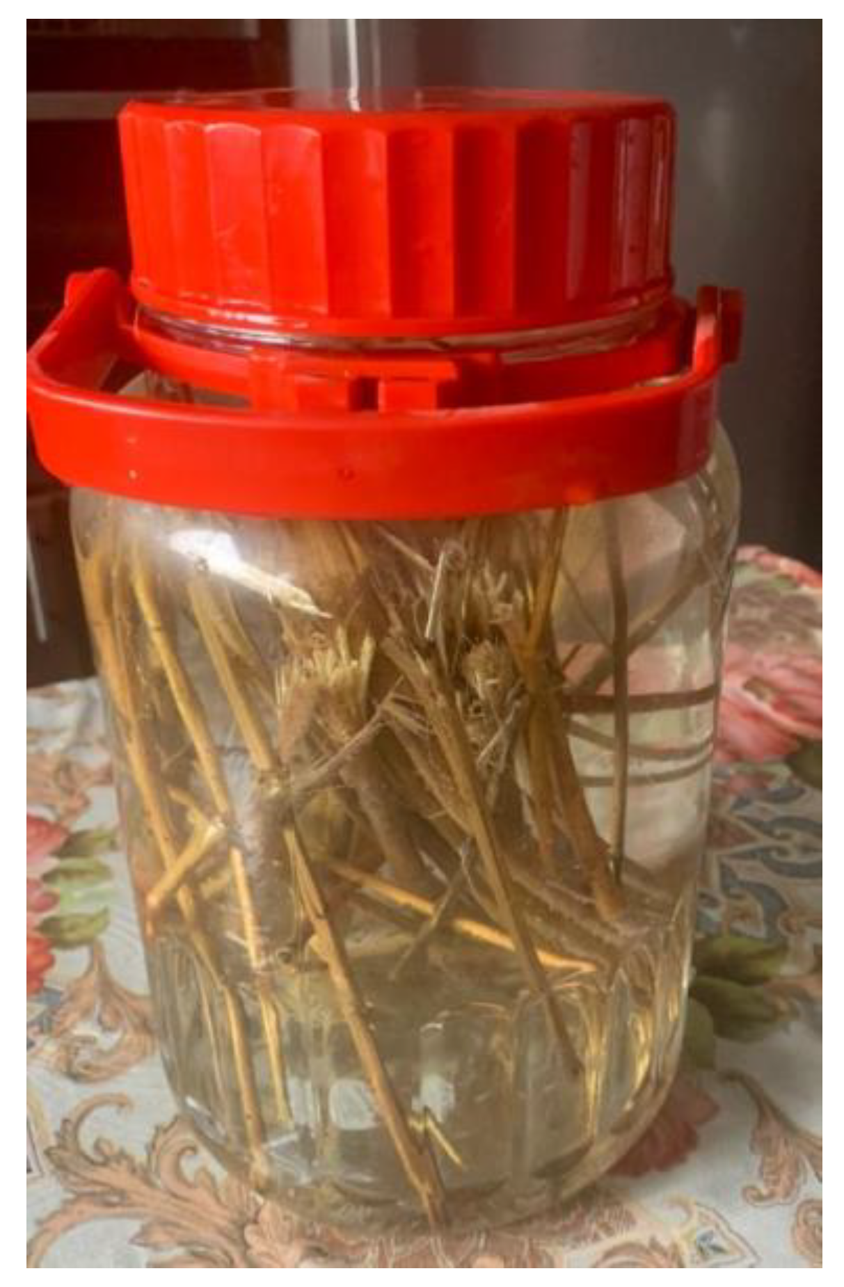
Figure 5.
Manual Extraction (Scraping Method) .
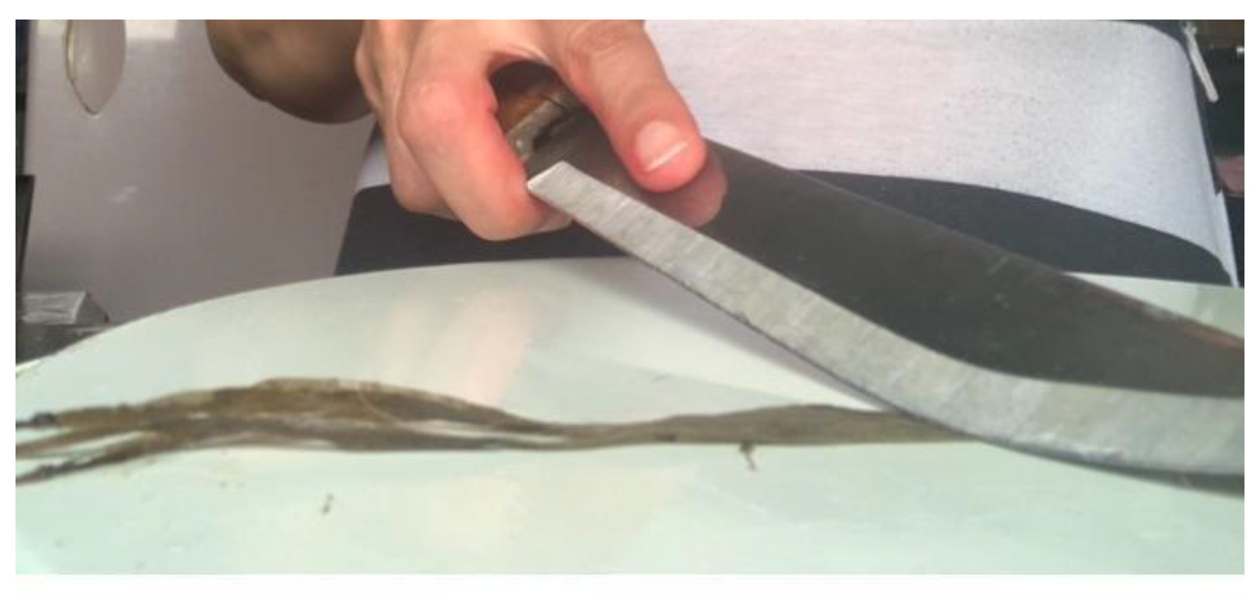
Figure 6.
Pictorial view of the process of extracting fibers (a) Collecting Molokhia plants (b) Remove the leaves and dry in the sun (c) Stems after drying (d) three different extraction methods (e) Skin degradation (f) Removal of skin to expose fibers. (g) Fibers obtained following the rinsing process (h) extracted fibers after Drying. (i) Combing the fiber. (j) extracted fibers after Combing.
Figure 6.
Pictorial view of the process of extracting fibers (a) Collecting Molokhia plants (b) Remove the leaves and dry in the sun (c) Stems after drying (d) three different extraction methods (e) Skin degradation (f) Removal of skin to expose fibers. (g) Fibers obtained following the rinsing process (h) extracted fibers after Drying. (i) Combing the fiber. (j) extracted fibers after Combing.
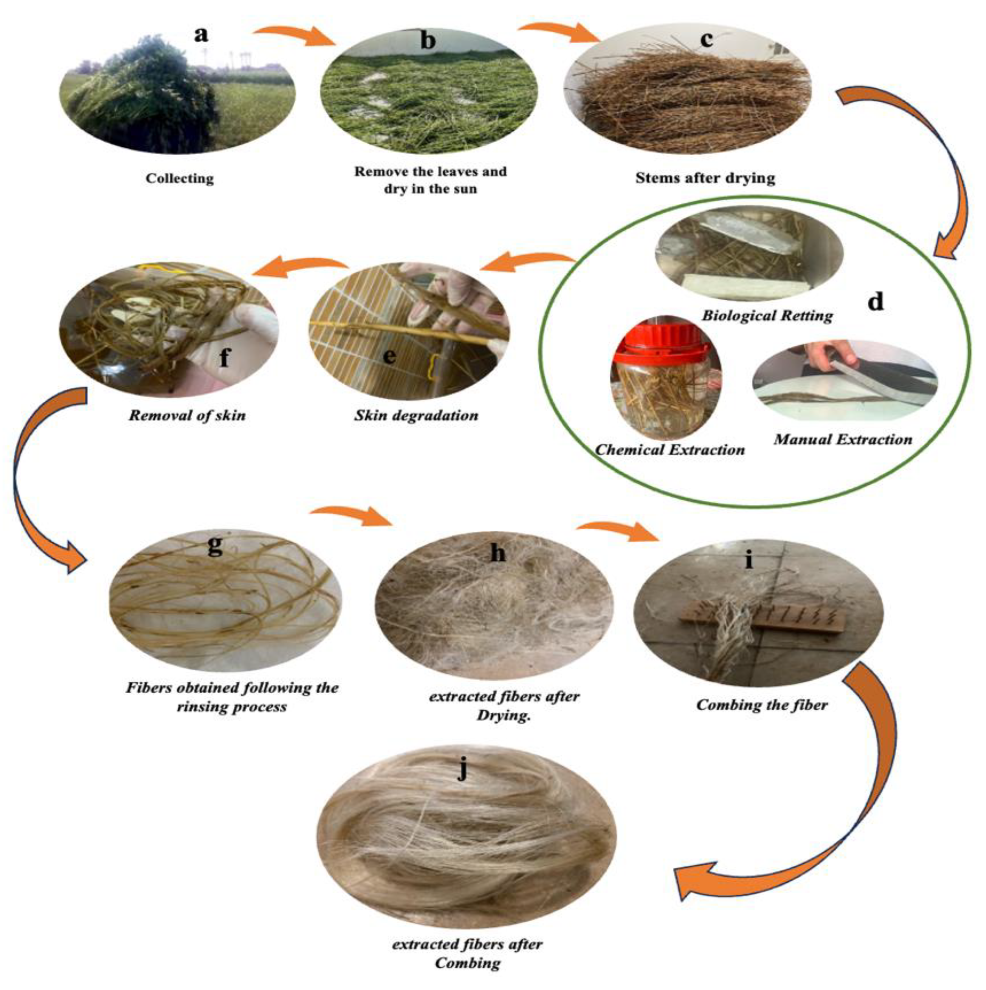
Figure 7.
SEM micrographs of Molokhia fibers obtained through different extraction methods. (a, b, e) Fibers extracted via biological retting, shown at longitudinal views with magnifications of 1000×, 2000×, and 70×, respectively. (c, f) Fibers extracted via chemical treatment, both captured at 1000× magnification. (d, g) Fibers extracted via manual scraping, displayed at 1000× and 200× magnifications, respectivel.
Figure 7.
SEM micrographs of Molokhia fibers obtained through different extraction methods. (a, b, e) Fibers extracted via biological retting, shown at longitudinal views with magnifications of 1000×, 2000×, and 70×, respectively. (c, f) Fibers extracted via chemical treatment, both captured at 1000× magnification. (d, g) Fibers extracted via manual scraping, displayed at 1000× and 200× magnifications, respectivel.
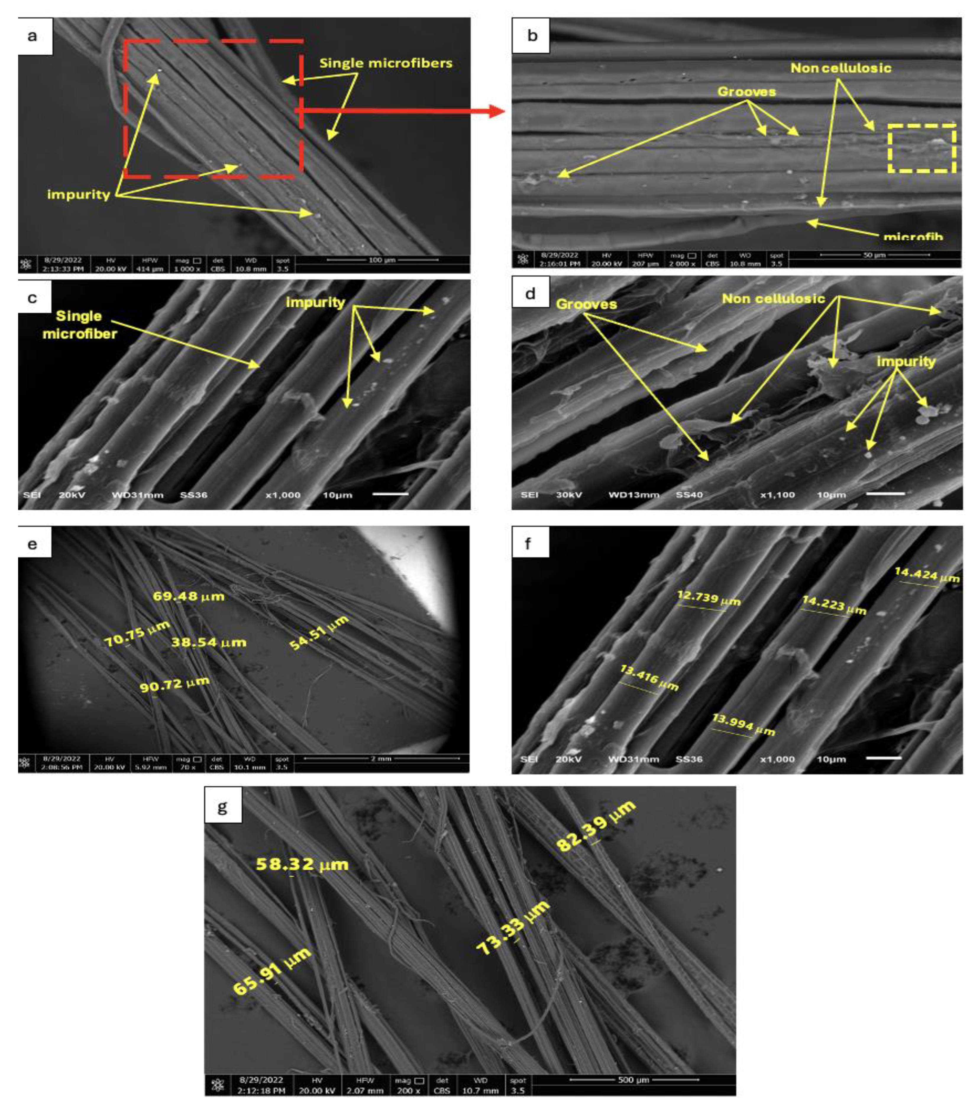
Figure 8.
FTIR spectra of (a) Egyptian raw molokhiya fibers biological extraction, (b) Egyptian raw Molokhia fibers chemical extraction, (c) Egyptian raw molokhiya fibers manual extraction.
Figure 8.
FTIR spectra of (a) Egyptian raw molokhiya fibers biological extraction, (b) Egyptian raw Molokhia fibers chemical extraction, (c) Egyptian raw molokhiya fibers manual extraction.
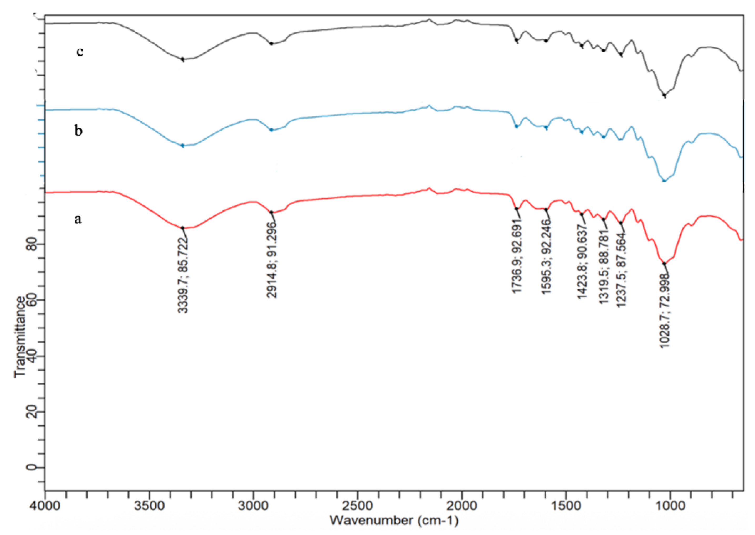
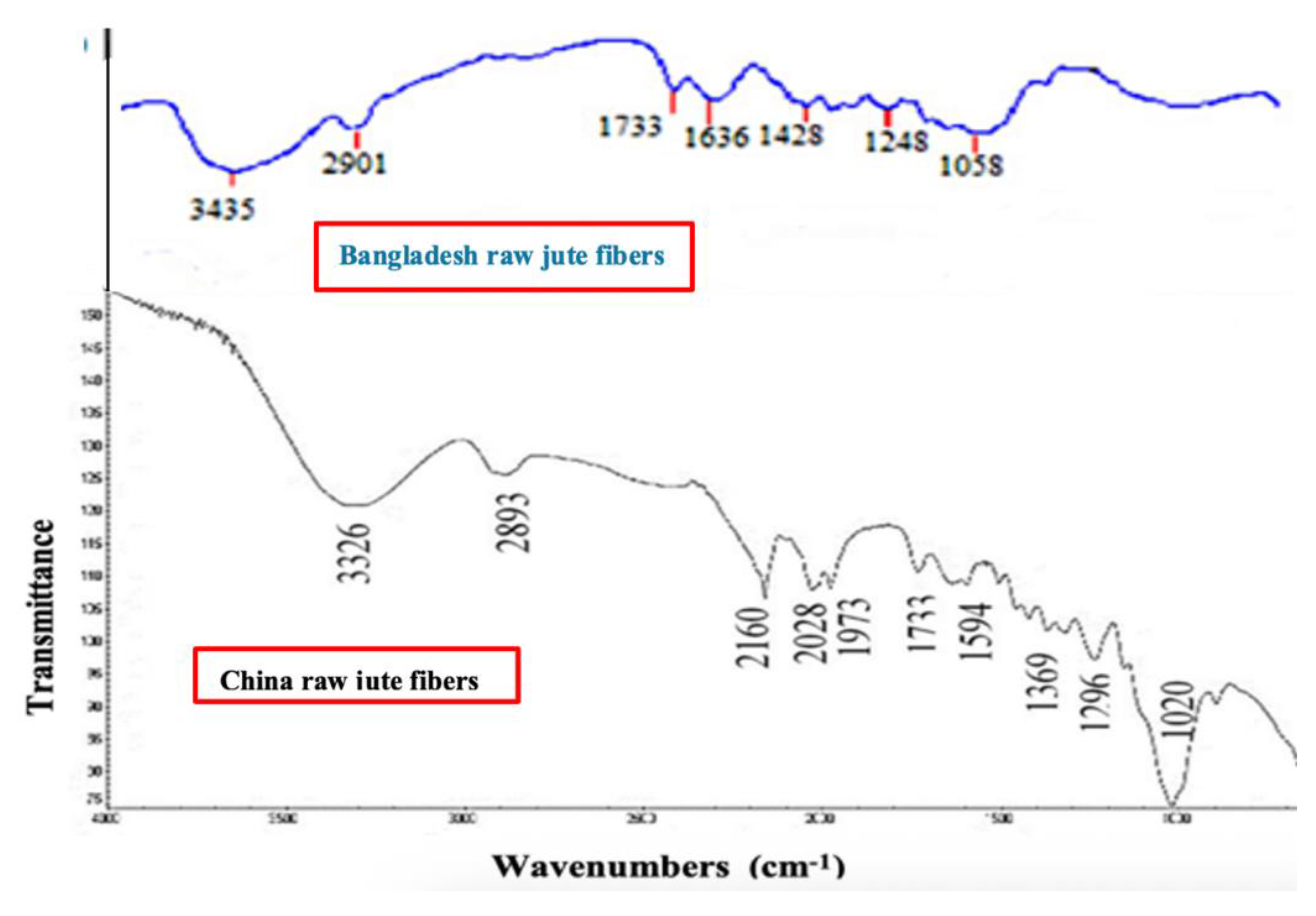
Figure 10.
Structure of jute (a) cellulose, (b) hemicellulose, and (c) lignin [40].
Figure 10.
Structure of jute (a) cellulose, (b) hemicellulose, and (c) lignin [40].
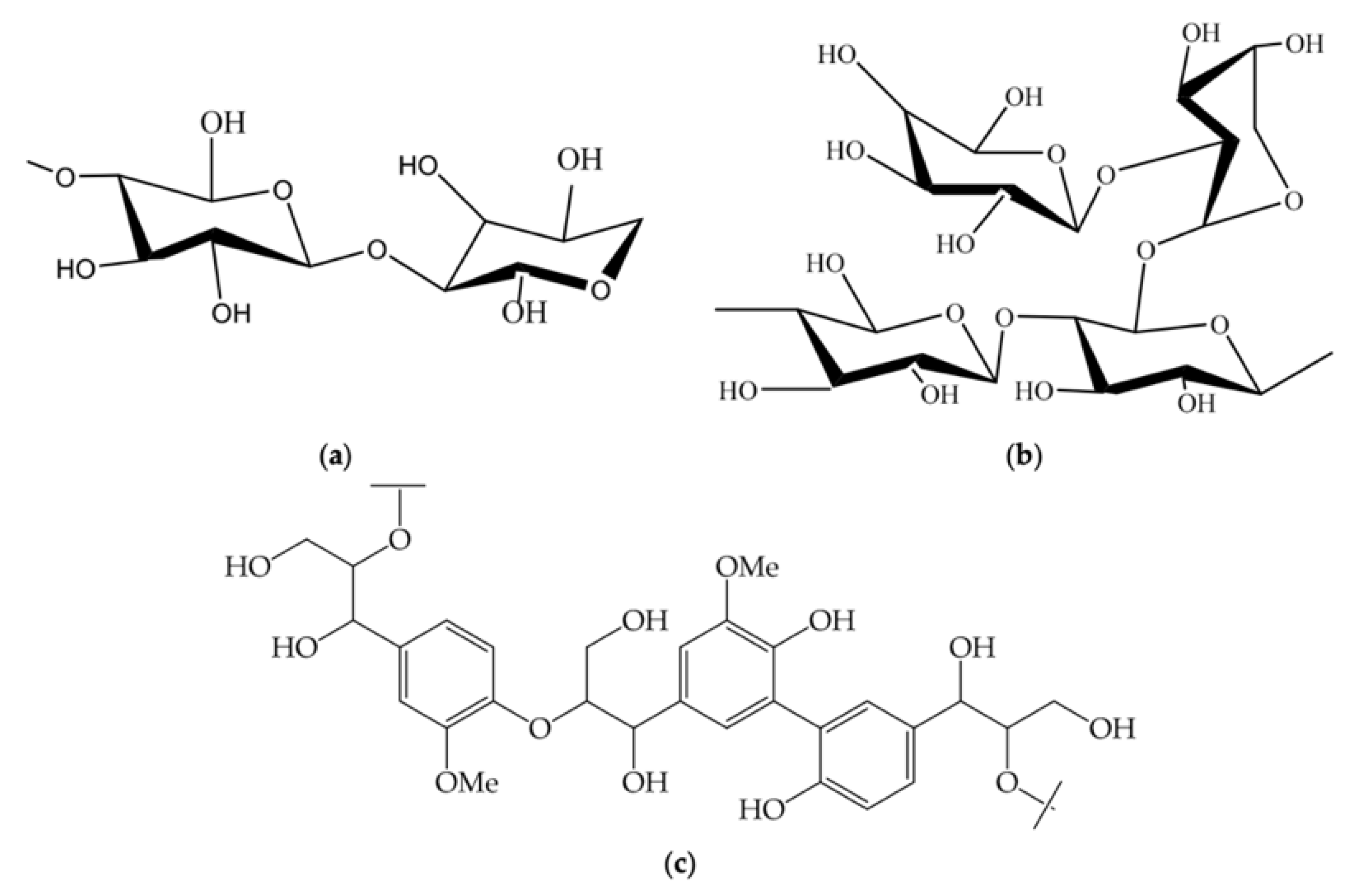
Figure 11.
X-ray diffractograms (XRD) of Egyptian molokhiya fibers (biological extraction, chemical extraction, manual extraction).
Figure 11.
X-ray diffractograms (XRD) of Egyptian molokhiya fibers (biological extraction, chemical extraction, manual extraction).
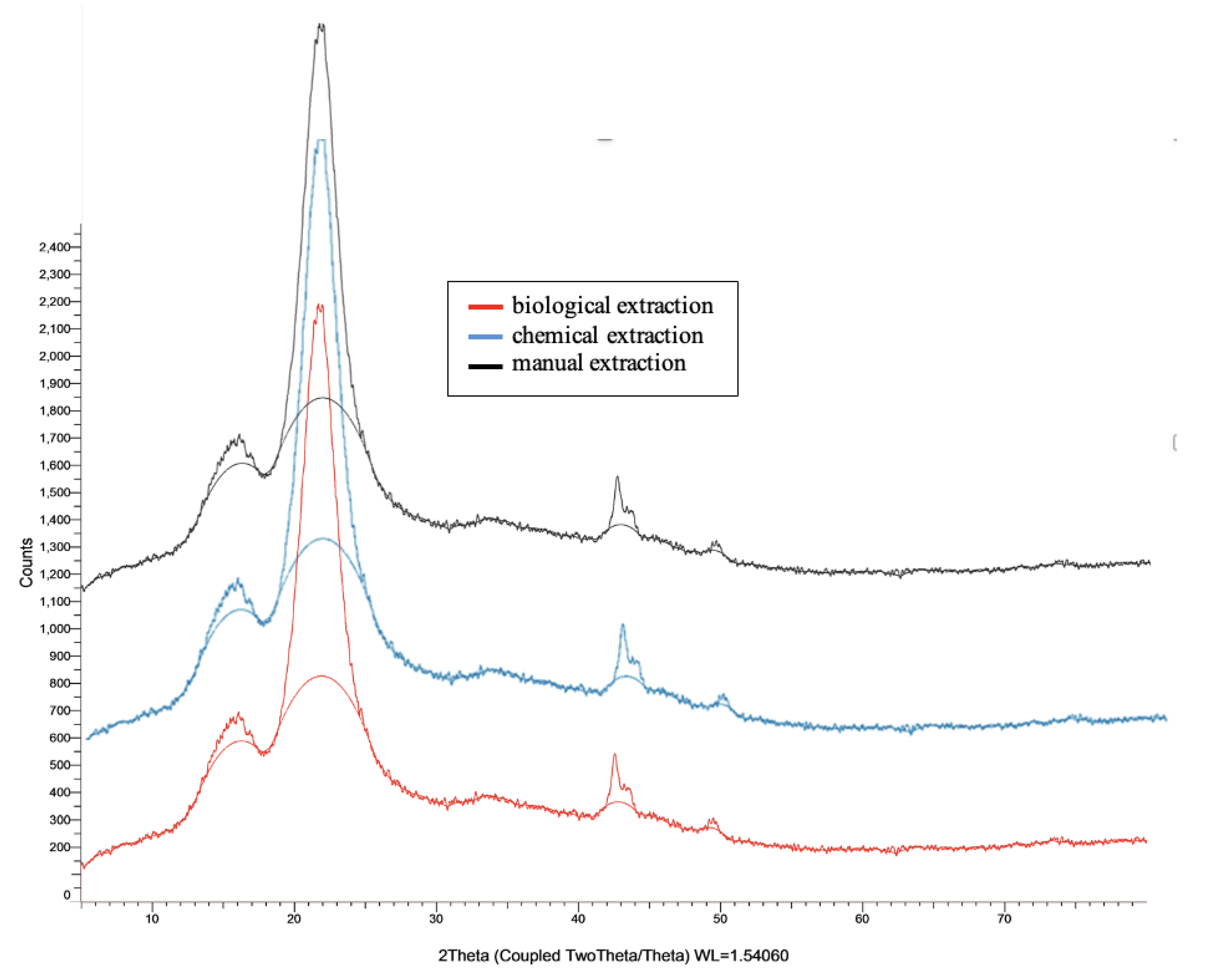
Figure 12.
X-Ray diffraction of (a) raw and treated jute fibers [61], (b) hemp fibers [62], and (c) pineapple raw fibers [63].
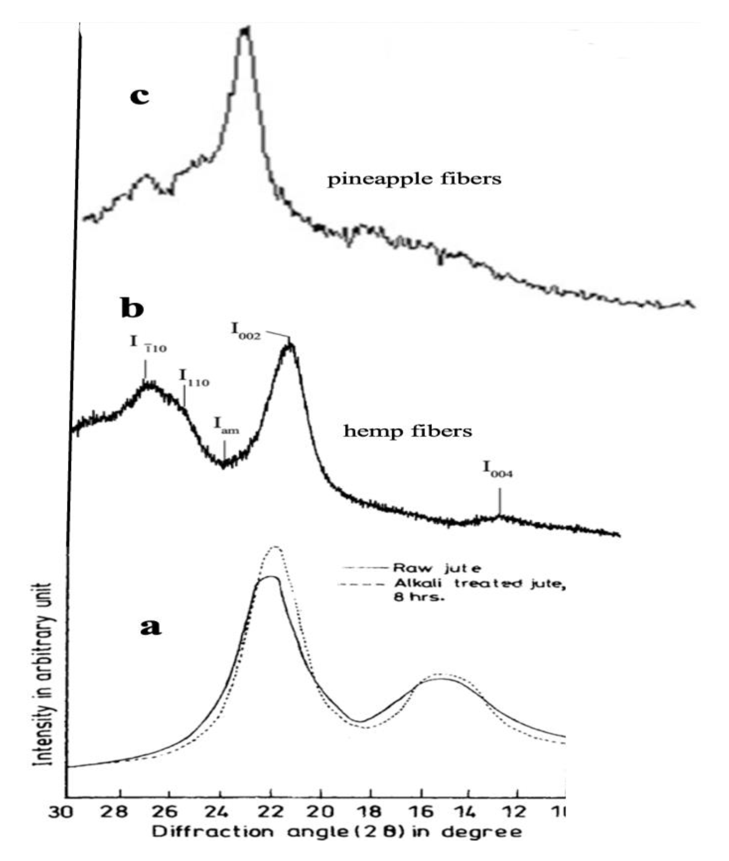
Figure 13.
distinct mean variations with limited dispersion (±SD) among groups.
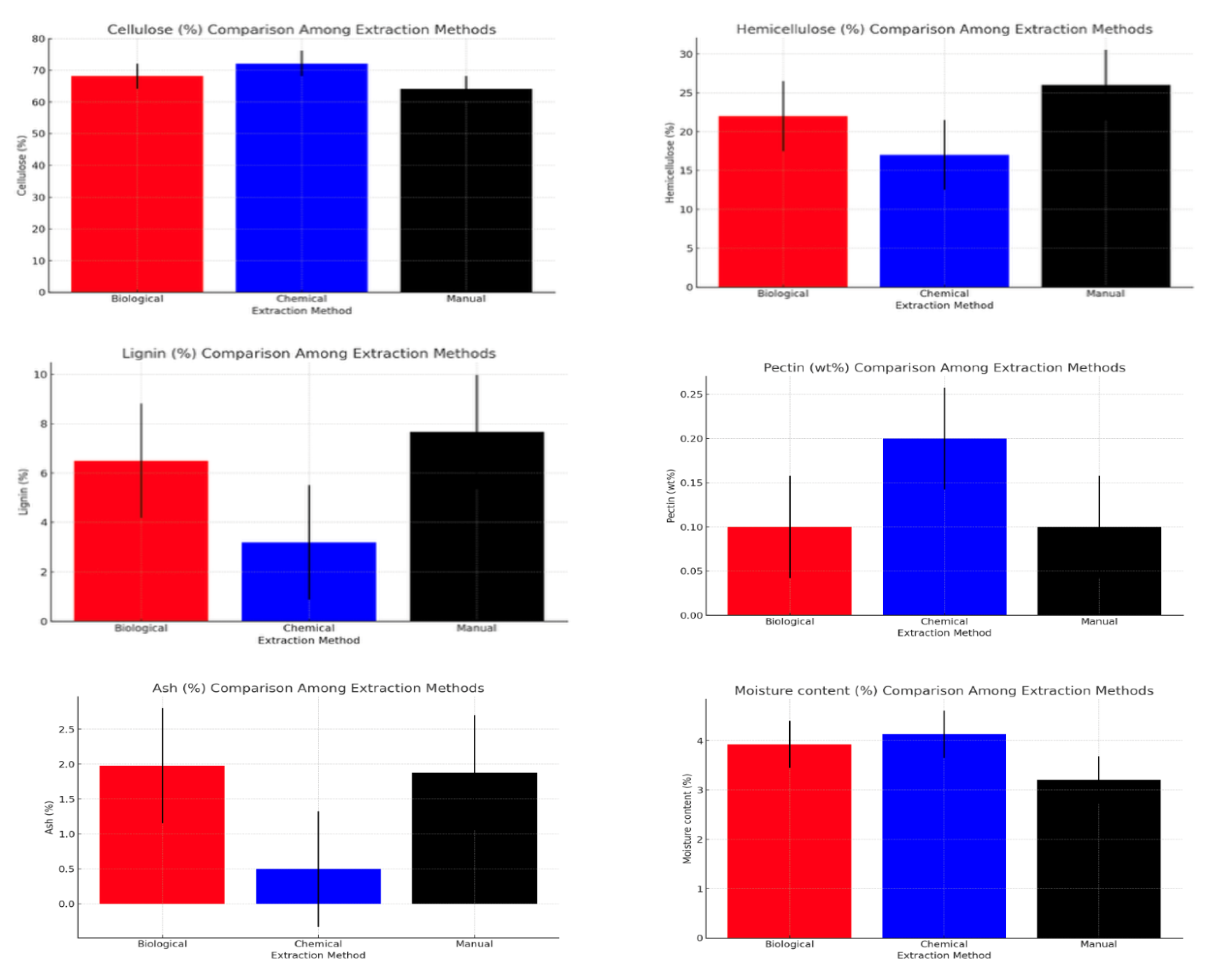
Figure 14.
the stress-strain curves of Egyptian Molokhia fibers extracted by three different methods.
Figure 14.
the stress-strain curves of Egyptian Molokhia fibers extracted by three different methods.
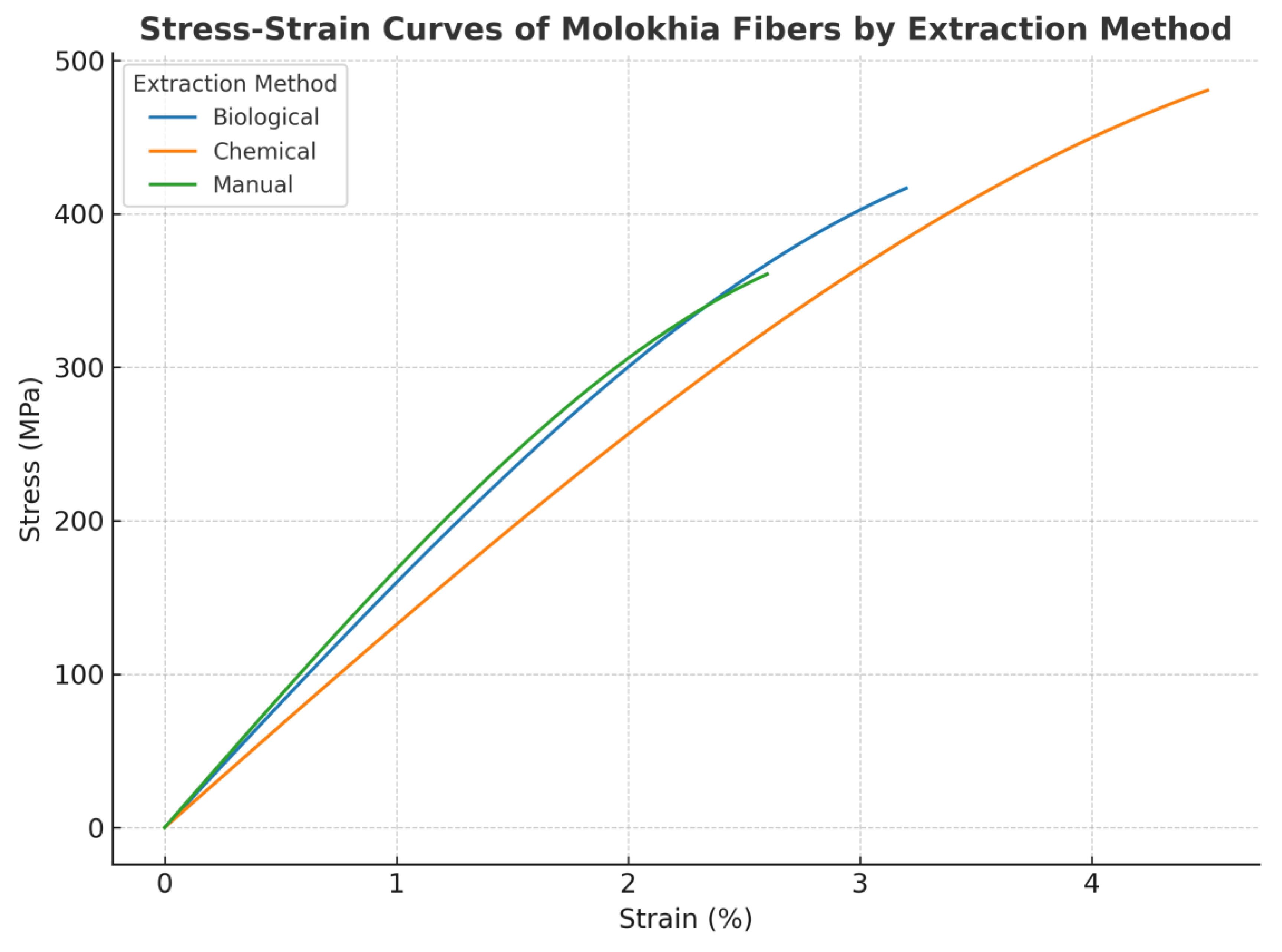
Figure 15.
boxplot charts (showing data distribution and variance) and bar charts (highlighting mean values with standard deviation).
Figure 15.
boxplot charts (showing data distribution and variance) and bar charts (highlighting mean values with standard deviation).
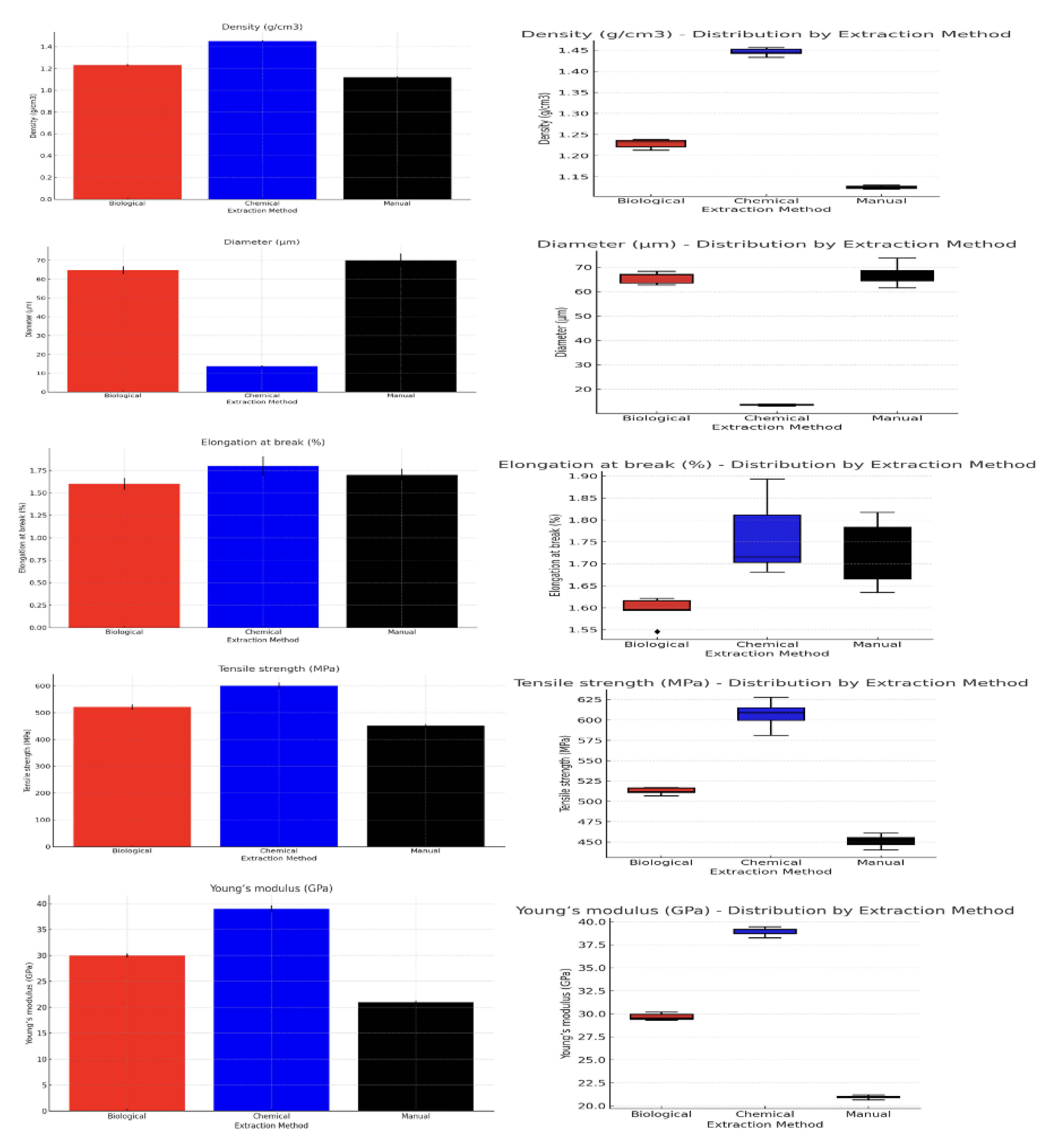
Figure 16.
Weight Loss by Extraction Method and Duration.
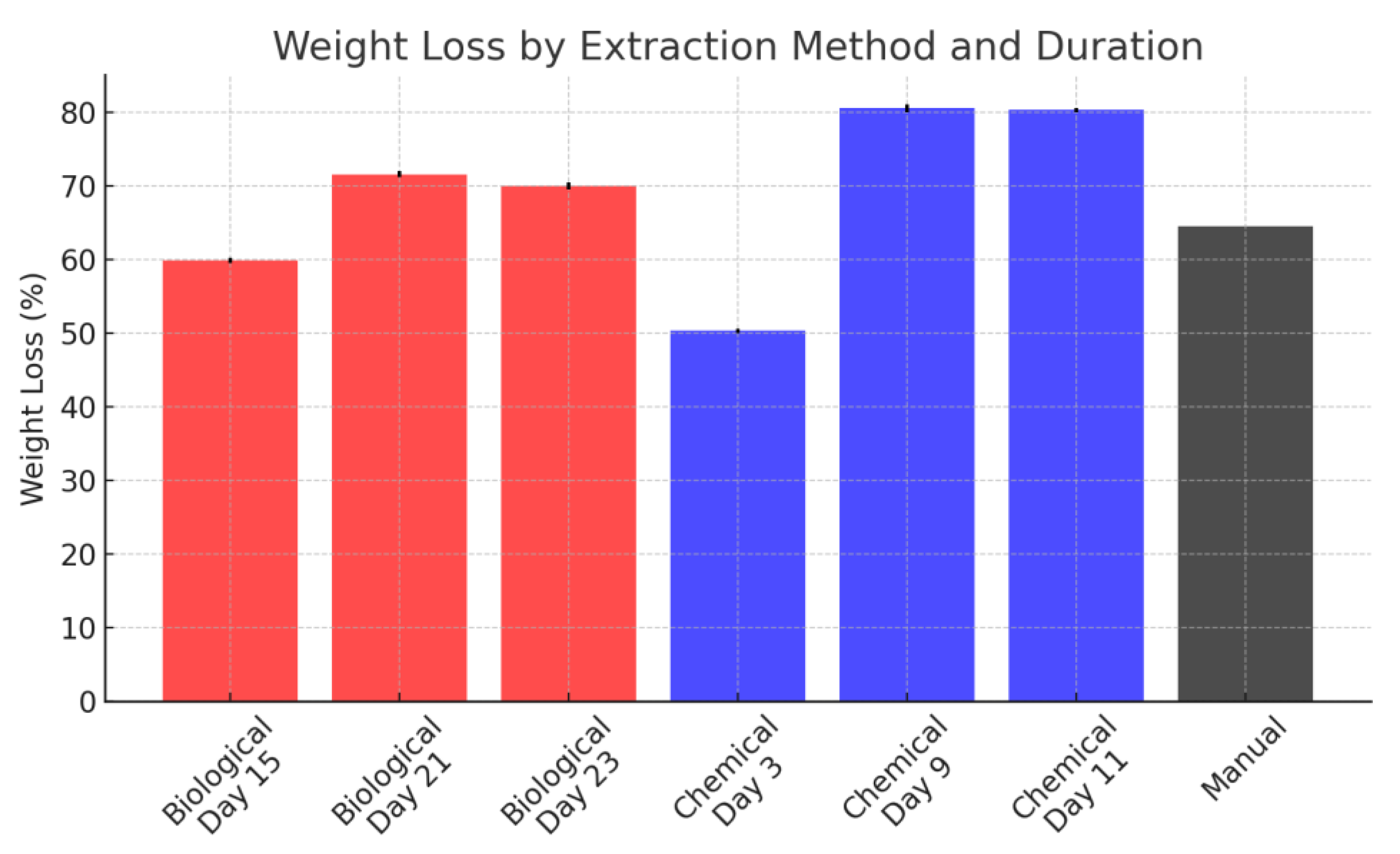
Figure 17.
Comparison of measured heavy metal concentrations in Molokhia bast fibers with the permissible limits defined by Oeko-Tex Standard 100.
Figure 17.
Comparison of measured heavy metal concentrations in Molokhia bast fibers with the permissible limits defined by Oeko-Tex Standard 100.
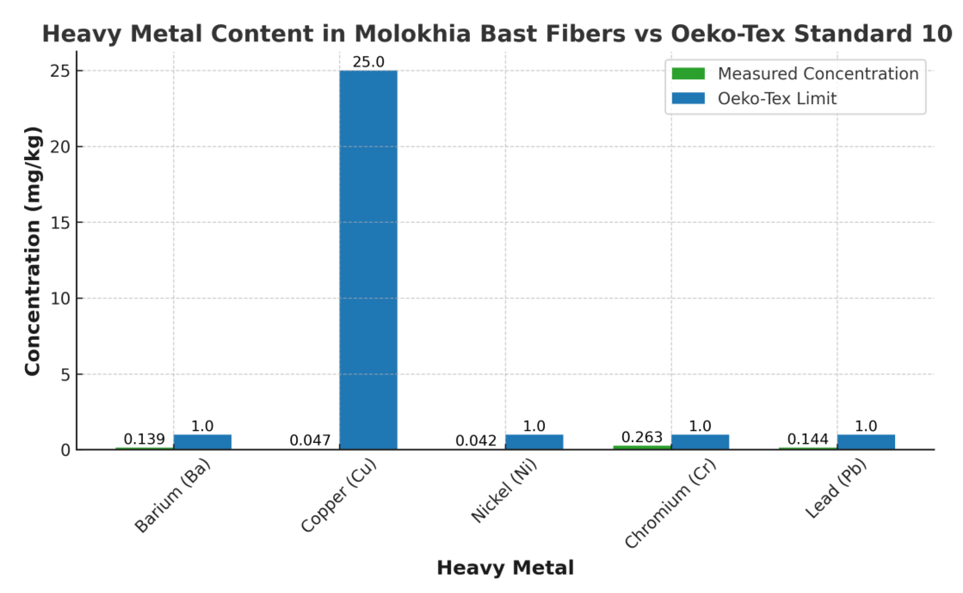
Figure 18.
Thermal Degradation Analysis of Molokhia Fibers.
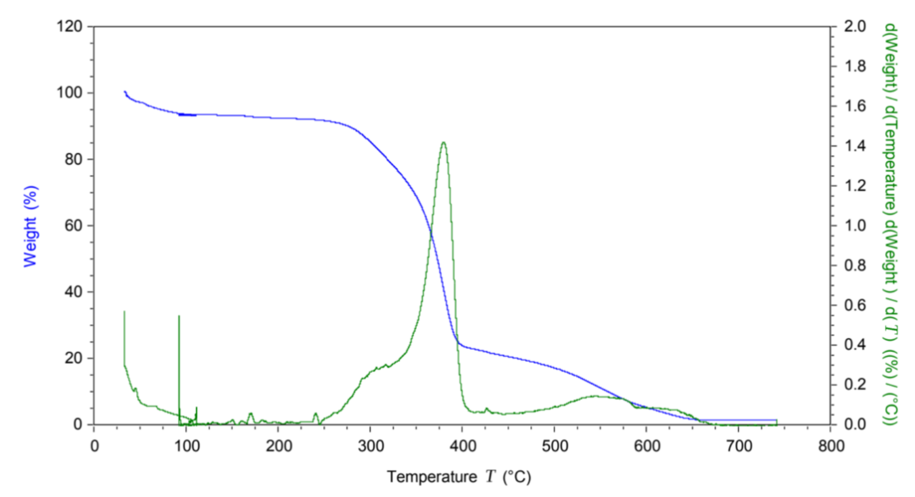
Figure 19.
Recommended applications for Molokhia fibers based on extraction method.
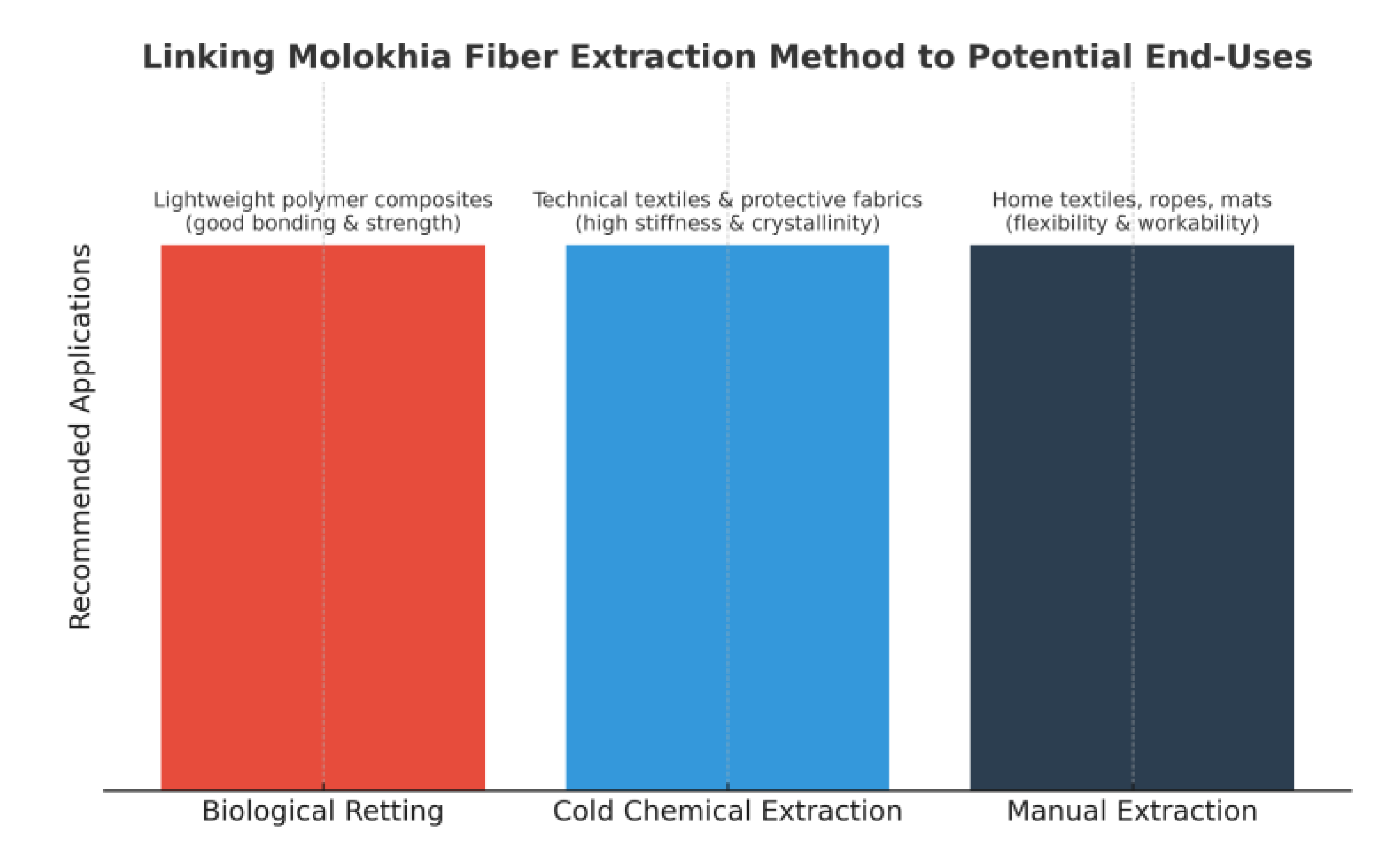

Table 1.
Oeko-Tex Standard 100 limit values (mg kg−1).
| Heavy metals | Baby wear |
With skin contact |
Without skin contact | Decoration material |
| Arsenic (As) | 0.2 | 1.0 | 1.0 | 1.0 |
| Barium (Ba) | 1000 | 1000 | 1000 | 1000 |
| Cadmium (Cd) | 0.1 | 0.1 | 0.1 | 0.1 |
| Cobalt (Co) | 1.0 | 4.0 | 4.0 | 4.0 |
| Cr (Vl) ( Chromium (Vl) / Chrom (Vl)) | 0.5 | 0.5 | 0.5 | 0.5 |
| Chromium (Cr) | 1.0 | 2.0 | 2.0 | 2.0 |
| Copper (Cu) | 25.0 | 50.0 | 50.0 | 50.0 |
| Mercury (Hg) | 0.02 | 0.02 | 0.02 | 0.02 |
| Nickel (Ni) | 1.0 | 4.07 | 4.0 | 4.0 |
| Lead (Pb) | 0.2 | 1.0 | 1.0 | 1.0 |
| Antimony (Sb) | 30.0 | 30.0 | 30.0 | |
| Se (Selenium) | 100 | 100 | 100 | 100 |
Table 2.
List of band assignments for FTIR spectra.
| Wavenumber cm−1 | Functional group | Assignment | References |
| 3339.69470 | ν(OH) (hydrogen bonded) | broad, strong bond from the cellulose, hemicelluloses, and lignin | [44] |
| 2914.77819 | C–H methyl and methylene groups Stretching vibrations of CH2 & CH3 | Cellulose | [44,45] |
| 1736.93943 | C=O stretching (lipids) ν(C=O) most probably from the lignin and hemicelluloses |
lignin, hemicelluloses | [37,46] |
| 1595.30059 | Ring C-C stretch of phenyl (2) | Lignin | [46] |
| 1423.84305 | δ (C–H) | cellulose, hemicelluloses | [47,48] |
| 1319.47759 | δ CH2 (wagging) at C–6 | Cellulose | [49] |
| 1237.47616 | PO2 asymmetric (phosphate I) δ COH in plane at C–6, O–H phenolic |
Cellulose | [46-49) |
| 1028.74524 | Glycogen absorption due to C-O and C-C stretching and C-O-H deformation motions | Cellulose | [45,49] |
Table 3.
Comparison of Crystallinity Index and Maximum XRD Peak Intensity for Molokhia fibers and Natural Fibers.
Table 3.
Comparison of Crystallinity Index and Maximum XRD Peak Intensity for Molokhia fibers and Natural Fibers.
| Fiber Type | Extraction Method | Crystallinity Index (%) | Max XRD Peak Intensity (Counts) | Ref. |
| Molokhia | Biological | 60.0 | ~2200 | |
| Molokhia | Cold Chemical | 70.0 | ~2500 | Present work |
| Molokhia | Manual | 64.0 | ~2700 | |
| Ramie | — | 74.5 | ~5200 | |
| Jute | — | 71.5 | ~5000 | |
| Flax | — | 65.2 | ~4700 | [56,57,58] |
| Hemp | — | 70.0 | ~4800 | |
| Kenaf | — | 59.3 | ~4300 | |
| Sisal | — | 67.0 | — (not reported) |
Table 4.
Comparison of the chemical composition of Egyptian Molokhia fibers with that of common natural lignocellulosic fibers.
Table 4.
Comparison of the chemical composition of Egyptian Molokhia fibers with that of common natural lignocellulosic fibers.
| Fiber | Cellulose (%) | Hemi cellulose (%) | Lignin (%) | Pectin (wt%) | Ash (%) | Moisture content (%) | Ref. |
| Molokhia Biological Retting Extraction | 68.17 ± 4.04 (n = 5) (b) | 22.00 ± 4.51 (n = 5) (b) | 6.50 ± 2.31 (n = 5) (b) | 0.10 ± 0.06 (n = 5) | 1.98 ± 0.83 (n = 5) | 3.93 ± 0.48 (n = 5) | Present work |
|
Molokhia Chemical Extraction |
72.23 ± 4.04 (n = 5) (a) | 17.00 ± 4.51 (n = 5) (c) | 3.20 ± 2.31 (n = 5) (c) | 0.20 ± 0.06 (n = 5) | 0.50 ± 0.83 (n = 5) | 4.13 ± 0.48 (n = 5) | |
| Molokhia Manual Extraction | 64.16 ± 4.04 (n = 5) (c) | 26.00 ± 4.51 (n = 5) (a) | 7.66 ± 2.31 (n = 5) (a) | 0.10 ± 0.06 (n = 5) | 1.88 ± 0.83 (n = 5) | 3.21 ± 0.48 (n = 5) | |
| Grewia flavescens | 58.46 | N/A | N/A | N/A | N/A | N/A | [12] |
| Ficus benjamina L. Aerial Root | 40.13 | 15.22 | 15.31 | 6.86 | N/A | N/A | [13] |
| Martynia annua stem | 52.56 | 12.34 | 11.56 | N/A | N/A | N/A | [14] |
| Cotton | 90 | 6 | 0 | 0-1 | 0 | 7.5 | [74,75] |
| Flax | 71-75 | 18.6- 20.6 | 2.2 | 2.3 | 2.3 | 10.0 | [76] |
| Hemp | 67- 75 | 16- 18 | 3.0- 5.0 | 0.9 | 1.23 | 10.4 | [77,78] |
| Nettle | 78- 85 | 9- 10 | 2-5 | 0.6 | - | 12.3 | [79] |
| Sisal | 60- 67 | 10- 15 | 8- 12 | 10 | 0.6- 1 | 9-11 | [80,81] |
| Ramie | 80- 85 | 3- 4 | 0.5 | 1.9 | - | 9.9 | [82,83] |
| Jute | 60 | 22.1 | 15.9 | 0.2 | 1.0 | 10.4 | [84,85] |
| Bamboo | 33-45 | 30& | 20- 25 | - | - | 10-16 | [86,87] |
| Banana | 60-65 | 6-8 | 5- 10 | - | 1.2 | 12.3 | [86,88] |
| Rice straw | 51- 70 | - | 12- 16 | - | 15- 20 | 18 | [89,90] |
| Pineapple | 80 | - | 12 | 0 | 11.8 | [91,92,93] | |
| Coir | 43-53 | 14.7 | 38.3- 40.7 | 5.2- 16 | - | 13 | [84,94] |
| Bagasse | 32-44 | 27- 32 | 19- 24 | - | 4.26 | 49 | [88] |
(N/A)not available,(n)Number of samples, Different letters (a–c) within the same column indicate statistically significant differences (p < 0.05) according to Tukey’s HSD test.
Table 5.
ANOVA Results for the chemical composition of Egyptian Molokhia fibers Extraction Methods.
| Property | F-Value | p-Value |
|---|---|---|
| Cellulose (%) | 16.08 | 0.001 |
| Hemicellulose (%) | 15.67 | 0.0011 |
| Lignin (%) | 15.26 | 0.0012 |
| Pectin (wt%) | 5.00 | 0.0397 |
| Ash (%) | 13.54 | 0.0016 |
| Moisture content (%) | 9.86 | 0.0044 |
Table 6.
Physical and mechanical properties of Egyptian molokhia fibers and various natural fibers.
| Type of fiber | Density (g/cm3) | Elongation at break (%) | Tensile strength (MPa) | Young’s modulus (GPa ) | Diameter (μm) | Ref. |
| Molokhia Biological Retting Extraction | 1.23 ±0.01 (n = 5) |
1.6 ±0.07 (n = 5) | 520.87 ± 9.58 (n = 5) (b) |
29.955 ± 0.39 (n = 5) (b) |
64.80 ±2.05 (n = 5) (b) |
Present work |
|
Molokhia Chemical Extraction |
1.4 5± 0.01. (n = 5) | 1.8 ±0.10 (n = 5) | 600.67± 11.73 (n = 5) (a) |
38.955 ± 0.64 (n = 5) (a) |
13.76±0.44 (n = 5) (a) |
|
| Molokhia Manual Extraction | 1.12 ± 0.01 (n = 5) | 1.7 ±0.07 (n = 5) | 450.89 ±7.34 (n = 5) (c) | 20.955 ±0.34 (n = 5) (c) | 69.99 ±3.59 (n = 5) (b) | |
| Inkberry | 1400 | 2.37 | 146.5 | 24.8 | N/A | [7] |
| Reddish shell bean | 1580 | 1.83 | 111 | 6.11 | 785.87 | [8] |
| Sambucus ebulus L. plant fiber | 1.080 | 7.72 | 50.68 | 6.56 | 0.76 | [9] |
| Beetroot plant | N/A | 7.72 | 50.68 | 52.39 | 284.04 | [10] |
| Alcea rosea L | N/A | 2.47 | 80.96 | 3.28 | 320 | [11] |
| Grewia flavescens | 1.1–1.3 | N/A | 50.3–73.1 | N/A | 507–629 | [12] |
| Ficus benjamina L. Aerial Root | 0.27 | N/A | 77.7 | N/A | N/A | [13] |
| Martynia annua stem | N/A | 1.17 ± 0.02 | 417.5 ± 7.1 | 17.5 ± 1.6 | N/A | [14] |
| Cotton | 1.21 | 3–10 | 287–597 | 5.5–12.6 | 12–22 |
[75,76,101,102,103] |
| Flax | 1.4–1.5 | 1.2–3.2 | 345–1500 | 27.6–80 | 15–20 | |
| Hemp | 1.48 | 1.6 | 550–900 | 70 | 16–50 | |
| Nettle | 1.51 | 1.7 | 650 | 38 | 20–40 | |
| Sisal | 1.33–1.5 | 2–14 | 400–700 | 9–38 | 100–300 | |
| Ramie | 1.5 | 2–3.8 | 220–938 | 44–128 | 25–30 | |
| Jute | 1.3–1.46 | 1.5–1.8 | 393–800 | 10–30 | 20–200 | |
| Bamboo | 0.6–1.1 | 1.3–8 | 140–441 | 11–36 | 10–20 | |
| Banana | 1.35 | 5–6 | 529–914 | 27–32 | 80–250 | |
| Abaca | 1.5 | 2.9 | 430–813 | 33.1–33.6 | 100–250 | |
| Pineapple | 1.5 | 1–3 | 170–1627 | 60–82 | 20–80 | |
| Coir | 1.2 | 15–30 | 175–220 | 4–6 | 100-450 | |
| Kenaf | 1.2 | 2.7–6.9 | 295–930 | 22–60 | 20–40 |
(N/A(not available, (MPa)Megapascal, (GPa) Gigapascal, (μm) Micron, (n) Number of samples. Different letters (a–c) within the same column indicate statistically significant differences (p < 0.05) according to Tukey’s HSD test.
Table 7.
ANOVA Results for the morphology and mechanical composition of Egyptian Molokhia fibers Extraction Methods.
Table 7.
ANOVA Results for the morphology and mechanical composition of Egyptian Molokhia fibers Extraction Methods.
| Property | F-Value | p-Value |
|---|---|---|
| Density (g/cm3) | 2131.890 | 4.441e-16 |
| Elongation at break (%) | 7.617 | 0.007317 |
| Tensile strength (MPa) | 297.430 | 5.978e-11 |
| Young’s modulus (GPa) | 1799.283 | 1.332e-15 |
| Diameter (μm) | 839.382 | 1.278e-13 |
Table 8.
weight loss percentages across various fiber extraction methods.
|
Molokhia Biological Retting Extraction |
Molokhia Chemical Extraction | Molokhia Manual Extraction | |||||
|---|---|---|---|---|---|---|---|
| Retting time (day) | 15 | 21 | 23 | 3 | 9 | 11 | - |
| weight loss% | 59.70 ±0.35 | 71.40 ±0.41 | 70.50 ±0.46 | 50.72 ±0.33 | 80.70 ±0.53 | 80.53 ±0.30 | 64.60 |
Table 9.
ANOVA analysis for weight loss percentage was analyzed across different extraction methods.
Table 9.
ANOVA analysis for weight loss percentage was analyzed across different extraction methods.
| Method | F-Value | p-Value |
|---|---|---|
| Biological | 1281.08 | 1.0263e-14 |
| Chemical | 13314.54 | 8.3517e-21 |
Table 10.
Heavy metal contents (mg kg−1) in Egyptian Molokhia fibers after wet digestion. (average of replicates ± standard deviation).
Table 10.
Heavy metal contents (mg kg−1) in Egyptian Molokhia fibers after wet digestion. (average of replicates ± standard deviation).
| Element | Heavy metal contents (mg kg−1) |
|---|---|
| Al | 1.419 ± 0.07 |
| B | nd |
| Ba | 0.1393 ± 0.002 |
| Cd | nd |
| Co | nd |
| Cr | 0.2633 ± 0.01 |
| Cu | 0.0473 ± 0.001 |
| Fe | 5.12 ± 0.30 |
| Mn | 0.0523 ± 0.001 |
| Mo | 0.0838 ± 0.002 |
| Ni | 0.0416 ± 0.001 |
| Pb | 0.1441 ± 0.002 |
| si | 0.7441 ± 0.04 |
| Sr | 11.16 ± 0.73 |
| V | 0.12 ± 0.002 |
| Zn | 0.2659 ± 0.01 |
| p | nd |
| Na | 8.12 ± 0.52 |
| K | 4.11± 0.28 |
nd: not detected.
Table 11.
Literature values of heavy metals (as mg kg−1) in textile fibers.
| Metals | Cotton | flax | hemp | Polyester | Nylon | Viscose | Ref. |
|---|---|---|---|---|---|---|---|
| Fe | 0.44–4.49 | — | — | 0.23–28.90 | 3.42–29.90 | 1.99–4.41 |
[115,116] |
| Cu | 0.26–0.78 | 2.1 ± 0.05 | 1.8 ± 0.08 | nd–0.36 | nd –0.48 | 0.26–11.20 | |
| Ni | 0.20–0.70 | — | — | 0.29–3.63 | 0.76–3.63 | 0.30–1.68 | |
| Pb | 0.28–0.30 | 7.2 ± 0.2 | 6.3 ± 0.2 | nd–0.76 | 0.31–3.76 | 0.40–0.80 | |
| Zn | 0.40–5.00 | 25.7 ± 0.4 | 18.9 ± 0.2 | 0.90–4.70 | nd–0.90 | 2.40–3.00 | |
| Cd | — | 3.34 ± 0.05 | 0.40 ± 0.002 | — | — | — | |
| Cr | 0.44–1.12 | — | — | nd–0.42 | nd | 0.22–0.90 |
[117] |
| Cu | 0.28–0.84 | — | — | 0.04–0.34 | 0.04–0.32 | 0.12–13.58 | |
| Ni | 0.24–1.52 | — | — | 0.20–0.24 | 0.24–0.26 | 0.16–0.98 | |
| Pb | 0.18–6.00 | — | — | 0.05–1.08 | 1.08–2.50 | 0.16–0.98 | |
| Zn | 0.42–2.16 | — | — | 0.92–4.04 | 0.14–0.92 | 1.48–3.46 | |
| Co | 0.04–0.96 | — | — | 0.08–0.16 | 0.12–0.16 | 0.01–0.06 | |
| Fe | 3.55–34.3 | — | — | — | — | — |
[118] |
| Cu | 0.76–341 | — | — | — | — | — | |
| Ni | 1.20–4.69 | — | — | — | — | — | |
| Zn | 0.63–4.84 | — | — | — | — | — | |
| Mn | 1.02–2.50 | — | — | — | — | — | |
| Cu | 0.05–0.21 | — | — | 0.05 | — | 0.05–0.06 |
[119] |
| Ni | 0.05–0.10 | — | — | 0.08 | — | 0.09–0.10 | |
| Mn | 0.03–0.05 | — | — | 1.17–2.17 | — | 0.31–0.36 | |
| Al | 0.11–0.17 | — | — | 0.27–0.29 | — | 0.21–0.29 |
nd: not detected; —: not examined.
Disclaimer/Publisher’s Note: The statements, opinions and data contained in all publications are solely those of the individual author(s) and contributor(s) and not of MDPI and/or the editor(s). MDPI and/or the editor(s) disclaim responsibility for any injury to people or property resulting from any ideas, methods, instructions or products referred to in the content. |
© 2026 by the authors. Licensee MDPI, Basel, Switzerland. This article is an open access article distributed under the terms and conditions of the Creative Commons Attribution (CC BY) license.
Copyright: This open access article is published under a Creative Commons CC BY 4.0 license, which permit the free download, distribution, and reuse, provided that the author and preprint are cited in any reuse.