Submitted:
17 April 2026
Posted:
21 April 2026
You are already at the latest version
Abstract
Reactive oxygen species (ROS)-mediated cancer therapy has attracted extensive attention due to its high spatiotemporal selectivity and minimum side-effects. Herein, we report a nonanuclear Pd-based coordination cage of Pd6-TMPP(Ni), constructed from Ni-chelated TMPP(Ni) as the metalloligand and Pd2+ ions (H2TMPP = meso-tetrakis (6-methylpyridin-3-yl) porphyrin). Pd6-TMPP(Ni) integrates dual ROS-generation for cancer therapy, viz., hydroxyl radical (•OH) production and photoinduced singlet oxygen (1O2) generation. In vitro cytotoxicity assays against cancer cell lines NCI-H82 (lung cancer), A549 (lung cancer), KYSE-510 (esophageal cancer), and Te-1 (esophageal cancer), reveal its potent dark toxicity (IC₅₀: 1.867–2.107 μmol L−1) and phototoxicity (IC₅₀: 0.835–1.528 μmol L−1), which is attributed to enhanced intracellular ROS accumulation. This work develops a versatile therapeutic platform that harnesses Ni-induced •OH for chemodynamic therapy (CDT) and porphyrin-generated ¹O₂ for photodynamic therapy (PDT), thereby mitigating the oxygen dependence of conventional PDT.
Keywords:
coordination cage
; reactive oxygen species
; photodynamic therapy
; chemodynamic therapy
; anticancer drugs
1. Introduction
Reactive oxygen species (ROS), particularly hydroxyl radical (•OH), singlet oxygen (1O2), and hydrogen peroxide (H2O2), have emerged as promising agents for cancer therapy due to their ability to induce oxidative stress and disrupt cellular homeostasis, offering distinct advantages in tumor selectivity and the potential to overcome conventional drug resistance [1,2]. Among various ROS-generating strategies, transition metal-catalyzed Fenton or Fenton-like reactions have garnered significant attention due to their ability to convert H2O2 into cytotoxic •OH [3,4,5]. Nickel complexes have demonstrated remarkable catalytic activity in this context [6,7,8]. The ability of Ni(II) to undergo reversible redox cycling with Ni(III) enables efficient decomposition of H2O2 to generate •OH radicals, a mechanism that has been extensively exploited in environmental remediation for the degradation of tough organic pollutants [7].
In the context of ROS-based cancer therapies, Ni(II)-induced •OH generation also offers an advantage as it can be produced by consumption of endogenously overexpressed H2O2 (typically 10–50 μM) in the tumor microenvironment (TME) [9,10]. This TME-dependent ROS production circumvents the O2 limitation and penetration depth limitations associated with conventional O2-dependent therapies such as type II photodynamic therapy (PDT) [11,12,13]. For example, NiCl2 has long been documented to induce lipid peroxidation and DNA damage via Fenton-like process [14]. Liu et al recently reported that nickel-based single-atom-metal-clusters (NSAMCs) exhibit peroxidase-like activity, catalyzing H2O2 in the TME to generate •OH, which synergizes with glutathione (GSH) depletion to boost ferroptosis [15].
Complementary to Ni-induced •OH production, photoinduced ROS generation, and 1O2 in particular, have become a cornerstone of PDT, an approach that relies on photosensitizers activated by specific light wavelengths [16,17]. Porphyrins and their derivatives are among the most widely used photosensitizers due to their strong absorption in the visible-near-infrared region and high 1O2 quantum yields [18,19]. However, their application is hindered by inherent limitations, including self-aggregation resulting from π–π stacking, poor water solubility, and low tumor targeting efficiency [20]. To address these challenges, researchers have integrated porphyrins into metal-organic frameworks or discrete coordination cages via coordination with metal ions [21,22,23,24]. These hybrid structures not only inhibit porphyrin stacking to preserve photosensitizer activity but also enhance ROS generation efficacy by optimizing light absorption, improving cellular uptake, and enabling controlled release of ROS in the TME [25,26,27].
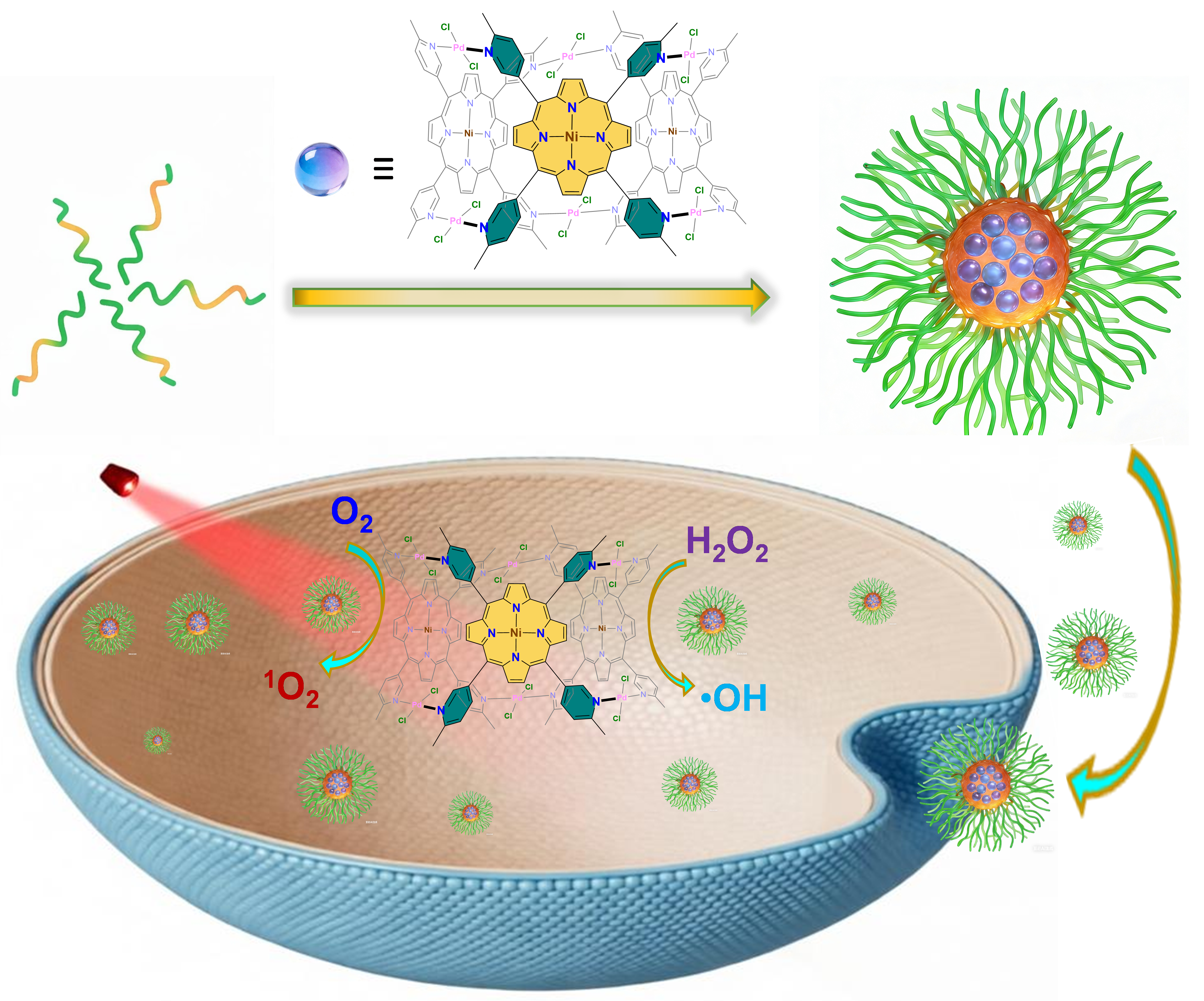
Herein, we report a coordination cage of Pd6-TMPP(Ni) constructed from Ni-chelated porphyrin metalloligand TMPP(Ni) and Pd2+ (H2TMPP = meso-tetrakis (6-methylpyridin-3-yl) porphyrin). Pd6-TMPP(Ni) features a hexagonal prismatic structure wherein the three TMPP(Ni) metalloligand occupy alternative lateral faces of the prism, juxtaposing three pairs of PdCl2 units via binding to the trans positions of the Pd2+ ion (Scheme 1 and Figure 1a). The TMPP(Ni) moieties in Pd6-TMPP(Ni) serve as both a catalyst for •OH production via Fenton-like reaction and a photosensitizer for light-induced 1O2 generation (Scheme 1b). Our in vitro cytotoxicity assays against multiple cancer cell lines NCI-H82 (lung cancer), A549 (lung cancer), KYSE-510 (esophageal cancer), and Te-1 (esophageal cancer) reveal potent dark toxicity of Pd6-TMPP(Ni) with half maximal inhibitory concentration (IC50) in the range of 1.867–2.107 μmol L−1, which can be further lowered to 0.835–1.528 μmol L−1 with the presence of 650 nm light irradiation. The cytotoxicity of Pd6-TMPP(Ni) outperforms that of free ligand H2TMPP and the Pd congener of Pd6-TMPP(Pd).
2. Results and Discussion
2.1. Synthesis and Structure of Pd6-TMPP(Ni)
The coordination cage of Pd6-TMPP(Ni) was directly obtained in 16.4% yield as red needle-like crystals from the solvothermal reaction of TMPP(Ni) and PdCl2 in CH2Cl2/MeOH in the presence of HAc as the modulator. Pd6-TMPP(Ni) is soluble in CH2Cl2, DMF, and DMSO, slightly soluble in MeOH and MeCN, but insoluble in H2O.
Single-crystal X-ray diffraction analysis revealed that Pd6-TMPP(Ni) crystallizes in the monoclinic P21/c space group (Table 1). The asymmetric unit of Pd6-TMPP(Ni) contains a full molecule (C1 point group, Figure 1a) instead of the expected D3h or C2v symmetry as that found for Pd6-TMPP(Au) [28].
In Pd6-TMPP(Ni), three pairs of PdCl2 units were juxtaposed by three TMPP(Ni) metalloligand to give a hexagonal prismatic structure. The side lengths of the two Pd-based triangles from the base planes, i.e., Pd1∙∙∙Pd2/Pd2∙∙∙Pd3/Pd1∙∙∙Pd3 being 12.20/11.99/13.52 Å, and Pd4∙∙∙Pd5/Pd5∙∙∙Pd6/Pd4∙∙∙Pd6 being 12.12/11.80/13.61 Å (Table S1), are similar but both deviate from an equilateral triangle or an isosceles triangle. In addition, the triangle formed by three Ni ions, viz., Ni1∙∙∙Ni2/Ni2∙∙∙Ni3/Ni1∙∙∙Ni3, is 10.30/10.66/12.46 Å (Table S1).
It is notable that the small ionic radius for Ni2+ (0.63 Å) also caused the saddle-shaped ruffling of the three porphyrin backbones (the twelve Ni−N bond distances are in the range of 1.904(12)−1.983(12) Å, Table S1) [29], featuring six pyrrole rings pointing inwards to the centers of the two base planes to create a hydrophobic pocket that may be suitable for storing small molecules [30]. Platon void calculation also suggested that the guest-accessible void of Pd6-TMPP(Ni) was sufficiently large, occupying 43.6% (8369.2 Å3 of 19210.0 Å3) of the total cell volume [31].
2.2. Spectroscopic and Spectrometric Characterization of Pd6-TMPP(Ni)
The 1H nuclear magnetic resonance (NMR) spectroscopy indicated that the characteristic peak of pyrrole protons at −2.81 ppm in H2TMPP disappeared after the formation of Pd6-TMPP(Ni) (Figure S1), which directly confirmed that Ni2+ had been successfully incorporated into the center of the porphyrin center [18,28]. The diffusion ordered spectroscopy (DOSY) for Pd6-TMPP(Ni) shows a narrow diffusion bands with diffusion coefficients of 3.83 × 10⁻¹⁰ m2 s−1 (Figure 1e), suggesting the formation of single assembles.
In the ultraviolet-visible (UV-Vis) absorption spectrum (Figure 1b), the Soret band of Pd6-TMPP(Ni) blue-shifted to 408 nm as compared to those of H2TMPP (420 nm) and Pd6-TMPP(Pd) (417 nm) [32]. Notably, the four Q bands of H2TMPP in the range of 500−700 nm coalesced into two peaks (525/559 nm) in Pd6-TMPP(Ni) (Figure 1b, inset) due to the metalation that elevated the symmetry of the porphyrin ring from the original D2h to D4h [29,33].
The X-ray photoelectron spectroscopy (XPS) shows that in the Pd 3d region, peaks at binding energies of 337.51 eV and 343.77 eV were assigned to Pd 3d5/2 and Pd 3d3/2 (Figure 1c), which were in line with the characteristic peaks of Pd2+ [34]. Meanwhile, in the Ni 2p region, peaks at binding energies of 855.34 eV and 872.46 eV corresponded to the spin-orbit split peaks of Ni 2p3/2 and Ni 2p1/2 (Figure 1d), consistent with those reported in the literature [35]. The above data collectively confirmed that both Pd and Ni existed in the +2 oxidation state in Pd6-TMPP(Ni).
In the matrix-assisted laser desorption/ionization time-of-flight mass spectrometry (MALDI-TOF MS) (Figure 1f and Figure S2), the peaks at a mass-to-charge ratio (m/z) of 3258.421 and 3280.443 were observed for Pd6-TMPP(Ni), consistent with those calculated for [M+H]+ (3258.706 m/z) and [M+Na]+ (3280.695 m/z).
The EDS analysis results (Figure S3) showed that the atomic ratio of Pd to Ni in Pd6-TMPP(Ni) was approximately 4.1 : 1.9 (equal to 2.2 : 1.0), which was close to the theoretical ratio in its molecular formula (2.0 : 1.0).
In the FT-IR spectra (Figure S4), the characteristic peak attributed to the in-plane vibration of N-H at the porphyrin center (964 cm−1) in H2TMPP disappeared in Pd6-TMPP(Ni), while new peaks appeared at 1002 cm−1 and 1012 cm−1, which directly confirmed that Ni2+ had replaced the protons at the porphyrin center [36,37]. In addition, the stretching vibration peak of the C=N bond in the porphyrin ring was also red-shifted from 1593 cm−1 in H2TMPP to 1610 cm−1 in Pd6-TMPP(Ni) due to the coordination of pyridine nitrogen atoms with Pd2+ [38,39].
2.3. Synthesis and Characterizations of Pd6-TMPP(Ni) Nanoparticles
Water-soluble nanoparticles were prepared by encapsulating H2TMPP, Pd6-TMPP(Pd), and Pd6-TMPP(Ni) with Pluronic F127, an FDA-approved triblock polymer for therapeutic use (Scheme 1a) [40,41], in a mixture of DMSO and H2O, followed by dialysis. The transmission electron microscopy (TEM) showed that the obtained nanoparticles H2TMPP-F127, Pd6-TMPP(Pd)-F127, and Pd6-TMPP(Ni)-F127 all exhibited regular spherical morphology and good dispersibility in water (Figure 2), with their diameters being 190 ± 15 nm, 140 ± 15 nm, and 230 ± 15 nm, respectively. The hydrodynamic diameters as revealed by dynamic light scattering (DLS) were marginally larger due to the hydration of these nanoparticles, featuring 231.9 nm for H2TMPP-F127, 184.6 nm for Pd6-TMPP(Pd)-F127, and 311.7 nm for Pd6-TMPP(Ni)-F127 (Figure S5).
The zeta potentials of these nanoparticles were 9.6 mV for H2TMPP-F127, 14.9 mV for Pd6-TMPP(Pd)-F127, and 27.4 mV for Pd6-TMPP(Ni)-F127, respectively (Figure S6). The highest zeta potential value of Pd6-TMPP(Ni)-F127, as compared to H2TMPP-F127 and Pd6-TMPP(Pd)-F127, indicates its best colloidal stability, which is critical during the drug delivery stage [42,43].
As shown in Figure S7a, the Soret bands of H2TMPP-F127, Pd6-TMPP(Pd)-F127, and Pd6-TMPP(Ni)-F127 remained constant when exposed to 650 nm laser for 10 min, suggesting that these particles are photostable. In addition, these particles were stable in H2O, PBS (0.1×), and RPMI 1640 (0.1×) for 0, 8, 12, and 24 h, indicated by only insignificant changes in particle size as revealed by DLS (Figure S8).
2.4. Detection of H2TMPP-F127, Pd6-TMPP(Pd)-F127, and Pd6-TMPP(Ni)-F127 Generation of ROS in Solution
The generation of •OH can be chemically assayed by 3, 3′, 5, 5′-tetramethylbenzidine (TMB) [22,44,45,46]. As shown in Figure S9, when H2TMPP-F127, Pd6-TMPP(Pd)-F127, and Pd6-TMPP(Ni)-F127 respectively mixed with a TMB solution containing 100 μM H2O2, H2TMPP-F127 produced no •OH within 3 h, while the amount of •OH generated by Pd6-TMPP(Pd)-F127 and Pd6-TMPP(Ni)-F127 increased with the rise of concentration. At the same TMPP concentration, Pd6-TMPP(Ni)-F127 produced significantly more •OH, which may be attributed to the fact that Ni(II) can activate the Fenton-like reaction to generate cytotoxic •OH [9].
As shown in the Figure 3a, the electron paramagnetic resonance (EPR) spectroscopy of both Pd6-TMPP(Pd)-F127 and Pd6-TMPP(Ni)-F127 exhibited four peaks with a intensity ratio of 1 : 2 : 2 : 1, which confirmed the presence of •OH induced both types of metal ions wherein Ni(II) outperforms Pd(II) [47,48,49]. The above experiments showed that both Pd6-TMPP(Ni)-F127 and Pd6-TMPP(Pd)-F127 could induce the chemodynamic therapeutic effect by generating •OH, whereas Pd6-TMPP(Ni)-F127 was superior to Pd6-TMPP(Pd)-F127 in this aspect.
We further used DPBF as a probe to detect and compare the ability of H2TMPP-F127, Pd6-TMPP(Pd)-F127, and Pd6-TMPP(Ni)-F127 to generate 1O2 in aqueous solutions [50,51]. As shown in Figures S7b−7f, the absorbance of DPBF decreased after 1 min of irradiation, with the decrease rate being similar among these particles.
2.5. Cell Cytotoxicity and in Vitro ROS Generation Assay
The cytotoxicity of H2TMPP-F127, Pd6-TMPP(Pd)-F127, and Pd6-TMPP(Ni)-F127 against A549 (Figure 4a and Figure 4b), KYSE-510 (Figure 4c and Figure 4d), NCI-H82 (Figure 4e and Figure 4f) and Te-1 (Figure 4g and Figure 4h) cells under light and light-free conditions was evaluated and compared. For all these cell lines, H2TMPP-F127 shows negligible cytotoxicity under both light and light-free conditions. In contrast, both Pd6-TMPP(Pd)-F127 and Pd6-TMPP(Ni)-F127 show concentration-dependent cytotoxicity against all these cell lines, with the exception that A549 is insensitive to Pd6-TMPP(Pd)-F127 treatment within the tested concentration range. Pd6-TMPP(Ni)-F127 also outperforms Pd6-TMPP(Pd)-F127 for all these cell lines in terms of both dark and light toxicities, with Pd6-TMPP(Ni)-F127 nearly killing all the cells with its TMPP concentrations above 3.33 μmol L−1 in the dark (equivalent to 1.11 μmol L−1 of Pd6-TMPP(Ni)). It is also feasible that the light irradiation further augments the toxicity of both Pd6-TMPP(Pd)-F127 and Pd6-TMPP(Ni)-F127, albeit for Pd6-TMPP(Ni)-F127, the light contribution to the toxicity against esophageal cancer cell lines KYSE-510 and Te-1 is insignificant. The low PDT contribution of porphyrin-chelated Ni(II) than Pd(II) can be explained by the fact that Ni2+ can quench the excited state of TMPP ligand via fast non-radiative decay due to the presence of metal-to-ligand charge transfer (MLCT) and d-d excited states that lie below or close to the π-π* states of TMPP [52,53].
The half maximal inhibitory concentration (IC50) for Pd6-TMPP(Ni)-F127 against cell lines NCI-H82 (dark: 2.015 μmol L−1; light: 1.100 μmol L−1), A549 (dark: 2.024 μmol L−1; light: 0.835 μmol L−1), KYSE-510 (dark: 2.107 μmol L−1; light: 1.528 μmol L−1), and Te-1 (dark: 1.867 μmol L−1; light: 1.194 μmol L−1) are comparable to those reported in the literature (Table 2).
2',7'-Dichlorodihydrofluorescein diacetate (DCFH-DA) was used as a fluorescent probe to estimate the ROS-generating capacity of the nanomaterials in Te-1 cells [54]. As shown in Figure 3b and Figure 3c, in the absence of laser irradiation, both Pd6-TMPP(Pd)-F127 and Pd6-TMPP(Ni)-F127 were able to generate a certain level of ROS. The presence of Ni in Pd6-TMPP(Ni)-F127 further enhanced this activity. After laser irradiation, the amount of ROS generated by Pd6-TMPP(Pd)-F127 and Pd6-TMPP(Ni)-F127 increased significantly, while H2TMPP-F127 produced almost no ROS.
2.6. Cellular Uptake of Pd6-TMPP(Pd)-F127 and Pd6-TMPP(Ni)-F127
The cellular uptake of Pd6-TMPP(Pd)-F127 and Pd6-TMPP(Ni)-F127 was evaluated by determining the Pd2+ via inductively coupled plasma mass spectrometry (ICP-MS). As shown in Table 3, both particles can be internalized by KYSE-510 cells in a time-dependent manner. It is also notable that the uptake efficiency of Pd6-TMPP(Ni)-F127 was higher than that of Pd6-TMPP(Pd)-F127, which probably also contributed to the higher cytotoxicity of Pd6-TMPP(Ni)-F127 observed in the cytotoxicity assays.
3. Materials and Methods
3.1. General
Ligand H2TMPP [28,66] and Pd6-TMPP(Pd)-F127 [32] were synthesized as described in our previous reports. PdCl2 (99%, TCI (Shanghai) Development Co., Ltd., Shanghai, China), Ni(NO3)2·6H2O (≥ 98%, Shanghai Aladdin Biochemical Technology Co., Ltd., Shanghai, China), Pluronic F-127 (MW: 2000, Shanghai Macklin Biochemical Co., Ltd, Shanghai, China), 3,3’,5,5’-tetramethylbenzidine (TMB, AR, Shanghai Yuanye Bio-Technology Co., Ltd., Shanghai, China), 1,3-diphenylisobenzofuran (DPBF, AR, Shanghai Maokang Biotechnology Co., Ltd., Shanghai, China), 5,5-dimethyl-1-pyrroline N-oxide (DMPO, 97%, Shanghai Aladdin Biochemical Technology Co., Ltd., Shanghai, China), and hydrogen peroxide (30%, ≥ 95%, Yonghua Chemical Co., Ltd., Shanghai, China) were available from the corresponding suppliers without further purification. MeOH, HAc, MeCN, Et2O, DMSO, and N, N-dimethylformamide (DMF), all in AR grade, were procured from respective sources and used as received.
NCI-H82, A549, KYSE-510, and Te-1 cell lines were purchased from the Shanghai Institute of Cell Biology, Chinese Academy of Sciences (Shanghai, China). Phosphate buffer solution (PBS), cell culturing medium RPMI 1640 (10% FBS + 1% P/S), RPMI 1640 (1% P/S), Ham’s F-12 (10% FBS + 1% P/S), Ham’s F-12 (1% P/S), and 0.25% trypsin solution (containing EDTA, dissolved in PBS) were purchased from Shanghai Basal Media Technologies Co., Ltd (Shanghai, China). The cell counting kit-8 (CCK-8) was available from APEXBIO. The reactive oxygen species detection kit and DCFH-DA were purchased from Shanghai Beyotime Biotechnology Co., Ltd (Shanghai, China).
1H nuclear magnetic resonance (NMR) spectra were recorded on a Bruker Avance III HD 400 MHz superconducting NMR spectrometer (Bruker AXS GmbH, Germany). Fourier-transform infrared (FT-IR) spectra were measured using a Bruker Vertex 70 FTIR-spectrometer with a Hyperion 2000 IR-Microscope (Bruker AXS GmbH, Germany), employing the attenuated total reflection (ATR) technique. Ultraviolet-visible (UV-Vis) spectra were acquired using a Varian Cary-50 UV-Vis spectrophotometer (Varian, Inc., Palo Alto, CA, USA). X-ray photoelectron spectroscopy (XPS) was performed on an Thermo Scientific EXCALAB 250 XI X-ray photoelectron spectrometer (Thermo Scientific, Waltham, MA, USA). Energy-dispersive X-ray spectroscopy (EDS) was conducted using a Zeiss EVO 18 scanning electron microscope (ZEISS Group, Oberkochen, Germany). Transmission electron microscopy (TEM) images were obtained using a Hitachi HT7700 transmission electron microscope (Hitachi, Japan), and samples were prepared by dropping aqueous solutions onto copper grids. Dynamic light scattering (DLS) and zeta potential measurements were performed using a Horiba LA-95052 laser particle size analyzer (Horiba, Kyoto, Japan). Matrix-assisted laser desorption/ionization time-of-flight mass spectrometry (MALDI-TOF MS) was carried out on a Bruker UltrafleXtreme MALDI-TOF/TOF (Bruker AXS GmbH, Germany). Electron paramagnetic resonance (EPR) spectroscopy was carried out on a JEOL JES-X320 electron spin resonance spectrometer (JEOL Ltd., Akishima, Japan). Inductively coupled plasma-mass spectrometry (ICP-MS) was performed with an iCAP PRO instrument (Thermo Scientific, Waltham, MA, USA). Photodynamic therapy experiments were conducted using a PR-CPC2-635NM cellular phototoxicity irradiator at 650 nm (PURI Materials, Guangdong, China). Cytotoxicity assays were performed on a Tecan M1000PRO microplate reader (Tecan, Switzerland) by measuring absorbance at 450 nm. Fluorescence imaging of reactive oxygen species was conducted using an MF52 inverted biological microscope (Guangzhou Micro-shot Technology Co., Ltd., Guangdong, China).
3.2. Synthesis of TMPP(Ni)
Ni(NO3)2·6H2O (2.0 mg, 6.88 μmol), H2TMPP (2.0 mg, 2.96 μmol) were introduced to a mixture of DMF/EtOH (v : v = 0.75 : 0.75 mL), and 2-pyridinemethanol (30 μL) was sequentially introduced as a modulator. The resulting mixture was transferred into a Pyrex glass tube, sealed, and transferred to a programmable oven. The temperature of the oven was gradually increased from r.t. to 120°C in 4 hours. This is followed by a stable heating at 120°C for 36 hours before cooling back to r.t. within 12 hours to yield purple plate crystals of TMPP(Ni). The tubes (50 parallel reactions) were carefully opened, and the resulting product was washed thoroughly with anhydrous ether and dried in an oven. Yield: 43.2 mg from 50 parallel reactions (39.8% based on Ni). FT-IR (ATR, cm−1): 3050 (m), 3011 (w), 2922 (w), 2852 (w), 2162 (w), 1667 (w), 1595 (m), 1557 (w), 1487 (w), 1366 (m), 1349 (m), 1295 (m), 1249 (w), 1204 (w), 1132 (m), 1081 (w), 1036 (w), 1020 (w), 1000 (s), 884 (w), 863 (w), 798 (s), 732 (w), 716 (s), 649 (w), 612 (w). 1H NMR (400 MHz, CDCl3, ppm): δ 9.07 (d, J = 4.0 Hz, 4H), 8.69 (s, 8H), 8.13 (q, J = 8.0 Hz, 4H), 7.47 (d, J = 8.0 Hz, 4H), 2.81 (s, 12H).
3.3. Synthesis of Pd6-TMPP(Ni)
TMPP(Ni) (1.5 mg, 2.10 μmol) and PdCl2 (1.0 mg, 5.63 μmol) were dissolved in a mixed solvent of CH2Cl2/MeOH (v : v = 0.5 : 1.5 mL). Acetic acid (HAc, 20 μL) was subsequently introduced as a modulator. The resulting solution was transferred into a Pyrex glass tube, sonicated for 30 min, and sealed. The tube was then transferred to a programmable oven, with its temperature gradually increased from r.t. to 120°C in 4 hours. This is followed by a stable heating at 120°C for 48 hours, before cooling back to r.t. within 24 hours to yield red needle-like crystals of Pd6-TMPP(Ni). The tubes (70 parallel reactions) were carefully opened, and the resulting crystals were filtered off and washed with diethyl ether 2−3 times to afford Pd6-TMPP(Ni). Yield: 25.6 mg from 70 parallel reactions (16.4% based on TMPP(Ni)). FT-IR (ATR, cm−1): 3323 (w), 3010 (w), 2923 (w), 1593 (m), 1548 (m), 1490 (s), 1471 (m), 1438 (w), 1384 (m), 1379 (m), 1338 (m), 1294 (m), 1240 (m), 1178 (m), 1151 (m), 1130 (m), 1085 (w), 1031 (m), 985 (m), 964 (s), 885 (m), 850 (m), 798 (s), 723 (s), 643 (s), 634 (m). UV-Vis (DMSO): λmax (log ε) 417 nm (4.81), 525 nm (3.65), 559 nm (3.11). MALDI TOF-MS: m/z calcd for [M+H]+: 3258.706, found 3258.421; calcd for [M+Na]+: 3280.725, found 3280.443. 1H NMR (400 MHz, CDCl3, ppm): δ 9.36 (s, 12H), 8.72 (s, 12H), 8.54 (s, 12H), 8.26 (d, J = 8.0 Hz, 12H), 7.59 (d, J = 8.0 Hz, 12H), 3.76 (s, 36H).
3.4. Single-crystal X-ray Crystallography
Diffraction data for Pd6-TMPP(Ni) were acquired on a Bruker APEX II CCD X-ray diffractometer using Ga-Kα (λ = 1.34139 Å) irradiation. Refinement and reduction of the collected data were achieved using the program SAINT, and absorption corrections were performed using a multi-scan method [67]. The crystal structure of Pd6-TMPP(Ni) was solved by direct methods and refined on F2 by full-matrix least-squares techniques with SHELXTL-2016 [68].
During refinement, some spatially delocalized electron density was observed within the crystal lattice, but it proved difficult to obtain satisfactory results. The solvent contribution was then modeled using SQUEEZE in the Platon program suite [69].
Crystallographic data for Pd6-TMPP(Ni) have been deposited in the Cambridge Crystallographic Data Center (CCDC) as supplementary publication numbers 2525223. These data can be obtained free of charge either from the CCDC via www.ccdc.cam.ac.uk/data_request/cif or from the Supporting Information. A summary of the key crystallographic data for Pd6-TMPP(Ni) is listed in Table 1.
3.5. The Formation of H2TMPP-F127, Pd6-TMPP(Pd)-F127, and Pd6-TMPP(Ni)-F127 Nanoparticles
The formation of H2TMPP-F127 follows our previously published procedures. [41] Specifically, Pluronic F-127 (10.6 mg, 5.30 μmol) was introduced to a 1 mL DMSO containing H2TMPP (5.00 mg, 7.41 μmol). Upon dissolution, the mixture was added dropwise into deionized water (10 mL) under tip sonication. The formed mixture was then dialyzed using a membrane with a molecular weight cutoff of 2000 Da for 24 h to obtain H2TMPP-F127 aqueous solution for subsequent use.
The preparation procedures for Pd6-TMPP(Pd)-F127 and Pd6-TMPP(Ni)-F127 were similar to that of H2TMPP-F127, except that Pd6-TMPP(Pd) (3.5 mg, 1.03 μmol) and Pd6-TMPP(Ni) (3.5 mg, 1.07 μmol) were used as the starting material.
3.6. Photostability Assay
H2TMPP-F127, Pd6-TMPP(Pd)-F127, and Pd6-TMPP(Ni)-F127 aqueous solutions with the same porphyrin concentration (7 μg mL−1) were prepared, and then irradiated with 650 nm laser (25 mW cm−2). The absorbance intensity at 416 nm was monitored over a period of 10 min, with data recorded every 1 min.
3.7. Singlet Oxygen (1O2) Detection
The same concentration of DPBF (33 μg mL−1) was added to aqueous solutions of H2TMPP-F127, Pd6-TMPP(Pd)-F127, and Pd6-TMPP(Ni)-F127 with an equivalent concentration of TMPP (7 μg mL−1) to give uniform mixtures. The solutions were then irradiated with a 650 nm laser at an intensity of 25 mW cm–2. After irradiation for every 1 min, the characteristic absorption intensity of DPBF at 416 nm was immediately measured.
3.8. Hydroxyl Radical (•OH) Detection
Chemical method: Different concentrations of H2TMPP-F127, Pd6-TMPP(Pd)-F127, and Pd6-TMPP(Ni)-F127 were added to TMB solution containing 100 μM H2O2. After reacting for 3 hours at r.t., the reaction solutions were centrifuged to remove the influence of nanoparticles on absorbance. The UV-Vis curve of the supernatant at the wavelength of 652 nm was determined.
Spectroscopic method: Pd6-TMPP(Pd)-F127 and Pd6-TMPP(Ni)-F127 with the equivalent concentration of TMPP (7 μg mL−1) were co-incubated with 100 μM H2O2, and after 3 hours, the spin trap 0.1 mL DMPO (97%) was added and stirred for 15 min. The resulting solution was then sealed in capillaries for EPR measurement.
3.9. In Vitro Cytotoxicity Evaluation by CCK-8 Assay
The NCI-H82 cell line (suspension-grown) was cultured in RPMI 1640 medium supplemented with 10% FBS, 1% P/S, or serum-free medium (RPMI 1640 + 1% P/S). Specifically, the suspension-grown cells were centrifuged, and the supernatant was discarded. The cells were re-suspended in serum-supplemented RPMI 1640 medium at a concentration of 1 × 10⁵ cells per milliliter. These cells were cultured at 37°C under a 5% CO2 atmosphere for the CCK-8 assays.
NCI-H82 cells were seeded at a density of 3.5 × 10⁴ cells per well in 100 µL of serum-free medium containing various concentrations of H2TMPP-F127, Pd6-TMPP(Pd)-F127, and Pd6-TMPP(Ni)-F127. All experiments were conducted with five replicates (n = 5), using untreated cells as the 100% cell viability control and cell-free medium (RPMI 1640 + 10 µL PBS + 1% P/S + CCK-8) as the blank.
For the PDT group, the cells were irradiated with a laser (650 nm, 25 mW cm−2) for 5 min after 24 hours of incubation, followed by an additional incubation time of 48 hours. By contrast, the non-PDT group was incubated continuously for 72 hours. After the incubation period, 10 µL FBS and 10 µL CCK-8 were added to the wells, and the plates were further incubated for 3.5 hours before analysis at 450 nm using a microplate reader. The relative cell viability (V%) was calculated using equation (1) as described below:
in which V% is the percentage of cell viability, [A]experimental is the absorbance of the wells culturing the treated cells, [A]blank is the absorbance of the blank, and [A]control is the absorbance of the wells culturing untreated cells.
The A549 cell line (adherent cells) was cultured in Ham’s F-12 medium supplemented with 10% FBS and 1% P/S, or serum-free medium (Ham’s F-12 + 1% P/S). Specifically, the adherent cells grew as a monolayer and were detached at confluence using trypsin (0.5% w/v in PBS). After trypsinization, the cells were incubated for 3 min, centrifuged, and the supernatant was discarded. A 3 mL portion of serum-supplemented culture medium was added to neutralize any residual trypsin. The cells were re-suspended in serum-supplemented Ham’s F-12 medium at a concentration of 1 × 10⁵ cells mL–1 and cultured under standard conditions (37°C, 5% CO2) for the CCK-8 studies.
A549 cells were seeded at a density of 1 × 10⁴ cells per well in 100 µL of culture medium and incubated for 24 hours at 37°C and 5% CO2 to allow cell attachment. The culture medium was then replaced with serum-free medium containing various concentrations of the H2TMPP-F127, Pd6-TMPP(Pd)-F127, and Pd6-TMPP(Ni)-F127. All experiments were performed with five replicates (n = 5), using untreated cells as the 100% cell viability control and cell-free medium (Ham’s F-12 + 10% FBS + 1% P/S + CCK-8) as the blank.
For the PDT group, the cells were irradiated with a laser (650 nm, 25 mW cm–2) for 5 minutes after 24 hours of incubation, followed by an additional incubation of 48 hours. For the non-PDT group, cells were directly incubated for 72 hours. After the incubation period, 100 µL of culture medium and 10 µL of CCK-8 were added to each well, and the plates were incubated for an additional 1 hour before being analyzed at 450 nm using a microplate reader. The relative cell viability (V%) was calculated using Equation 1 above.
The cytotoxicity assessment for the KYSE-510 (adherent cells) and Te-1 (adherent cells) cell lines follow a similar protocol to that of the A549 cells, except that the culture media are different: KYSE-510 cells were cultured in a 1 : 1 (v : v) mixture of RPMI 1640 and Ham’s F-12 media, while Te-1 cells were cultured in RPMI 1640 medium. The exact compositions were: RPMI 1640 : Ham’s F-12 = 1 : 1 + 10% FBS + 1% P/S and RPMI 1640 : Ham’s F-12 = 1 : 1 + 1% P/S for KYSE-510; RPMI 1640 + 10% FBS + 1% P/S and RPMI 1640 + 1% P/S for Te-1.
3.10. Detection of Intracellular Reactive Oxygen Species
Te-1 cells were seeded in a 6-well plate at a density of 8 × 10⁵ cells per well in 2 mL culture medium (RPMI 1640 : Ham’s F-12 = 1 : 1 + 10% FBS + 1% P/S). The cells were incubated at 37°C under 5% CO2 for 24 hours to allow proper attachment. After incubation, the culture medium was removed, and the wells were replenished with 1.5 mL serum-free culturing medium (RPMI 1640 : Ham’s F-12 = 1 : 1 + 1% P/S) containing H2TMPP-F127, Pd6-TMPP(Pd)-F127, and Pd6-TMPP(Ni)-F127 at a concentration of 10 μmol L−1.
For the PDT group, one plate of cells was irradiated with a 650 nm laser at a light intensity of 25 mW cm−2 for 5 min, while cells in the non-PDT group were not subjected to laser irradiation. After irradiation, all cells were further incubated for 24 h, followed by the addition of 2 mL of fresh medium to each well. In the negative control wells, 1.0 μL of PBS was added, while 1.0 μL of Rosup-containing medium was added to the positive control wells. After 30 min of incubation, 1 μL of DCFH-DA was added to each well, and the culture was continued for another 20 min. The cells were then rinsed three times with serum-free medium to remove uninternalized reagents. Finally, the fluorescent performance of the cells was observed and recorded under an inverted fluorescence microscope.
3.11. Cellular Uptake
KYSE-510 cells were prepared in a 1 : 1 mixture of Ham’s F-12 and RPMI 1640 medium, and seeded into cell culture dishes (9 parallel dishes) with a volume of 10 mL medium each containing a density of 1 × 10⁷ cells per dish. After the cells adhered to the dish and proliferated to 95% confluency, the medium was replaced with serum-free medium. Subsequently, Pd6-TMPP(Pd)-F127 and Pd6-TMPP(Ni)-F127 were added directly to the medium at a porphyrin concentration of 10 μmol L−1. After drug addition, the cells were incubated for 2 h, 4 h, and 6 h, with three replicate groups (n = 3) for each time point.
After incubation, the cells were washed three times with PBS, harvested by trypsin digestion, transferred to 15 mL centrifuge tubes, and centrifuged at 900 rpm for 3 min. The collected cells were rinsed twice with PBS, centrifuged, and reserved for subsequent use. The cells were lysed with 1 mL of nitric acid, then diluted to 10 mL with deionized water and filtered. The intracellular contents of Pd were analyzed by ICP-MS.
4. Conclusion
In this work, we synthesized a nonanuclear Pd-based coordination cage of Pd6-TMPP(Ni) that elicits its anticancer effect via dual ROS generation strategies, viz., catalytic •OH via the Ni2+ center and the photoinduced 1O2 generation via the TMPP ligand. These results validate that Pd6-TMPP(Ni) is a promising candidate for hypoxia-tolerant cancer therapy. Our future work is to equip Pd6-TMPP(Ni) with targeting functions via conjugation with tumor-specific ligands for more effective cancer therapies.
Supplementary Materials
The following supporting information can be downloaded at: www.mdpi.com/xxx/s1, Additional structure diagrams, spectroscopies, and table.
Author Contributions
Conceptualization, W.H.Z., W.L., and Y.M.; methodology, M.L.D. and Y.N.; validation, M.L.D. and Y.H.J.; formal analysis, M.L.D.; data curation, M.L.D. and Y.N.; writing—original draft preparation, M.L.D.; writing—review and editing, W.H.Z., W.L., and Y.M.; supervision, W.H.Z.; project administration, W.H.Z.; funding acquisition, W.L., and Y.M. All authors have read and agreed to the published version of the manuscript.:
Funding
This work was financially supported by Suzhou Municipal Science and Technology Innovation Program for Applied Basic Research (Healthcare) (SYW2024063), Suzhou Municipal Program for Strengthening Healthcare through Science and Education (MSXM2024077), and Gusu Health Talent Program of Suzhou (GSWS2023073, MR-32-25-016221).
Conflicts of Interest
The authors declare no conflict of interest.
References
- Yang, B.; Chen, Y.; Shi, J. Reactive oxygen species (ROS)-based nanomedicine. Chem. Rev. 2019, 119, 4881−4985. [Google Scholar] [CrossRef] [PubMed]
- Dickinson, B.C.; Chang, C.J. Chemistry and biology of reactive oxygen species in signaling or stress responses. Nat. Chem. Biol. 2011, 7, 504−511. [Google Scholar] [CrossRef] [PubMed]
- Li, S.-L.; Chu, X.; Dong, H.-L.; Hou, H.-Y.; Liu, Y. Recent advances in augmenting Fenton chemistry of nanoplatforms for enhanced chemodynamic therapy. Coord. Chem. Rev. 2023, 479, 215004. [Google Scholar] [CrossRef]
- Wang, Y.; Gao, F.; Li, X.; Niu, G.; Yang, Y.; Li, H.; Jiang, Y. Tumor microenvironment-responsive Fenton nanocatalysts for intensified anticancer treatment. J. Nanobiotechnology 2022, 20, 69. [Google Scholar] [CrossRef]
- Tang, Z.; Zhao, P.; Wang, H.; Liu, Y.; Bu, W. Biomedicine meets Fenton chemistry. Chem. Rev. 2021, 121, 1981–2019. [Google Scholar] [CrossRef]
- Wang, F.; Tong, S.; Ma, X.; Yang, H.; Zhang, T.; Wu, K.; Wu, J. Nickel nanoparticles: A novel platform for cancer-targeted delivery and multimodal therapy. Front. Drug Deliv. 2025, 5, 1627556. [Google Scholar] [CrossRef]
- Oh, H.; Kim, J.-Y.; Chae, K.H.; Kim, J.; Yun, E.-T.; Lee, Y.; Lee, C.; Moon, G.-H.; Lee, J. Oxyanion-sensitive catalytic activity of Ni(II)/oxyanion systems for heterogeneous organic degradation: Differential oxidizing capacity of Ni(III) and Ni(IV) as high-valent intermediates. Environ. Sci. Technol. 2024, 58, 16642−16655. [Google Scholar] [CrossRef]
- Goyal, S.; Kumar, P.; Kumar, G.; Soni, A.; Nemiwal, M. Nickel-based metal-organic frameworks as versatile heterogeneous catalysts: A comprehensive exploration in diverse organic transformations. Tetrahedron 2024, 158, 133979. [Google Scholar] [CrossRef]
- Zhang, R.; Xu, H.; Yao, Y.; Ran, G.; Zhang, W.; Zhang, J.; Sessler, J.L.; Gao, S.; Zhang, J.-L. Nickel(II) phototheranostics: A case study in photoactivated H2O2-enhanced immunotherapy. J. Am. Chem. Soc. 2023, 145, 23257−23274. [Google Scholar] [CrossRef]
- Chu, Z.; Yang, J.; Zheng, W.; Sun, J.; Wang, W.; Qian, H. Recent advances on modulation of H2O2 in tumor microenvironment for enhanced cancer therapeutic efficacy. Coord. Chem. Rev. 2023, 481, 215049. [Google Scholar] [CrossRef]
- Wang, X.; Zhong, X.; Liu, Z.; Cheng, L. Recent progress of chemodynamic therapy-induced combination cancer therapy. Nano Today 2020, 35, 100946. [Google Scholar] [CrossRef]
- Zhao, Y.-Y.; Xu, Y.; Zhang, X.; Chen, Z.; Kim, H.; Li, X.; Yoon, J. A hypoxia-triggered bioreduction of hydrophilic type I photosensitizer for switchable in vivo photoacoustic imaging and high-specificity cancer phototherapy. Angew. Chem. Int. Ed. 2025, 64, e202506412. [Google Scholar] [CrossRef] [PubMed]
- Zhang, C.; Hu, X.; Jin, L.; Lin, L.; Lin, H.; Yang, Z.; Huang, W. Strategic design of conquering hypoxia in tumor for advanced photodynamic therapy. Adv. Healthc. Mater. 2023, 12, 2300530. [Google Scholar] [CrossRef] [PubMed]
- Stinson, T.J.; Jaw, S.; Jeffery, E.H.; Plewa, M.J. The relationship between nickel chloride-induced peroxidation and DNA strand breakage in rat liver. Toxicol. Appl. Pharmacol. 1992, 117, 98−103. [Google Scholar] [CrossRef]
- Liu, H.; Yu, B.; Zhou, C.; Deng, Z.; Wang, H.; Zhang, X.; Wang, K. Nickel atom-clusters nanozyme for boosting ferroptosis tumor therapy. Mater. Today Bio 2024, 27, 101137. [Google Scholar] [CrossRef]
- Dolmans, D.E.J.G.J.; Fukumura, D.; Jain, R.K. Photodynamic therapy for cancer. Nat. Rev. Cancer 2003, 3, 380−387. [Google Scholar] [CrossRef]
- Fan, W.; Huang, P.; Chen, X. Overcoming the Achilles' heel of photodynamic therapy. Chem. Soc. Rev. 2016, 45, 6488−6519. [Google Scholar] [CrossRef]
- Wang, X.; Feng, J.-H.; Zeng, C.-M.; Zhang, Z.-S.; Cao, F.-L.; Zhang, W.-H.; Chen, J.-X.; Young, D.J. [FeIIICl(TMPPH2)][FeIIICl4]2: A stand-alone molecular nanomedicine that induces high cytotoxicity by ferroptosis. Molecules 2024, 29, 2495. [Google Scholar] [CrossRef]
- Zou, Y.-M.; Li, R.-T.; Yu, L.; Huang, T.; Peng, J.; Meng, W.; Sun, B.; Zhang, W.-H.; Jiang, Z.-H.; Chen, J.; et al. Reprogramming of the tumor microenvironment using a PCN-224@IrNCs/d-Arg nanoplatform for the synergistic PDT, NO, and radiosensitization therapy of breast cancer and improving anti-tumor immunity. Nanoscale 2023, 15, 10715−10729. [Google Scholar] [CrossRef]
- Attar, G.S.; Bhalla, V.; Kumar, M. Nanoscale metal−organic frameworks: An emerging versatile tool for next-generation photodynamic therapy. Chem. Asian J. 2025, 20, e202500079. [Google Scholar] [CrossRef]
- Lismont, M.; Dreesen, L.; Wuttke, S. Metal−organic framework nanoparticles in photodynamic therapy: Current status and perspectives. Adv. Funct. Mater. 2017, 27, 1606314. [Google Scholar] [CrossRef]
- Li, Q.; Xu, B.-W.; Zou, Y.-M.; Niu, R.-J.; Chen, J.-X.; Zhang, W.-H.; Young, D.J. Nanoscale two-dimensional FeII- and CoII-based metal−organic frameworks of porphyrin ligand for the photodynamic therapy of breast cancer. Molecules 2023, 28, 2125. [Google Scholar] [CrossRef]
- Tuo, W.; Xu, Y.; Fan, Y.; Li, J.; Qiu, M.; Xiong, X.; Li, X.; Sun, Y. Biomedical applications of Pt(II) metallacycle/metallacage-based agents: From mono-chemotherapy to versatile imaging contrasts and theranostic platforms. Coord. Chem. Rev. 2021, 443, 214017. [Google Scholar] [CrossRef]
- Cook, T.R.; Vajpayee, V.; Lee, M.H.; Stang, P.J.; Chi, K.-W. Biomedical and biochemical applications of self-assembled metallacycles and metallacages. Acc. Chem. Res. 2013, 46, 2464−2474. [Google Scholar] [CrossRef] [PubMed]
- Hu, J.; Wu, W.; Qin, Y.; Liu, C.; Wei, P.; Hu, J.; Seeberger, P.H.; Yin, J. Fabrication of glyco-metal-organic frameworks for targeted interventional photodynamic/chemotherapy for hepatocellular carcinoma through percutaneous transperitoneal puncture. Adv. Funct. Mater. 2020, 30, 1910084. [Google Scholar] [CrossRef]
- Li, W.; Li, R.; Ye, Q.; Zou, Y.; Lu, X.; Zhang, W.; Chen, J.; Zhao, Y. Mn3O4 nanoshell coated metal–organic frameworks with microenvironment-driven O2 production and GSH exhaustion ability for enhanced chemodynamic and photodynamic cancer therapies. Adv. Healthc. Mater. 2023, 12, 2202280. [Google Scholar] [CrossRef]
- Wang, D.; Wu, H.; Lim, W.Q.; Phua, S.Z.F.; Xu, P.; Chen, Q.; Guo, Z.; Zhao, Y. A mesoporous nanoenzyme derived from metal–organic frameworks with endogenous oxygen generation to alleviate tumor hypoxia for significantly enhanced photodynamic therapy. Adv. Mater. 2019, 31, 1901893. [Google Scholar] [CrossRef]
- Cao, F.-L.; Zhang, Z.-S.; Dong, M.-L.; Ning, Y.; Zhang, W.-H.; Mao, Y.; Young, D.J. A high-entropy coordination cage featuring an Au-porphyrin metalloligand for the photodynamic therapy of liver cancer. Chem. Commun. 2025, 61, 6663−6666. [Google Scholar] [CrossRef]
- La, T.; Richards, R.A.; Lu, R.S.; Bau, R.; Miskelly, G.M. Solution chemistry and crystal structure of nickel tetrakis(2,3,5,6-tetrafluoro-N,N,N-trimethyl-4-aniliniumyl)porphyrin trifluoromethanesulfonate (NiTF4TMAP(CF3SO3)4). Inorg. Chem. 1995, 34, 5632−5640. [Google Scholar] [CrossRef]
- Schindler, J.; Kupfer, S.; Ryan, A.A.; Flanagan, K.J.; Senge, M.O.; Dietzek, B. Sterically induced distortions of nickel(II) porphyrins – Comprehensive investigation by DFT calculations and resonance Raman spectroscopy. Coord. Chem. Rev. 2018, 360, 1−16. [Google Scholar] [CrossRef]
- Spek, A.L. Single-crystal structure validation with the program PLATON. J. Appl. Cryst. 2003, 36, 7−13. [Google Scholar] [CrossRef]
- Hu, Q. Nanomaterials of TMPP-based functional coordination complex and their antitumor properties; Soochow University, 2022. [Google Scholar]
- Nurco, D.J.; Smith, K.M.; Fajer, J. Conformational landscape surfing induced by off–on π–π stacking in a porphyrin–quinone dyad. Chem. Commun. 2002, 2982−2983. [Google Scholar] [CrossRef]
- Wang, X.; Chen, J.; Zeng, J.; Wang, Q.; Li, Z.; Qin, R.; Wu, C.; Xie, Z.; Zheng, L. The synergy between atomically dispersed Pd and cerium oxide for enhanced catalytic properties. Nanoscale 2017, 9, 6643−6648. [Google Scholar] [CrossRef]
- Bagus, P.S.; Nelin, C.J.; Brundle, C.R.; Crist, B.V.; Ilton, E.S.; Lahiri, N.; Rosso, K.M. Main and satellite features in the Ni 2p XPS of NiO. Inorg. Chem. 2022, 61, 18077−18094. [Google Scholar] [CrossRef] [PubMed]
- Zhao, Y.; Wang, J.; Pei, R. Micron-sized ultrathin metal–organic framework sheet. J. Am. Chem. Soc. 2020, 142, 10331−10336. [Google Scholar] [CrossRef] [PubMed]
- Wang, Y.-R.; Liu, M.; Gao, G.-K.; Yang, Y.-L.; Yang, R.-X.; Ding, H.-M.; Chen, Y.; Li, S.-L.; Lan, Y.-Q. Implanting numerous hydrogen-bonding networks in a Cu-porphyrin-based nanosheet to boost CH4 selectivity in neutral-media CO2 electroreduction. Angew. Chem. Int. Ed. 2021, 60, 21952−21958. [Google Scholar]
- Armaghan, M.; Niu, R.-J.; Liu, Y.; Zhang, W.-H.; Hor, T.S.A.; Lang, J.-P. Zn-based metal–organic frameworks (MOFs) of pyridinemethanol–carboxylate conjugated ligands: Deprotonation-dependent structures and CO2 adsorption. Polyhedron 2018, 153, 218−225. [Google Scholar] [CrossRef]
- Drelinkiewicz, A.; Hasik, M.; Quillard, S.; Paluszkiewicz, C. Infrared and Raman studies of palladium—nitrogen-containing polymers interactions. J. Mol. Struct. 1999, 511-512, 205−215. [Google Scholar] [CrossRef]
- Cacaccio, J.; Durrani, F.; Cheruku, R.R.; Borah, B.; Ethirajan, M.; Tabaczynski, W.; Pera, P.; Missert, J.R.; Pandey, R.K. Pluronic F-127: An efficient delivery vehicle for 3-(1'-hexyloxy)ethyl-3-devinylpyropheophorbide-a (HPPH or Photochlor). Photochem. Photobiol. 2020, 96, 625−635. [Google Scholar] [CrossRef]
- Wang, P.; Wang, J.-W.; Zhang, W.-H.; Bai, H.; Tang, G.; Young, D.J. In vitro anticancer activity of nanoformulated mono- and di-nuclear Pt compounds. Chem. Asian J. 2021, 16, 2993−3000. [Google Scholar] [CrossRef]
- Maeda, H.; Nakamura, H.; Fang, J. The EPR effect for macromolecular drug delivery to solid tumors: improvement of tumor uptake, lowering of systemic toxicity, and distinct tumor imaging in vivo. Adv. Drug Deliv. Rev. 2013, 65, 71−79. [Google Scholar] [CrossRef]
- Zhang, L.; Liu, Y.; Liu, G.; Xu, D.; Liang, S.; Zhu, X.; Lu, Y.; Wang, H. Prolonging the plasma circulation of proteins by nano-encapsulation with phosphorylcholine-based polymer. Nano Res. 2016, 9, 2424−2432. [Google Scholar] [CrossRef]
- Hou, Y.-K.; Zhang, Z.-J.; Li, R.-T.; Peng, J.; Chen, S.-Y.; Yue, Y.-R.; Zhang, W.-H.; Sun, B.; Chen, J.-X.; Zhou, Q. Remodeling the tumor microenvironment with core–shell nanosensitizer featuring dual-modal imaging and multimodal therapy for breast cancer. ACS Appl. Mater. Interfaces 2023, 15, 2602−2616. [Google Scholar] [CrossRef] [PubMed]
- Hu, J.-J.; Yu, X.-Z.; Zhang, S.-Q.; Zhang, Y.-X.; Chen, X.-L.; Long, Z.-J.; Hu, H.-Z.; Xie, D.-H.; Zhang, W.-H.; Chen, J.-X.; et al. Hydrogel with ROS scavenging effect encapsulates BR@Zn-BTB nanoparticles for accelerating diabetic mice wound healing via multimodal therapy. iScience 2023, 26, 106775. [Google Scholar] [CrossRef] [PubMed]
- Zhang, X.; Yang, Q.; Lang, Y.; Jiang, X.; Wu, P. Rationale of 3,3′,5,5′-tetramethylbenzidine as the chromogenic substrate in colorimetric analysis. Anal. Chem. 2020, 92, 12400−12406. [Google Scholar] [CrossRef]
- Liu, T.Z.; Lin, T.F.; Chiu, D.T.Y.; Tsai, K.-J.; Stern, A. Palladium or platinum exacerbates hydroxyl radical mediated DNA damage. Free Radic. Biol. Med. 1997, 23, 155−161. [Google Scholar] [CrossRef]
- Athar, M.; Hasan, S.K.; Srivastava, R.C. Evidence for the involvement of hydroxyl radicals in nickel mediated enhancement of lipid peroxidation: Implications for nickel carcinogenesis. Biochem. Biophys. Res. Commun. 1987, 147, 1276−1281. [Google Scholar] [CrossRef]
- Malec, D.; Warszyńska, M.; Repetowski, P.; Siomchen, A.; Dąbrowski, J.M. Enhancing visible-light photocatalysis with Pd(II) porphyrin-based TiO2 hybrid nanomaterials: Preparation, characterization, ROS generation, and photocatalytic activity. Molecules 2023, 28. [Google Scholar] [CrossRef]
- Entradas, T.; Waldron, S.; Volk, M. The detection sensitivity of commonly used singlet oxygen probes in aqueous environments. J. Photochem. Photobiol. B 2020, 204, 111787. [Google Scholar] [CrossRef]
- Carloni, P.; Damiani, E.; Greci, L.; Stipa, P.; Tanfani, F.; Tartaglini, E.; Wozniak, M. On the use of 1,3-diphenylisobenzofuran (DPBF). Reactions with carbon and oxygen centered radicals in model and natural systems. Res. Chem. Intermed. 1993, 19, 395−405. [Google Scholar] [CrossRef]
- Antipas, A.; Gouterman, M. Porphyrins. 44. Electronic states of cobalt, nickel, rhodium, and palladium complexes. J. Am. Chem. Soc. 1983, 105, 4896−4901. [Google Scholar] [CrossRef]
- Shelby, M.L.; Lestrange, P.J.; Jackson, N.E.; Haldrup, K.; Mara, M.W.; Stickrath, A.B.; Zhu, D.; Lemke, H.T.; Chollet, M.; Hoffman, B.M.; et al. Ultrafast Excited State Relaxation of a Metalloporphyrin Revealed by Femtosecond X-ray Absorption Spectroscopy. J. Am. Chem. Soc. 2016, 138, 8752−8764. [Google Scholar] [CrossRef] [PubMed]
- Yu, D.; Zha, Y.; Zhong, Z.; Ruan, Y.; Li, Z.; Sun, L.; Hou, S. Improved detection of reactive oxygen species by DCFH-DA: New insight into self-amplification of fluorescence signal by light irradiation. Sens. Actuators B Chem. 2021, 339, 129878. [Google Scholar] [CrossRef]
- Pandey, S.K.; Kumar, S.; Singh, S.; Patel, A.K.; Gond, M.K.; Acharya, A.; Bharty, M.K. Synthesis, structural characterisation, and anticancer potential of mono and dinuclear Pd(II) complexes of N-(2-pyridyl)thiourea. Dalton Trans. 2025, 54, 1139−1149. [Google Scholar] [CrossRef] [PubMed]
- Dingiswayo, S.; Babu, B.; Burgess, K.; Mack, J.; Nyokong, T. Photodynamic anticancer and antibacterial activities of Sn(IV) N-confused meso-tetra(methylthiophenyl)porphyrin. Photochem 2023, 3, 313−326. [Google Scholar] [CrossRef]
- Hu, X.; Ogawa, K.; Kiwada, T.; Odani, A. Water-soluble metalloporphyrinates with excellent photo-induced anticancer activity resulting from high tumor accumulation. J. Inorg. Biochem. 2017, 170, 1−7. [Google Scholar] [CrossRef]
- Tong, K.-C.; Hu, D.; Wan, P.-K.; Lok, C.-N.; Che, C.-M. Anticancer gold(III) compounds with porphyrin or N-heterocyclic carbene ligands. Front. Chem. 2020, 8, 587207. [Google Scholar] [CrossRef]
- Zhang, Q.; He, J.; Yu, W.; Li, Y.; Liu, Z.; Zhou, B.; Liu, Y. A promising anticancer drug: A photosensitizer based on the porphyrin skeleton. RSC Med. Chem. 2020, 11, 427−437. [Google Scholar] [CrossRef]
- Bora, B.; Das, N.; Sultana, J.P.; Raza, M.K.; Goswami, T.K. Mn(III) porphyrins as photosensitizers: Structural, photophysical and anticancer studies. Dalton Trans. 2025, 54, 11743−11756. [Google Scholar] [CrossRef]
- Zhang, Q.; Yu, W.; Liu, Z.; Li, H.; Liu, Y.; Liu, X.; Han, Z.; He, J.; Zeng, Y.; Guo, Y.; et al. Design, synthesis, antitumor activity and ct-DNA binding study of photosensitive drugs based on porphyrin framework. Int. J. Biol. Macromol. 2023, 230, 123147. [Google Scholar] [CrossRef]
- Li, H.; Tang, C.; Liu, Z.; Tian, Z.; Shi, L.; Yang, L.; He, J.; Ai, W.; He, X.; Liu, Y. Synthesis and antitumor activity of photosensitizer eugenol porphyrin derivatives: A combination therapy of chemotherapy and photodynamic therapy. Appl. Organomet. Chem. 2025, 39, e7759. [Google Scholar] [CrossRef]
- Guo, L.; Li, P.; Jing, Z.; Gong, Y.; Lai, K.; Fu, H.; Dong, H.; Yang, Z.; Liu, Z. Iminoamido chelated iridium(III) and ruthenium(II) anticancer complexes with mitochondria-targeting ability and potential to overcome cisplatin resistance. J. Inorg. Biochem. 2024, 258, 112631. [Google Scholar] [CrossRef] [PubMed]
- Li, Y.; Gao, Z.; Chen, F.; You, C.; Wu, H.; Sun, K.; An, P.; Cheng, K.; Sun, C.; Zhu, X.; et al. Decoration of cisplatin on 2D metal–organic frameworks for enhanced anticancer effects through highly increased reactive oxygen species generation. ACS Appl. Mater. Interfaces 2018, 10, 30930−30935. [Google Scholar] [CrossRef] [PubMed]
- Panicker, R.R.; John, M.L.; N, D.M.; Yogendra Varma, P.; S, D.; Pandya, C.; Anand, A.S.V.; Mondal, J.; Sivaramakrishna, A. Square planar mononuclear Ni(II) complexes of functionalized 2,2′:6′,2′′-terpyridines: BSA/DNA binding and anticancer activity. New J. Chem. 2025, 49, 5883−5900. [Google Scholar] [CrossRef]
- Niu, R.-J.; Zhou, W.-F.; Liu, Y.; Yang, J.-Y.; Zhang, W.-H.; Lang, J.-P.; Young, D.J. Morphology-dependent third-order optical nonlinearity of a 2D Co-based metal–organic framework with a porphyrinic skeleton. Chem. Commun. 2019, 55, 4873−4876. [Google Scholar] [CrossRef]
- Sheldrick, G.M. SADABS (Version 2.03): Program for empirical absorption correction of area detector data; University of Göttingen, Germany. 1996.
- Sheldrick, G.M. Crystal structure refinement with SHELXL. Acta Crystallogr., Sect. C 2015, 71, 3−8. [Google Scholar] [CrossRef]
- Spek, A.L. PLATON SQUEEZE: A tool for the calculation of the disordered solvent contribution to the calculated structure factors. Acta Crystallogr., Sect. C 2015, 71, 9−18. [Google Scholar] [CrossRef]
Scheme 1.
The assembly process of Pd6-TMPP(Ni) with Pluronic F127 to give Pd6-TMPP(Ni)-F127 (a), and an illustration of the anticancer mechanism of Pd6-TMPP(Ni)-F127 (b).
Scheme 1.
The assembly process of Pd6-TMPP(Ni) with Pluronic F127 to give Pd6-TMPP(Ni)-F127 (a), and an illustration of the anticancer mechanism of Pd6-TMPP(Ni)-F127 (b).

Figure 1.
The X-ray structure of Pd6-TMPP(Ni) (a), the UV-Vis spectra of H2TMPP, Pd6-TMPP(Pd), and Pd6-TMPP(Ni) (b; inset: the amplified absorption in the range of 475−700 nm), the Pd 3d5/2, 3/2 (c) and Ni 2p3/2, 1/2 (d) XPS spectra of Pd6-TMPP(Ni), the hydrogen atoms are omitted for clarity. the 1H DOSY spectra of Pd6-TMPP(Ni) (298 K, CDCl3; e), and the experimental (orange yellow) and calculated (light green) MALDI-TOF MS spectrometry of Pd6-TMPP(Ni) (f). For (a), the hydrogen atoms are omitted for clarity. Color legend: Pd (dark magenta), Ni (brown), Cl (green), N (blue), C (black).

Figure 2.
The TEM images of H2TMPP-F127 (a), Pd6-TMPP(Pd)-F127 (b), and Pd6-TMPP(Ni)-F127 (c).

Figure 3.
The electron paramagnetic resonance spectra for •OH as induced by Pd6-TMPP(Pd)-F127 and Pd6-TMPP(Ni)-F127, using DMPO as a spin trap (a). Comparison of the corresponding mean fluorescence intensity (MFI) of Pd6-TMPP(Pd)-F127 (denoted as Pd6-Pd) and Pd6-TMPP(Ni)-F127 (denoted as Pd6-Ni) after 24 hours of culture in Te-1 cells with/without 650 nm light irradiation, using PBS as a negative control (b). Comparison of the intracellular ROS-generating ability of H2TMPP-F127, Pd6-TMPP(Pd)-F127, and Pd6-TMPP(Ni)-F127 in Te-1, in the presence/absence of laser irradiation (c). Data are presented as mean±s.d. *p < 0.05, **p < 0.01, ***p < 0.001.

Figure 4.
Cell viability and data curves of A549 (a, b), KYSE-510 (c, d), NCI-H82 (e, f), and Te-1 (g, h) incubated with H2TMPP-F127, Pd6-TMPP(Pd)-F127, and Pd6-TMPP(Ni)-F127 at gradient concentrations under conditions with/without laser irradiation (650 nm, 25 mW cm−2). Data are presented as mean ± s.d. *p < 0.05, **p < 0.01, ***p < 0.001.


Table 1.
Crystal data and structure refinement parameters for Pd6-TMPP(Ni).
| Pd6-TMPP(Ni) | |
| Formula | C132H96Cl12N24Ni3Pd6 |
| Formula Weight | 3258.25 |
| Crystal System | monoclinic |
| Space Group | P21/c |
| a/Å | 23.923(2) |
| b/Å | 52.525(4) |
| c/Å | 15.4675(13) |
| α/° | 90 |
| β/° | 98.748(3) |
| γ/° | 90 |
| V/Å3 | 19209(3) |
| Z | 4 |
| ρcalc/(g cm–3) | 1.127 |
| F(000) | 6480 |
| Total Reflections | 226697 |
| Unique Reflections | 29517 |
| Observations Reflections | 18465 |
| Parameters | 1510 |
| Rint | 0.2495 |
| Ra (I ≥ 2σ (I)) | 0.1056 |
| wRb (I ≥ 2σ (I)) | 0.2724 |
| cGOF | 1.073 |
| a R1 = Σ||Fo|−|Fc||/Σ|Fo|, b wR2 = {Σ[w(Fo2−Fc2)2]/Σ[w(Fo2)2]}1/2, c GOF = {Σ[w(Fo2−Fc2)2]/(n−p)}1/2, where n is the number of reflections and p is total number of parameters refined. | |
Table 2.
IC50 values (μmol L−1) of H2TMPP-F127, Pd6-TMPP(Pd)-F127, and Pd6-TMPP(Ni)-F127 against different cell lines in the presence/absence of laser irradiation.
Table 2.
IC50 values (μmol L−1) of H2TMPP-F127, Pd6-TMPP(Pd)-F127, and Pd6-TMPP(Ni)-F127 against different cell lines in the presence/absence of laser irradiation.
| Entry | Compound | Cell Line | Dark/Light | IC50 (µM) | Reference | |
| 1 | [PdLaCl]2 | MCF-7 | Dark | 26.10 | [55] | |
| 2 | SnLbCl2 | MCF-7 | Light | 3.90 | [56] | |
| 3 | GaLc | Colon 26a | Light | 14.11 | [57] | |
| 4 | AuLd | A2780 | Dark | 0.16 | [58] | |
| 5 | ZnLe | HeLa | Dark | 8.83 | [59] | |
| 6 | MnLf(H2O)2 | HeLa | Light | 6.25 | [60] | |
| 7 | ZnLg | A549 | Light | 160.00 | [61] | |
| 8 | ZnLh | A549 | Light | 184.57 | [62] | |
| 9 | RuLjLkCl | A549 | Light | 12.74 | [63] | |
| 10 | Cu-TCPP(Fe) | A549 | Dark | 194.50 | [64] | |
| 11 | NiLl | HepG2 | Dark | 21.46 | [65] | |
| 12 | Pd6-TMPP(Ni)-F127 | NCI-H82 | Dark | 2.015 | This work | |
| 13 | Pd6-TMPP(Ni)-F127 | NCI-H82 | Light | 1.100 | This work | |
| 14 | Pd6-TMPP(Ni)-F127 | A549 | Dark | 2.024 | This work | |
| 15 | Pd6-TMPP(Ni)-F127 | A549 | Light | 0.835 | This work | |
| 16 | Pd6-TMPP(Ni)-F127 | KYSE-510 | Dark | 2.107 | This work | |
| 17 | Pd6-TMPP(Ni)-F127 | KYSE-510 | Light | 1.528 | This work | |
| 18 | Pd6-TMPP(Ni)-F127 | Te-1 | Dark | 1.867 | This work | |
| 19 | Pd6-TMPP(Ni)-F127 | Te-1 | Light | 1.194 | This work | |
| La = N-methyl-N'-(3-methylpyridin-2-yl)-1-(l1-sulfaneyl)methanediamine, Lb = 5,10,15,20-tetrakis(4-(methylthio)phenyl)porphyrin, Lc = 5,10,15,20-tetra(pyridin-4-yl)porphyrin, Ld = 5,10,15,20-tetraphenylporphyrin, Le = N-((9,10-dioxo-9,10-dihydroanthracen-1-yl)carbamoyl)-2-(4-(10,15,20-tri-o-tolylporphyrin-5-yl)phenoxy)acetamide, Lf = 5,10,15,20-tetra-p-tolylporphyrin, Lg = 4-(10,15,20-tris(4-chlorophenyl)porphyrin-5-yl)phenyl 2-((4-oxo-2-phenyl-4H-chromen-6-yl)oxy)acetate, Lh = 4-(10,15,20-tris(4-chlorophenyl)porphyrin-5-yl)phenyl 2-(4-allyl-2-methoxyphenoxy)propanoate, Lj = (E)-5-methyl-2-((phenylimino)methyl)aniline, Lk = p-cymene, Ll = 3,5-dimethoxy-5'-phenyl-[1,1':3',1''-terphenyl]-4-ol. | ||||||
Table 3.
The time-dependent Pd concentrations (ppb) in KYSE-510 cells (1 × 10⁷ cells) with the administration of Pd6-TMPP(Pd)-F127 and Pd6-TMPP(Ni)-F127 at the TMPP concentration of 10 μmol L−1.
Table 3.
The time-dependent Pd concentrations (ppb) in KYSE-510 cells (1 × 10⁷ cells) with the administration of Pd6-TMPP(Pd)-F127 and Pd6-TMPP(Ni)-F127 at the TMPP concentration of 10 μmol L−1.
| Pd6-TMPP(Pd)-F127 | Pd6-TMPP(Ni)-F127 | |
| 2 h | 25.325 | 45.065 |
| 4 h | 38.432 | 76.425 |
| 6 h | 50.285 | 96.370 |
Disclaimer/Publisher’s Note: The statements, opinions and data contained in all publications are solely those of the individual author(s) and contributor(s) and not of MDPI and/or the editor(s). MDPI and/or the editor(s) disclaim responsibility for any injury to people or property resulting from any ideas, methods, instructions or products referred to in the content. |
© 2026 by the authors. Licensee MDPI, Basel, Switzerland. This article is an open access article distributed under the terms and conditions of the Creative Commons Attribution (CC BY) license (http://creativecommons.org/licenses/by/4.0/).
Copyright: This open access article is published under a Creative Commons CC BY 4.0 license, which permit the free download, distribution, and reuse, provided that the author and preprint are cited in any reuse.


