

Submitted:
20 April 2026
Posted:
21 April 2026
You are already at the latest version
Abstract
Metabolic disorders, including obesity, type 2 diabetes mellitus (T2DM), dyslipidemia, and metabolic dysfunction–associated fatty liver disease (MAFLD), represent a major and escalating global health burden. These conditions are now recognized as systemic disorders arising from dysregulated inter-organ communication among metabolically active tissues. Central mechanisms include insulin resistance, chronic low-grade inflammation, oxidative stress, mitochondrial dysfunction, and neuroendocrine dysregulation. Exercise is increasingly recognized as a potent multisystem therapeutic intervention. Beyond energy expenditure, it induces coordinated molecular adaptations across tissues, including improved mitochondrial function, reduced inflammation, and enhanced metabolic flexibility. Exercise-induced signaling molecules (exerkines) and gut microbiota remodeling further mediate systemic metabolic benefits. This review synthesizes current evidence on exercise as an integrative therapy for metabolic disorders, with emphasis on molecular mechanisms, organ-specific adaptations, and clinical applications. Emerging roles of membrane microdomains such as caveolae are discussed as potential regulators of metabolic signaling, although their role in exercise adaptation remains incompletely defined.
Keywords:
exercise therapy
; exerkines
; metabolic syndrome
; metaflammation
; inflammaging
; exercise prescription
1. Introduction
Metabolic disorders encompass a broad spectrum of conditions characterized by dysregulated energy homeostasis and nutrient metabolism. These disorders, including obesity, insulin resistance, type 2 diabetes mellitus (T2DM), dyslipidemia, and metabolic syndrome, are increasing in prevalence worldwide and represent a major public health challenge [1]. Their pathogenesis reflects a complex interplay between genetic predisposition, environmental influences, and lifestyle factors such as physical inactivity and energy-dense diets [2,3,4]].
At the cellular level, metabolic dysfunction arises from impaired enzymatic activity, disrupted hormonal signaling, and altered nutrient handling, leading to the accumulation of toxic metabolites and progressive cellular and tissue stress [5]. Clinically, these conditions frequently coexist, and their clustering markedly increases the risk of cardiovascular disease (CVD), chronic kidney disease (CKD), and metabolic dysfunction–associated fatty liver disease (MAFLD) [6,7,8]. This convergence highlights the need to view metabolic disorders as systemic, interconnected conditions rather than isolated organ-specific diseases [8].
Mechanistically, chronic low-grade inflammation or metaflammation, oxidative stress, and mitochondrial dysfunction represent central features of metabolic disorders, contributing to impaired inter-organ communication and the development of insulin resistance [9,10]. In parallel, neuroendocrine dysregulation, including activation of the hypothalamic–pituitary–adrenal (HPA) axis and the sympathetic nervous system, further exacerbates metabolic imbalance and disease progression [11]. Increasing evidence also highlights the role of plasma membrane caveolae and caveolin-1 as key regulators of metabolic signaling. By organizing signaling molecules such as insulin receptors and endothelial nitric oxide synthase, caveolae provide a structural platform for coordinating metabolic responses across tissues [12].
Understanding these interconnected mechanisms is critical for developing effective therapeutic strategies. Lifestyle interventions, particularly exercise and dietary modification, have been shown to improve insulin sensitivity, attenuate inflammation, and restore metabolic homeostasis, demonstrating the potential reversibility of metabolic dysfunction even in established disease [13,14]. However, the complex integration of metabolic, cardiovascular, and renal systems [4] necessitates a holistic and systems-based approach to both research and clinical management.
This review provides an integrated overview of the role of exercise as a multisystem therapy for metabolic disorders. We first summarize the interconnected pathophysiology of major metabolic diseases, then examine the molecular and physiological mechanisms underlying exercise-induced metabolic adaptations, including inflammatory regulation, mitochondrial remodeling, and gut microbiota interactions. Finally, we discuss clinical implications for exercise prescription and underline future directions for personalized and mechanism-based exercise interventions.
2. Defining Metabolic Disorders and Their Pathophysiology
Metabolic disorders comprise a broad spectrum of conditions characterized by impaired regulation of energy homeostasis and nutrient metabolism. These disturbances involve dysregulation of carbohydrate, lipid, and protein metabolism and arise from complex interactions among genetic susceptibility, environmental exposures, and lifestyle factors, particularly physical inactivity and excess caloric intake [6,7].
At a mechanistic level, these disorders are defined by alterations in hormonal signaling, enzymatic activity, and cellular metabolic pathways, leading to imbalances in energy storage and utilization. In rare inherited metabolic diseases, single-gene defects result in enzyme deficiencies and the accumulation of toxic intermediates [8]. In contrast, common cardiometabolic disorders, including obesity, T2DM, dyslipidemia, metabolic syndrome, and MAFLD, are polygenic and multifactorial, involving systemic metabolic dysregulation [15].
These prevalent disorders frequently coexist and share core pathogenic mechanisms, including insulin resistance, chronic low-grade inflammation, mitochondrial dysfunction, and lipid dysregulation [13,16]. Rather than representing discrete disease entities, they constitute a continuum of metabolic dysfunction with overlapping clinical phenotypes and molecular signatures, contributing collectively to increased cardiometabolic risk [14].
Emerging evidence suggests that plasma membrane microdomains, particularly caveolae, may contribute to the spatial organization of metabolic signaling pathways. Caveolin-1–enriched caveolae have been implicated in the regulation of insulin receptor signaling and lipid homeostasis, supporting their potential role in coordinating metabolic responses across tissues [12,17]. However, their contribution to the integrated pathogenesis of metabolic syndrome remains incompletely defined and warrants further investigation.
2.1. Common Metabolic Disorders
Metabolic disorders range from rare inherited enzyme deficiencies to highly prevalent lifestyle-related syndromes (Table 1). These conditions can be broadly classified according to the primary metabolic pathway affected:
2.2. Integrated Pathophysiology
Although metabolic disorders were historically conceptualized as organ-specific conditions, such as skeletal muscle insulin resistance in T2DM or hepatic steatosis in fatty liver disease, current evidence supports their classification as systemic disorders arising from dysregulated inter-organ communication among skeletal muscle, liver, adipose tissue, pancreas, vasculature, and central and autonomic nervous systems [28]. Integrative frameworks, such as cardiovascular–kidney–metabolic (CKM) syndrome, further emphasize the networked and multisystem nature of metabolic dysfunction [29].
Key mechanistic drivers include:
- Insulin resistance: Impaired insulin signaling reduces glucose uptake in skeletal muscle and adipose tissue while increasing hepatic glucose production [30].
- Chronic low-grade inflammation (Metaflammation): Persistent activation of pro-inflammatory cytokines and adipokines promotes vascular dysfunction and metabolic injury [31].
- Oxidative stress: Excess reactive oxygen species (ROS) impair cellular function and amplify inflammatory signaling cascades [32].
- Mitochondrial dysfunction: Impaired mitochondrial bioenergetics and redox imbalance exacerbate metabolic inefficiency and insulin resistance [33].
- Neuroendocrine dysregulation: Autonomic imbalance and activation of the hypothalamic–pituitary–adrenal (HPA) axis can contribute to systemic metabolic disturbances [34].
These processes are highly interconnected, forming a self-reinforcing network in which dysfunction in one tissue propagates abnormalities across multiple organ systems. This integrated pathophysiology underlies the progression to T2DM, cardiovascular disease, and MAFLD [35].
2.3. Caveolae and Metabolic Signaling
Caveolae, flask-shaped invaginations of the plasma membrane enriched in caveolin proteins, are increasingly recognized as critical regulators of metabolic signaling [17]. These specialized membrane microdomains function as organizational hubs that compartmentalize and coordinate key signaling pathways involved in glucose, NO and lipid metabolism [36,37]. Disruption of caveolae structure or caveolin-1 expression has been linked to insulin resistance, hypertensin, dyslipidemia, and other features of metabolic syndrome, highlighting their importance in maintaining metabolic homeostasis [26,38].
Mechanistically, caveolae facilitate the spatial organization of signaling molecules, including insulin receptors, AMP-activated protein kinase (AMPK), and endothelial nitric oxide synthase (eNOS), thereby enhancing signaling efficiency and specificity [39].
In addition to their role in biochemical signaling, caveolae act as mechanosensitive structures that respond to changes in membrane tension and shear stress under exercise, linking cellular mechanotransduction to systemic metabolic regulation [40]. Through these combined functions, caveolae provide a structural and functional interface between cellular signaling networks and whole-body metabolic homeostasis. However, despite accumulating evidence, the precise role of caveolae in coordinating multisystem metabolic responses, particularly during exercise, remains incompletely defined and requires further investigation.
4. Interconnection of Metabolic Disorders
Metabolic disorders, including obesity, T2DM, dyslipidemia, and cardiovascular and renal diseases, do not occur as isolated conditions but instead form a highly interconnected network driven by shared molecular and physiological mechanisms. Among these, obesity, particularly the expansion of visceral adipose tissue, plays a central pathogenic role by promoting chronic low-grade inflammation and disrupting insulin signaling pathways [26]. Adipose tissue dysfunction is characterized by increased secretion of pro-inflammatory cytokines and adipokines, which contribute to systemic insulin resistance, altered glucose metabolism, and dysregulated lipid handling [13].
Metabolic syndrome represents the clinical manifestation of this clustering, defined by the coexistence of central obesity, hyperglycemia, hypertension, and dyslipidemia. This constellation markedly increases the risk of cardiovascular morbidity and mortality and reflects the convergence of metabolic and vascular dysfunction [41]. At the molecular level, insulin resistance and ectopic lipid accumulation act as key drivers linking these abnormalities across tissues. Emerging evidence suggests that membrane microdomains, including caveolae, may contribute to the spatial organization of signaling molecules involved in metabolic regulation [42]. However, their role as central integrators of metabolic syndrome in humans remains incompletely defined and requires further validation.
T2DM further amplifies this interconnected pathology. Chronic hyperglycemia induces endothelial dysfunction, promotes oxidative stress, and accelerates atherosclerotic processes, thereby increasing the risk of cardiovascular complications such as coronary artery disease and stroke [14]. Concurrently, dyslipidemia, characterized by elevated triglycerides and low-density lipoprotein (LDL) cholesterol, together with reduced high-density lipoprotein (HDL) cholesterol, exacerbates vascular injury and promotes plaque formation [22].
Chronic inflammation and oxidative stress represent common mechanistic threads linking these disorders. Persistent activation of inflammatory signaling pathways and increased production of ROS contribute to progressive metabolic dysfunction, endothelial injury, and organ damage [16]. These processes establish a self-reinforcing cycle in which metabolic and vascular abnormalities amplify one another over time.
Metabolic disorders can be conceptualized as an integrated pathophysiological network rather than isolated disease entities. Viewing them through this interconnected lens underscores the importance of therapeutic approaches that target shared underlying mechanisms, such as insulin resistance, chronic inflammation, and mitochondrial dysfunction, instead of treating each condition in isolation. Moreover, treating each condition separately often leads to polypharmacy, which not only increases healthcare costs but also heightens the risk of medication-induced complications.
5. Mechanisms of Action: How Exercise Improves Metabolic Health
Physical exercise represents a potent non-pharmacological stimulus that induces coordinated adaptations across multiple organ systems, extending far beyond its role in energy expenditure. These adaptations are mediated through integrated molecular and physiological processes, including modulation of inflammatory signaling, enhancement of mitochondrial function, regulation of inter-organ communication via exerkines, and remodeling of the gut microbiota [43]. These mechanisms can enhance metabolic flexibility and confer systemic metabolic resilience.
5.1. Modulation of Inflammation and Oxidative Stress
Chronic low-grade inflammation and oxidative stress are central features of metabolic disorders and contribute to insulin resistance, endothelial dysfunction, and tissue injury [44]. Exercise acts as a controlled physiological stressor that transiently increases ROS production, thereby activating adaptive cellular stress response pathways.
Among these, heat shock proteins (Hsps) play a critical cytoprotective role. Exercise induces the expression of Hsp families, including Hsp60 and Hsp70, through stimuli such as increased temperature, intracellular calcium flux, and oxidative stress [45,46,47]. These proteins facilitate protein folding, stabilize cellular structures, and suppress inflammatory signaling pathways. Exercise-induced Hsp expression has been associated with reduced activation of pro-inflammatory mediators and improved vascular function [45].
In contrast, metabolic disorders are characterized by sustained oxidative stress and dysregulated activation of stress-response pathways, including heat shock factor 1 (HSF1) signaling, which may contribute to chronic inflammation and vascular injury [48,49]. Emerging evidence suggests that HSF1/Hsp signaling exerts context-dependent effects: transient activation during exercise promotes adaptive anti-inflammatory responses, whereas chronic activation under metabolic stress may exacerbate inflammatory processes [50].
Regular physical activity attenuates systemic inflammation by reducing circulating levels of pro-inflammatory cytokines such as tumor necrosis factor-α (TNF-α) and C-reactive protein (CRP), while enhancing anti-inflammatory mediators including interleukin-10 (IL-10) and adiponectin [51,52]. These effects contribute to the mitigation of metaflammation and inflammaging, thereby improving metabolic homeostasis.
5.2. Exerkines and Inter-Organ Communication
Exercise-induced metabolic adaptations are mediated in part by exerkines, a diverse group of signaling molecules released from metabolically active tissues, including skeletal muscle, liver, and adipose tissue [53,54]. These factors act through endocrine, paracrine, and autocrine pathways to coordinate systemic metabolic responses.
Hepatokines represent an important class of exerkines. Molecules such as fibroblast growth factor 21 (FGF21), angiopoietin-like protein 4 (ANGPTL4), and follistatin are released in response to exercise and regulate lipid metabolism, glucose homeostasis, and inflammatory signaling [55]. Similarly, skeletal muscle–derived myokines contribute to systemic metabolic regulation. For example, irisin promotes browning of white adipose tissue, enhances energy expenditure, and improves metabolic flexibility [56].
In addition to protein mediators, exerkine induces widespread changes in circulating metabolites. Metabolomic studies demonstrate that physical activity alters pathways related to amino acid metabolism, lipid oxidation, and redox balance, extending the systemic effects of exercise beyond traditional cardiometabolic markers such as glucose and triglycerides [57,58].
These exerkine-mediated networks provide a mechanistic basis for inter-organ communication, linking skeletal muscle activity to metabolic regulation in distant tissues, including the liver, adipose tissue, vasculature, and central nervous system.
5.3. Gut Microbiota and the Gut–Liver Axis
The gut microbiota has emerged as a key regulator of metabolic health. Exercise modulates both the composition and diversity of the gut microbiome, with downstream effects on host metabolism and immune function [59,60]. Regular physical activity is associated with increased microbial diversity and enrichment of metabolically beneficial taxa.
Exercise-induced alterations in gut microbiota contribute to reduced systemic inflammation, partly through decreased circulating lipopolysaccharide (LPS) levels and modulation of immune signaling pathways. In addition, exercise enhances intestinal barrier integrity by upregulating tight junction proteins and promoting mucus production, thereby limiting endotoxin translocation [61]. These microbial adaptations have important implications for the gut–liver axis. By influencing bile acid metabolism, short-chain fatty acid production, and inflammatory signaling, exercise improves hepatic lipid metabolism and insulin sensitivity [62]. Clinical studies demonstrate that both moderate-intensity continuous training and high-intensity interval training reduce hepatic fat content and improve metabolic parameters in individuals with MAFLD [63].
Mechanistically, these effects are supported by exercise-induced changes in gastrointestinal physiology, including increased intestinal motility, altered blood flow, and modulation of mucosal immune function [59]. Comparative studies further demonstrate distinct gut microbiota profiles between physically active and sedentary individuals, reinforcing the role of exercise as a regulator of host–microbiome interactions [64].
5.4. Integrated Multisystem Adaptations
The metabolic benefits of exercise arise from the integration of these mechanisms across multiple organ systems. Improvements in mitochondrial function enhance cellular energy metabolism, while reductions in inflammation and oxidative stress restore insulin sensitivity and vascular function. Concurrently, exerkine signaling and gut microbiota remodeling facilitate coordinated inter-organ communication. Eventually, these adaptations shift the organism toward a metabolically flexible and resilient state, enabling more efficient substrate utilization and improved responses to metabolic stress. This systems-level reprogramming distinguishes exercise from single-target pharmacological therapies and underlies its broad efficacy in the prevention and management of metabolic disorders.
6. Systemic and Organ-Specific Effects of Exercise
Regular physical activity induces coordinated, multisystem adaptations that can enhance metabolic homeostasis. Rather than targeting a single pathway, exercise exerts integrated effects across organ systems, improving insulin sensitivity, vascular function, energy metabolism, and inflammatory regulation.
Pancreas and Glucose Regulation
Exercise improves whole-body glucose homeostasis by enhancing insulin sensitivity and supporting pancreatic β-cell function. Increased skeletal muscle glucose uptake occurs via both insulin-dependent and contraction-mediated pathways, reducing glycemic load and β-cell stress [65]. These adaptations contribute to improved glycemic control in individuals with insulin resistance and T2DM.
Cardiovascular and Vascular System
Exercise induces robust cardiovascular and vascular adaptations. Regular physical activity improves lipid profiles by lowering triglycerides and LDL cholesterol while increasing HDL cholesterol [66]. It also reduces blood pressure and systemic inflammation, thereby lowering cardiovascular risk.
At the vascular level, exercise-induced increases in blood flow generate shear stress, activating endothelial mechanotransduction pathways such as AMP-activated protein kinase (AMPK) and nitric oxide signaling [67]. These pathways enhance endothelial function, promote vasodilation, and strengthen antioxidant defenses.
Caveolae have been implicated in endothelial mechanosensing. Experimental evidence suggests a role in shear stress–mediated nitric oxide production; however, their contribution to exercise-induced vascular adaptations in humans remains incompletely defined [67]. Exercise also enhances myocardial metabolism by promoting mitochondrial biogenesis, regulating autophagy, and improving substrate utilization, thereby conferring protection against diabetic cardiomyopathy [68].
Respiratory System
Exercise improves pulmonary function through both mechanical and molecular adaptations. Enhanced ventilation efficiency, tidal volume, and respiratory muscle strength facilitate oxygen uptake and delivery [69]. Concurrently, reducing pulmonary inflammation and oxidative stress restores endothelial nitric oxide signaling, which in turn improves pulmonary vascular function, enhances alveolar gas-exchange capacity, and ultimately supports more efficient systemic oxygen transport. Together, diminished inflammatory and oxidative burden and increased nitric-oxide bioavailability strengthen pulmonary vascular performance and promote effective whole-body oxygen delivery [70].
Nervous System and Autonomic Regulation
Metabolic disorders are frequently associated with autonomic imbalance, characterized by heightened sympathetic activity and reduced parasympathetic tone. Regular exercise restores autonomic balance by improving baroreflex sensitivity and modulating autonomic outflow [71].
These adaptations contribute to improved cardiovascular regulation, enhanced metabolic control, and reduced risk of complications such as diabetic neuropathy [72]. Exercise also supports central and peripheral nervous system function, influencing behavioral regulation and long-term adherence to physical activity.
Skeletal Muscle and Adipose Tissue
Skeletal muscle is a primary site of metabolic adaptation to exercise. Training enhances mitochondrial biogenesis, increases oxidative capacity, and improves lipid oxidation, thereby reducing intramyocellular lipid accumulation and improving insulin sensitivity [73].
In adipose tissue, exercise promotes browning of white adipose tissue and increases thermogenic activity, contributing to greater energy expenditure and reductions in visceral fat mass [74]. These changes are accompanied by improved adipokine profiles and reduced inflammatory signaling.
Exercise also induces persistent adaptations in muscle function (“muscle memory”), facilitating sustained metabolic benefits and improved tolerance to repeated physical activity [75].
Intestinal System and Gut Barrier Function
Exercise exerts significant effects on intestinal physiology and gut barrier integrity. It enhances tight junction protein expression, increases mucus production, and reduces intestinal permeability, thereby limiting translocation of pro-inflammatory molecules such as LPS [76]. In parallel, exercise-induced modulation of the gut microbiota alters the production of metabolites such as short-chain fatty acids, which contribute to systemic metabolic regulation and cardiometabolic protection [77]
Liver and Hepatic Metabolism
Exercise improves hepatic metabolism by increasing fatty acid oxidation, reducing triglyceride accumulation, and enhancing insulin sensitivity. These effects are particularly important in MAFLD, where exercise reduces hepatic fat content independently of weight loss. These benefits arise from both direct hepatic adaptations and indirect mechanisms, including improved adipose tissue function and gut microbiota-mediated signaling [78].
Integrated Systemic Effects
The metabolic benefits of exercise emerge from the integration of organ-specific adaptations. Enhanced skeletal muscle glucose uptake improves systemic glucose disposal, while vascular and autonomic adaptations optimize tissue perfusion and metabolic regulation. Concurrent reductions in inflammation and oxidative stress, together with gut microbiota remodeling, further reinforce metabolic homeostasis. This coordinated, systems-level response distinguishes exercise from single-target pharmacological interventions and underpins its broad efficacy in preventing and managing metabolic disorders. Maximizing these benefits requires individualized exercise prescriptions that consider modality, intensity, duration, and long-term adherence [43].
7. Practical Application: Exercise Prescription
Regular physical activity is a cornerstone intervention for preventing and managing metabolic disorders. Extensive evidence demonstrates that exercise improves insulin sensitivity, lipid metabolism, vascular function, body composition, and chronic low-grade inflammation [79]. Importantly, many benefits occur independently of significant weight loss, highlighting the intrinsic metabolic adaptations elicited by physical activity. Both aerobic and resistance training provide complementary benefits, with combined programs typically producing the most consistent improvements in cardiometabolic outcomes [80].
7.1. Exercise Across Major Metabolic Disorders
Exercise is a core therapeutic strategy across a range of metabolic conditions. In T2DM and insulin-resistant states, physical activity enhances both insulin-dependent and insulin-independent glucose uptake in skeletal muscle, increasing peripheral glucose disposal and supporting pancreatic β-cell function [81]. Sustained lifestyle interventions incorporating structured exercise may contribute to partial disease remission in some individuals [82].
Current consensus guidelines recommend at least 150–300 minutes of moderate-intensity aerobic activity per week, or 75–150 minutes of vigorous-intensity activity, combined with resistance training two to three times weekly [83]. These regimens are associated with significant reductions in glycated hemoglobin (about 0.5-1%) and improvements in insulin sensitivity [81].
In obesity, exercise increases total energy expenditure, preferentially reduces visceral adiposity, and preserves lean body mass during caloric restriction [84]. While exercise alone often produces modest weight loss, it is critical for long-term weight maintenance and prevention of weight regain, typically requiring sustained, high levels of physical activity [85].
Exercise also improves key cardiometabolic risk factors, including hypertension and dyslipidemia. Regular aerobic activity reduces both systolic and diastolic blood pressure, with effect sizes comparable to first-line antihypertensive therapies in some populations [78]. These effects are mediated through improved endothelial function, enhanced nitric oxide bioavailability, reduced arterial stiffness, and decreased sympathetic activity. In dyslipidemia, exercise lowers circulating triglycerides and modestly increases HDL cholesterol, reflecting enhanced lipid oxidation and lipoprotein metabolism [86]. In MAFLD, both aerobic and resistance training reduce hepatic fat content and improve insulin sensitivity, often independently of weight loss [62]. These benefits reflect coordinated adaptations across skeletal muscle, adipose tissue, and liver [87].
7.2. Exercise Modalities and Physiological Adaptations
Different exercise modalities elicit distinct but complementary physiological responses. Aerobic exercise enhances cardiorespiratory fitness, mitochondrial function, and metabolic flexibility while improving glucose and lipid metabolism [88]. Resistance training increases muscle mass and strength, supports basal metabolic rate, and improves glycemic control and insulin sensitivity [89]. Combined aerobic and resistance training generally yields the greatest overall metabolic benefit. High-intensity interval training (HIIT) offers a time-efficient strategy to improve cardiorespiratory fitness and metabolic health, though its superiority over moderate-intensity continuous training for glycemic outcomes remains variable [90].
7.3. FITT-VP Framework for Exercise Prescription
Exercise interventions are often structured according to the FITT-VP principle: frequency, intensity, time, type, volume, and progression, which provides a practical framework for individualized prescription [83,91].
- Frequency: Aerobic exercise is recommended 3–5 days per week, avoiding prolonged inactivity (>2 consecutive days), particularly in individuals with diabetes.
- Intensity: Moderate intensity corresponds approximately to 40–59% of VO₂ reserve, while vigorous intensity corresponds to 60–84%.
- Time and Volume: Weekly targets of 150–300 minutes of moderate-intensity or 75–150 minutes of vigorous-intensity activity can be accumulated through continuous or intermittent sessions.
- Type: A combination of aerobic activities (e.g., walking, cycling, swimming) and resistance training (multi-joint exercises using free weights, machines, or bodyweight) is recommended.
- Progression: Gradual increases in intensity, duration, or frequency are essential to sustain adaptation while minimizing injury risk.
Programs should be individualized based on baseline fitness, comorbidities, age, and patient preferences to maximize adherence and long-term effectiveness.
7.4. Clinical Implementation and Safety Considerations
Effective exercise implementation requires attention to safety and patient-specific factors. Pre-exercise medical evaluation is recommended for individuals with cardiovascular disease, diabetes, or other high-risk conditions. Patients with diabetes should monitor blood glucose before and after exercise and adjust medications or carbohydrate intake to reduce the risk of hypoglycemia [92], and individuals with markedly elevated blood glucose (e.g., >16 mmol/L) should avoid vigorous exercise until glycemic control is achieved [84].
Exercise prescriptions should be modified for individuals with complications such as peripheral neuropathy, retinopathy, or frailty. Resistance training should avoid the Valsalva maneuver, and balance or mobility limitations should be considered when selecting exercise modalities [94]. In addition to structured exercise, reducing sedentary behavior and increasing daily physical activity contribute significantly to metabolic health [95].
Multidisciplinary approaches involving clinicians, exercise physiologists, and allied health professionals improve adherence, safety, and long-term outcomes.
7.5. Emerging and Adjunct Approaches
Physiological and environmental modifiers can influence exercise-induced metabolic adaptations. For example, hypoxic training may induce transient oxidative stress, followed by adaptive improvements in mitochondrial efficiency and metabolic resilience [96]. Acute exercise also increases circulating cell-free DNA, reflecting immune activation and potential interactions with inflammatory pathways [97].
Alternative exercise modalities, including dance- or group-based programs, have demonstrated improvements in cardiometabolic risk factors and may enhance adherence through enjoyment and social engagement [98]. Tailoring exercise interventions to individual preferences and behavioral factors supports long-term sustainability.
8. Challenges and Future Directions
Despite the well-established benefits of exercise for preventing and managing metabolic disorders, translating this evidence into sustained real-world practice remains difficult. Barriers arise at individual, clinical, and societal levels, limiting both the initiation and long-term adherence to physical activity interventions.
8.1. Barriers to Implementation and Adherence
At the individual level, adherence is often hindered by limited time, low motivation, fatigue, and physical discomfort—challenges that are especially pronounced in individuals with obesity or chronic disease. Psychological factors such as depression, low self-efficacy, and fear of hypoglycemia in diabetes further reduce engagement, while insufficient knowledge about safe and effective exercise practices contributes to avoidance.
Within healthcare settings, exercise counseling remains underutilized. Many clinicians lack the time, training, or resources needed to deliver individualized exercise prescriptions, resulting in generalized rather than structured guidance [99]. Broader environmental and social determinants, including limited access to safe exercise spaces, inadequate infrastructure, and lack of social support, also suppress participation [100]. Weight stigma and bias may further discourage engagement and undermine effective clinical communication [101].
Emerging approaches should focus on precision exercise prescription and the integration of multi-omics with digital phenotyping to guide individualized responses and variability.
8.2. Inter-Individual Variability and the Need for Personalization
A major challenge in exercise therapy is the substantial inter-individual variability in metabolic responses. Individuals completing similar exercise regimens may show markedly different improvements in insulin sensitivity, body composition, and cardiovascular fitness, reflecting differences in genetics, baseline metabolic status, age, sex, comorbidities, and environmental exposures [102].
Emerging precision-exercise approaches aim to address this variability by tailoring prescriptions to physiological and molecular characteristics. Objective physiological thresholds, such as ventilatory and lactate thresholds, offer more accurate intensity prescription than generalized heart-rate targets [103]. Integration of molecular biomarkers, including exerkines and metabolomic signatures, may further refine individualized strategies [104].
8.3. Technological Integration and Digital Health
Advances in digital health technologies provide new opportunities to enhance adherence and safety. Wearable devices and mobile health applications now enable continuous monitoring of physical activity, heart rate, and glucose levels, offering real-time feedback and personalized adjustment of exercise intensity [105]. Artificial-intelligence systems are being developed to predict individual responses and mitigate risks such as exercise-induced hypoglycemia, transforming exercise prescription into a dynamic, data-driven intervention [106].
8.4. Mechanistic Gaps and Research Priorities
Despite progress, key mechanistic questions remain unresolved. The relative contributions of inflammation, mitochondrial remodeling, exerkine signaling, and gut microbiota dynamics to the metabolic benefits of exercise are not fully delineated. The temporal coordination of these adaptations and their cross-tissue interactions also require deeper investigation [107]. Emerging evidence suggests that cellular structures such as caveolae may participate in metabolic signaling, but their role in exercise-induced adaptations remains insufficiently characterized in humans [108]. Similarly, causal links between exercise-induced microbiome changes and metabolic improvements remain an active area of research [109].
Priority areas for future work include:
- Longitudinal, well-controlled human studies
- Integration of multi-omics approaches
- Identification of causal pathways linking exercise to metabolic outcomes
8.5. Integration with Multimodal Therapies
Exercise is increasingly incorporated into multimodal therapeutic strategies. Combining exercise with pharmacological treatments, dietary interventions, or metabolic surgery may yield additive or synergistic benefits. Exercise-induced improvements in insulin sensitivity and mitochondrial function may complement glucose-lowering or lipid-modifying therapies. Understanding how exercise interacts with emerging pharmacotherapies, such as incretin-based agents, represents an important future direction.
8.6. Public Health and Systems-Level Approaches
At the population level, increasing physical activity requires structural and policy-level interventions. Urban design that promotes walkability, access to green spaces, and safe environments can facilitate behavioral change. Community-based programs and peer-support initiatives may further enhance engagement and long-term adherence. Within healthcare systems, greater integration of exercise medicine into routine care is needed. This includes improved clinician education, standardized exercise-prescription protocols, and incorporation of exercise professionals into multidisciplinary teams. Scalable models that combine digital health tools with community-based support may offer effective pathways for broad implementation.
Ultimately, advancing the role of exercise in metabolic health will require a shift from generalized recommendations to precision-based, mechanism-informed interventions. Integrating physiological, molecular, and behavioral data has the potential to transform exercise into a personalized therapeutic modality. Addressing the global burden of metabolic disorders will depend not only on mechanistic advances but also on the successful translation of exercise science into sustainable, accessible, and equitable interventions at both individual and population levels.
Figure 1.
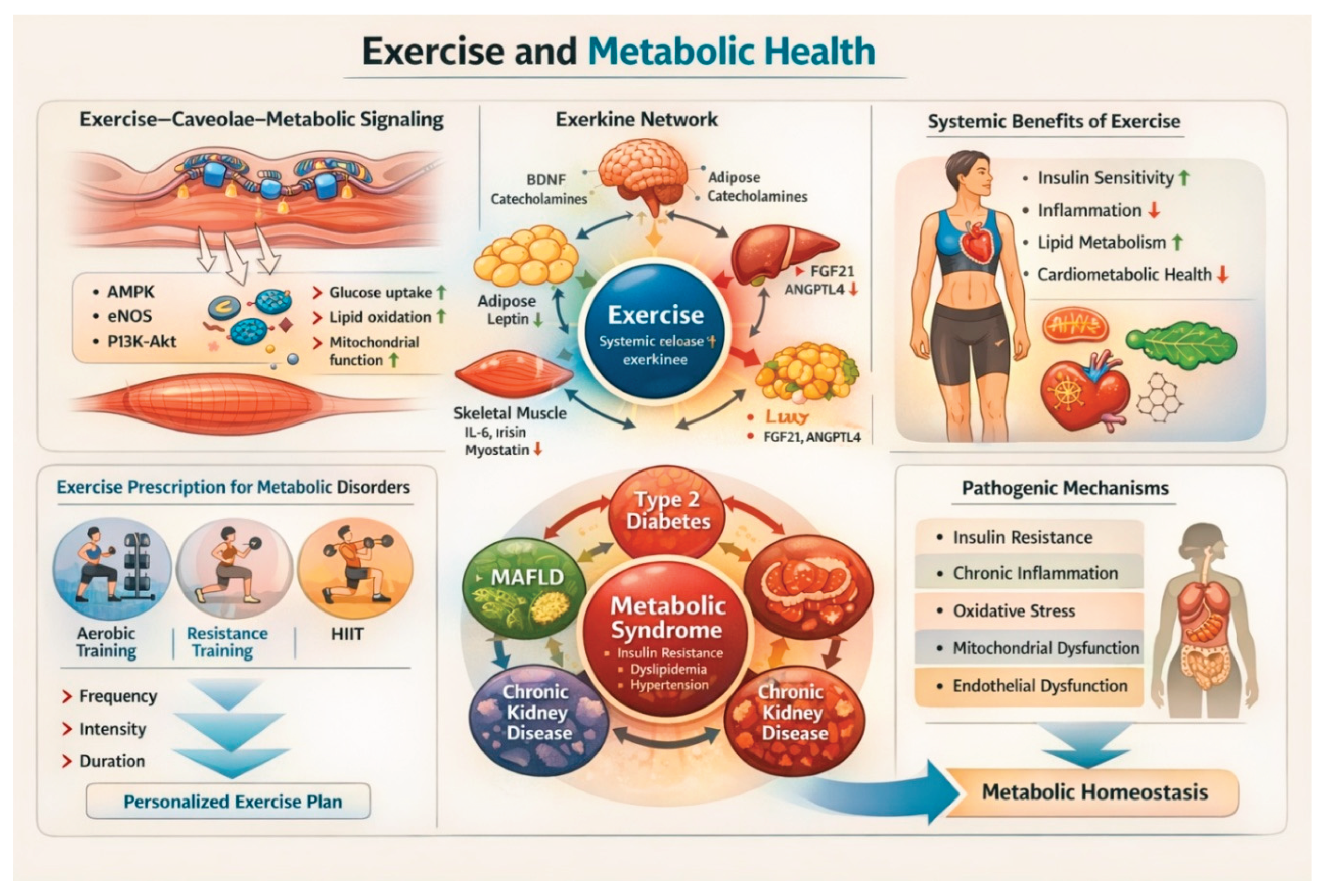
Exercise and Metabolic Health: Exercise enhances metabolic health via caveolae-mediated signaling (AMPK, eNOS, PI3K–Akt) and exerkine-driven inter-organ communication. These adaptations improve insulin sensitivity, lipid metabolism, and mitochondrial function while reducing inflammation and oxidative stress. Tailored exercise modalities help prevent metabolic syndrome and related diseases, ultimately restoring metabolic homeostasis.
Figure 1.
Exercise and Metabolic Health: Exercise enhances metabolic health via caveolae-mediated signaling (AMPK, eNOS, PI3K–Akt) and exerkine-driven inter-organ communication. These adaptations improve insulin sensitivity, lipid metabolism, and mitochondrial function while reducing inflammation and oxidative stress. Tailored exercise modalities help prevent metabolic syndrome and related diseases, ultimately restoring metabolic homeostasis.
9. Conclusions
Metabolic disorders represent a complex and escalating global health burden driven by interconnected disturbances in energy metabolism, inflammation, oxidative stress, and inter-organ communication. Rather than discrete disease entities, conditions such as obesity, T2DM, dyslipidemia, metabolic syndrome, and MAFLD form a unified pathophysiological network in which dysfunction in one tissue propagates across multiple organ systems.
Exercise represents a unique multisystem therapy capable of targeting the interconnected mechanisms underlying metabolic disorders. Its effects extend beyond energy balance to include coordinated molecular, cellular, and systemic adaptations. Through mechanisms including exerkine-mediated signaling, modulation of inflammatory and stress-response pathways, enhancement of mitochondrial function, and remodeling of the gut microbiota, exercise restores metabolic flexibility and improves systemic metabolic homeostasis.
Importantly, these benefits occur across a broad range of metabolic conditions and are often independent of substantial weight loss, highlighting exercise as a fundamental regulator of metabolic health. However, variability in individual responses and challenges in long-term adherence remain significant barriers to implementation.
Future progress will depend on advancing precision exercise medicine through integration of physiological, molecular, and digital health approaches, and on embedding exercise more effectively within clinical care pathways. As a scalable and cost-effective intervention, sustained physical activity represents a cornerstone strategy for reducing the global burden of metabolic disease and improving population health.
References
- Janssen JAMJL. The Causal Role of Ectopic Fat Deposition in the Pathogenesis of Metabolic Syndrome. Vol. 25, International Journal of Molecular Sciences. 2024. p. 13238.
- Godoy-Matos AF, Valério CM, Júnior WSS, de Araujo-Neto JM, Sposito AC, Suassuna JHR. CARDIAL-MS (CArdio-Renal-DIAbetes-Liver-Metabolic Syndrome): a new proposition for an integrated multisystem metabolic disease. Diabetol Metab Syndr [Internet]. 2025;17(1):218. [CrossRef]
- Katzmarzyk PT, Friedenreich C, Shiroma EJ, Lee IM. Physical inactivity and non-communicable disease burden in low-income, middle-income and high-income countries. Br J Sports Med. 2022;56(2).
- Ndumele CE, Neeland IJ, Tuttle KR, Chow SL, Mathew RO, Khan SS, et al. A Synopsis of the Evidence for the Science and Clinical Management of Cardiovascular-Kidney-Metabolic (CKM) Syndrome: A Scientific Statement From the American Heart Association. Circulation [Internet]. 2023 Nov 14;148(20):1636–64. [CrossRef]
- Liu H, Wang S, Wang J, Guo X, Song Y, Fu K, et al. Energy metabolism in health and diseases. Signal Transduct Target Ther [Internet]. 2025;10(1):69. [CrossRef]
- Pandey R, Pharasi N, Kaur P, Kaur L. Inborn errors of metabolism: From toxic, metabolic, and physical insults to neurodegenerative disorders. In: Evidence-Based Neurological Disorders: Symptoms, Causes, and Therapy. 2024.
- Ren Z, Fan H, Xue Y, Yang X, Liu X, Luo J, et al. Mediational role of metabolic syndrome between physical activity, sedentary behavior and non-alcoholic fatty liver disease: a cross-sectional study. BMC Public Health [Internet]. 2025;25(1):1661. [CrossRef]
- Ijaz A, Abbas S, Shabbir M, Badshah Y, Abid F, Afsar T, et al. Inherited metabolic disorders: presentation, clinical types, laboratory diagnosis and genetic markers. Orphanet J Rare Dis [Internet]. 2025;20(1):422. [CrossRef]
- Xu X, Shao X, Hou FF. Risk stratification of metabolic disorder-associated kidney disease. Kidney Int. 2025 Jun;107(6):1002–10.
- Kim ME, Lim Y, Lee JS. Mitochondrial Dysfunction and Metabolic Reprogramming in Chronic Inflammatory Diseases: Molecular Insights and Therapeutic Opportunities. Vol. 47, Current Issues in Molecular Biology. 2025. p. 1042.
- Ring M. An Integrative Approach to HPA Axis Dysfunction: From Recognition to Recovery. Am J Med [Internet]. 2025 Oct 1;138(10):1451–63. [CrossRef]
- Zhang WZ. Chapter 7 - PharmacolZhang, W. Z. (2026). Chapter 7 - Pharmacologically targeting caveolae in metabolic diseases (M. R. B. T.-P. T. in M. D. de Oliveira (ed.); pp. 127–139). Academic Press. https://doi.org/10.1016/B978-0-443-27370-4.00012-. In: de Oliveira MRBTPT in MD, editor. Academic Press; 2026. p. 127–39. Available from: https://www.sciencedirect.com/science/article/pii/B9780443273704000123.
- Dutta B, Tripathy A, Archana PR, Kamath SU. Unraveling the complexities of diet induced obesity and glucolipid dysfunction in metabolic syndrome. Diabetol Metab Syndr [Internet]. 2025;17(1):292. [CrossRef]
- Karakasis P, Theofilis P, Patoulias D, Vlachakis PK, Antoniadis AP, Fragakis N. Diabetes-Driven Atherosclerosis: Updated Mechanistic Insights and Novel Therapeutic Strategies. Vol. 26, International Journal of Molecular Sciences. 2025.
- Pasookhush P, Surawit A, Suta S, Pumeiam S, Mongkolsucharitkul P, Pinsawas B, et al. Transferability of polygenic risk scores for metabolic and cardiovascular traits in an underrepresented population. npj Genomic Med [Internet]. 2025;10(1):76. [CrossRef]
- Abtin S, Ziveh T, Rezaee-Tavirani M. The complicated relationship between inflammation and metabolic dysfunction. J Diabetes Metab Disord [Internet]. 2025;24(2):237. [CrossRef]
- Stea DM, D’Alessio A. Caveolae: Metabolic Platforms at the Crossroads of Health and Disease. Vol. 26, International Journal of Molecular Sciences. 2025.
- Chen W, Kahn CR. Insulin. Trends Endocrinol Metab [Internet]. 2025 Oct 1;36(10):968–9. [CrossRef]
- Geramizadeh B, Ezgu F, Beyzaei Z. Glycogen storage disorder types IX: the mutation spectrum and ethnic distribution. Orphanet J Rare Dis [Internet]. 2024;19(1):475. [CrossRef]
- Zimran A, Revel-Vilk S, Dinur T, Istaiti M, Botha J, Lukina E, et al. Evaluation of Lyso-Gb1 as a biomarker for Gaucher disease treatment outcomes using data from the Gaucher Outcome Survey. Orphanet J Rare Dis [Internet]. 2025;20(1):43. [CrossRef]
- Mengel E, Patterson MC, Da Riol RM, Del Toro M, Deodato F, Gautschi M, et al. Efficacy results from a 12-month double-blind randomized trial of arimoclomol for treatment of Niemann-Pick disease type C (NPC): Presenting a rescored 4-domain NPC Clinical Severity Scale. Mol Genet Metab Reports [Internet]. 2025;43:101233. Available from: https://www.sciencedirect.com/science/article/pii/S2214426925000485.
- Dakal TC, Xiao F, Bhusal CK, Sabapathy PC, Segal R, Chen J, et al. Lipids dysregulation in diseases: core concepts, targets and treatment strategies. Lipids Health Dis [Internet]. 2025;24(1):61. [CrossRef]
- Nulmans I, Lequeue S, Desmet L, Neuckermans J, De Kock J. Current state of the treatment landscape of phenylketonuria. Orphanet J Rare Dis [Internet]. 2025;20(1):281. [CrossRef]
- Socha P, Jańczyk W, Zanetto A, Burra P, Czlonkowska A, Debray D, et al. EASL-ERN Clinical Practice Guidelines on Wilson’s disease. J Hepatol. 2025;82(4).
- Spodymek A, Lachowicz J, Szymczyk-Nużka M. Hemochromatosis — an insidious genetic disease. J Transfus Med Hemost [Internet]. 2025;18(0):93–102. [CrossRef]
- Zinna L, Verde L, Tolla MF Di, Barrea L, Parascandolo A, D’Alterio F, et al. Chronodisruption enhances inflammatory cytokine release from visceral adipose tissue in obesity. J Transl Med [Internet]. 2025;23(1):231. [CrossRef]
- Thyfault JP, Bergouignan A. Exercise and metabolic health: beyond skeletal muscle. Diabetologia [Internet]. 2020;63(8):1464–74. [CrossRef]
- Tokizane K, Imai S ichiro. Inter-organ communication is a critical machinery to regulate metabolism and aging. Trends Endocrinol Metab [Internet]. 2025 Aug 1;36(8):756–66. [CrossRef]
- Iacoviello M, Gori M, Grandaliano G, Minutolo R, Pitocco D, Trevisan R. A holistic approach to managing cardio-kidney metabolic syndrome: insights and recommendations from the Italian perspective. Front Cardiovasc Med [Internet]. 2025;Volume 12. Available from: https://www.frontiersin.org/journals/cardiovascular-medicine/articles/10.3389/fcvm.2025.1583702.
- Burchfield JG, Diaz-Vegas A, James DE. The insulin signalling network. Vol. 7, Nature Metabolism. 2025.
- Michalak KP, Michalak AZ. Understanding chronic inflammation: couplings between cytokines, ROS, NO, Cai2+, HIF-1α, Nrf2 and autophagy. Front Immunol [Internet]. 2025;Volume 16. Available from: https://www.frontiersin.org/journals/immunology/articles/10.3389/fimmu.2025.1558263.
- Xu X, Pang Y, Fan X. Mitochondria in oxidative stress, inflammation and aging: from mechanisms to therapeutic advances. Signal Transduct Target Ther [Internet]. 2025;10(1):190. [CrossRef]
- Chen Y, Liu X, Liu Y, Li Y, Li D, Mei Z, et al. Mitochondrial quality control in diabetes mellitus and complications: molecular mechanisms and therapeutic strategies. Cell Death Dis [Internet]. 2025;16(1):652. [CrossRef]
- Joyce Zhang and Wei Zheng Zhang. Stress-Induced Metabolic Disorders: Mechanisms, Pathologies, and Prospects. Am J Biomed Sci Res. 2025;26(6):834–42.
- Xourafa G, Korbmacher M, Roden M. Inter-organ crosstalk during development and progression of type 2 diabetes mellitus. Vol. 20, Nature Reviews Endocrinology. 2024.
- Zhang C, Hu W, Zhang J, Wang Z, Lu H. Role of Caveolae and Caveolin in Vascular Physiology and Pathology. J Am Heart Assoc [Internet]. 2026 Feb 17;15(4):e047590. [CrossRef]
- Zhang WZ. Chapter 7 - Pharmacologically targeting caveolae in metabolic diseases. In: de Oliveira MRBTPT in MD, editor. Academic Press; 2026. p. 127–39. Available from: https://www.sciencedirect.com/science/article/pii/B9780443273704000123.
- Zhang WZ. The link between caveolae, metabolic syndrome, and cataractogenesis: A mechanistic hypothesis. Exp Eye Res [Internet]. 2026;267:110942. Available from: https://www.sciencedirect.com/science/article/pii/S0014483526000989.
- Zemljic-Harpf AE, Bonds JA, Zuniga-Hertz JP, Patel HH. Role of membrane microdomains in cardiac protection: strategies for diabetic cardiomyopathy. Vol. 329, American Journal of Physiology - Heart and Circulatory Physiology. 2025.
- Singh V, Breton V, Viaris de Lesegno C, Macé AS, Bun P, Blouin CM, et al. Spatiotemporal coupling of caveolae mechanosensing and RhoA-GEFs regulates cell polarity and directional migration. Nat Commun [Internet]. 2025;17(1):398. [CrossRef]
- Gupta A, Shah K, Gupta V. Interconnected epidemics: obesity, metabolic syndrome, diabetes and cardiovascular diseases—insights from research and prevention strategies. Discov Public Heal [Internet]. 2025;22(1):106. [CrossRef]
- Zhang W zheng. An association of metabolic syndrome constellation with cellular membrane caveolae. Pathobiol Aging Age-related Dis. 2014;4(1).
- Hoseini R, Rahim HA, Saifalddin DL, Kareem DA, Fatah AM. Exercise intensity-mediated regulation of gut epithelial cells and immune function in gut microbiota dysbiosis. J Transl Med [Internet]. 2025;24(1):10. [CrossRef]
- Majeed K, Ali H. Association of low-grade inflammation and oxidative stress with metabolic dysfunction in healthy obese individuals. AIMS Allergy Immunol [Internet]. 2025;9(2):56–69. Available from: https://www.aimspress.com/article/doi/10.3934/Allergy.2025004.
- Orsatti FL, de Queiroz Freitas AC, Borges AVB e., Santato AS, de Oliveira Assumpção C, Souza MVC, et al. Unveiling the role of exercise in modulating plasma heat shock protein 27 levels: insights for exercise immunology and cardiovascular health. Mol Cell Biochem [Internet]. 2025;480(3):1381–401. [CrossRef]
- Marino Gammazza A, Macaluso F, Di Felice V, Cappello F, Barone R. Hsp60 in Skeletal Muscle Fiber Biogenesis and Homeostasis: From Physical Exercise to Skeletal Muscle Pathology. Vol. 7, Cells. 2018.
- Ayubi N, Wibawa JC, Callixte C. The mechanism of physical exercises increases heat shock protein 70 (HSP70) (a systematic review). Medicni Perspekt [Internet]. 2024 Dec 26;29(4 SE-THEORETICAL MEDICINE):14–22. Available from: https://journals.uran.ua/index.php/2307-0404/article/view/319168.
- Mikhailova E, Sokolenko A, Combs SE, Shevtsov M. Modulation of Heat Shock Proteins Levels in Health and Disease: An Integrated Perspective in Diagnostics and Therapy. Vol. 14, Cells. 2025.
- Gruper Y, Ben-Shmuel A, Scherz-Shouval R. HSF1 renders NK cells too stressed to respond. Nat Cell Biol [Internet]. 2024;26(10):1630–1. [CrossRef]
- Kumari B. Cellular Stress Responses and Associated Diseases: A Focus on Heat Shock Proteins. Cell Biochem Biophys [Internet]. 2025;83(3):2793–804. [CrossRef]
- Malandish A, Gulati M. The impacts of exercise interventions on inflammaging markers in overweight/obesity patients with heart failure: A systematic review and meta-analysis of randomized controlled trials. IJC Hear Vasc [Internet]. 2023;47:101234. Available from: https://www.sciencedirect.com/science/article/pii/S2352906723000659.
- Crasan IM, Tanase M, Delia CE, Gradisteanu-Pircalabioru G, Cimpean A, Ionica E. Metaflammation’s Role in Systemic Dysfunction in Obesity: A Comprehensive Review. Vol. 26, International Journal of Molecular Sciences. 2025.
- Jin L, Diaz-Canestro C, Wang Y, Tse MA, Xu A. Exerkines and cardiometabolic benefits of exercise: from bench to clinic. EMBO Mol Med [Internet]. 2024;16(3):432–44. [CrossRef]
- Safdar A, Saleem A, Tarnopolsky MA. The potential of endurance exercise-derived exosomes to treat metabolic diseases. Nat Rev Endocrinol [Internet]. 2016;12(9):504–17. [CrossRef]
- Ennequin G, Sirvent P, Whitham M. Role of exercise-induced hepatokines in metabolic disorders. Vol. 317, American Journal of Physiology - Endocrinology and Metabolism. 2019.
- Mohammed SN, Jasim MH, Mahmood SH, Saleh EN, Hashemzadeh A. The role of irisin in exercise-induced muscle and metabolic health: a narrative review. Naunyn Schmiedebergs Arch Pharmacol [Internet]. 2025;398(9):11463–91. [CrossRef]
- He Y, Zhang Y, Lai J, Ma S, Yin P, Wu Z, et al. Plasma metabolomics dataset of race-walking athletes illuminating systemic metabolic reaction of exercise. Sci Data [Internet]. 2025;12(1):448. [CrossRef]
- Guo X, Zhou Z, Wang Y, Sun H, Liu S, He Y, et al. Exercise-induced metabolomics and its association with metabolic health in adolescents. Nutr Metab (Lond) [Internet]. 2025;22(1):48. [CrossRef]
- Hawley JA, Forster SC, Giles EM. Exercise, the Gut Microbiome and Gastrointestinal Diseases: Therapeutic Impact and Molecular Mechanisms. Vol. 169, Gastroenterology. 2025.
- Sohail MU, Yassine HM, Sohail A, Al Thani AA. Impact of physical exercise on gut microbiome, inflammation, and the pathobiology of metabolic disorders. Rev Diabet Stud. 2019;15(1).
- Rengasamy Balakrishnan Ji-Yeon Lee, Yang-Kook Rho, Byoung-Kook Kim, Dong-Kug Choi SIK. Gut Microbiota-Immune System Interactions in Health and Neurodegenerative Diseases: Insights into Molecular Mechanisms and Therapeutic Applications. Vol. 16, Aging and disease. p. 3421–52.
- Shi J, Cui J, Zheng T, Han X, Wang B, Wang W, et al. Comparative effects of aerobic and resistance exercise on bile acid profiles and liver function in patients with non-alcoholic fatty liver disease. BMC Gastroenterol [Internet]. 2025;25(1):239. [CrossRef]
- Franklin BA, Zhu W. High-Intensity Interval Training: Benefits, Risks, and Clinical Implications. J Sci Sport Exerc [Internet]. 2026;8(1):1–8. [CrossRef]
- Boelius HM, Aatsinki AK, Heiskanen MA, Haapala EA, Munukka E, Mykkänen J, et al. Association of leisure time physical activity with gut microbiota composition in early adulthood. Sci Rep [Internet]. 2025;15(1):19697. [CrossRef]
- Havers T, Held S, Schönfelder M, Geisler S, Wackerhage H. Effects of Skeletal Muscle Hypertrophy on Fat Mass and Glucose Homeostasis in Humans and Animals: A Narrative Review with Systematic Literature Search. Sport Med [Internet]. 2025;55(8):1867–85. [CrossRef]
- Smart NA, Downes D, van der Touw T, Hada S, Dieberg G, Pearson MJ, et al. The Effect of Exercise Training on Blood Lipids: A Systematic Review and Meta-analysis. Sport Med [Internet]. 2025;55(1):67–78. [CrossRef]
- Maufroy E, Baeyens N, Deboeck G. Mechanoreceptor-mediated adaptations to physical exercise: from acute responses to long-term training effects. Cell Mol Life Sci [Internet]. 2026. [CrossRef]
- Li J, Bai J, Liu G, Zhu Z, Cao C. Exercise Intervention in Autonomic Function, Immunity, and Cardiovascular Health: A Precision Medicine Approach. Vol. 12, Journal of Cardiovascular Development and Disease. 2025.
- Liu X, Liu H, Chen L, Duan J, Ran H, Chen L, et al. Volume OXygenation index in predicting the efficacy of early non-invasive ventilation in patients with acute hypoxic respiratory failure: A multicenter study. Hear Lung J Cardiopulm Acute Care [Internet]. 2025 Jul 1;72:42–7. [CrossRef]
- Ichikawa T, Sugiura H. Role of Nitrosative Stress: What Is the Potential Clinical Implication? BT - Asthma-COPD Overlap: Updated Concept, Pathophysiology, Diagnosis and Treatment. In: Nagase H, Sugiura H, Shirai T, editors. Singapore: Springer Nature Singapore; 2024. p. 111–25. [CrossRef]
- de Matos DG, de Santana JL, Aidar FJ, Cornish SM, Giesbrecht GG, Nunes-Silva A, et al. Changes in Autonomic Balance, Cardiac Parasympathetic Modulation, and Cardiac Baroreflex Gain in Older Adults Under Different Orthostatic Stress Conditions. Healthc. 2025;13(19).
- Wang J, Zhang J, Zhang H, Yu F, Tian Z, Jia D. Exercise modulates redox homeostasis in cardiovascular and metabolic diseases: from bench to clinic. Cardiovasc Diabetol [Internet]. 2026. [CrossRef]
- Botella J, Perri E, Caruana NJ, López-Calcerrada S, Brischigliaro M, Jamnick NA, et al. Sprint interval exercise disrupts mitochondrial ultrastructure driving a unique mitochondrial stress response and remodelling in men. Nat Commun [Internet]. 2025;17(1):71. [CrossRef]
- Satheesan A, Kumar J, Leela KV, Lathakumari RH, Angelin M, Murugesan R, et al. The multifaceted regulation of white adipose tissue browning and their therapeutic potential. J Physiol Biochem [Internet]. 2025;81(4):925–47. [CrossRef]
- Mallett G. The effect of exercise and physical activity on skeletal muscle epigenetics and metabolic adaptations. Eur J Appl Physiol [Internet]. 2025;125(3):611–27. [CrossRef]
- Heo JW, Kim MJ, Yang YJ, Choi HN, Kim KY, Oh TW, et al. The role of tight junctions in the pathogenesis of inflammatory bowel disease: immune modulation and barrier dysfunction. Mol Cell Toxicol [Internet]. 2025;21(3):495–506. [CrossRef]
- Finderle J, Schleicher VS, Schleicher LMS, Krsek A, Braut T, Baticic L. Exercise-Induced Modulation of the Gut Microbiota: Mechanisms, Evidence, and Implications for Athlete Health. Gastrointest Disord. 2025;8(1).
- Wang J feng, Mao S jie, Xia F, Li X lin. Effects of aerobic and resistance exercise on patients with hypertension: a systematic review and meta-analysis focusing on the sympathetic nervous system. Front Cardiovasc Med [Internet]. 2025;Volume 12. Available from: https://www.frontiersin.org/journals/cardiovascular-medicine/articles/10.3389/fcvm.2025.1569638.
- Magni O, Arnaoutis G, Panagiotakos D. The impact of exercise on chronic systemic inflammation: a systematic review and meta–meta-analysis. Sport Sci Health [Internet]. 2025;21(3):1405–17. [CrossRef]
- Mengistu FA, Lake YA, Andualem ME, Miherete YD, Zewdie SA. Impact of aerobic, resistance, and combined training on cardiometabolic health-related indicators in inactive middle-aged men with excess body weight and obesity. Front Physiol [Internet]. 2025;Volume 16. Available from: https://www.frontiersin.org/journals/physiology/articles/10.3389/fphys.2025.1519180.
- Garcia SP, Cureau FV, Iorra F de Q, Bottino LG, R. C. Monteiro LE, Leivas G, et al. Effects of exercise training and physical activity advice on HbA1c in people with type 2 diabetes: A network meta-analysis of randomized controlled trials. Diabetes Res Clin Pract [Internet]. 2025 Mar 1;221. [CrossRef]
- Powell LH, Berkley-Patton J, Drees BM, Karavolos K, Lohse B, Masters KS, et al. Lifestyle Intervention for Sustained Remission of Metabolic Syndrome: A Randomized Clinical Trial. JAMA Intern Med [Internet]. 2026 Jan 1;186(1):67–77. [CrossRef]
- American College of Sports Medicine. ACSM publishes updated resistance training guidelines. [Internet]. 2026. Available from: https://acsm.org/resistance-training-guidelines-update-2026/.
- Lin X, Li G, Xie J, Wang S. Effects of concurrent training on fat mass and its distribution in individuals with overweight or obesity: a systematic review, meta-analysis, and exploratory regression analysis. BMC Public Health [Internet]. 2026. [CrossRef]
- van Baak MA, Mariman ECM. Physiology of Weight Regain after Weight Loss: Latest Insights. Curr Obes Rep [Internet]. 2025;14(1):28. [CrossRef]
- Smart NA, Wood GN, Pearson MJ, Dieberg G, van der Touw T, Kostner K. Exercise training for the management of dyslipidaemia. A position statement from Exercise and Sports Science Australia (ESSA). J Sci Med Sport [Internet]. 2026 Mar 24. [CrossRef]
- Huang W, He Y, Chen R, Huo C, Xie Y, Lin Y, et al. Independent effects of exercise intensity on hepatic fat reduction in adults with metabolic dysfunction–associated steatotic liver disease: a randomized controlled trial protocol. Front Endocrinol (Lausanne) [Internet]. 2026;Volume 17. Available from: https://www.frontiersin.org/journals/endocrinology/articles/10.3389/fendo.2026.1711420.
- Cefis M, Marcangeli V, Hammad R, Granet J, Leduc-Gaudet JP, Gaudreau P, et al. Impact of physical activity on physical function, mitochondrial energetics, ROS production, and Ca2+ handling across the adult lifespan in men. Cell Reports Med [Internet]. 2025 Feb 18;6(2). [CrossRef]
- Bärg M, Idiart-Borda Polotto V, Geiger S, Held S, Brinkmann C. Effects of home- and gym-based resistance training on glycemic control in patients with type 2 diabetes mellitus—a systematic review and meta-analysis. Diabetol Metab Syndr [Internet]. 2025;17(1):228. [CrossRef]
- Al-Mhanna SB, Poon ETC, Franklin BA, Tarnopolsky MA, Hawley JA, Jakicic JM, et al. Comparative effectiveness of high-intensity interval training and moderate-intensity continuous training on cardiometabolic health in patients with diabesity: a systematic review and meta-analysis of randomized controlled trials. Diabetol Metab Syndr [Internet]. 2025;17(1):331. [CrossRef]
- AUSactive. Cardiovascular exercise prescription for healthy adults (Version 2). [Internet]. 2024. Available from: https://ausactive.org.au/wp-content/uploads/2024/06/Cardiovascular-Exercise-Prescription-for-Healthy-Adults-v2.pdf.
- American Diabetes Association Professional Committee. 6. Glycemic goals and hypoglycemia: Standards of care in diabetes—2025. Diabetes Care. 2025;48.
- Michielsen M, Yagiz J, Hanssens M, Claes J, Geuns L, Gojevic T, et al. The effect of exercise characteristics on HbA1c and other cardiovascular risk factors in adults with type 2 diabetes: a systematic review and meta-analysis of randomised controlled trials. Cardiovasc Diabetol. 2025.
- Yang Y, Peng Y, Min Q, Lian Y, Wang D, Song X, et al. Effects of combined high-intensity interval and moderate-intensity continuous training vs. moderate-intensity continuous training alone in male adolescents: a randomized controlled trial. Sci Rep [Internet]. 2025;15(1):13353. [CrossRef]
- Lewis BA, Napolitano MA, Buman MP, Williams DM, Nigg CR. Physical activity interventions: an update on advancing sedentary time, technology, and dissemination and implementation research. J Behav Med [Internet]. 2025;48(1):99–110. [CrossRef]
- Jasbi P, Mohr AE, Murthy MHS, Klein-Seetharaman J. Understanding metabolic resilience by unraveling temporal dynamics of cellular responses. Trends Endocrinol Metab [Internet]. 2025 Dec 1;36(12):1084–98. [CrossRef]
- Rodrigues KB, Weng Z, Graham ZA, Lavin K, McAdam J, Tuggle SC, et al. Exercise intensity and training alter the innate immune cell type and chromosomal origins of circulating cell-free DNA in humans. Proc Natl Acad Sci [Internet]. 2025 Jan 21;122(3):e2406954122. [CrossRef]
- Dube A, Shaw I, Mathunjwa ML, Shaw BS. Impact of Traditional Dance and Games on Cardiovascular Health: A Scoping Review of Outcomes Across Diverse Low- and Middle-Income Countries. Vol. 22, International Journal of Environmental Research and Public Health. 2025.
- Fominiene VB, Fominaite M, Sipaviciene S. Physicians’ opinions on barriers to patient counseling on physical activity in primary care: focus on physicians’ healthy exercise habits and knowledge about physical activity. Front Med [Internet]. 2026;Volume 13. Available from: https://www.frontiersin.org/journals/medicine/articles/10.3389/fmed.2026.1711438.
- Zhao K, Chen Z, Huang Q, Li S, Tan G, Guo K, et al. Multi-level determinants of physical activity and sports participation among adults during COVID-19 pandemic: an interpretable machine learning approach. Front Psychol [Internet]. 2026;Volume 16. Available from: https://www.frontiersin.org/journals/psychology/articles/10.3389/fpsyg.2025.1701201.
- Bannuru RR, ElSayed NA, Aroda VR, Aronne LJ, Balapattabi K, Bennett AK, et al. Weight stigma and bias: standards of care in overweight and obesity-2025. Vol. 13, BMJ Open Diabetes Research and Care. 2025.
- Noone J, Mucinski JM, DeLany JP, Sparks LM, Goodpaster BH. Understanding the variation in exercise responses to guide personalized physical activity prescriptions. Cell Metab [Internet]. 2024 Apr 2;36(4):702–24. [CrossRef]
- Hansen D, Junior GC, Milani JGPO, Milani M, Gojevic T, Machado FVC, et al. Advancing Aerobic Exercise Training Intensity Prescription in Health and Disease Beyond Standard Recommendations: A Call to Action. Vol. 55, Sports Medicine. 2025.
- Marques de Sá e Silva D, Avgerinou G, Petridou A, Theodoridis G, Mougios V, Gika H. Metabolomic signatures of high-intensity and sprint interval exercise/training in humans: a systematic review. Metabolomics [Internet]. 2026;22(2):25. [CrossRef]
- Kumar KV, Yerraguntla KR, Jenne MP, Gadi A, Sepoori A, Gunda A, et al. Advancements in Continuous Glucose Monitoring: a Revolution in Diabetes Management. Biomed Mater Devices [Internet]. 2025. [CrossRef]
- Ma S, Coopergard R, Clements M, Chow L. Managing Exercise-Related Glycemic Events in Type 1 Diabetes: Development and Validation of Predictive Models for a Practical Decision Support Tool. JMIR Diabetes [Internet]. 2025;10:e68948. Available from: https://diabetes.jmir.org/2025/1/e68948.
- Jacques M, Landen S, Sharples AP, Garnham A, Schittenhelm R, Steele J, et al. Molecular landscape of sex- and modality-specific exercise adaptation in human skeletal muscle through large-scale multi-omics integration. Cell Rep [Internet]. 2025 Jun 24;44(6). [CrossRef]
- Osman Y, Akimbekov NS, El-Zayat EM, Hassan N, Digel I. Cavin gene family and caveolae-related disorders: pathogenetic roles and possible mechanisms. Cell Commun Signal [Internet]. 2026;24(1):136. [CrossRef]
- Martin D, Bonneau M, Orfila L, Horeau M, Hazon M, Demay R, et al. Atypical gut microbial ecosystem from athletes with very high exercise capacity improves insulin sensitivity and muscle glycogen store in mice. Cell Rep [Internet]. 2025 Apr 22;44(4). [CrossRef]

Table 1.
Common Metabolic Disorders.
| Disorder Category | Examples | Pathophysiology Highlights |
|---|---|---|
| Carbohydrate Metabolism Disorders | Diabetes mellitus (T1DM, T2DM), glycogen storage diseases [18,19] | Impaired insulin secretion or action, or enzyme deficiencies leading to dysregulated glucose homeostasis. |
| Lipid Metabolism Disorders | Dyslipidemia, Gaucher disease, Niemann–Pick disease [20,21,22] | Defects in lipid transport, storage, or catabolism resulting in abnormal lipid accumulation in circulation or tissues. |
| Amino Acid Metabolism Disorders | Phenylketonuria (PKU) [23] | Enzyme deficiencies impair amino acid metabolism, leading to accumulation of toxic intermediates. |
| Metal Metabolism Disorders | Hemochromatosis, Wilson’s disease [24,25] | Impaired metal transport or excretion results in toxic accumulation in organs such as the liver and brain. |
| Metabolic Syndrome | Central obesity, hypertension, insulin resistance, dyslipidemia [13,26,27] | Systemic metabolic dysregulation driven by insulin resistance, chronic inflammation, and altered lipid metabolism; emerging evidence suggests a potential role for caveolae and caveolin-1 in coordinating metabolic signaling. |
Disclaimer/Publisher’s Note: The statements, opinions and data contained in all publications are solely those of the individual author(s) and contributor(s) and not of MDPI and/or the editor(s). MDPI and/or the editor(s) disclaim responsibility for any injury to people or property resulting from any ideas, methods, instructions or products referred to in the content. |
© 2026 by the authors. Licensee MDPI, Basel, Switzerland. This article is an open access article distributed under the terms and conditions of the Creative Commons Attribution (CC BY) license (http://creativecommons.org/licenses/by/4.0/).
Copyright: This open access article is published under a Creative Commons CC BY 4.0 license, which permit the free download, distribution, and reuse, provided that the author and preprint are cited in any reuse.


