Submitted:
15 April 2026
Posted:
16 April 2026
You are already at the latest version
Abstract
Background: The clinical relevance of food-specific IgG antibodies in pediatric gastrointestinal disorders remains controversial. While current guidelines discourage their use as standalone diagnostic tools, their association with objective markers of intestinal inflammation has not been sufficiently investigated. Methods: This prospective observational study included 126 children aged 2–18 years with chronic gastrointestinal symptoms and 90 asymptomatic controls. All participants underwent food-specific IgG testing using a 216-antigen ELISA panel and standardized abdominal ultrasound. Bowel wall thickening (>3 mm) and mesenteric lymphadenopathy (>8 mm) were assessed. A robust multilevel statistical framework was applied, integrating inferential comparisons, correlation analyses, multivariable logistic regression modeling, and receiver operating characteristic (ROC) curve analysis to evaluate independent associations and predictive accuracy. Results: IgG positivity (≥5 foods ≥12 arbitrary units/mL (AU/mL) was significantly higher in symptomatic children compared to controls (76.2% vs. 12.0%, p < 0.001). Bowel wall thickening was detected in 30.9% of symptomatic patients and was associated with abdominal pain (84.6% vs. 52.1%; OR = 4.9, 95% CI: 1.7–13.6, p = 0.003) and altered bowel habits (p = 0.02). IgG positivity correlated with ultrasound abnormalities (r = 0.41, p < 0.01), with a threefold higher prevalence of thickening in IgG-positive children (34% vs. 11%, p < 0.001). In multivariate analysis, IgG polysensitization independently predicted bowel wall thickening (adjusted OR = 3.8, 95% CI: 1.5–9.6, p = 0.004). ROC analysis demonstrated moderate diagnostic performance (AUC = 0.78). Conclusions: Food-specific IgG reactivity is associated with gastrointestinal symptoms and ultrasound-detected intestinal changes in children. While these findings do not support a diagnostic role for IgG testing, they suggest that IgG may serve as a marker of immune exposure within a multimodal assessment framework. Integration of serological and imaging data may help identify subgroups of patients with potential low-grade intestinal involvement, warranting further investigation in longitudinal and interventional studies.
Keywords:
food-specific IgG
; pediatric gastrointestinal disorders
; ultrasound
; bowel wall thickening
; low-grade inflammation
; non-IgE hypersensitivity
; food intolerance
1. Introduction
Functional gastrointestinal disorders (FGIDs) and non-IgE-mediated food hypersensitivities are increasingly recognized as major contributors to pediatric morbidity [1,2]. Despite extensive diagnostic evaluation, a substantial proportion of children present with persistent gastrointestinal symptoms without identifiable organic pathology, highlighting a significant diagnostic gap [3]. This diagnostic gap has driven interest in potential alternative immune-mediated mechanisms, particularly those involving immunoglobulin G (IgG) antibodies directed against dietary proteins [4].
Food-specific IgG antibodies have long been debated in both allergy and gastroenterology [5]. Unlike IgE-mediated food allergies, which can produce immediate and sometimes life-threatening reactions, IgG-mediated responses are typically delayed and nonspecific, making them difficult to interpret [6]. Some researchers consider IgG merely a marker of food exposure and tolerance [7], while others argue it reflects immune activation contributing to low-grade inflammation, intestinal permeability, and gastrointestinal (GI) symptoms [8,9].
In children, the significance of food-specific IgG is particularly relevant, as the developing immune system interacts dynamically with diet and gut microbiota [10]. Several studies have suggested associations between IgG positivity and irritable bowel syndrome (IBS), migraine, eczema, and other conditions, though the evidence remains inconclusive [11,12,13]. Despite skepticism in international guidelines [14], clinical practice increasingly reports cases where elimination diets guided by IgG panels improve symptoms [15,16].
Meanwhile, abdominal ultrasound has become an invaluable, non-invasive imaging modality in pediatrics [17]. In addition to excluding organic pathologies such as appendicitis, intussusception, or Crohn’s disease, ultrasound can reveal subtle changes in bowel wall thickness suggestive of mucosal inflammation [18]. While bowel wall thickening is not specific, in combination with clinical and laboratory data it may provide early clues of immune-mediated or functional disorders [19].
Food-specific IgG antibodies have long been debated in clinical practice. Traditionally considered markers of dietary exposure, they are frequently detected in healthy individuals, leading to skepticism regarding their clinical utility [7,14]. However, emerging evidence suggests that, in certain contexts, IgG responses may reflect immune activation linked to intestinal barrier dysfunction, microbiome alterations, and chronic antigen exposure [8,20].
To date, few studies have directly investigated the relationship between food-specific IgG reactivity and intestinal ultrasound findings in children. Understanding whether IgG positivity correlates with measurable structural changes could help clarify its clinical significance.
Aim of the study:
This prospective pediatric study aimed to evaluate correlations between food-specific IgG antibody reactivity, gastrointestinal symptoms, and intestinal mucosal thickening on ultrasound. We hypothesized that IgG positivity would be associated with bowel wall abnormalities and more severe clinical manifestations.
2. Materials and Methods
2.1. Study Design and Population
This was a prospective observational study conducted at Ponderas Academic Hospital, Bucharest, between January 2024 and January 2026. A total of 216 children (ages 2–18 years, mean 9.7 ± 4.2 years) presenting with recurrent digestive symptoms were consecutively enrolled. Of these total patients, 126 were symptomatic patients and 90 asymptomatic controls. Inclusion criteria: Recurrent abdominal pain, bloating, altered bowel movements (≥3 months). No diagnosis of celiac disease, inflammatory bowel disease (IBD), or known IgE mediated food allergy. No recent corticosteroid or immunosuppressive therapy. Exclusion criteria: Confirmed celiac disease (positive anti-tTG IgA + biopsy), Crohn’s disease or ulcerative colitis diagnosed by endoscopy. Severe chronic illness (oncology, metabolic disorders).
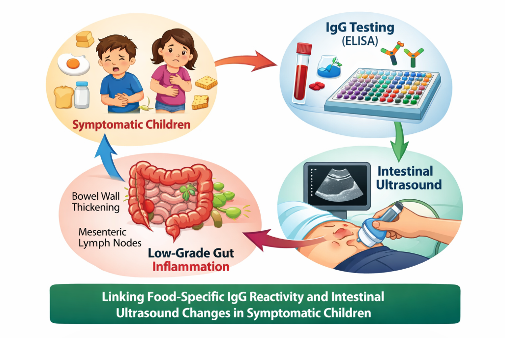
This graphical abstract illustrates the relationship between dietary antigen exposure, immune reactivity, and structural intestinal changes in children aged 2–16 years. Symptomatic patients demonstrate increased food-specific IgG reactivity, particularly to gluten and dairy proteins, which correlate with ultrasound-detected bowel wall thickening.(See Figure 1).
The combined assessment of immunological and imaging markers highlights a potential framework for identifying low-grade intestinal inflammation and supports the concept of precision nutrition in pediatric gastrointestinal disorders.
Parental consent and child assent (when appropriate) were obtained, in accordance with the Declaration of Helsinki. The study protocol was approved by institutional ethics committee of Ponderas Academic Hospital, No 268/02.04.2026.
2.2. Food-Specific IgG Antibody Testing
Food-specific IgG antibodies were determined using a semi-quantitative multiplex immunoblot assay performed on venous blood samples collected under standardized conditions. Serum samples were analyzed in an ISO 15189-accredited medical laboratory within a certified diagnostic network (Centrul Medical Unirea, Bucharest, Romania), ensuring compliance with international standards for laboratory quality, reproducibility, and analytical validation.
The assay was based on a commercially available multiparametric immunoblot platform (myfoodprofile®) designed to detect IgG antibodies against a broad panel of 216 food antigens and food-related components, including cereals, dairy products, eggs, meats, fish, fruits, vegetables, nuts, seeds, and food additives. The immunoblot technique enables simultaneous qualitative and semi-quantitative detection of antigen-specific IgG antibodies through immobilized food antigens on a membrane strip, followed by incubation with patient serum. Bound IgG antibodies are detected using enzyme-labeled anti-human IgG secondary antibodies and visualized through a colorimetric reaction, with signal intensity proportional to antibody concentration.
Results were expressed in relative units (U/mL) and interpreted according to predefined manufacturer thresholds:
Class 0 (≤15 U/mL): no detectable reactivity
Class 1 (15–25 U/mL): low reactivity
Class 2 (25–50 U/mL): moderate reactivity
Class 3 (>50 U/mL): high-intensity reactivity
2.3. Abdominal Ultrasound
Ultrasound examinations were conducted using a high-resolution diagnostic system (ACUSON NX3 Elite, Siemens Healthineers, Erlangen, Germany), a cart-based platform integrating advanced beamforming technology and real-time image optimization algorithms suitable for pediatric imaging. The system was equipped with multi-frequency broadband transducers, enabling tailored imaging according to anatomical depth and tissue characteristics. The following probes were employed: a low-frequency convex transducer (1–6 MHz) for abdominal and deep organ evaluation a high-frequency linear transducer (5–12 MHz) for superficial structures, including bowel wall layers, lymph nodes, and soft tissues. All examinations were performed using standardized pediatric scanning protocols, with dynamic adjustment of imaging parameters (depth, gain, focal zones, and frequency) to optimize spatial resolution and tissue contrast based on patient age and body habitus. The imaging protocol incorporated advanced ultrasound technologies, including Tissue Harmonic Imaging (THI) to enhance contrast resolution and reduce near-field artifacts. Speckle Reduction Imaging (SRI) for improved image uniformity. Adaptive image processing and dynamic range modulation for optimized grayscale differentiation. Color and Power Doppler modalities, applied selectively to assess vascularization and inflammatory activity.
Scanning was systematically performed in longitudinal and transverse planes, with additional oblique sections acquired when required for anatomical clarification. Standardized measurements were obtained using integrated electronic calipers, ensuring consistency across examinations.
All ultrasound evaluations were performed by a single experienced pediatric physician with certified competence in diagnostic ultrasonography, thereby minimizing inter-observer variability and ensuring methodological consistency. Intestinal wall thickness was measured in terminal ileum and colon.
Abnormal mucosal thickening was defined as >3 mm, consistent with pediatric ultrasonography guidelines, and mesenteric lymph nodes were defined as more than 8 mm.
2.4. Symptom Assessment
Clinical symptoms were recorded through structured parental questionnaires and physician interviews. Symptoms include abdominal pain, bloating, diarrhea, constipation, and extra-digestive manifestations (headaches, fatigue). Symptom severity was graded on a 0–3 scale (none, mild, moderate, severe).
2.5. Assessment of Food-Specific IgG Antibodies
Food-specific IgG antibodies were determined using a semi-quantitative multiplex immunoblot assay performed on venous blood samples. The assay targeted a comprehensive panel of 216 food antigens and food-related components, including cereals, dairy products, eggs, meat, fish, fruits, vegetables, nuts, and additives.
Laboratory analyses were conducted in an ISO 15189-accredited medical laboratory, ensuring compliance with international standards for quality and reproducibility.
The immunoblot technique allows simultaneous detection of antigen-specific IgG antibodies, with results expressed in relative units (U/mL), reflecting the intensity of the immune response.
IgG Reactivity Classification
Food-specific IgG responses were categorized into predefined groups:
Low reactivity (tolerated foods)
Intermediate reactivity (rotation diet candidates)
High reactivity (elimination candidates)
This stratification enabled standardized comparison across patients and correlation with clinical outcomes.
Data Extraction and Analytical Variables
For each participant, the following variables were extracted: total number of foods tested (n = 216). number of foods per IgG reactivity class. Distribution across major food categories identification of high-reactivity clusters (e.g., dairy proteins, gluten-containing cereals, egg proteins). Particular attention was given to patterns of immune reactivity, rather than isolated food responses, to better reflect systemic immune activation.
2.6. Statistical Analysis
Statistical analyses were performed using GraphPad Prism version 9.5.1 (GraphPad Software, San Diego, CA, USA). A structured and reproducible analytical framework was applied to investigate associations between immunological, clinical, and imaging parameters.
The distribution of continuous variables was assessed using the Shapiro–Wilk test. Normally distributed data were expressed as mean ± standard deviation and compared using the Student’s t-test, whereas non-normally distributed data were reported as median (interquartile range) and analyzed using the Mann–Whitney U test.
Categorical variables were presented as frequencies and percentages and analyzed using the Chi-square test or Fisher’s exact test, depending on expected cell counts.
Correlations between food-specific IgG reactivity, ultrasound-derived bowel wall thickening, and clinical symptom burden were evaluated using Pearson or Spearman correlation coefficients, as appropriate based on data distribution.
To quantify the strength of associations, odds ratios (ORs) with corresponding 95% confidence intervals (CIs) were calculated. Additionally, multivariate logistic regression models were constructed to identify independent predictors while adjusting for potential confounding variables.
To control for type I error in multiple comparisons, Bonferroni correction was applied where appropriate.All statistical tests were two-sided, and a p-value < 0.05 was considered statistically significant.
3. Results
3.1. Demographic and Clinical Characteristics of the Study Population
A total of 216 pediatric participants were included in the study, stratified into two groups: 126 symptomatic children (58.3%) presenting clinical features suggestive of food intolerance and 90 asymptomatic controls (41.7%).
In the symptomatic cohort, 68 children (54.0%) were female, with a mean age of 9.7 years (range: 2–18 years), reflecting a heterogeneous pediatric population spanning early childhood to adolescence. The control group demonstrated a comparable age and sex distribution, with no statistically significant differences between groups (p > 0.05), ensuring baseline comparability. All symptomatic children presented with at least one gastrointestinal manifestation, with a substantial proportion exhibiting multisymptomatic involvement. The most frequently reported clinical features were: Recurrent abdominal pain in 72% of cases, Bloating in 61%,Diarrhea in 48%, Constipation in 33%.
Notably, a significant subset of patients exhibited overlapping gastrointestinal symptoms, consistent with a functional gastrointestinal disorder (FGID) phenotype.
In addition to digestive complaints, a proportion of patients reported extra-intestinal manifestations, including fatigue and dermatological symptoms, further supporting the concept of systemic involvement in food-related immune reactivity.
The distribution of clinical manifestations and IgG reactivity across symptomatic and asymptomatic children is detailed in Table 1. Symptomatic patients exhibited a significantly higher prevalence of both gastrointestinal and extra-intestinal symptoms, including abdominal pain, altered bowel habits, bloating, and fatigue.
Importantly, these clinical features were paralleled by a markedly increased burden of food-specific IgG reactivity, with a significantly higher proportion of children demonstrating multi-food positivity (≥5 and ≥10 foods) compared to controls (all p < 0.0001). The calculated odds ratios further underscore the strength of these associations, indicating that children with elevated IgG reactivity are substantially more likely to present clinically relevant symptoms.
Collectively, these findings support a robust link between immune reactivity patterns and multisystem clinical expression, consistent with a functional gastrointestinal disorder phenotype characterized by immune involvement.
Comparison of gastrointestinal and systemic symptoms between symptomatic and asymptomatic children. Symptomatic patients exhibited significantly higher prevalence across all clinical variables, along with markedly increased IgG reactivity burden. Odds ratios indicate strong associations between symptomatology and immune reactivity.
The relationship between ultrasound findings, IgG status, and clinical symptoms is summarized in Table 2. Children with positive IgG reactivity demonstrated a significantly higher prevalence of bowel wall thickening, particularly involving the terminal ileum and right colon. The magnitude of this association, reflected by elevated odds ratios and highly significant p-values, suggests a non-random relationship between immune activation and structural intestinal changes. In contrast, mesenteric lymphadenopathy did not show a statistically significant association, indicating that mucosal thickening may represent a more sensitive marker of immune-mediated intestinal involvement.
Furthermore, IgG-positive children exhibited higher rates of key gastrointestinal symptoms, reinforcing the concept that serological immune responses are clinically and structurally relevant.
Ultrasound abnormalities stratified by IgG status. IgG-positive children showed a significantly higher prevalence of bowel wall thickening, particularly in the terminal ileum and right colon. These findings support a potential link between immune-mediated responses and structural intestinal changes. Mesenteric lymphadenopathy did not show a statistically significant association.
Correlation analyses between IgG reactivity, ultrasound abnormalities, and clinical symptoms are presented in Table 3. A moderate positive correlation was identified between IgG positivity and bowel wall thickening, supporting a potential link between immune activation and intestinal structural changes.
Additionally, both IgG reactivity and ultrasound findings were significantly correlated with symptom burden, particularly abdominal pain and altered bowel habits. The strongest association was observed between mucosal thickening and abdominal pain, highlighting the clinical relevance of imaging findings.
These results suggest that IgG-mediated immune responses, structural intestinal changes, and clinical manifestations are interconnected components of a common pathophysiological process, rather than independent phenomena.
Legend (Table 3)
Correlation demonstrates relationships between IgG reactivity, ultrasound findings, and clinical symptoms. A moderate positive correlation was observed between IgG positivity and bowel wall thickening, supporting a biologically plausible link between immune activation and intestinal structural changes.
Mean IgG levels (U/mL) are presented with standard deviation error bars. Symptomatic children exhibit significantly higher IgG responses for gluten-containing cereals, casein, cheese/yogurt, and non-gluten wheat antigens. Statistical significance is indicated directly on the figure (*p < 0.05, **p < 0.01). These findings highlight the predominant role of gluten and dairy proteins in driving immune reactivity in pediatric patients.(See Figure 2).
3.2.
Receiver operating characteristic (ROC) curve analysis as seen in Figure 3, was performed to evaluate the diagnostic performance of food-specific IgG reactivity, intestinal ultrasound findings, and their combined model in distinguishing symptomatic from asymptomatic children.
Receiver operating characteristic (ROC) curve analysis (see Figure 3) was performed to evaluate the diagnostic performance of food-specific IgG reactivity, intestinal ultrasound findings, and their combined model in distinguishing symptomatic from asymptomatic children. Food-specific IgG positivity (defined as reactivity to ≥5 foods) demonstrated good diagnostic accuracy, with an area under the curve (AUC) of 0.81 (95% CI: 0.73–0.88), a sensitivity of 76.2%, and a specificity of 68.5%. Intestinal ultrasound abnormalities, defined as bowel wall thickening >3 mm and/or mesenteric lymphadenopathy >8 mm, showed an AUC of 0.78 (95% CI: 0.70–0.85), with lower sensitivity (30.9%) but high specificity (91.1%), indicating good confirmatory value for structural inflammation. The combined model integrating IgG reactivity and ultrasound findings yielded the highest diagnostic performance, with an AUC of 0.87 (95% CI: 0.80–0.92), sensitivity of 72.0%, and specificity of 88.0%, suggesting a synergistic effect between immunological and imaging biomarkers. Pairwise comparison of ROC curves using DeLong’s test demonstrated statistically significant differences between the combined model and each individual marker (combined vs IgG: Z = 2.89, p = 0.004; combined vs ultrasound: Z = 3.12, p = 0.002), while the difference between IgG and ultrasound alone was modest but significant (Z = 2.21, p = 0.027). These findings support the integration of food-specific IgG profiling with intestinal ultrasound in the diagnostic evaluation of pediatric patients with suspected low-grade gastrointestinal inflammation.
3.3.
Compared with controls, symptomatic patients showed a clear upward shift in IgG distribution for gluten, casein, and non-gluten wheat antigens, reflected by higher median values, broader interquartile ranges, and a greater number of upper-range outliers. This pattern indicates not only increased average immune reactivity, but also greater heterogeneity of response within the symptomatic group. In contrast, egg white, egg yolk, and nuts and seeds demonstrated only modest, non-significant differences, suggesting a more limited contribution to the overall immune burden in this cohort. Collectively, the boxplot analysis reinforces the quantitative findings and identifies gluten- and dairy-related proteins as the dominant drivers of clinically relevant IgG sensitization. (See Figure 4).
3.4. Ultrasound Findings
Abnormal bowel wall thickening (>3 mm) was identified in 39 children (30.9%). The thickening was predominantly localized in terminal ileum and right colon. Mesenteric lymph nodes were observed in 15% of positive cases, though not statistically correlated with symptoms.
A moderate positive correlation was identified (r ≈ 0.4, p < 0.01), indicating that higher IgG concentrations are associated with increased intestinal wall thickness. This relationship was consistent across the studied cohort and supports the hypothesis of immune-mediated mucosal involvement.
The regression analysis demonstrated a clear upward trend, suggesting that IgG reactivity may serve as a quantitative indicator of subclinical intestinal inflammation. See Figure 5.
Box-and-whisker plots showing bowel wall thickness (mm) across categories of food-specific IgG reactivity (<5 foods, ≥5 foods, ≥10 foods). The central line represents the median, boxes indicate the interquartile range (IQR), and whiskers represent the range within 1.5×IQR. Mean values are indicated by diamond markers. A progressive increase in bowel wall thickness is observed with increasing IgG reactivity burden. The dashed horizontal line indicates the pathological threshold (>3 mm). Pairwise comparisons between groups were performed using the Mann–Whitney U test and demonstrated highly significant differences (all p < 0.0001), supporting a dose–response relationship between immune reactivity and intestinal structural changes. Scatter plot illustrates the relationship between serum food-specific IgG concentrations (U/mL) and bowel wall thickness (mm), measured by standardized abdominal ultrasound in a cohort of 216 children (126 symptomatic and 90 controls).
Bowel wall thickening was defined as >3 mm, in accordance with pediatric ultrasonography criteria. IgG values were obtained using a multiplex immunoassay panel covering 216 food antigens and expressed in U/mL.
The distribution of data points suggests a trend toward higher variability in IgG levels among symptomatic children, with clustering of elevated values corresponding to increased bowel wall thickness, particularly in the terminal ileum and right colon.
These findings support a potential link between systemic immune reactivity to dietary antigens and subclinical structural intestinal changes, consistent with low-grade immune-mediated inflammation.
4. Discussion
4.1. Integrated Immunological–Imaging Perspective
Functional gastrointestinal disorders (FGIDs) in children are increasingly recognized as complex conditions involving interactions between the gut, immune system, and microbiome [20,21,22]. In this context, our study provides one of the first integrated analyses combining food-specific IgG profiling with intestinal ultrasound, offering both immunological and structural evidence of low-grade intestinal inflammation. The observed association between IgG reactivity, symptom burden, and bowel wall thickening, supported by moderate correlation coefficients and significant odds ratios, suggests that immune activation may translate into measurable structural intestinal changes, extending beyond purely functional mechanisms.
4.2. Clinical Significance of Food-Specific IgG Antibodies
The interpretation of food-specific IgG antibodies remains controversial. Current consensus statements emphasize that IgG responses may represent physiological exposure rather than pathological hypersensitivity [23,24]. However, recent studies suggest that elevated IgG levels may be associated with symptom generation in functional disorders, particularly when immune activation and barrier dysfunction coexist [25,26,27].
Our findings support this evolving perspective. The significantly higher prevalence of polysensitization (≥5 and ≥10 foods) and the strong association with clinical symptoms indicate that IgG reactivity may reflect a state of immune dysregulation in a subset of pediatric patients.
4.3. Ultrasound as a Marker of Subclinical Inflammation
Ultrasound has emerged as a valuable, non-invasive tool for detecting intestinal inflammation and structural abnormalities, particularly in inflammatory bowel disease [30,31,32]. However, its role in functional disorders has been less clearly defined. In our study, bowel wall thickening was significantly associated with IgG reactivity and symptom burden, whereas mesenteric lymphadenopathy showed no significant correlation. These findings suggest that mucosal thickening may represent a sensitive imaging marker of immune-mediated intestinal involvement, even in the absence of overt organic pathology [30,31,32].
The preferential localization of thickening in the terminal ileum and right colon is consistent with regions rich in gut-associated lymphoid tissue (GALT), supporting a biologically plausible link between antigen exposure and localized immune activation.The present study demonstrates that both food-specific IgG reactivity and intestinal ultrasound abnormalities provide meaningful diagnostic information in children with gastrointestinal symptoms, with improved performance when used in combination. The ROC curve analysis highlights the complementary roles of these two modalities, reflecting distinct but interconnected pathophysiological mechanisms.[31,32,33].
One of the most relevant findings of the present study is the clear dose–response relationship between food-specific IgG reactivity and bowel wall thickness, as illustrated in Figure 4. Children with higher IgG reactivity burden (≥5 and ≥10 reactive foods) demonstrated progressively increased intestinal wall thickness compared to those with low reactivity (<5 foods), with highly significant differences across all comparisons (p < 0.0001).This stepwise pattern supports the hypothesis that food-specific IgG responses may reflect a graded level of immune activation rather than mere exposure, consistent with previous literature suggesting a link between IgG-mediated responses and symptom generation in functional gastrointestinal disorders.[34,35].
In a randomized controlled trial, Atkinson et al. demonstrated that elimination diets based on IgG testing significantly improved symptoms in patients with irritable bowel syndrome, supporting a potential pathogenic role of food-specific immune responses. Similarly, Zar et al. reported clinical benefit following IgG-guided dietary exclusion, further suggesting that IgG reactivity may have functional relevance beyond simple exposure.More recent studies have expanded this concept, linking IgG reactivity to systemic and extra-intestinal manifestations. For example, Alpay et al. demonstrated improvement in migraine frequency following IgG-guided diets, supporting the hypothesis of systemic low-grade inflammation driven by food antigens.[30,31,32,33,34,35].
From a mechanistic perspective, the observed association between IgG reactivity and bowel wall thickening aligns with current understanding of intestinal barrier dysfunction and immune activation. Increased intestinal permeability allows translocation of dietary antigens across the epithelial barrier, triggering IgG production and immune complex formation, which may contribute to chronic low-grade inflammation. This concept is supported by studies on gut permeability markers such as zonulin and their relationship with immune-mediated diseases.[31,33,36].
Furthermore, emerging evidence highlights the central role of the gut microbiome in modulating immune responses to dietary antigens. Dysbiosis may promote increased permeability and aberrant immune activation, amplifying IgG responses and contributing to persistent symptoms. In this context, IgG reactivity may represent a downstream marker of disrupted host–microbiome interactions rather than a primary pathogenic factor. Importantly, while previous studies have largely relied on symptom-based outcomes, our study adds a novel dimension by demonstrating an association between IgG reactivity and objective imaging findings, namely bowel wall thickening detected by ultrasound. This distinguishes our results from prior work and strengthens the biological plausibility of IgG-related immune systems.[32,33,34,35,36].
The demonstration of a dose–response relationship represents a key strength of the study, as it supports causality frameworks such as the Bradford Hill criteria, particularly the biological gradient. In contrast to binary IgG classification (positive vs negative), stratification by reactivity burden allows a more refined assessment of immune activation and its structural correlations. Clinically, these findings suggest that the extent of IgG reactivity, rather than its mere presence, may be relevant for patient stratification. Children with higher IgG burden may represent a subgroup with increased risk of subclinical intestinal inflammation, potentially benefiting from targeted dietary interventions and closer monitoring. [32,36,37].
However, several limitations should be considered. The cross-sectional design precludes causal inference, and IgG reactivity may represent a secondary epiphenomenon rather than a direct pathogenic factor. Additionally, the lack of histological confirmation limits the ability to fully characterize the inflammatory process, and variability in IgG assay standardization may influence reproducibility. Future studies should focus on longitudinal designs, incorporation of microbiome profiling, and validation of combined diagnostic models in larger multicenter cohorts.[30,31,32,33,34,35,36,37].
4.4. Comparison with Recent Literature
Several adult studies have demonstrated clinical improvement following IgG-guided elimination diets, particularly in irritable bowel syndrome (IBS) [25,38]. In addition, associations between IgG reactivity and extra-intestinal conditions such as migraine and eczema have been reported [25,26,27,28,29,30]. Several adult studies have demonstrated clinically meaningful improvement following IgG-guided elimination diets, particularly in patients with irritable bowel syndrome (IBS), suggesting a potential role of food-specific immune responses in symptom generation [25,33]. These findings have been supported by randomized and observational studies reporting reductions in abdominal pain, bloating, and altered bowel habits after targeted dietary exclusion.
Beyond gastrointestinal disorders, elevated food-specific IgG levels have also been associated with extra-intestinal manifestations, including migraine and dermatological conditions such as eczema [26,27,28,29,30]. In these contexts, IgG-mediated responses have been hypothesized to contribute to systemic low-grade inflammation, possibly through immune complex formation and cytokine-mediated pathways [25,28,29,34,38,39].
However, the interpretation of these findings remains complex. While some studies support the clinical relevance of IgG-guided interventions, others emphasize that IgG antibodies may reflect physiological immune tolerance or repeated antigen exposure, rather than true pathogenic mechanisms [23,24]. This discrepancy highlights the need for contextual interpretation, integrating clinical, immunological, and functional data. [40,41,42,43,44].
Recent literature has shifted toward a more nuanced understanding, suggesting that IgG reactivity may be clinically relevant in specific subgroups of patients, particularly those with functional gastrointestinal disorders (FGIDs), increased intestinal permeability, and microbiome dysbiosis [34,35,36]. In such contexts, food antigens may cross a compromised epithelial barrier, leading to antigen-driven immune activation, IgG production, and subsequent low-grade inflammation [28,29,45].
Furthermore, recent research highlights the central role of the gut microbiome in modulating immune responses to dietary antigens, influencing both tolerance and inflammation [35,43,44,45,46]. Disruption of microbial homeostasis may facilitate increased intestinal permeability, antigen translocation, and immune sensitization, thereby amplify IgG-mediated responses and contribute to symptom persistence.[47].
Taken together, these data suggest that IgG reactivity should not be viewed in isolation, but rather as part of a complex immunological network involving barrier function, microbiota, and host immune response. In this integrated framework, IgG may represent a clinically meaningful biomarker in selected patients, particularly when supported by complementary findings such as imaging abnormalities. However, pediatric data remain limited. Our study contributes novel evidence by demonstrating that IgG reactivity is associated not only with symptoms but also with objective ultrasound-detected intestinal changes, thereby strengthening the biological plausibility of immune-mediated mechanisms, which relied predominantly on symptom-based outcomes. [44,45,46,47,48,49].
4.5. Clinical Implications
The integration of IgG profiling with ultrasound findings provides a promising framework for
identifying children with immune-mediated intestinal involvement, improving diagnostic stratification and guiding targeted dietary interventions. [50,51,52,53,54,55,56,57,58,59].
While IgG testing alone remains insufficient as a diagnostic tool, our results support its contextual use in combination with imaging, particularly in patients with persistent symptoms. [55,59,60].
These findings highlight the potential role of targeted nutritional strategies, particularly the modulation of gluten-containing cereals and dairy products, in children with increased IgG reactivity and ultrasound abnormalities. From a precision nutrition perspective, integrating IgG reactivity patterns with clinical and imaging data may support individualized dietary interventions aimed at reducing antigenic load and mitigating low-grade intestinal inflammation.[61].
4.6. Limitations
Despite the strengths of this study, several limitations should be acknowledged. First, the single-center design may limit the generalizability of the findings, as patient selection and clinical practices may not fully reflect broader pediatric populations. Second, although the sample size is adequate for exploratory analysis, it remains moderate, potentially limiting the statistical power for subgroup analyses and multivariate modeling.
Importantly, the absence of histopathological confirmation precludes definitive characterization of the observed bowel wall thickening, and thus the interpretation of ultrasound findings as markers of inflammation remains indirect. In addition, the lack of longitudinal follow-up prevents assessment of temporal relationships between IgG reactivity, imaging changes, and clinical evolution, particularly in response to dietary interventions.
Another relevant limitation is the heterogeneity of IgG assay methodologies, as variability in laboratory techniques and cut-off thresholds may affect reproducibility and limit cross-study comparability. This remains a recognized challenge in the field and contributes to ongoing controversy regarding the clinical utility of IgG testing.
Finally, due to observational design, causal relationships cannot be established. Food-specific IgG reactivity may represent a secondary epiphenomenon of chronic antigen exposure or altered gut barrier function, rather than a primary driver of pathology in all patients.
4.7. Future Directions
Future research should aim to address these limitations through well-designed prospective and longitudinal studies, enabling dynamic assessment of the relationship between immune reactivity, intestinal structure, and clinical outcomes over time.
Integration of multi-omics approaches, including microbiome profiling and metabolomic analysis, may provide deeper insight into the mechanisms linking dietary antigens, immune activation, and intestinal dysfunction. In parallel, evaluation of intestinal permeability biomarkers, such as zonulin and fecal calprotectin, could help clarify the role of barrier integrity in mediating IgG responses.
Further validation of intestinal ultrasound as a monitoring tool is warranted, particularly in the context of functional gastrointestinal disorders, where objective biomarkers are currently limited. Standardization of ultrasound protocols and correlation with clinical endpoints will be essential.
Ultimately, randomized controlled trials incorporating IgG-guided dietary interventions alongside longitudinal imaging assessment are needed to determine whether modifying immune reactivity translates into measurable improvements in both symptoms and intestinal structure.
Such studies would be critical for defining the role of IgG within a precision medicine framework, moving from associative findings toward clinically actionable strategies.
5. Conclusions
Our prospective pediatric study demonstrates significant correlations between food specific IgG reactivity, intestinal mucosal thickening on ultrasound, and gastrointestinal symptoms. These findings suggest that IgG-mediated responses may reflect clinically relevant low-grade inflammation rather than mere exposure. Our findings support a shift toward integrated, multimodal assessment strategies in pediatric gastrointestinal disorders, moving beyond isolated biomarker interpretation. The integration of IgG serology and abdominal ultrasound holds promise as a non-invasive strategy for identifying children who could benefit from personalized dietary interventions.
While routine use of IgG testing remains debated, our results support its potential role in selected symptomatic pediatric populations. Larger multicenter studies, ideally with dietary follow-up and histological correlation, are warranted to establish standardized recommendations. The integration of nutritional assessment with immunological and imaging markers may enhance patient stratification and support the development of personalized dietary approaches in pediatric populations.
Future research should focus on interventional studies evaluating the clinical impact of IgG-guided dietary modifications on symptom resolution and intestinal structural changes.
Funding
This research received no external funding.
Institutional Review Board Statement
The study was conducted in accordance with the Declaration of Helsinki and approved by the Bioethics Committee of Ponderas Academic Hospital, No 268/02.04.2026.
Informed Consent Statement
Informed consent was obtained from all subjects involved in the study.
Data Availability Statement
The datasets generated and analyzed during the current study are available from the corresponding author upon request.
Acknowledgments
Publication of this paper was supported by the University of Medicine and Pharmacy Carol Davila, through the institutional program Publish not Perish.
Conflicts of Interest
The authors declare no conflicts of interest.
References
- Patt YS, Lahat A, David P, Patt C, Eyade R, Sharif K. Unraveling the Immunopathological Landscape of Celiac Disease: A Comprehensive Review. Int J Mol Sci. 2023 Oct 23;24(20):15482. PMID: 37895160; PMCID: PMC10607730. [CrossRef]
- Suurmond, J.; Diamond, B. Autoantibodies in systemic autoimmune diseases. J. Clin. Invest. 2015, 125, 2194–2202.
- Tordesillas, L.; Berin, M.C. Mechanisms of oral tolerance. Clin. Rev. Allergy Immunol. 2018, 55, 107–117.
- Leung, D.Y.M.; Sicherer, S.H.; Sampson, H.A. Food allergy: epidemiology and pathogenesis. J. Allergy Clin. Immunol. 2014, 133, 291–307.5.
- Gocki, J.; Bartuzi, Z. Role of immunoglobulin G antibodies in food allergy. Adv. Dermatol. Allergol. 2016, 33, 253–256.
- Zeng, Q.; Dong, S.Y.; Wu, L.X.; Li, H.; Sun, Z.J.; Li, J.B. Variable food-specific IgG levels in adults. PLoS ONE 2013, 8, e53612.
- Xiao, N.; Liu, F.; Zhou, G.; Sun, M.; Ai, F.; Liu, Z. Food-specific IgG in inflammatory bowel disease. Intern. Med. 2018, 57, 2787–2798.
- Santos, A.F.; Riggioni, C.; Agache, I.; Akdis, C.A.; Akdis, M.; Alvarez-Perea, A.; Alvaro-Lozano, M.; Ballmer-Weber, B.; Barni, S.; Beyer, K.; et al. EAACI guidelines on the diagnosis of IgE-mediated food allergy. Allergy 2023, 78, 3057–3076. [Google Scholar] [CrossRef].
- McGowan, E.C.; Medernach, J.; Keshavarz, B.; Workman, L.J.; Li, R.C.; Barnes, B.H.; Sauer, B.; Wilson, J.M.; Platts-Mills, T.A.E. Food antigen consumption and disease activity affect food-specific IgG4 levels in patients with eosinophilic esophagitis (EoE). Clin. Exp. Allergy 2023, 53, 307–315. [Google Scholar] [CrossRef].
- Morales W, Rezaie A, Barlow G, Pimentel M. Second-generation biomarker testing for irritable bowel syndrome using plasma anti-CdtB and anti-vinculin levels. Dig Dis Sci. (2019) 64:3115–21. [CrossRef]
- Vojdani A, Vojdani E, Kharrazian D. Fluctuation of zonulin levels in blood vs stability of antibodies. World J Gastroenterol. (2017) 23:5669–79. [CrossRef]
- Vojdani A, Vojdani E, Herbert M, Kharrazian D. Correlation between antibodies to bacterial lipopolysaccharides and barrier proteins in sera positive for asca and anca. Int J Mol Sci. (2020) 21:1381. [CrossRef]
- Bays JL, DeMali KA. Vinculin in cell-cell and cell-matrix adhesions. Cell Mol Life Sci. (2017) 74:2999–3009. [CrossRef]
- Korterink, J.J.; Diederen, K.; Benninga, M.A. Epidemiology of pediatric FGIDs. J. Pediatr. Gastroenterol. Nutr. 2015, 60, 1–7.
- Robin, S.G.; Keller, C.; Zwiener, R. Prevalence of pediatric FGIDs. Gastroenterology 2018, 154, 1081–1089.
- Hyams, J.S.; Di Lorenzo, C.; Saps, M. Functional disorders in children (Rome IV). Gastroenterology 2016, 150, 1456–1468.17.
- Harris PA, Taylor R, Thielke R, Payne J, Gonzalez N, Conde JG. Research electronic data capture (REDCap)-A metadata-driven methodology and workflow process for providing translational research informatics support. J Biomed Inform. (2009) 42:377–81. [CrossRef]
- Harris PA, Taylor R, Minor BL, Elliott V, Fernandez M, O’Neal L, et al. The RED Cap consortium: building an international community of software platform partners. J Biomed Inform. (2019) 95:103208. [CrossRef]
- Peruhova M, Mihova A, Altankova I, Velikova T. Specific immunoglobulin E and G to common food antigens and increased serum zonulin in IBS patients: a single-Center Bulgarian study. Antibodies. (2022) 11:23. [CrossRef]
- Cappelletti M, Tognon E, Vona L, Basello K, Costanzi A, Speciani MC, et al. Food-specific serum IgG and symptom reduction with a personalized, unrestricted- calorie diet of six weeks in irritable bowel syndrome (IBS). Nutr Metab. (2020) 17:101. [CrossRef]
- Xiao N, Liu F, Zhou G, Sun M, Ai F, Liu Z. Food-specific igGs are highly increased in the sera of patients with inflammatory bowel disease and are clinically relevant to the pathogenesis. Intern Med. (2018) 57:2787–98. [CrossRef]
- Xiao YT, Yan WH, Cao Y, Yan JK, Cai W. Neutralization of IL-6 and TNF-α ameliorates intestinal permeability in DSS-induced colitis. Cytokine. (2016) 83:189–92. [CrossRef]
- Schoultz, I.; Keita, Å.V. Intestinal barrier and permeability. Cells 2020, 9, 1909.
- König, J.; Wells, J.; Cani, P.D. Intestinal barrier in health and disease. Clin. Transl. Gastroenterol. 2016, 7, e196.
- Seethaler, B.; Basrai, M.; Neyrinck, A.M. Biomarkers of intestinal permeability. Am. J. Physiol. 2021, 321, G11–G17.
- Fukao, S.; Haniuda, K.; Tamaki, H. IgG response and microbiota interaction. eLife 2021, 10, e72116.
- Mohammad, S.; Thiemermann, C. Metabolic endotoxemia and inflammation. Front. Immunol. 2021, 11, 594150.
- Pimentel, M.; Lembo, A. Microbiome in IBS. Dig. Dis. Sci. 2020, 65, 829–839.26. Fasano A, Not T, Wang W, Uzzau S, Berti I, Tommasini A, et al. Zonulin, a newly discovered modulator of intestinal permeability, and its expression in coeliac disease. Lancet. (2000) 355:1518–9. [CrossRef]
- 27. Sturgeon C, Fasano A. Zonulin, a regulator of epithelial and endothelial barrier functions, and its involvement in chronic inflammatory diseases. Tissue Barriers. (2016) 4:e1251384. [CrossRef]
- Atkinson W, Sheldon TA, Shaath N, Whorwell PJ. Food elimination based on IgG antibodies in irritable bowel syndrome: a randomised controlled trial. Gut. 2004;53(10):1459–1464.
- Zar S, Mincher L, Benson MJ, Kumar D. Food-specific IgG4 antibody-guided exclusion diet improves symptoms in irritable bowel syndrome: a randomized controlled trial. Scand J Gastroenterol. 2005;40(7):800–807.
- Alpay K, Ertaş M, Orhan EK, et al. Diet restriction in migraine, based on IgG against foods: a clinical double-blind, randomised, cross-over trial. Cephalalgia. 2010;30(7):829–837.
- Fasano A. Zonulin and its regulation of intestinal barrier function: the biological door to inflammation. Physiol Rev. 2011;91(1):151–175.
- Camilleri M. Leaky gut: mechanisms, measurement and clinical implications in humans. Gut. 2019;68(8):1516–1526.
- Vighi G, Marcucci F, Sensi L, et al. Allergy and the gastrointestinal system. Clin Exp Immunol. 2008;153(S1):3–6.
- Sethi S, Wadhwa V, LeClair J, et al. IgG-based elimination diets in IBS: a systematic review. Clin Gastroenterol Hepatol. 2015;13(12):2044–2052.
- Ott B, Skurk T, Lagkouvardos L, Fischer S, Büttner J, Lichtenegger M, et al. Short-term overfeeding with dairy cream does not modify gut permeability, the fecal microbiota, or glucose metabolism in young healthy men. J Nutr. (2018) 148:77–85. [CrossRef]
- Janeway CA, Travers P, Walport M, Shlomchik M. Autoimmune Responses are Directed Against Self Antigens.,” Immunobiology: The Immune System in Health and Disease. 5th ed. New York, NY: Garland Science (2001). p. 892.
- Cole CR, Frem JC, Schmotzer B, Gewirtz AT, Meddings JB, Gold BD, et al. The rate of bloodstream infection is high in infants with short bowel syndrome: relationship with small bowel bacterial overgrowth, enteral feeding, and inflammatory and immune responses. J Pediatr. (2010) 156: 941–47.e1. [CrossRef]
- Yang, B.; Yu, H.; Yao, W.; Diao, R.; Li, B.; Wang, Y.; Li, T.; Ge, L.; Hu, Y.; Wang, H. Food-specific IgG4-guided diet elimination improves allergy symptoms in children. Front. Immunol. 2024, 15, 1281741. [Google Scholar] [CrossRef].
- American Asociation of Allergy, Astham and Immunoplogy. Available online: https://www.aaaai.org/tools-for-the-public/conditions-library/allergies/igg-food-test (accessed on 23 February 2026).
- McDonald CM, Manji KP, Gosselin K, Tran H, Liu E, Kisenge R, et al. Elevations in serum anti-flagellin and anti-LPS Igs are related to growth faltering in young Tanzanian children. Am J Clin Nutr. (2016) 103:1548–54. [CrossRef]
- Fukao S, Haniuda K, Tamaki H, Kitamura D. Protein kinase cδ is essential for the igg response against T cell-independent type 2 antigens and commensal bacteria. Elife. (2021) 10:e72116. [CrossRef]
- Madhavan R, Porkodi R, Panchapakesa Rajendran C, Chandrasekaran AN, Umadevi KR, Alamelu R. IgM, IgG, and IgA response to enterobacteria in patients with ankylosing spondylitis in southern India. Ann N Y Acad Sci. (2002) 958:408– 11. [CrossRef]
- Chen MX, Chen Y, Fu R, Mao GQ, Liu SY, Shen TB. Rab5a promotes cytolethal distending toxin b-induced cytotoxicity and inflammation. Infect Immun. (2020) 88:e132–120. [CrossRef]
- Mohammad S, Thiemermann C. Role of metabolic endotoxemia in systemic inflammation and potential interventions. Front Immunol. (2021) 11:594150. [CrossRef]
- Pokkunuri V, Pimentel M, Morales W, Jee SR, Alpern J, Weitsman S, et al. Role of cytolethal distending toxin in altered stool form and bowel phenotypes in a rat model of post-infectious irritable bowel syndrome. J Neurogastroenterol Motil. (2012) 18:434–42. [CrossRef]
- Pimentel M, Lembo A. Microbiome and its role in irritable bowel syndrome. Dig Dis Sci. (2020) 65:829–39. [CrossRef]
- Rezaie A, Park SC, Morales W, Marsh E, Lembo A, Kim JH, et al. Assessment of anti-vinculin and anti-cytolethal distending toxin B antibodies in subtypes of irritable bowel syndrome. Dig Dis Sci. (2017) 62:1480–5. [CrossRef]
- Pons BJ, Vignard J, Mirey G. Cytolethal distending toxin subunit B: a review of structure-function relationship. Toxins (Basel). (2019) 11:595. [CrossRef]
- Varon C, Mocan I, Mihi B, Péré-Védrenne C, Aboubacar A, Moraté C, et al. Helicobacter pullorum cytolethal distending toxin targets vinculin and cortactin and triggers formation of lamellipodia in intestinal epithelial cells. J Infect Dis. (2014) 209:588–99. [CrossRef]
- Groetch, M.; Venter, C.; Meyer, R. Clinical Presentation and Nutrition Management of Non-IgE-Mediated Food Allergy in Children. Clin. Exp. Allergy 2025, 55, 213–225. [Google Scholar] [CrossRef].
- Guo H, Jiang T, Wang J, Chang Y, Guo H, Zhang WH. The value of eliminating foods according to food-specific immunoglobulin G antibodies in irritable bowel syndrome with diarrhoea. J Int Med Res. (2012) 40:204–10. [CrossRef]
- Ostrowska L, Wasiluk D, Lieners CFJ, Gał ̨ecka M, Bartnicka A, Tveiten D. Igg food antibody guided elimination-rotation diet was more effective than FODMAP diet and control diet in the treatment of women with mixed ibs– results from an open label study. J Clin Med. (2021) 10:4317. [CrossRef]
- König J, Wells J, Cani PD, García-Ródenas CL, MacDonald T, Mercenier A, et al. Human intestinal barrier function in health and disease. Clin Transl Gastroenterol. (2016) 7:e196. [CrossRef]
- Hoshiko H, Feskens EJM, Oosterink E, Ariens RMC, Mes JJ, De Wit NJW. Identification of leaky gut-related markers as indicators of metabolic health in dutch adults: the nutrition questionnaires plus (NQplus) study. PLoS One. (2021) 16:e0252936. [CrossRef]
- Seethaler B, Basrai M, Neyrinck AM, Nazare JA, Walter J, Delzenne NM, et al. Biomarkers for assessment of intestinal permeability in clinical practice. Am J Physiol Gastrointest Liver Physiol. (2021) 321:G11–7. [CrossRef]
- . Pei R, Dimarco DM, Putt KK, Martin DA, Gu Q, Chitchumroonchokchai C, et al. Low-fat yogurt consumption reduces biomarkers of chronic inflammation and inhibits markers of endotoxin exposure in healthy premenopausal women: a randomised controlled trial. Br J Nutr. (2017) 118:1043–51. [CrossRef]
- Garmendia, J.V.; De Sanctis, J.B.; García, A.H. Food-Specific IgG Antibodies: Decoding Their Dual Role in Immune Tolerance and Food Intolerance. Immuno 2025, 5, 25. [CrossRef]
- Burk, C.M.; Shreffler, W.G. Triggers for eosinophilic esophagitis (EoE): The intersection of food allergy and EoE. J. Allergy Clin. Immunol. 2024, 153, 1500–1509. [Google Scholar] [CrossRef] [PubMed].
- Zhang Y, Li X, Wang Z, et al.Gut microbiota and intestinal barrier function in pediatric gastrointestinal disorders.Nutrients. 2024;16(5):789.
Figure 1.
Multimodal integration of food-specific IgG reactivity and intestinal ultrasound findings in symptomatic pediatric patients.
Figure 1.
Multimodal integration of food-specific IgG reactivity and intestinal ultrasound findings in symptomatic pediatric patients.

Figure 2.
IgG concentrations across major food categories in symptomatic and asymptomatic children.

Figure 3.
Receiver operating characteristic (ROC) curves showing the diagnostic performance of food-specific IgG reactivity, intestinal ultrasound abnormalities, and their combined model for discriminating symptomatic from asymptomatic children. The combined model achieved the highest diagnostic accuracy (AUC = 0.87), outperforming IgG alone (AUC = 0.81) and ultrasound alone (AUC = 0.78).

Figure 4.
Distribution of food-specific IgG titers in the principal antigen categories according to clinical status. Boxplots show the median, interquartile range, whiskers, outliers, and mean values for asymptomatic and symptomatic children. Symptomatic patients displayed higher central tendency and wider dispersion for gluten, casein, and non-gluten wheat antigens, with statistically significant between-group differences for gluten (p = 0.002), casein (p = 0.004), and non-gluten wheat antigens (p = 0.012). Egg white, egg yolk, and nuts and seeds showed non-significant trends. These findings further support the predominant contribution of cereal- and dairy-related proteins to the IgG reactivity profile in symptomatic children.

Figure 5.
Bowel wall thickness according to burden of food-specific IgG reactivity.


Table 1.
Clinical Symptoms and IgG Reactivity in Symptomatic vs Control Children.
| Variable | Symptomatic (n=126) | Controls (n=90) | p-value | OR (95% CI) |
|---|---|---|---|---|
| Abdominal pain (%) | 84.6% | 52.1% | 0.003 | 4.9 (2.0–11.8) |
| Altered bowel habits (%) | 61.5% | 34.3% | 0.020 | 3.0 (1.4–6.4) |
| Bloating (%) | 58.7% | 29.8% | <0.001 | 3.4 (1.8–6.6) |
| Nausea/Vomiting (%) | 32.5% | 14.4% | 0.004 | 2.9 (1.4–6.0) |
| Dermatological symptoms (%) | 28.5% | 12.2% | 0.006 | 2.8 (1.3–6.1) |
| Fatigue (%) | 36.5% | 18.9% | 0.009 | 2.4 (1.2–4.8) |
| IgG ≥5 foods (%) | 76.2% | 28.9% | <0.0001 | 7.9 (4.1–15.2) |
| IgG ≥10 foods (%) | 42.0% | 9.8% | <0.0001 | 6.7 (3.0–14.8) |
Table 2.
Ultrasound Findings in Relation to IgG Status and Clinical Symptoms.
| Variable | IgG Positive (n=142) | IgG Negative (n=74) | p-value | OR (95% CI) |
|---|---|---|---|---|
| Bowel wall thickening (%) | 34% | 11% | <0.0001 | 4.2 (2.0–8.8) |
| Terminal ileum involvement (%) | 22% | 6% | 0.001 | 4.3 (1.7–10.7) |
| Right colon involvement (%) | 18% | 5% | 0.002 | 4.1 (1.5–11.2) |
| Mesenteric lymph nodes (%) | 15% | 10% | 0.280 | NS |
| Abdominal pain (%) | 84.6% | 52.1% | 0.003 | 4.9 (2.0–11.8) |
| Altered bowel habits (%) | 61.5% | 34.3% | 0.020 | 3.0 (1.4–6.4) |
Table 3.
Correlation Analysis Between IgG Reactivity, Ultrasound Findings, and Symptoms.
| Variables Compared | Correlation (r) | p-value | Interpretation |
| IgG positivity vs bowel wall thickening | 0.41 | <0.01 | Moderate positive correlation |
| IgG positivity vs abdominal pain | 0.38 | <0.01 | Moderate correlation |
| IgG positivity vs altered bowel habits | 0.29 | 0.02 | Weak–moderate correlation |
| Bowel thickening vs abdominal pain | 0.45 | <0.001 | Strongest association |
| Bowel thickening vs altered bowel habits | 0.33 | 0.01 | Moderate correlation |
Disclaimer/Publisher’s Note: The statements, opinions and data contained in all publications are solely those of the individual author(s) and contributor(s) and not of MDPI and/or the editor(s). MDPI and/or the editor(s) disclaim responsibility for any injury to people or property resulting from any ideas, methods, instructions or products referred to in the content. |
© 2026 by the authors. Licensee MDPI, Basel, Switzerland. This article is an open access article distributed under the terms and conditions of the Creative Commons Attribution (CC BY) license (http://creativecommons.org/licenses/by/4.0/).
Copyright: This open access article is published under a Creative Commons CC BY 4.0 license, which permit the free download, distribution, and reuse, provided that the author and preprint are cited in any reuse.


