Submitted:
14 April 2026
Posted:
15 April 2026
You are already at the latest version
Abstract
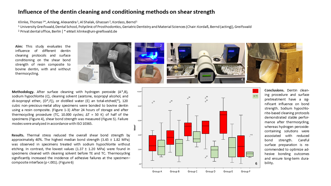
(1) Background: This article evaluates the influence of different dentin cleaning protocols and surface conditioning on the shear bond strength of resin composite to bovine dentin, with and without thermocycling. (2) Methods: 120 cubic alloy specimens were bonded to bovine dentin using a resin composite. Dentin surfaces were treated with one of four cleaning protocols (hydrogen peroxide, sodium hypochlorite, cleaning solvent (acetone, isopropyl alcohol, and di-isopropyl ether), or distilled water) and either subjected to total-etch conditioning (TE) or left unetched. Shear bond strength was measured after 24 hours of storage and after thermocycling (TC; ΔT=50 K). Statistical analysis was performed at a significance level of α=0.05. (2) Results: TC reduced the overall shear bond strength by approximately 40%. The highest median bond strength (3.45±1.82 MPa) was observed in specimens treated with sodium hypochlorite without total-etching. In contrast, the lowest values (1.37±1.20 MPa) were found in specimens cleaned with cleaning solvent before TE and TC. Thermocycling significantly increased the incidence of adhesive failures at the specimen–composite interface (p
Keywords:
restorative dentistry
; FDPs
; surface preparation
; surface conditioning
; total-etch
; shear bond strength
1. Introduction
Adhesive and bonding techniques constitute a fundamental basis for the placement of FDPs (tooth-colored restorations and the luting of prosthetic reconstructions) across multiple fields of dentistry. The primary objective of these procedures is to achieve durable adhesion to the organic tooth substrate, which requires the establishment of a clean, dry, and selectively etched surface to ensure adequate and system-compatible adhesive performance.
The major challenge arises from the structural and chemical complexity of dentin, which consists of up to 50 vol% organic components and contains approximately 21 vol% water. In contrast, enamel, predominantly inorganic in composition, exhibits a water content of less than 10 vol%. [1] According to Cardoso et al., the hydrophilic nature of dentin contributes to reduced bond strength, as the dentinal tubules of vital teeth filled with dentinal fluid [2] impede effective interaction with hydrophobic resin composites. [3,4] An additional limitation to adhesive performance is the smear layer generated during tooth preparation, the characteristics of which have been shown by Tani and Finger to correlate with bur grit size and the resulting surface roughness. [1,5] According to Ding and Nakabayashi et al, adhesive bond strength has been shown in in-vitro investigations to depend on hybrid layer formation, resin penetration into dentinal tubules resulting in resin tag formation, and potential chemical interactions at the adhesive–dentin interface. [6,7,8,9,10] These conclusions are primarily derived from tensile and micro tensile bond strength testing combined with ultrastructural analyses using scanning and transmission electron microscopy. However, the functional relevance of adhesive penetration and resin tag formation within pretreated dentinal tubules remains incompletely understood, as morphological findings do not consistently correlate with mechanical performance outcomes.
Adequate surface cleaning prior to adhesive application is generally regarded as a prerequisite for reproducible bonding, whereas Kenshima et al. and Saikaew et al. using standardized in vitro bond strength models, reported no significant influence of the smear layer on measured bond strength values. [9,10] Furthermore, Ding et al demonstrated that nano-leakage, assessed by tracer penetration techniques, does not exhibit a statistically significant association with bond strength, whereas hybrid layer integrity and resin tag formation were positively correlated with adhesive performance under laboratory conditions. [6] The contribution of resin tag formation to adhesive bond strength remains controversial in the scientific literature. [11] Gwinnett reported an increase in bond strength of up to 30%, based on in vitro bond strength testing combined with morphological analysis of resin tag formation. [12] In contrast, other investigators have challenged this interpretation, demonstrating no significant interaction between resin tag formation and measured bond strength values, despite the presence of well-defined tags. [13,15] Several studies have instead identified adhesive penetration into dentinal tubules as a primary determinant of bond strength, with conclusions derived from micro tensile bond strength testing and ultrastructural evaluation using scanning and transmission electron microscopy. [14,16]
Within this context, resin tag length and density have been proposed as morphological parameters indicative of adhesive system efficacy; however, their relevance appears to be system-dependent and does not consistently translate into improved mechanical performance. [15,17] For surface cleaning of (root) dentin and restorative materials, specific cleaning solutions are commonly recommended to facilitate degreasing and removal of contaminants. These formulations typically consist of solvent mixtures containing acetone for the removal of organic residues and smear layer components, isopropyl alcohol and di-isopropyl ether to promote disinfection and accelerated drying, and petroleum ether as a nonpolar hydrocarbon for surface degreasing. To date, no controlled comparative studies have systematically evaluated the effect of such solvent-based cleaning agents on adhesive performance or bond strength using standardized laboratory or clinical protocols.
To ensure durable adhesion of restorative and luting materials to the organic surfaces of dentin and enamel, effective debris removal ̶ including smear layer, blood, and other contaminants ̶ and surface degreasing prior to conditioning is considered essential. Traditional cleaning approaches using hydrogen peroxide (H₂O₂) and alcohols (ethanol [CH₃CH₂OH] or isopropanol [(CH₃)₂CHOH]) are discouraged, as H₂O₂ may inhibit polymerization of composite materials via oxygen release, and rapid evaporation of alcohols can induce pulpal stress (evaporative cooling) while potentially compromising adhesive performance. Chlorhexidine (CHX) solutions have been proposed as an alternative, functioning as antimicrobial detergents widely used in dentistry. In vitro studies on bovine dentin demonstrated that CHX-based cleaning, as well as the application of hemostatic agents, partially restores bond strength in dentin contaminated with blood. [17] Moreover, CHX application following phosphoric acid etching has been shown to delay adhesive degradation through inhibition of matrix metalloproteinases (MMPs). [18,19,20,21] These findings support the recommendation of CHX-containing solutions for dentin surface treatment prior to bonding.
Based on this background, the present study hypothesizes that the application of a solvent mixture composed of acetone, isopropyl alcohol, petroleum ether, and di-isopropyl ether to dentin surfaces has no significant effect on bond strength. It is further hypothesized that none of the tested cleaning protocols will increase bond strength compared with untreated control surfaces, and that hydrothermal aging (thermocycling) will not influence shear bond strength under laboratory conditions.
2. Materials and Methods
Cubic specimens (N = 120, 6 × 6 mm) were fabricated from a non-precious-metal alloy (Wirobond® C, BEGO GmbH & Co. KG, Bremen, Germany). The specimens were first modeled in casting wax (Cervikalwachs Thowax, Yeti Dentalprodukte GmbH, Engen, Germany), embedded in investment material (SHERAFINA-RAPID, SHERA Werkstoff GmbH, Lemförde, Germany) using a mechanical mixing device (Twister, Renfert GmbH, Hilzingen, Germany), and subsequently cast using a vacuum-pressure casting system (Heracast IQ, Heraus Kulzer GmbH, Bremen, Germany) according to the lost-wax principle. Surface preparation included airborne-particle abrasion with 110 µm corundum (Korox 110, BEGO GmbH & Co. KG, Bremen, Germany) at 2 bar using a sandblasting device (P-G 400, Harnisch+Rieth GmbH & Co. KG, Winterbach, Germany), followed by finishing, polishing, and ultrasonic cleaning for 10 minutes. For dentin substrates, caries-free, freshly extracted bovine incisors (N = 120, Rocholl GmbH, Eschelbronn, Germany) were used. Roots and pulpal tissue were removed shortly after extraction in accordance with ISO/TS 11405:2003(E). To prevent dehydration, teeth were stored in 0.9% saline at room temperature until use. The labial enamel surfaces were removed using water-cooled diamond burs (ISO 806 314 289 524 012; ISO 806 312 290 534 014) to expose a flat dentin surface. The dentin surfaces were then sequentially ground under water cooling using silicon carbide abrasive papers of 180 and 600 grit to achieve a standardized, planar substrate suitable for adhesive testing. The surfaces were cleaned and degreased with a foam pellet (Pele Tim, 750793, Voco Dental GmbH, Cuxhaven, Germany) using various cleaning solutions. (Table 1).
Specimens in Groups A and D were prepared for adhesive bonding using a total-etch protocol following surface cleaning. The bonding surfaces were etched with 37% phosphoric acid gel (Phosphoric Acid 37.5%, Kerr GmbH, Herzogenrath, Germany) for 15 seconds, rinsed under running water for 10 seconds, and air-dried for 5 seconds, in accordance with recommendations by Pioch et al. and Perdigão. [22,23] Subsequently, primer (Optibond FL, Kerr GmbH, Herzogenrath, Germany) was applied using a microbrush (Microbrush Applicators, MicroBrush International, Heidelberg, Germany) and gently air-thinned. Light curing was performed for 40 seconds using a polymerization lamp (Elipar Freelight 2, 3M ESPE, Seefeld, Germany). In all other groups (B, C, E, and F), surface conditioning via total-etch was omitted. After 24 hours of storage, 60 specimens (N = 10 per group) underwent artificial aging in a thermal cycling regimen (10,000 cycles, ΔT = 50 K, dwell time 30 s, transfer time 3 s) using a thermocycler (Thermocycler 1100, SD Mechatronik, Westerham, Germany) according to ISO 10477 [24,25].
Shear bond strength was subsequently determined using a universal testing machine (Zwick Z050, Zwick/Roell AG, Ulm, Germany) in a modified punch-shear configuration following Schmidt-Schulmeyer according to ISO 10365. [26] Specimens were secured in a custom jig, and the adhesive interface was loaded at a crosshead speed of 2 mm/min until failure. Force data (N) were continuously recorded via a 1 kN load cell and converted to shear stress (MPa) based on the bonded area using TestXpert III software (Zwick/Roell, Ulm, Germany). Fracture surfaces were analyzed under a stereomicroscope (SZH10, Olympus, Japan) at 10× magnification. Fracture patterns were classified according to ISO 10365 [25] based on three parameters: adhesive, cohesive, or mixed failure. (Table 2)
The statistical analysis and presentation of the results were performed using descriptive statistics and non-parametric methods, using mean values, minimum and maximum values, medians, and standard deviations with a statistics program (SPSS v24, IBM Statistics). A one-factor analysis of variance (ANOVA) and the Mann-Whitney U test were used to test for significant differences between the measurement series. The significance level was set at p=0.05.
3. Results
The bonded area of the specimens (N = 120) varied, with a mean surface area of 41.14 mm² (range: 39.56 – 43.54 mm²; SD = 0.54 mm²). In over 90 % of failures, the predominant failure mode was adhesive failure within the bonding interface between the metallic specimen and the luting composite.
3.1. Fracture Analysis
Fracture mode analysis revealed that 90.0 % of failures occurred as adhesive failure between the specimen and the luting composite (Type 1), 2.5 % as adhesive failure between the luting composite and dentin (Type 2), and 7.5 % could not be unambiguously classified. (Table 1 and Table 2) When considering surface conditioning via total-etch (TE) treatment, etched surfaces (N = 20) exhibited 92.5 % Type 1, 2.5 % Type 2, and 5.0 % mixed or unclassifiable failures (Type 3). Non-etched surfaces (N = 80) showed 88.8 % Type 1, 2.5 % Type 2, and 8.8 % Type 3 failures (Table 3).
These results indicate that total-etch surface treatment slightly increased the proportion of adhesive failures at the metallic-composite interface (Type 1), while failures involving the dentin-composite interface (Type 2) remained low. The majority of failures were concentrated at the specimen–composite interface regardless of surface conditioning. (Table 1 and Table 2)
3.2. Failure Mechanisms and Shear Bond Strength
Of the 120 specimens, shear bond strength testing was successfully performed on 92.5 % (N = 111). Nine specimens (7.5 %) could not be evaluated: six specimens (5 %) failed prematurely during thermocycling, and three specimens (2.5 %) exhibited bond failure during 24-hour storage in distilled water. Thermocycling (TC) reduced shear bond strength across all groups significantly (p<.001). The highest mean shear strength was observed in Group E (control, without TC) at 6.14 ± 1.87 MPa (Table 4). The lowest mean shear strength was measured in Group F following TC, with a mean of 1.68 ± 1.08 MPa. The influence of surface conditioning (TE) on bond strength in relation to thermocycling is summarized in Table 3. Thermocycling reduced median bond strength by 2.28 MPa in TE-treated surfaces, compared with 1.45 MPa in non-treated surfaces. The application of TE without thermocycling increased bond strength only marginally by 0.17 MPa (p < 0.01). In contrast, thermocycling in combination with TE decreased bond strength by 0.66 MPa (p<0.1). Overall, thermocycling decreased mean and median bond strength as follows: in etched surfaces (total-etch, TE), the mean reduction was 2.36 ± 1.20 MPa and the median 2.28 MPa; in non-etched surfaces, the mean reduction was 2.94 ± 0.69 MPa and the median 1.45 MPa (Table 4).
These results indicate that thermocycling has a measurable impact on adhesive bond performance, with a slightly stronger reduction observed in specimens that underwent total-etch surface treatment. The data also suggest that TE increases bond strength only minimally under non-aged conditions.
Fracture analysis showed that the majority of failures occurred at the specimen–composite interface (Type 1). In etched surfaces (N = 20), 92.5% of fractures were Type 1, 2.5% Type 2, and 5.0% Type 3. In non-etched surfaces (N = 80), 88.8% were Type 1, 2.5% Type 2, and 8.7% Type 3 (Table 1). Overall, TE increased the proportion of Type 1 fractures slightly, whereas Type 2 failures remained low. Thermocycling slightly amplified the reduction of bond strength in TE-treated surfaces (median decrease 2.28 MPa) compared with non-treated surfaces (median decrease 1.45 MPa). The thermal cycling load (TC) reduces the shear strength. The highest shear strength was observed in group E without TC with a mean and SD of 6.14 MPa ± 1.87 MPa. (Table 4) The lowest shear strength was measured in group F with a mean value of 1.68 MPa ± 1.08 MPa after TC. On average and median, TC reduces shear strength with TE by 2.36 MPa ± 1.20 MPa and 2.28 MPa, respectively, and without TE by 2.94 MPa ± 0.69 MPa and 1.45 MPa, respectively. (Table 4)
The influence of surface cleaning with surface conditioning with total-etch (TE) depending on the TC is shown in Table 4. The TC reduces the median bond strength of the samples with TE by 2.28 MPa, and in the group without surface conditioning by only 1.45
MPa on average. The bond strength can only be increased insignificantly by 0.17 N/mm² (p<0.1) with TE and without TC, while with TC and TE the bond strength is reduced by 0.66 MPa (p<0.1). The highest bonding strength after TC was 3.85 ± 1.40 MPa for group C (NaClO w/o TE) with a median of 3.45 MPa and an interquartile distance of 1.82 MPa, and a mean (MW) ± standard deviation (SD) of 3.85 ± 1.40 MPa. For group B (H2O2 w/ TE) with MW of 2.57 ± 1.72 MPa and a median of 2.76 MPa. (Figure 1, Table 4) After thermal stress by TC, the shear strength in group A decreased to MW 2.50 ± 1.41 MPa, significantly lower than in group C with TC at MW 3.85 MPa ± 1.40 MPa (p=0.041). The shear strength in group A without TC achieved a significantly lower shear strength at MW of 3.15 MPa ± 2.46 MPa; p=0.012 compared to the in vitro reference (group E: distilled water) without TC at a mean of 6.14 MPa ± 1.87 MPa. Group A achieved adhesion values in the MW of 2.50 ± 1.41 MPa with a median of 2.40 MPa, group E: 2.48 ± 1.72 MPa and a median of 2.23 MPa, and finally group D: 1.79 ± 0.93, with a median of 1.93 MPa, and finally group F with 1.68 ± 1.08 MPa and a median of 1.37 MPa. (Figure 1, Table 4).
4. Discussion
This study investigated the shear bond strength of composites on dentin surfaces depending on cleaning methods and surface treatment. The reported bond strengths on etched dentin surfaces range from 15 to 30 N/mm², which is primarily due to the formation of a hybrid layer. Hydrothermal aging has been shown to reduce bond strength by 35–70 %, resulting in values between 3.75 and 10.5 N/mm².
In the present study, the mean bond strengths ranged from 3.45 MPa (group C) to 1.37 MPa (group F). The reduced values can be attributed in part to differences in the coefficient of thermal expansion (CTE) between the substrate and the composite, which generate temperature-induced stresses within the bonded interface during thermocycling, negatively affecting shear strength and leading to premature failure. [24] While the organic dentin matrix remains relatively stable under thermal stress, cohesive fractures were observed within the adhesive surface. Similar observations were reported by Cattani-Lorente et al., who found reduced fracture strength in ZrO₂ ceramic implants after hydrothermal aging, which was attributed to phase transformation. [28] The formation of retention-promoting plastic branches further supports the adhesive bond. [11] As Gwinett emphasized, the penetration of the adhesive into the dentinal tubules is essential for an effective bond, which requires unobstructed tubules and maximum branch length. [12,16] The flow properties of the adhesive are of crucial importance in this context. Conversely, studies by Coelho et al. and Ercan et al. reported a negative influence of hydrogen peroxide (H₂O₂) on shear strength, which was attributed to the release of oxygen, which impairs the infiltration of the adhesive and the polymerization of the composite by radical scavengers. [29,30,31] Pre-treatment with hypochlorite has been shown to improve adhesion. Nakamura et al. demonstrated that treatment with 10 % NaOCl after etching with 40 % phosphoric acid increased the bond strength by about 1.5 times compared to acid-etched dentin without NaOCl pre-treatment, even after thermocycling. [32] Prati et al. confirmed that treatment of phosphoric acid-etched dentin with NaOCl (1.5 % for 2 minutes or 10 % for 120 hours) significantly improved shear bond strength compared to untreated, etched dentin. [33]
In the present study, the relatively low bond strength values observed may limit direct clinical extrapolation. Because shear bond strength was evaluated exclusively on cleaned dentin substrates, enamel adhesion was not assessed and should be considered in clinical interpretation. Furthermore, the use of bovine dentin restricts transferability to intraoral conditions, and even dentin obtained from extracted human teeth represents an inherent methodological limitation of this study. However, thermocycling reduced the overall shear bond strength by almost 40 %. The greatest reduction was observed in the in vitro reference group (group E, 67.1 %) and in group D (63.2 %). The smallest change, reflecting the effect of total etching (TE), was observed in group A (0.05 MPa). Surfaces without preparation showed the greatest influence of TE in combination with thermocycling, with a reduction of 4.55 MPa. These results suggest that both chemical adhesion mechanisms and thermally induced stresses contribute to the observed reduction in bond strength.
5. Conclusions
The findings of this study suggest that effective cleaning and degreasing of dentin surfaces are critical for optimal adhesive bonding, particularly with regard to smear layer removal. The use of hydrogen peroxide, containing solutions was associated with significantly lower bond strength values, potentially due to adverse interactions with the composite adhesive system. Cleaning solutions based on acetone, isopropyl alcohol, and diisopropyl ether did not result in a significant increase in shear bond strength. In contrast, the literature shows that chlorhexidine-containing cleaning solutions in combination with an appropriate surface pretreatment strategy (total-etch technique), demonstrated the most favorable bond strength outcomes and may provide a more reliable basis for durable composite restorations.
Author Contributions
Conceptualization, T.K., A.A.; methodology, T.K.; formal analysis, T.K., G.AS.; investigation, T.K.; resources, B.K.; data curation, T.K. G. AS.; writing—original draft preparation, T.K.; writing—review and editing, A.A., G. AS.; visualization, T.K.; project administration, T.K., B.K.; All authors have read and agreed to the published version of the manuscript.
Funding
This research was not financially supported by any institutional, private, or corporate sources. The datasets used and/or analyzed during the current study are available from the corresponding author upon reasonable request.
Acknowledgments
During the preparation of this manuscript, the author(s) used Chat GPT Plus for the purposes of English grammar and interpunction correction. The authors have reviewed and edited the output and take full responsibility for the content of this publication.
Conflicts of Interest
The authors declare no conflicts of interest.
Abbreviations
The following abbreviations are used in this manuscript:
| K | Kelvin |
| (CH₃)₂CHOH | Iso-propanol |
| ANOVA | Analysis if Variance |
| CH₃CH₂OH | Ethanol |
| CHX | Chlorhexidine |
| FDP | Fixed-partial denture |
| H₂O₂ | Hydro peroxide |
| ISO | International Standard Organisazion |
| MMP | matrix metalloproteinase |
| MPa | Megapascal, N/mm² |
| MW | Mean |
| SD | Standard deviation |
| TC | Thermocycling |
| TE | Total-etch |
References
- Van Landuyt, K.L., et al., Bonding to Dentin: Smear Layer and the Process of Hybridization, in Dental Hard Tissues and Bonding, D.C.W. G. Eliades, T. Eliades, Editor. 2005, Heidelberg: Springer. p. 89-116.
- Cardoso, M.V. , et al., Current aspects on bonding effectiveness and stability in adhesive dentistry. Aust Dent J 2011, 56, 31–44. [Google Scholar] [CrossRef]
- Aminoroaya, A. , et al., A review of dental composites: Challenges, chemistry aspects, filler influences, and future insights. Composites Part B: Engineering 2021, 216, 108852. [Google Scholar] [CrossRef]
- Wysokińska-Miszczuk, J. , et al., Composite Materials Used for Dental Fillings. Materials 2024, 17(19), 4936. [Google Scholar] [CrossRef] [PubMed]
- Tani, C.; Finger, W.J. Effect of smear layer thickness on bond strength mediated by three all-in-one self-etching priming adhesives. J Adhes Dent 2002, 4(4), 283–289. [Google Scholar]
- Ding, P.G.F. , et al., Relationship between microtensile bond strength and nanoleakage at the composite-dentin interface. Dent Mater 2009, 25(1), 135–41. [Google Scholar] [CrossRef] [PubMed]
- Ding, P.G.F. , et al., Relationship between microtensile bond strength and submicron hiatus at the composite–dentin interface using CLSM visualization technique. Dental Materials 2010, 26(3), 257–263. [Google Scholar] [CrossRef]
- Nakabayashi, N. Bonding of restorative materials to dentine: the present status in Japan. Int Dent J 1985, 35(2), 145–54. [Google Scholar]
- Kenshima, S. , et al., Effect of smear layer thickness and pH of self-etching adhesive systems on the bond strength and gap formation to dentin. J Adhes Dent 2005, 7(2), 117–26. [Google Scholar] [PubMed]
- Saikaew, P. , et al., Effect of smear layer and surface roughness on resin-dentin bond strength of self-etching adhesives. Dental materials journal 2018, 37(6), 973–980. [Google Scholar] [CrossRef]
- Giachetti, L.; Bertini, F.; Scaminaci Russo, D. Investigation into the nature of dentin resin tags: a scanning electron microscopic morphological analysis of demineralized bonded dentin. J Prosthet Dent 2004, 92(3), 233–8. [Google Scholar] [CrossRef]
- Gwinnett, A.J. Quantitative contribution of resin infiltration/hybridization to dentin bonding. Am J Dent 1993, 6(1), 7–9. [Google Scholar] [PubMed]
- Tao, L.; Pashley, D.H. Shear bond strengths to dentin: effects of surface treatments, depth and position. Dental Materials 1988, 4(6), 371–378. [Google Scholar] [CrossRef] [PubMed]
- Albaladejo, A. , et al., Hybrid layers of etch-and-rinse versus self-etching adhesive systems. Med Oral Patol Oral Cir Bucal 2010, 15(1), e112–8. [Google Scholar] [CrossRef]
- Van Meerbeek, B. , et al., Morphological aspects of the resin-dentin interdiffusion zone with different dentin adhesive systems. J Dent Res 1992, 71(8), 1530–40. [Google Scholar] [CrossRef] [PubMed]
- Dagostin, A.; Ferrari, M. In vivo bonding mechanism of an experimental dual-cure enamel-dentin bonding system. Am J Dent 2001, 14(2), 105–8. [Google Scholar]
- Pucci, C.R. , et al., Effects of Contamination by Hemostatic Agents and Use of Cleaning Agent on Etch-and-Rinse Dentin Bond Strength. Braz Dent J 2016, 27(6), 688–692. [Google Scholar] [CrossRef]
- Almahdy, A. , et al., Effects of MMP inhibitors incorporated within dental adhesives. J Dent Res 2012, 91(6), 605–11. [Google Scholar] [CrossRef]
- Dietrich, T.; Kraemer, M.L.J.; Roulet, J.-F. Blood contamination and dentin bonding—effect of anticoagulant in laboratory studies. Dental Materials 2002, 18(2), 159–162. [Google Scholar] [CrossRef]
- Ricci, H.A. , et al., Chlorhexidine increases the longevity of in vivo resin-dentin bonds. Eur J Oral Sci 2010, 118(4), 411–6. [Google Scholar] [CrossRef]
- Zhang, S.C.; Kern, M. The role of host-derived dentinal matrix metalloproteinases in reducing dentin bonding of resin adhesives. Int J Oral Sci 2009, 1(4), 163–76. [Google Scholar] [CrossRef] [PubMed]
- Pioch, T. , et al., Influence of different etching times on hybrid layer formation and tensile bond strength. Am J Dent. 1998, 11, 202 ̶ 206. [Google Scholar]
- Perdigão, J.; Lopes, M. The effect of etching time on dentin demineralization. Quintessence Int. 2001, 32(1), 19 ̶ 26. [Google Scholar]
- Keul, C. , et al., The effect of ceramic primer on shear bond strength of resin composite cement to zirconia: a function of water storage and thermal cycling. J Am Dent Assoc. 2013, 144(11), 1261–71. [Google Scholar] [CrossRef]
- Schwickerath, H.; Coca, I. Einzelkronen aus Glaskeramik. Phillip Journal fur Restaurative Zahnmedizin 1987, 4(6), 336–338. [Google Scholar] [PubMed]
- ISO. 2022. Adhesives — Designation of main failure patterns.
- Inoue, T.; Miyazaki, T.; Nishimura, F. Tensile strength and durability of bovine dentin. Dent Mater J 2007, 26(3), 348–54. [Google Scholar] [CrossRef] [PubMed]
- Cattani-Lorente, M. , et al., Effect of different surface treatments on the hydrothermal degradation of a 3Y-TZP ceramic for dental implants. Dent Mater 2014, 30(10), 1136–46. [Google Scholar] [CrossRef] [PubMed]
- Coelho, A. , et al., Effect of Cavity Disinfectants on Dentin Bond Strength and Clinical Success of Composite Restorations—A Systematic Review of In Vitro, In Situ and Clinical Studies. International Journal of Molecular Sciences 2021, 22(1), 353. [Google Scholar] [CrossRef] [PubMed]
- Coelho, A. , et al., Effect of Different Cavity Disinfectants on Adhesion to Dentin of Permanent Teeth. Journal of Functional Biomaterials 2022, 13(4), 209. [Google Scholar] [CrossRef]
- Ercan, E. , et al., Effect of different cavity disinfectants on shear bond strength of composite resin to dentin. J Adhes Dent 2009, 11(5), 343–6. [Google Scholar]
- Nakamura, T. , et al., Mechanical properties of new self-adhesive resin-based cement. J Prosthodont Res 2010, 54(2), 59–64. [Google Scholar] [CrossRef]
- Prati, C.; Chersoni, S.; Pashley, D.H. Effect of removal of surface collagen fibrils on resin–dentin bonding. Dental Materials 1999, 15(5), 323–331. [Google Scholar] [CrossRef] [PubMed]
Figure 1.
Boxplot showing shear bond strength before and after thermocycling. Median, 25–75% quartiles, and outliers are displayed. Group C (NaOCl, non-etched) retains the highest values; Group F (Fokaldry®, non-etched) shows the lowest.
Figure 1.
Boxplot showing shear bond strength before and after thermocycling. Median, 25–75% quartiles, and outliers are displayed. Group C (NaOCl, non-etched) retains the highest values; Group F (Fokaldry®, non-etched) shows the lowest.


Table 1.
Group classification and surface cleaning and conditioning methods and fracture type frequency. Group E was defined as the in vitro reference group (*), whereas Group A served as the in vivo reference (**).
Table 1.
Group classification and surface cleaning and conditioning methods and fracture type frequency. Group E was defined as the in vitro reference group (*), whereas Group A served as the in vivo reference (**).
| Group | Surface cleaning and conditioning method | Number | Fracture analysis | ||
|---|---|---|---|---|---|
| Type 1 | Type 2 | Type 3 | |||
| A** | Hydrogen peroxide (H2O2) w/ surface conditioning (TE) | 20 | 18 | 1 | 1 |
| B | Hydrogen peroxide (H2O2) w/o TE | 20 | 14 | 1 | 5 |
| C | Sodium hypochlorite (NaClO, 3 %) w/o TE | 20 | 20 | 0 | 0 |
| D | Acetone, isopropyl alcohol and di-isopropyl ether solvent (Fokaldry®) w/ TE | 20 | 19 | 0 | 1 |
| E* | Distilled water w/o TE | 20 | 19 | 0 | 1 |
| F | Acetone, isopropyl alcohol and di-isopropyl ether solvent (Fokaldry®) w/ TE | 20 | 18 | 1 | 1 |
Table 2.
Fracture types.
| Fracture type | Fractur localization |
|---|---|
| 1 | Adhesive fracture > 75% within the bonded zone between the test specimen and the luting composite |
| 2 | Adhesive fracture > 75% within the bonded zone between the luting composite and the dentin |
| 3 | Cohesive fracture with < 75% of the surface covered, no clear classification possible. |
Table 3.
Influence of thermo cycling (TC, p<0.001) on bond strength depending on surface conditioning (TE).
Table 3.
Influence of thermo cycling (TC, p<0.001) on bond strength depending on surface conditioning (TE).
| Surface conditioning (total-etch, TE) |
w/ TE (N=38) | w/o TE (N=73) | ||
|---|---|---|---|---|
| Thermo cycling (TC) | w/o TC (N=20) | w/ TC (N=18) |
w/o TC (N=39) |
w/ EC (N=34) |
| Mean ± SD | 4.50 ± 2.43 | 2.14 ± 1.23 | 4.60 ± 2.35 | 2.69 ± 1.66 |
| Confidence interval (95% CI) | 2.97 – 5.59 | 1.57 – 2.72 | 3.83 – 5.37 | 2.11 – 3.27 |
| Median | 4.38 | 2.10 | 4.21 | 2.76 |
| Interquartile distance | 3.84 | 1.57 | 3.53 | 2.60 |
| Standard error (SE) | 0.62 | 0.27 | 0.38 | 0.28 |
Table 4.
Bond strength depending on cleaning methods and thermal cycling (TC).
| Cleaning method | Group A | Group B | Group C | |||
| w/o TC | w/ TC | w/oi TC | w/ TC | w/o TC | w/ TC | |
| Mean ± SD | 3.15 ± 2.46 | 2.50 ± 1.41 | 4.14 ± 2.25 | 2.57 ± 1.72 | 5.43 ± 2.00 | 3.85 ± 1.40 |
| Confidence interval (95% CI) |
1.09 – 5.21 | 1.49 – 3.52 | 2.53 – 5.76 | 1.34 – 3.81 | 3.89 – 6,97 | 2.68 – 5.02 |
| Median | 2.45 | 2.40 | 3.68 | 2.76 | 5.,19 | 3.45 |
| Interquartile distance | 2.51 | 2.10 | 2.84 | 2.49 | 3.27 | 1.82 |
| Standard error | 0.87 | 0.44 | 0.71 | 0.54 | 0.66 | 0.49 |
| Cleaning method | Group D | Group E | Group F | |||
| w/o TC | w/ TC | w/o TC | w/ TC | w/o TC | w/ TC | |
| Mean ± SD | 5.17 ± 2.52 | 1.79 ± 0.93 | 6.14 ± 1.87 | 2.48 ± 1.72 | 2.76 ± 1.99 | 1.68 ± 1.08 |
| Confidence interval (95% CI) |
3.36 – 6.98 | 1.12 – 2.45 | 4.80 -7.49 | 1.24 – 3.71 | 1.34 – 4.19 | 0.54 – 2.82 |
| Median | 5.25 | 1.93 | 6.78 | 2.23 | 2.42 | 1.37 |
| Interquartile distance | 2.96 | 1.66 | 3.16 | 2.88 | 3.12 | 1.20 |
| Standard error | 0.79 | 0.29 | 0.59 | 0.54 | 0.63 | 0.44 |
Disclaimer/Publisher’s Note: The statements, opinions and data contained in all publications are solely those of the individual author(s) and contributor(s) and not of MDPI and/or the editor(s). MDPI and/or the editor(s) disclaim responsibility for any injury to people or property resulting from any ideas, methods, instructions or products referred to in the content. |
© 2026 by the authors. Licensee MDPI, Basel, Switzerland. This article is an open access article distributed under the terms and conditions of the Creative Commons Attribution (CC BY) license (http://creativecommons.org/licenses/by/4.0/).
Copyright: This open access article is published under a Creative Commons CC BY 4.0 license, which permit the free download, distribution, and reuse, provided that the author and preprint are cited in any reuse.


