Preprint
Article

This version is not peer-reviewed.

Transfemoral Valve-in-Valve TAVI with MyVal for Failed Surgical Aortic Bioprostheses: Procedural Outcomes, Serial Hemodynamics, and Anatomy-Based Determinants of Residual Gradient

Submitted:

07 April 2026

Posted:

08 April 2026

You are already at the latest version

Abstract

Background/Objectives: Valve-in-valve transcatheter aortic valve implantation (ViV-TAVI) is an established treatment for failed surgical aortic bioprostheses, but dedicated data on the MyVal platform remain limited. We evaluated outcomes, hemodynamics, residual gradient, and an exploratory matched comparison with ACURATE neo2. Methods: This prospective, single-center cohort included consecutive patients with failed surgical aortic bioprostheses treated with MyVal ViV-TAVI between July 2022 and June 2025. Outcomes were reported according to VARC-3. Results: Sixty-eight patients were included (age 77 ± 7 years; 51.5% women; EuroSCORE II 7.3 ± 1.8%). Technical success was 100%, with no 30-day death, stroke, myocardial infarction, second-valve implantation, or emergency surgical conversion. Mean gradient decreased from 38.0 ± 9.5 mmHg at baseline to 6.7 ± 2.1 mmHg post-procedure and remained low at 1 year (8.1 ± 2.5 mmHg; overall p<0.001). AVA increased from 0.80 ± 0.23 cm² to 1.98 ± 0.19 cm² post-procedure and was 1.86 ± 0.23 cm² at 1 year (overall p<0.001). Smaller true internal diameter independently predicted elevated 1-year gradient (adjusted OR per 1-mm decrease 1.33, 95% CI 1.04–1.78; p=0.028). In the exploratory matched comparison, safety and hemodynamic outcomes did not differ significantly between MyVal and ACURATE neo2. At a median follow-up of 12.8 months, all-cause mortality and heart failure hospitalization were each 4.4%. Conclusions: MyVal ViV-TAVI showed high procedural success and durable 1-year hemodynamic performance, with residual gradient driven mainly by small surgical valve true internal diameter.

Keywords: 
;  ;  ;  ;  ;  ;  ;  

1. Introduction

Bioprosthetic surgical aortic valve replacement is being used in an increasingly broad patient population, and structural deterioration of these prostheses is therefore becoming a more frequent clinical problem. For selected patients with failed surgical bioprosthetic aortic valves, valve-in-valve transcatheter aortic valve implantation (ViV-TAVI) has emerged as an established alternative to redo surgery, particularly in older patients or in those at increased operative risk and is now incorporated into contemporary guideline-based decision making [1,2,3,4].
Despite these advances, aortic ViV-TAVI remains a distinct procedural entity rather than a simple extension of native-valve TAVI. The major limitation of ViV-TAVI is residual hemodynamic burden, because the pre-existing surgical sewing ring and prosthetic frame may constrain transcatheter heart valve expansion and leave a relatively small effective orifice despite technically successful implantation [1,2,4]. This issue is especially relevant in small surgical bioprostheses, in stenotic modes of structural valve degeneration, and in the presence of pre-existing prosthesis-patient mismatch (PPM) [1,5,6,7]. Importantly, prior registry analyses have shown that smaller failed bioprostheses, predominant surgical valve stenosis, and severe pre-existing PPM are associated with less favorable outcomes after aortic ViV procedures [1,2,5,6,7]. More recently, elevated postprocedural gradients after TAVI-in-surgical bioprostheses have been linked to worse 1-year outcomes at higher gradient ranges, reinforcing the concept that residual obstruction is not merely an echocardiographic finding but a clinically relevant therapeutic target [8].
Accordingly, contemporary ViV-TAVI requires meticulous preprocedural planning and selective use of adjunctive optimization strategies. Cardiac computed tomography plays a central role in defining surgical valve type, true internal diameter, leaflet position, coronary ostial height, sinus and sinotubular junction anatomy, and valve-to-coronary relationships [3,9,10]. Coronary obstruction remains one of the most feared complications of ViV-TAVI, and CT-based frameworks such as the VIVID classification have been developed to improve procedural risk stratification and identify patients who may require coronary protection or leaflet-modification strategies [9,10]. At the same time, preservation of future coronary access has become increasingly important, particularly as transcatheter therapies are being applied in younger patients and in patients with longer anticipated life expectancy [11]. In parallel, bioprosthetic valve fracture (BVF) has been introduced as a hemodynamic optimization strategy intended to enlarge the constrained surgical valve frame and reduce residual gradients, particularly in small surgical valves [12,13].
The MyVal transcatheter heart valve is a newer balloon-expandable platform with design characteristics that may be particularly relevant in the ViV setting. Its expanded sizing matrix, including intermediate 1.5-mm size increments, was developed to permit more granular valve selection and potentially improve device-to-prosthesis matching in anatomically constrained procedures [14,15]. In native aortic stenosis, MyVal has shown encouraging safety and performance in both observational and randomized comparative studies [16,17]. However, MyVal-specific evidence in failed surgical bioprostheses remains limited. The earliest published experience consisted of a small case series, and subsequent multicenter data combined valve-in-valve and valve-in-ring procedures across left-sided valve positions rather than focusing specifically on serial hemodynamic performance after transfemoral aortic ViV-TAVI [18,19]. As a result, dedicated data on procedural outcomes, longitudinal valve performance, residual gradient burden, and the interaction between small-valve anatomy and optimization strategies in MyVal aortic ViV-TAVI remain sparse. On this background, the present study evaluated consecutive patients undergoing transfemoral MyVal aortic ViV-TAVI at a high-volume structural heart center, with prespecified analyses focused on procedural safety, serial hemodynamic performance, anatomy-based determinants of residual obstruction, and an exploratory internal comparison with ACURATE neo2.

2. Materials and Methods

2.1. Study Design

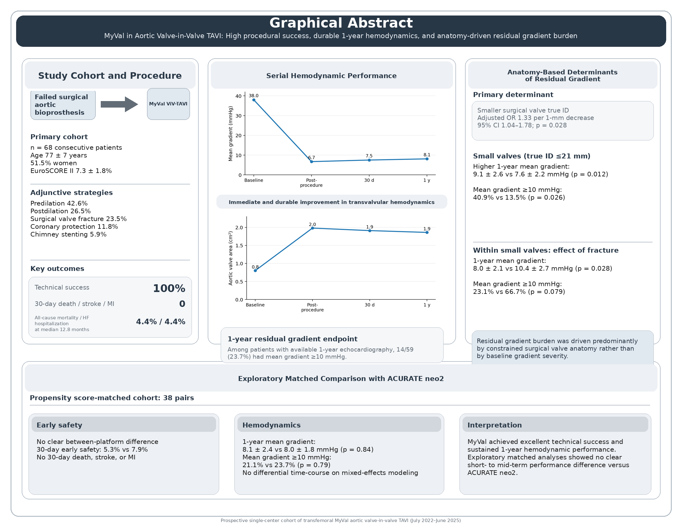
This was a prospective, single-center, observational cohort study conducted at a high-volume structural heart disease center. The primary study cohort comprised consecutive adult patients with symptomatic structural degeneration of a previously implanted surgical bioprosthetic aortic valve who underwent transfemoral valve-in-valve (ViV) transcatheter aortic valve implantation (TAVI) with the MyVal transcatheter heart valve between July 2022 and June 2025. The objectives of the present analysis were to evaluate procedural success, early safety, serial hemodynamic performance, and 1-year clinical outcomes after MyVal ViV-TAVI, with prespecified analyses focused on residual transvalvular gradients, anatomy-based subgroup effects, and an exploratory internal device-platform comparison.
All cases were evaluated by a multidisciplinary Heart Team that included interventional cardiologists, cardiac surgeons, cardiac imaging specialists, and cardiac anesthesiologists. Treatment decisions were based on clinical status, bioprosthetic failure mechanism, anatomical feasibility, procedural risk, and overall comorbidity burden in accordance with contemporary guideline-based practice. The study was conducted in accordance with the Declaration of Helsinki and was approved by the local institutional ethics committee. Written informed consent for the procedure and institutional use of clinical data was obtained according to local policy.

2.2. Patient Population

Eligible patients were adults with symptomatic dysfunction of a surgical bioprosthetic aortic valve who were treated with transfemoral ViV-TAVI using the MyVal platform during the study period. Structural valve degeneration was established using integrated clinical and imaging assessment and was classified according to the predominant failure mechanism as stenosis, regurgitation, or mixed dysfunction. The primary analysis was restricted to the consecutive MyVal-treated transfemoral ViV-TAVI cohort. Non-MyVal ViV procedures were not included in the primary cohort analysis, but an internal ACURATE neo2 cohort was used separately for the exploratory matched comparative analysis.
Patients were not excluded from the primary MyVal cohort on the basis of small surgical valve size, coronary obstruction-risk anatomy, or anticipated need for adjunctive procedural strategies such as coronary protection, chimney stenting, or surgical valve fracture, because these features were intrinsic to the real-world anatomical complexity that this study sought to evaluate.

2.3. Preprocedural Assessment and Anatomical Planning

All patients underwent structured preprocedural clinical and imaging evaluation, including transthoracic echocardiography and invasive coronary angiography when clinically indicated. Contrast-enhanced electrocardiographically gated cardiac computed tomography was used for ViV planning whenever feasible and clinically appropriate. Preprocedural imaging was used to characterize aortic root anatomy, identify the failed surgical bioprosthesis, assess coronary ostial height and sinus/sinotubular junction dimensions, and inform procedural planning for transcatheter valve sizing, implantation strategy, and coronary protection when necessary.
Surgical valve true internal diameter (true ID) was determined from the known surgical prosthesis model and labeled size using manufacturer specifications and standard ViV planning references, with CT-based anatomical confirmation when available. For prespecified subgroup analyses, small surgical valves were defined as true ID ≤21 mm.

2.4. Procedure

All procedures were performed via transfemoral access under fluoroscopic guidance according to institutional practice. The anesthesia strategy, intraprocedural echocardiographic support, vascular access management, and pacing strategy were selected according to case complexity and operator judgment. Valve sizing, implantation depth, and overall deployment strategy were based on preprocedural imaging, surgical valve identification, and anatomical risk assessment.
The procedural approach was anatomy-driven and allowed selective use of adjunctive techniques when indicated, including balloon predilation, postdilation, coronary protection, chimney stenting, and surgical valve fracture. These adjunctive strategies were used at operator discretion to optimize valve expansion, minimize coronary risk, and reduce residual hemodynamic burden in anatomically constrained ViV procedures. Procedural variables prospectively recorded included procedure duration, fluoroscopy time, contrast volume, use of adjunctive techniques, and intraprocedural complications. Because preservation of future coronary access formed part of the procedural planning framework, implantation orientation was optimized whenever feasible on the basis of anatomical considerations and device positioning strategy.

2.5. Follow-Up and Data Collection

Clinical and echocardiographic follow-up were prospectively scheduled at baseline, early post-procedure, 30 days, and 1 year. Collected variables included baseline demographics, comorbidities, surgical valve characteristics, procedural details, in-hospital events, 30-day outcomes, survival status, heart failure hospitalization, repeat coronary angiography and/or percutaneous coronary intervention after ViV-TAVI, and New York Heart Association (NYHA) functional class at follow-up. Follow-up data were obtained from institutional records, outpatient assessments, and structured follow-up documentation.

2.6. Echocardiographic Assessment

Transthoracic echocardiography was performed according to routine institutional standards and interpreted by experienced echocardiographers using standard multiparametric prosthetic valve assessment principles. For the present analysis, the prespecified core hemodynamic variables were mean transvalvular gradient and aortic valve area (AVA).
Mean transvalvular gradient was derived from continuous-wave Doppler interrogation of the aortic prosthesis using standard Bernoulli-based methods. AVA was calculated by the continuity equation. Measurements were recorded at baseline, post-procedure, 30 days, and 1 year. At 1 year, valve competence was additionally described by grading total aortic regurgitation and paravalvular leak (PVL), and the proportions with greater-than-mild regurgitation or PVL were reported descriptively. For echocardiographic follow-up endpoints, percentages were calculated using the number of patients with available echocardiography at the relevant time point. This available-case approach also applied to subgroup analyses of 1-year echocardiographic outcomes.

2.7. Outcomes and Endpoint Definitions

Procedural and early clinical outcomes were assessed using Valve Academic Research Consortium-3 (VARC-3) definitions where applicable. Technical success was reported according to VARC-3 criteria [20]. Early safety at 30 days was summarized using a VARC-3-based composite, and the individual 30-day adverse events reported in the present manuscript included all-cause mortality, stroke, myocardial infarction, major vascular complications, major or life-threatening bleeding, acute kidney injury stage 2-3, new permanent pacemaker implantation, valve dysfunction requiring repeat procedure, and valve-related reintervention.
The primary hemodynamic effectiveness analyses focused on serial changes in mean transvalvular gradient and AVA from baseline to post-procedure, 30 days, and 1 year. A prespecified residual hemodynamic endpoint for risk-factor analysis was mean transvalvular gradient ≥10 mmHg at 1-year echocardiography.
Clinical follow-up outcomes included all-cause mortality, heart failure hospitalization, endocarditis, valve thrombosis, valve-related reintervention, and NYHA functional class. Post-ViV coronary access preservation was assessed pragmatically as the feasibility of selective coronary angiography and/or PCI in cases in which coronary access was attempted during follow-up.

2.8. Prespecified Subgroup Analyses

Two prespecified subgroup analyses were performed to evaluate anatomy- and strategy-related determinants of residual hemodynamic burden. First, the primary MyVal cohort was stratified according to surgical valve true ID (≤21 mm vs >21 mm) to examine differences in serial gradients, 1-year echocardiographic valve performance, and clinical outcomes. Second, within the small-valve subgroup (true ID ≤21 mm), outcomes were compared between patients treated with surgical valve fracture and those treated without fracture to explore the hemodynamic impact of fracture-enabled optimization in constrained anatomies.
Given the limited subgroup sample sizes and event counts, these analyses were considered mechanistic and hypothesis-generating. Emphasis was therefore placed on effect size, directionality, and internal consistency across related hemodynamic endpoints rather than on isolated nominal p-values alone.

2.9. Exploratory Internal Device-Platform Comparison

To contextualize MyVal performance within local ViV-TAVI practice, an exploratory internal comparative analysis was performed against a non-randomized institutional cohort of transfemoral ViV-TAVI procedures treated with ACURATE neo2. Because device selection was influenced by anatomy and treatment era, crude comparisons were considered susceptible to confounding by indication. Propensity score methods were therefore used to improve comparability between device groups before exploratory comparative analyses were performed. A detailed description of propensity score estimation, matching strategy, covariate balance assessment, and post-matching diagnostics is provided in the Supplementary Methods.

2.10. Statistical Analysis

All analyses were performed using R (R Foundation for Statistical Computing, Vienna, Austria). Continuous variables are reported as mean ± standard deviation or median with interquartile range, as appropriate, and categorical variables as counts and percentages. All statistical tests were two-sided, and a p-value <0.05 was considered statistically significant. Because subgroup and device-platform comparisons were exploratory, p-values from these analyses were interpreted descriptively and were not adjusted for multiple comparisons.
For between-group comparisons in the primary cohort and prespecified subgroup analyses, continuous variables were compared using Student’s t-test or Welch’s t-test when assumptions for parametric testing were judged acceptable and by Mann-Whitney U testing otherwise. Categorical variables were compared using the chi-square test or Fisher’s exact test, as appropriate according to cell counts.
Serial hemodynamic outcomes, including mean transvalvular gradient and AVA at baseline, post-procedure, 30 days, and 1 year, were analyzed using linear mixed-effects models with a patient-level random intercept to account for within-patient correlation across repeated measurements. Time was modeled as a categorical fixed effect. Overall time effects and prespecified pairwise comparisons between post-procedure, 30-day, and 1-year time points were derived from model-based marginal means.
Predictors of residual hemodynamic burden were evaluated using logistic regression for the endpoint of mean transvalvular gradient ≥10 mmHg at 1 year among patients with available 1-year echocardiography. Because of the limited number of events, Firth penalized logistic regression was used to reduce small-sample bias and instability related to sparse data or quasi-separation. Univariable models were followed by a prespecified multivariable model including surgical valve true ID as a continuous variable, baseline mean transvalvular gradient, failure mechanism category, and surgical valve fracture strategy. Odds ratios (ORs) with 95% confidence intervals (CIs) are reported, and the effect of true ID is expressed per 1-mm decrease. An exploratory true ID × fracture interaction was examined in a secondary model.
For the exploratory MyVal versus ACURATE neo2 comparison, propensity scores for treatment with MyVal were estimated using multivariable logistic regression including clinically relevant baseline, anatomical, and treatment-era variables associated with device selection and outcomes. One-to-one nearest-neighbor matching without replacement was performed on the logit of the propensity score using a caliper width of 0.2 standard deviations of the logit of the propensity score and restriction to the region of common support. Covariate balance after matching was assessed using standardized mean differences, with an absolute standardized mean difference <0.10 considered acceptable.
In the matched cohort, continuous fixed-time outcomes were analyzed using matched-pair methods and are reported as mean differences with 95% CIs. Binary fixed-time outcomes were analyzed using matched-data methods appropriate for paired samples, with effect estimates reported as ORs when estimable. Longitudinal matched hemodynamic trajectories were additionally examined using mixed-effects models including fixed effects for time, device platform, and the time-by-device interaction. Time-to-event outcomes during follow-up, including all-cause mortality, heart failure hospitalization, and the composite of all-cause mortality or heart failure hospitalization, were analyzed exploratorily using Cox proportional hazards models stratified by matched pair, with hazard ratios and 95% CIs reported. Given the low number of clinical follow-up events, these time-to-event estimates were considered exploratory and interpreted descriptively.

2.11. Missing Data

Missing data were handled using an analysis-specific available-data approach. Serial hemodynamic analyses were performed using linear mixed-effects models estimated with maximum likelihood, thereby incorporating all available repeated measurements without requiring complete follow-up at every time point under a missing-at-random assumption. Cross-sectional follow-up analyses, including the 1-year residual gradient endpoint and subgroup-specific 1-year echocardiographic comparisons, were conducted as available-case analyses using the corresponding time-point denominators. No imputation was performed.

3. Results

3.1. Baseline Characteristics

A total of 68 patients (51.5% female) underwent transfemoral ViV-TAVI with MyVal. Mean age at baseline was 77 ± 7 years (Table 1). The cohort was at high surgical risk, with a mean EuroSCORE II of 7.3 ± 1.8%, and had a substantial comorbidity burden; hypertension (77.9%) and dyslipidemia (66.2%) were the most prevalent baseline conditions. At presentation, 26/68 patients (38.0%) were in NYHA class IV. Stenosis was the predominant mechanism of surgical bioprosthetic failure, accounting for 34/68 cases (50.0%). Mean surgical valve true internal diameter (true ID) was 22 ± 3 mm, and 24/68 patients (35.3%) had small surgical valves (true ID ≤21 mm).

3.2. Procedural Characteristics

All procedures were performed through transfemoral access (100%) (Table 2). Predilation and postdilation were used in 42.6% and 26.5% of cases, respectively, whereas surgical valve fracture was performed in 23.5%, predominantly in small-valve anatomies. Coronary protection and chimney stenting were used selectively in 11.8% and 5.9% of procedures, respectively. No patient required second-valve implantation or emergency surgical conversion, and no unresolved intraprocedural coronary flow compromise occurred.

3.3. Procedural Success and 30-Day Outcomes

Procedural success was high, with VARC-3 technical success achieved in all 68/68 patients (Table 3). The VARC-3-based early safety composite at 30 days was 4/68 (5.9%). Individual 30-day adverse events were infrequent: there were no deaths, strokes, or myocardial infarctions, while major vascular complications, major/life-threatening bleeding, and AKI stage 2-3 each occurred in 1/68 patients (1.5%), and new permanent pacemaker implantation occurred in 2/68 patients (2.9%).

3.4. Serial Hemodynamic Performance

Serial echocardiography showed marked immediate hemodynamic improvement after MyVal ViV-TAVI, with preservation of valve performance through 1 year. Mean transvalvular gradient decreased from 38.0 ± 9.5 mmHg at baseline to 6.7 ± 2.1 mmHg post-procedure and remained low at follow-up, measuring 7.5 ± 2.4 mmHg at 30 days and 8.1 ± 2.5 mmHg at 1 year (overall p<0.001) (Figure 1). The small numerical increases observed after the post-procedural study did not translate into significant pairwise differences between post-procedure and 30 days (p=0.053), post-procedure and 1 year (p=0.051), or 30 days and 1 year (p=0.91). In parallel, AVA increased from 0.80 ± 0.23 cm² at baseline to 1.98 ± 0.19 cm² post-procedure and remained preserved at 30 days (1.91 ± 0.21 cm²) and 1 year (1.86 ± 0.23 cm²; overall p<0.001; post-procedure vs 30 days p=0.07, post-procedure vs 1 year p=0.052, and 30 days vs 1 year p=0.79) (Figure 2).
Among patients with available 1-year echocardiography (n=59), 14/59 (23.7%) had a mean gradient ≥10 mmHg. Significant regurgitation remained uncommon, with greater-than-mild total aortic regurgitation in 2/59 patients (3.4%) and greater-than-mild PVL in 1/59 (1.7%). At a median follow-up of 12.8 months, all-cause mortality and heart failure hospitalization were each 4.4%, and no cases of endocarditis, valve thrombosis, or valve-related reintervention were recorded. Functional status improved substantially over follow-up; whereas all patients were in NYHA class III-IV at baseline, the recorded follow-up distributions were confined to NYHA class I-II at both 30 days (69% class II, 31% class I) and 1 year (60% class II, 40% class I) (Figure 3).

3.5. Subgroup Analysis According to Surgical Valve True Internal Diameter

Surgical valve true internal diameter (true ID) was significantly associated with residual hemodynamic burden (Table 4). Patients with small surgical valves (true ID ≤21 mm) had higher 1-year mean transvalvular gradients than those with larger valves (9.1 ± 2.6 vs 7.6 ± 2.2 mmHg, p=0.012) and more frequently exhibited a mean gradient ≥10 mmHg at 1 year (9/22 [40.9%] vs 5/37 [13.5%], p=0.026). Figure 4 illustrates that the true ID ≤21 mm subgroup had a distribution of 1-year mean gradients centered at higher values, with a greater proportion of observations above the 10 mmHg threshold. Surgical valve fracture was performed more frequently in small-valve anatomies (58.3% vs 4.5%, p<0.001), consistent with the anatomy-driven procedural strategy. Despite the higher residual gradient burden in patients with small surgical valves, 30-day early safety and follow-up clinical event rates did not differ significantly between the true ID subgroups.

3.6. Small-Valve Subgroup: Fracture Versus no Fracture

Within the small-valve subgroup (true ID ≤21 mm), surgical valve fracture was associated with a more favorable hemodynamic profile (Table 5, Figure 5). Patients undergoing fracture had lower mean gradients immediately after the procedure (6.1 ± 1.7 vs 7.6 ± 2.0 mmHg, p=0.041) and at 1 year (8.0 ± 2.1 vs 10.4 ± 2.7 mmHg, p=0.028). The 30-day mean gradient also tended to be lower in the fracture group (7.2 ± 2.0 vs 9.1 ± 2.6 mmHg, p=0.056). The proportion of patients with mean gradient ≥10 mmHg at 1 year was numerically lower after fracture, although this difference did not reach conventional statistical significance (3/13 [23.1%] vs 6/9 [66.7%], p=0.079). Clinical event rates remained low in both groups.

3.7. Predictors of Elevated Residual Gradient at 1 Year

The anatomy-centered signal was reinforced in multivariable modeling (Table 6). In Firth penalized logistic regression for elevated 1-year gradient (≥10 mmHg), smaller true ID remained the only independent predictor after adjustment for baseline gradient, failure mechanism, and fracture strategy (adjusted OR per 1-mm decrease 1.33, 95% CI 1.04-1.78, p=0.028). Baseline mean gradient was not independently associated with the outcome, and fracture showed a directionally protective but non-significant association. In exploratory testing, the true ID × fracture interaction was not significant (pinteraction=0.12).

3.8. Exploratory Matched Comparison with ACURATE Neo2

To contextualize MyVal performance within local ViV practice, exploratory matched analyses were performed against an internal ACURATE neo2 cohort (Table 7). In the matched cohort of 38 pairs, procedural success and 30-day safety remained high in both groups. No 30-day deaths, strokes, or myocardial infarctions occurred in either platform group. There was no clear between-platform difference in fixed-time hemodynamic outcomes, with post-procedure mean gradients of 6.7 ± 2.0 versus 6.9 ± 1.6 mmHg (p=0.62), 30-day mean gradients of 7.5 ± 2.2 versus 7.8 ± 1.9 mmHg (p=0.48), and 1-year mean gradients of 8.1 ± 2.4 versus 8.0 ± 1.8 mmHg (p=0.84) for MyVal and ACURATE neo2, respectively. The proportion with mean gradient ≥10 mmHg at 1 year was likewise similar (21.1% vs 23.7%, p=0.79). Longitudinal trajectories remained closely overlapping, and mixed-effects modeling showed no evidence of differential gradient evolution over time between platforms. Follow-up clinical comparisons were limited by low event counts and wide confidence intervals, but no statistically significant between-platform differences were observed. Post-ViV coronary access was feasible in all 14 attempted MyVal cases and in 10 of 11 attempted ACURATE neo2 cases (p=0.26).
Figure 6. Serial mean transvalvular gradient in the matched MyVal and ACURATE neo2 cohorts. Mean transvalvular gradient at baseline, discharge, 30 days, and 1 year in the propensity score-matched MyVal and ACURATE neo2 cohorts after valve-in-valve TAVI. Values are shown as mean ± SD. The time×device p value derives from the mixed-effects model for longitudinal gradient trajectories.
Preprints 206976 g006

4. Discussion

The principal findings of the present study are fourfold. First, in a prospective, consecutive real-world cohort, MyVal valve-in-valve transcatheter aortic valve implantation (ViV-TAVI) was associated with excellent procedural success and a very low burden of early adverse events despite substantial anatomic complexity, including a meaningful proportion of small surgical bioprostheses and selective use of coronary protection, chimney stenting, and surgical valve fracture. Second, MyVal implantation yielded an immediate and durable hemodynamic improvement, with a marked fall in mean transvalvular gradient, preservation of aortic valve area through 1 year, and parallel improvement in functional status. Third, residual hemodynamic burden was driven primarily by the structural constraint imposed by the failed surgical bioprosthesis, with true internal diameter (true ID) emerging as the dominant determinant of 1-year residual gradient, whereas baseline gradient severity was not independently predictive after adjustment. Fourth, within the limits of an exploratory matched comparison, MyVal and ACURATE neo2 showed similar early safety, similar 1-year hemodynamic performance, and no evidence of differential gradient evolution over time. Taken together, these findings suggest that MyVal is a credible balloon-expandable platform for contemporary ViV-TAVI, while reinforcing the broader principle that post-ViV physiology is determined more by anatomy than by device label alone [1,2,4,18,19].
From a safety standpoint, the present data compare favorably with the major historical ViV-TAVI experiences. In the Global Valve-in-Valve Registry, procedural success was 93.1% and 30-day mortality was 8.4%, with device malposition, coronary obstruction, and residual gradients identified as the central limitations of early ViV practice [1]. The STS/ACC Registry subsequently confirmed the effectiveness of ViV-TAVI at scale, and the PARTNER 2 aortic ViV registry showed sustained clinical and echocardiographic benefit at 5 years in high-risk patients [2,4]. In that context, the present study’s absence of 30-day death, stroke, or myocardial infarction and the absence of second-valve implantation or emergency conversion is consistent with the progressive maturation of ViV practice through better computed tomography planning, improved transcatheter heart valve technology, more selective use of adjunctive techniques, and greater operator familiarity with bioprosthetic failure phenotypes [1,2,4].
The hemodynamic data are particularly important because residual obstruction remains the Achilles heel of ViV-TAVI. In the present cohort, mean gradient fell from 38.0 mmHg at baseline to 6.7 mmHg post-procedure and remained low at 30 days and 1 year, while aortic valve area nearly doubled immediately after implantation and was preserved over follow-up. These values compare favorably with early ViV experience, including the Global Valve-in-Valve Registry, in which postprocedural mean gradients were substantially higher overall, and they also extend the very limited MyVal-specific ViV literature, which previously consisted mainly of an initial case series and a later multicenter left-sided ViV/valve-in-ring report that was not designed as a dedicated transfemoral aortic hemodynamic study [1,18,19]. The present results therefore add granularity that was previously missing from the MyVal literature: not only was implantation feasible, but the achieved valve performance was sustained through serial follow-up rather than being confined to the index procedure alone [1,18,19].
Small surgical bioprostheses (true ID ≤21 mm) had clearly higher 1-year gradients and a substantially greater frequency of gradient ≥10 mmHg, and true ID was the only independent predictor of residual gradient in the multivariable Firth model. This is highly consistent with previous ViV literature. Dvir et al. showed that stenotic failure modes and smaller surgical valves were associated with less favorable post-ViV physiology and survival [6]. Pibarot et al. further demonstrated that pre-existing severe prosthesis-patient mismatch of the failed surgical valve is independently associated with excess mortality after ViV implantation and with a higher frequency of high postprocedural gradients [7]. This observation is consistent with the underlying hemodynamic framework of valve-in-valve intervention, in which transcatheter implantation can reduce baseline obstruction yet remains constrained by the fixed internal dimensions of the failed surgical bioprosthesis unless additional expansion strategies are employed [6,7]. The absence of an independent association between baseline gradient and 1-year residual gradient in the adjusted analysis further reinforces the concept that final hemodynamic performance after valve-in-valve implantation is determined primarily by the structural constraints of the host bioprosthesis rather than by the severity of the baseline Doppler gradient [6,7].
The fracture analysis should be interpreted in the same anatomy-first framework. Within small surgical valves, fracture was associated with lower post-procedural and 1-year mean gradients and with a numerically lower proportion of patients crossing the ≥10 mmHg threshold. This direction of effect is concordant with the original bioprosthetic valve fracture literature, which showed that fracturing the surgical valve ring can expand the effective orifice and improve post-ViV hemodynamics [13]. However, contemporary registry data have appropriately tempered indiscriminate enthusiasm. In the TVT Registry analysis limited to balloon-expandable SAPIEN 3/Ultra ViV-TAVR, attempted bioprosthetic valve fracture was associated with higher in-hospital mortality and life-threatening bleeding, while the hemodynamic gains, although real, were modest overall [12]. Within small surgical bioprostheses, fracture was associated with lower residual gradients, consistent with a potential role in mitigating anatomically constrained post-valve-in-valve hemodynamics. These findings should not be interpreted as supporting routine fracture, but rather as suggesting benefit in selected high-risk anatomies. Given the limited subgroup size and the absence of a significant interaction, the results remain exploratory and should be viewed as hypothesis-generating rather than definitive [12,13].
The exploratory matched comparison with ACURATE neo2 offers additional perspective on the hemodynamic profile of MyVal in valve-in-valve TAVI. A supra-annular self-expanding platform would theoretically be expected to provide lower residual gradients than a balloon-expandable device, particularly in small failed surgical bioprostheses. Prior comparative studies and the LYTEN program support this expectation, having shown lower postprocedural and 1-year gradients with self-expanding valves, although without clear differences in short- to intermediate-term clinical outcomes [21,22,23,24]. Against this background, the similar hemodynamic performance observed with MyVal and ACURATE neo2 in the matched cohort is notable. This may reflect more granular prosthesis matching enabled by the intermediate MyVal sizes, anatomy-driven use of adjunctive optimization strategies, and the limited ability of a modest matched analysis to fully account for confounding by indication in a procedure where device choice is closely linked to surgical valve type, coronary anatomy, and operator preference [16,17,21,22,23]. These findings therefore support the feasibility of achieving comparable hemodynamic results with MyVal in selected anatomies but should not be interpreted as establishing equivalence between balloon-expandable and supra-annular self-expanding platforms in valve-in-valve TAVI [16,17,21,22,23].
The favorable coronary results in the present cohort occurred within a contemporary CT-guided, anatomy-based ViV-TAVI strategy. Because coronary obstruction and loss of future coronary access remain major limitations of valve-in-valve intervention, detailed preprocedural assessment of valve-to-coronary relationships, root anatomy, and surgical prosthesis characteristics is essential for procedural planning and for selection of preventive measures [9,10]. Within this framework, the absence of unresolved intraprocedural coronary compromise and the high feasibility of coronary access in attempted cases are most likely attributable to systematic risk stratification and selective use of adjunctive techniques, including coronary protection, chimney stenting, and alignment-focused implantation [9,10,11,25].
Several limitations should frame interpretation of these findings. This was a single-center observational study without external adjudication or core-laboratory echocardiography, and although the cohort was prospectively assembled, the sample size remains modest for subgroup inference. The fracture analysis and the matched device-platform comparison were exploratory and underpowered for clinical endpoints, as reflected by wide confidence intervals. One-year echocardiographic analyses were available-case rather than complete-case analyses. In addition, the matched MyVal versus ACURATE neo2 comparison, while methodologically preferable to crude comparison, cannot fully eliminate residual confounding because device choice in ViV-TAVI is inherently anatomy dependent. Finally, the present study was not designed to assess valve durability beyond the first year, and longer follow-up will be essential.

5. Conclusions

In this prospective real-world cohort, transfemoral MyVal valve-in-valve TAVI was associated with favorable procedural safety and durable 1-year hemodynamic performance. Residual hemodynamic burden was driven primarily by surgical valve true internal diameter, supporting an anatomy-based valve-in-valve strategy with selective use of adjunctive optimization in constrained anatomies.

Supplementary Materials

The following supporting information can be downloaded at the website of this paper posted on Preprints.org.

Author Contributions

Conceptualization, G.P. and V.N.; methodology, G.P. and V.N.; software, G.P. and V.N.; validation, G.P. and V.N.; formal analysis, G.P. and V.N.; investigation, I.N., S.E., A.I., A.N., and V.N.; resources, G.P. and V.N.; data curation, G.P. and V.N.; writing—original draft preparation, G.P.; writing—review and editing, V.N.; visualization, G.P. and V.N.; supervision, V.N.; project administration, V.N. All authors have read and agreed to the published version of the manuscript.

Funding

This research received no external funding.

Institutional Review Board Statement

The study was conducted in accordance with the Declaration of Helsinki, and approved by the Institutional Review Board of Interbalkan Medical Center (protocol code 3435/11.06.2022).

Data Availability Statement

The data presented in this study are available on request from the corresponding author. The data are not publicly available due to ethical restrictions.

Conflicts of Interest

The authors declare no conflicts of interest.

Abbreviations

The following abbreviations are used in this manuscript:
AKI Acute kidney injury
AVA Aortic valve area
BVF Bioprosthetic valve fracture
CABG Coronary artery bypass grafting
CI Confidence interval
COPD Chronic obstructive pulmonary disease
CT Computed tomography
DVI Doppler velocity index
eGFR Estimated glomerular filtration rate
EOAi Effective orifice area index
HF Heart failure
HR Hazard ratio
ID Internal diameter
LVEF Left ventricular ejection fraction
MD Mean difference
NYHA New York Heart Association
OR Odds ratio
PCI Percutaneous coronary intervention
PPM Prosthesis-patient mismatch
PVL Paravalvular leak
SAVR Surgical aortic valve replacement
SD Standard deviation
TAVI Transcatheter aortic valve implantation
TIA Transient ischemic attack
VARC-3 Valve Academic Research Consortium-3
ViV Valve-in-valve
ViV-TAVI Valve-in-valve transcatheter aortic valve implantation
VIVID Valve-in-Valve International Data

References

  1. Dvir, D; Webb, J; Brecker, S; Bleiziffer, S; Hildick-Smith, D; Colombo, A; et al. Transcatheter aortic valve replacement for degenerative bioprosthetic surgical valves: results from the global valve-in-valve registry. Circulation 2012, 126(19), 2335–44. [Google Scholar] [CrossRef]
  2. Hahn, RT; Webb, J; Pibarot, P; Ternacle, J; Herrmann, HC; Suri, RM; et al. 5-Year Follow-Up From the PARTNER 2 Aortic Valve-in-Valve Registry for Degenerated Aortic Surgical Bioprostheses. JACC Cardiovasc Interv. 2022, 15(7), 698–708. [Google Scholar] [CrossRef]
  3. Praz, F; Borger, MA; Lanz, J; Marin-Cuartas, M; Abreu, A; Adamo, M; et al. 2025 ESC/EACTS Guidelines for the management of valvular heart disease. Eur Heart J. 2025, 46(44), 4635–736. [Google Scholar] [CrossRef]
  4. Tuzcu, EM; Kapadia Samir, R; Vemulapalli, S; Carroll John, D; Holmes David, R; Mack Michael, J; et al. Transcatheter Aortic Valve Replacement of Failed Surgically Implanted Bioprostheses. JACC 2018, 72(4), 370–82. [Google Scholar] [CrossRef]
  5. Bleiziffer, S; Simonato, M; Webb, JG; Rodés-Cabau, J; Pibarot, P; Kornowski, R; et al. Long-term outcomes after transcatheter aortic valve implantation in failed bioprosthetic valves. Eur Heart J. 2020, 41(29), 2731–42. [Google Scholar] [CrossRef] [PubMed]
  6. Dvir, D; Webb, JG; Bleiziffer, S; Pasic, M; Waksman, R; Kodali, S; et al. Transcatheter aortic valve implantation in failed bioprosthetic surgical valves. Jama 2014, 312(2), 162–70. [Google Scholar] [CrossRef]
  7. Pibarot, P; Simonato, M; Barbanti, M; Linke, A; Kornowski, R; Rudolph, T; et al. Impact of Pre-Existing Prosthesis-Patient Mismatch on Survival Following Aortic Valve-in-Valve Procedures. JACC Cardiovasc Interv. 2018, 11(2), 133–41. [Google Scholar] [CrossRef]
  8. Kherallah, RY; Suffredini, JM; Rahman, F; Eng, MH; Kleiman, N; Manandhar, P; et al. Impact of Elevated Gradients After Transcatheter Aortic Valve Implantation for Degenerated Surgical Aortic Valve Bioprostheses. Circ Cardiovasc Interv. 2024, 17(7), e013558. [Google Scholar] [CrossRef]
  9. Tang, GHL; Komatsu, I; Tzemach, L; Simonato, M; Wolak, A; Blanke, P; et al. Risk of coronary obstruction and the need to perform BASILICA: the VIVID classification. EuroIntervention 2020, 16(9), e757–e9. [Google Scholar] [CrossRef] [PubMed]
  10. Tomii, D; Okuno, T; Lanz, J; Stortecky, S; Reineke, D; Windecker, S; et al. Valve-in-valve TAVI and risk of coronary obstruction: Validation of the VIVID classification. J Cardiovasc Comput Tomogr. 2023, 17(2), 105–11. [Google Scholar] [CrossRef] [PubMed]
  11. Tarantini, G; Nai Fovino, L; Scotti, A; Massussi, M; Cardaioli, F; Rodinò, G; et al. Coronary Access After Transcatheter Aortic Valve Replacement With Commissural Alignment: The ALIGN-ACCESS Study. Circ Cardiovasc Interv. 2022, 15(2), e011045. [Google Scholar] [CrossRef]
  12. Chhatriwalla, AK; Allen, KB; Depta, JP; Rodriguez, E; Thourani, VH; Whisenant, BK; et al. Outcomes of Bioprosthetic Valve Fracture in Patients Undergoing Valve-in-Valve TAVR. JACC Cardiovasc Interv. 2023, 16(5), 530–9. [Google Scholar] [CrossRef]
  13. Chhatriwalla, AK; Allen, KB; Saxon, JT; Cohen, DJ; Aggarwal, S; Hart, AJ; et al. Bioprosthetic Valve Fracture Improves the Hemodynamic Results of Valve-in-Valve Transcatheter Aortic Valve Replacement. Circ Cardiovasc Interv. 2017, 10(7). [Google Scholar] [CrossRef]
  14. Kawashima, H; Serruys, PW; Mylotte, D; Rosseel, L; Amat-Santos, IJ; Rao, RS; et al. Operator preference and determinants of size selection when additional intermediate-size aortic transcatheter heart valves are made available. Int J Cardiol. 2021, 338, 168–73. [Google Scholar] [CrossRef]
  15. Seth, A; Kumar, V; Singh, VP; Kumar, D; Varma, P; Rastogi, V. Myval: A Novel Transcatheter Heart Valve for the Treatment of Severe Aortic Stenosis. Interv Cardiol. 2023, 18, e12. [Google Scholar] [CrossRef]
  16. Baumbach, A; van Royen, N; Amat-Santos, IJ; Hudec, M; Bunc, M; Ijsselmuiden, A; et al. LANDMARK comparison of early outcomes of newer-generation Myval transcatheter heart valve series with contemporary valves (Sapien and Evolut) in real-world individuals with severe symptomatic native aortic stenosis: a randomised non-inferiority trial. Lancet 2024, 403(10445), 2695–708. [Google Scholar] [CrossRef] [PubMed]
  17. Terkelsen, CJ; Freeman, P; Dahl, JS; Thim, T; Nørgaard, BL; Mogensen, NSB; et al. SAPIEN 3 versus Myval transcatheter heart valves for transcatheter aortic valve implantation (COMPARE-TAVI 1): a multicentre, randomised, non-inferiority trial. Lancet 2025, 405(10487), 1362–72. [Google Scholar] [CrossRef]
  18. Ielasi, A; Buono, A; Medda, M; Casilli, F; Bande, M; Pellicano, M; et al. Transcatheter Valve-in-Valve Implantation With a Novel Balloon-Expandable Device in Patients With Bioprosthetic Heart Valve Failure: A Case Series. Cardiovasc Revasc Med. 2021, 28s, 98–101. [Google Scholar] [CrossRef] [PubMed]
  19. Moscarella, E; Ielasi, A; Mussayev, A; Montorfano, M; Mullassari, A; Martin, P; et al. Transcatheter valve-in-valve or valve-in-ring implantation with a novel balloon-expandable device in patients with bioprosthetic left side heart valves failure: 1-year follow-up from a multicenter experience. Int J Cardiol. 2023, 376, 35–45. [Google Scholar] [CrossRef]
  20. Généreux, P; Piazza, N; Alu, MC; Nazif, T; Hahn, RT; Pibarot, P; et al. Valve Academic Research Consortium 3: updated endpoint definitions for aortic valve clinical research. Eur Heart J. 2021, 42(19), 1825–57. [Google Scholar] [CrossRef] [PubMed]
  21. Nuche, J; Abbas, AE; Serra, V; Vilalta, V; Nombela-Franco, L; Regueiro, A; et al. Balloon- vs Self-Expanding Transcatheter Valves for Failed Small Surgical Aortic Bioprostheses: 1-Year Results of the LYTEN Trial. JACC Cardiovasc Interv. 2023, 16(24), 2999–3012. [Google Scholar] [CrossRef] [PubMed]
  22. Ochiai, T; Yoon, SH; Sharma, R; Miyasaka, M; Nomura, T; Rami, T; et al. Outcomes of Self-Expanding vs. Balloon-Expandable Transcatheter Heart Valves for the Treatment of Degenerated Aortic Surgical Bioprostheses - A Propensity Score-Matched Comparison. Circ J. 2018, 82(10), 2655–62. [Google Scholar] [CrossRef] [PubMed]
  23. Rodés-Cabau, J; Abbas Amr, E; Serra, V; Vilalta, V; Nombela-Franco, L; Regueiro, A; et al. Balloon- vs Self-Expanding Valve Systems for Failed Small Surgical Aortic Valve Bioprostheses. JACC 2022, 80(7), 681–93. [Google Scholar] [CrossRef]
  24. Papadopoulos, GE; Ninios, I; Evangelou, S; Ioannidis, A; Nikitopoulos, A; Giannakoulas, G; et al. Efficacy and Safety of ACURATE neo2 in Valve-in-Valve TAVI: A Prospective Single-Center Study. J Clin Med. 2025, 14(13). [Google Scholar] [CrossRef]
  25. Khan, JM; Greenbaum, AB; Babaliaros, VC; Rogers, T; Eng, MH; Paone, G; et al. The BASILICA Trial: Prospective Multicenter Investigation of Intentional Leaflet Laceration to Prevent TAVR Coronary Obstruction. JACC Cardiovasc Interv. 2019, 12(13), 1240–52. [Google Scholar] [CrossRef] [PubMed]
Figure 1. Serial mean transvalvular gradient after MyVal ViV-TAVI.Mean transvalvular gradient at baseline, post-procedure, 30 days, and 1 year after transfemoral MyVal valve-in-valve transcatheter aortic valve implantation. Values are shown as mean ± SD. Overall time effect: p<0.001. Pairwise comparisons: post-procedure vs 30 days, p=0.053; post-procedure vs 1 year, p=0.051; 30 days vs 1 year, p=0.91.
Preprints 206976 g001
Figure 2. Serial aortic valve area after MyVal ViV-TAVI. Aortic valve area at baseline, post-procedure, 30 days, and 1 year after transfemoral MyVal valve-in-valve transcatheter aortic valve implantation. Values are shown as mean ± SD. Overall time effect: p<0.001. Pairwise comparisons: post-procedure vs 30 days, p=0.07; post-procedure vs 1 year, p=0.052; 30 days vs 1 year, p=0.79.
Preprints 206976 g002
Figure 3. Distribution of NYHA functional class at baseline and follow-up after MyVal ViV-TAVI. Stacked percentage distribution of NYHA functional class at baseline, 30 days, and 1 year after transfemoral MyVal valve-in-valve transcatheter aortic valve implantation, showing a shift from baseline NYHA III-IV to follow-up NYHA I-II.
Preprints 206976 g003
Figure 4. Distribution of 1-year mean transvalvular gradient according to surgical valve true internal diameter. Violin plot with overlaid boxplot and individual observations showing the distribution of 1-year mean transvalvular gradient in patients with surgical valve true ID ≤21 mm versus >21 mm after MyVal ViV-TAVI. The dashed line indicates the 10 mmHg threshold for elevated residual gradient. Patients with true ID ≤21 mm had higher 1-year mean gradients than those with true ID >21 mm (9.1 ± 2.6 vs 7.6 ± 2.2 mmHg; p=0.012).
Preprints 206976 g004
Figure 5. Effect of surgical valve fracture on residual hemodynamic burden in small surgical valves (true ID ≤21 mm). (A) Serial mean transvalvular gradient at discharge, 30 days, and 1 year in patients treated with and without surgical valve fracture after MyVal ViV-TAVI. Values are shown as mean ± SD. The dashed line indicates the 10 mmHg threshold. The p value refers to the between-group comparison of 1-year mean gradient. (B) Proportion of patients with 1-year mean transvalvular gradient ≥10 mmHg according to fracture strategy. The p value refers to the between-group comparison of the binary 1-year residual gradient endpoint.
Preprints 206976 g005
Table 1. Baseline characteristics of the MyVal ViV-TAVI cohort.
Variable Overall (n=68)
Age, years 77 ± 7
Female sex 35/68 (51.5%)
Body mass index, kg/m² 27.8 ± 4.6
EuroSCORE II, % 7.3 ± 1.8
Hypertension 53/68 (77.9%)
Dyslipidemia 45/68 (66.2%)
Diabetes mellitus 21/68 (30.9%)
Coronary artery disease 39/68 (57.4%)
Prior PCI 18/68 (26.5%)
Prior CABG 11/68 (16.2%)
Prior myocardial infarction 9/68 (13.2%)
Prior stroke/TIA 6/68 (8.8%)
Atrial fibrillation 24/68 (35.3%)
COPD 14/68 (20.6%)
Chronic kidney disease (eGFR <60 mL/min/1.73 m²) 27/68 (39.7%)
eGFR, mL/min/1.73 m² 61 ± 18
Prior permanent pacemaker 7/68 (10.3%)
NYHA class III 42/68 (62%)
NYHA class IV 26/68 (38%)
Time from SAVR to degeneration, years 10.6 ± 3.9
Failure mechanism: stenosis 34/68 (50.0%)
Failure mechanism: regurgitation 15/68 (22.1%)
Failure mechanism: mixed 19/68 (27.9%)
Surgical valve true internal diameter (ID), mm 22 ± 3
True ID ≤21 mm 24/68 (35.3%)
True ID >21 mm 44/68 (64.7%)
Baseline mean gradient, mmHg 38.0 ± 9.5
Baseline peak gradient, mmHg 63.2 ± 15.1
Baseline AVA, cm² 0.80 ± 0.23
Baseline DVI 0.22 ± 0.06
Baseline EOAi, cm²/m² 0.45 ± 0.12
Baseline LVEF, % 53 ± 10
Footnote: Values are presented as mean ± SD or n/N (%). Abbreviations: AVA, aortic valve area; CABG, coronary artery bypass grafting; COPD, chronic obstructive pulmonary disease; DVI, Doppler velocity index; eGFR, estimated glomerular filtration rate; EOAi, effective orifice area index; ID, internal diameter; LVEF, left ventricular ejection fraction; NYHA, New York Heart Association; PCI, percutaneous coronary intervention; SAVR, surgical aortic valve replacement; SD, standard deviation; TIA, transient ischemic attack.
Table 2. Procedural characteristics and intraprocedural findings.
Variable Overall (n=68)
Access route: transfemoral 68/68 (100%)
General anesthesia 19/68 (27.9%)
Predilation 29/68 (42.6%)
Postdilation 18/68 (26.5%)
Surgical valve fracture 16/68 (23.5%)
Coronary protection used 8/68 (11.8%)
Chimney stenting performed 4/68 (5.9%)
Second valve implantation 0/68 (0.0%)
Emergency conversion to surgery 0/68 (0.0%)
Unresolved intraprocedural coronary flow compromise 0/68 (0.0%)
Procedure duration, min 74 ± 20
Fluoroscopy time, min 16.7 ± 6.5
Contrast volume, mL 108 ± 36
Footnote: Values are presented as mean ± SD or n/N (%) Abbreviations: mL, milliliters; min, minutes; SD, standard deviation; TAVI, transcatheter aortic valve implantation; ViV, valve-in-valve.
Table 3. VARC-3 procedural success and 30-day outcomes.
Outcome Value (n=68)
Technical success (VARC-3) 68/68 (100.0%)
Early safety composite at 30 days (VARC-3-based composite) 4/68 (5.9%)
All-cause mortality (30 days) 0/68 (0.0%)
Stroke (all) 0/68 (0.0%)
Myocardial infarction 0/68 (0.0%)
Major vascular complication 1/68 (1.5%)
Major/life-threatening bleeding 1/68 (1.5%)
AKI stage 2–3 1/68 (1.5%)
New permanent pacemaker implantation 2/68 (2.9%)
Valve dysfunction requiring repeat procedure 0/68 (0.0%)
Valve-related reintervention (30 days) 0/68 (0.0%)
Footnote: Values are presented as n/N (%). Abbreviations: AKI, acute kidney injury; VARC-3, Valve Academic Research Consortium-3.
Table 4. Subgroup analysis by surgical valve true ID (≤21 mm vs >21 mm).
Variable True ID ≤21 mm (overall n=24) True ID >21 mm (overall n=44) p-value
Baseline mean gradient, mmHg 39.6 ± 9.8 37.1 ± 9.3 0.31
Post-procedure mean gradient, mmHg 7.2 ± 2.2 6.4 ± 2.0 0.14
30-day mean gradient, mmHg 8.2 ± 2.5 7.1 ± 2.2 0.08
1-year mean gradient, mmHg* 9.1 ± 2.6 7.6 ± 2.2 0.012
Mean gradient ≥10 mmHg at 1 year* 9/22 (40.9%) 5/37 (13.5%) 0.026
Surgical valve fracture performed 14/24 (58.3%) 2/44 (4.5%) <0.001
Coronary protection used 4/24 (16.7%) 4/44 (9.1%) 0.44
Chimney stenting 2/24 (8.3%) 2/44 (4.5%) 0.61
30-day early safety composite 2/24 (8.3%) 2/44 (4.5%) 0.61
All-cause mortality during follow-up 1/24 (4.2%) 2/44 (4.5%) 1.00
HF hospitalization during follow-up 2/24 (8.3%) 1/44 (2.3%) 0.28
Composite of all-cause mortality or HF hospitalization during follow-up† 3/24 (12.5%) 3/44 (6.8%) 0.66
Footnote: Values are presented as mean ± SD or n/N (%). *One-year echocardiographic endpoints are reported using patients with available 1-year echocardiography in each subgroup. †Composite endpoint denotes all-cause mortality or heart failure hospitalization during follow-up. Abbreviations: HF, heart failure; ID, internal diameter; SD, standard deviation.
Table 5. Small-valve subgroup (true ID ≤21 mm): fracture vs no fracture.
Variable Fracture (overall n=14) No fracture (overall n=10) p-value
Post-procedure mean gradient, mmHg 6.1 ± 1.7 7.6 ± 2.0 0.041
30-day mean gradient, mmHg 7.2 ± 2.0 9.1 ± 2.6 0.056
1-year mean gradient, mmHg* 8.0 ± 2.1 10.4 ± 2.7 0.028
Mean gradient ≥10 mmHg at 1 year* 3/13 (23.1%) 6/9 (66.7%) 0.079
Coronary protection used 3/14 (21.4%) 1/10 (10.0%) 0.61
30-day early safety composite 1/14 (7.1%) 1/10 (10.0%) 1.00
All-cause mortality during follow-up 0/14 (0.0%) 1/10 (10.0%) 0.42
HF hospitalization during follow-up 1/14 (7.1%) 1/10 (10.0%) 1.00
Composite of all-cause mortality or HF hospitalization during follow-up† 1/14 (7.1%) 2/10 (20.0%) 0.55
Footnote: Values are presented as mean ± SD or n/N (%). *One-year echocardiographic endpoints are reported using patients with available 1-year echocardiography in each subgroup. †Composite endpoint denotes all-cause mortality or heart failure hospitalization during follow-up. Abbreviations: HF, heart failure; ID, internal diameter; SD, standard deviation.
Table 6. Predictors of elevated residual gradient at 1 year (mean gradient ≥10 mmHg): Firth logistic regression (n=59, events=14).
Variable Univariable OR (95% CI) p-value Multivariable adjusted OR (95% CI) p-value
True ID (per 1 mm decrease) 1.38 (1.10–1.79) 0.010 1.33 (1.04–1.78) 0.028
True ID ≤21 mm 3.80 (1.19–12.6) 0.025
Baseline mean gradient (per 5 mmHg increase) 1.12 (0.82–1.53) 0.46 1.09 (0.77–1.50) 0.61
Failure mechanism (stenosis vs non-stenosis) 1.29 (0.42–4.08) 0.66 1.21 (0.37–4.01) 0.75
Surgical valve fracture (yes vs no) 0.54 (0.16–1.73) 0.29 0.49 (0.12–1.71) 0.27
Postdilation 0.73 (0.21–2.36) 0.60
True ID × fracture interaction (exploratory) p(interaction)=0.12
Footnote: Values are reported as ORs with 95% CIs. The multivariable model included true ID, baseline mean transvalvular gradient, failure mechanism category, and surgical valve fracture strategy. True ID was parameterized per 1-mm decrease. Firth penalized logistic regression was used because of the limited number of events. The exploratory true ID × fracture interaction was evaluated in a secondary model and was not included in the primary adjusted model. Abbreviations: CI, confidence interval; ID, internal diameter; OR, odds ratio.
Table 7. Matched comparative outcomes: MyVal vs ACURATE neo2 (38 matched pairs).
Endpoint MyVal (n=38) ACURATE neo2 (n=38) Effect estimate (95% CI) p-value
Technical success 38 (100.0%) 37 (97.4%) OR not estimable (near-complete success) 0.31
30-day early safety composite 2 (5.3%) 3 (7.9%) OR 0.65 (0.09–3.96) 0.64
30-day all-cause mortality 0 (0.0%) 0 (0.0%)
30-day stroke 0 (0.0%) 0 (0.0%)
30-day myocardial infarction 0 (0.0%) 0 (0.0%)
New permanent pacemaker implantation 1 (2.6%) 0 (0.0%) OR 3.1 (0.12–170) 1.00
Major vascular complication 1 (2.6%) 0 (0.0%) OR 3.1 (0.12–170) 1.00
Coronary protection used 6 (15.8%) 2 (5.3%) OR 3.38 (0.62–23.0) 0.16
Chimney stenting 3 (7.9%) 2 (5.3%) OR 1.54 (0.20–14.9) 0.65
Post-procedure mean gradient, mmHg 6.7 ± 2.0 6.9 ± 1.6 MD −0.2 (−1.0 to 0.6) 0.62
30-day mean gradient, mmHg 7.5 ± 2.2 7.8 ± 1.9 MD −0.3 (−1.2 to 0.6) 0.48
1-year mean gradient, mmHg 8.1 ± 2.4 8.0 ± 1.8 MD +0.1 (−0.9 to 1.1) 0.84
Mean gradient ≥10 mmHg at 1 year 8 (21.1%) 9 (23.7%) OR 0.86 (0.28–2.63) 0.79
>mild PVL at 1 year 1 (2.6%) 1 (2.6%) OR 1.00 1.00
1-year survival 94.7% 97.4% Exploratory HR 1.86 (0.17–20.6) 0.62
HF hospitalization 2 (5.3%) 1 (2.6%) Exploratory HR 2.03 (0.18–22.8) 0.56
Composite death/HF hospitalization 4 (10.5%) 2 (5.3%) Exploratory HR 2.01 (0.37–10.8) 0.41
Post-ViV coronary access feasibility (attempted) 14/14 (100.0%) 10/11 (90.9%) OR not stable (sparse) 0.26
Footnote: Values are presented as mean ± SD, n/N (%), or Kaplan–Meier estimates. Effect estimates are reported as matched ORs for binary fixed-time outcomes, paired MDs for continuous fixed-time outcomes, and exploratory HRs for time-to-event outcomes. Time-to-event analyses were performed using Cox proportional hazards models stratified by matched pair. One-year survival values represent Kaplan–Meier estimates. Abbreviations: CI, confidence interval; HF, heart failure; HR, hazard ratio; MD, mean difference; OR, odds ratio; PCI, percutaneous coronary intervention; PVL, paravalvular leak; SD, standard deviation; ViV, valve-in-valve.
Disclaimer/Publisher’s Note: The statements, opinions and data contained in all publications are solely those of the individual author(s) and contributor(s) and not of MDPI and/or the editor(s). MDPI and/or the editor(s) disclaim responsibility for any injury to people or property resulting from any ideas, methods, instructions or products referred to in the content.
Copyright: This open access article is published under a Creative Commons CC BY 4.0 license, which permit the free download, distribution, and reuse, provided that the author and preprint are cited in any reuse.
Prerpints.org logo

Preprints.org is a free preprint server supported by MDPI in Basel, Switzerland.

Subscribe

Disclaimer

Terms of Use

Privacy Policy

Privacy Settings

© 2026 MDPI (Basel, Switzerland) unless otherwise stated