

Submitted:
31 March 2026
Posted:
31 March 2026
You are already at the latest version
Abstract
Background/Objectives: Neonatal sepsis remains a major contributor to neonatal morbidity and mortality worldwide, yet diagnostic uncertainty and heterogeneous clinical presentation continue to challenge early recognition and management. Early onset sepsis (EOS), typically arising within the first 72 hours of life, is strongly influenced by maternal and perinatal factors. Limited data exist on the temporal evolution of infec-tion foci during the first week of life. Methods: To identify the predominant infection localization sites in preterm and term neonates with suspected or confirmed sepsis and to determine maternal and neonatal risk factors associated with early disease severity, persistent sepsis, and adverse outcomes .Results: A total of 297 neonates met inclusion criteria. Most infants (99.3%) were admitted before 72 hours of life. Respiratory involvement was the predominant infection focus on Day 1 (57.2%) and remained common through Day 3. CNS, gastrointestinal, and skin involvement were infrequent. Lower gestational age (p=0.035) and pro-longed rupture of membranes >18 hours (p=0.043) independently predicted sepsis at Day 1. Advanced sep-sis at admission was associated with lower birth weight, lower gestational age, older maternal age, and ab-sence of intrapartum antibiotics (all p≤0.001). Persistent sepsis at Day 7 was linked to prematurity (p=0.008), higher mortality (p< 0.001), and prolonged hospitalization (p=0.001). Conclusions: Respiratory involvement is the most common early infection focus in neonates with EOS. Prematurity, low birth weight, prolonged rupture of membranes, and maternal intrapartum infection significantly increase the risk of severe disease.
Keywords:
neonatal sepsis
; infection foci
; risk factors
1. Introduction
Neonatal sepsis remains a major global challenge for healthcare systems, contributing substantially to neonatal morbidity and mortality [1,2,3,4]. It is typically categorized as early-onset sepsis (EOS), occurring within the first 72 h of life and primarily resulting from vertical maternal transmission, or late-onset sepsis (LOS), occurring after 72 h of life and usually reflecting nosocomial or community-acquired infection. The Global Burden of Disease (GBD) Study estimates approximately 1.3 million annual cases of neonatal sepsis worldwide, with global deaths ranging between 400.000 and 700.000 each year [5,6].
Defining neonatal sepsis remains challenging, as many studies highlight the lack of a universally accepted diagnostic standard. The most commonly used definition includes the presence of clinical signs of infection combined with a positive blood culture and/or positive cultures from sterile body fluids such as cerebrospinal fluid, urine, or peritoneal fluid. Suspected sepsis is generally defined as clinical signs of infection accompanied by a positive sepsis screen [4,7,8,9,10]. However, blood cultures may yield false-positive or false-negative results, and clinical signs alone lack diagnostic specificity [7]. This diagnostic uncertainty has contributed to widespread empirical antibiotic use, which is associated with both short- and long-term adverse outcomes in childhood, including asthma, obesity, and inflammatory bowel disease [11,12].
The majority of neonatal sepsis cases occur in premature infants (<37 weeks of gestation) and in very low birth weight (VLBW) neonates (<1500 g) [6,7,8,9,13,14]. Established risk factors include premature rupture of membranes (PROM), chorioamnionitis, intrapartum fever, meconium-stained amniotic fluid, and the use of central venous catheters in the Neonatal Intensive Care Unit(NICU) [4,14,15]. In developed countries, Group B Streptococcus (GBS) and Escherichia coli remain the predominant pathogens responsible for EOS. Universal antenatal GBS screening, recommended by the American College of Obstetricians and Gynecologists (ACOG) between 36 0/7 and 37 6/7 weeks of gestation, has significantly reduced the incidence of GBS-related EOS.16 Nevertheless, GBS continues to contribute to neonatal morbidity and mortality in the United States and other high-income settings [9,12,16].
Management strategies differ between EOS and LOS due to variation in their underlying pathogens. Rising antimicrobial resistance has led to deviations from recommended treatment protocols, underscoring the need for robust antibiotic stewardship [17]. The introduction of the Sepsis Risk Calculator (SRC) for EOS has been associated with reduced antibiotic exposure and improved clinical decision-making [11]. The American Academy of Pediatrics now recommends the use of the SRC for managing neonates ≥34 weeks of gestation [12]. This low-cost, web-based tool integrates maternal and neonatal risk factors to estimate EOS probability and guide clinical management [17,18]. Studies evaluating the SRC have demonstrated reductions in antibiotic use, shorter hospital stays, and decreased healthcare costs [12,17,18].
This retrospective single-center study aims to identify the primary foci of infection in both preterm and term neonates diagnosed with sepsis and to explore maternal and neonatal predisposing factors associated with its development.
2. Materials and Methods
2.1. Study Design and Setting
This retrospective observational study was conducted in the Level II NICU of Venizeleio General Hospital of Heraklion, Crete, and included all neonates managed between January 2016 and December 2019. According to national census data, 25.377 live births occurred on the island of Crete during this four-year period, distributed among the three NICUs operating on the island.
2.2. Study Population
A total of 458 neonates were diagnosed with suspected or confirmed sepsis in our NICU during the study period. Of these, 297 neonates met the inclusion criteria, namely hospitalization within the first week of life and availability of a complete medical record. Neonates with incomplete documentation or transferred after the first week of life were excluded.
2.3. Data Collection
Data were extracted from a structured questionnaire completed by attending neonatologists at admission and updated daily throughout hospitalization. Maternal and perinatal variables included GBS screening status, duration of membrane rupture, intrapartum fever >38 °C, clinical or histological chorioamnionitis, and mode of delivery. Neonatal variables included sex, gestational age, birth and admission dates, transfer origin, and age at admission. Clinical and microbiological data included empirical and targeted antibiotic regimens, clinical signs of early or advanced sepsis (including indicators of septic shock), identified focus of infection, culture results from blood or other sterile sites, and clinical outcomes.
2.4. Statistical Analysis
Statistical analysis was performed using IBM SPSS Statistics 25.0 (SPSS Inc., IL, USA). Continuous variables were summarized as means with standard deviations or medians with interquartile ranges, as appropriate. Categorical variables were expressed as frequencies and percentages. Group comparisons were performed using the independent samples t-test for two-group comparisons and one-way ANOVA with Tukey’s post hoc analysis for comparisons involving more than two groups. Associations between variables were assessed using Pearson’s correlation. Variables with p<0.10 in univariate analysis were entered into a multivariate logistic regression model. A p-value<0.05 was considered statistically significant.
2.5. Ethical Approval
The study protocol was reviewed and approved by the Ethics Committee for Human Research of Venizeleio General Hospital of Crete. All procedures complied with institutional and national ethical standards..
3. Results
3.1. Study Population
During the four-year study period, 458 neonates were evaluated for suspected or confirmed sepsis in our NICU. A total of 297 infants met the inclusion criteria, having been admitted within the first week of life and possessing complete medical records. Most neonates (99.3%, 295/297) were admitted before 72 h of life, while only 0.7% (2/297) were admitted between 72 h and 7 days, consistent with the EOS definition applied during the study period [19].
Of the included infants, 190 (64%) were male and 107 (36%) female. The majority were of Greek ethnicity (83.6%). Neonates were admitted from the hospital’s maternity unit (2.2%), other public hospitals (60.4%), private maternity units (37.4%), or directly from the community.
The mean gestational age was 33.8 ± 4.42 weeks (range 23–41 weeks). Cesarean section was the mode of delivery in 75.8% (225/297). Prolonged rupture of membranes (>18 h) occurred in 16.9% of pregnancies, 12.8% of mothers received intrapartum antibiotics ≥4 h before delivery, and 4.40% had intrapartum fever >38 °C and/or clinical chorioamnionitis.
3.2. Clinical Presentation
Clinical signs of sepsis were most prominent on Day 1 and declined over time. Respiratory involvement was the predominant localization throughout the first week. Table 1 summarizes the distribution of clinical signs and infection sites at Days 1, 3, and 7 post-admission.
Antibiotic Therapy and Clinical Outcomes
All neonates received empirical broad-spectrum antibiotics. The most common regimen was ampicillin plus gentamicin (93.3%), followed by ampicillin plus cefotaxime (5.7%). Three infants with skin infections received empirical cloxacillin, and Staphylococcus epidermidis was isolated in all cases. The initial empirical regimen was continued in 93.9% of infants, while 5.7% required targeted therapy.
Most neonates (96.7%) were discharged in good clinical condition. Nine infants (3%) died due to sepsis and complications related to prematurity. The mean NICU length of stay was 27.8 ± 31.1 days (range 0–182 days). A total of 6.40% were transferred to another hospital for further management.
Factors associated with sepsis severity and outcomes
- A.
- Factors associated with Sepsis at Day 1
Multivariate analysis identified several independent predictors of sepsis on Day 1: lower gestational age (p = 0.035) and prolonged rupture of membranes >18 h (p = 0.043) Infants presenting with advanced clinical signs of sepsis at admission had a distinct risk profile characterized by: lower birth weight (p = 0.001), lower gestational age (p < 0.001), older maternal age (p = 0.001) and absence of intrapartum antibiotic administration ≥4 h before delivery (p = 0.001). A concurrent identifiable focus of infection at Day 1 was strongly associated with prolonged rupture of membranes (p < 0.001) and maternal intrapartum fever >38 °C and/or chorioamnionitis (p = 0.016). Figure 1 correlates advanced sepsis at Day 1 with death rates.
- B. Factors associated with persistent sepsis at Day 7
Infants who continued to exhibit clinical signs of sepsis at Day 7 were significantly more premature (p = 0.008). Persistent sepsis was also associated with higher mortality risk (p < 0.001), longer NICU stay (p = 0.001) and prolonged antibiotic therapy (p = 0.003). These findings indicate that early disease severity strongly predicted a prolonged clinical course.
- C. Factors associated with adverse outcomes
Total NICU length of stay was significantly influenced by lower birth weight (p < 0.001), lower gestational age (p < 0.001), referral from another public hospital (p = 0.002) and presence of advanced sepsis at Day 1 (p = 0.002). Neonates who died were significantly more likely to have been born by vaginal delivery (p = 0.011), present with advanced sepsis at Day 1 (p < 0.001), exhibit persistent sepsis through Day 7 and have a concurrent focus of infection identified at Day 3 (p = 0.022). Figure 2 correlates the percentage of death with the delivery mode.
4. Discussion
In this retrospective cohort, the respiratory tract emerged as the most frequent site of pathogen localization on the first day of NICU admission in neonates with EOS. This finding is consistent with the pathophysiology of early-onset disease, where early respiratory colonization, perinatal aspiration, and immature pulmonary immune defenses predispose neonates to respiratory involvement. The high prevalence of respiratory distress in the first 72 h of life, as reported in previous studies, further supports this observation and underscores the importance of prompt respiratory assessment and targeted microbiological evaluation upon admission [20,21]. Central nervous system, gastrointestinal, and skin involvement were less commonly identified at presentation, aligning with existing literature suggesting that these sites typically manifest later in the disease course or in association with specific risk factors. For example, meningitis has been reported in a minority of late-onset sepsis cases, while gastrointestinal complications such as necrotizing enterocolitis (NEC) are more frequently associated with low birth weight and hypoalbuminemia [22,23]. Similarly, skin involvement in neonatal sepsis has been linked to immature epidermal barriers and invasive procedures [24].
To our knowledge, this is the first study to systematically map the evolution of infection localization sites during the first week of life in neonates with early-onset sepsis, using prospectively structured daily clinical assessments. By capturing dynamic changes in clinical presentation rather than relying solely on admission findings, this study provides novel insight into early disease trajectories that has not been previously described in the literature.
Empirical antibiotic therapy in our cohort followed international recommendations, with most neonates receiving ampicillin in combination with gentamicin. This regimen proved effective, as the majority of infants demonstrated clinical improvement and were no longer septic at discharge [25,26]. Evidence from multicenter studies supports the efficacy of this combination for early-onset sepsis, particularly in settings where Group B Streptococcus and Escherichia coli predominate [27]. In cases where Staphylococcus epidermidis was isolated, transitioning from broad-spectrum therapy to targeted treatment with cloxacillin was considered appropriate and minimized unnecessary antibiotic exposure. However, susceptibility patterns for S. epidermidis vary, and subinhibitory antibiotic concentrations may promote biofilm formation, highlighting the need for culture-guided therapy [28,29].
Our findings reinforce the well-established association between prematurity, low birth weight, and increased vulnerability to severe neonatal sepsis [8,27,30]. Premature infants lack adequate transplacental transfer of maternal IgG, a process that accelerates during the third trimester, rendering them more susceptible to invasive infections. Emerging evidence also suggests that low 25-hydroxyvitamin D levels in preterm neonates may further compromise immune function, supporting recommendations for maternal vitamin D supplementation during pregnancy.[30,31,32,33] In addition, prolonged rupture of membranes and maternal intrapartum fever were associated with a concurrent focus of infection in our cohort, consistent with previous studies demonstrating increased sepsis risk under these conditions.4,34 The maternal urogenital tract remains a major reservoir for pathogens implicated in early-onset disease, and prolonged membrane rupture facilitates ascending infection and fetal exposure [33].
The appearance of a new pathogen localization site by the third day of hospitalization was strongly associated with mortality, observed in 3% of our cohort. Most of these infants were extremely preterm with extremely low birth weight (ELBW), a population known to be at heightened risk for late-onset sepsis due to prolonged hospitalization, central line use, and mechanical ventilation [5]. This observation aligns with previous reports indicating that infants who develop late-onset sepsis have significantly higher mortality compared with uninfected peers [35]. These findings highlight the need for intensified infection-prevention strategies in high-risk neonates, including optimized central line care, judicious use of invasive procedures, and enhanced surveillance for early signs of clinical deterioration.
This study has several notable strengths. It includes a relatively large cohort of neonates evaluated for suspected or confirmed sepsis over a four-year period, providing a robust dataset for identifying clinically meaningful associations. The use of a structured, prospectively completed questionnaire ensured systematic and consistent documentation of maternal, perinatal, and neonatal variables, reducing information variability. The study also benefits from detailed daily clinical updates recorded by attending neonatologists, allowing for precise characterization of disease progression across the first week of life. Furthermore, the focus on early-onset sepsis within a well-defined NICU population enhances internal validity and supports reliable interpretation of temporal patterns in clinical presentation and outcomes.
Several limitations should also be acknowledged. The retrospective design inherently restricts the completeness and accuracy of available data, and missing information may have influenced certain analyses. Infants who were transferred to other hospitals could not be followed longitudinally, limiting the ability to assess their subsequent clinical course and outcomes. As a single-center study, the generalizability of the findings may be limited, particularly in settings with different microbial epidemiology, obstetric practices, or NICU protocols. Finally, although the cohort was large, certain subgroups—such as infants with specific infection foci or advanced sepsis—were relatively small, which may have reduced the statistical power of some analyses.
5. Conclusions
This study highlights the substantial burden of neonatal sepsis and reinforces the importance of established risk factors such as prematurity, low birth weight, prolonged rupture of membranes, and maternal intrapartum infection. The respiratory tract was the most common site of pathogen localization at admission, emphasizing the need for early respiratory evaluation in suspected cases. Continued efforts to refine risk stratification, optimize empirical antibiotic use, and strengthen antibiotic stewardship programs are essential. Multicenter prospective studies are warranted to further elucidate pathogen patterns, refine predictive models, and guide individualized management strategies for neonatal sepsis.
Author Contributions
Conceptualization.: M.P., methodology: M.P., software: M.M; validation: F.L. and M.M formal analysis: M.M. investigation: C.T and M..P. , resources: C.T and M.P. data curation, C.T and M.P., writing—F.L and A.D, writing—review and editing: A.D, M.M, F.L., C.T, M.K., and M.P, visualization: M.P. supervision: F.L, M.K and M.P. project administration, M.P., Funding acquisition: No funding. All authors have read and agreed to the published version of the manuscript.
Funding
This research received no external funding.
Institutional Review Board Statement
The study was conducted in accordance with the Declaration of Helsinki, and approved by Ethics Committee for Human Research of Venizeleio General Hospital of Crete (approval number 56, date: April 23, 2024).
Informed Consent Statement
Informed consent was obtained from parents or guardians of neonates participating in the study.
Data Availability Statement
Data available upon reasonable request.
Acknowledgments:. We would like to sincerely thank all co-authors that contributed time and effort to this research.
Conflicts of Interest
The authors declare no conflicts of interest.
References
- Cailes, B; Kortsalioudaki, C; Buttery, J; Pattnayak, S; Greenough, A; Matthes, J; et al. Epidemiology of UK neonatal infections: the neonIN infection surveillance network. Arch Dis Child Fetal Neonatal Ed. 2018, 103(6), F547-F553. [Google Scholar] [CrossRef]
- Dong, Y; Basmaci, R; Titomanlio, L; Sun, B; Mercier, JC. Neonatal sepsis: within and beyond China. Chin Med J (Engl) 2020, 133(18), 2219-2228. [Google Scholar] [CrossRef]
- Li, J; Shen, L; Qian, K. Global, regional and national incidence and mortality of neonatal sepsis and other neonatal infections, 1990–2019. Front Public Health 2023, 11, 1139832. [Google Scholar] [CrossRef]
- Shifera, N; Dejenie, F; Mesafint, G; Yosef, T. Risk factors for neonatal sepsis among neonates in the neonatal intensive care unit at Hawassa University Comprehensive Specialized Hospital and Adare General Hospital in Hawassa City, Ethiopia. Front Pediatr. 2023, 11, 1092671. [Google Scholar] [CrossRef] [PubMed]
- Fleischmann, C; Reichert, F; Cassini, A; Horner, R; Harder, T; Markwart, R; et al. Global incidence and mortality of neonatal sepsis: a systematic review and meta-analysis. Arch Dis Child. 2021, 106(8), 745-752. [Google Scholar] [CrossRef] [PubMed]
- World Health Organization. Global report on the epidemiology and burden of sepsis; WHO: Geneva, 2026; Available online: https://www.who.int/publications/i/item/9789240010789.
- Hayes, R; Hartnett, J; Semova, G; Murray, C; Murphy, K; Caroll, L; et al. Neonatal sepsis definitions from randomized clinical trials. Pediatr Res. 2023, 93(5), 1141-1148. [Google Scholar] [CrossRef] [PubMed]
- Da Palma Afonso, E; Blot, S. Effect of gestational age on the epidemiology of late-onset sepsis in neonatal intensive care units: a review. Expert Rev Anti Infect Ther. 2017, 15(10), 917-924. [Google Scholar] [CrossRef]
- Pace, E; Yanowitz, T. Infections in the NICU: neonatal sepsis. Semin Pediatr Surg. 2022, 31(4), 151200. [Google Scholar] [CrossRef]
- Sultan, HM; Ibrahim, AM; Elmahdy, MAA. Neonatal sepsis: a review of current management strategies. J Neonatal Nurs. 2024, 30, 539-551. [Google Scholar] [CrossRef]
- Odabasi, IO; Bulbul, A. Neonatal sepsis. SisliEtfal Hastan Tip Bul 2020, 54(2), 142-158. [Google Scholar] [CrossRef]
- Rallis, D; Balomenou, F; Karantanou, K; Kappatou, K; Tzoufli, M; Giapros, V. A comparison between risk-factor guidance for neonatal early-onset sepsis and Kaiser Permanente sepsis risk calculator in a Greek cohort. Early Hum Dev. 2021, 155, 105331. [Google Scholar] [CrossRef]
- Wynn, J. Defining neonatal sepsis. Curr Opin Pediatr. 2016, 28(2), 135-140. [Google Scholar] [CrossRef]
- Araujo, BC; Guimaraes, H. Risk factors for neonatal sepsis: an overview. J Pediatr Neonatal Individ Med. 2020, 9(2), e090206. [Google Scholar] [CrossRef]
- Beck, C; Gallagher, K; Taylor, LA; Goldstein, JA; Mithal, LB; Gernand, A; et al. Chorioamnionitis and risk for maternal and neonatal sepsis: a systematic review and meta-analysis. Obstet Gynecol. 2021, 137(6), 1007-1022. [Google Scholar] [CrossRef] [PubMed]
- American College of Obstetricians and Gynecologists. Prevention of group B streptococcal early-onset disease in newborns. ACOG Committee Opinion. 2019. Available online: https://www.acog.org/clinical/clinical-guidance/committee-opinion/articles/2020/02/prevention-of-group-b-streptococcal-early-onset-disease-in-newborns. (accessed on 2025 Jan 6).
- Kopsidas, I; Molocha, NM; Kourkouni, E; Coffin, S; Gkentzi, D; Chorianopoulou, E; et al. Potential benefit from the implementation of the Kaiser Permanente neonatal early-onset sepsis calculator on clinical management of neonates with presumed sepsis. Eur J Pediatr. 2022, 181(3), 1001-1008. [Google Scholar] [CrossRef]
- Gannon, J; Strunk, T; Friesen, N; Saw, C. Neonatal early-onset sepsis calculator: impact on antibiotic use in a level II neonatal unit in Western Australia. PediatrNeonatol 2024, 65(1), 71-75. [Google Scholar] [CrossRef]
- Dorrough, S; Ryan, K. Early-onset sepsis. Pediatr Clin North Am. 2025, 72(4), 623-637. [Google Scholar] [CrossRef]
- Hematyar, M; Najibpour, R; Bayesh, S; Hojjat, A; Farshad, A. Assessing the role of clinical manifestations and laboratory findings in neonatal sepsis. Arch Pediatr Infect Dis. 2016, 5(1), e29985. [Google Scholar] [CrossRef]
- Kidszun, A; Hansmann, A; Winter, J; Gröndahl, B; Knuf, M; Weise, K; et al. Detection of respiratory viral infections in neonates treated for suspicion of nosocomial bacterial sepsis: a feasibility study. Pediatr Infect Dis J. 2014, 33(1), 102-104. [Google Scholar] [CrossRef] [PubMed]
- Wu, IH; Tsai, MH; Lai, MY; Hsu, LF; Chiang, MC; Lien, R; et al. Incidence, clinical features, and implications on outcomes of neonatal late-onset sepsis with concurrent infectious focus. BMC Infect Dis. 2017, 17, 465. [Google Scholar] [CrossRef] [PubMed]
- Wang, ZL; An, Y; He, Y; Hu, XY; Guo, L; Li, QY; et al. Risk factors of necrotizing enterocolitis in neonates with sepsis: a retrospective case-control study. Int J Immunopathol Pharmacol. 2020, 34, 2058738420963818. [Google Scholar] [CrossRef]
- Harrison, IS; Monir, RL; Neu, J; Schoch, JJ. Neonatal sepsis and the skin microbiome. J Perinatol. 2022, 42(11), 1429-1433. [Google Scholar] [CrossRef] [PubMed]
- Mahmoud, HAH; Parekh, R; Dhandibhotla, S; Sai, T; Pradhan, A; Alugula, S; et al. Insight into neonatal sepsis: an overview. Cureus 2023, 15(9), e45530. [Google Scholar] [CrossRef] [PubMed]
- Okomo, U; Akpalu, ENK; Le Doare, K; Roca, A; Cousens, S; Jarde, A; et al. Aetiology of invasive bacterial infection and antimicrobial resistance in neonates in sub-Saharan Africa: a systematic review and meta-analysis. Lancet Infect Dis. 2019, 19(11), 1219-1234. [Google Scholar] [CrossRef] [PubMed]
- Vergnano, S; Menson, E; Kennea, N; Embleton, N; Bedford Russell, A; Watts, T; et al. Neonatal infections in England: the NeoIN surveillance network. Arch Dis Child Fetal Neonatal Ed. 2011, 96(1), F9-F14. [Google Scholar] [CrossRef]
- Deng, LJ; Luh, KT; Hsieh, WC; Ho, SW. Antibiotic susceptibility patterns of Staphylococcus aureus and Staphylococcus epidermidis. Zhonghua Min Guo Wei Sheng Wu Ji Mian Yi Xue Za Zhi 1981, 14(1), 1-9. [Google Scholar]
- Mirzaei, R; Yousefimashouf, R; Arabestani, MR; Sedighi, I; Alikhani, MY. Methicillin-resistant Staphylococcus epidermidis biofilm formation is induced by subinhibitory concentrations of cloxacillin, cefazolin, and clindamycin. PLoS One 2022, 17(11), e0277287. [Google Scholar] [CrossRef]
- Shane, AL; Sanchez, PJ; Stoll, BJ. Neonatal sepsis. Lancet 2017, 390(10104), 1770-1780. [Google Scholar] [CrossRef]
- Dogan, P; Ozkan, H; Koksal, N; Celebi, S; Bagci, O; Topcu, M; et al. The role of low 25-hydroxyvitamin D levels in preterm infants with late-onset sepsis. FetalPediatrPathol 2021, 40(6), 571-580. [Google Scholar] [CrossRef]
- Glaser, MA; Hughes, LM; Jnah, A; Newberry, D. Neonatal sepsis: a review of pathophysiology and current management strategies. AdvNeonatalCare 2021, 21(1), 49-60. [Google Scholar] [CrossRef]
- Moyo, GPK; Essomba, AA; Komba, D; Ngando, VK; Bege, MP; Eteme, A; et al. Neonatal sepsis: highlights and controversies. J PediatrNeonatal 2022, 4(1), 1-5. [Google Scholar] [CrossRef]
- Ocviyanti, D; Wahono, WT. Risk factors for neonatal sepsis in pregnant women with premature rupture of membranes. J Pregnancy 2018, 2018, 4823404. [Google Scholar] [CrossRef] [PubMed]
- Stoll, BJ; Hansen, N; Fanaroff, AA; Wright, LL; Carlo, WA; Ehrenkranz, RA; et al. Late-onset sepsis in very low birth weight neonates: the experience of the NICHD Neonatal Research Network. Pediatrics 2002, 110(2), 285-291. [Google Scholar] [CrossRef] [PubMed]
Figure 1.
Correlation of advanced sepsis at Day1 with death.
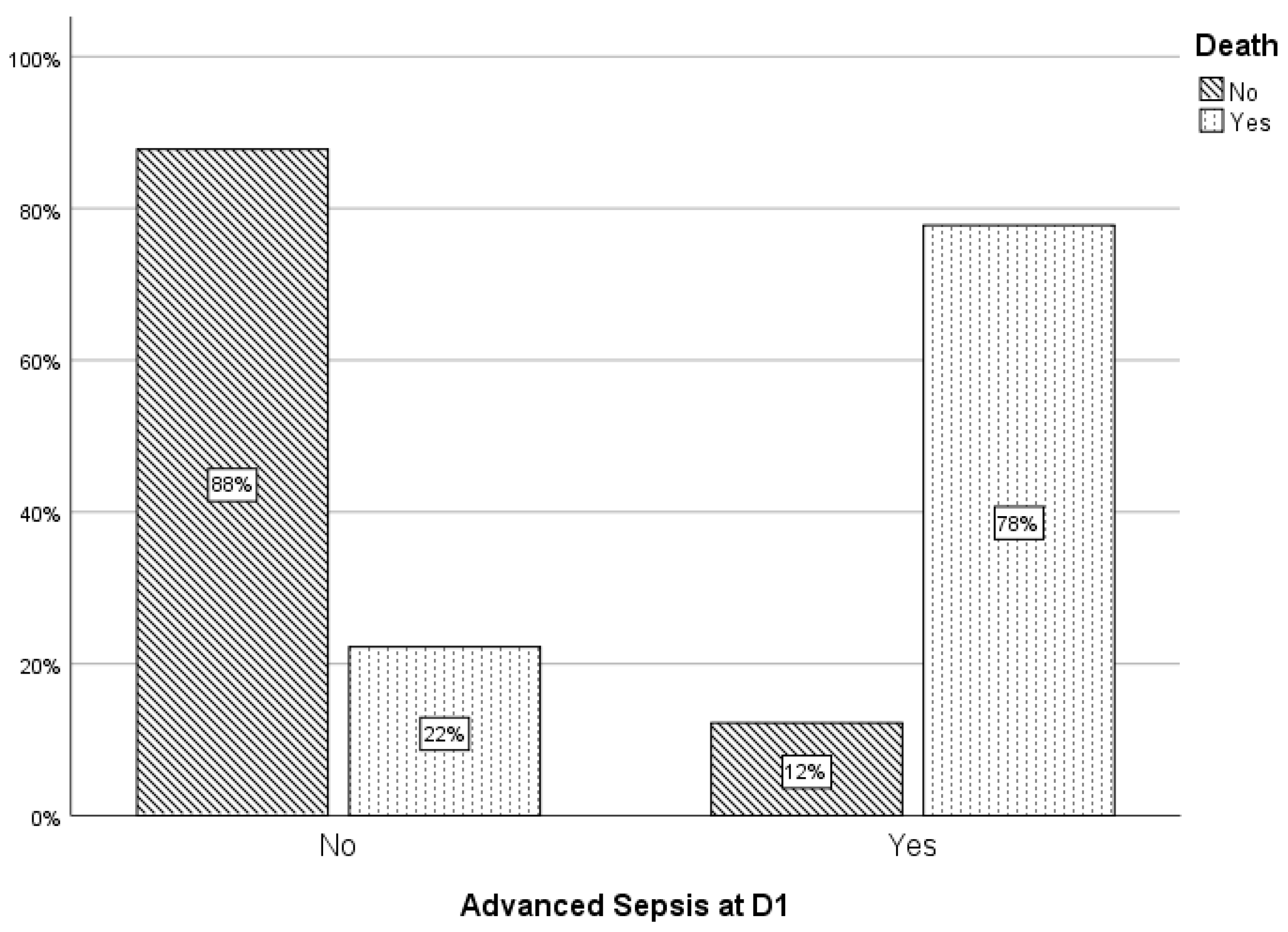
Figure 2.
Percentage of death in relation to delivery mode.
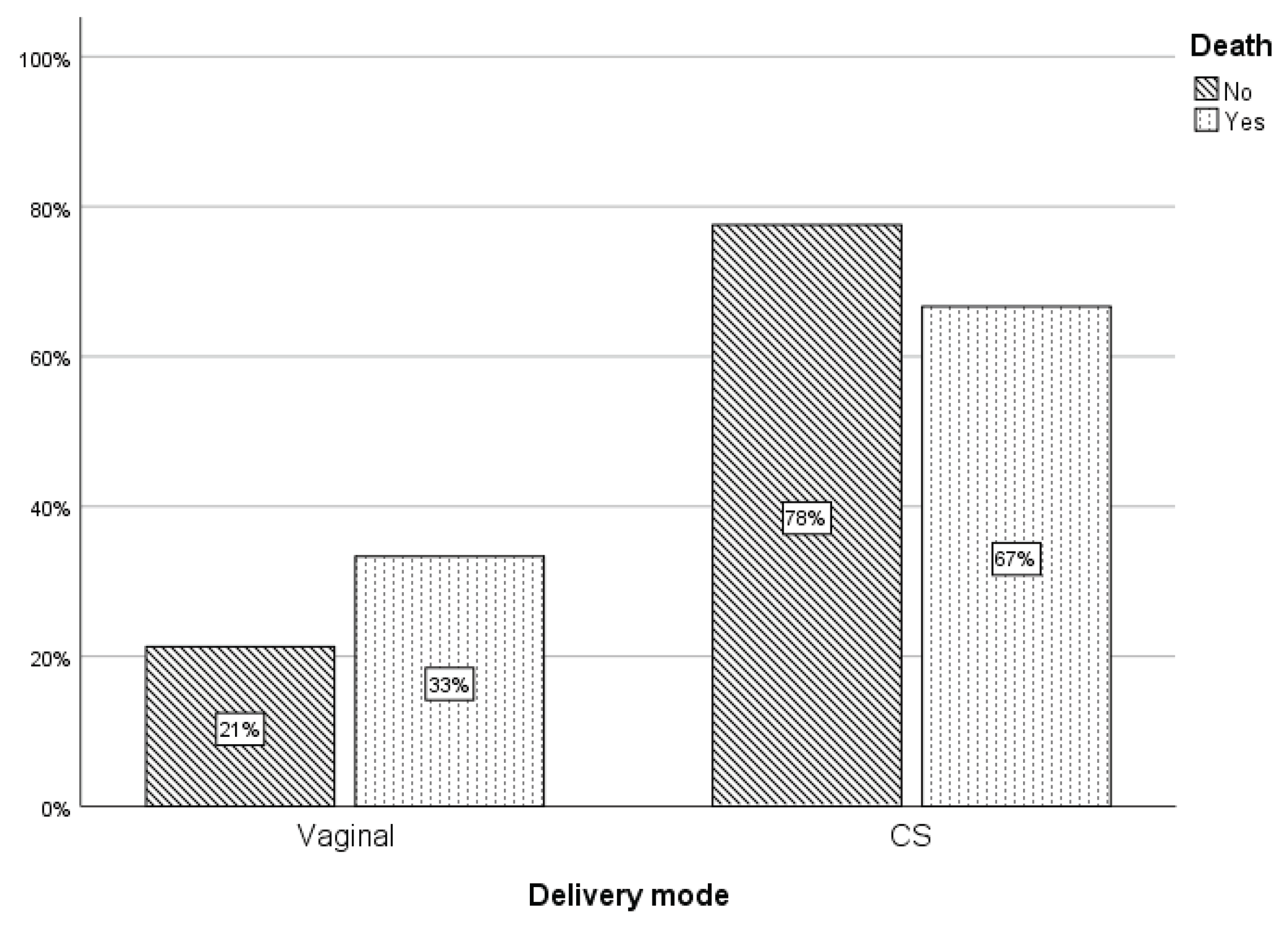

Table 1.
Neonates with incipient sepsis signs at three post-admission time points and pathogen localization sites. CNS: Central nervous system, GI: Gastrointestinal tract.
Table 1.
Neonates with incipient sepsis signs at three post-admission time points and pathogen localization sites. CNS: Central nervous system, GI: Gastrointestinal tract.
|
Day 1 |
Day 3 |
Day 7 |
|
|---|---|---|---|
| Incipient sepsis signs | 89.2% | 50.2% | 6.5% |
| Respiratory tract infection | 57.2% | 58% | 24.5% |
| CNS involvement | 5% | 0 % | 0 % |
| GI tract involvement | 2% | 0 % | 0 % |
| Skin involvement | 1.6% | 0 % | 0 % |
Disclaimer/Publisher’s Note: The statements, opinions and data contained in all publications are solely those of the individual author(s) and contributor(s) and not of MDPI and/or the editor(s). MDPI and/or the editor(s) disclaim responsibility for any injury to people or property resulting from any ideas, methods, instructions or products referred to in the content. |
© 2026 by the authors. Licensee MDPI, Basel, Switzerland. This article is an open access article distributed under the terms and conditions of the Creative Commons Attribution (CC BY) license (http://creativecommons.org/licenses/by/4.0/).
Copyright: This open access article is published under a Creative Commons CC BY 4.0 license, which permit the free download, distribution, and reuse, provided that the author and preprint are cited in any reuse.


