

Submitted:
26 March 2026
Posted:
30 March 2026
You are already at the latest version
Abstract
Background/Objectives: Excessive dietary inorganic phosphate (Pi) as a food additive poses potential health risks. Methods: This study investigates its impact on intestinal and immune homeostasis in mice using gradient Pi exposure combined with an inflammatory model. Results: Pi overload induced atrophy in the thymus, spleen, and kidney, damaged the intestinal barrier, reduced villus height‑to‑crypt depth ratio, and decreased goblet cell numbers. Altered levels of serum sIgA and IgE, as well as intestinal IgA, IgG, IgE, and IgM, together with decreased IFNα, indicated disrupted immune balance caused by Pi treatment. Proteomic analysis revealed differential expression of key proteins, including CNTFR and Bcl2l1 in the JAK‑STAT pathway, and metabolic regulators CPT1α and IDH1, compared Pi treated mice with the control group. Conclusions: These findings suggest Pi may affect immune and neuronal functions through tumor‑related signaling and mitochondrial pathways, providing insight into the health implications of Pi overconsumption.
Keywords:
inorganic phosphate
; intestinal mucosa
; immune disfunction
; IgE
1. Introduction
Phosphorus is an essential nutrient for the human body, playing a critical role in key physiological processes such as energy metabolism, bone tissue formation, maintenance of cell membrane structure, and intracellular signal transduction[1,2,3]. Dietary phosphorus is mainly divided into two categories: organic phosphorus and inorganic phosphorus. In natural foods, phosphorus primarily exists in organic forms, with an absorption rate of approximately 10%~30%. In contrast, inorganic phosphates, widely used as food additives, have a bioavailability of nearly 100%[4,5]. Inorganic phosphates are extensively applied in the food industry, pharmaceuticals, and animal feed. According to statistics, over 70% of frozen and dried foods, more than 65% of pre-packaged meat products, over 55% of baked goods, and more than 50% of soups and yogurts in supermarkets contain inorganic phosphorus additives[6,7]. It is estimated that thousands of food products containing phosphorus additives are available for human consumption. Currently, the global average per capita phosphorus intake is nearly twice the Recommended nutritional intake (RNI), with a trend toward further increase[8]. Excessive intake of inorganic phosphorus can lead to elevated serum phosphorus levels. It was reported that 10.1% of Japanese adolescents have serum phosphorus levels exceeding the healthy threshold of 4.5 mg/dL[9].
Excessive intake of inorganic phosphorus poses multiple health risks, such as adverse mental health symptoms, disrupting calcium-phosphorus balance, impairing bone and liver function, and being associated with coronary and heart valve calcification, worsening renal function, reduced exercise tolerance, and increased all-cause mortality[10,11,12]. The pathogenesis of that high phosphorus intake promotes cardiac valve calcification by inducing osteogenic differentiation of a subset of valvular interstitial cells[13]. Calcium/phosphorus balance in human body is regulated by FGF23/α-klotho signaling pathway. Higher phosphate intake raises FGF23, without parallel increase of α-klotho, then increases the ratio of FGF23/α-klotho. Increased serum FGF23 concentration or FGF23/α-klotho ratio was positively linearly related to the diabetes and cardiovascular diseases[14,15].
The intestine, as the primary site of digestion and absorption, is also an important immune organ in the human body. However, the effects of excessive inorganic phosphate intake on the intestine and its related immune systems have not yet been reported. This study employed a mouse inflammation model stimulated with low dose of LPS, and the impact of high inorganic phosphorus diet on the intestinal immune barrier and systemic immune function were investigated. The detective methods included organ indices, intestinal morphology, ELISA of immunoglobulin expression, and proteomic changes. The findings aim to provide a theoretical basis for the scientific regulation of dietary phosphorus intake and perhaps offer a hypothesis that the occurrence of food hypersensitivity reactions is related to excessive intake of inorganic phosphorus.
2. Materials and Methods
2.1. Materials
A total of 40 SPF-grade female BALB/c mice were selected as experimental subjects. The mice weighed 20±2g and were 6-8 weeks old, supplied by the Guangdong Provincial Medical Laboratory Animal Center (Experimental Animal License No.: Yue Shi Zheng (2019) 05073). LPS (lipopolysaccharide) (Sigma), trisodium phosphate food additive (Zhengzhou Gaoyan Biotechnology Co., Ltd.), Carnoy's fixative, HE staining kit, AB-PAS staining kit (Servicebio), IL-1β ELISA kit (Yikexai Biotechnology Co., Ltd.), IFN-α ELISA kit, IgG ELISA kit (Huamei Bio), sIgA ELISA Kit, IgA ELISA Kit, IgE ELISA Kit (Shanghai Sangon), Proteinase Inhibitor (Thermo Fisher Scientific (A32953)), Sodium Deoxycholate (Sigma-Aldrich (30970)), Chloroacetamide (Sigma-Aldrich (22790)), Tri(2-carboxyethyl)phosphine (Sigma-Aldrich (C4706)) . Microplate reader (Thermo Fisher Scientific, USA), Omni Bead Ruptor Elite 24 bead homogenizer (OMNI, USA), nanoElute liquid chromatography (Bruker Corporation), timsTOF Pro mass spectrometer (Bruker Corporation).
2.1.1. Animal Experiments
The recommended dietary phosphorus intake (RNI) for adults is approximately 700 mg/day. In the United States, the average daily phosphorus intake for adult males is 1600 mg/d~1800 mg/d, while the Tolerable Upper Intake Level (UL) for dietary phosphorus is 4000 mg/day. Therefore, the three trisodium phosphate dose gradients (100 mg/kg, 200 mg/kg, 400 mg/kg) used in the mouse gavage experiment correspond to daily dietary phosphorus intakes of 1665 mg/day, 2610 mg/day, and 4500 mg/day for an adult, respectively.
During animal experiments, mice were labeled using a staining method. Forty mice were randomly divided into eight groups: blank control group (Control), LPS model group (LPS), low-dose phosphate group (LIP), medium-dose phosphate group (MIP), high-dose phosphate group (HIP), low-dose phosphate-LPS group (LIP-LPS), medium-dose phosphate-LPS group (MIP-LPS), and high-dose phosphate-LPS group (HIP-LPS). The Control and LPS model groups received daily oral administration of 0.9% sodium chloride solution at 0.2 mL/10 g body weight. The Low-dose trisodium phosphate and Low-dose trisodium phosphate-LPS groups received daily oral administration of trisodium phosphate solution at 100 mg/kg body weight via 0.2 mL/10 g body weight. The medium-dose trisodium phosphate group and medium-dose trisodium phosphate-LPS group received 0.2 mL/10 g body weight of 200 mg/kg trisodium phosphate solution daily; the high-dose trisodium phosphate group and high-dose trisodium phosphate-LPS group received 0.2 mL/10 g body weight of 400 mg/kg trisodium phosphate solution daily. Gavage was administered once daily for 15 consecutive days. On day 15, mice received their final gavage. Three hours post-gavage, LPS solution (0.05 mg/mL) was intraperitoneally injected into LPS, LIP-LPS, MIP-LPS, and HIP-LPS groups, respectively. During the experiment, mice were monitored for behavioral changes. Mouse body weights were weighed and recorded daily. Twenty-four hours after the final gavage, the experimental mice were euthanized by cervical dislocation. Their small intestines, livers, thymuses, and spleens were collected for subsequent experimental studies.
2.1.2. Preparation of Paraffin Sections
After obtaining small intestinal tissue from experimental mice, the intestinal contents were thoroughly rinsed with PBS solution or normal saline. The small intestinal tissue was fixed in Carnoy's fixative for at least 4 hours. The fixed intestinal tissue was then removed and cut into segments approximately 1 cm in length. The intestinal segments were sequentially dehydrated in ethanol solution containers. The dehydration ethanol concentration was as follows: first immersed in 85% ethanol solution for 1 hour, then in 95% ethanol solution for 1 hour (repeated twice), and finally in anhydrous ethanol solution for 1 hour (repeated twice). The dehydrated intestinal tissue was then transparentized. After completion of the transparentization process, paraffin embedding was performed. The transparentization and embedding steps were as follows: the tissue was immersed in two xylene solution containers for 30 minutes each, followed by immersion in three molten paraffin containers for 30 minutes, 25 minutes, and 20 minutes, respectively. The paraffin-embedded small intestinal tissue was placed in the embedding container. During the rapid pouring of molten paraffin, efforts were made to minimize bubble formation. The paraffin was then placed in an ice bath to accelerate solidification. Once the paraffin was fully solidified and hardened, the tissue embedding was complete. The processed paraffin blocks were sectioned. The paraffin blocks were placed in a tissue slicer for sectioning. The cut paraffin sections were transferred to a constant-temperature water bath for slow unfolding. Unfolded tissue sections were removed from the water bath using a slide, ensuring that the small intestinal tissue sections were completely adhered to the slide.
2.1.3. Preparation of Intestinal HE-Stained Sections
Preparation method for HE-stained tissue sections: Paraffin sections were immersed twice in xylene solution (20 minutes each time) and then twice in anhydrous ethanol (5 minutes each time). Subsequently, they were immersed in 75% ethanol for 5 minutes. Finally, the sections were rinsed with tap water. After dewaxing, the sections were stained with hematoxylin for approximately 4 minutes. Following hematoxylin staining, the sections were rinsed with tap water and then decolorized with a decolorizing solution. After decolorization, the sections were rinsed again with running water. The sections were then re-stained with a re-staining solution and rinsed with running water. For eosin staining of hematoxylin-stained sections: The sections were first dehydrated with 85% ethanol for 5 minutes, then with 95% ethanol for 5 minutes, followed by 5 minutes of eosin staining. After eosin staining, the sections were dehydrated by soaking in anhydrous ethanol three times (5 minutes each time). Subsequently, they were immersed twice in xylene solution (5 minutes each time) until the sections became transparent and decolorized. After completing the above steps, the sections were sealed. After sealing the small intestinal tissue sections with HE staining, the staining changes were observed under a forward optical microscope. Randomly selected 3-5 mouse intestinal villi were photographed to measure villus length and crypt depth, and the ratio of villus length to crypt depth was calculated.
2.1.4. Preparation of AB-PAS-Stained Intestinal Sections
Mouse small intestinal paraffin sections were routinely deparaffinized to water, then placed in 1% Alcian Blue acetic acid staining solution for 15 min at room temperature, followed by thorough rinsing with running tap water until the rinse water was colorless. Subsequent to Alcian Blue staining, the sections were transferred to 1% periodic acid solution for 15 min incubation at room temperature, quickly rinsed once with tap water and then washed with distilled water to remove residual periodic acid. After that, the sections were incubated in Schiff's staining solution for 30 min at room temperature in the dark, rinsed with running tap water, immersed in hematoxylin staining solution for 2 min counterstaining, and then rinsed with distilled water. Finally, the sections were dehydrated, cleared and mounted using the same routine method as HE-stained sections; after preparation, the morphological changes of small intestinal tissues were observed under an inverted light microscope, with intestinal lumen villi photographed and goblet cells on the villi counted and analyzed.
2.1.5. ELISA Assay for Detecting Serum sIgA and IgE Levels in Mice
Prior to the assay, standard solutions designated as S1 to S8 (unit: ng/ml) were prepared, with S8 serving as the sample diluent and the concentrations being 10, 5, 2.5, 1.25, 0.63, 0.31, 0.16, and 0 ng/ml, respectively; the reagents and materials used included the aforementioned standard solutions, biotin-labeled secretory immunoglobulin antibody working solution, horseradish peroxidase (HRP)-labeled streptavidin working solution, wash buffer, chromogenic reagent, stop solution, 96-well microplate, and a pre-warmed microplate reader for absorbance measurement. For the experimental procedures, standard wells (with duplicate wells to ensure reproducibility) and sample wells were first set up on the 96-well microplate, followed by adding 100 μl of standard working solution or 100 μl of each serum sample to the corresponding wells; after loading, the microplate was sealed and incubated at 37°C for 90 minutes, and the liquid in each well was discarded thereafter with the microplate gently tapped on absorbent paper to remove residual liquid. Subsequently, 100 μl of biotin-labeled secretory immunoglobulin antibody working solution was added to each reaction well, the microplate was resealed and incubated at 37°C for 1 hour, and the first washing step was performed by adding 350 μl of pre-prepared wash buffer to each well (standing for 2 minutes before discarding), repeating this step 4 times with the microplate tapped dry to remove excess wash buffer prior to each subsequent wash. After washing, 100 μl of HRP-labeled streptavidin working solution was added to each well, the microplate was sealed and incubated at 37°C for 30 minutes, followed by a second washing step where 300 μl of wash buffer was added to each reaction well (standing for 30 seconds before discarding), repeating this procedure 4 times and thoroughly removing residual wash buffer by vortexing after the final wash. Under dark conditions, 90 μl of pre-prepared chromogenic reagent was then added to each well, the microplate was sealed and incubated at 37°C in the dark for approximately 15 minutes to facilitate color development, and immediately after color development, 50 μl of stop solution was added to each well to terminate the reaction. Finally, the optical density (OD) values of the standard solutions and samples were measured at 450 nm using the pre-warmed microplate reader, and the actual concentrations of sIgA and IgE in each serum sample were calculated based on the standard curve generated from the standard solution assay results.
2.1.6. ELISA Assay for Detecting IFN-α and IL-1β Levels, as well as IgE, IgG, IgM, and IgA Expression in Mouse Small Intestinal Tissue
Tissue samples were homogenized into a homogeneous slurry, followed by ultrasonic disruption for complete lysis; after lysis, the samples were centrifuged at 5000 × g for 5–10 min, and the supernatant was collected and stored appropriately for subsequent assay, while standard stock solutions were serially diluted to generate a series of gradient-concentration working standards that were aliquoted and preserved under optimal conditions until use; for the assay, a 96-well reaction plate was prepared with blank wells and sample wells arranged according to the experimental design, 100 μL of each sample supernatant or gradient standard was added to the corresponding wells, subsequently 50 μL of biotinylated antibody working solution was added to each well, and the plate was sealed and incubated at 20–25 °C with shaking at 300 rpm on a microplate shaker for 120 min; after the first incubation, the plate was subjected to washing by dispensing 300 μL of wash buffer into each well, incubating for 10 s, then completely removing the liquid by gentle shaking, a procedure repeated 5 times, followed by adding 100 μL of enzyme conjugate working solution to all wells except the blank wells, resealing the plate and incubating at 20–25 °C with shaking at 300 rpm for 60 min; a second washing step was then performed by adding 300 μL of wash buffer to each well, incubating for 10 s, thoroughly removing residual wash buffer by centrifugation, and repeating this step 5 times to eliminate non-specific binding; under dark conditions, 100 μL of chromogenic substrate was added to each reaction well, the plate was sealed immediately after reagent addition and incubated at 20–25 °C in the dark for 15 min to allow color development, and upon completion of color development, 100 μL of stop solution was promptly added to each well to terminate the reaction; finally, the optical density (OD) values of the standards and test samples were measured at a wavelength of 450 nm using a pre-warmed microplate reader, and the actual concentrations of interferon-α (IFN-α), interleukin-1β (IL-1β), immunoglobulin E (IgE), immunoglobulin G (IgG), immunoglobulin M (IgM), and immunoglobulin A (IgA) in each small intestinal tissue sample were calculated based on the standard curve constructed from the standard OD values.
2.1.7. Immunofluorescence Detection of IgA Cell Expression in the Intestinal Tract of Experimental Mice
An appropriate volume of environmentally friendly dewaxing solution and anhydrous ethanol were separately prepared in different containers. Paraffin sections were first immersed in three sequential containers of environmentally friendly dewaxing solution for 10 min each, then transferred to three sequential containers of anhydrous ethanol for 5 min each. After this treatment, the sections were rinsed with distilled water prior to antigen retrieval. Antigen retrieval was performed under the following conditions: microwave irradiation at medium power for 8 min in citric acid buffer (pH 6.0), followed by standing for 8 min, and subsequent microwave irradiation at low-medium power for 7 min. After fixation, the sections were placed in phosphate-buffered saline (PBS, pH 7.4) and washed on a decolorizing shaker for 5 min per wash, with this washing step repeated three times. For serum blocking, a histochemical pen was used to draw circles around the treated tissue, followed by dropwise addition of bovine serum albumin (BSA) for blocking at room temperature for 30 min. Subsequent to serum blocking, primary antibody was pipetted onto the tissue sections, and the sections were incubated overnight at 4 °C. The incubated sections were then washed in PBS (pH 7.4) on a decolorizing shaker three times, 5 min per wash. Next, the corresponding secondary antibody was added to the sections, which were then incubated at room temperature in the dark for 50 min. After secondary antibody incubation, the sections were washed in PBS (pH 7.4) three times for 5 min each on the decolorizing shaker. Nuclear staining was performed using 4',6-diamidino-2-phenylindole (DAPI) reagent: DAPI solution was added dropwise to the sections, followed by incubation at room temperature in the dark for 10 min. The stained sections were then washed again in PBS (pH 7.4) three times, 5 min per wash. Subsequently, pre-prepared autofluorescence quencher was added to the sections and allowed to stand for 5 min. Finally, the sections were rinsed with running water, and the processed sections were mounted using anti-autofluorescence mounting medium. Following these procedures, image acquisition was conducted. Images were captured at the excitation wavelengths for DAPI (330–380 nm), 488 nm, CY3 (465–495 nm), and CY5 (515–555 nm), as well as their corresponding emission wavelengths: 510–560 nm, 590 nm, 608–648 nm, and 672–712 nm.
2.1.8. Proteomics Sample Preparation
Approximately 20 mg of mouse spleen tissue was weighed and mixed with phosphate-buffered saline (PBS) containing protease inhibitors, followed by thorough vortexing. Lysis buffer was then added, and the mixture was incubated in a 95 °C water bath for 5 min. The sample was transferred to a grinding tube and homogenized using a bead mill homogenizer under the following conditions: speed 7.1 m/s, with cycles of 20 s on and 20 s off, repeated for five cycles. After homogenization, the sample was transferred to a new centrifuge tube, and contact ultrasonication was performed on ice for protein extraction at 30% energy (4 s on, 8 s off) for a total duration of 3 min. The extracted protein was centrifuged at 16,000 × g for 5 min, and the supernatant was collected for protein concentration determination via the bicinchoninic acid (BCA) assay. Following concentration determination, 100 μg of protein was diluted and mixed with Tris-HCl buffer (pH 8.8). Trypsin was added at a protein-to-enzyme ratio of 25:1; the mixture was vortexed thoroughly and incubated at 37 °C on a shaking mixer (1000 rpm) for 16 h. The pH of the mixture was adjusted to 3 using 10% formic acid (FA), and the sample was centrifuged at 16,000 × g for 5 min. The resulting supernatant was collected, desalted using an automated desalting system, and dried at 45 °C. The dried sample was resuspended in 30 μL of 0.1% FA, the peptide concentration was measured, and the sample was prepared for instrumental analysis.
2.1.9. LC-MS/MS Analysis
Cohort samples were analyzed using a Bruker nanoElute liquid chromatography system coupled to a Bruker timsTOF Pro mass spectrometer. A 30 cm × 75 μm column packed with 1.9 μm C18 particles (120 Å, Dr. Maisch GmbH) was employed. The nanoElute gradient was set as follows: 2%-4% mobile phase B for the first 5 min, 4%-22% B for 5-70 min, 22%-35% B for 70-90 min, 35%-80% B for 90-100 min, and 80% B for 100-110 min. Mobile phase composition: Phase A consists of water and 0.1% formic acid solution; Phase B consists of acetonitrile and 0.1% formic acid solution. The overall flow rate is 300 nL/min, but increases to 500 nL/min during the 5 minutes preceding formal detection. Bruker timsTOF Pro mass spectrometer parameters: Ion source voltage set to 4.5 kV, ion source temperature set to 180°C, ion source flow rate 3 L/min. Data acquisition mode employed DIA-PASEF. The primary mass spectrum m/z range was 300–1500, with ion drift set to 1/K0. The scanning range was 0.75–1.40 V s/cm², and the ramp-up time was set to 100 ms. For secondary spectrum acquisition, the peptide m/z range was 400–1200, charge state set to 0-5, mass isolation window set to 25 Th, DIA-PASEF window count set to 64, and total method cycle time set to 1.17 s. To prevent duplicate peptide scanning, the dynamic exclusion time (Release after) for tandem mass spectrometry was set to 30 s.
2.1.10. Statistical Analysis
Data obtained from this experiment were analyzed using SPSS 26.0 software. GraphPad Prism 8.0 was employed for statistical graphing of results. P < 0.05 was considered statistically significant. Protein-protein interaction network analysis was visualized using the String website. Data analysis and quality control were performed using Python 3.8 algorithms. Differential protein analysis was conducted with R software (R_4.1), expressing the abundance differences of all detected proteins via Fold Change (FC) values. Differential proteins were selected based on log2 FC > 2 and P< 0.05.
3. Results
3.1. High Inorganic Phosphate Diet Impairs the Organ Indices and the Integrity of the Intestinal Mucosal Barrier in Mice
The intestine is the primary site for digestion and absorption, and it also represents the largest and most complex immune organ in the human body, undertaking the critical functions. The intestinal mucosa acts as a crucial barrier separating the internal milieu from the luminal environment. Compromise of its integrity may permit the invasion of exogenous harmful substances, triggering inflammation and tissue damage. This study employed a 15-day intragastric administration of varying doses of inorganic phosphate (100, 200, 400 mg/kg) to mice, followed by intraperitoneal injection of LPS to establish an acute inflammation model, aiming to investigate the impact of the food additive inorganic phosphate on the immune system and organ function under inflammatory challenge. Body weight monitoring during the experiment revealed an increase in weight across all phosphate-dosed groups compared to pre-administration levels, whereas no significant change was observed in the blank control group (Figure 1A). However, upon LPS-induced inflammatory challenge, all experimental groups exhibited a significant decrease in body weight (Figure 1B). Analysis of organ indices indicated that inorganic phosphate intake led to a decrease in the thymus index and spleen index, suggesting that phosphate overload may cause immunosuppression and organ damage (Figure 1C, D). The kidney index also showed a dose-dependent decrease, indicating adverse effects of high phosphate intake on renal health (Figure 1E). Notably, under the acute inflammatory state induced by LPS, the spleen and kidney indices showed a recovery, providing further macroscopic evidence supporting the inference that inorganic phosphate may possess immunosuppressive or immunomodulatory effects (Figure 1C-E). These results suggest that inorganic phosphate may exert potential physiological regulatory effects on the organism by influencing the development of immune organs and metabolic functions.
The effects of a high inorganic phosphate diet on the intestinal barrier structure were systematically evaluated further via histomorphological analysis. H&E staining results showed that compared to the blank control group, all phosphate-intervention groups exhibited morphological alterations including shortened average intestinal villi height and increased crypt depth, with the group of 100 mg/kg phospate treatment showing a significant reduction of near 50% in villus length (Figure 2A-C). The ratio of villus height to crypt depth (V/C ratio) was significantly decreased in all treatment groups, indicating villial atrophy and crypt hyperplasia (Figure 2C). AB-PAS staining and cell counting analysis demonstrated that the number of goblet cells in the phosphate-intervention groups was significantly lower than in the control group (Figure 2D, E). These results collectively indicate that inorganic phosphate intake disrupts the integrity of the intestinal structure and immune barriers by inducing villus atrophy and crypt hyperplasia, and reducing the number of goblet cells, thereby disturbing normal intestinal physiological function.
In summary, excessive intake of inorganic phosphate as a food additive not only exerts negative effects on organs such as the thymus, spleen, and kidneys but also impairs the structure and function of the intestinal mucosal barrier, highlighting its potential risks in terms of immunomodulation and intestinal health.
3.2. Effects of a High Inorganic Phosphate Diet on the Intestinal Immune Environment in Mice
There is a significant correlation between damage to the intestinal mucosal barrier and food allergies. On the basis of detecting the structural damage caused by sodium phosphate to the intestinal mucosa, we measured the protein expression levels of immunoglobulins (IgA, sIgA, IgE, IgM, IgG) and pro-inflammatory cytokines (IL-1 β, IFN - α) in the blood and small intestine of mice. The results showed that gavage of sodium phosphate significantly reduced the levels of sIgA in mouse serum (Figure 3A) and IgA in mouse small intestine (Figure 3C), and the number of IgA secreting cells in the small intestine wall also decreased (Figure 3G and Figure 3H).Gavage of sodium phosphate also significantly reduced the protein expression of IgE (Figure 3D), IgM (Figure 3E), and IgG (Figure 3F) in the small intestine of mice.
Although gavage of sodium phosphate alone did not significantly alter the expression of IgE in mouse serum (Figure 3B), induction with adjuvant LPS led to a significant increase in the expression of IgE (Figure 3D), IgM content (Figure 3E), IFN - α (Figure 3I), and IL-1 β (Figure 3J) in the small intestine tissue of mice administered with 200mg/kg sodium phosphate (MIP-LPS group).
In addition, gavage of 200 mg/kg sodium phosphate significantly inhibited LPS induced IgG levels (Figure 3F), reflecting a decrease in humoral immune function and possible impairment of the body's ability to resist pathogens.
In summary, inorganic phosphate can disrupt intestinal immune balance and normal inflammatory responses by changing the expression of immunoglobulins (IgA, sIgA, IgE, IgG and IgM) and pro-inflammatory cytokines (IL-1β, IFN-α). This study provides evidence that inorganic phosphate may promote immune evasion and intestinal immune dysregulation, suggesting that long-term or excessive intake of inorganic phosphate food additives may pose potential risks to immune function.
3.3. Proteomic Analysis of the Effects of A High-Phosphate Diet on the Mice Immune System
Based on Data-Independent Acquisition (DIA) quantitative proteomics technology, this study systematically analyzed the impact of a high-phosphate diet on the protein expression profile in the mouse spleen. Quality control of the data analysis showed low coefficients of variation and high consistency among samples in each group (Supplementary Figure S1A), with significant correlations in protein quantification between samples (Supplementary Figure S1B), indicating reliable data quality. Through differential expression analysis (screening criteria: |log2FC| > 2 and P < 0.05), a number of significantly differentially expressed proteins (DEPs) were identified across different comparison groups. Specifically, 187, 132, and 77 DEPs were identified when comparing the blank control group with the low-, medium-, and high-dose phosphate groups, respectively. Conversely, comparisons between the LPS model group and the corresponding phosphate+LPS groups yielded 85, 88, and 91 DEPs, respectively. Common DEPs across the different phosphate+LPS dose groups compared to the LPS model group were also identified (Supplementary Figure S2).
Gene Ontology (GO) functional annotation analysis revealed that the DEPs were significantly enriched in biological processes such as immune-inflammatory response, cellular stress response, and metabolic regulation (Figure 4A-C). The biological process, such as response to oxidative stress, cellular response to chemical stress, mitochondrial gene expression, cellular response to oxygen radical, respiratory electron trasport chain, posttranscriptional regulation of gene expression, negative regulation of response to endoplasmic reticulum stress, response to oxygen radical, establishment of organelle localization, ribonucleoprotein complex biogenesis could be disturbed by excessive Na3PO4 treatment; the cell components, such as ribosome, ribosomal subunit, polysomal ribosome, mitochondrial protein containing complex, polysome, respirasome, organellar ribosome, Smn Sm protein complex, triglyceride rich plasma lipoprotein particle, small ribcsomal subunit could be damaged by excessive Na3PO4 treatment; and molecular function, such as structural constituent of ribosome, regulatory RNA binding, MiRNA binding, oxidoreductase activity acting on NADPH quinone or similar compound as acceptor, cysteine type endopeptidase regulator activity involved in apoptotic process, cysteine type endopeptidase inhibitor activity involved in apoptotic process, oxidoreduction driven active transmembrane transporter activity, nuclease activity, Snare binding, catalytic activity acting on RNA could be changed by excessive Na3PO4 treatment (Figure 4B). Kyoto Encyclopedia of Genes and Genomes (KEGG) pathway enrichment analysis indicated that these proteins are primarily involved in signaling pathways including neutrophil degranulation, the ribosome pathway, and DNA damage repair (Figure 4D-F). Of particular note, within the JAK-STAT signaling pathway, ciliary neurotrophic factor receptor (CNTFR) protein expression was significantly up-regulated, while Bcl2l1 protein expression was significantly down-regulated (Supplementary Figure S4A). Protein-protein interaction (PPI) network analysis further elucidated the regulatory relationships among the DEPs (Supplementary Figure S4B).
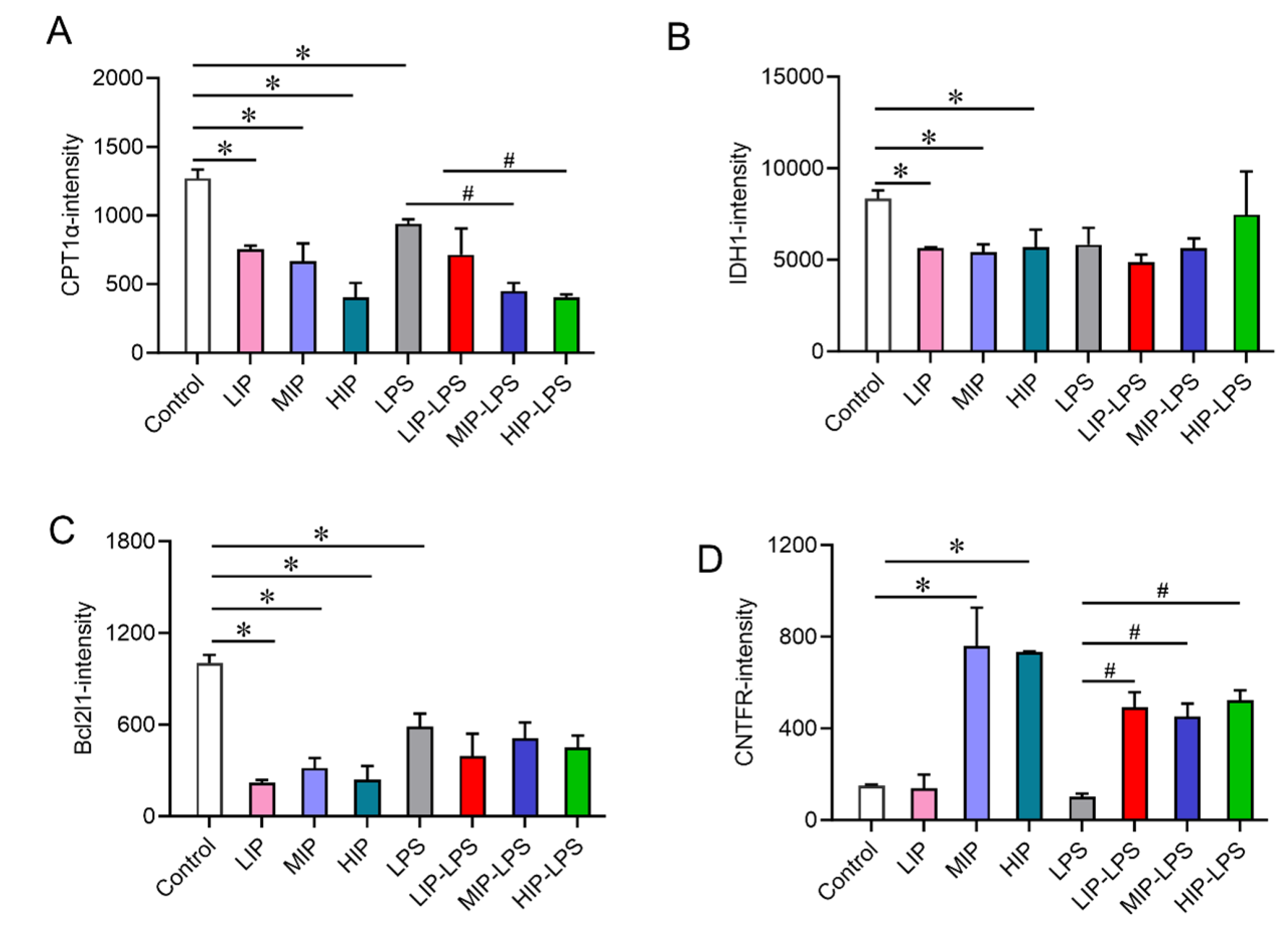
In-depth analysis of key functional proteins revealed that the expression of palmitoyltransferase1 α 1(CPT1α) and socitric Dehydrogenase1(IDH1) were significantly down-regulated in the phosphate treatment groups, showing a dose-dependent change (Figure 5A-B). The expression of Bcl2l1 protein was significantly down-regulated across all treatment groups (Figure 5C), whereas CNTFR protein expression was significantly up-regulated (Figure 5D). These results indicate that a high-phosphate diet can alter the splenic protein expression profile, affecting signaling pathways related to immune regulation, cellular metabolism, and apoptosis, thereby potentially disrupting immune homeostasis. This study provides molecular-level evidence for a deeper understanding of the immunoregulatory mechanisms of a high-phosphate diet and offers a scientific basis for assessing the safety of dietary phosphate additives.
4. Discussion
This study systematically investigated the comprehensive effects of high inorganic phosphorus diet on the intestinal mucosal barrier, immune environment, and spleen protein expression profiles in mice through integrated in vivo animal models and proteomic analysis. Our findings demonstrate that excessive inorganic phosphorus intake not only reduces indicators in immune organs such as the thymus and spleen but also causes structural damage to intestinal villi, decreases goblet cell numbers, and thins the mucus layer, indicating significant impairment of both the intestinal physical structure and critical immune organs. Further analysis of immunoglobulins and cytokines revealed widespread reductions in key immunoglobulins (sIgA, IgA, IgM, IgG) and suppressed IFN-α expression. Secretory immunoglobulin sIgA, serving as the frontline defense of the body's mucosal system, encapsulates bacteria, viruses, and toxins to prevent pathogen adhesion and penetration of mucosal epithelial cells, thereby avoiding hypersensitivity reactions. The reduced sIgA expression in small intestinal tissues indicates that excessive inorganic phosphate intake disrupt the mucosal immune function of the small intestine. LPS can act as an adjuvant in inducing food hypersensitivity reactions. Our results showed that the IgE content in the small intestine of mice was significantly increased after intragastric administration of 200 mg/kg sodium phosphate for 15days combined with LPS stimulation. As the primary indicator of food hypersensitivity, IgE demonstrates strong correlation with impaired intestinal mucosal barriers[16]. The elevated IgE in this experiment may result from the combined action of sodium phosphate and LPS on the small intestinal mucosa, leading to intestinal leakage that allows macromolecular allergens from food to enter the bloodstream, promoting antibody formation and ultimately triggering food hypersensitivity reactions. The significant and complex changes of immunoglobulin (IgA, sIgA, IgE, IgM, IgG) and pro-inflammatory cytokines (IL-1β, IFN-α) in blood and small intestine of mice caused by oral administration of inorganic phosphate indicated that the immune response mechanism and immune defense function of intestinal mucosa were impaired by exessive inorganic phosphate intake.
At the molecular mechanism level, proteomic results revealed alterations in multiple key proteins and signaling pathways in mouse spleen tissues. Phosphorus overload affected various pathways, including oxidative stress, mitochondria gene expression, respiratory electron transport chain, post-transcriptional regulation of gene expression, endoplasmic reticulum stress, and the biosynthesis of ribonucleoprotein complexes. Structural damage to organelles such as ribosomes, respirasomes, Smn Sm protein complexes, and triglyceride-rich plasma lipoprotein particles was observed due to phosphorus overload. Additionally, multiple biological functions were disrupted by phosphorus overload, including regulation of RNA binding, MiRNA binding, activity of oxidoreductases acting on NADPH quinone or similar compound receptors, regulation of cysteine endopeptidase activity involved in apoptosis, activity of redox-driven active transmembrane transporters, and nuclease activity. These findings suggest that phosphorus overload induces systemic and holistic changes in the organism, analogous to the global environmental non-point source pollution currently experienced. Currently, farmland and water bodies worldwide face significant pressure from excessive nitrogen (N) and phosphorus (P) levels, and the organism may similarly encounter comparable challenges.
Furthermore, this study revealed upregulation of CNTFR and downregulation of Bcl2l1 in the JAK-STAT pathway, with significant downregulation of metabolic-related proteins such as CPT1α and IDH1. CNTFR is a neurotrophic factor receptor, and its signaling pathway is critical for the survival, development, and repair of motor neurons. In certain disease states (e.g., amyotrophic lateral sclerosis [ALS]), the upregulation of CNTFR is often regarded as a compensatory response. The nervous system attempts to counteract neuronal damage and death by enhancing neurotrophic support. Bcl2l1 is a key anti-apoptotic protein in the Bcl-2 family, playing a central role in the mitochondrial apoptosis pathway. Its downregulation significantly impairs the anti-apoptotic capacity of cells (particularly neurons), making them more susceptible to programmed cell death under stress. CPT1α is a rate-limiting enzyme in fatty acid β-oxidation, located in the outer mitochondrial membrane, responsible for transporting long-chain fatty acids into mitochondria for oxidative energy production. Its downregulation indicates a reduced ability of cells to utilize fatty acids for energy production, leading to energy metabolism disorders and lipid accumulation. IDH1 catalyzes the oxidative decarboxylation of isocitrate to produce α-ketoglutarate (α-KG) in the cytoplasm, while also generating NADPH. Its downregulation results in a decrease in the cellular antioxidant NADPH, making cells more vulnerable to oxidative stress damage; it also reduces α-KG levels, disrupting various cellular metabolic and epigenetic regulatory processes.
The core characteristic of amyotrophic lateral sclerosis (ALS) is the selective loss of motor neurons, with its pathological mechanisms clearly including: dysregulation of neurotrophic factor signaling, mitochondrial dysfunction, abnormal energy metabolism, increased oxidative stress, and activation of apoptosis pathways. Multiple studies have confirmed compensatory alterations in the CNTFR pathway, imbalances in Bcl-2 family protein expression, abnormal fatty acid metabolism, and IDH1-associated oxidative stress responses in ALS patients and models[17]. The results detected in this trial are highly consistent with these findings. Phosphorus overload-induced changes encompass nearly all these critical pathways, suggesting that excessive intake of inorganic phosphate may also contribute to the risk of ALS.
Organic phosphorus in food is also absorbed as phosphate after being cleaved by intestinal alkaline phosphatases or by bacteria-derived phytases, or being oxidized in the intestine[18]. Our results demonstrate that phosphorus overload exerts holistic and systemic disturbances on physiological states, analogous to the destructive impacts of non-point source pollution on the global environment[19,20]. Currently, farmland and water bodies worldwide are under immense pressure due to excessive nitrogen (N) and phosphorus (P) content, and animals, including humans, may also face similar challenges.
This study elucidates the adverse effects of high inorganic phosphate diets on the intestinal mucosal barrier and systemic immune function in mice. It suggests that the JAK-STAT signaling pathway and cellular metabolic reprogramming may play potential roles in these effects. The findings provide experimental evidence for the safety evaluation of inorganic phosphate food additives and offer new insights for the development of prevention and rehabilitation strategies for phosphorus overload-related chronic diseases.
Supplementary Materials
The following supporting information can be downloaded at the website of this paper posted on Preprints.org, Figure S1: Analysis of proteomics data quality control results; Figure S2: Analysis of differentially expressed proteins; Figure S3: Analysis of differentially expressed proteins volcano plot of differentially expressed proteins and hierarchical clustering analysis of differentially expressed proteins, Figure S4: JAK-STAT pathway and DEP interaction network analysis.
Author Contributions
Conceptualization, Y.Z.; methodology, Z.S., Y.W.; software, Y.Z.; validation, Y.L.; formal analysis, S.H.; investigation, R.W.; resources, Y.Z.; data curation, Z.S.; writing—original draft preparation, Y.Z.; writing—review and editing, S.H.; visualization, W.W.; supervision, J.L.; project administration, D.H.; funding acquisition, Y.Z. All authors have read and agreed to the published version of the manuscript.
Funding
This work was supported by the Central Government-funded Project to Guide Local Scientific and Technological Development: Development and Clinical Application of Special Dietary Foods Based on Precision Nutrition and Marine Polysaccharides (Grant No. YDZX2025062).
Institutional Review Board Statement
This animal study protocol has been approved by the Science and Technology Ethics Committee of University of Health and Rehabilitation (Protocol Number: KFDX:NO.2025-1241, Approval Date: February 28, 2025).
Data Availability Statement
The data reported in this paper have been deposited in the OMIX, China National Center for Bioinformation / Beijing Institute of Genomics, Chinese Academy of Sciences (https://ngdc.cncb.ac.cn/omix: accession no.OMIX014618) [21,22].
Acknowledgments
The author acknowledges technical support from Lingqiang Zhang of the Guangdong International Institute of Smart Medicine for the proteomics-related content in this paper.
Conflicts of Interest
The authors declare no conflicts of interest.
References
- Bird, R.P.; Eskin, N.A.M. The emerging role of phosphorus in human health . Adv Food Nutr Res 2021, 96, 27–88. [Google Scholar]
- Razzaque, M.S.; Wimalawansa, S.J. Minerals and Human Health: From Deficiency to Toxicity . Nutrients 2025, 17(3). [Google Scholar] [CrossRef]
- Chen, Y. Phosphorus modifies the association between body mass index and uric acid: Results from NHANES 2007-2018 . PLoS One 2024, 19(10), e0306383. [Google Scholar] [CrossRef]
- Ma, B. A combined approach to evaluate total phosphorus/inorganic phosphate levels in plants . STAR Protoc 2022, 3(3), p. 101456. [Google Scholar] [CrossRef] [PubMed]
- Yang, B. Phospholipase-mediated phosphate recycling during plant leaf senescence . Genome Biol 2024, 25(1), p. 199. [Google Scholar] [CrossRef] [PubMed]
- Dunford, E.K.; Calvo, M.S. Phosphate-based additives in processed foods: is excess exposure a cause for concern? A cross-sectional examination of the United States packaged food supply . Am J Clin Nutr 2025, 121(4), 873–881. [Google Scholar] [CrossRef]
- Pizzorno, L. Canaries in the Phosphate-Toxicity Coal Mines . Integr Med (Encinitas) 2014, 13(6), 24–32. [Google Scholar]
- Borgi, L. Inclusion of Phosphorus in the Nutrition Facts Label . Clin J Am Soc Nephrol 2019, 14(1), 139–140. [Google Scholar] [CrossRef]
- Saito, Y. Habitual confectionery intake is associated with serum phosphorus levels: A cross-sectional study on healthy subjects . J Med Invest 2019, 66(1.2), 134–140. [Google Scholar] [CrossRef] [PubMed]
- Valle-Hita, C. Associations between ultra-processed food consumption and kidney function in an older adult population with metabolic syndrome . Clin Nutr 2023, 42(12), 2302–2310. [Google Scholar] [CrossRef]
- MS, C.; EK, D. Industrial Use of Phosphate Food Additives: A Mechanism Linking Ultra-Processed Food Intake to Cardiorenal Disease Risk? Nutrients 2023, 15(16). [Google Scholar] [CrossRef]
- Hecht, E.M. Cross-sectional examination of ultra-processed food consumption and adverse mental health symptoms . Public Health Nutr 2022, 25(11), 3225–3234. [Google Scholar] [CrossRef]
- Yang, R. The Pathogenic Roles of Local Vitamin D Metabolism Defect in Valve Inflammation and Calcification . Adv Sci (Weinh) 2025, 12(48), p. e01250. [Google Scholar] [CrossRef]
- Franco, L.P. Young adults' circulating FGF23 and alpha-klotho and their relationship with habitual dietary acid load and phosphorus intake during growth . Sci Rep 2024, 14(1), p. 27784. [Google Scholar] [CrossRef] [PubMed]
- Bi, J. Relationships of serum FGF23 and alpha-klotho with atherosclerosis in patients with type 2 diabetes mellitus . Cardiovasc Diabetol 2024, 23(1), p. 128. [Google Scholar] [CrossRef] [PubMed]
- Plum, T. Mast cells link immune sensing to antigen-avoidance behaviour . Nature 2023, 620(7974), 634–642. [Google Scholar] [CrossRef] [PubMed]
- Dohner, H.; Wei, A.H.; Lowenberg, B. Towards precision medicine for AML . Nat Rev Clin Oncol 2021, 18(9), 577–590. [Google Scholar] [CrossRef]
- Wagner, C.A. The basics of phosphate metabolism . Nephrol Dial Transplant 2024, 39(2), 190–201. [Google Scholar] [CrossRef]
- Wang, Y.; Xiong, H.; Chen, C. Agricultural non-point source pollution and health of the elderly in rural China . PLoS One 2022, 17(10), e0274027. [Google Scholar] [CrossRef]
- Paudel, J.; Crago, C.L. Environmental Externalities from Agriculture: Evidence from Water Quality in the United States . American Journal of Agricultural Economics 2020, 103(1), 185–210. [Google Scholar] [CrossRef]
- Zhang, S. The GSA Family in 2025: A Broadened Sharing Platform for Multi-omics and Multimodal Data . Genomics Proteomics Bioinformatics 2025, 23(4). [Google Scholar] [CrossRef] [PubMed]
- Members, C.-N. and Partners, Database Resources of the National Genomics Data Center, China National Center for Bioinformation in 2025. Nucleic Acids Res 2025, 53(D1), D30–D44. [Google Scholar]
Figure 1.
Effects of a high inorganic phosphorus diet on body weight and organ Index in mice. A) Trend chart of body weight changes in mice receiving low, medium, and high doses of phosphate via gastric lavage for 15 days. B) Rate of change in body weight of mice in the inflammation model group. C) Effect of high inorganic phosphorus diet on thymus index in mice. D) Effect of high inorganic phosphorus diet on spleen index in mice. E) Effect of high inorganic phosphorus diet on kidney index in mice. *Compared with the control (C, D, E), P<0.05 was considered statistically significant. #Compared with the LPS (D), P<0.05 was considered statistically significant.
Figure 1.
Effects of a high inorganic phosphorus diet on body weight and organ Index in mice. A) Trend chart of body weight changes in mice receiving low, medium, and high doses of phosphate via gastric lavage for 15 days. B) Rate of change in body weight of mice in the inflammation model group. C) Effect of high inorganic phosphorus diet on thymus index in mice. D) Effect of high inorganic phosphorus diet on spleen index in mice. E) Effect of high inorganic phosphorus diet on kidney index in mice. *Compared with the control (C, D, E), P<0.05 was considered statistically significant. #Compared with the LPS (D), P<0.05 was considered statistically significant.
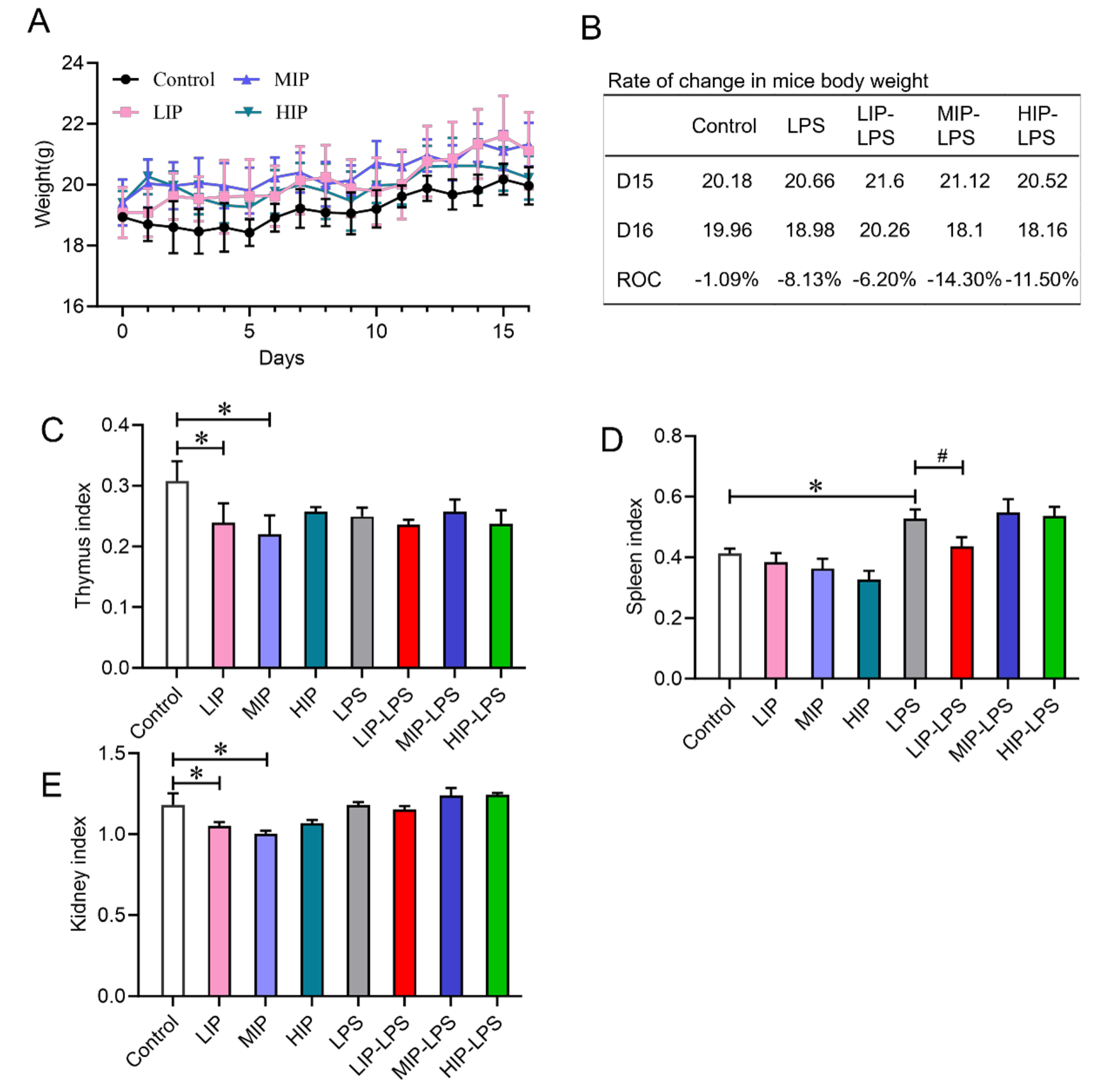
Figure 2.
A high-inorganic-phosphorus diet induces intestinal injury and impairs the repair of the mucosal barrier in mice. A) Pathological observation of HE-stained sections of mouse small intestine tissue (200×). B) Histogram of mouse jejunal villi length and crypt depth. C) Histogram of the ratio of villi length to crypt depth in the small intestine of mice. D) Mouse small intestine tissue AB-PAS stained sections were observed (200×). E) Histogram of mouse jejunal goblet cell count.
Figure 2.
A high-inorganic-phosphorus diet induces intestinal injury and impairs the repair of the mucosal barrier in mice. A) Pathological observation of HE-stained sections of mouse small intestine tissue (200×). B) Histogram of mouse jejunal villi length and crypt depth. C) Histogram of the ratio of villi length to crypt depth in the small intestine of mice. D) Mouse small intestine tissue AB-PAS stained sections were observed (200×). E) Histogram of mouse jejunal goblet cell count.
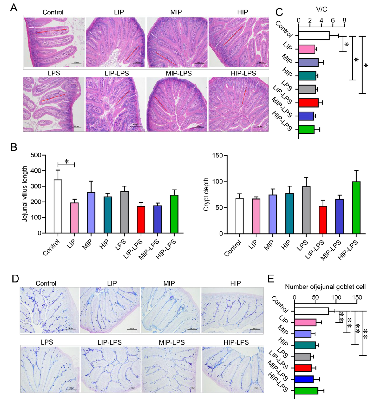
Figure 3.
Inorganic phosphates impair the intestinal immune environment in mice by affecting immunoglobulins and pro-inflammatory cytokines. A) Expression of sIgA in Mouse Serum. B) Expression of IgE in mouse serum. C) IgA content in the mouse small intestine. D) IgE content in the mouse small intestine. E) IgM content in the mouse small intestine. F) IgG content in the mouse small intestine. G) Immunofluorescence was used to observe the secretion of IgA cells in the intestinal lumen of mice (200×). H) Histogram of the number of IgA-secreting cells in the lumen of the jejunum of mice (IHC,×200). I) Expression of IFN-α in Mouse Small Intestinal Tissue. J) Expression of Il-1β in mouse small intestinal tissue.
Figure 3.
Inorganic phosphates impair the intestinal immune environment in mice by affecting immunoglobulins and pro-inflammatory cytokines. A) Expression of sIgA in Mouse Serum. B) Expression of IgE in mouse serum. C) IgA content in the mouse small intestine. D) IgE content in the mouse small intestine. E) IgM content in the mouse small intestine. F) IgG content in the mouse small intestine. G) Immunofluorescence was used to observe the secretion of IgA cells in the intestinal lumen of mice (200×). H) Histogram of the number of IgA-secreting cells in the lumen of the jejunum of mice (IHC,×200). I) Expression of IFN-α in Mouse Small Intestinal Tissue. J) Expression of Il-1β in mouse small intestinal tissue.
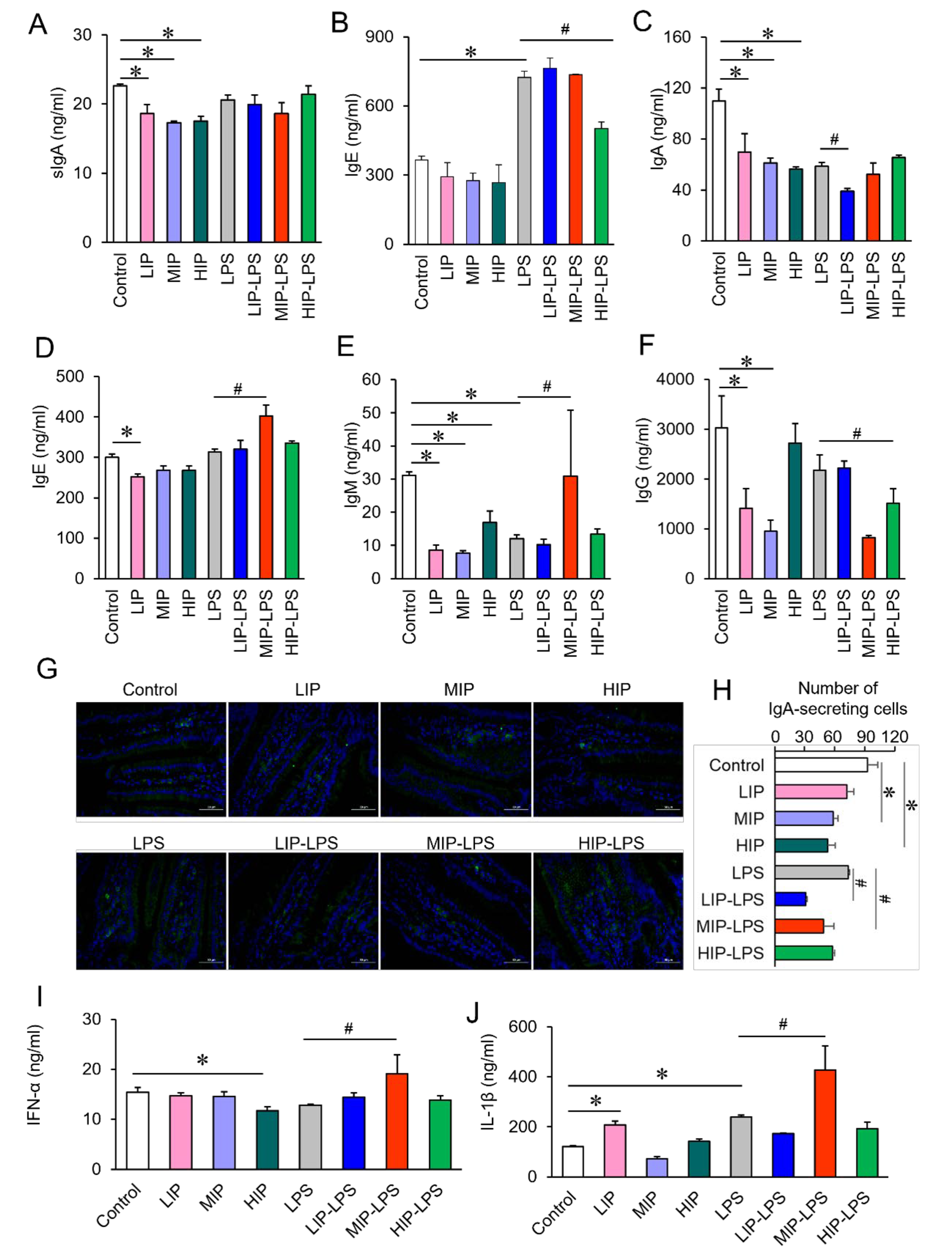
Figure 4.
Proteomic analysis of the effects of a high-phosphorus diet on the immune system in mice. A) Top 10 GO functional entries ranked by significance between the blank control group and the LPS model group. B) Top 10 GO functional entries with significant differences between the blank control group and the phosphate group. C) Top 10 GO functional entries ranked by significance in the LPS model group and phosphate-LPS group. D) KEGG pathway enrichment analysis of blank control group and LPS model group. E) KEGG pathway enrichment analysis of the blank control group and phosphate group. F) KEGG pathway enrichment analysis of LPS model group vs. phosphate-LPS group.
Figure 4.
Proteomic analysis of the effects of a high-phosphorus diet on the immune system in mice. A) Top 10 GO functional entries ranked by significance between the blank control group and the LPS model group. B) Top 10 GO functional entries with significant differences between the blank control group and the phosphate group. C) Top 10 GO functional entries ranked by significance in the LPS model group and phosphate-LPS group. D) KEGG pathway enrichment analysis of blank control group and LPS model group. E) KEGG pathway enrichment analysis of the blank control group and phosphate group. F) KEGG pathway enrichment analysis of LPS model group vs. phosphate-LPS group.
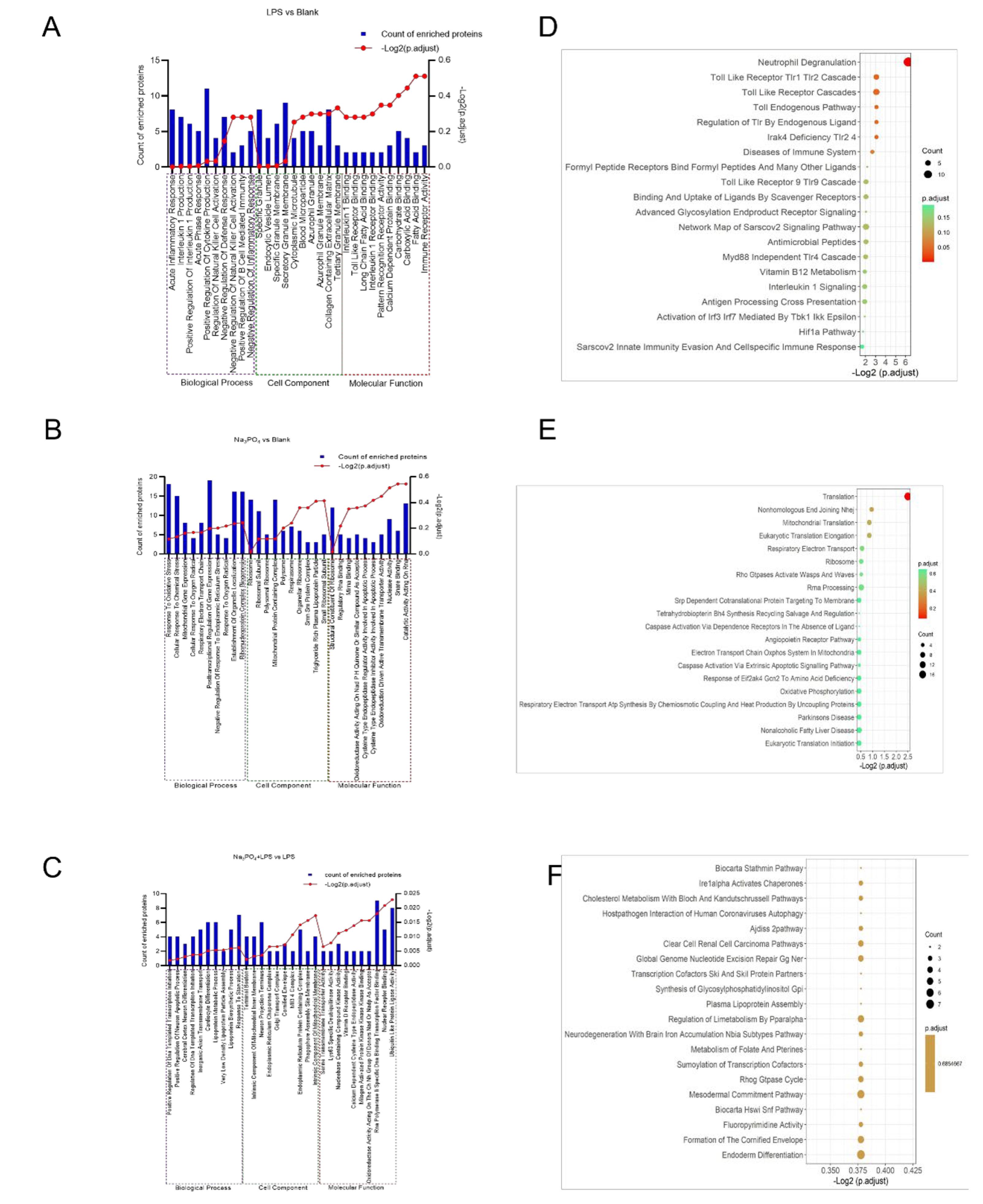
Figure 5.
Effects of different phosphate concentrations and LPS stimulation on differentially expressed proteins. A) Changes in CPT1α protein. B) Changes in IDH1 protein. C) Changes in Bcl2l1 protein. D) Changes in CNTFR protein.
Figure 5.
Effects of different phosphate concentrations and LPS stimulation on differentially expressed proteins. A) Changes in CPT1α protein. B) Changes in IDH1 protein. C) Changes in Bcl2l1 protein. D) Changes in CNTFR protein.
Disclaimer/Publisher’s Note: The statements, opinions and data contained in all publications are solely those of the individual author(s) and contributor(s) and not of MDPI and/or the editor(s). MDPI and/or the editor(s) disclaim responsibility for any injury to people or property resulting from any ideas, methods, instructions or products referred to in the content. |
© 2026 by the authors. Licensee MDPI, Basel, Switzerland. This article is an open access article distributed under the terms and conditions of the Creative Commons Attribution (CC BY) license.
Copyright: This open access article is published under a Creative Commons CC BY 4.0 license, which permit the free download, distribution, and reuse, provided that the author and preprint are cited in any reuse.


