

Submitted:
25 March 2026
Posted:
25 March 2026
You are already at the latest version
Abstract
It is hypothesized that in most cases of sporadic late-onset Alzheimer’s disease (LOAD), the abnormally-elevated cholesterol level in brain neurons represents a critical causative factor that drives the pathogenic processes of LOAD. Specifically, it is hypothesized that the abnormally-elevated neuronal cholesterol will disrupt mitochondrial structure and metabolic activity, resulting in ATP deficiency as well as reduced formation of neuroactive metabolic intermediates (such as mevalonate and geranylgeraniol) along the cholesterol synthesis pathway in brain neurons. In addition, the abnormally-elevated neuronal cholesterol will cause direct neuronal damage as well as other pathogenic changes in the brain, including increased formation and aggregation of amyloid (Aβ) plaques. It is speculated that Aβ accumulation and plaque formation in a majority of LOAD cases only represent a characteristic secondary pathological change, and are usually not the driving force in the pathogenesis of LOAD. As discussed in this paper, the abnormally-elevated neuronal cholesterol in conjunction with ATP deficiency and lack of neuroactive metabolic intermediates will not only cause learning and memory impairment, but will also reduce the formation of cholinergic vesicles and induce tauopathy. It is expected that these pathogenic changes are more readily seen initially in ischemia-sensitive neurons in the hippocampus and posterior parietal cortex, which is then followed by progressive neurodegenerative and atrophic changes in many other brain regions along with progressive cognitive decline. As explained in this paper, ApoE4 is a major risk factor in LOAD because ApoE4 has a drastically reduced ability than ApoE2 and ApoE3 to efflux excess cholesterol out of neurons. Overall, there is a large body of direct, indirect and circumstantial clinical and experimental evidence which jointly supports the cholesterol-centered hypothesis on the etiology and pathogenesis of LOAD.
Keywords:
Alzheimer’s disease
; pathogenic mechanism
; cholesterol
; neuronal cholesterol dysregulation
; apolipoprotein E
; amyloid β
; tauopathy
1. Introduction
Alzheimer’s disease (AD), first described by German physician Alois Alzheimer in the early part of the 20th century [1], is now recognized as the most common cause of dementia among the elderly [2]. The brain region most vulnerable to neuronal dysfunction and cell loss in AD is the medial temporal lobe, including entorhinal cortex and hippocampus. AD usually begins in the entorhinal cortex and proceeds to the hippocampus, a waystation important in memory formation. As the hippocampal neurons degenerate, short-term memory falters. Often the ability to perform routine tasks begins to deteriorate as well. It then gradually spreads to other regions of the brain, particularly the cerebral cortex, which is the outer area of the brain and involved in functions such as language and reasoning. Disturbing behaviors, such as wandering and agitation, beset many people as the disease progresses. In its final stages, AD wipes out the ability to recognize even close family members or to communicate in any way. All sense of self seems to vanish, and the individual becomes completely dependent on others for care. Patients often live for years with this condition, and death usually ensues from a complication of immobility such as pneumonia or pulmonary embolism.
The pathological hallmarks of AD are amyloid plaques, which are the extracellular accumulations of amyloid β (Aβ), and intracellular neurofibrillary tangles (NFTs) composed of the microtubule-associated protein tau [3,4,5]. The development of amyloid plaques usually occurs earlier, and tangle burden accrues over time in a manner that correlates more closely with the development of cognitive impairment. Although many older individuals develop some plaques and tangles as a consequence of normal ageing, the AD brains have a far higher likelihood of developing plaques and tangles in relevant brain regions [6]. The mechanisms by which Aβ and tau induce neuronal dysfunction and death are still not fully clear at present, but many mechanisms have been suggested, which include direct impairment of synaptic transmission and plasticity, excitotoxicity, oxidative stress, neuroinflammation, and others.
Except for 1–5% cases where genetic differences have been identified, the cause for most AD cases, in particular those sporadic late-onset AD (LOAD) cases, is still unknown, and is believed to be multifactorial. Many interacting risk factors, genetic and environmental, jointly contribute to the onset of the disease. The genetic heritability of AD, based on studying twins and families, range from 49–79% [7,8]. Around 0.1% of the cases are familial forms of autosomal dominant inheritance, which have an onset before age 65 [9]. This form of the disease is known as early-onset familial AD. Studies have revealed that most of the autosomal dominant familial AD cases are attributed to mutations in one of three genes: those encoding amyloid-β precursor protein (APP) and presenilins 1 and 2 [10]. Most mutations in the APP and presenilin genes increase the production of a small protein fragment called Aβ42 (the β-amyloid peptide containing 1‒42 amino acid residues), which is also a main component of the senile plaques [11]. Notably, the genetic evidence, coupled with the fact that Aβ accumulates in the AD brain, forms the basis for the famous amyloid hypothesis of AD pathogenesis [12,13,14]. In addition, many other genes have been identified as having alleles that increase AD risk. By far the most important of these is APOE, which encodes the apolipoprotein E (ApoE) [9,15]. Individuals inheriting the ε4 allele of APOE have a 3-fold or higher risk for developing AD [9]. While these individuals make up less than one-fourth of the population, they account for more than half of all AD cases.
In addition to genetic factors, it has been suggested that the cellular homeostasis of ionic copper, iron and zinc is disrupted in AD. For instance, it was hypothesized that dietary copper excess and zinc deficiency may play a causal role in AD [16]. Presently, it remains unclear whether the observed changes in metals are produced by or causes for alterations in relevant neuronal proteins, such as APP, tau and ApoE [17,18].
In this paper, a unifying hypothesis is proposed, which postulates that in most sporadic LOAD cases, abnormally-elevated cholesterol in brain neurons constitutes a crucial causative factor, driving many key pathogenic processes of LOAD, including learning and memory impairment, cholinergic deficiency, Aβ plaque formation, tauopathy, and ultimately, neuronal death. An in-depth analysis of the proposed hypothesis along with a discussion of the supporting experimental and clinical evidence is provided below.
2. Hypothesis
Cholesterol is involved in many important functions in the central nervous system (CNS), such as learning and memory formation [19,20,21,22], synaptogenesis [94], axonal growth and neuronal regeneration [95]. During these processes, brain neurons require an increased supply of cholesterol. It is generally thought that the neighboring astrocytes are the main source of cholesterol for brain neurons [23,24], although neurons also have the ability to synthesize and supply small amounts of cholesterol for their own needs under certain conditions. The cholesterol molecules synthesized in brain astrocytes are carried mostly in lipidated ApoE particles, although other apolipoproteins such as ApoA-I, ApoA-II, ApoA-IV, ApoJ, ApoD and ApoH [25,26] are also involved in carrying, to varying degrees, cholesterol in the brain. These lipoproteins are delivered to neurons through receptor-mediated endocytosis. A number of receptors and proteins are involved in mediating the endocytosis of lipidated ApoE particles by neurons (discussed later).
As cholesterol is required for maintaining the normal structure and function of brain neurons, it is understood that if neuronal cholesterol supply becomes severely inadequate, some of its functions will be compromised. In line with this suggestion, an earlier study reported that when cholesterol content in hippocampal neurons is reduced by statin treatment, synaptic density is reduced and synaptic vesicular release is impaired [27]. Similarly, selective loss of astrocytic cholesterol synthesis, which is the chief source of cholesterol in the brain [23,24], alters brain development and synaptic functions, including reduced synaptic vesicle numbers and defective synaptic plasticity [28,29].
Here, it should also be clearly pointed out that when neuronal cholesterol content is abnormally elevated, it can become strongly cytotoxic [30,31,32,33]. It is hypothesized that abnormally-elevated cholesterol content in brain neurons constitutes a major causative factor, which drives the pathogenic processes in most cases of LOAD (schematically depicted in Figure 1). Specifically, based on the observations made recently [30] and earlier [31,32,33], it is hypothesized that elevated free cholesterol (i.e., unmetabolized cholesterol) can disrupt mitochondrial structure and function, resulting in ATP deficiency in neurons as well as other cells. In addition, high levels of neuronal cholesterol will suppress the cholesterol synthesis pathway through feedback inhibition of the HMG-CoA reductase (HMG-CoA-R or HMGR) [34], which then reduces the formation of key neuroactive metabolic intermediates (such as mevalonate and geranylgeraniol) in neuronal cells [35]. These neuroactive metabolic intermediates play a vital role in protein prenylation and synaptogenesis [36]. Jointly, these biochemical changes due to elevated neuronal cholesterol level will gradually lead to learning and memory impairment and tauopathy (explained in latter sections). Additionally, abnormally-elevated neuronal cholesterol is known to drive the formation and aggregation of Aβ42 and Aβ40 (i.e., amyloid plaque formation) in the brain (reviewed in [37]. As discussed in latter sections, Aβ accumulation and amyloid plaque formation in most LOAD cases only represent characteristic secondary pathological changes, and are not the dominant force that drives the pathogenic process of LOAD.
The above-proposed mechanistic hypothesis on the pathogenesis of LOAD has the following major elements (a detailed discussion of the supporting experimental evidence for each hypothetical element is provided separately in sections 3‒9):
i. It is hypothesized that in brain neurons, chronically-elevated cholesterol (in particular mitochondrial cholesterol) will cause disruption of mitochondrial structure and metabolic function, resulting in reduced ATP synthesis. This hypothesis is proposed on the basis of recent experimental findings [30].
It is known that adequate supply of cellular ATP is important for maintaining cognitive function and new memory formation in the brain. It is hypothesized that cholesterol-induced decrease in mitochondrial ATP synthesis will create an energy deficiency in neurons, particularly in certain regions of the brain where neurons have a higher demand for oxygen and ATP supply to maintain their normal physiological functions. Inhibition of neuronal mitochondrial metabolic activity by cholesterol is an important early event that subsequently triggers a series of pathogenic changes (discussed in detail later), culminating in learning and memory impairment. Based on this understanding, it is speculated that most LOAD cases begin in brain regions that have a particularly-high demand for oxygen and energy supply (ATP synthesis), and then gradually spread to other brain regions with a relatively lower demand for energy supply, and eventually to most regions of the brain.
Figure 1.
A hypothesis on the etiological role of neuronal cholesterol dysregulation in LOAD. As depicted in the upper left (in black color), cholesterol is synthesized using acetyl-CoA as the initial substrate (all the enzymes involved in cholesterol synthesis and metabolism are separately listed in Figure 2). It is hypothesized that elevated free cholesterol (i.e., unmetabolized cholesterol) can disrupt mitochondrial structure and function, resulting in ATP deficiency in brain neurons as well as other cells. In addition, high levels of neuronal cholesterol will also suppress the cholesterol synthesis pathway through feedback inhibition of HMG-CoA reductase (HMG-CoA-R or HMGR), which then decreases the formation of key neuroactive metabolic intermediates (such as mevalonate and geranylgeraniol) in neurons. These neuroactive metabolic intermediates play a vital role in protein prenylation and synaptogenesis. Collectively, these biochemical changes resulting from elevated neuronal cholesterol will gradually lead to learning and memory impairment (explained in detail later). Additionally, abnormally-elevated neuronal cholesterol will drive the formation and aggregation of Aβ42 and Aβ40 (i.e., amyloid plaque formation) in the brain. Mitochondrial dysfunction and ATP deficiency will also contribute to reduced degradation of the hyperphosphorylated tau proteins as well as reduced synthesis and release of the cholinergic vesicles. Please refer to the manuscript for a detailed explanation of each of the above hypothetical elements.
Figure 1.
A hypothesis on the etiological role of neuronal cholesterol dysregulation in LOAD. As depicted in the upper left (in black color), cholesterol is synthesized using acetyl-CoA as the initial substrate (all the enzymes involved in cholesterol synthesis and metabolism are separately listed in Figure 2). It is hypothesized that elevated free cholesterol (i.e., unmetabolized cholesterol) can disrupt mitochondrial structure and function, resulting in ATP deficiency in brain neurons as well as other cells. In addition, high levels of neuronal cholesterol will also suppress the cholesterol synthesis pathway through feedback inhibition of HMG-CoA reductase (HMG-CoA-R or HMGR), which then decreases the formation of key neuroactive metabolic intermediates (such as mevalonate and geranylgeraniol) in neurons. These neuroactive metabolic intermediates play a vital role in protein prenylation and synaptogenesis. Collectively, these biochemical changes resulting from elevated neuronal cholesterol will gradually lead to learning and memory impairment (explained in detail later). Additionally, abnormally-elevated neuronal cholesterol will drive the formation and aggregation of Aβ42 and Aβ40 (i.e., amyloid plaque formation) in the brain. Mitochondrial dysfunction and ATP deficiency will also contribute to reduced degradation of the hyperphosphorylated tau proteins as well as reduced synthesis and release of the cholinergic vesicles. Please refer to the manuscript for a detailed explanation of each of the above hypothetical elements.
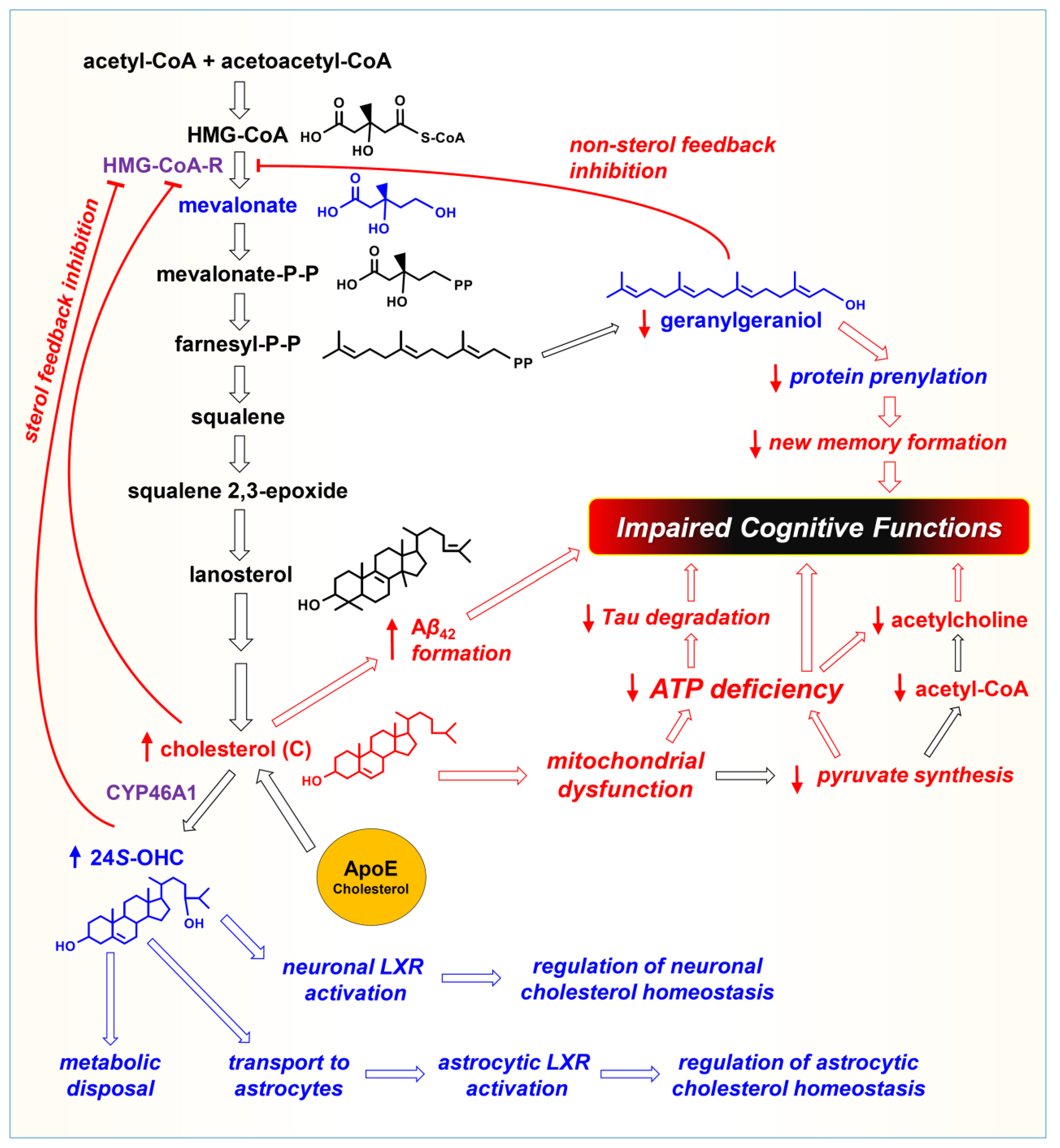
Figure 2.
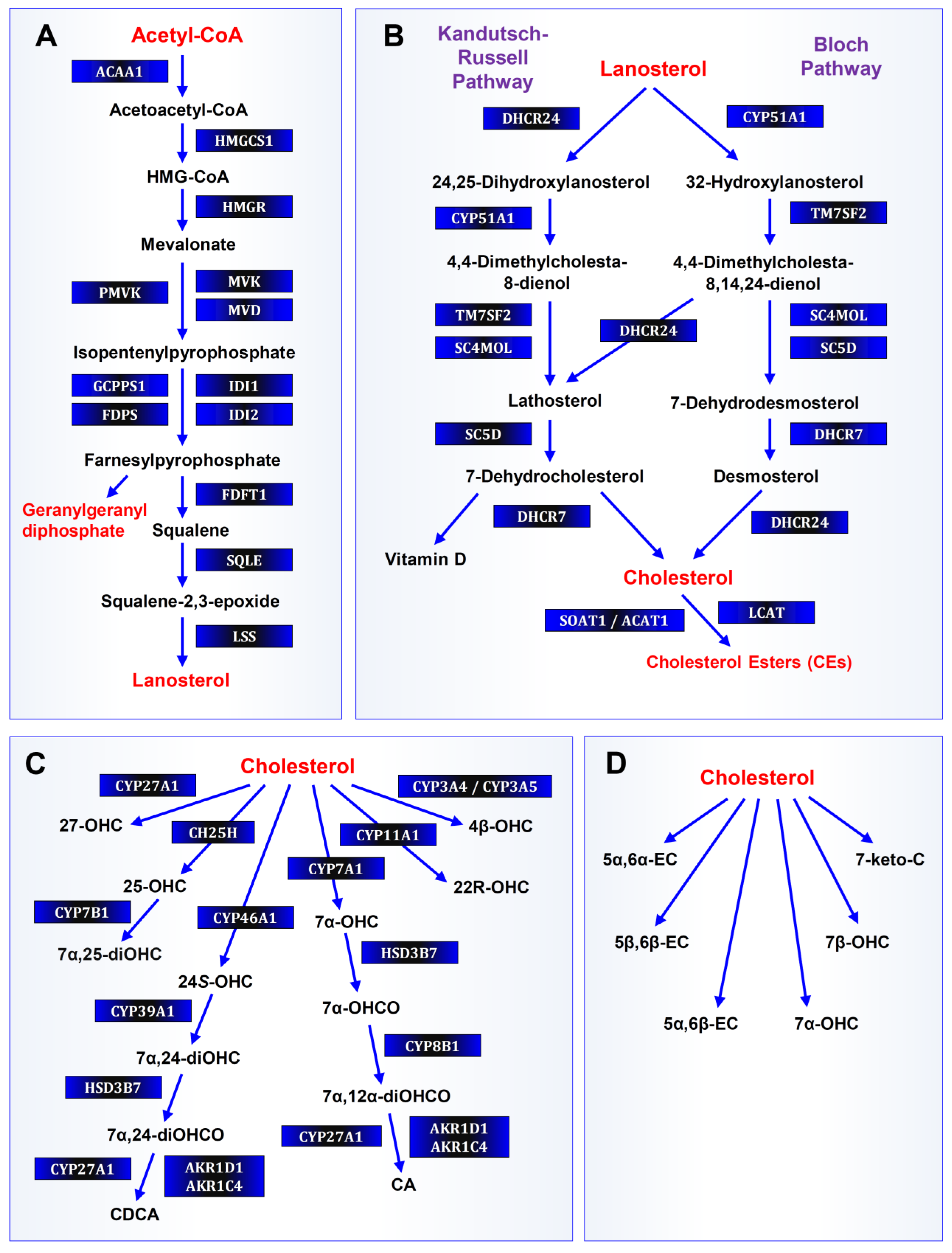
Cholesterol biosynthetic and metabolic pathways. (A). De novo cholesterol biosynthesis (pre-squalene mevalonate pathway). Acetyl-CoA: acetyl-coenzyme A; ACAA: acetyl-coenzyme A acetyltransferase; acetoacetyl-CoA: acetoacetyl-coenzyme A; HMGCS1: 3-hydroxy-3-methylglutaryl-coenzyme A synthase 1; HMG-CoA: 3-hydroxy-3-methylglutaryl-coenzyme A; HMGR: HMG-CoA reductase; PMVK: phosphomevalonate kinase; MVK: mevalonate kinase; GGPPS1: geranylgeranyl diphosphate synthase 1; IDI1: isopentenyl-diphosphate delta isomerase 1; FDPS: farnesyl-diphosphate synthase; IDI2: isopentenyl-diphosphate delta isomerase 2; FDFT1: farnesyl-diphosphate farnesyltransferase 1; SQLE: squalene epoxidase; LSS: lanosterol synthase. (B). De novo cholesterol biosynthesis (post-squalene mevalonate pathway, including the Bloch and Kandutsch–Russell pathways) and cholesterol esterification. DHCR24: 24-dehydrocholesterol reductase; CYP51A1: cytochrome P450 51A1; 24,25 DHLan: 24,25-dihydrolanosterol; TM7SF2: transmembrane 7 superfamily member 2; SC4MOL: methylsterol monooxygenase 1; SC5D: sterol-C5-desaturase; DHCR7: 7-dehydrocholesterol reductase; SOAT1 (also called ACAT1): sterol O-acyltransferase 1 (acyl-CoA:cholesterol acyltransferase 1); LCAT: lecithin:cholesterol acyltransferase. (C). Enzymatic cholesterol catabolism. CYP27A1: cytochrome P450 27A1; CYP3A4 or CYP3A5: cytochrome P450 3A4 or 3A5, respectively; 4β-OHC: 4β-hydroxycholesterol; 27-OHC: 27-hydroxycholesterol; CH25H: cholesterol 25-hydroxylase; CYP11A1: cytochrome P450 11A1; 22R-OHC: 22R-hydroxycholesterol; 25-OHC: 25-hydroxycholesterol; CYP7B1: cytochrome P450 7B1; 7α,24-diOHC: 7α,24-dihydroxycholesterol; CYP46A1: cytochrome P450 46A1; CYP7A1: cytochrome P450 7A1; CYP7B1: cytochrome P450 7B1; 24S-OHC: 24S-hydroxycholesterol; CYP39A1: cytochrome P450 39A1; 7α-OHC: 7α-hydroxycholesterol; CYP8B1: cytochrome P450 8B1; 7α,12α-diOHCO: 7α,12α-dihydroxycholestenone; HSD3B7: 3β-hydroxysteroid dehydrogenase type 7; 7α-OHCO: 7α-hydroxycholestenone; CA: cholic acid; CDCA, chenodeoxycholic acid. (D). Non-enzymatic cholesterol catabolism. 7β-OHC: 7β-hydroxycholesterol; 5α,6α-EC: 5α,6α-epoxycholesterol; 5β,6β-EC: 5β,6β-epoxycholesterol; 5α,6β-EC: 5α,6β-epoxycholesterol.
Figure 2.
Cholesterol biosynthetic and metabolic pathways. (A). De novo cholesterol biosynthesis (pre-squalene mevalonate pathway). Acetyl-CoA: acetyl-coenzyme A; ACAA: acetyl-coenzyme A acetyltransferase; acetoacetyl-CoA: acetoacetyl-coenzyme A; HMGCS1: 3-hydroxy-3-methylglutaryl-coenzyme A synthase 1; HMG-CoA: 3-hydroxy-3-methylglutaryl-coenzyme A; HMGR: HMG-CoA reductase; PMVK: phosphomevalonate kinase; MVK: mevalonate kinase; GGPPS1: geranylgeranyl diphosphate synthase 1; IDI1: isopentenyl-diphosphate delta isomerase 1; FDPS: farnesyl-diphosphate synthase; IDI2: isopentenyl-diphosphate delta isomerase 2; FDFT1: farnesyl-diphosphate farnesyltransferase 1; SQLE: squalene epoxidase; LSS: lanosterol synthase. (B). De novo cholesterol biosynthesis (post-squalene mevalonate pathway, including the Bloch and Kandutsch–Russell pathways) and cholesterol esterification. DHCR24: 24-dehydrocholesterol reductase; CYP51A1: cytochrome P450 51A1; 24,25 DHLan: 24,25-dihydrolanosterol; TM7SF2: transmembrane 7 superfamily member 2; SC4MOL: methylsterol monooxygenase 1; SC5D: sterol-C5-desaturase; DHCR7: 7-dehydrocholesterol reductase; SOAT1 (also called ACAT1): sterol O-acyltransferase 1 (acyl-CoA:cholesterol acyltransferase 1); LCAT: lecithin:cholesterol acyltransferase. (C). Enzymatic cholesterol catabolism. CYP27A1: cytochrome P450 27A1; CYP3A4 or CYP3A5: cytochrome P450 3A4 or 3A5, respectively; 4β-OHC: 4β-hydroxycholesterol; 27-OHC: 27-hydroxycholesterol; CH25H: cholesterol 25-hydroxylase; CYP11A1: cytochrome P450 11A1; 22R-OHC: 22R-hydroxycholesterol; 25-OHC: 25-hydroxycholesterol; CYP7B1: cytochrome P450 7B1; 7α,24-diOHC: 7α,24-dihydroxycholesterol; CYP46A1: cytochrome P450 46A1; CYP7A1: cytochrome P450 7A1; CYP7B1: cytochrome P450 7B1; 24S-OHC: 24S-hydroxycholesterol; CYP39A1: cytochrome P450 39A1; 7α-OHC: 7α-hydroxycholesterol; CYP8B1: cytochrome P450 8B1; 7α,12α-diOHCO: 7α,12α-dihydroxycholestenone; HSD3B7: 3β-hydroxysteroid dehydrogenase type 7; 7α-OHCO: 7α-hydroxycholestenone; CA: cholic acid; CDCA, chenodeoxycholic acid. (D). Non-enzymatic cholesterol catabolism. 7β-OHC: 7β-hydroxycholesterol; 5α,6α-EC: 5α,6α-epoxycholesterol; 5β,6β-EC: 5β,6β-epoxycholesterol; 5α,6β-EC: 5α,6β-epoxycholesterol.
ii. The normal learning and memory process requires the supply of certain neuroactive metabolic intermediates, such as mevalonate and geranylgeraniol [35], which are involved in the prenylation of proteins [36]. Offering support for the important role of these neuroactive metabolic intermediates in learning and memory, an earlier study demonstrated that impairments in learning and memory functions in an animal model can be restored by supplying geranylgeraniol, but not cholesterol [35].
Notably, both mevalonate and geranylgeraniol are metabolic intermediates formed in brain neurons as part of the cholesterol synthesis pathway [34,38,39] (see Figure 1). High cholesterol level in neurons will inhibit HMGR and thus suppress the cholesterol synthesis pathway. As a result, it will markedly reduce the levels of these neuroactive metabolic intermediates in brain neurons. Understandably, it is crucial that the upper half of the cholesterol synthesis pathway (depicted in Figure 1) in brain neurons need to remain active for normal learning and memory function as it will provide key neuroactive metabolic intermediates required for the formation of new synaptic connections. Here, it should be noted that activation of the entire cholesterol synthesis pathway will lead to the production of cholesterol, which may be used by neurons to fulfill certain physiological functions when it is so needed; however, when cholesterol is actually not needed by neurons or is already adequately supplied by the neighboring astrocytes, the upper half of the cholesterol synthesis pathway in brain neurons may still need to remain active, which is not for the purpose of synthesizing more cholesterol, but for the purpose of synthesizing neuroactive metabolic intermediates. To achieve this unique function without producing excess cholesterol in neurons, the upper half of the cholesterol synthesis pathway can be, in fact, effectively regulated by geranylgeraniol, which serves as a non-sterol feedback inhibitor of HMGR [34,38] (see Figure 1). In this way, when neurons have already synthesized sufficient amount of mevalonate and geranylgeraniol for protein prenylation, it can effectively shut down HMGR when further synthesis of cholesterol is not needed.
Understandably, when cholesterol level in a neuron is abnormally elevated (for whatever reasons), it will always impose an inhibition of HMGR, a rate-limiting enzyme of the cholesterol synthesis pathway. This inhibition will not only suppress the synthesis of cholesterol, but will also suppress the production of mevalonate and geranylgeraniol. Additionally, elevated neuronal cholesterol will also disrupt mitochondrial structure and metabolic function, and inhibit ATP synthesis (already described above). These pathogenic effects caused by elevated neuronal cholesterol will jointly hamper the normal process of learning and memory formation.
iii. It is known that when the cholesterol level in brain neurons becomes abnormally elevated, it will also lead to increased formation of Aβ plaques (mechanistic explanation is discussed later). In addition, it is known that elevated extracellular Aβ42 and Aβ40 levels can disrupt mitochondrial function, which contributes to reduced mitochondrial ATP synthesis [40,41].
iv. It is well known that the intracellular protein tau is involved in AD pathogenesis by forming intracellular neurofibrillary tangles (discussed in section 6). Tau protein degradation in neurons is mediated by the ubiquitin system in an ATP-dependent manner (discussed in section 6). Elevated cholesterol level in neurons will lead to reduced ATP synthesis, and it is, therefore, hypothesized that severe cellular ATP deficiency will not only disrupt the process of neurotransmission, but will also slow down proteasome-mediated degradation of tau proteins, which eventually results in accumulation of hyperphosphorylated tau proteins in neurons, resulting in the formation of characteristic intracellular neurofibrillary tangles.
v. It is known that the cholinergic neurotransmitter vesicles contain high levels of ATP, which is a key component of the cholinergic vesicles [42,43]. It is hypothesized that severe deficiency of cholinergic vesicles seen in AD patients is largely due to disruption
of mitochondrial metabolic activity by cholesterol, which reduces ATP synthesis, and the reduced neuronal ATP level is an important factor for the reduced formation of
Cholinergic vesicles. In addition, it is hypothesized that disruption of mitochondrial metabolic activity by cholesterol also reduces the synthesis and cross-mitochondrial transport of acetyl-CoA, a precursor for the synthesis of acetylcholine. It is speculated that these two factors jointly contribute to the reduced synthesis of acetylcholine and the reduced formation and release of cholinergic vesicles in many LOAD cases. Provided below is an explanation of each of the hypothetical elements briefly described above, along with a critical analysis of the available supporting evidence, most of which is scattered in unrelated scientific literature in bits and pieces. In addition, efforts are made trying to apply the proposed mechanistic hypothesis to provide a tentative explanation for some of the interesting, yet poorly-understood, experimental and/or clinical observations related to AD (mostly LOAD).
3. Role of Cholesterol in LOAD
3.1. Cholesterol in Normal Brain Function: A Brief Overview
A very brief review of the relevant knowledge points related to the synthesis, regulation and function of cholesterol in the CNS is provided in this section; it is intended to help non-expert readers more readily understand the proposed hypothesis and the relevant explanations provided in latter sections.
3.1.1. Cholesterol in the Brain
The human brain is enriched with cholesterol compared with other tissues. It is estimated that while the brain only makes up, on average, 2.1% of body mass, it contains 23% of total body cholesterol [44,45,46,47]. The cholesterol levels in most animal tissues are around 2 mg/g tissue, but its level in the brain is 15–20 mg/g tissue [45].
The majority (70–90%) of cholesterol in the CNS is associated with myelin that surrounds axons. Cholesterol synthesis in the brain is highest in oligodendrocytes during developmental stages involving active myelination, and decreases by approximately 90% in adult brain after myelination is completed [45,48].
It is generally thought that due to the presence of the blood-brain barrier (BBB), cholesterol in the brain does not readily equilibrate with cholesterol bound to lipoproteins in the blood [49,50]. Therefore, cholesterol in the brain is believed to be produced locally [44,45,51], and is synthesized primarily in glial cells, such as astrocytes and oligodendrocytes, although cholesterol can also be synthesized in smaller quantities in neurons [47,52]. Cholesterol transport among different cell types in the CNS is mostly carried out by ApoE-containing lipoproteins [53] (discussed in section 4.1).
Figure 3.
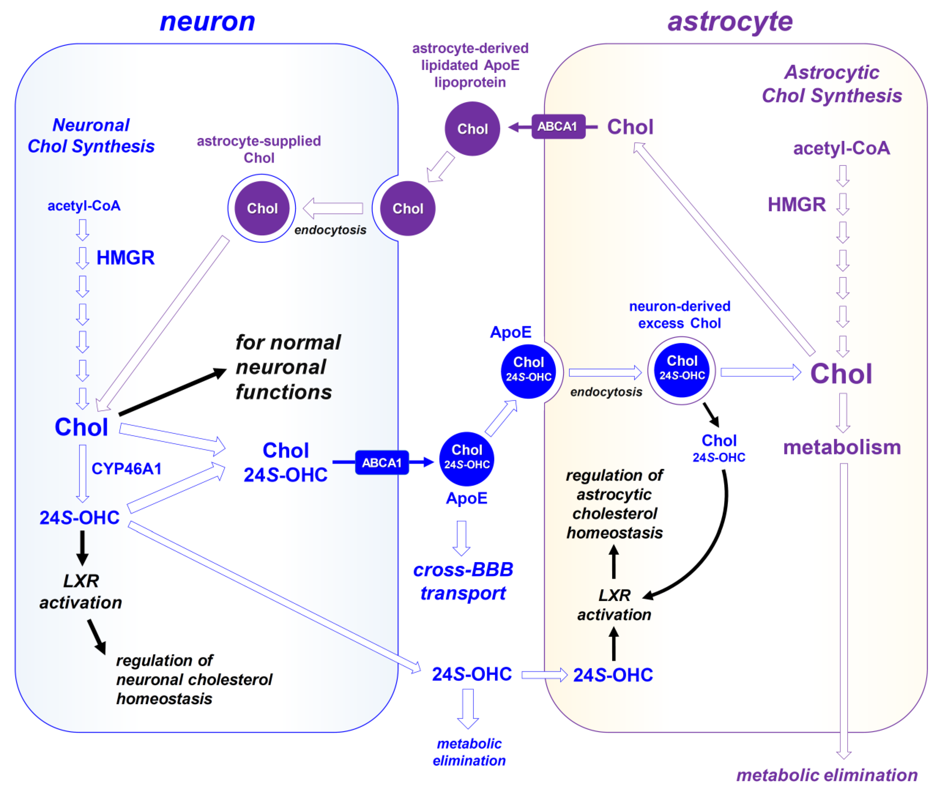
Regulation of cholesterol synthesis and transport in brain neurons and astrocytes. As depicted in the right panel, astrocytes are the main site in the brain for the de novo synthesis of cholesterol (abbreviated as Chol) using acetyl-CoA as the starting material. In addition, brain neurons also have the ability to perform de novo synthesis of a smaller amount of cholesterol using acetyl-CoA as the starting material (depicted in the left panel). Brain ApoE-containing lipoproteins are the main carriers that can transport astrocyte-derived lipids (rich in cholesterol and CEs) to neurons through ApoE receptor-mediated endocytosis. Once internalized, ApoE lipoprotein particles supplies cholesterol (and other lipids) to neurons, which are usually required for fulfilling certain neuronal functions. Importantly, ApoE can also efflux excess neuronal cholesterol to astrocytes for disposal in an ABCB1-dependent manner. As depicted, neurons selectively express CYP46A1; when neuronal cholesterol supply is in excess, CYP46A1 expression will be upregulated, which will convert cholesterol to 24S-OHC. 24S-OHC is a crucial neuronal regulator, which can activate neuronal LXR to regulate neuronal cholesterol homoestasis. Specifically, under the feedback regulation of 24S-OHC, neurons will stop endocytosing astrocyte-produced, cholesterol-rich ApoE particles, and in the meantime, they will enhance the efflux of excess neuronal cholesterol in an ABCA1/ApoE-dependent manner. While these effluxed neuronal cholesterol (in ApoE lipoprotein particles) can be taken up by astrocytes for metabolism and disposal, they can also undergo cross-BBB transport (discussed in section 8.1). As an important part of the feedback regulation, the neuronally-produced 24S-OHC can be transported to the neighboring astrocytes (via simple diffusion or ApoE/ABCA1-dependent efflux), which will then activate the LXR in astrocytes to suppress the astrocytic synthesis and release of cholesterol and also will enhance cholesterol metabolism and disposition by astrocytes.
Figure 3.
Regulation of cholesterol synthesis and transport in brain neurons and astrocytes. As depicted in the right panel, astrocytes are the main site in the brain for the de novo synthesis of cholesterol (abbreviated as Chol) using acetyl-CoA as the starting material. In addition, brain neurons also have the ability to perform de novo synthesis of a smaller amount of cholesterol using acetyl-CoA as the starting material (depicted in the left panel). Brain ApoE-containing lipoproteins are the main carriers that can transport astrocyte-derived lipids (rich in cholesterol and CEs) to neurons through ApoE receptor-mediated endocytosis. Once internalized, ApoE lipoprotein particles supplies cholesterol (and other lipids) to neurons, which are usually required for fulfilling certain neuronal functions. Importantly, ApoE can also efflux excess neuronal cholesterol to astrocytes for disposal in an ABCB1-dependent manner. As depicted, neurons selectively express CYP46A1; when neuronal cholesterol supply is in excess, CYP46A1 expression will be upregulated, which will convert cholesterol to 24S-OHC. 24S-OHC is a crucial neuronal regulator, which can activate neuronal LXR to regulate neuronal cholesterol homoestasis. Specifically, under the feedback regulation of 24S-OHC, neurons will stop endocytosing astrocyte-produced, cholesterol-rich ApoE particles, and in the meantime, they will enhance the efflux of excess neuronal cholesterol in an ABCA1/ApoE-dependent manner. While these effluxed neuronal cholesterol (in ApoE lipoprotein particles) can be taken up by astrocytes for metabolism and disposal, they can also undergo cross-BBB transport (discussed in section 8.1). As an important part of the feedback regulation, the neuronally-produced 24S-OHC can be transported to the neighboring astrocytes (via simple diffusion or ApoE/ABCA1-dependent efflux), which will then activate the LXR in astrocytes to suppress the astrocytic synthesis and release of cholesterol and also will enhance cholesterol metabolism and disposition by astrocytes.
Cholesterol serves as a precursor for the synthesis of neurosteroids, oxysterols and bile acids in the brain [54,55,56]. Importantly, cholesterol also functions as a membrane reinforcer, regulating cell signaling via lipid rafts in neuronal cells. The cholesterol that binds tightly with sphingolipids forms a sterol/sphingolipid-rich domain, commonly referred to as the lipid raft domain, serves as a platform to host various membrane proteins involved in cell signaling and neurotransmission [57,58]. Notably, the amyloid precursor protein (APP) is also richly contained in the lipid raft domain, where enzymatic cleavage of APP by different secretases takes place [59]. In fact, it is known that increasing neuronal cholesterol level can alter APP cleavage, favoring the formation of Aβ42 and Aβ40 fragments (discussed in detail in section 5).
3.1.2. Cholesterol Biosynthesis and Regulation
Cholesterol synthesis in the brain is precisely regulated [34,38]. Cholesterol is synthesized from acetyl-CoA via a complex pathway involving over 30 enzymatic steps (depicted in Figure 1 and Figure 2). Acetyl-CoA, which is transported from mitochondria to cytoplasm, is the building block for cholesterol synthesis. The enzyme acetyl-CoA acyltransferase (ACAA) utilizes acetyl-CoA to produce acetoacetyl-CoA, which is then converted to HMG-CoA by the cytosolic HMG-CoA synthase. The next step, i.e., the reduction of HMG-CoA to mevalonic acid, is catalyzed by HMG-CoA reductase (HKG-CoA-R or simply as HMGR), which is a rate-limiting step in sterol synthesis. Mevalonic acid contains five carbons and is converted through a series of reactions to lanosterol, which is the first sterol precursor with 30 carbons. Through several additional enzymatic reactions that occur mostly at the endoplasmic reticulum (ER) membrane, lanosterol is converted to cholesterol, a sterol with 27 carbons. In addition, mevalonic acid is a precursor for the synthesis of geranylgeraniol, which is needed for covalent modifications of many macromolecules via enzymatic prenylation [36]. Mevalonic acid and geranylgeraniol are considered crucial neuroactive metabolic intermediates [39], and play an important role in learning and memory formation (discussed later).
Important feedback control in cholesterol synthesis involves sterol and nonsterol-mediated rapid degradation of HMGR (Figure 1; reviewed in [60,61]). HMGR is a multi-span membrane protein residing in the ER, and its degradation occurs when sterols accumulate in the ER membranes, which triggers the binding of HMGR to a pair of ER membrane proteins called Insig-1 and Insig-2. Insig binding leads to ubiquitination of HMGR, which undergoes proteasome-mediated degradation in the cytosol.
In addition to sterol’s regulation of HMGR degradation, the sterol-dependent transcription factor (SREBP2) is also regulated by sterols (e.g., cholesterol), causing down-regulation of genes involved in sterol synthesis (e.g., HMGR) and transport (e.g., the LDL receptor LDLR) [62]. Geranylgeraniol, which is derived from mevalonate, is a nonsterol that also exerts feedback inhibition of HMGR. In this way, the cholesterol synthesis pathway is precisely regulated when it is activated solely for the purpose of producing neuroactive metabolic intermediates (such as mavalonic acid and geranylgeraniol) but not for the synthesis of more cholesterol.
Additionally, the HMGR activity can be regulated by cellular metabolic state in a sterol-independent manner: the enzyme can be phosphorylated or dephosphorylated in a reversible manner through the actions of protein kinases and phosphoprotein phosphatases, resulting in enzyme inactivation and activation [63].
3.1.3. Sources of Neuronal Cholesterol and Its Intracellular Trafficking
As depicted in Figure 3, cholesterol in brain neurons is synthesized primarily in glial cells (e.g., astrocytes and oligodendrocytes) [44,45,51], although smaller quantities of cholesterol can also be synthesized in neurons [47,52]. Cholesterol transport among different cell types in the CNS is mostly carried out by ApoE-containing lipoproteins [53] (discussed in section 4.1). The movement of cholesterol through different subcellular compartments inside a neuron involve multiple metabolic pathways, resulting in different intracellular cholesterol pools that are in slow equilibrium with one another.
i. Supply by Cholesterol-Rich Lipoproteins. In the brain, ApoE is a major cholesterol carrier, and astrocyte-derived ApoE lipoproteins enter a neuron through receptor-mediated endocytosis [34,38,64] (Figure 3). After endocytosis, lipoproteins first enter a distinct early endocytic compartment which is rich in acid lipase for catalyzing the hydrolysis of cholesterol esters (CEs) [65]. Cholesterol released from lipoprotein-derived CEs then moves to the endosomes that contain a pair of cholesterol-binding proteins designated as Niemann-Pick type C1 (NPC1) and NPC2. NPC2, a soluble protein present in the luminal region of the endosomes and lysosomes [66], binds cholesterol and transfers it to NPC1 [67,68], a protein that contains multiple transmembrane domains, including a sterol-sensing domain [69]. NPC1 then exports cholesterol to the exterior of the late endosomes and lysosomes. Cholesterol exiting from the late endosomes arrives at other membrane compartments, via various transport mechanisms. These recipient membrane compartments include the plasma membrane [70,71], ER [72,73,74], trans-Golgi network [75,76,77], mitochondria and peroxisomes.
ii. De Novo Biosynthesis. Inside a neuron, the majority of newly-synthesized sterols, including cholesterol, lanosterol and other precursor sterols, move quickly from the ER to the plasma membrane after synthesis. Upon arriving at the plasma membrane, a significant amount of the synthesized sterols, especially lanosterol, is released to the exterior by the sterol efflux process that depends on ABCA1 and apolipoproteins [78,79]. The sterols (including cholesterol, lanosterol and other precursor sterols) remaining at the plasma membrane will recycle between the plasma membrane and various internal compartments, including endosomes and lysosomes [80,81].
iii. SOAT1-Mediated Esterification. Cholesterol inside a neuron is also a substrate for sterol O-acyltransferase 1 (SOAT1), which is also called acyl-CoA:cholesterol acyltransferase 1 (ACAT1), resulting in the formation of CEs (Figure 2B). CEs are then sequestered in cellular lipid droplets, which are subject to hydrolysis by enzymes collectively designated as CE hydrolases [82]. When SOAT1 is inhibited, part of the cholesterol pool destined for storage as CEs may be shuffled to the plasma membrane where cholesterol may serve as substrate for ABCA1-mediated lipid efflux [79].
iv. Cholesterol in Mitochondria. Mitochondrial cholesterol comes from at least three sources: The first source is the plasma membrane. The molecular nature of the plasma membrane‒mitochondria cholesterol movement is not clear at present, but this process does not require NPC2 [83]. At the mitochondria, the transfer of cholesterol from the outer membrane to the inner membrane is mainly mediated by steroidogenic acute regulatory (StAR) protein [84]. The second source is from late endosomes and lysosomes. In cells that express low levels of StAR protein, the StAR-related lipid transfer protein domain 3 (STARD3) present in the late endosomes [85,86] works along with the NPC2 protein to transport cholesterol from the late endosomes and lysosomes to the mitochondria [87,88]. This process explains the observation that, in cells with mutant NPC1, cholesterol overloading occurs in mitochondria [89,90]. This information is crucial to understanding the mitochondrial toxicity and cellular ATP depletion as crucial causative pathogenic changes in the NCP disease (discussed in section 8.4). The third source of mitochondrial cholesterol comes from a specialized membrane region designated as the mitochondria-associated membranes, which are part of the ER membranes in close physical contact with the mitochondrial membrane [91]. These membranes are enriched with cholesterol and the enzyme SOAT1 for converting cholesterol to CEs [92,93].
3.1.4. Cholesterol and Normal Brain Functions
Cholesterol is involved in many important brain functions, such as synthesis of neurosteroids [54,55,56], learning and memory formation [19,20,21,22], synaptogenesis [94] and axonal growth [95,96]. Being an important component of the plasma membrane,
cholesterol is also critically involved in regulating ion permeability [97] and signal transduction in neurons [98]. An earlier study showed that depletion of cholesterol in cultured rat hippocampal neurons by methyl-β-cyclodextrin, a cholesterol-sequestering agent, leads to reduced excitatory postsynaptic currents (EPSCs) and diminished long-term potentiation (LTP) [21]. Other studies also reported that dysregulation of brain cholesterol homeostasis affects synaptic functions [99,100,101].
In the brain, astrocytes are the main source of neuronal cholesterol [23,24] (depicted in Figure 3). Studies have shown that the selective loss of astrocytic cholesterol synthesis can alter brain development and synaptic functions in vivo, with reduced synaptic vesicle numbers and defective synaptic plasticity [28,29]. As cholesterol is enriched in myelin [102] and required for its growth [103,104], oligodendrocytes support neuronal function partly by enwrapping neuronal axons with cholesterol-enriched myelin.
3.2. Neuronal Cholesterol Dyshomeostasis in LOAD
As discussed above, cholesterol has many important functions in the brain. Therefore, some of cholesterol’s neuronal functions will be compromised if the supply of astrocyte-derived cholesterol is severely inadequate (more discussion on this subject is provided later). On the other hand, when cholesterol level inside neurons is abnormally elevated, it will also become pathogenic. There is evidence linking elevated blood cholesterol levels to increased risk of LOAD in the elderly [105,106,107,108,109]. Similarly, cholesterol metabolism in LOAD brains is altered compared to normal brains [107,110], and alterations in brain cholesterol metabolism are associated with an increased risk of LOAD [111,112].
Consistent with the human observations, animal studies have more clearly demonstrated that feeding rabbits a cholesterol-rich diet can cause learning and memory impairment, along with increased Aβ production in their brains [113].
The association between impaired cholesterol homeostasis in the brain and neurodegeneration is perhaps best exemplified in the NPC disease, which is caused by mutations in either the NPC1 or NPC2 gene. NPC1 and NPC2 each can bind to cholesterol and act in tandem in late endosomes and lysosomes to mediate the egress of CEs derived from endocytosed lipoproteins [114,115]. Consequently, in NPC1- or NPC2-deficient cells, including neurons [116] and glial cells [117,118], unesterified cholesterol and other lipids become sequestered in late endosomes and lysosomes, and the amount of cholesterol in the plasma membrane and ER (the cellular sites at which cholesterol homeostasis are regulated) is reduced [119]. In the NPC1–/– neurons, this defect in cholesterol export from late endosomes and lysosomes to other cellular membrane structures results in a higher-than-normal cholesterol content in neuronal cell bodies (especially inside the mitochondria) and a decreased cholesterol content in the distal axons [116,120].
While the NCP disease is a relatively rare (1/150,000 live births) autosomal recessive inherited disorder in humans, it causes progressive neurodegeneration and premature death (often accompanied by hepatosplenomegaly and lung disease) [121]. A characteristic histological finding in the brain is a massive loss of neurons, particularly Purkinje cells in the cerebellum, consistent with an impairment of motor functions in these individuals [122], although neurons in other regions of the brain are also affected to varying degrees. The precise mechanism underlying the pathogenesis of the NCP disease is still not clear at present, but one thing is certain that dysregulation of the neuronal cholesterol homeostasis (e.g., transport) is a fundamental pathogenic change that can directly result in massive neuronal injury and death. (A more detailed mechanistic explanation on the NCP disease is provided in section 8.4).
In addition to AD and NPC disease, other neurodegenerative diseases, such as Huntington’s disease and Smith-Lemli-Opitz syndrome, are also associated with cholesterol dyshomeostasis [123,124]. The apparent link between these disease conditions and neuronal cholesterol abnormality underscores the clinical relevance of the proposed cholesterol-centered hypothesis on pathogenesis. A mechanistic explanation on the pathogenic role of cholesterol in LOAD is provided below.
3.3. Mechanistic Explanation
In addition to its involvement in neurosteroid synthesis [54,55,56], synaptogenesis [94] and axonal growth [95,96], cholesterol also has a unique role in regulating neuronal membrane functions, including membrane fluidity, vesicle formation and fusion, ion channel function, and formation of specialized microdomains involved in neural communication [125,126]. Indeed, cholesterol is required presynaptically for formation of neurotransmitter vesicles [127], and postsynaptically for the clustering and stability of neurotransmitter receptors [125]. As changes in the strength of synaptic connections may alter learning and memory formation [19,20], dysregulation of neuronal cholesterol homeostasis had been suggested to affect learning and memory through modulating presynaptic and/or postsynaptic processes of neurotransmission [128]. In addition, neuronal cholesterol abnormalities can alter enzymatic formation of Aβ peptides and amyloid plaques [129,130,131,132,133].
In this paper, a new hypothesis is proposed, which suggests that in most cases of LOAD, abnormally-elevated cholesterol in brain neurons constitutes a major causative factor, which drives the pathogenic processes of LOAD. Mechanistically, it is hypothesized that the elevated neuronal cholesterol (most likely mitochondrial cholesterol) will disrupt mitochondrial structure and metabolic activity, resulting in reduced synthesis of ATP and important neuroactive metabolic intermediates along the cholesterol synthesis pathway which are required for normal neuronal functions of the brain (in particular, learning and memory). These metabolic changes are important initial events that will trigger a series of pathogenic changes culminating in the gradual development of LOAD (depicted in Figure 1). Specifically, elevated neuronal cholesterol will increase the formation of Aβ42 and Aβ40 (discussed in section 5); and neuronal ATP deficiency will enhance tauopathy (discussed in section 6) and decrease the formation of cholinergic vesicles (discussed in section 7). Additionally, elevated brain cholesterol will suppress macrophage response (due to reduced glial ATP levels), which contributes to the development of neuroinflammation in LOAD.
Offering partial support for this hypothesis, a recent study has shown that exposure of cultured neuronal cells to even very low concentrations of free cholesterol can disrupt mitochondrial structure and metabolic activity (for ATP synthesis) [30]. As shown, a strong disruptive effect of cholesterol on cellular and mitochondrial functions was readily observed when cholesterol was present at 0.1–1 µM, which is really intriguing. These observations are somewhat in line with an earlier study which used a genetic mouse model that selectively overloaded cholesterol in the mitochondria and then examined the effect of mitochondrial cholesterol on Aβ neurotoxicity and AD pathology [134]. Additionally, it was shown earlier that the isolated mitochondria from the cortical neurons of transgenic mice which overexpressed SREBP-2 or lacked NPC1 exhibited mitochondrial cholesterol accumulation, mitochondrial glutathione reduction, and increased susceptibility to Aβ42-induced oxidative stress and release of apoptogenic proteins [134]. Consistent with these observations, the APP/PS1 transgenic AD mice displayed mitochondrial cholesterol loading, glutathione depletion, and functional deficits, and the degree of deficits correlated with the degree of Aβ accumulation [74].
As mentioned earlier, the learning and memory formation needs mevalonate, which serves as a precursor for the subsequent synthesis of geranylgeraniol. Geranylgeraniol plays a key role in the prenylation of proteins in neurons [36]. Both mevalonate and geranylgeraniol are important neuroactive metabolic intermediates that are formed during cholesterol biosynthesis in brain neurons [35] (Figure 1). High levels of cellular cholesterol in neurons will inhibit the cholesterol synthesis pathway, which will then result in reduced biosynthesis of these key neuroactive metabolic intermediates. In support of this hypothesis, an earlier study reported that the learning and memory function could be effectively restored in the AD animal model by giving geranylgeraniol [35].
Based on the above discussion, it is understood that the upper half of the cholesterol synthesis pathway in brain neurons needs to be kept active during the normal process of memory formation as it produces crucial neuroactive metabolic intermediates (mevalonate and geranylgeraniol) required for the formation of new synaptic connections. As such, even if when cholesterol is not needed by the neurons (for instance, when cholesterol is already adequately supplied by the neighboring astrocytes), the upper half of the cholesterol synthesis pathway in neurons may still need to remain active, which is not for the purpose of synthesizing more cholesterol, but for the synthesis of neuroactive metabolic intermediates (Figure 1).
In order to keep the upper half of the cholesterol synthesis pathway active in brain neurons, it is important to keep free cholesterol levels relatively low in these cells at all time. When neuronal cholesterol level is abnormally elevated, it will suppress the cholesterol synthesis pathway through feedback inhibition of HMGR. Notably, statins are known to exert their cholesterol-lowering effect through inhibiting HMGR. Based on the above discussion, it is understood that centrally-active statins will not be as beneficial (most likely harmful) for AD because they will always concomitantly reduce the levels of both cholesterol and neuroactive metabolic intermediates in brain neurons. While reductions in neuronal cholesterol likely will be beneficial most of the time, the reduced synthesis of neuroactive metabolic intermediates in neurons will always be harmful for learning and memory formation. Based on the available clinical observations, the net effect of centrally-active statins is mixed, likely depending on the statin doses used and the degree of neuronal HMGR inhibition produced. While most studies reported a lack of improvement in memory function with centrally-active statins, it appears the use of peripherally-acting statins is more likely to have a beneficial effect. More detailed discussion on the complex effects of statins in LOAD is provided in section 8.3.
Based on the above explanation, it is also understood that alterations in many factors that affect cholesterol metabolism and transport likely will also alter the risk for LOAD. For instance, CYP46A1, which is also called “cholesterol 24(S)-hydroxylase” [51,135,136], catalyzes the conversion of cholesterol to 24(S)-hydroxycholesterol (24S-OHC) [137,138], which is the most abundant oxysterol found in human brain [139]. This metabolic pathway plays an important role in regulating neuronal cholesterol disposition and homeostasis, and abnormalities are associated with an increased risk for LOAD [137]. Similarly, alterations in factors like ApoE4, ABCA1 and ApoE receptors, which are involved in cholesterol transport (particularly neuronal cholesterol efflux), and SOAT1, which catalyzes CE formation, will also alter the risk for LOAD. Provided below is a discussion of the roles of CYP46A1 and SOAT1 in cholesterol homeostasis and their association with LOAD, while the discussion of other relevant factors (such as ApoE4, ABCA1, lipoprotein receptors, etc.) and their respective pathogenic contribution to LOAD is provided separately in relevant latter sections.
3.3.1. Role of CYP46A1 in LOAD
Neurons are the primary cell type in the brain that expresses CYP46A1 [140]. CYP46A1 oxidizes cholesterol to 24S-OHC and helps maintain cholesterol homeostasis in the brain [137]. It was suggested by some researchers that for cholesterol to be transported across the BBB, it needs to be first converted to 24S-OHC by CYP46A1 [141]. 24S-OHC then diffuses out of cells and crosses the BBB, and is finally cleared in the liver [142]. In addition, it has also been suggested that 24S-OHC can be removed from brain neurons in an ABCA1/ApoE-dependent manner (depicted in Figure 3, left panel). As discussed below, the newly-proposed hypothesis (i.e., elevated neuronal cholesterol is a key pathogenic factor in LOAD) will help better understand the role of CYP46A1-mediated cholesterol metabolism in both AD animal models and LOAD patients.
i. It has been reported [143] that the adenovirus-mediated selective overexpression of CYP46A1 in AD animal models can reduce AD severity, such as memory and learning impairment and amyloid accumulation. Similarly, it is predicted that selective knockdown of CYP46A1 will increase AD severity in experimental AD animal models (if we assume that everything else is unchanged). In fact, the observations made earlier [144] agreed well with the predicted outcomes based on the proposed hypothesis.
The mechanistic explanation for these experimental observations or predictions is quite straightforward. The protective effect of the CYP46A1-mediated cholesterol metabolism in AD animal models likely results from two mechanisms: One is CYP46A1-mediated metabolic disposition of excess neuronal cholesterol. As CYP46A1 is selectively expressed in brain neurons, animals with CYP46A1 knockdown are expected to have a drastically-lower ability to metabolize neuronal cholesterol and thus will result in higher levels of cholesterol inside brain neurons. Alternatively, if the brain neurons have a heightened ability to metabolize cholesterol, then neuronal cholesterol level will be reduced, which is beneficial for neuronal survival. The other mechanism is related to the activation of the nuclear receptor LXR system by 24S-OHC, a cholesterol metabolite formed by CYP46A1 [145,146,147]. Activation of LXR by 24S-OHC can regulate a number of genes associated with cholesterol homeostasis [146,148,149], such as reduced expression of HMGR (which reduces cholesterol synthesis) and increased expression of the cholesterol efflux transporters and ApoE (which jointly mediate cholesterol efflux).
ii. While the above explanation of the results from animal studies appears to be quite straightforward and readily understood, there were clinical observations that are more complex than what are seen in animal models. Many earlier human studies reported that the elevated 24S-OHC levels in cerebrospinal fluid (CSF) were associated with neurodegenerative diseases (reviewed in [150]). For instance, studies have shown that the CSF 24S-OHC levels were higher in LOAD patients, in patients with vascular dementia, and in patients with mild cognitive impairment compared to the control subjects [151,152,153]. The higher CSF 24S-OHC levels in LOAD patients [151,153] were also correlated with elevated CSF levels of ApoE, cholesterol and tau [142,150]. In these clinical studies, there was a clear positive correlation between increased levels of 24S-OHC and the risk of LOAD, which gives a false impression that 24S-OHC is a causative factor in LOAD. For a better understanding of these human observations, a little more explanation is needed here. In most LOAD patients, it is expected that their average neuronal cholesterol levels are higher than those in non-demented healthy individuals. The higher neuronal cholesterol levels will lead to increased metabolic disposition catalyzed by CYP46A1 (along with activation of other metabolic pathways). As a result, the oxysterol metabolites are higher in their CSF as well as blood circulation, which actually reflects an enhanced effort of the bodies of LOAD patients to dispose the excess neuronal cholesterol. Here, it should be noted that the observed higher levels of 24S-OHC found in the CSF and/or blood of LOAD patients compared to non-demented human subjects are largely because a majority of the LOAD patients are expected to have higher levels of neuronal (and blood) cholesterol before the onset of clinical LOAD compared to non-demented control subjects. If two groups of LOAD patients (or two groups of non-demented control subjects) which have the same initial neuronal cholesterol levels are compared, it is quite certain that the expected observations will be opposite, i.e., those who have a higher ability to metabolically convert neuronal cholesterol to 24S-OHC (i.e., with higher neuronal CYP46A1 activity) will be associated with a reduced risk for developing LOAD.
iii. In addition to CYP46A1-mediated formation of 24S-OHC, there are other enzymes that can also catalyze the formation of other oxysterols (e.g., 27-hydroxycholesterol) and bile acids (Figure 2C). Based on the understanding that elevated neuronal cholesterol is a causative factor in LOAD, it is readily understood that increased metabolic conversion of neuronal cholesterol to other oxysterols and bile acids will also be beneficial for reducing AD risk in animal models, which will be similar to the beneficial effects of enhanced metabolic conversion of cholesterol to 24S-OHC seen in animal models (as discussed above).
However, clinical studies again reported an opposite finding, i.e., there appeared to have a “positive correlation” between brain 27-hydroxycholesterol levels and LOAD risk [154]. The levels of 27-hydroxycholesterol in the blood, brain and CSF were found to be markedly elevated in individuals with LOAD [154]. These observations are very similar to the above case with 24S-OHC. It is speculated that the increased blood levels of 27-hydroxycholesterol only indirectly reflect an enhanced effort of the LOAD patient’s body to metabolically dispose elevated neuronal cholesterol.
Besides, alterations in brain tissue levels of nonenzymatically-produced oxysterols were also observed in LOAD, which include 7α-hydroxycholesterol (which can also be formed enzymatically by CYP7A1) [155], 7β-hydroxycholesterol, 5α,6α-epoxycholesterol, 5α,6β-dihydroxycholestanol, and 5β,6β-epoxycholesterol (Figure 2D). These observations suggest that there is an increased level of free, unmetabolized cholesterol available for both enzymatic and nonenzymatic conversions to various oxysterol derivatives in these patients.
3.3.2. Role of SOAT1 in LOAD
Besides the conversion of cholesterol to 24S-OHC by CYP46A1, a fraction of cholesterol is esterified for storage by two enzymes: SOAT1 and LACT (lecithin:cholesterol acyltransferase) [156,157]. It is of note that when a mouse model of AD was cross-bred with mice lacking SOAT1, the amyloid pathology was attenuated in these animals [158]. Similarly, human studies have shown that polymorphism in SOAT1 gene was associated with reduced brain amyloid load and reduced CSF cholesterol content [159], whereas elevated SOAT1 expression was associated with more severe amyloid load [160].
How to explain the observed relationship between SOAT1 inhibition and reduced amyloid load? As explained later in section 5, enhanced cleavage of APP by β-/γ-secretases resulting in the production of Aβ42 and Aβ40 (major fragments contained in amyloid plaques) is believed to takes place in the lipid raft regions of the cell membrane when neuronal cholesterol level is elevated. The above experimental observations suggest that the form of cholesterol contained in the lipid rafts of neuronal cells is mostly CEs rather than the free cholesterol. When SOAT1 is absent, the content of CEs in the lipid rafts will not increase proportionally to the elevated cellular level of free cholesterol, and thus will not lead to a proportional increase in APP cleavage through the β-/γ-secretase pathways and a more severe amyloid pathology. A net increase in the cellular free cholesterol pool will naturally lead to increased metabolic disposition by CYP46A1 (due to increased availability of the substrate), thus resulting in increased formation of 24S-OHC [158]. Increased formation of 24S-OHC will then activate the expression of other genes that further help reduce neuronal cholesterol levels. In addition, an increase in cellular free cholesterol pool may serve as substrate for ABCA1-mediated lipid efflux [79]. However, it should be noted that the learning and memory performance of the SOAT1-deficient transgenic mice (which have a markedly-reduced amyloid pathology) may not be proportionally better as the levels of free cholesterol inside their brain neurons may not be markedly lower. If free cholesterol levels in neurons remain markedly elevated, which will disrupt mitochondrial function and inhibit the synthesis of ATP and neuroactive metabolic intermediates, it is expected that the learning and memory performance of these animals will still be impaired.
Notably, inhibition of SOAT1 enzymatic activity has received attention as a potential therapeutic strategy in LOAD as it can reduce amyloidogenic processing of APP through reducing the formation of CEs plus potentially increasing the conversion of unesterified cholesterol to 24S-OHC by CYP46A1 [158]. However, the real therapeutic benefits of SOAT1 inhibitors used alone are expected to be limited since, as discussed above, the free cholesterol levels in brain neurons may not be proportionally reduced as a result of SOAT1 inhibition alone.
3.4. Section Summary
As cholesterol has important physiological functions in the brain, lack of adequate cholesterol supply will impede certain neuronal functions. However, from a pathogenic perspective, it is hypothesized that it is equally or even more important when cholesterol level in neurons is abnormally elevated, because cholesterol can disrupt mitochondrial structure and function and inhibit the synthesis of ATP and neuroactive metabolic intermediates [30]. In addition, elevated neuronal cholesterol is a major cause for increased formation of Aβ42 and amyloid plaques (discussed later in section 5), and a reduction in neuronal ATP will lead to the development of tauopathy (discussed in section 6.4) and reduced formation of cholinergic vesicles (discussed in section 7.2). Therefore, a new hypothesis is proposed here which speculates that in most cases of LOAD, chronically-elevated cholesterol inside brain neurons constitutes a major causative factor, which drives the pathogenic process (depicted in Figure 1). There is mounting evidence from human epidemiological studies as well as experimental studies using both animal models and cultured neuronal cells which jointly demonstrate that elevated neuronal cholesterol is associated with mitochondrial inadequacy, neuronal injury, and increased risk of AD. In line with this mechanistic suggestion, an earlier study in a mouse model of AD showed that, in the early stage of AD development, synaptic mitochondria exhibit significant functional deficits, and the degree of deficits correlates positively with the degree of Aβ accumulation [74,161].
Based on the mechanistic understanding discussed in this section, it is speculated that neuronal cholesterol is elevated in LOAD patients, which is an important initial change. Subsequently, the elevated cholesterol will suppress neuronal cholesterol synthesis pathway, and will lead to increased metabolic formation of 24S-OHC, which will suppress astrocytic cholesterol synthesis, along with increased efflux of cholesterol out of neurons and across the BBB (discussed in section 8.1). As a result of these feedback regulations, the net cholesterol level in the brains of LOAD patients often may not be overly too high compared to non-demented control subjects. Here, it is important to point out that while this might be the case, the neuroactive metabolic intermediates in these patients likely are markedly reduced compared to non-demented individuals. The main reason for their decrease is because their synthesis will be suppressed most of the time due to the presence of feedback inhibition by elevated neuronal cholesterol. A recent targeted metabolomic study demonstrated that the concentration of the cholesterol precursor lanosterol, but not free cholesterol, was clearly lower in the brains of LOAD patients than in the brains of control subjects [160]. Similar observations were made with the levels of the enzymes involved in cholesterol synthesis [160], indicating a decrease in major cholesterol-synthesizing enzymes in relevant brain regions of LOAD patients. Notably, the expression of these enzymes was not similarly altered in the substantia nigra in Parkinson’s patients, suggesting that these changes are relatively specific to the brain regions of LOAD patients [160].
Lastly, it is known that astrocytes are the main site for cholesterol synthesis in the brain [45,51]. An earlier animal study reported that that while selective loss of brain astrocytic cholesterol synthesis in vivo significantly alters brain development, the learning ability of these mice appeared to be largely unaffected (although their memory is reduced) [28]. This observation is intriguing, and suggests that the lack of astrocytic cholesterol supply mostly affects brain development, a process that heavily involves myelination (and also requires a lot of astrocyte-supplied cholesterol), but not learning ability. In fact, the lack of astrocytic supply of cholesterol supposedly will reduce neuronal cholesterol levels, which may actually help maintain a higher level of ATP and neuroactive metabolic intermediates in these neurons, and these changes supposedly are beneficial for the learning process. On the other hand, the long-term memory formation in these animals is still affected, likely resulting from the lack of astrocytic supply of cholesterol which is required for learning-induced de novo myelination, an essential process in long-term memory formation [162].
4. Role of ApoE in LOAD Pathogenesis
4.1. ApoE in Normal Brain Function: A Brief Overview
Human ApoE is a 34-kD glycoprotein present in the CNS and periphery, serving as a lipid carrier [163,164]. It consists of 299 amino acids, encoded by the APOE gene located on chromosome 19q13.32. Human ApoE is polymorphic, with three major alleles, APOE-ε2 (ApoE2: cys112, cys158), APOE-ε3 (ApoE3: cys112, arg158), and APOE-ε4 (ApoE4: arg112, arg158) [165,166]. Structurally, ApoE has three main domains: a N-terminal (4 helices) receptor-binding domain; a C-terminal (3 helices) lipid-binding domain; and an intervening flexible hinge region [167,168,169,170].
In the periphery, ApoE is synthesized and secreted mostly by hepatocytes and macrophages, and is associated with various lipoproteins, ranging from small plasma HDL particles (7–14 nm in size) [171] to the larger polyhedral VLDL particles (30–100 nm in size) [88], to the very large chylomicrons (75–1200 nm) [172,173,174]. Its main function is to transport lipids and regulate plasma lipid levels, and is also involved in immune modulation [175,176].
In the CNS, ApoE is the most abundant apolipoprotein [163], although other apolipoproteins are also present, including the more abundant ApoA-I and the less abundant apolipoproteins such as ApoJ, ApoA-II, ApoA-IV, ApoD and ApoH [25,26]. The majority of ApoE is believed to be produced in astrocytes, while microglia, vascular mural cells, choroid plexus and neurons can also produce ApoE [163,177,178,179,180].
One of the best-studied functions of brain ApoE proteins is their transport of astrocyte-derived lipids (rich in cholesterol and CEs) to neurons through ApoE receptor-mediated endocytosis [94,163,181,182,183]. A number of ApoE receptors have been identified, and they belong to the LDL receptor family, such as the LDL receptor, LDL receptor-related protein 1 (LRP1) and ApoE receptor 2 (ApoER2) [184,185,186,187,188]. Once internalized, ApoE supplies cholesterol (and other lipids) to neurons, which are required for certain neuronal processes, such as synaptogenesis [94], elongation of axons [95,189,190,191] and synaptic plasticity [191,192]. In addition, it was reported that ApoE affects the functions of certain membrane receptors of postsynaptic neurons. For example, the functions of the postsynaptic N-methyl-D-aspartate (NMDA) receptor in the hippocampus and cortex [193,194], which is important for controlling synaptic plasticity and memory formation (reviewed in [195]), are altered by ApoE4 and cholesterol [196]. Similarly, the functions of some other membrane receptors, channels, and transporters are also affected by ApoE through changes in cholesterol and CE content in neuronal membranes [197].
Offering support for the notion that the astrocyte-derived ApoE in the brains plays an important role in normal cognitive functions, earlier studies have shown that the ApoE knockout mice display learning and memory deficits [198,199,200]. This phenotype of defective memory function is also evident in mice with selective ApoE knockdown in astrocytes [201].
In addition to supplying neurons with astrocyte-derived cholesterol and CEs, another important function of the brain ApoE is to carry out the efflux of excess neuronal cholesterol. It is known that ApoE2 can promote significantly more cholesterol efflux from both astrocytes and neurons than ApoE3, and ApoE3 can promote more cholesterol efflux than ApoE4 [202,203]. Differences in the lipoprotein-binding preference of the ApoE isoforms are also observed in the periphery: while ApoE3 associates preferentially with the protein-rich HDL particles, ApoE4 associates more effectively to the lipid-rich VLDL particles [169,170,202,204]. These intriguing observations suggest that ApoE3 is more apt to efflux lipids (including cholesterol and CEs) out of neurons, which is akin to HDL’s function in the periphery, whereas ApoE4 appears to have a reduced ability to efflux lipids but a good ability to supply lipids to neurons, which is somewhat similar to the function of VLDL in the periphery.
At the molecular level, the three ApoE isoforms differ at positions 112 and 158 in the N-terminal domain, which are cysteine–arginine substitutions, altering the charge of the protein and its ability to form cysteine–cysteine dimers [166]. While ApoE4 contains no cysteine residues throughout the protein, ApoE3 contains one cysteine residue (cys112) and ApoE2 contains two cysteine residues (cys112 and cys158). ApoE3 can form ApoE−ApoE and ApoE‒ApoA-II dimers through its cys112 residue, and similarly, ApoE2 can more readily form respective dimers as it has two cysteine residues; in comparison, ApoE4 cannot form dimers at all [205,206,207]. Since the dimeric ApoE2 and ApoE3 can elicit higher lipid efflux than their monomeric forms [202], the inability of ApoE4 to form dimers significantly reduces its ability to efflux excess cholesterol out of neurons.
Consistent with the differential ability of the three ApoE isoforms in neuronal cholesterol efflux, many studies have reported that they also have different levels of lipidation in the CNS [208,209,210]. While ApoE2 and ApoE3 are better lipidated (with ApoE2 best lipidated), ApoE4 is markedly less lipidated [208,210]. Analysis of the CSF collected from middle-aged and older cognitively-normal individuals showed that ApoE4 is less lipidated than ApoE2 and ApoE3 [208,210]. Selective expression of the human form of ApoE2, ApoE3 or ApoE4 in mice with viral constructs confirmed that ApoE4 forms less lipidated ApoE particles compared to ApoE2 and ApoE3 particles [209].
ApoE-mediated lipid efflux in the CNS takes place in conjunction with the ATP-binding cassette (ABC) proteins, such as ABCA1 and ABCG1, ABCG4 and ABCA7 [211,212]. These proteins are embedded in cell membrane and act to pump lipid molecules into the extracellular space, where they bind apolipoproteins such as ApoE, ApoJ and ApoA-I [212]. Like ApoE, the expression of ABCA1 and ABCG1 is increased following activation of the nuclear receptor LXR system, either directly [18] or indirectly [213], to promote ApoE-mediated lipid efflux [211].
Figure 4.
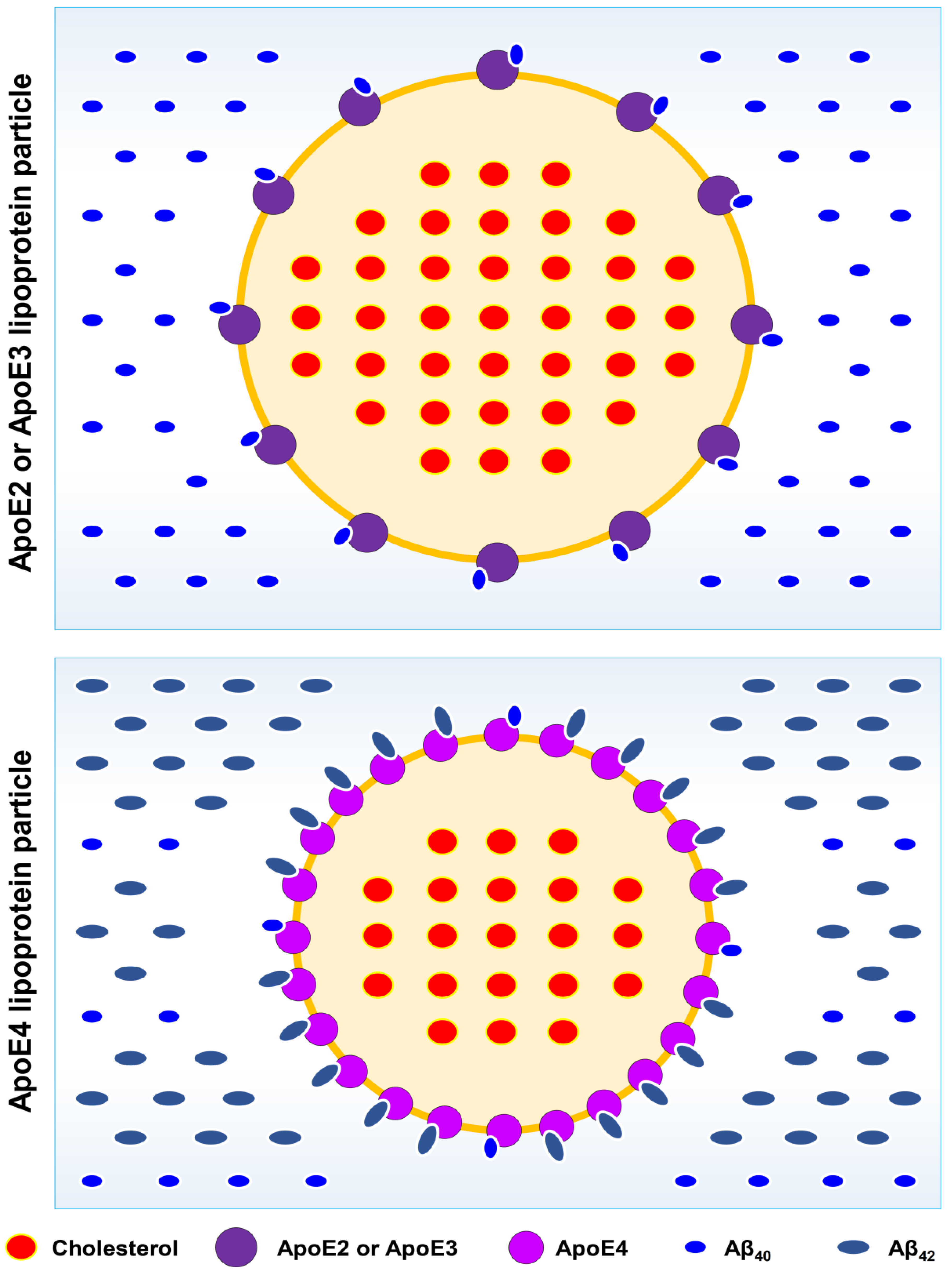
The relative size and density of ApoE2, ApoE3, and ApoE4 molecules in their respective lipoprotein particles. It is known that while the ApoE4 lipoprotein particles have smaller average size than the ApoE2- or ApoE3-containing particles, the ApoE4 particles contain approximately twice the amount of the ApoE4 molecules per particle compared to the E2- or E3 particles. Additionally, ApoE2 and ApoE3 have high binding affinity for Aβ40 but very low binding affinity for Aβ42 (A). In an opposite fashion, ApoE4 has very low affinity for Aβ40 binding but high affinity for Aβ42 binding (B). Because of these unique properties, each ApoE4 lipoprotein particle will bind more Aβ42 fragments. It is hypothesized that the higher density of Aβ42 bound to each ApoE4 lipoprotein particle will accelerate the deposition of the Aβ42-bound ApoE4 particles in the brain (discussed in section 5), thereby resulting lower ApoE4 levels in the CSF.
Figure 4.
The relative size and density of ApoE2, ApoE3, and ApoE4 molecules in their respective lipoprotein particles. It is known that while the ApoE4 lipoprotein particles have smaller average size than the ApoE2- or ApoE3-containing particles, the ApoE4 particles contain approximately twice the amount of the ApoE4 molecules per particle compared to the E2- or E3 particles. Additionally, ApoE2 and ApoE3 have high binding affinity for Aβ40 but very low binding affinity for Aβ42 (A). In an opposite fashion, ApoE4 has very low affinity for Aβ40 binding but high affinity for Aβ42 binding (B). Because of these unique properties, each ApoE4 lipoprotein particle will bind more Aβ42 fragments. It is hypothesized that the higher density of Aβ42 bound to each ApoE4 lipoprotein particle will accelerate the deposition of the Aβ42-bound ApoE4 particles in the brain (discussed in section 5), thereby resulting lower ApoE4 levels in the CSF.
4.2. Pathogenic Role of ApoE4 in LOAD
4.2.1. Observations from Human Studies
ApoE was first identified in 1993 as a genetic risk factor for sporadic LOAD [214,215]. Extensive epidemiological, clinical and pathological studies have since then established the APOE gene as perhaps the most important genetic risk factor for sporadic LOAD [216,217,218,219]. While ApoE3 is the most common isoform in the general population, ApoE4 occurs in 40‒80% percent of all sporadic LOAD patients who possess at least one copy of the APOE-ε4 allele [16]. People who inherit one copy of the APOE-ε4 allele is about three times more likely to develop AD, and people who have two copies of the APOE-ε4 allele (one from the mother and one from the father) are at least eight times more likely to develop AD than those who have two copies of the APOE-ε3 allele [9]. Conversely, the ApoE2 variant appears to be protective in LOAD. People with one copy each of the APOE-ε2 and APOE-ε3 allele have only one-fourth the risk of developing AD as people with two copies of the APOE-ε3 allele [220,221,222].
Human studies have shown that the APOE4 genotype is associated with an earlier onset of LOAD [217]. The PET scan [223] and post-mortem analysis [3,224,225,226] have shown that AD patients with the APOE4 genotype have an earlier appearance and more amyloid deposits in their brains.
Functional studies have shown that ApoE4 human individuals are unable to efficiently regulate cerebral glucose metabolism and oxygen utilization compared to ApoE4-negative individuals [227]. Similarly, the glucose uptake (based on FDG-PET scan) was lower in ApoE4 individuals in certain regions of the brain, such as the posterior cingulate, parietal, temporal and prefrontal cortex [228]. Post-mortem analysis of brains from young individuals showed that the APOE4 genotype is associated with reduced levels of brain glucose and lactate transporters and mitochondrial electron transport proteins [229], clearly suggesting a reduced mitochondrial metabolic activity. Behavioral analysis also showed that the APOE4 genotype is associated with reduced verbal memory [230] in asymptomatic carriers and reduced visual recall and memory retention even in children [231].
4.2.2. Observations from Animal Studies
Some of the more convincing evidence on the pathogenic role of ApoE4 came from detailed analyses of the transgenic AD mice. These mice selectively expressed the human APOE genes from their endogenous APOE promoter [232], with the expected glial expression of human ApoE isoforms [233]. Compared to APOE3 mice, APOE4 mice were impaired in spatial learning [193,234,235,236,237,238,239]. They were also impaired in other memory-related functions [193,236,237,240]. More pronounced deficits in behavior and brain functions were observed in older APOE4 mice [237,241], which are consistent with clinical observations.
The APOE4 mice also had altered neuronal activity. The electrophysiology of amygdala neurons showed reduced excitatory transmission in the APOE4 mouse brains [242]. Evoked release of acetylcholine from hippocampal neurons was reduced in older APOE4 mice [243]. The hippocampal neurotransmission also had fewer shortwave ripples and reduced slow gamma wave activity in aged APOE4 mice [244].
Structural analysis showed that the brains of APOE4 mice have simpler structures compared to APOE3 mice [242,245,246,247], including less branching or reduced spine densities. Decreased complexity of neurons in APOE4 brains was seen in the entorhinal cortex [248,249], consistent with the altered functions of that brain region often seen in human LOAD [250]. In older mice, the APOE brains appeared to have a lower vascular density, along with white matter damage [251] and smaller hippocampal regions [235].
Here, it is of note that the APOE-associated differences in behavior and brain functions in transgenic animals often occurred in the absence of overt amyloid pathology, suggesting that alterations in ApoE-mediated neuronal cholesterol efflux affects cognitive function more predominantly over amyloid pathology.
4.2.3. Mechanistic Explanation
Most previous studies have focused on the role of ApoE in delivering astrocytic cholesterol to neurons. For instance, it was speculated earlier that ApoE isoform-specific effects on learning and memory are partly due to the reduced ability of ApoE4 than ApoE3 in supplying astrocytic cholesterol to neurons and thus a decreased capacity in supporting synaptic functions [252,253]. Recently, an ApoE cascade hypothesis in AD pathogenesis was proposed [254]. Here, a different explanation is entertained. It is hypothesized that the differential ability of the ApoE isoforms in effluxing cholesterol out of neurons plays a more critical role in determining the pathogenesis of sporadic LOAD because a hampered neuronal cholesterol efflux will lead to elevated cellular cholesterol levels, which will disrupt mitochondrial function and result in reduced formation of neuronal ATP and neuroactive metabolic intermediates, along with increased Aβ formation, tauopathy and acetylcholine deficiency. Provided below is a discussion of the available experimental and clinical observations that support the notion that the APOE4 genotype is a key risk factor for LOAD on the basis of its decreased ability to efflux excess neuronal cholesterol.
i. It is known that ApoE4 has a reduced ability to efflux cholesterol out of neurons in the CNS compared to ApoE3 and ApoE4 [202,255,256]. For instance, ApoE3 has 2.5- to 3.9-fold higher ability to efflux cholesterol than ApoE4. It is known that the dimeric ApoE can induce higher lipid efflux than its monomeric form [202]. As ApoE3 (and also ApoE2) can readily form dimeric forms, ApoE4 cannot form dimers. The inability of ApoE4 to form dimer is a major cause for its reduced ability to remove excess cholesterol from neurons.
Consistent with the reduced ability of ApoE4 to efflux cholesterol, individuals with the APOE4 genotype also have smaller ApoE-containing lipid particles plus more lipid-depleted particles in the CSF, whereas those with the APOE3 genotype have larger lipidated ApoE particles [208,257,258]. Similar observations were also made in the transgenic APOE mice [176,210,259]. While the APOE4 mice were associated with smaller ApoE4 lipoprotein particles plus more lipid-depleted particles in their brains [209,236,260,261], the APOE2 or APOE3 mice were associated with larger lipidated ApoE particles [209].
ii. Interestingly, studies have shown that humans with the APOE4 genotype have lower levels of ApoE in their CSF than those with the APOE3 genotype [208,257,258]. Similarly, the ApoE4 mice also have the lowest levels of ApoE in extracts from relevant brain regions, such as the frontal cortex and hippocampus, whereas the ApoE2 mice have the highest ApoE levels in these brain regions [210].
To explain why people with the APOE4 allele have low levels of ApoE4 protein in their CSF, earlier studies suggested that ApoE4 proteins can undergo more rapid degradation than ApoE2 and ApoE3 proteins in the brain [210,262]. The potential reasons for the rapid degradation of the ApoE4 particles may include the following: First, despite the smaller size, each ApoE4 particle is known to contain approximately twice the amount of the ApoE4 molecules compared to the ApoE3-containing particles [263,264] (depicted in Figure 4). As such, the relative density of ApoE4 molecules per lipoprotein particle is actually much higher. In addition, it is known that ApoE3 (and also ApoE2) has very high affinity for Aβ40 binding but low affinity for Aβ42 binding; in contrast, ApoE4 has very low affinity for Aβ40 binding but high affinity for Aβ42 binding [214,258,265]. As a result, while most of the ApoE3 or ApoE2 particles are bound with Aβ40 instead of Aβ42, the majority of the ApoE4 particles are bound with Aβ42 instead of Aβ40 (compare Figure 4A with Figure 4B). Due to the higher ApoE4 density per particle, each ApoE4-containing lipoprotein particle will be bound with a lot more Aβ42 peptides. It is speculated that the higher density of Aβ42 bound to each ApoE4 lipoprotein particle will accelerate the deposition of the cytotoxic Aβ42 peptides in the brain (discussed later in section 5). In the meanwhile, these Aβ42-bound ApoE4 lipoprotein particles will stimulate their removal and disposition by different types of cells in the brain, and may even stimulate the cross-BBB transport of these particles, as part of the mechanism that would help remove excess Aβ42 peptides (discussed later in section 8.1). As a result of these accelerated removal processes, people with the APOE4 genotype actually will end up with lower levels of ApoE4 in their CSF than those with the APOE3 (or APOE2) genotype.
iii. It has been reported that in general, lower levels of ApoE in the brain are often correlated with an elevated risk of LOAD [266]. Based on the discussion provided above, this phenomenon might be quite readily understood. First, in the case of individuals with the APOE4 genotype, it is already explained above that these individuals will be associated with lower levels of ApoE4 proteins in their CSF, and they are also associated with an increased risk for LOAD. Second, if two individuals with the same APOE3 (or APOE2) genotype are compared, it is reasonable to suggest that the one with a lower brain ApoE protein level will have a higher risk for LOAD as this individual will have a reduced capacity of ApoE-mediated cholesterol efflux, which will result in elevated neuronal cholesterol (assuming that everything else is the same in these two individuals). Offering partial support for this explanation, an earlier study has shown that administration of bexarotene (an agonist of the RXR nuclear receptor) which can robustly increase ApoE content (likely along with other components of the cholesterol efflux machinery plus cholesterol-metabolizing enzymes) in the hippocampus and cortex of a mouse AD model was associated with improved memory and cognition and reduced Aβ plaques in these animals [267]. These beneficial effects were not observed in ApoE-deficient mice [267].
Therefore, based on the mechanistic explanations provided above, it is understood that individuals with the APOE4 genotype will have overall clinical manifestations highly similar to those with abnormally-elevated neuronal cholesterol. With this understanding in mind, a brief discussion of a few other risk-modifying factors in LOAD, including ApoE2, ApoJ, cholesterol-efflux transporters (ABCA1, ABCG1, ABCG4 and ABCA7) and the ApoE receptors is provided below. The role of TREM2 in LOAD is discussed separately in section 8.1.1.
4.3. Protective Effect of ApoE2 in LOAD
4.3.1. Observations from Human and Animal Studies
The earlier clinical observations that the APOE-ε2 allele was under-represented in LOAD patients [268,269] had led to the suggestion that the APOE2 genotype likely is protective in LOAD patients. Compared to APOE-ε3/ε3 homozygotes, the risk of LOAD in APOE-ε2 carriers is approximately 50% less [221,270]. Moreover, LOAD patients who are APOE-ε2 carriers exhibit slower cognitive decline compared with non-carriers [271]. In Down’s syndrome patients who have triplicate APP gene, APOE-ε2 protects against the development of AD [272], and also delays the age of AD onset [273,274,275].
Postmortem LOAD brains from APOE-ε2 carriers have lower densities of amyloid plaques than those from APOE-ε3/ε3 individuals [276,277,278]. PET imaging also showed that the amyloid load in non-demented brains accumulates at a slower pace during aging and has a later onset of amyloid positivity in APOE-ε2 carriers than in APOE-ε3/ε3 homozygotes [223]. APOE-ε2 affects not only the global amyloid load but also the region-specific Aβ deposition [279]. In general, the protective effect of APOE-ε2 is more pronounced in pathologically-confirmed LOAD than in clinically-diagnosed cases [280].
4.3.2. Mechanistic Explanation
A number of mechanisms have been proposed in the past to explain the protective effect of the APOE2 genotype. For instance, it was suggested that hyperlipidation of ApoE2 is a central mechanism for the protective effect of ApoE2, and the main reason for this suggestion is that ApoE2 is capable of transporting more astrocytic cholesterol to neurons. In addition, the lipidation state of ApoE2 affects its binding by Aβ peptides [281,282,283] and thus may affect Aβ catabolism [284]. Additionally, it has been suggested previously that the protective effect of ApoE2 may be achieved through ApoE2-activated neuroprotective signaling pathways [285,286,287].
In this article, a new mechanistic explanation is tendered. It is hypothesized that individuals with the APOE2 genotype will have a reduced level of neuronal cholesterol, which will be protective against the development of LOAD. This mechanistic explanation is rather straightforward, and is supported by a number of experimental and clinical observations, as briefly elaborated below.
First, people with the APOE2 genotype have a higher overall ability to efflux neuronal cholesterol in an ABC transporter-dependent manner, and this ability is jointly determined by the following two factors: i. While ApoE4 cannot form dimers, ApoE2 (like ApoE3) can form dimers [205,207], which is associated with a higher ability to carry out neuronal cholesterol efflux, in the rank order of ApoE2 > ApoE3 >> ApoE4 [181,202,288]. In addition, studies have shown that the APOE2 and APOE3 mice are associated with larger lipoprotein particles, with ApoE2 particles largest and APOE4 smallest [289,290,291]. ii. Studies have shown that humans with the APOE2 genotype have higher levels of ApoE in their CSF than those with the APOE4 genotype [257,258]. Similar observations were also made in the transgenic mice expressing different human ApoE proteins [210].
Second, in addition to the higher ability of ApoE2 to efflux neuronal cholesterol, ApoE2 appears to have a lower ability to deliver cholesterol to neurons compared to ApoE3 and ApoE4, for the following reasons: i. ApoE2 has far lower binding affinity (approximately 1%) for LDLR (the LDL receptor) than ApoE3 and ApoE4 [264,292,293]. Accordingly, it is speculated that the lipidated ApoE2 particles will have a markedly reduced ability than ApoE3 and ApoE4 to deliver lipids (including cholesterol and CEs) to neurons via LDLR-mediated endocytosis. ii. LRP1 is another ApoE receptor that mediates the endocytosis of lipidated ApoE particles, and ApoE2 also has a lower binding affinity for LRP1 than ApoE3 and ApoE4 [294]. In fact, the lipidated ApoE4 can bind to LRP1 with a high affinity [295], which further supports the speculation that ApoE4 is highly capable of delivering astrocytic cholesterol to neurons (likely more capable than ApoE3 and ApoE2). iii. Aβ40 has a much higher binding affinity for ApoE2 than for ApoE3 and ApoE4; in comparison, ApoE4 has much higher binding affinity for Aβ42 than for Aβ40 (depicted in Figure 4) [214,258,265,296]. As a result, ApoE2 lipoprotein particles will be mostly bound by Aβ40, whereas ApoE4 will be mostly bound by Aβ42 (Figure 4). As explained in detail later (section 5.3), when the APP-binding site on ApoE2 is bound by Aβ40, ApoE2 is effectively blocked by Aβ40 and thus no longer be able to perform its cholesterol-delivering function through endocytosis, but the Aβ40-bound ApoE2 lipoprotein can still perform its cholesterol efflux function. As a result, a larger fraction of the ApoE2 particles will be in the cholesterol efflux mode, rather than in the cholesterol-delivery model (i.e., endocytosis mode). This property of ApoE2 also contributes to the reduced ability of the lipidated ApoE2 lipoprotein particles to undergo endocytosis to deliver cholesterol to neurons.
Because of these reasons, it is clear that ApoE2 not only has a higher ability to efflux cholesterol out of neurons, but it may also have a reduced ability to supply astrocytic cholesterol to neurons. A combination of these two properties will beneficially result in reduced brain neuronal cholesterol in individuals with the APOE2 genotype compared with those with the APOE3 and APOE4 genotypes. In agreement with this speculation, it was reported earlier that aged APOE2 mice had lower cortical cholesterol levels than aged APOE3 or APOE4 mice [297].
According to the explanation that ApoE2’s neuroprotection results from reduced neuronal cholesterol levels, it is predicted that ApoE2 will be associated with improved mitochondrial metabolic activity (i.e., higher ATP level) and higher levels of neuroactive metabolic intermediates in brain neurons. In addition, the reduced neuronal cholesterol level will also favorably reduce the formation of Aβ42 (discussed in section 5); and the improved neuronal ATP level will help reduce tauopathy (discussed in section 6) and improve the formation of cholinergic vesicles (discussed in section 7). Additionally, reduced neuronal cholesterol level will also have a reduced macrophage response in the brain [298,299], which may partly explain why the APOE2 genotype is associated with reduced neuroinflammation ― another contributing factor in ApoE2’s protective function in LOAD.
Lastly, it should be noted that ApoE2 not only has a protective effect against LOAD [297,298,299,300,301], but it is also associated with longevity [302,303,304,305,306]. It is hypothesized that the improved mitochondrial function and cellular ATP level in neurons (as well as in other somatic cells) resulting from reduced cellular cholesterol levels are important underlying factors contributing to longevity. This notion is certainly in line with the long-held view that healthy mitochondrial function is crucial for general health and longevity [307,308].
4.4. Role of APOJ Genotype in LOAD
Besides APOE, genome-wide association studies [309,310] have found a statistically-significant association between a SNP of the APOJ gene and the risk of AD [311]. APOJ is not as strong a genetic risk factor as APOE; the polymorphic site in APOJ has an odds ratio of approximately 0.9 for the APOJ allele, compared with an odds ratio of approximately 5 for the APOE-ε4 allele [312].
ApoJ is a component of the CNS lipoproteins [313]. It is functionally similar to ApoE, and involved in neuronal cholesterol efflux [206,314] in an ABCA1-dependent manner [315]. It has been suggested that ApoE and ApoJ can interact with lipid debris in the brain, and are involved in removing degenerating membranes after neuronal injury [316]. It is hypothesized that the polymorphic ApoJ has a reduced ability to efflux neuronal cholesterol and thus increases the risk for LOAD. Given that ApoJ likely plays a lesser role in CNS cholesterol efflux than ApoE, this may determine why the APOJ genotype is not as strong a genetic risk factor as the APOE4 genotype.
Studies have shown that people with LOAD often have more ApoJ in their blood and its levels affect the regional distribution of Aβ [317]. However, ApoJ levels alone cannot be used to predict the onset of LOAD [318]. These observations likely suggest that ApoJ may play a complementary role with ApoE in removing excess neuronal cholesterol. In people with AD conditions, which usually is associated with abnormally-elevated neuronal cholesterol (often associated with the APOE4 genotype which has a reduced overall ability to efflux cholesterol), it is understood that the body may try to increase ApoJ levels as a functional compensation. Understandably, for a LOAD patient with a faster cognitive decline, it likely means that this patient has much higher neuronal cholesterol levels than usual, and as a result, the body may try to express a lot more ApoJ proteins (along with other brain apolipoprotein isoforms) as a means to help to efflux neuronal cholesterol.
Similarly, it was reported that the brain ApoJ level is elevated in proportion to the APOE-ε4 allele dose level. The explanation for this phenomenon is related to the fact that the higher APOE-ε4 allele dose level is associated with lower CSF ApoE4 protein level, and that ApoE4 has a very low ability to efflux neuronal cholesterol, and as a result, a stronger induction of ApoJ in the APOE-ε4 individuals is required to provide a bigger compensation to efflux neuronal cholesterol.
As both ApoE and ApoJ are involved in neuronal cholesterol efflux, they certainly will also jointly contribute to reducing Aβ deposition in the brain. If an individual with the APOE4 genotype plus a mutant ApoJ, this person is expected to have a higher chance to have more severe neuronal accumulation of cholesterol, which will be associated with more severe Aβ buildup. The above speculation is supported by earlier studies [319,320]. In addition, earlier studies have suggested that ApoE and ApoJ may also modify Aβ clearance across the BBB (more discussion on this subject is provided in section 8.1.1).
4.5. Role of Brain Cholesterol Efflux Transporters in LOAD
The ATP-binding cassette transporters A1 and G1 (i.e., ABCA1 and ABCG1, respectively) are important ApoE-lipidating proteins in the CNS [321,322]. Other members in this transporter family (such as ABCG4 and ABCA7) may also be involved in this process. Earlier studies have shown that while ABCA1 preferentially activates the efflux of cholesterol and its binding to unlipidated ApoE, ABCG1 is more selective for partially-lipidated lipoprotein complexes [323,324]. Studies have suggested that ABCA1 and ABCG1 may work in concert to control the efflux of cholesterol and its oxysterol metabolites such as 24S-OHC [256,325].
Since these transporters are all involved in neuronal cholesterol efflux, it is understood that a reduced function of any of these transporters likely will affect neuronal cholesterol level, to varying degrees. As ABCA1 is an important and well-studied member of this class, a more detailed discussion of its role in LOAD will be given here as an example. Alterations in the expression of ABCA1 were found to be linked to LOAD (reviewed by [182,326]). Studies of mice with brain-specific deficiency of ABCA1 manifested mild impairments in neurite morphology, synaptic structures, motor activity and memory formation [327,328,329]. As ABCA1 is required for the normal acquisition of cholesterol by ApoE, it is expected that the lipidation state of the ApoE particles will be reduced in ABCA1-knockout mice [321,322].
When ABCA1 knockout mice were crossed with the mouse model of AD, it is expected that the AD conditions, including amyloid load and the learning and memory deficits, will be more severe as neuronal cholesterol efflux is more severely hampered [330]. On the other hand, overexpression of ABCA1 in these AD mice is expected to help alleviate amyloid deposition and improve brain function, as observed in an earlier study [330].
It is of note that Tangier disease is caused by mutations in the ABCA1 gene [331]. In the periphery, these mutations prevent ABCA1 from effectively transporting cholesterol and phospholipids out of cells for pickup by ApoA-I in the bloodstream. Tangier disease is characterized by severe plasma deficiency or absence of HDL, apolipoprotein A-I (ApoA-I, the major HDL apolipoprotein) and accumulation of CEs in many tissues throughout the body. Additionally, the buildup of cholesterol in cells can be toxic, causing cell death or impaired functions. These combined factors lead to the peripheral signs and symptoms of Tangier disease. In the CNS, the absence of ABCA1 is associated with poorly-lipidated ApoE particles, along with elevated neuronal cholesterol, which impairs neuronal functions.
While it has been suggested that 24S-OHC may leave neurons and eventually reach peripheral circulation through simple diffusion (due to its slightly higher water solubility than cholesterol), it was also reported that 24S-OHC can be secreted by neurons in an ApoE- and ABCA1-dependent manner [256,325]. As the lipidated ApoE particles can be taken up by astrocytes for metabolic disposition, this process may help deliver 24S-OHC to astrocytes where it can exert its important regulatory functions. It is known that in astrocytes, 24S-OHC can down-regulate the expression of cholesterol synthesis genes while activate the expression of APOE, ABCA1 and TREM2 [311]. Mechanistically, the nuclear receptor LXR mediates the action of 24S-OHC to regulate the expression of these genes [148,332,333]. An earlier study has reported that treatment of APP/PS1-transgenic mice with bexarotene (an agonist of RXR) can stimulate the synthesis of ABCA1, ABCG1 and ApoE, which is associated with enhanced clearance of the brain Aβ and reversal of cognitive deficits [267].
As ABCA1 is involved in the efflux of 24S-OHC [256,325], it is expected that neuronal secretion of 24S-OHC will be reduced when ABCA1 is deficient, and as a result, astrocytic expression of ApoE (a process regulated by 24S-OHC-activated LXR) will be drastically reduced. This may explain why ApoE levels in the brain of ABCA1-knockout mice are 80% lower than in wild-type control mice [321,334].
Deletion of the ABCA1 gene not only decreases the ApoE level [321], but also increases Aβ deposition [335] in the brain; in comparison, ABCA1 overexpression in the AD mouse model decreases Aβ deposition [330]. These changes are readily understood on the basis of the expected changes in neuronal cholesterol level under these experimental conditions. Deletion of the ABCA1 gene will be associated with increased neuronal cholesterol level, whereas ABCA1 overexpression will reduce neuronal cholesterol level. The change in neuronal cholesterol level will then alter the content of CEs in the lipid rafts, which then alters the catalytic activity of β-/γ-secreatases as well as the formation of Aβ42 and Aβ40 (detailed explanation is provided in section 5).
In addition to ABCA1, other ABC transporters (ABCG1, ABCG4 and ABCA7) may also be similarly involved in neuronal cholesterol efflux in the CNS [336,337]. In the light of the functional role of the ABCA1 in neuronal cholesterol efflux and LOAD as discussed above, it is expected that abnormalities in the functions of other CNS-resident lipid transporters may also similarly lead to increased neuronal cholesterol levels, along with other accompanying effects. For instance, studies have shown that mice deficient in ABCG1 exhibit increased neuronal cholesterol level and memory deficits, similar to ABCA1-deficient mice [338,339]. In addition, ABCA7 has also been identified as one of the LOAD susceptibility genes [340], and as expected, studies have shown that deletion of ABCA7 increases Aβ formation and accumulation in an AD mouse model [341,342].
4.6. Role of ApoE Receptors in LOAD
When ApoE performs its cholesterol delivery functions in the CNS, it needs to bind to its cell surface receptors, such as LDLR [343], VLDL receptor, LRP1, ApoER2 (also known as LRP8), or heparan sulfate proteoglycans (HSPGs) [53,344,345]. The interactions between ApoE and its receptors have clear isoform preference and are affected by the ApoE lipidation status [283,346,347]. For instance, it is known that LDLR is only recognized by lipidated ApoE [283,346,347], indicating that LDLR is chiefly involved in the supply of lipids to brain neurons through endocytosis. This suggestion is consistent with the known functions of LDLR in the periphery, i.e., to deliver lipids to recipient cells. As mentioned in section 4.3.2., earlier studies have shown that ApoE2 has a markedly weaker binding affinity for LDLR compared to ApoE3 and ApoE4 [292,293], indicating that lipidated ApoE2 lipoprotein particles have a lower ability to deliver lipids to neurons than ApoE3 and ApoE4.
In addition to LDLR, LRP1 is also known to mediate neuronal endocytosis of lipidated ApoE particles [348,349]. It is speculated that lipidated ApoE2 particles have a reduced ability to deliver cholesterol to neurons via LRP1-mediated endocytosis. There is some experimental evidence for this suggestion: First, ApoE2 has a lower binding affinity for LRP1 compared to ApoE3 and ApoE4 [294]. Second, Aβ40 is known to have a much higher binding affinity (approximately 20-fold higher affinity) for ApoE2 than for ApoE3 and ApoE4 [214,258,265,296], and as such, the APP-binding site on ApoE2 (which is required for endocytosis; discussed later in section 5) likely will be effectively blocked by Aβ40 and thus will reduce the chances for lipidated ApoE2 particles to undergo endocytosis. This explanation is somewhat in line with the known functional profiles of LPR1 in mediating the endocytosis of various ApoE-containing chylomicron remnants in the periphery (reviewed in [187,350].
At present, much less is known about the receptors involved in neuronal cholesterol efflux. It is speculated that LRP1 may be involved in the efflux of cholesterol, in addition to its ability to mediate endocytosis of ApoE-rich lipoproteins. Similarly, ApoER2 may also be involved in neuronal cholesterol efflux. These are purely speculations, and the experimental evidence for these speculations is mostly lacking at present, except the observations that polymorphisms in LRP1 [288,351,352] and ApoER2 [348] were associated with increased incidence of LOAD.
4.7. Section Summary
ApoE lipoproteins in the brain are responsible for carrying astrocyte-derived cholesterol (and other lipids) to neurons, and they are also responsible for effluxing excess neuronal cholesterol. Past studies have mostly focused on the function of ApoE in delivering astrocytic cholesterol to neurons. Here, a new hypothesis is proposed, which suggests that the differential ability of the ApoE isoforms in effluxing neuronal cholesterol plays a key role in the pathogenesis of sporadic LOAD, as a retarded cholesterol efflux will lead to cellular and mitochondrial cholesterol elevation, which will then disrupt mitochondrial structure and function, and reduce the synthesis of ATP and neuroactive metabolic intermediates, along with increased Aβ formation, tauopathy, and cholinergic deficiency.
ApoE4 is known to have a markedly lower ability than ApoE2 and ApoE3 to efflux cholesterol out of neurons, partly due to its inability to form dimers. The reduced ability of ApoE4 to efflux excess neuronal cholesterol will result in markedly elevated cholesterol level in neurons and thus elevated LOAD risk. Based on this mechanistic explanation, it is also understood that individuals with the APOE4 genotype will have overall clinical manifestations highly similar to those with abnormally-elevated neuronal cholesterol levels.
As ApoE4 has very low affinity for binding Aβ40 but higher affinity for binding Aβ42, the ApoE4 particles will be bound with Aβ42 instead of Aβ40. Due to the higher density of the ApoE4 molecules per particle, each ApoE4-containing lipoprotein particle will bind with more Aβ42 fragments. The higher density of Aβ42 bound to each ApoE4 particle will accelerate the deposition of Aβ42 peptides in the brain (discussed later in section 5.3). By contrast, ApoE2 has a far better ability to promote cholesterol efflux from both astrocytes and neurons than ApoE3 and ApoE4. Besides, ApoE2 has a lower ability to deliver astrocyte-derived cholesterol to neurons than ApoE3 and ApoE4. As a result, it is expected that the cholesterol levels in neurons will be lowest with the APOE2 genotype, median with the APOE3 genotype, and highest with the APOE4 genotype. The low neuronal cholesterol level associated with the APOE2 genotype is an important mechanistic basis for its protective effect against the development of LOAD.
As the APOE2 genotype is associated with lower neuronal cholesterol level, it is readily understood that ApoE2 will be associated with improved mitochondrial metabolic activity (i.e., higher ATP levels) and higher levels of neuroactive metabolic intermediates in brain neurons. Additionally, lower neuronal cholesterol content will favorably decrease the formation of Aβ42 (discussed in section 5); and the improved neuronal ATP levels will help reduce tauopathy (discussed in section 6) and improve the formation of cholinergic vesicles (discussed in section 7). Lastly, the new mechanism proposed here also offers a good explanation for the observation that ApoE2 not only reduces the risk of LOAD, but it will also promote longevity.
ApoJ is another member of the CNS lipoproteins, and is functionally similar to ApoE in neuronal cholesterol efflux. It is understood that the polymorphic ApoJ with a reduced ability to efflux neuronal cholesterol will be associated with an increased risk for LOAD. Similarly, as the ATP-binding cassette transporters (such as ABCA1 and ABCG1) are involved in neuronal cholesterol efflux, reduced functions of any of these transporters will also affect neuronal cholesterol levels to varying degrees, thus affecting the risk of LOAD.
5. How Do Neuronal Cholesterol and ApoE4 Accelerate Amyloid Plaque Formation?
5.1. Structure and Function of APP
In 1987, APP was cloned and found to be linked to the pathogenesis of familial early-onset AD [353]. APP is a type I transmembrane protein with a large extracellular domain and a small cytoplasmic domain, resembling a transmembrane receptor for an unidentified ligand. The cytoplasmic domain contains an NPXY sequence, which is a consensus binding sequence for a number of adaptor proteins [354]. There are two proteins highly homologous to APP: the amyloid precursor-like proteins 1 and 2 (APLP1 and APLP2) [355].
Presently, the function of APP is still not fully clear. Studies have shown that Drosophila lacking their single APP-like protein display impaired synapse formation in neuromuscular junctions [356]. APP knockout mice are viable, but show reduced long-term potentiation in the brain [357], consistent with learning and memory deficits and loss of hippocampal synapses [172,358,359]. APP/APLP2 double knockout mice have impaired neuromuscular junctions [360].
It has been extensively characterized that APP is cleaved by a number of proteolytic enzymes which affect the release of the Aβ peptides (depicted in Figure 5A). The α-secretases cleave the extracellular domain of APP’s twelve amino acids away from the membrane, and is the quantitatively and functionally most important proteolytic cleavage of APP, which releases the extracellular sAPPα domain, along with the membrane-bound C-terminal fragment CTFα. The β-secretase was identified as the β-site APP cleaving enzyme (BACE1) [361], which cleaves the extracellular domain of APP farther way from the membrane. While α-secretase activity occurs primarily at or near the cell surface, β-secretase is more favored in endosomes, consistent with the low pH optimum of BACE1 [361]. The β-cleavage of APP results in the release of the soluble ectodomain sAPPβ, along with the membrane-bound C-terminal fragment CTFβ. The γ-secretase, which was identified as a complex of proteins containing the presenilins [362], further cleaves CTFα or CTFβ (following cleavage of APP by α- or β-secretase).
As depicted in Figure 5A, the sequential cleavage of APP by α-secretase followed by γ-secretase will generate three fragments: sAPPα, a small p3 fragment, and the APP intracellular domain (AICD). The α-secretase pathway also operates in many non-neuronal cell types, generating shorter fragments that are thought to be nonamyloidogenic. sAPPα has been suggested to exhibit neuroprotective and synapse-promoting activities [363]. It was shown that mice that only produce sAPPα did not exhibit the various phenotypes caused by a full APP knockout, including disturbed LTP and memory function. This observation suggests that sAPPα may mediate some of the functions of the APP holoprotein [357].
The β- and γ-secretases cleave APP in the so-called amyloidogenic pathway (Figure 5A,B). β-Secretase releases the ectodomain sAPPβ, and the remaining CTFβ is subsequently cleaved by γ-secretase liberating the Aβ peptide(s) and the AICD. C-Terminal heterogeneity is generated by the γ-secretase itself. This protease cleaves APP at different positions, generating a variety of peptides, of which Aβ43, Aβ42, Aβ40, Aβ38 and Aβ37 variants are detected in cell culture and body fluids (Figure 5B). Aβ40 is continuously and abundantly produced in both healthy and AD-afflicted brain tissues, whereas other Aβ peptides are produced at lower levels. Aβ42 is the most famous and best studied Aβ peptide, and may serve as a core for the formation of amyloid plaques.
The causative mutations in the APP and presenilin genes which alter APP processing were identified many years ago. Moreover, transgenic APP animal models were successfully developed that can produce pathogenic Aβ deposits [364,365].
Interestingly, ApoE receptors also partly share with APP the pattern of proteolysis [366,367,368]. ApoE receptors undergo surface cleavage to generate soluble (shed) forms of the receptors [369,370,371]. Several ApoE receptors have been identified as substrates for β-/γ-secretases, including LRP1 [372,373], and apoER2 [374] and VLDLR [375]. Furthermore, some of the intracellular cytoplasmic adaptor proteins that interact with APP also interact with ApoE receptors [367,368].
5.2. Neuronal Cholesterol and Aβ Production
One of the best-known pathological characteristics of AD is the deposition of extracellular Aβ in neuritic plaques [376]. Cholesterol has been shown to play a crucial role in affecting Aβ production and deposition [132,377]. The first evidence for the role of cholesterol in Aβ production in the brain came from a study demonstrating that dietary cholesterol increased Aβ accumulation in rabbits [113]. Although it is difficult to study the phenomenon in humans, limited human results support the observations made in animal models. For instance, in autopsied AD brains, more severe Aβ deposition was found to be associated with higher levels of blood cholesterol measured earlier during life.
Mechanistically, studies have revealed that the cholesterol content in the membrane (particularly in the lipid raft microdomains) is an important factor that regulates the catalytic activity of β- and γ-secretases [37,129,130,131,132]. This finding provides a mechanistic basis for the causal relationship between elevated neuronal cholesterol and increased Aβ production [129,131,133]. Provided below is a discussion on how ApoE4 promotes the formation of Aβ and amyloid plaques.
5.3. Mechanistic Explanation
When neuronal cholesterol level is abnormally elevated, which most likely is due to increased supply of astrocytic cholesterol, a number of regulatory mechanisms will be activated to help its removal. First, elevation in neuronal cholesterol will lead to increased metabolic disposition via CYP enzyme-mediated cholesterol oxidation to 24S-OHC and other oxysterols, simply due to the availability of more substrate molecules. Since 24S-OHC is an activator of LXR in astrocytes, neurons and other brain cells, its increased levels will activate a number of regulatory pathways in different cell types, which will jointly help reduce neuronal cholesterol. For instance, 24S-OHC can increase the expression of its own metabolizing enzyme CPY46A1, the lipid carrier protein ApoE, the lipid transporter ABCB1, and others. In addition to 24S-OHC, other oxysterols formed in neuronal cells can also serve as LXR activators, and will have similar regulatory functions.
Figure 6.
Role of ApoE-containing lipoprotein particles in neuronal transport (i.e., supply and efflux) of cholesterol. As depicted in (A), ApoE-mediated delivery of astrocytic cholesterol to neurons is carried out via endocytosis, which involves the binding of lipidated ApoE particles to specific receptors on neuronal surface. It is hypothesized that the intact APP protein is required for the endocytosis of ApoE particles into neurons, and it serves as a “permissive” signal to the neurons. (Here, it is of note that the cleaved CTFα fragment of APP or the APP-related proteins may also share a similar function as the intact APP in carrying out this “permissive” role.) As depicted in (B), when the ApoE molecules in the lipoprotein particle are already bound by Aβ fragments (Aβ40 or Aβ42), the particle can only bind to its receptor but cannot bind simultaneously to APP (due to competitive blockage). As a result, endocytosis will not be allowed to take place; but under this condition, the ApoE lipoprotein particle can still mediate the efflux of neuronal cholesterol. Therefore, the ApoE particle can be regulated in a precise manner such that it can effectively switch from the function of supplying astrocytic cholesterol to neurons (when neurons are in need of more cholesterol for physiological functions) to the function of removing excess cholesterol from neurons.
Figure 6.
Role of ApoE-containing lipoprotein particles in neuronal transport (i.e., supply and efflux) of cholesterol. As depicted in (A), ApoE-mediated delivery of astrocytic cholesterol to neurons is carried out via endocytosis, which involves the binding of lipidated ApoE particles to specific receptors on neuronal surface. It is hypothesized that the intact APP protein is required for the endocytosis of ApoE particles into neurons, and it serves as a “permissive” signal to the neurons. (Here, it is of note that the cleaved CTFα fragment of APP or the APP-related proteins may also share a similar function as the intact APP in carrying out this “permissive” role.) As depicted in (B), when the ApoE molecules in the lipoprotein particle are already bound by Aβ fragments (Aβ40 or Aβ42), the particle can only bind to its receptor but cannot bind simultaneously to APP (due to competitive blockage). As a result, endocytosis will not be allowed to take place; but under this condition, the ApoE lipoprotein particle can still mediate the efflux of neuronal cholesterol. Therefore, the ApoE particle can be regulated in a precise manner such that it can effectively switch from the function of supplying astrocytic cholesterol to neurons (when neurons are in need of more cholesterol for physiological functions) to the function of removing excess cholesterol from neurons.
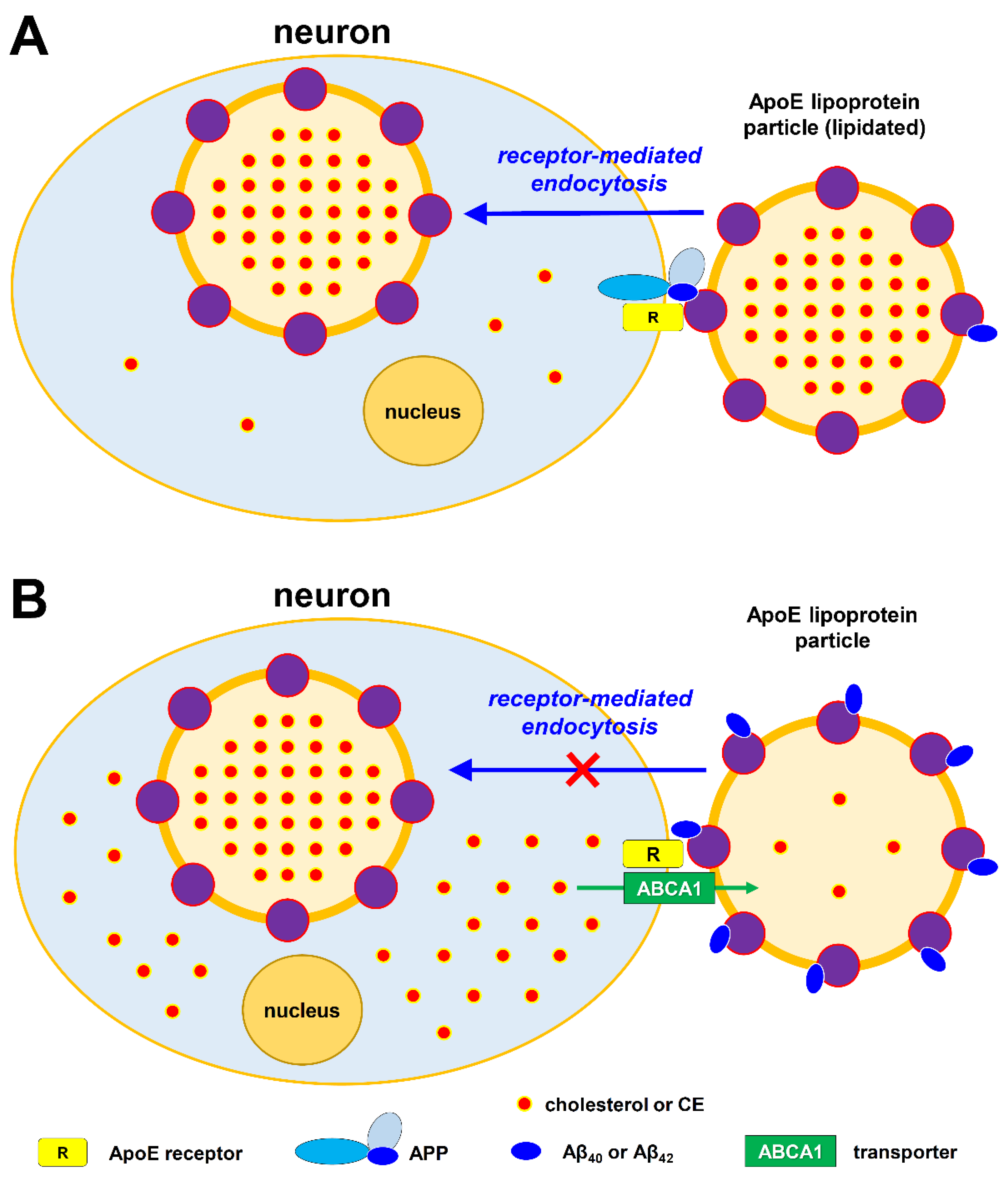
Another important mechanism to effectively reduce neuronal cholesterol is to activate its efflux transport via the ApoE lipoprotein particles in an ABCB1-dependent manner. As discussed in sections 4.2 & 4.3, it is known that the unlipidated ApoE2 and ApoE3 particles are highly capable of effluxing cholesterol out of neurons, but ApoE4 is markedly less capable in this respect, which is an important factor determining the pathogenic activity of ApoE4 in LOAD. Here, I will specifically discuss how changes in factors affecting ApoE-mediated neuronal cholesterol efflux contribute to Aβ formation and deposition.
The ApoE-mediated delivery of cholesterol to neurons via endocytosis requires the binding of lipidated ApoE particles to their specific receptors present on neuronal surface (here I will use LRP1 as an example for explanation). ApoE is known to bind to APP [378,379]. It is hypothesized that the intact, uncleaved APP protein is involved in the endocytosis of a lipidated ApoE particle into a neuron by playing a “permissive” role, which means that only when the lipidated ApoE particle simultaneously binds to its specific receptor (LRP1) and APP, then its endocytosis will be permitted to take place. Notably, it is speculated that the cleaved CTFα fragment of APP may also share this permissive role. However, when the lipidated ApoE particle only binds to LRP1 but not simultaneously to APP (or CTFα), then the endocytosis will not be permitted to take place (as depicted in Figure 6A). In so doing, an ApoE particle can be effectively regulated in a precise manner that it can readily switch from its function of supplying cholesterol to a neuron when the neuron is in need of more cholesterol to its function of removing excess cholesterol from a neuron. It is speculated that the APP-related proteins may share a similar function as APP, as the APP knockout mice do not display severe defects unless the APP-related protein APP1 is also concomitantly knocked out.
It is known that increased endocytosis of lipidated ApoE particles by a neuron will result in increased supply of cholesterol, which is usually for the purpose of fulfilling certain normal functional needs of this neuron. When neuronal cholesterol level is relatively low, the APP proteins will be cleaved predominantly by α-secretase (which forms Aβ17-40 fragment following further cleavage by γ-secretase). However, when elevated cholesterol level is present in a neuron, its APP will undergo enzymatic cleavages by β- and γ-secretases, resulting in increased production of Aβ40 and Aβ42 [37,129]. Aβ40 and Aβ42 fragments are known to bind to ApoE [214,215,258,265,296] (as depicted in Figure 5). Moreover, Aβ40 can bind effectively to both lipidated and non-lipidated ApoE3 or ApoE2 particles. As depicted in Figure 6B, it is speculated that the binding of Aβ40 to these ApoE3 and ApoE2 particles will prevent them from further binding to APP or APP-related proteins present on neuronal surface, thereby preventing these particles from undergoing endocytosis and thus preventing the further rise in neuronal cholesterol levels. However, the binding of Aβ40 to non-lipidated (or less-lipidated) ApoE2 orApoE3 particles will not affect their ability to carry out the cholesterol efflux function, which can help neurons to remove excess intracellular cholesterol. Cholesterol efflux from a neuron is dependent on the ABC transporter ABCA1, although other transporters (such as ABCD1 and ABCA7) may also help with this process, likely to lesser degrees.
At present, it is not known which specific receptors are involved in mediating neuronal cholesterol efflux by the astrocyte-produced ApoE. It is speculated that LRP1 likely is one of the ApoE receptors involved in cholesterol efflux, in addition to its known function in mediating the endocytosis of ApoE-containing lipoprotein particles. As discussed in section 4.6, there is clear evidence that LRP1 can mediate ApoE endocytosis; in comparison, its involvement in cholesterol efflux is far less clear (discussed later in section 8.1).
LOAD patients with the ApoE4 genotype are known to be associated with more Aβ plaques compared to ApoE2 and ApoE3 carriers. It was suggested earlier that ApoE is involved in the deposition of Aβ through direct protein‒protein interactions, and the lipidated ApoE4 binds preferentially to an intermediate, aggregated form of Aβ with a higher affinity than the lipidated ApoE2 or ApoE3 [282]. Here, a different explanation is provided. It is known that Aβ40 has a higher binding affinity for the lipidated ApoE2 or ApoE3 particles than for the lipidated ApoE4 particles [214,258,265]. In addition, the ApoE2 dimer can form a complex with Aβ40 more efficiently than the ApoE3 dimer [296]. By contrast, ApoE4 binds very poorly to Aβ40, but binds more tightly to Aβ42 [214,258,265]. It is known that for each individual ApoE-containing lipoprotein particle, the amount of ApoE4 molecules is about twice the amount of ApoE3 or ApoE2 [263,264]. Therefore, in the case of ApoE4 lipoprotein particles, when the same concentration of Aβ40 is present in the SCF, it is expected that there are still a lot of ApoE4 proteins not bound (and thus not blocked) by Aβ40. These unblocked ApoE4 lipoprotein particles can still be endocytosed into neuronal cells, which may cause a further rise in neuronal cholesterol level. As such, the neuronal cholesterol level will be higher with ApoE4 than with ApoE3 or ApoE2 (in the order of ApoE4 > ApoE3 > ApoE2). Elevated neuronal cholesterol is known to increase the expression of APP, which will then lead to increased enzymatic cleavage of APP by β- and γ-secretases to form more Aβ40 within the cholesterol/CE-enriched lipid raft microdomains of the plasma membrane [37,129,131]. Increased formation and release of Aβ40 may partially compensate for its lower affinity for binding to the ApoE4 particles. Such regulatory mechanisms through increased expression of APP and increased formation of Aβ40 may or may not be sufficient to effectively bring down the elevated neuronal cholesterol level to a range that is physiologically healthy to neurons. However, if such regulatory measures are still inadequate to bring down neuronal cholesterol level to a healthy range, then it will result in further rises in neuronal cholesterol content (including in neuronal cell membranes). Elevated cholesterol content in lipid raft microdomains will favor the binding of cholesterol to APP, which then increases the formation of Aβ42 instead of the usual Aβ40 [37,129,131]. In fact, there were both in vitro and in vivo studies showing that neuronal cholesterol accumulation can induce Aβ42 formation and accumulation in the brain [37,129,130]. Since Aβ42 has a significantly higher binding affinity than Aβ40 for the lipidated ApoE4 particles [214,258,265], the increased production of Aβ42 is expected to help more effectively block the binding sites on the ApoE4 particles and thus prevent the lipidated ApoE4 particles from endocytosis into neurons. Therefore, it is understood that when ApoE4 is the apolipoprotein, the brain neurons will be forced to produce a lot more Aβ peptides, including both Aβ42 and Aβ40 (particularly Aβ42), to help prevent ApoE4 particles from endocytosis (Figure 4 and Figure 6B).
Regarding the mechanism by which higher neuronal cholesterol level facilitates enzymatic production of Aβ42, it is known that the β- and γ-secretases that convert APP to Aβ fragments are predominantly localized to the cholesterol-enriched lipid raft microdomains of the plasma membrane [37,129]. It is speculated that increased intracellular levels of cholesterol (likely in the form of CEs) will increase the formation of lipid rafts in the plasma membrane of neuronal cells, which then favorably leads to the activation of enzymatic cleavage of APP catalyzed by β-/γ-secretases, resulting in increased formation of the Aβ42 peptide (in addition to Aβ40). Moreover, decrease in cellular cholesterol levels will increase APP cleavage by α-secretase, thereby decreasing the processing of APP into Aβ42 and Aβ40 peptides that accumulate in amyloid plaques [37,111,132]. Similarly, studies using both in vitro and in vivo models have shown that the use of statins was strongly associated with reduced levels of Aβ42 and Aβ40 [380,381]. By reducing cholesterol levels in living hippocampal neurons by 70% with lovastatin and methyl-β-cyclodextrin, the formation of Aβ was almost completely inhibited whereas the usual (normal) catalytic process of forming sAPP is unperturbed [382].
Here, it is of interest to note that when neuronal cholesterol level is low, Aβ17–40 is the major Aβ peptide formed and the amount of neuronal cholesterol exported by ApoE decreases. Based on this observation, it is tentatively speculated that Aβ17–40 may be able to bind preferentially to unlipidated ApoE, and the Aβ17–40-bound unlipidated ApoE may not be favored to bind to the ApoE receptors to perform the cholesterol efflux function. This is purely a speculation, and it will be of interest to experimentally examine this possibility in the future.
In addition to LRP1, other ApoE receptors, such as LDLR and VLDLR, can also mediate ApoE endocytosis. As these two ApoE receptors likely are not involved in ApoE-mediated cholesterol efflux, it is uncertain whether the endocytosis mediated by these receptors also requires the binding of APP (or similar proteins) to provide a “permissive signal”. If, in case, such a permissive signal is not needed, then it is speculated that these ApoE receptors should have effective mechanism(s) for their inactivation when neuronal cholesterol has reached an adequate level; only in this way the undesired further increase in neuronal cholesterol can be prevented in a timely and effective manner. One of the potential mechanisms may be through the secretase (or other proteinase)-mediated cleavage of these membrane-bound ApoE receptors in a manner that their functions can be very sensitively and effectively terminated when adequate amount of cholesterol is already supplied to neurons. There is some indirect evidence in line with speculation—it was shown earlier that the ApoE receptors can be enzymatically cleaved just like APP by secretases [375].
It is known that ApoE4 is selectively bound with a lot of Aβ42 peptides, and the ApoE4-bound Aβ42 peptides are insoluble and highly cytotoxic. A number of cell types in the CNS were reported to have the ability to internalize ApoE-bound Aβ, including astrocytes [265], microglia [383,384,385] and neurons [386], albeit with varying capacities. Immunohistological studies of human AD brains using antibodies against various Aβ epitopes have identified the N-terminal-truncated fragments of Aβ40 and Aβ42 inside the lipofuscin-like granules of astrocytes [387,388]. Similarly, in vitro studies using primary astrocytes in culture showed that these cells can internalize and degrade both soluble [389,390] and insoluble Aβ [391]. Interestingly, the ability of astrocytes to engulf and degrade Aβ is compromised in APOE knockout astrocytes, or in wild-type astrocytes upon the addition of an antibody against Aβ or ApoE, or an LDLR family antagonist [265]. These results confirm an essential role of ApoE in a receptor-mediated uptake of Aβ-bound ApoE particles by astrocytes. Primary hippocampal neurons were also reported to internalize Aβ in the presence of ApoE [392], but neurons appear to be less efficient in degrading Aβ, resulting in the formation of high molecular weight Aβ aggregates in their endosomal vesicles [393]. Additionally, Aβ can be taken up by smooth muscle cells delineating the arterioles or by endothelial cells that constitute the BBB [394,395,396,397,398]. It was suggested that a fraction of the Aβ fragments is transcytosed and secreted into the bloodstream [399], which may serve as an effective mean of clearance (discussed later in section 8.1).
The normal clearance rate for Aβ (likely including both soluble and ApoE-bound Aβ) appears to be very fast, estimated at approximately 8% per hour [394]. Physiological factors, such as the blood and CSF flux rates in the brain, together with a series of clearance receptors, including LRP1 and VLDLR, are all implicated in the removal of Aβ. However, in the case of ApoE4 which are usually bound with a lot more Aβ42, the clearance rate may be reduced when neuronal ATP supply is inadequate, which will gradually lead to Aβ deposition, often associated with heightened neuroinflammation.
5.4. Section Summary
One of the best-known pathological characteristics of AD is the deposition of extracellular Aβ in neuritic plaques. The β- and γ-secretases cleave APP in the amyloidogenic pathway, resulting in the formation of Aβ42, which may serve as a core for the formation of amyloid plaques. Neuronal membrane cholesterol content has been shown to play a crucial role in regulating the catalytic activity of β- and γ-secretases and thus affecting Aβ production and deposition.
When neuronal cholesterol level is abnormally elevated, a number of regulatory mechanisms will be activated to remove the excess cholesterol in neurons. One mechanism is through increasing metabolic disposition via CYP enzyme-mediated cholesterol oxidation; the other mechanism is through the efflux transport of excess cholesterol via the ApoE particles in an ABCB1-dependent manner. The unlipidated ApoE2 and ApoE3 particles have a very good ability to effectively efflux neuronal cholesterol, but ApoE4 has a sharply-reduced ability to efflux cholesterol, which is a key factor contributing to its pathogenic activity in LOAD.
In addition, Aβ40 and Aβ42 fragments are known to bind to ApoE, including both lipidated and non-lipidated ApoE3 or ApoE2 particles. The binding of Aβ40 to these ApoE3 and ApoE2 particles will prevent them from further binding to APP or APP-related proteins present on cell surface, thereby blocking these particles from undergoing endocytosis and thus limiting the further rise in neuronal cholesterol level. Notably, the binding of Aβ40 to non-lipidated (or less-lipidated) ApoE3 or ApoE2 particles will not prevent them from carrying out the cholesterol efflux function, which will help neurons to remove excess intracellular cholesterol. Notably, Aβ40 has a higher binding affinity for lipidated ApoE2 or ApoE3 particles than for lipidated ApoE4 particles. In fact, ApoE4 binds very poorly to Aβ40, but it can bind more tightly to Aβ42. In addition, each individual ApoE4-containing particle contains about twice the amount of ApoE4 molecules compared to that of ApoE3 or ApoE2 particles. Therefore, in the case of ApoE4 lipoprotein particles, when the same level of Aβ40 is present in the SCF, there are a lot of ApoE4 proteins still unblocked by Aβ40. These unblocked ApoE4 lipoprotein particles can still be endocytosed into neurons, thus causing further rises in neuronal cholesterol. Elevated neuronal cholesterol will lead to increased expression of APP, which then leads to increased enzymatic cleavage of APP by β- and γ-secretases to form more Aβ42, which has a much higher affinity to bind and thus block ApoE4 than does Aβ40. Therefore, Aβ42 is formed as a preferred alternative product when ApoE4 is the apolipoprotein.
While increased formation of large amounts of Aβ is an important initial cause in familial early-onset AD cases, it is understood that Aβ accumulation and plaque formation only represent a secondary pathological change in most cases of LOAD, and is not the dominant force driving the disease pathogenesis and progression. It is clear that cholesterol dysregulation in neuronal cells in ApoE4 carriers is a key factor that drives the Aβ formation and deposition.
6. What Causes Tauopathy in LOAD?
6.1. The Tau Hypothesis
It is generally believed that the microtubules in neurons can serve as tracks, guiding the transport of subcellular organelles and macromolecules from the soma of a neuron to the ends of the axon and back. Additionally, microtubules are involved in other important neuronal functions, such as axonal and dendrite outgrowth, and perhaps neurotransmission. Tau, one of the important microtubule-associated proteins (MAPs), affects microtubule stability and dynamics [400]. A salient feature of many neurodegenerative diseases is the formation of pathological tau aggregates, leading to the formation of NFTs, which mostly contain hyperphosphorylated tau [401].
The tau hypothesis proposes that tau protein abnormalities initiate the AD cascade [402]. In this model, pathologically-phosphorylated tau binds to normal tau and inhibits its functions [403,404]. Eventually, they form NFTs inside nerve cell bodies. When this occurs, the microtubules disintegrate, destroying the structure of the cell’s cytoskeleton which would collapse the neuron’s transport system. More recently, it was suggested that abnormal tau phosphorylation may subsequently trigger Aβ accumulation in LOAD [405].
While tau is mostly a cytosolic protein, it is also present in the nucleus [406]. Interestingly, the nuclear tau may have a DNA-protective function, which is impaired when tau aggregates [407,408,409]. In line with this suggestion, tau oligomerization is associated with increased DNA damage [410] and cell death [409]; furthermore, expression of a mutant tau is associated with chromosomal instability [411].
Notably, NFTs have been found in other neurodegenerative conditions including Pick’s disease, progressive supranuclear palsy, corticobasal degeneration, and others. The causal relationship between tau dysfunction and neurodegeneration is supported by the identification of over 40 mutations in the tau gene that cause familial frontotemporal dementia with Parkinsonism linked to chromosome 17 (FTDP-17) [412,413]. As discussed in detail below, it is speculated that when tau-based NFTs occur in other neurodegenerative diseases, there may exist severe neuronal energy deficiency as a major underlying cause.
6.2. Hyperphosphorylation and Aggregation of Tau
Tau in tangles is characterized by a high degree of phosphorylation on many residues (45 serines, 35 threonines and 5 tyrosines) [414]. Some of these sites are typically phosphorylated during embryogenesis or under specific physiological conditions (such as hibernation in certain species), whereas other sites are highly pathological.
In AD, tau phosphorylation is increased and the phosphorylation pattern is also altered, as opposed to normal physiological phosphorylation [415]. Phosphorylation of tau is carried out by multiple enzymes [416,417].
Phosphorylation of tau reduces its binding affinity for microtubules, which is associated with reduced microtubule stability and loss of synaptic functions [418]. In addition, hyperphosphorylated tau proteins are prone to aggregation [416], by a process that is not fully understood yet. Aggregated tau proteins would form oligomers, and eventually form fibrils, which assemble into paired helical filaments (PHFs) and neurofibrillary tangles (NFTs); these are hallmarks of AD [419].
A number of factors have been reported to induce tau hyperphosphorylation, such as dysregulation of kinase‒phosphatase activity that can lead to hyperphosphorylation. Several other factors have also been reported to induce tau hyperphosphorylation, such as glycation [420]. However, the mechanism underlying the induction of tau hyperphosphorylation is not clear at present.
6.3. Ubiquitin Degradation of Tau Protein is an ATP-Dependent Process
The 26S proteasome catalyzes the great majority (>80%) of the protein degradation in growing mammalian cells [421], in an ATP-dependent manner. ATP is consumed to make the protein degradation process highly selective through ubiquitination, and also enables substrate unfolding and translocation into an isolated chamber within the proteasome. The rate of hydrolysis of ubiquitinated proteins by the 26S is proportional to its rate of ATP hydrolysis, and the total ATP consumed in degrading ubiquitinated proteins is surprisingly large [422]. Besides, substrate size is also a factor that determines the amount of time and energy needed for proteolysis. The breakdown of large multi-domain proteins will consume much greater time and ATP for the 26S to unwind and degrade each successive domain [249].
Importantly, protein hyperphosphorylation can alter proteasome activity. There is growing evidence that the proteasome’s capacity to degrade ubiquitinated proteins is defective in neurodegenerative diseases [423,424,425]. As the proteasome function decreases, ubiquitinated proteins would accumulate in neurons along with phosphorylated tau.
6.4. Mechanistic Explanation
Neurons are terminally-differentiated, polarized cells, and require a well-developed cytoskeleton and motor proteins to facilitate the trafficking of various vesicles and organelles. Microtubules are also required for neuronal migration, differentiation, axonal extension, and neurotransmission. These important functions of microtubules are heavily dependent on cellular ATP for energy.
As discussed earlier, elevated neuronal cholesterol will lead to reduced neuronal biosynthesis of ATP. It is hypothesized that when there is a severe shortage of cellular ATP supply, the transport of cellular components will not be able to proceed normally, and as a result, some of the transport functions of the microtubules might have to be put on hold to save cellular energy for other, perhaps more important, neuronal processes.
As discussed above, tau protein degradation is mediated by the ubiquitin system present in neurons, which requires large amounts of cellular ATP to degrade unwanted proteins. High cholesterol levels in neurons will lead to reduced synthesis of ATP, and it is hypothesized that when cellular ATP is severely deficient, it will lead to a slowdown in the ubiquitination-associated degradation of tau proteins. When tau proteins cannot be enzymatically degraded in a timely manner, they will accumulate inside neurons. In order to prevent these tau proteins to perform their normal cellular functions (which will further consume cellular ATP), they are extensively phosphorylated in certain ways, as hyperphosphorylated tau proteins will have reduced functions. However, hyperphosphorylation of tau proteins also facilitates their aggregation. Therefore, based on the proposed pathogenic mechanism, it is speculated that tau accumulation mostly results from the reduced ability of the ubiquitin-proteosome system to effectively degrade tau due to severe deficiency of neuronal ATP, which results from abnormal elevation of neuronal cholesterol levels.
In support of the above mechanistic explanation, an earlier study has reported that there is a buildup of cholesterol in tangle-bearing neurons, suggesting that increased cellular cholesterol promotes tau phosphorylation [426]. Also, it was shown that synaptic accumulation of hyperphosphorylated tau oligomers in AD is associated with dysfunction of the ubiquitin-proteasome system [427].
Here it is of interest to note that in an Aβ vaccination trial, there were two patients who were almost entirely devoid of Aβ deposition, and yet displayed signs of severe pathology associated with NFTs (Braak stage VI) [428]. As expected, the intellectual capacities of these patients were severely affected, and the clinical progression of mental decline was not significantly different from untreated AD patients [428]. This observation is consistent with the hypothesis that Aβ formation and accumulation often is a secondary event accompanying neuronal cholesterol elevation, which then results in deficiency in neuronal ATP and neuroactive metabolic intermediates. In these two patients, although their Aβ deposition is almost completely removed by Aβ vaccines, if their neuronal cholesterol remains at high levels, then their cognitive functions as well as tauopathy will remain very severe, which are determined by the severity of neuronal ATP deficiency and are largely independent of the severity of Aβ deposition.
In the light of the suggestion that NFT formation is the result of severe neuronal ATP deficiency, it is reasonable to suggest that the appearance and spread of NFTs throughout the brain will closely reflect the degree of ATP deficiency in brain neurons, which can be caused by abnormally-elevated neuronal cholesterol. In other words, the degree of tauopathy is expected to be significantly less severe if neuronal ATP synthesis is not severely inhibited in those AD cases. In agreement with this suggestion, earlier studies have shown that dementia symptoms in AD correlate better with the appearance and spread of NTFs throughout the brain than with the deposition of Aβ in senile plaques [429,430].
Based on the explanation provided above, it is further speculated that factors that can inhibit neuronal ATP synthesis likely will also induce tau hyperphosphorylation and subsequently certain degrees of tauopathy. In partial support of this speculation, earlier studies have shown that oxidative stress and environmental toxins (e.g., arsenite) that are capable of inhibiting mitochondrial metabolic function [431,432] can induce tau hyperphosphorylation [433,434]. Similarly, methanol has also been reported to induce tau hyperphosphorylation and decrease the cognitive ability in animal models [435]. Mechanistically, methanol is known to be metabolically converted to formic acid in the body, which is a strong inhibitor of mitochondrial oxidative phosphorylation [436], thus reducing cellular ATP synthesis. Notably, an earlier study reported that Aβ can increase tau hyperphosphorylation in neuronal cells that take up these secreted peptides [437]. This observation likely is due to the fact that endocytosis of Aβ fragments is usually associated with the endocytosis of Aβ-bound ApoE particles (enriched with cholesterol and other lipids), which will disrupt mitochondrial function and reduce ATP production in neurons.
It is known that the two classic lesions of AD (i.e., Aβ deposits and neuritic tangles) can occur independently in other brain diseases. NFTs composed of tau aggregates that are similar to or indistinguishable from those found in AD have been described in many less common neurodegenerative diseases (e.g., frontotemporal dementia, subacute sclerosing panencephalitis and progressive supranuclear palsy) that essentially lack Aβ protein deposits and neuritic tangles. Conversely, diffuse Aβ deposits can be seen in aged “normal” brains with almost no neuritic tangles. The fact that NFTs composed of aggregated forms of tau proteins occur in certain diseases in the absence of Aβ protein deposition is not surprising as severe ATP deficiency is a main driving force for the formation of NFTs in brain neurons. Besides elevated neuronal cholesterol levels, other factors can also cause neuronal ATP deficiency. However, when neuronal cholesterol levels are abnormally elevated, increased Aβ production and deposition may be more readily seen, often jointly with the accumulation of NFTs. During this process, the degree of impairment of memory functions will largely depend on the degree of deficiency of neuronal ATP and neuroactive metabolic intermediates, both of which are the consequences of elevated neuronal cholesterol. In those aged AD patients that lack significant tauopathy, it is expected that their neuronal cholesterol levels are only modestly elevated, which will slowly but chronically increase Aβ formation and deposition, and eventually may end up with rather heavy buildup of amyloid plaques (if their removal is not as efficient). Because neuronal ATP deficiency in these patients may not be as severe, significant tauopathy may not develop as a result; usually these patients are also expected to have slightly better cognitive brain functions.
In line with the above explanations, many earlier studies have noted significant perturbations of neuronal proteolytic degradation machinery in AD. In fact, a striking morphological alteration in neurons of the AD brains is the accumulation of autophagosomes, autolysosomes, and lysosomal dense bodies in dystrophic neurites [438,439,440]. Similar alterations are also observed in APP and/or PS transgenic AD mouse models [441], even before amyloid deposition is observed. When autophagy is compromised in the brain of an APP-overexpressing AD mouse model following inactivation of the Beclin-1/BECN1 gene (BECN1+/− mice), enhanced Aβ deposition and loss of synapses and neurons are observed, which suggests that decreased autophagy can aggravate or even independently contribute to the overall neurodegenerative process [442]. Interestingly, expression of Beclin-1 using lentiviral vectors attenuated the amyloid load in the injected mouse brain areas.
6.5. Section Summary
As discussed in earlier sections, elevated neuronal cholesterol can lead to increased formation of amyloid plaques. In addition, elevated neuronal cholesterol can reduce the biosynthesis of ATP and its cellular levels, and severe ATP insufficiency then results in tau hyperphosphorylation and aggregation (i.e., the formation of NFTs). Based on this mechanistic explanation, tauopathy will be more readily seen in LOAD as opposed to early-onset familial AD. In early-onset familial AD, neuronal cholesterol elevation and ATP deficiency are not the initial driving causes, therefore it is speculated that hyperphosphorylation of tau likely may not be as severe initially compared to the situation in sporadic LOAD.
According to the proposed hypothesis, modest reduction in neuronal ATP levels is a relatively early event resulting from elevated neuronal cholesterol. As tau accumulation is the result of ATP deficiency, this process will occur relatively early in AD pathogenic process, which is consistent with clinical observations. Notably, a more severe reduction in ATP supply is usually experienced in brain regions that have higher levels of neural activity and thus higher demands for energy supply, such as the medial temporal lobe [443,444]. In fact, this brain region is known to be an early site of tau accumulation (in the absence of evident amyloid accumulation), and its dysfunction may underlie episodic-memory decline commonly seen in aging and dementia [443,444].
While elevated neuronal cholesterol level will increase Aβ42 formation, there are also other factors affecting Aβ42 formation and deposition. These are the main reasons why the degree of mental functional decline often is not tightly correlated with Aβ pathology. On the other hand, mental functional decline is more directly associated with degree of neuronal ATP deficiency. Since tau accumulation is the result of neuronal ATP deficiency, it is not surprising that mental functional declines are more closely correlated with tau accumulation. Offering partial support for this suggestion, human [18F]flortaucipir PET studies have shown strong associations between regional tau and cognitive decline and neurodegeneration [445].
7. What Causes Acetylcholine Deficiency in LOAD?
7.1. The Cholinergic Hypothesis
Acetylcholine is a critical neurotransmitter in the brain that is involved in the formation of memories. Moreover, this neurotransmitter is widely used by neurons in the hippocampus and cerebral cortex, regions mostly devastated by AD. In the 1970s, it was found that acetylcholine levels fall somewhat during normal aging, but drop by approximately 90% in people with AD [446,447]. In addition, other neurotransmitters have also been implicated in AD. For example, the levels of serotonin, somatostatin and norepinephrine are also reduced in some AD patients, and deficits in these substances have been suggested to partially contribute to sensory disturbance, aggressive behavior, and neuronal death.
The cholinergic hypothesis is perhaps the oldest hypothesis concerning the potential causes of AD, which postulates that AD is caused by reduced biosynthesis of the neurotransmitter acetylcholine. In fact, some of the currently available drug therapies for AD are based on the cholinergic hypothesis [446,447]. Overall, medications that treat acetylcholine deficiency have not been very effective clinically.
7.2. Mechanistic Explanation
The synthesis of acetylcholine neurotransmitter is a single step reaction catalyzed by the enzyme acetyl-CoA:choline O-acetyltransferase (ChAT):
choline + acetyl-CoA ⇌ acetylcholine + CoA
ChAT is found in the nervous system specifically at sites where acetylcholine synthesis takes place. Within cholinergic neurons, ChAT is concentrated in nerve terminals. In subcellular fractionation studies, ChAT was recovered in the synaptosomal fraction, and within synaptosomes it was primarily in the cytoplasmic fraction. In comparison, acetylcholinesterase (AChE), the enzyme responsible for degradation of acetylcholine, is produced by cells that contain cholinoreceptive sites, although it is also produced by cholinergic neurons.
Under usual conditions, synthesis of acetylcholine is mostly determined by the intracellular availability of choline, which is a limiting step determined by the uptake of choline into the nerve ending. Choline is supplied to neurons either from blood circulation or through metabolism of choline-containing compounds. At least half of the choline used in acetylcholine synthesis comes directly from recycling of released acetylcholine, which is hydrolyzed to choline by AChE. Presumably, uptake of this metabolically-derived choline occurs rapidly, before these molecules diffuse away from the synaptic cleft. Another source of choline is the breakdown of phosphatidylcholine. Choline derived from these two sources becomes available in the extracellular space and is then subject to high-affinity uptake into the nerve ending. In the CNS, these metabolic sources of choline are particularly important as choline in the plasma cannot penetrate the BBB. Thus, in the CNS, the high-affinity uptake of choline into cholinergic neurons may be saturated, and as such, acetylcholine synthesis can be limited by the supply of choline, at least during sustained neuroelectrical activity. This suggestion is in line with the observation that acetylcholine stores in the brain are subject to variation, whereas acetylcholine stores in other places such as ganglia and muscles remain relatively constant.
However, under conditions of AD, it is hypothesized that acetylcholine deficiency is mainly due to elevations of neuronal cholesterol levels, which will result in reduced mitochondrial metabolic activity as well as reduced ATP synthesis in neurons and nerve terminals. Since acetyl-CoA is formed as a metabolic intermediate inside the mitochondria, suppression of mitochondrial metabolic activity is expected to result in reduced formation of acetyl-CoA. In addition, since the transport of acetyl-CoA from mitochondria to cytoplasmic compartment is an ATP-dependent process, reduced mitochondrial ATP synthesis resulting from elevated neuronal cholesterol may also reduce the transport of mitochondrial acetyl-CoA to cytoplasmic compartment where acetylcholine biosynthesis takes place.
In addition to the above two factors, it is important to note that the cholinergic vesicles in nerve terminals not only store acetylcholine, but also ATP and Ca2+ [42]. ATP is a required component of the cholinergic vesicles, and the release of ATP has been shown to accompany acetylcholine release from these vesicles during neurotransmission [42]. Under conditions of severe ATP shortage, formation of acetylcholine vesicles is expected to be drastically reduced.
Lastly, the normal process of synaptic neurotransmission itself also demands high ATP supply [43]. When there is an ATP shortage in cholinergic neurons or nerve terminals, the activity of neurotransmission will be significantly reduced, and the level of reduction likely depends on the degree of ATP deficiency in these neurons and their nerve terminals.
8. Explanation of Relevant Clinical and Experimental Observations Based on the Proposed New Hypothesis
8.1. The BBB May Enable Active Transport of CNS Lipidated ApoE Particles into the Peripheral Compartment
It has been generally believed that due to the presence of BBB, cholesterol within the CNS does not readily equilibrate with cholesterol in peripheral circulation [46]. Similarly, it is believed that the peripheral and CNS ApoEs do not cross the BBB and do not exchange their ApoE-containing lipoproteins [448]. Despite these earlier suggestions, it was, however, estimated that a relatively small but significant fraction (up to 0.4%) of the total brain cholesterol pool is “excreted” from the brain every day [45].
To explain the cholesterol turnover in the CNS, it was reported earlier that knockout of CYP46A1 gene in mice results in over 50% reduction in brain cholesterol excretion and approximately 40% reduction in brain cholesterol synthesis [137]. Based on this observation, it was suggested that CYP46A1 in mice may be directly responsible for approximately 40% turnover of total brain cholesterol [137]. While this explanation is not unreasonable, a different possibility is suggested here. It is speculated that the reduced brain cholesterol turnover in CYP46A1-knockdout mice may mostly result from the reduced cross-BBB transport of cholesterol-enriched ApoE and ApoJ lipoprotein particles. It is known that the major components involved in this cross-BBB transport, such as ApoE, ApoJ, ABCA1 transporter and TREM2, are all up-regulated by 24S-OHC, which is formed by CYP46A1. As such, when CYP46A1 is deficient in mice, it not only decreases CYP46A1-mediated metabolic disposition of cholesterol, but it also drastically decreases the cross-BBB transport of cholesterol, due to the absence of the key regulatory factor 24S-OHC.
The above explanation is reasonable from a theoretical point of view. It is speculated that neuron-expressed CYP46A1 may mostly serve the function of producing a very small quantity of the signaling molecule 24S-OHC, for the purpose of regulating neuronal cholesterol homeostasis. The actual metabolic capacity of brain neurons (i.e., the metabolic capacity of CYP46A1) likely is very limited, as these cells are highly specialized for complex and important neuronal functions. If the neuronal CYP46A1-mediated metabolic disposition of cholesterol per se were chiefly responsible for brain cholesterol turnover every day, it probably would be more reasonable that some other major cell types in the brain (such as microglia) play a dominant role in this task. This would be akin to the situation that astrocytes, rather than neurons themselves, are largely responsible for the synthesis of lipids required for fulfilling many neuronal functions in the brain. However, if the brain has the ready means to actively transport excess cholesterol across the BBB into the peripheral circulation, then it is readily understood that the selective presence of CYP46A1 in brain neurons (but not in astrocytes) is mostly for the purpose of producing a signaling molecule in very small quantities for regulating neuronal cholesterol homeostasis.
Here, it is of interest to note that earlier studies found that after ApoE particles are secreted by astrocytes, they are initially associated with smaller amounts of lipids forming small discoid particles (8–15 nm in diameter), then they increase in size, becoming spherical as they accumulate more lipids, and eventually they are enriched with lipids (12–20 nm with a fraction up to 30 nm) and flow into the CSF [449,450,451]. These interesting observations have led to the speculation that there might exist a specific mechanism responsible for the active transport of cholesterol-rich ApoE particles in the CSF across the BBB into the peripheral circulation for disposal (note that these ApoE particles are usually also bound with Aβ fragments to prevent them from endocytosis into neurons). Specifically, it is postulated that the BBB can routinely enable the transport of cholesterol-enriched ApoE particles (likely together with ApoJ) into the peripheral circulation in such a fashion that excess neuronal cholesterol can be effectively removed. Mechanistically, it is hypothesized that TREM2 present in microglia is involved in the efflux (transcytosis) of Aβ-bound, ApoE/ApoJ-containing lipid particles across the BBB. In addition, the LRP1 may also mediate the cross-BBB transport of Aβ-bound ApoE particles. Furthermore, under certain conditions, the BBB may even become selectively “leaky”, enabling some of the peripheral apolipoproteins to gain access into the CNS to aid in the removal of excess neuronal cholesterol. Some of the supporting observations which are scattered in the literature are briefly discussed below.
8.1.1. Role of TREM2 in Cross-BBB Transport of Cholesterol
Studies have shown that microglial dysfunction is an important pathogenic factor in LOAD [452,453,454,455]. Microglia are actively involved in phagocytosis-mediated cholesterol clearance in the brain [456], which is perhaps one of the most important functions of microglia. In fact, it has been suggested that the brain microglia need exogenous cholesterol to maintain their phagocytic capacity and even promote their survival, as the presence of cholesterol will activate the expression of several microglial signature genes associated with the so-called “AD signature phenotype” [457]. Functionally, microglia-mediated myelin remodeling through phagocytosis of lipids (including cholesterol) is part of the process required for memory formation [458].
Large-scale genetic studies have uncovered variants in LOAD risk-associated genes that are highly expressed in microglia [459,460], one of which encodes the triggering receptor expressed on myeloid cells 2 (TREM2), a single-pass transmembrane immune receptor selectively expressed in brain microglia. Individuals carrying rare heterozygous variants of TREM2, such as R47H, have a higher risk for LOAD (average odds ratio of approximately 4.5) [461,462,463]. Notably, the rare homozygous TREM2 loss-of-function mutations are known to cause Nasu-Hakola disease, a syndrome characterized by myelin loss, neurodegeneration, and early-onset dementia plus other pathologies such as recurrent bone fractures [464].
Consistent with results from human studies, animal studies have more clearly shown that TREM2 knockout mice have learning and memory deficits, and transgenic mice that overexpress TREM2 show significant improvements in these functions [465,466]. Additional animal studies have further revealed that the loss-of-function TREM2 variants are associated with increased AD risks [467].
Regarding the protective function of TREM2 in LOAD, it is hypothesized that the observed “microglial clearance” of ApoE and ApoJ [468,469,470,471] is a process associated with the cross-BBB transport of Aβ-bound ApoE/ApoJ lipoprotein particles. It is speculated that many of these ApoE/ApoJ particles are cholesterol-enriched, although TREM2 is capable of transporting both lipidated and unlipidated particles. A schematic representation of the proposed hypothesis is depicted in Figure 7. It is hypothesized that TREM2 plays a more important role in guiding the phagocytosed vesicle to re-merge with the cell membrane on the peripheral side. When this function of TREM2 is partially or fully lost (such as due to the loss-of-function mutations), the cross-BBB transport of cholesterol-enriched Aβ-bound ApoE/ApoJ particles will be significantly reduced, which will then lead to elevated brain cholesterol levels, particularly inside microglia, subsequently increasing the risk of LOAD. As discussed below, there are experimental observations offering partial support for the above mechanistic explanation regarding the role of TREM2 in LOAD.
i. The ectodomain (ECD) of TREM2 is a receptor for binding different lipoproteins (e.g., ApoE, ApoJ, ApoA1, and LDL), although ApoE has been the best-studied ligand [468,472,473]. This binding is independent of the ApoE isoforms [468,472,473,474] and their lipidation states [472,473,474]. Earlier studies have suggested that ApoJ likely is involved in helping the transport of ApoE-rich lipoprotein particles across the BBB [468,470,471,472]. Studies have shown that AD-associated TREM2 variants (such as R47H and R62H) indirectly affect the binding of TREM2 with its ligands [463,468,472,473,475,476]. Detailed analysis has revealed the R47H mutation significantly reduces the binding of TREM2 with ApoE [473], including all three isoforms [472] with varying lipidation states [468], with other apolipoproteins [468,473] and lipids [477,478]. It is expected that the reduced ability of TREM2 variants for ligand binding will increase the risk of LOAD [468,472,473,478] as these variants are expected to increase cholesterol levels in the brain, especially in microglial cells.
Notably, studies have revealed that the amyloid mouse models lacking ApoE and the mouse models lacking TREM2 each display similar pathologies in amyloid plaques and microgliosis [479,480]. Their similarities are readily understood as both situations will lead to elevated cholesterol in neurons and microglia. Similarly, it is also understood that individuals simultaneously carrying the R47H variant and the APOE4 genotype would have a markedly elevated risk for developing LOAD [481].
ii. Studies have shown that under normal homeostatic conditions, TREM2 is primarily located intracellularly [482], in association with the trans-Golgi network [483,484,485] and in
Exocytic vesicles [483]. These TREM2-containing vesicles continuously shuttle to the membrane, a process which can be rapidly induced by increases in intracellular Ca2+ [483]. TREM2 is recycled from the membrane in clatherin-coated vesicles in a beclin-1 [486] and Vps35-dependent manner [486,487]. Vps35 mediates recycling of TREM2 from the membrane via retromer complexes [487]. In the case of loss-of-function variants [475,488,489,490], TREM2 will have a reduced presence on cell surface but an increased association with lysosomes and is degraded [487].
Consistent with the hypothesis that TREM2 is critically involved in the transcytosis of lipoprotein particles, animal studies have shown that microglia lacking TREM2 have a drastic increase in intracellular vesicles, which resemble autophagic vesicles [487]. This observation was also made in humans, as microglia in AD patients carrying the loss-of-function TREM2 variants have more intracellular vesicles than microglia in AD patients with the common TREM2 variant. These observations support the notion that TREM2 plays a crucial role in guiding the phagocytosed vesicles to re-merge with the cell membrane on the other side. When this function of TREM2 is lost, then it would have a reduced presence on cell surface but an increased association with intracellular vesicles (such as lysosomes) and is quickly degraded [487]. As these vesicles are loaded with cholesterol, it is expected that the microglia with mutant TREM2 will have very high levels of cholesterol inside, which will then inhibit microglial functions and induce neuroinflammation.
iii. It is known that TREM2 serves as a major sensor for extracellular lipids, and mediates cholesterol clearance during demyelination. It detects demyelination by sensing the lipidic components of myelin and promotes myelin debris removal via lipid transport and catabolism [491]. Interestingly, it appears that chronic myelin phagocytosis in Trem2–/– brain is not affected, but the lack of TREM2 causes accumulation of CEs and oxidized CEs in the TREM2–/– brains. These observations agree with the known functions of TREM2, i.e., it plays a more important role in the transcytosis, i.e., the re-merging of the phagocytosed vesicles with the cell membrane on the peripheral side, whereas its role in mediating the initial phagocytic process is less important.
Studies from recent years have identified the disease-associated microglia (DAM) phenotype, which is a transcriptionally-distinct microglial profile and closely associated with LOAD pathogenesis [452]. In addition to the expression of the microglial marker gene TREM2, this subpopulation of microglia is also defined by the expression of genes involved in lipid metabolism and transport, such as APOE [452,457]. In fact, increased expression of TREM2 along with other components is widely considered a characteristic shift from the homeostatic phenotype of microglia to the neurodegenerative phenotype [453].
Mechanistically, some earlier studies have suggested that TREM2 may directly control microglial gene expression, resulting in the development of the disease-associated microglia phenotype. While this possibility cannot be ruled out, it is speculated the upregulation of gene expression involved in lipid transport and metabolism most likely is an indirect effect resulting from the functional deficiency of TREM2. It is hypothesized that when neuronal cholesterol is abnormally elevated, the astrocytes will synthesize more ApoE and ApoJ to help efflux cholesterol (and its metabolite 24S-OHC). Under conditions when the removal of excess cholesterol is still insufficient (such as due to the presence of ApoE4), more ApoE and ApoJ will be produced. In these situations, there will be a heightened reliance on TREM2-mediated microglial transport of Aβ-bound ApoE/ApoJ particles across the BBB. As such, microglia will be regulated by elevated cellular cholesterol and 24S-OHC, further stimulating the expression of TREM2, cholesterol transporters, apolipoproteins (such as ApoE and ApoJ), and metabolizing enzymes (such as SOAT1 and LCAT1), thereby manifesting as the DAM phenotype.
iv. It is known that loss-of-function TREM2 variants can increase the risk of LOAD [479]. These variants under certain conditions (such as chronic demyelination) will lead to accelerated microglial accumulation of intracellular cholesterol in a storage form (i.e., CEs) without altering microglial phagocytic capacity [467]. CEs are known to accumulate in the brains of LOAD patients and AD mouse models [492,493,494,495], and the LOAD-linked TREM2 variants [463] are associated with robust intracellular accumulation of CEs. It is of note that TREM2-deficient and ApoE-deficient microglia show similar abnormalities in cholesterol metabolism, including CE accumulation [479]. Mechanistically, it has been suggested that CE accumulation may mediate cytotoxicity via enzymatic or non-enzymatic formation of oxidized metabolites and lipid peroxides from the mono- and polyunsaturated fatty acid chains the CEs harbor [496,497].
Lastly, it is of note that TREM2–/–macrophages also exhibit a similar CE storage disorder, resulting in the formation of macrophage foam cells commonly seen in atherosclerotic lesions [498]. Earlier studies of foam cells revealed that cholesterol dyshomeostasis resulting in excess CE accumulation is closely associated with pro-inflammatory responses [496,498,499]. Therefore, it is speculated that abnormal accumulation of cholesterol and CEs in the brain (likely mostly in microglia) is an important factor in neuronal inflammation.
v. Cyclocreatine is a potent bioenergetic agent that helps maintain higher cellular ATP levels during ischemia [500]. Interestingly, dietary administration of cyclocreatine in 5XFAD mice lacking TREM2 prevented the buildup of intracellular vesicles, increased microglia numbers, enhanced clustering around Aβ plaques, and mitigated plaque-associated neurite dystrophy [501]. This observation points to a central role of brain ATP deficiency (presumably caused by elevated neuronal cholesterol resulting from TREM2 deficiency) in microglial dysfunctions and their role in LOAD pathogenesis. This observation also suggests that there are other mechanisms which may compensate for the lack of TREM2 function if adequate microglial ATP supply is maintained.
8.1.2. Role of TREM2 in the Cross-BBB Transport of Aβ
It is known that microglia can directly internalize and degrade Aβ [502]. Studies have shown that TREM2 can serve as a microglial receptor for Aβ, and the Aβ oligomers can bind to TREM2 with higher affinity than the Aβ monomers [503]. In the AD mouse model, elevated microglial TREM2 gene dosing decreases the Aβ burden, along with improved memory performance [466]. On the other hand, TREM2–/– and TREM2-R47H mutant microglia fail to surround and clear amyloid plaques in vivo, resulting in Aβ plaque buildup and accumulation of dystrophic neurites near plaques [463,476,504,505,506,507,508,509].
As discussed in the preceding sections, the microglial TREM2 likely is involved in transporting the cholesterol-rich ApoE/ApoJ lipoprotein particles across the BBB. It is expected that when these ApoE/ApoJ particles are being transported across the BBB, they would also take along with them considerable amount of Aβ fragments that are tightly bound to these particles. In support of this suggestion, earlier animal studies have shown that ApoJ can bind directly to the soluble form of Aβ in a specific and reversible manner, forming complexes that have been shown to cross the BBB [510]. A high-affinity transport system for ApoJ (Kd of 0.2‒0.5 nM) was identified at the BBB and the choroid epithelium in vivo, and the ApoJ‒Aβ40 complex had 2.4- to 10.2-fold higher affinity than ApoJ itself for the same transport system [320].
8.1.3. Role of LRP1/2 in the Cross-BBB Transport of Aβ and/or Aβ-Bound ApoE or ApoJ Particles
In addition to TREM2, earlier studies have indicated that LRP1 is also involved in the vascular transport of Aβ peptides across the BBB [53]. For instance, it was reported that intracerebrally-microinjected 125I-Aβ40 was rapidly removed from the brain of young mice (t1/2 ≤ 25 min), largely through vascular transport across the BBB. The efflux transport system for 125I-Aβ40 at the BBB was half saturated at 15.3 nM, and the maximal transport capacity was reached between 70 and 100 nM. 125I-Aβ40 clearance was substantially inhibited by the receptor-associated protein, and by antibodies against LRP1 and α2-macroglobulin (α2M). There was no evidence that Aβ was metabolized in brain interstitial fluid and degraded to smaller peptide fragments and amino acids before its transport across the BBB into the circulation. Similarly, another study suggested that BBB-associated pericytes can clear Aβ aggregates via an LRP1/ApoE isoform-specific mechanism [511].
In addition, an earlier study [512] reported that Aβ40 binds to immobilized LRP clusters II and IV with high affinity (Kd = 0.6‒1.2 nM) compared to Aβ42 and mutant Aβ. Transgenic mice expressing low LRP-clearance mutant Aβ develop robust Aβ cerebral accumulation much earlier than Tg-2576 Aβ-overproducing mice. Aβ40 is cleared rapidly across the BBB via LRP1, but Aβ42 is removed across the BBB at a slower rate (1.9-fold slower) compared with Aβ40.
The transport pathways for clearance of human Aβ and ApoE/ApoJ in the mouse CNS have been reported [513]. ApoE3 is cleared slowly across the BBB, and after lipidation its transport at the BBB becomes barely detectable. ApoJ is eliminated rapidly across the BBB via LRP2. Aβ40 binding to apoE3 reduced its efflux rate at the BBB by 5.7-fold, but Aβ42 binding to ApoJ enhanced Aβ42 BBB clearance rate by 83%.
8.1.4. The Leaky BBB Phenotype
An earlier study by Zlokovic and colleagues [514] reported that unlike normal mice, the ApoE knockout mice exhibit a leaky BBB phenotype. Interestingly, selective expression of human ApoE2 or human ApoE3, but not human ApoE4, rescued the leaky BBB phenotype of the mouse. It is speculated that the above experimental observations likely are related to the ability of ApoE2 and ApoE3 to remove excess cholesterol from neuronal cells through the ApoE-mediated cholesterol efflux. Under the pathogenic conditions of complete ApoE deficiency, the brains of transgenic mice would not be able to effectively remove excess cholesterol accumulated inside their CNS neurons. As a result, through certain feedback regulatory processes, the brain of these mice would selectively increase the “permeability” of their BBB such that some of the peripheral apolipoproteins may be allowed into the brain to help remove the excess cholesterol from neuronal cells. While the exact mechanism by which peripheral apolipoproteins are allowed into the brain is not clear at present, studies have shown that under certain conditions, the vascular endothelial cells of the BBB can permit the transcytosis of certain peripheral macromolecules (such as apolipoproteins) to cross the BBB [515]. However, when these ApoE-deficient mice are knocked in to express the human ApoE2 or ApoE3, then the functional deficiency in neuronal cholesterol efflux is restored—understandably, the “leaky BBB phenotype” is no longer be needed. However, knock-in of a gene that selectively expresses the human Apo-E4 will not be as helpful since it has a far lower ability to efflux cholesterol out of neuronal cells.
There are some experimental observations offering partial support for the above suggestion that the BBB can become selectively leaky under certain conditions. For instance, it is known that cells in the brain do not produce ApoA1, but the cerebrospinal fluid can acquire a significant amount of ApoA1 under certain conditions, most likely from the blood via unknown mechanism(s) [515].
8.2. Why Hypercholesteremia Is Associated with Increased Risk of LOAD?
Cholesterol present in the CNS is mostly synthesized locally in astrocytes, which is delivered to neurons through endocytosis of lipidated ApoE-rich particles. For years, it is widely believed that the two pools of cholesterol and ApoE, i.e., one in the periphery and one in the CNS, are separated, and there is no exchange between these two pools of cholesterol due to the presence of the BBB. However, epidemiological studies have clearly shown that hypercholesteremia is associated with an increased risk for AD [516]. Similarly, in animal studies, it was shown that a cholesterol-rich diet increases Aβ production in the brains of these animals, and an opposite effect was observed in some of the studies when the animals were treated with cholesterol-lowering drugs [113,380,517,518].
The mechanism by which hypercholesterinemia increases the risk of AD is not clearly understood at present. It was speculated earlier that the observed association between hypercholesterinemia and AD likely is because the impermeability of the BBB is compromised in individuals with AD such that cholesterol can be transported into the brain from the plasma [519]. A slightly different explanation is provided here. It is speculated that hypercholesterolemia may largely affect the removal of excess cholesterol from neuronal cells across the BBB into the peripheral circulation. In other words, when the plasma cholesterol level is elevated, it will slow down the net cross-BBB transport of excess cholesterol carried by brain ApoE particles into the peripheral circulation. As a result, reductions in the cross-BBB transport of excess cholesterol will facilitate cholesterol buildup in the CNS (particularly inside neurons) and ultimately result in reduced neuronal synthesis of both ATP and neuroactive metabolic intermediates (mevalonate and geranylgeraniol), contributing to memory and learning deficits. In addition, elevated neuronal cholesterol is expected to facilitate the formation and deposition of Aβ fragments, and the reduced neuronal ATP level will increase tauopathy and decrease the formation of cholinergic vesicles. All these effects will jointly enhance the pathogenesis of AD, in particular LOAD.
8.3. How to Explain the Complex Effects of Statins in AD?
Statins have been used successfully to treat patients with dyslipidemic cardiovascular diseases [520]. They work by reducing cholesterol synthesis through inhibiting HMGR, a rate-limiting enzyme involved in endogenous sterol synthesis [521]. In general, most statins can inhibit HMGR with a very high potency (Ki or EC50 <1 nM) [521].
Because high levels of plasma cholesterol are associated with increased incidence of AD (discussed in the preceding section), the use of statins has been suggested as a potential treatment for AD. Studies using both in vitro and in vivo models have shown that statins can reduce the levels of Aβ peptides, including both Aβ42 and Aβ40 [380,381]. Based on these observations, many human studies had been initiated to investigate the effect of statins on dementia and cognitive functions. While some human studies have noted a decreased risk of dementia or AD in statin users [522,523,524,525,526,527,528,529,530,531], there were also studies reporting the lack of a clear relationship between the incidence of AD and statin use [532,533,534].
The potential benefits of statin use in AD have been controversial for the following reasons: i. Some researchers felt that the conclusion regarding a beneficial effect of a peripherally-acting statin in AD itself is somewhat “conceptually” controversial, as it has been long held the view that the plasma lipoproteins do not cross the BBB, and that there is no exchange between the central and peripheral pools of cholesterol due to the presence of the BBB. ii. There are disagreements as to whether it is the peripheral or central lipid-lowering actions of statins that really contribute to the benefits seen in AD. Some researchers strongly felt that if the use of statins is indeed beneficial in AD, then the use of only those statins that can readily cross the BBB will be indicated, rather than the use of peripherally-acting statins for this purpose. iii. The last and most important controversy centers around the question of whether the use of a statin drug is really effective or beneficial in AD, as there are many clinical and animal studies reporting opposite findings. Also, doubts are raised as to whether the potential benefits of statins in AD are really attributable to their cholesterol-lowering effect or might actually be due to some other potential actions of the statin compounds (such as anti-inflammatory and/or antioxidant properties).
As discussed below, the beneficial effects of the statin drugs in AD, and in particular the complexity of their actions in AD, might be more readily understood in the light of the new pathogenic hypothesis proposed in this paper.
First, based on the discussion provided in earlier sections, it is clear that selective inhibition of the peripheral cholesterol synthesis (such as in liver and other peripheral organs) with a statin drug will result in decreased cholesterol levels in circulation, which will be beneficial for the cross-BBB transport of excess neuronal cholesterol to the periphery. Based on this mechanistic understanding, it is clear that the use of a peripherally-acting statin will be beneficial for reducing the risk of AD.
Second, when the HMGR in neurons and astrocytes is inhibited by a centrally-active statin in addition to their inhibition of the peripheral HMGR, the net effect with respect to AD risk will be more complex, as already noted in some of the earlier studies [535], and the underlying reasons for the complexity are explained below.
It is clear that the elevated neuronal cholesterol is “bad” for AD, not only because it will reduce mitochondrial metabolic activity and ATP level in neurons, but it will also inhibit the neuronal HMGR activity, which subsequently reduces the synthesis of important neuroactive metabolic intermediates. Besides, high levels of neuronal cholesterol will also increase Aβ40 and Aβ42 formation and deposition in the brain. These effects jointly determine the pathogenic role of elevated neuronal cholesterol in AD. Accordingly, reducing neuronal cholesterol levels through reduced cholesterol uptake (influx), increased cholesterol efflux and/or increased cholesterol metabolism will all be beneficial for restoring altered neuronal functions.
However, reducing neuronal cholesterol levels through the use of a centrally-active statin drug will not achieve the same beneficial outcomes as it will constantly suppress cholesterol synthesis even when the neurons need it. Worst of all, the much-needed neuroactive metabolic intermediates that are formed along the cholesterol synthesis pathway will always be suppressed by the presence of a centrally-active statin drug, which is detrimental to learning and memory formation. These combined effects of the centrally-active statins are the underlying reasons for their differential and complex effects on amyloid plaque formation and cognitive functions of the brain. During the use of a centrally-active statin, a strong reduction in neuronal cholesterol level will help decrease amyloid plaque formation, but a decrease in the formation of the neuroactive metabolic intermediates will impair cognitive functions.
Based on the above explanations, it is understood that in real-world clinical settings, the outcomes of a statin drug in AD patients will, in part, depend on the doses of the statin drug used and whether the drug can effectively cross the BBB to exert a central effect. It is speculated that the use of low-dose, peripherally-acting statin drugs is more likely to produce a beneficial clinical outcome for AD. However, some of the peripherally-acting statins may also exert partial central activity when used at higher doses, and under such conditions, the clinical outcomes likely will be mixed. As predicted, many randomized, double-blind placebo-controlled clinical studies reported a lack of significant beneficial effect for most centrally-active statins on the progression of AD symptoms despite significant decreases in plasma cholesterol levels [535,536,537], while some other studies suggested a potential benefit (such as reduced amyloid load) of the centrally-active statins in AD [107,380,537].
The available results from animal studies have also reflected this complexity. For instance, some studies showed that simvastatin administration ameliorated learning and memory deficits [538,539,540], opposing observations were also reported [541].
Third, it is of note that when statins were used at super-high concentrations (1,000 times higher than the Ki value), they might have off-target effects that are independent of HMGR inhibition [542]. It has been suggested that neuroprotective effect of statins against AD may also be partly attributable to the anti-inflammatory and/or antioxidant properties of the statins [543,544].
Lastly, it is of note that different from statin drugs, the effect of methyl-β-cyclodextrin (which causes acute cholesterol depletion) [545] is expected to be different, since it only helps to reduce cellular cholesterol level without affecting the formation of the neuroactive metabolic intermediates in neurons.
8.4. Pathogenic Mechanism of the NPC Disease: A Tentative Explanation
The NPC disease is a relatively rare autosomal recessive inherited disorder (1/150,000 live births) that causes progressive neurodegeneration and premature death, and is often accompanied by hepatosplenomegaly and lung disease [546]. A characteristic histological feature of the brains of the NPC disease is a massive loss of neurons, particularly Purkinje cells in the cerebellum, consistent with the impairment of motor function in these individuals [122], although neurons in other parts of the brain are also affected to varying degrees.
The NPC disease is caused by mutations in either the NPC1 or NPC2 gene. NPC1 and NPC2 each bind to cholesterol and act in tandem in late endosomes and/or lysosomes to mediate the exit of unesterified cholesterol derived from endocytosed lipoproteins [67,114]. Consequently, in NPC1- or NPC2-deficient cells, including neurons [116] and glial cells [117,118], unesterified cholesterol and other lipids accumulate in late endosomes and/or lysosomes. Accordingly, the amount of cholesterol in the plasma membrane and ER (the cellular site at which cholesterol homeostasis is regulated) is reduced [119]. In NPC1–/– neurons, this defect in cholesterol export from late endosomes and/or lysosomes likely results in higher-than-normal cholesterol content in neuronal cell bodies and reduced cholesterol content in the distal axons [116,120]. It has been suggested earlier that some of the neurological deficits in the NPC disease might be attributable to a deficiency of cholesterol in axons.
It is speculated that the pathogenesis of NPC disease likely is due to the toxic levels of free cholesterol accumulating in neuronal cells, which will inhibit mitochondrial function and respiration, significantly reduce ATP levels. Therefore, the most pronounced damaging effect is expected to be seen in neurons with a very high density of mitochondria in their cell body. Indeed, recent studies using cultured neuronal cells and cell type-specific NPC1 knockout mice have demonstrated that the primary cause of neurodegeneration in the NPC disease is due to NPC1 protein deficiency specifically in neurons, rather than in astrocytes or microglia [118,547,548]. It is understood that brain neuronal cells are more sensitive than astrocytes and microglia to the cytotoxicity of abnormal cholesterol accumulation because neurons usually have a higher density of mitochondria and a higher demand for cellular ATP supply to fulfill their neuronal functions. Purkinjie cells in the cellebellum may be especially sensitive to the cytotoxicity of abnormal cholesterol accumulation as these large neuronal cells contain a high density of mitochondria in their cell body [549].
It has been observed that the morphology and composition of synaptic vesicles are altered by NPC1 deficiency [120]. In addition, the NPC1–/– neurons have a reduced exocytosis of synaptic vesicles [550]. It is speculated that the following two factors might be part of the reasons contributing to the observed changes in synaptic functions: one is the reduced cholesterol content in presynaptic membranes, as it is known that NPC1 deficiency will reduce the cholesterol content in the plasma membranes of a neuron, which will then affect the recycling and regeneration of the synaptic vesicles. The other factor is the severely-reduced ATP biosynthesis in neuronal cells and particularly in the nerve terminals due to the highly-elevated intraneuronal cholesterol levels resulting from NPC1 deficiency. As discussed earlier, neurotransmission and synaptic functions require large amounts of cellular ATP, and when cellular ATP is deficient, vesicular regeneration and release will be severely hampered.
As explained in section 6.4, the formation of neurofibrillary tangles and tauopathy are closely associated with severe ATP deficiency in neuronal cells. Also, because of the abnormal accumulation of cholesterol in brain neurons of NCP patients, the neuronal ATP levels likely are severely reduced, and as a result, the formation of neurofibrillary tangles and tauopathy will be markedly increased. Indeed, a number of studies have reported the formation of neurofibrillary tangles and tauopathy in many brain regions of the NPC disease [551,552]. These observations also agree with the suggestion that neurofibrillary tangles and tauopathy are caused by cholesterol-elicited neuronal ATP deficiency.
Based on the proposed mechanistic explanation, it is understood that amyloid pathology will not be as severe in most cases of NPC disease, which will be in contrast to the severe neurofibrillary tangles and tauopathy seen in this patient. It is known that elevated cholesterol content in the plasma membrane will lead to increased enzymatic formation of Aβ40 and Aβ42 and amyloid deposition in the brain. However, in NPC1/2-deficient neurons, it is expected that while the cholesterol level inside the cells is elevated, its level in the plasma membrane will not be similarly elevated. As a result, the enzymatic formation of Aβ40 and Aβ42 and the subsequent amyloid deposition in the brain likely will also not be similarly elevated. In fact, this explanation fully agrees with the clinical observations.
Although the amyloid pathology may not be severe in NPC patients, the NPC patients still suffer severe deficits in memory and other cognitive functions [553]. Mechanistically, it is understood that the reduced levels of neuronal ATP and neuroactive metabolic intermediates resulting from high intra-neuronal cholesterol levels in NPC disease are important causes for impaired learning and memory functions. In addition, deficiency in NPC1 results in failed localization of cholesterol in the synaptic membrane, which is involved in synaptic functions, is another contributing mechanism for memory impairments [553].
In addition, as discussed in section 8.11, abnormally-elevated cholesterol inside brain microglia will lead to microglial activation (i.e., formation of a neurodegenerative phenotype); in addition, ATP deficiency in brain microglia will also trigger inflammatory responses in the CNS. Certainly, these changes will contribute to neurodegeneration and memory impairments in NPC patients. Indeed, studies have shown that like many other neurodegenerative diseases, neuroinflammation is pronounced in the brains of NPC patients as well as in the brains of mouse models of the disease [554].
At present, no effective treatment is available for the NPC disease. However, recent experiments have suggested a therapeutic approach for the NPC disease. A single subcutaneous injection of the cholesterol-binding compound cyclodextrin into 7-day-old NPC1–/– mice significantly slowed the neurodegeneration and extended the lifespan of the mice by approximately 50% [555]. Moreover, the direct intra-thecal delivery of cyclodextrin into the CNS of NPC1–/– mice resulted in a concentration of cyclodextrin of 0.1‒0.2 mM in the brain [556], and this concentration produced the same beneficial effects on cholesterol homeostasis as observed in neurons and glial cells isolated from NPC1–/– mice [557].
Lastly, since the NPC1 and NPC2 proteins are ubiquitously present in all tissues in animals and humans, why only the brain tissue is most severely affected by their deficiency? It is speculated that brain neurons are especially vulnerable to abnormal cholesterol accumulation and its associated damages resulting from NPC1 and NPC2 deficiency; in comparison, the peripheral cells are less vulnerable to the damages caused by cholesterol accumulation as there might be other ways that can more readily help dispose accumulated cholesterol in peripheral cells.
8.5. Why Age Is One of the Most Important Risk Factors in LOAD?
Age remains the most important risk factor for LOAD [558]. Aging is a complex progressive process involving every organ and cell in the body that can span decades. A number of factors that have been suggested to be potential contributors to brain aging and LOAD, which include glucose hypometabolism and mitochondria dysfunction, innate immune and inflammatory reactions, β-amyloid processing, dysregulation of cholesterol homeostasis, white matter degeneration and decline in regenerative capacity.
Based on the newly-proposed pathogenic mechanism of LOAD, it is hypothesized that the age-associated hypercholesteremia is an important risk factor in LOAD. In partial support of this hypothesis, many human epidemiological studies have reported that elevated levels of total serum cholesterol measured at midlife is associated with an increased risk of late-life dementia and late-life cognitive decline [105,559,560,561,562,563,564]. Among the human subjects in a population-based study in Eastern Finland, higher cholesterol levels, measured at a mean age of 50, are associated with an increased risk of dementia, mild cognitive impairment and AD during a 21-year follow-up [565]. In another large observational study, the risk of late-life dementia is increased by 50% among subjects with midlife total serum cholesterol level above 6.2 mM compared to subjects with cholesterol level below 6.2 mM [560]. A recent study performed on twins showed that in twin pairs discordant for dementia, higher cholesterol levels are found in the twins who develop dementia [563]. Most cross-sectional studies showed a correlation between higher HDL levels and lower prevalence of dementia, better cognitive performance, and milder AD pathology [566,567,568,569,570]; in comparison, lower levels of serum HDL and Apo-AI are correlated with a more severe AD condition [566].
Lastly, it is speculated that while the need to remove excess cholesterol from the brain may increase with age as neuronal loss increases with age, the real ability of the brain to remove excess neuronal cholesterol may reduce with aging. The combination of these two factors may also contribute to the accumulation of brain cholesterol in the elderly.
8.6. The Relative Importance of the Amyloid Hypothesis in LOAD Pathogenesis
In 1991, the amyloid hypothesis speculated that the extracellular Aβ deposits are the fundamental cause of AD, and believed that these deposits play a crucial role in all cases of AD [12,13,14]. Specifically, it was postulated that Aβ42 may form aggregates which then initiate a pathogenic cascade ultimately resulting in neuronal loss and dementia. Genetic analysis of the rare familial autosomal dominant AD with early onset has led to the identification of mutations in three genes (i.e., the genes encoding APP, presenilin1 and presenilin2) which are associated with AD and can all increase Aβ42 production [4]. Studies have shown that Aβ42 is abundantly contained in amyloid plaques of both sporadic and familial AD patients [4,571,572], and it might provide a nidus for amyloid formation [573]. In addition, inheritance of ApoE4 polymorphism was suggested to enhance the stability of Aβ and aid in its accumulation. Based on the similarities in pathology and clinical presentations of familial early-onset AD and LOAD, it became widely accepted that Aβ42 accumulation also plays a central role in LOAD.
Regarding the mechanism by which Aβ42 causes AD, it was suggested, mostly based on in vitro cell culture studies, that Aβ peptides may enter the cells via multiple mechanisms. One mechanism depends on ApoE and the ApoE receptors, especially the LDLR and LRP1. The Aβ peptides may bind to ApoE lipoproteins first [574,575], and then endocytosed into neurons and other cell types in the brain. Once inside the cells, the oligomeric Aβ peptides may cause numerous functional disturbances, including alterations in mitochondrial morphology and oxidative stress [576,577,578]; alterations in Golgi morphology by causing fragmentation and malfunctions [579,580]; alterations in mitochondria-associated membranes (ER/mitochondria contact sites) [581,582]; alterations in cellular cholesterol metabolism [583]; and alterations in synaptic organelle transport [584]. In addition, Aβ peptides may also bind to cholesterol directly [585]. Lastly, there were also studies reporting that at high concentrations, Aβ peptides (in particular Aβ42) are cytotoxic to neuronal cells in culture and in the brain in vivo [586,587,588,589].
Based on the above discussion, it is clear that Aβ or Aβ aggregates are cytotoxic to brain neurons when they are present at high concentrations. However, the real contribution of Aβ or Aβ aggregates to the pathogenesis of AD in vivo appears less convincing [590]. As discussed below, there were some clinical and animal studies suggesting that the abundance of amyloid deposition and plaque formation often do not correlate closely with the severity of memory deficits or neuronal toxicity:
i. Human studies have shown that the number of Aβ plaques in the brain does not correlate with the severity of cognitive decrements in patients, and amyloid plaques sometimes are present many years before clinical symptoms are observed. In the process of normal human aging, large amounts of amyloid plaques often are also observed in their brains but with minor neuronal functional alterations, indicating that the relationship between Aβ accumulation and Aβ toxicity is not straightforward [591,592].
ii. In transgenic AD mouse models that overexpress APP and/or presenilin, there lacked a clear relationship between amyloid plaques and cognitive alterations or neurodegenerative changes [593,594].
iii. Animal studies have shown that the transgenic human ApoE4 elicits age-dependent learning and memory impairments in the absence of overt amyloidopathy [234,244,311]. On the other hand, while APOE-ε2 has a protective effect against Aβ deposition in AD patients, non-demented APOE-ε2 carriers over 90 years of age (oldest old) have a higher burden of neuritic plaques relative to non-carriers [595,596]. It appeared that APOE-ε2 carriers are more resilient to Aβ pathology than non-carriers such that the “oldest old” individuals can survive better from Aβ toxicity and thus have their cognitive functions preserved.
iv. An earlier study has sought to determine whether Aβ deposition into plaques is the main mechanism by which ApoE isoforms affect AD. The researchers analyzed the murine ApoE-deficient transgenic mice expressing in their brains the human APP and Aβ together with ApoE3 or ApoE4 [597]. It was found that the cognitive decline in AD correlates better with decreases in synaptophysin-immunoreactive presynaptic terminals, choline acetyltransferase (ChAT) activity, and ChAT-positive fibers than with Aβ plaque load [597].
Based on the literature information reviewed above, it is quite clear that there lacks a clear relationship between the severity of Aβ accumulation in the brain and the severity of cognitive alterations in experimental and clinical situations. To better account for the apparent discrepancy between amyloid plaque load and mental functional decline, a modified amyloid hypothesis was later proposed by some researchers, which speculates that the oligomeric forms of Aβ peptides are probably the more toxic molecular species that cause synaptic loss [598,599]. Over 30 years ago, it was demonstrated in vitro that the nontoxic monomeric Aβ could be converted into a more toxic species after incubation for a few days in buffer [600]. The concept of “Aβ-derived diffusible ligands” (ADDL) [586] or “soluble toxic oligomers” [586,587,589] had received a lot of attention because it provided a potential explanation for the toxicity of the extracellular Aβ peptides and particularly for the lack of a correlation between the deposition of the insoluble Aβ plaques and neuronal loss [601]. Several different oligomeric assemblies of the Aβ peptide have been described, which were generated in vitro [602], or isolated from transfected CHO cells as stable dimers, trimers, and multimers [603], or from transgenic mouse brains as a 56-kDa oligomer [587]. Various oligomeric species have also been isolated from the brains of AD patients; the smallest toxic isolate was reported to be comprised of a dimeric structure [604]. It was reported that the Aβ40 dimers, trimers and tetramers are 3-, 8- and 13-fold more cytotoxic, respectively, than Aβ40 monomers. Despite all these earlier studies, no consensus exists as to which toxic Aβ assembly is most relevant in vivo, i.e., which oligomeric Aβ form contributes to AD development.
While many of the clinical and animal studies discussed above appear to question the amyloid plaque buildup in the brain as a direct driving force for AD development, there is no denial that Aβ42 fragments are involved in the pathogenic process. In fact, it is known that in cases of familial early-onset AD, the aberrant formation and accumulation of Aβ42 are the single cause driving the pathogenic process. Here the question is: How does the increased formation and accumulation of Aβ alone initiates the pathogenic process of familial early-onset AD? It is known that in these familial AD cases, there is a marked increase in the formation of Aβ42 instead of Aβ40 due to mutations in related genes. Using individuals of the homozygous APOE3 (ԑ3/ԑ3) genotype as example, it is known that Aβ40 has a high binding affinity for ApoE3-containing lipidated lipoprotein particles but Aβ42 only has a low binding affinity for these particles; as such, it will lead to more ApoE3-containing particles unbound with Aβ42 and thus still available for endocytosis by neuronal cells, leading to the supply of excess astrocyte-derived cholesterol. Consequently, the neuronal cholesterol levels in these individuals with the familial early-onset AD will be abnormally elevated, and this is believed to be an important causative factor that drives the development of AD symptoms, in addition to further enhancing Aβ formation and plaque formation. Since ApoE2 has a similar binding profile for Aβ40 and Aβ42 as ApoE3, similar outcomes as described above for the homozygous APOE3 genotype are expected for individuals carrying the homozygous APOE2 genotype (ԑ2/ԑ2) or the APOE2/E3 mixed genotype (ԑ2/ԑ3).
However, if the familial early-onset AD patient happens to have a homozygous APOE4 genotype (ԑ4/ԑ4), a different situation is involved. The increased formation of Aβ42 in this individual will lead to most ApoE4-ciontaining lipoprotein particles (either lipidated and less lipidated) tightly bound with Aβ42; when the ApoE4 particles are bound with Aβ42, they cannot be efficiently endocytosed (discussed in section 5.3). As a result, when the astrocytes need to supply cholesterol to neurons in need, they will have to produce more ApoE-containing particles in order to fulfill this purpose. It is speculated that the amount of excess ApoE4 produced by astrocytes likely will be closely associated with the excess amount of Aβ42 being produced by brain neurons. It is known that the presence of ApoE4 can serve as an anchor that binds Aβ42 and facilitates the formation of amyloid plaques. In addition, the heavy buildup of cholesterol-containing ApoE4 particles (produced by astrocytes) will overburden the brain microglia and vascular cells to transport these Aβ42-bound cholesterol-containing lipoprotein particles across the BBB. Depending on the excess amount of Aβ42 and Aβ40 being formed, the efflux capacity of the brain will be overwhelmed to varying degrees. When that happens, a fraction of the Aβ-bound, cholesterol-enriched ApoE4 lipoprotein particles will be stuck in the CSF, which will induce damage because of the excess cholesterol they carry.
Based on the above explanations, it is understood that in the familial early-onset AD individuals carrying the “worst genetic background” (i.e., carrying the homozygous APOE4 genotype + gene mutations for increased Aβ42 formation), their neuronal cholesterol levels actually will be among the lowest compared to individuals with all other genetic background. As a result, these individuals may have exceptional I.Q. levels (particularly in terms of their memory function and related cognitive abilities). The reason is mostly because the astrocyte-produced ApoE4 (and cholesterol) cannot be efficiently delivered to brain neurons, and as a result, neurons in these individuals will have very low levels of cholesterol and thus higher levels of ATP and neuroactive metabolic intermediates. At present, although there lacks rigorous clinical data to offer a strong endorsement of this speculation, many clinical practitioners have the impression that there is a fraction of familial early-onset AD patients that have exceptional intelligence levels before their AD diagnosis. Based on the above explanations, only those individuals who carry the so-called “worst combination” of genetic background (i.e., carrying the homozygous APOE4 genotype + gene mutations for elevated Aβ42 formation) may have the exceptional I.Q. levels, but not the other cases.
In summary, if we put aside the notion that Aβ plaque formation is the driving force in AD development, and if we add the component of neuronal cholesterol in AD pathogenesis, then most of the puzzle pieces appear to fit together much better, i.e., the mental functional decline is mostly caused by cholesterol-associated deficiency of ATP and neuroactive metabolic intermediates in brain neurons, whereas Aβ accumulation and plaque formation often may represent a secondary accompanying event, rather than an initial driving force in the pathogenesis of most LOAD cases. As discussed above, even in familial LOAD cases, abnormal cholesterol buildup in brain neurons is still a key pathogenic determinant.
9. Potential Strategies for Treatment and Prevention of AD
As the number of AD cases keeps rising worldwide, particularly in developed countries, the unmet medical needs for disease-modifying pharmacotherapy continue to grow. Presently, there is no cure for AD; the available treatments offer relatively small symptomatic benefits but remain palliative in nature [605,606,607]. The widely-used medications for treating the cognitive problems of AD include: acetylcholinesterase inhibitors (e.g., tacrine, rivastigmine, galantamine and donepezil) and NMDA receptor antagonists (e.g., memantine). These agents temporarily relieve some of the AD symptoms for a period of time in a subset of patients, but they do not address the underlying pathological process or substantially slow down clinical progression. The overall benefits from their use are relatively small [605,606,607], and presently there is no medication that can clearly delay or halt the progression of the disease.
It is of note that much of the past AD research has focused on the amyloid cascade hypothesis. The identification of β- and γ-secretases that generate Aβ42 had boosted a race to develop selective chemical inhibitors, antibodies or vaccines in the past two to three decades. However, based on the new understanding that elevated neuronal cholesterol is the key pathogenic factor in LOAD, these treatments, even if developed, may not be as effective as we have hoped for since Aβ42 may only represent a secondary change in the pathogenic process of most LOAD.
Provided below is a discussion of some of the potential strategies for treating and/or preventing AD in light of the new pathogenic hypothesis developed in this paper.
9.1. Centrally-Acting Nuclear Receptor Agonists and CYP46A1 Inducers or Activators
9.1.1. LXR Agonists
ApoE, ABCA1 and related macromolecules work together to help remove excess cholesterol from neuronal cells. These macromolecules are regulated by the nuclear receptor system consisting of LXR and RXR in neuronal cells. As such, CNS-acting LXR/RXR agonists are attractive candidates for AD prevention and treatment [608]; these agents can activate the expression of ApoE, ABCA1 and ABCG1 [609], resulting in enhanced neuronal cholesterol efflux. Reduction in neuronal cholesterol will not only improve ATP levels, but also improve the synthesis of neuroactive metabolic intermediates. Moreover, it is expected that reductions in neuronal cholesterol will help reduce the formation of amyloid/neuritic plaques. Indeed, studies have shown that LXR agonists (such as bexarotene) can effectively increase ApoE and ABCA1 levels in the AD mouse brains, which is coupled with improved synaptic plasticity [610] and behavior [611,612] as well as reduced Aβ levels [149,210,267,611,613]. As expected, these beneficial protective effects are only seen in animals that carry both APOE and ABCA1 genes [614]. Similarly, studies have demonstrated that the RXR agonist bexarotene and its derivative OAB-14 each can effectively rescue impaired ApoE4 lipidation and reverse behavioral deficits in APOE4 mice [236,615]. Based on these observations, it is quite evident that the brain-penetrable LXR/RXR agonists or modulators might be of therapeutic value for LOAD treatment and/or prevention.
Here, it is of note that while 24S-OHC is an endogenous ligand (activator) of the brain LXR and can effectively regulate the expression of ApoE, ABCA1 and other proteins involved in cholesterol efflux, it is ineffective in improving AD symptoms, instead it elicits deficits in learning and memory in a rat model [616]. This observation fully agrees with the proposed hypothesis as 24S-OHC is an endogenous cholesterol derivative and can strongly inhibit neuronal HMGR, which will then inhibit the formation of neuroactive metabolic intermediates in neurons and thereby inhibit cognitive function and memory formation.
9.1.2. CYP46A1 Inducers or Activators
CYP46A1 catalyzes the metabolic conversion of cholesterol to 24S-OHC [136,139]. Earlier studies have shown that overexpression of CYP46A1 ameliorates amyloid pathology [143] and tauopathy [617] in two different mouse AD models. In addition, overexpression of CYP46A1 in a mouse model for Huntington’s disease also decreased neuronal atrophy and improved motor neuron deficits [618]. It is hypothesized that the mechanistic basis for the neuroprotective benefits of CYP46A1 overexpression is due to increased metabolic formation of 24S-OH, which then activates LXR and increases the expression of ApoE, ApoJ, ABCB1 and ABCB7, ultimately enhancing the efflux of neuronal cholesterol.
Based on the above discussion, it is reasonable to suggest that selective induction of neuronal CYP46A1 in humans by centrally-active inducers may provide therapeutic benefits in AD and other related neurodegenerative diseases. While 24S-OHC is an endogenous inducer of CYP46A1 in human brains, this oxysterol is not suitable for this particular purpose because it can also inhibit neuronal HMGR. Theoretically, centrally-active synthetic inducers of CYP46A1 which cannot directly inhibit the HMGR would be potentially-useful drug candidates for this particular purpose.
In addition to inducing CYP46A1 expression, studies have shown that the catalytic activity of this neuronal CYP isoform can be activated by some chemicals. Efavirenz, a non-nucleoside reverse transcriptase inhibitor [619], was reported to activate the enzymatic activity of CYP46A1 for cholesterol metabolism [620,621]. An interesting earlier study in iPSC-derived neurons showed that reducing CE levels through CYP46A1 activation by efavirenz can reduce both p-tau and Aβ secretion [622]. Efavirenz has also been evaluated in LOAD patients with mild cognitive impairment [623,624].
9.2. Peripherally-Acting Cholesterol-Lowering Drugs
In light of the proposed hypothesis, and also according to many earlier clinical and animal studies (discussed in section 8.3), it is suggested that the use of low-dose, peripherally-acting statin drugs which aims to improve peripheral hypercholesterolemia will be of some benefit for reducing the risk of LOAD. In addition, it is expected that their use may also be of benefit for slowing down the progression of LOAD or improving the clinical symptoms. By contrast, the use of centrally-active statin drugs should be avoided because these agents would constantly inhibit the formation of neuroactive metabolic intermediates which are critically needed for learning and memory formation. Moreover, to avoid the potential central effects of statins, it is recommended that relatively-lower doses of the peripherally-acting statins be used.
9.3. Some of the Presently-Approved AD Drugs
9.3.1. Cholinergic Replacement Therapy
The discovery that the acetylcholine neurotransmitter was drastically reduced in the AD brain had led to the earlier hypothesis that replacing acetylcholine would be of some benefit for improving AD symptoms. Many researchers have searched for compounds that can increase the levels of acetylcholine, replace it, or slow its breakdown in affected brain regions.
One obvious target is the cholinesterase, which breaks down acetylcholine after its release (discussed in section 7). Many of the AD drugs developed to date are cholinesterase inhibitors, which are designed to suppress cholinesterase such that acetylcholine will not be degraded as quickly, thereby increasing its levels in the brain to compensate for its shortage [625]. There is considerable evidence for their clinical efficacy in mild to moderate AD [626,627], and some evidence for their use in advanced stages. At present, several acetylcholinesterase inhibitors (i.e., tacrine, rivastigmine, galantamine and donepezil) are approved for clinical use for mild to moderate AD, and donepezil is also indicated for advanced AD dementia [628]. The use of these drugs in AD patients with mild cognitive impairment has not shown any efficacy in delaying the onset of the disease [629]. The most common side effects are nausea and vomiting, both of which are linked to cholinergic excess. These side effects arise in 10–20% of users, are mild to moderate in severity, and can be managed by slowly adjusting medication doses [630].
9.3.2. NMDA Receptor Antagonists
Glutamate is an excitatory neurotransmitter of the nervous system, although excessive release in the brain can lead to cell death through a process called excitotoxicity resulting from overstimulation of glutamate receptors [631]. Memantine is a noncompetitive NMDA receptor antagonist first used as an anti-influenza agent. It acts on the glutamatergic system by blocking the NMDA receptor and reducing its overstimulation by glutamate [631]. Memantine is moderately efficacious in improving the symptoms of patients with moderate to severe AD [632]. Reported adverse events with memantine are infrequent and relatively mild, including hallucinations, confusion, dizziness, headache and fatigue [633]. The combination of memantine and donepezil was shown to have marginal effectiveness clinically [634].
9.4. Candidate Drugs Targeting the Formation and Aggregation of Aβ and Tau
As mentioned earlier, much of the past AD research has focused on the amyloid cascade hypothesis. As a result, developing prospective mechanism-based treatments such as inhibitors of the β- and γ-secretases and immunotherapeutics (e.g., anti-Aβ monoclonal antibodies and vaccines) has become a major research focus in the past.
9.4.1. β-Secretase as a Drug Target for AD
Studies have shown that knockout of Bace1 (a gene for β-secretase) in mice drastically reduced Aβ production and reduced amyloid plaque load [635,636,637], and also improved AD-related symptoms in the AD mouse models [638,639]. Therefore, β-secretase was viewed as a drug target for AD. Orally-effective β-secretase inhibitors have been reported earlier [640]. Notably, the relatively-mild phenotypes in mice deficient for β-secretase compare quite favorably with the severe Notch-related phenotypes caused by broad spectrum γ-secretase inhibition (e.g., gastrointestinal bleedings, autoimmune phenotypes). Additionally, a theoretical advantage of blocking β-secretase instead of γ-secretase is that this would not result in abnormal accumulation of the APP-CTFβ.
9.4.2. γ-Secretase as a Drug Target for AD
In the past, several approaches to increase the therapeutic window, i.e., to find compounds that can efficiently block Aβ production yet without affecting the Notch signaling, have been explored. It was estimated that if the γ-secretase inhibitors could decrease Aβ production by 30‒40%, they likely would not detrimentally interfere with the Notch signaling pathway.
A series of non-transition-state γ-secretase inhibitors have been described such as peptide-based inhibitors (DAPT), sulfonamides, and benzodiazepines (compound E). When these inhibitors are used at low concentrations, the ζ-site cleavage is not affected, but the γ-site cleavages are potently inhibited, which is associated with decreased formation of Aβ40 and Aβ42 [641].
It is of note that an unexpected turn in the development of γ-secretase inhibitors is the finding that these inhibitors represent a viable therapeutic strategy for certain cancers in which the Notch signaling is overly activated, such as T-cell acute lymphoblastic leukemia [642], lung cancer [643] and precancerous adenoma [644].
9.4.3. Aβ Vaccines
It was proposed earlier that an alternative potential approach to secretase inhibition is to use small molecules that can bind to Aβ monomers and prevent their aggregation into potentially neurotoxic oligomers. However, from a theoretical point of view, if an anti-aggregating compound solely blocks amyloid fibril formation, this might actually facilitate the accumulation of the intermediates, such as oligomers, and this effect could potentially aggravate neurotoxicity. Alternatively, an immunologic approach to lowering the levels of Aβ protein monomers, oligomers, and higher aggregates was thus proposed. Studies using the APP transgenic mice have shown that parenteral immunization of mice with synthetic human Aβ42 initially led to an antibody response associated with striking clearance of Aβ deposits [645]. Subsequent studies have confirmed and extended this approach by showing that Aβ immunization can indeed lower brain Aβ protein burden in mice, and may also improve their learning deficits [645].
A number of potential mechanisms have been suggested for the beneficial effects of active immunization [645]. First, the anti-Aβ protein antibodies may cross the BBB in small amounts and bind to Aβ protein, followed by gradual clearing of the resultant Aβ protein-antibody complexes by local microglia. Second, high titers of anti-Aβ protein antibodies in peripheral circulation may bind and sequester Aβ protein in that compartment, resulting in a gradual redistribution of Aβ protein from brain parenchyma to CSF to plasma. Third, the anti-Aβ protein antibodies might bind to soluble Aβ protein oligomers in the brain and neutralize their synaptotoxic effects.
While no untoward antigen-antibody reactions were reported in active vaccination experiments in the mouse models [645], administration of a Aβ42 peptide vaccine (with an adjuvant) to humans with mild to moderate AD resulted in approximately 6% of the patients developing an inflammatory reaction in the CNS that resembled a postvaccinal meningoencephalitis [646,647].
Anti-Aβ monoclonal antibodies such as Aducanumab, Lecanemab and Donanemab were developed as a potential therapy for AD [646,647]. They were reported to reduce Aβ levels in animal and human subjects, and their use was associated with modest improvements in cognitive decline in AD patients. However, there are significant side effects associated with monoclonal antibody therapy. The inflammatory responses produced by monoclonal antibodies on the brain vasculature is associated with the development of edema and hemorrhage within the parenchyma and sulcal spaces.
Based on the understanding that Aβ accumulation and aggregation may only represent a secondary accompanying change in most cases of AD, the expected benefits of β-secretase inhibitors or Aβ vaccines which aim at reducing the overall amyloid plaque load may actually have rather limited clinical benefits in improving the overall cognitive functions in AD patients.
9.4.4. Tau as a Drug Target for AD
Since tau is a major player in AD pathology, research efforts to develop inhibitors of tau aggregation have also been actively considered [648], in the hope that inhibitors of tau aggregation might help reduce NFT formation. Several small molecule inhibitors have shown promising results in laboratory conditions but produced mixed results in clinical trials [648]. Tau aggregation inhibitors can be divided into two classes—covalent and noncovalent. Covalent inhibitors include polyphenols from plants, such as oleocanthal [649]. Non-covalent inhibitors interact with tau through a different mechanism. Methylene blue, a dye, was found to inhibit tau aggregation, with a Ki of 120 nM in a cell-based aggregation system [650]. In a study performed on transgenic AD mice, treatment with methylene blue derivatives resulted in a reduction of tau pathology [651]. A phase II clinical trial reported significant improvements after 24 weeks of treatment [652].
Activity-dependent neuroprotective protein is a peptide that is essential for proper brain function. A small segment of this peptide called NAP (NAPVSIPQ) is thought to have a significant neuroprotective ability and is in phase II clinical trials for schizophrenia [653]. In a mouse model of AD, NAP treatment was shown to reduce the level of hyperphosphorylated tau and to improve behavioral symptoms [654]. NAP also protected microtubules against nocodazole-induced disassembly and stimulated the polymerization of microtubules in cultured cells [655]. Since NAP can reduce the hyperphosphorylated tau and protect microtubules against disassembly, NAP is considered a potential therapeutic agent for AD.
Similar to β-/γ-secretase inhibitors and Aβ vaccines, the expected potential benefits of tau aggregation inhibitors might be limited in AD patients given that tau aggregation is considered a consequence of severe neuronal ATP deficiency. If the situation of neuronal ATP deficiency (resulting from elevated neuronal cholesterol) persists, even a significant reduction in tau aggregation may not improve learning and memory functions of the AD brain.
9.5. Other Potential Candidate Drugs for AD
9.5.1. ApoA1 Mimetic Peptides
Earlier studies have shown that the synthetic ApoA1 mimetic peptides can mimic the effects of ApoE and ApoA1 in stimulating ABCA1-dependent cellular cholesterol efflux (reviewed in [656]). When one of these mimetic peptides, CS-6253, was directly injected into the brains of young ApoE4 mice, it increased the lipidation of the ApoE4-rich lipoproteins [260]. CS-6253 also significantly reversed ApoE4-associated pathology, including Aβ accumulation and tau hyperphosphorylation in hippocampal neurons, as well as synaptic impairments and cognitive deficits. These results suggest that increasing the lipidation of ApoE4-containing lipoprotein particles by using synthetic ApoA1 mimetic peptides may become a potential strategy to combating LOAD in APOE4 patients.
9.5.2. SOAT1 Inhibitors
The CE levels in mouse and human brains under normal conditions are very low, making up <1% of the free unesterified cholesterol. However, in the vulnerable brain regions (entorhinal cortex) from AD patients, CE levels are increased by 1.8-fold [493]. In the brains of three different AD mouse models (which express mutant human APP or mutant APP + mutant presenilin 1), the CE levels were elevated 3- to 11-fold compared to the control group [493,657]. In addition, under high-fat diet, the brain CE content in ApoE4 mice was significantly elevated over ApoE3 mice [658]. Together, these results suggest that elevated CE content correlates positively with AD development. In the mouse models for AD, both pharmacological [659,660,661] and genetic approaches [158,662] showed that inhibition of SOAT1 reduced amyloid plague load and restored cognitive deficits [663].
9.6. NSAIDs
Epidemiological studies have repeatedly shown that the early use of nonsteroidal anti-inflammatory drugs (NSAIDs) is associated with reduced risk of AD in humans [664,665,666], but they have been unsuccessful in treating AD in clinical trials [667], or preventing AD in short-term prevention trials of the elderly [668]. Interestingly, the preventive effect of NSAIDs appeared to be more pronounced in those with the APOE4 genotype [666,669,670,671], and before the appearance of overt neuropathological changes of LOAD [668]. Similarly, animal studies have also shown that ibuprofen can rescue the effect of APOE4 genotype on reduced dendritic spine density [248].
The mechanism by which NASIDs protect against LOAD in humans is not quite understood at present. It has also been suggested that some NSAIDs (such as ibuprofen) which have a protective effect against AD may exert its effect through inhibition of the γ-secretase-mediated processing of APP to Aβ42 [672]. Here, it is speculated that inhibition of the inflammatory responses resulting from cholesterol-induced neuronal ATP deficiency and cellular damage in microglia likely is a major mechanism underlying NASIDs’ neuroprotection in AD.
9.7. Dietary/Nutritional Supplements and Other Natural Neuroprotective Compounds
9.7.1. Boosters of the Synthesis of Neuronal ATP and Neuroactive Metabolic Intermediates
Based on the proposed hypothesis, it is apparent that dietary or nutritional supplements that can safely supply or boost the synthesis of neuronal ATP and/or neuroactive metabolic intermediates (such as mevalonate and geranylgeraniol) will be of great value in helping reduce the risk of LOAD. These food supplements may be potentially used as an adjuvant therapy for LOAD. This is a potentially fruitful area of biomedical research that is presently under-explored.
It is of interest to note that dietary administration of cyclocreatine, a chemical capable of increasing cellular ATP levels [500], was found to effectively mitigate Aβ plaque-associated neurite dystrophy as well as other injuries in TREM2-deficient mice [501]. Similarly, an earlier study reported that impairments in learning and memory functions in an AD animal model can be restored by administration of geranylgeraniol, a neuroactive metabolic intermediate that can readily across the BBB [35].
9.7.2. Acetylcholine Synthesis Enhancers
The earlier finding of drastic acetylcholine deficits in LOAD also raised hope that dietary supplementation of choline and lecithin may help alleviate the conditions in LOAD patients. These two nutrients are used by the body to synthesize acetylcholine. Clinical trials with these two substances have been disappointing so far: while choline supplements showed no effect on cognitive function, lecithin only had a slight effect in a few patients. Researchers are still searching for other substances that may promote the synthesis and availability of acetylcholine in the brain of LOAD patients.
Studies have shown that acetyl-l-carnitine, a synthetic compound, may activate cholinergic neural transmission and enhance neuronal metabolism in the mitochondria [673] and thus may improve dementia [674] and prevent neuronal degeneration [675]. The exact mechanism by which acetyl-l-carnitine exerts its beneficial biological action is currently unclear [673]. Acetyl-l-carnitine may enhance the synthesis of acetylcholine [674,676] by facilitating the uptake of acetyl-CoA into the mitochondria during fatty acid oxidation, and stimulate the synthesis of proteins and membrane phospholipids [677,678]. Notably, it was also reported that the plasma l-carnitine levels are inversely associated with cognitive impairment in patients with acute ischemic stroke [679].
9.7.3. Estrogens
Gender is an important risk factor in LOAD, as two-thirds of the LOAD patients are women [680]. This proportion is partly attributed to the greater longevity of women over men, making them more susceptible to this and other age-associated diseases [681]. In addition, it has been suggested that the drastic decline in sex hormone levels in women at older age may be an important factor contributing to the increased risk of LOAD in elderly women [682].
In 1993, estrogen made headlines when researchers reported a possible link between estrogen and LOAD. In a study of thousands of women in a southern California retirement community, those who had taken estrogen after menopause had a lower incidence of LOAD than those who had not taken estrogen. However, the neuroprotective effect of estrogens has been controversial. Earlier studies that sought connections between estrogen and mental skills showed mixed results [683,684]. While some studies reported a beneficial effect on cognition in women receiving hormone replacement therapy at different ages after menopause [685,686,687,688], other studies reported a lack of beneficial effect on reducing the risk for LOAD [689,690]. Because of the inconsistent epidemiological findings, plus concerns over increased risk for thrombosis, estrogen-based hormone replacement therapy is presently not recommended as a preventive measure for cognitive decline and LOAD [691].
Mechanistically, it was reported that estrogens have neuroprotective effects [692,693] and may prevent mitochondrial dysfunction in nerve cells [693]. Recent studies showed that the 4-hydroxyestrone, an endogenous estrogen metabolite selectively formed in the brain [694], has a far stronger neuroprotective effect than its parent hormones 17β-estradiol and estrone [694,695]. The protective effects of endogenous estrogens and some of their metabolic derivatives (neuroestrogens) may partially explain their beneficial actions in LOAD. In addition, it is hypothesized that the strong cholesterol-modulating effects of estrogens may also contribute to their overall benefits in LOAD [696]. Here it is of note that some of the endogenous estrogen metabolites (e.g., 4-methoxyestrogens) are devoid of meaningful estrogenic activity in vitro and in vivo, but still retain strong cholesterol-lowering activity [696].
9.7.4. Protein Disulfide Isomerase Inhibitors
Recent studies have shown that some of the endogenous estrogen metabolites (e.g., 4-hydroxyestrone) are strong inhibitors of protein disulfide isomerase (PDI) [697], and they exhibit strong neuroprotection against oxidative neuronal death through PDI inhibition [694,695]. Besides, these estrogen metabolites also have strong cholesterol-lowering effects [696], which may jointly contribute to their overall benefits in LOAD.
Recent studies have shown that bazedoxifene and raloxifene, two FDA-approved selective estrogen receptor modulators, are also strong inhibitors of PDI, and they can strongly protect against oxidative neuronal death in vitro and in vivo [698,699]. Additionally, recent studies found that N-methyldopamine (an endogenous metabolite of dopamine) and ibopamine (a prodrug which can release N-methyldopamine in vivo) each can rescue chemically-induced oxidative ferroptosis [700,701]. These chemicals may hold promise for prevention of oxidative neuronal death.
9.7.5. Antioxidants
The body has certain lines of defense against oxygen free radicals. Enzymes like superoxide dismutase (SOD) and catalase can disarm the damaging oxygen radicals. Antioxidant vitamins C and E and β-carotene also counter free radicals. The potential protective effect of antioxidants in LOAD has received considerable interest in the past [702,703], but the clinical effectiveness has been controversial.
9.7.6. Vitamin D
Vitamin D is a fat-soluble hormone that is naturally synthesized in the body through subcutaneous synthesis upon exposure to sunlight. This vitamin plays a crucial role in maintaining the health of bones and muscles, and also prevents various diseases such as cancer, diabetes, cardiovascular diseases, and autoimmune diseases. Epidemiological studies have observed a relationship between reduced serum vitamin D levels (especially 25-hydroxyvitamin D) and LOAD risk [704,705,706]. Vitamin D is an important steroid-derived hormone that acts on calcium metabolism and bone regulation, and also plays a role in the brain in regulating neurotrophic factors, calcium homeostasis, oxidative stress, immune system function and inflammation [707]. In the case of neuroinflammation, it is suggested that vitamin D deficiency can activate the amyloidogenic pathway, resulting in elevation of BACE1 and APP cleavage and decrease of Aβ degradation [708,709]; vitamin D supplementation in elderly rats was reported to reduce BACE1 and Aβ formation. It has also been observed that vitamin D can activate macrophages for clearance of Aβ peptides [710,711]. In AD patients, mutations were also observed in vitamin D receptor (VDR) gene, favoring the onset of the disease [712].
In this paper, it is hypothesized that vitamin D may elicit its beneficial effects on LOAD by reducing blood cholesterol levels. Decreased blood cholesterol levels would help reduce neuronal cholesterol in the brain (discussed in section 8.2), which is beneficial to the treatment and prevention of AD. Offering partial support for this hypothesis, there were studies in the literature reporting that high blood vitamin D levels are associated with lower LDL levels and higher levels of HDL, but the results from clinical and epidemiological studies are not uniform at present [713]. The mechanism by which vitamin D regulates blood lipid levels is presently also unclear.
To date, there is no large randomized clinical trial to study the effect of vitamin D supplementation on the cognitive functions of AD patients. However, in smaller or cohort studies, the results on the use of high-dose vitamin D and cognitive improvement are divergent [714,715]. It is recommended that vitamin D deficiency should be screened in the elderly population, and vitamin D supplementation would be of value in hypercholesterolemia patients with vitamin D insufficiency and at elevated risk for cardiovascular diseases and AD.
9.8. Physical and Mental Activities
Physical activity is crucial to health regulation since it is significantly linked to obesity, metabolic disease, and atherosclerotic cardiovascular disease [717,718]. The inverse relationship between physical activity and the risk of suffering cognitive decline has been widely documented. Regular physical activity is associated with significant reductions in the risk of developing AD, although there are also discrepancies. Mechanistically, it has been suggested that exercise may cause changes in the brain at the anatomic, cellular, and molecular levels that promote angiogenesis, neurogenesis, synaptogenesis, and stimulation of neurotrophic factors, thereby improving learning, memory, and brain plasticity [719]. It has been reported that exercise increases the gray and white matter of the brain [720], increases cerebral blood flow [721], and reduces Aβ formation and tau phosphorylation [722,723].
Clinical studies have shown that physical activity is associated with reduced risk for LOAD by as much as 45% [724,725]. This protective effect is related to several mechanisms, such as reduction of blood pressure, obesity and proinflammatory activity besides the improvement in lipid profile and endothelial function. In addition, adaptations that occur in response to exercise can lead to a better cerebral blood flow and, consequently, better oxygenation of important areas for cognitive function [724]. It has also been suggested that physical activity can prevent LOAD by increasing neurotrophic factors such as BDNF (brain-derived neurotrophic factor), IGF-1 (insulin-like growth factor) and VEGF (vascular endothelial growth factor); stimulating neurogenesis and synaptic plasticity; and lowering free radicals in the hippocampus and increasing superoxide dismutase and eNOS [726]. Studies have shown that physical activities increase hippocampal volume, in addition to increasing plasma BDNF levels in healthy elderly, indicating a possible neuroprotective effect. It was also reported that in the LOAD elderly, physical activity correlates positively with levels of BDNF [727], a growth factor associated with neuronal survival [728]. Recently, it was reported that the liver-derived exercise factor (exerkine) may reverse aging- and AD-related memory loss by targeting the brain vasculature [734].
Notably, earlier studies have shown that physical activity is associated with an elevated release of the neurotransmitters norepinephrine and dopamine in the CNS [729]. Recent studies have revealed that norepinephrine and dopamine have a strong protective effect against chemically-induced oxidative ferroptosis in cultured hippocampal neuronal cells [700,701,730]. Mechanistically, it was shown that PDI plays an important role in mediating chemically-induced ferroptotic cell death through catalyzing NOS activation (i.e., dimerization), which then leads to sequential accumulation of cellular NO, ROS and lipid-ROS, and ultimately, the induction of oxidative ferroptosis [700,701]. Interestingly, norepinephrine and dopamine neurotransmitters are capable of binding to PDI, likely through covalent interactions, and inhibit its catalytic activity, thus abrogating glutathione depletion-associated oxidative neuronal death [700].
Lastly, it is of note that physical activity may exert its beneficial effect on LOAD partly through reducing blood cholesterol levels. A decrease in total blood cholesterol levels would help reduce neuronal cholesterol in the brain, which is beneficial to the prevention and treatment of AD. This may be another reason for the beneficial effects of physical activity in AD. In line with this hypothesis, it is well known that physical activity can help bring favorable changes in plasma lipid profiles, whereas prolonged physical inactivity is associated with a rise in both total cholesterol and LDL-cholesterol [731,732].
9.9. Section Summary
At present, there is still no cure for AD. The available treatments (e.g., cholinesterase inhibitors, NMDA receptor antagonists, and anti-Aβ antibodies) offer relatively small symptomatic benefits but remain palliative in nature. These agents temporarily relieve some of the AD symptoms in a subset of patients, but do not address the underlying pathological process or substantially slow down clinical progression. There is no medication at present that can delay or halt the progression of the disease.
Past AD research has focused heavily on the amyloid cascade hypothesis, and developing inhibitors of the β- and γ-secretases and anti-Aβ immunotherapeutics has become a major research focus in the past. Based on the understanding that Aβ accumulation and aggregation may only represent a secondary change in AD, the expected benefits of β/γ--secretase inhibitors or Aβ vaccines may have rather limited clinical benefits in improving the overall cognitive functions in AD patients. Similarly, the expected potential benefits of tau aggregation inhibitors may also be limited in AD patients given that tau aggregation is a consequence of neuronal ATP deficiency. If neuronal ATP deficiency (resulting from high neuronal cholesterol levels) persists, even a significant reduction in the overall amyloid plaque load and tau aggregation may still not fundamentally improve learning and memory functions of the AD brain.
On the other hands, strategies that aim at reducing neuronal cholesterol level may be highly beneficial in AD prevention and treatment. For instance, agonists for nuclear receptor LXR may activate the expression of ApoE, ABCA1 and ABCG1, resulting in enhanced neuronal cholesterol efflux. CYP46A1 inducers may also have a similar beneficial effect. While 24S-OHC is an endogenous LXR activator and inducer of CYP46A1 in human brains, this oxysterol is not suitable for this purpose as it also strongly inhibits HMGR in the brain. Theoretically, some of the centrally-active synthetic inducers of CYP46A1 which do not inhibit HMGR likely will serve this particular purpose far better.
It is suggested that the use of low-dose, peripherally-acting statin drugs to improve hypercholesterolemia will be of benefit for reducing the risk for LOAD. Their use may also be of benefit to slow down the progression of LOAD or to improve its clinical symptoms. In this context, it is of note that vitamin D may also exert its beneficial effects on LOAD through reducing blood cholesterol levels.
Based on the proposed hypothesis, it is apparent that dietary or nutritional supplements that can safely supply or booster the synthesis of neuronal ATP and/or neuroactive metabolic intermediates (such as mevalonate and geranylgeraniol) will be of great potential in reducing the risk of LOAD. These food supplements may even be of use as an adjuvant therapy for treating LOAD.
Endogenous estrogens have long been considered a protective factor in AD. Recent studies have shown that some of the endogenous estrogen metabolites (e.g., 4-hydroxyestrone) which have very weak estrogenic activity are strong inhibitors of PDI, and they have strong neuroprotective effect against oxidative neuronal death through PDI inhibition. Besides, these estrogen metabolites also have strong cholesterol-lowering effects, which may jointly contribute to their overall benefits in LOAD. In this context, it is of note that recent studies have shown that bazedoxifene and raloxifene, two FDA-approved selective estrogen receptor modulators, are also strong inhibitors of PDI, and can strongly prevent oxidative neuronal death. These chemicals may hold promise for AD treatment and particularly prevention in women.
Lastly, it is of note that physical activity is associated with a drastically-reduced risk for LOAD. Studies have shown that physical activity is associated with heightened release of the neurotransmitters norepinephrine and dopamine in the CNS, which have a strong protective effect against oxidative neuronal death. In addition, physical activity may exert its beneficial effects on LOAD partly through reducing blood cholesterol levels.
10. Concluding Remarks
Few diagnoses in medicine are more dispiriting for patients and their families than AD. This disease, which causes insidious dissolution of one’s most human qualities, namely, reasoning, abstraction, language and memory, now affects over 30 million individuals worldwide. Despite the fact that substantial consensus has been developed that certain biochemical changes in hippocampus and the association cortices occur many years before clinical symptoms, overall the study of AD is presently still fraught with mechanistic ignorance and therapeutic nihilism. Agreements on the temporal sequence of the molecular, biochemical and cellular events leading to dementia and which steps are most amenable to effective intervention have been difficult to achieve.
In this paper, a new hypothesis is developed, which speculates that in most cases of sporadic LOAD, the abnormally-elevated cholesterol level in brain neurons represents a crucial causative factor that drives the pathogenic processes of LOAD. Specifically, the elevated neuronal cholesterol will disrupt mitochondrial structure and metabolic activity, resulting in ATP deficiency as well as reduced formation of neuroactive metabolic intermediates along the cholesterol synthesis pathway in brain neurons. In addition, the abnormally-elevated neuronal cholesterol will cause direct neuronal damage as well as other pathogenic changes in the brain, including increased formation and aggregation of Aβ plaques and tauopathy as well as reduced formation of cholinergic vesicles. It is further hypothesized that Aβ accumulation and plaque formation in most LOAD cases only represent a characteristic secondary pathological change, and are usually not the dominant force that drives the pathogenesis of LOAD.
Genetic factors play a crucial role in the pathogenesis of sporadic LOAD. ApoE is now recognized as the most important genetic risk factor in sporadic LOAD. One of the best-characterized functions of the brain ApoE proteins is the delivery of astrocyte-derived cholesterol to neurons. Another crucial function of the brain ApoE proteins is their ability to remove excess neuronal cholesterol. It is hypothesized that the differences in ApoE’s ability to efficiently remove excess neuronal cholesterol together with its differential ability to bind Aβ40 and Aβ42 jointly determines its pathogenic role in many LOAD cases. There are many good reasons for this assertion. First, alterations in ApoE’s ability to efflux excess neuronal cholesterol will result in elevation of neuronal cholesterol, which suppresses mitochondrial metabolic activity, accompanied by reduced levels of ATP and neuroactive metabolic intermediates. These effects are detrimental to the normal functioning of neurons as well as their survival. Second, elevations of neuronal membrane cholesterol will lead to activation of the β-/γ-secreatases, resulting in increased formation of Aβ42 and Aβ40 peptides and subsequently amyloid plaque formation. Additionally, different ApoE isoforms have different ability to bind Aβ42 and Aβ40 peptides, which also affects amyloid deposition. Lastly, severe deficiency of neuronal ATP will reduce the production of cholinergic vesicles as well as the formation of tauopathy. These pathogenic effects jointly contribute to the development of LOAD. As discussed in considerable detail in this paper, the proposed pathogenic mechanism of AD offers a good explanation for the clinical observations of an elevated AD risk for the APOE4 genotype, and a reduced risk for the APOE2 genotype.
It is known that many other genetic risk factors also affect neuronal cholesterol homeostasis and thus contribute, directly or indirectly, to the pathogenesis of LOAD (discussed in ref. [96]). The relationship between these genetic factors and cholesterol dyshomeostasis is really intriguing, which offers additional support for a pivotal pathogenic role of neuronal cholesterol dyshomeostasis in LOAD.
Notably, brain neurons are among a group of cells in the body that have the highest demand for oxygen and energy supply, and adequate supply of cellular ATP in neurons is vital for normal cognitive function and memory formation. Significant reductions in mitochondrial ATP synthesis, which will lead to neuronal energy deficit, are believed to play a crucial role in driving the pathogenic process of all forms of AD. Accordingly, it is speculated that AD usually begins with neuronal injury in brain regions that likely have the highest demand for energy supply (ATP synthesis), and then gradually spreads to other brain regions with a relatively lower demand for energy supply, and eventually to the whole brain. In line with this suggestion, AD usually begins in the entorhinal cortex and then proceeds to the hippocampus, a waystation important in memory formation. As the hippocampal neurons degenerate, short-term memory falters. Often the ability to perform routine tasks begins to deteriorate as well. It then gradually spreads to other regions, particularly the cerebral cortex which is the outer area of the brain involved in functions such as language and reasoning. In the diseased regions, the neurons degenerate, lose their connections or synapses with other neurons, and some of the neurons die as a result.
Since the introduction of the amyloid hypothesis in 1991, it has attracted enormous research interest. It was postulated that Aβ42 can readily form aggregates which then initiate a pathogenic cascade ultimately resulting in neuronal loss and dementia. Over the years, there were many clinical and animal studies showing that the abundance of amyloid deposition and plaque formation often does not correlate with the severity of memory deficits or neuronal toxicity. As discussed in this paper, if we put aside the notion that Aβ plaque formation is a driving force in AD development, and if we add the neuronal cholesterol as a key component in the pathogenic process, then most of the puzzle pieces appear to fit together much better, i.e., the mental functional decline is mostly caused by cholesterol-induced deficits in ATP and neuroactive metabolic intermediates in brain neurons, whereas Aβ accumulation and plaque formation often only represent a secondary event, rather than the initial driving force in LAOD pathogenesis. As discussed in this paper, even in familial early onset AD cases, abnormal cholesterol buildup in brain neurons is still a key player in the pathogenic process.
It is known that increases in the content of cholesterol (particularly CEs) in neuronal membranes increase Aβ formation and deposition, and it is hypothesized that tau accumulation is mostly the result of neuronal ATP deficits. At present, it is difficult to precisely pinpoint which of these two events occurs earlier—it likely depends on the brain regions affected as they have different levels of energy demand. As elevated neuronal cholesterol levels can directly alter β-/γ-secretase activities, it is generally true that increased Aβ formation is usually seen very early on in most LOAD patients. Similarly, tau accumulation may also occur relatively early in selected brain regions where neurons have a particularly high demand for energy supply, and neuronal ATP deficiency resulting from elevated cholesterol will be preferentially experienced and thus will trigger the accumulation of tau. Overall, as mental functions are more closely associated with the degree of neuronal ATP deficiency, this is the main reason why mental functional decline often is better correlated with tauopathy rather than amyloid accumulation in the brain.
Chronic neuroinflammation is a salient feature of AD, which plays a crucial role in AD progression. It is speculated that neuroinflammation is largely caused by two major pathogenic changes: one is neuronal injury and death, which results from elevated cholesterol and reduced ATP synthesis in brain neurons. The other major cause is the accumulation of cholesterol in other brain cells, such as microglial cells, astrocytes and cells that are actively involved in the disposition of cholesterol-loaded, Aβ-bound ApoE particles. An increase in cholesterol levels in these cells is expected to cause mitochondrial dysfunction, energy deficiency, and cell death, which is one of the root causes for neuroinflammation.
At present, there is still no cure for AD. The available treatments offer relatively small symptomatic benefits, but do not address the underlying pathological process or substantially slow down clinical progression. As mentioned earlier, developing inhibitors of the β- and γ-secretases and Aβ-based immunotherapeutics has been a major research focus in the past few decades. However, in light of the new understanding that Aβ accumulation may only represent a secondary change in most LOAD cases, the expected benefit of β- and γ-secretase inhibitors or Aβ vaccines may be very limited for improving the cognitive functions of LOAD. Similarly, the expected benefit of tau inhibitors will also be limited given that tau aggregation is a consequence of severe neuronal ATP deficiency. If the situation of neuronal cholesterol elevation and ATP deficiency persist, even a significant reduction in amyloid plaque load and tau aggregation likely may still not fundamentally improve memory deficits in AD, not to mention the significant side effects associated with these treatments.
On the other hands, strategies aiming at reducing neuronal cholesterol levels may be of great benefits in AD treatment and prevention. For instance, the LXR agonists will activate the expression of ApoE, ABCA1 and ABCG1, resulting in enhanced neuronal cholesterol efflux. CYP46A1 inducers may also have a similar effect. Theoretically, the centrally-active synthetic inducers of CYP46A1 which do not directly inhibit the HMGR would serve this particular purpose much better. Similarly, it is suggested that the use of low-dose, peripherally-acting statins which aim to improve hypercholesterolemia would also be of some benefit for reducing the risk for LOAD. Their use may also be of benefit to slow down the progression of LOAD or improve its clinical symptoms. Based on the proposed hypothesis, it is apparent that dietary or nutritional supplements that can safely booster the synthesis of neuronal ATP and/or neuroactive metabolic intermediates (such as mevalonate and geranylgeraniol) will be of great potential as adjuvant therapies in reducing the risk of developing LOAD. Additionally, recent studies have found that some of the endogenous estrogen metabolites (e.g., 4-hydroxyestrone) which have very weak estrogenic activity are strong inhibitors of PDI and have a strong neuroprotective effect against oxidative neuronal death. Furthermore, these estrogen metabolites also have a strong cholesterol-lowering effect, which may jointly contribute to their overall benefit in LOAD patients. Lastly, it is of note that physical activity is associated with a drastically-reduced risk for LOAD. Physical activity may exert its beneficial effects on LOAD partly through reducing blood cholesterol, in addition to increasing the release of brain neurotransmitters dopamine and norepinephrine, which have a strong protective effect against oxidative neuronal death through inhibition of neuronal PDI.
In closing, it is of interest to note that during the initial characterization of AD pathology by Dr. Alzheimer, he also noted the accumulation of “adipose inclusions,” likely neutral lipids, in glial cells from postmortem brain samples of patients with dementia (discussed in [733]). The presently-proposed cholesterol-centered hypothesis on AD pathogenesis may finally shed a dawning light on the initial careful observations made by Dr. Alzheimer concerning the potential pathogenic roles of the adipose-like inclusions in various brain cells of dementia patients.
Author Contributions
BTZ is the sole contributor to this paper.
Funding
This research received no external funding.
Institutional Review Board Statement
Not applicable.
Informed Consent Statement
Not applicable.
Data Availability Statement
No new data were created or analyzed in this study. Data sharing is not applicable to this article.
Conflicts of Interest
The author declares no conflicts of interest.
Abbreviations
The following abbreviations are used in this manuscript:
| AD | Alzheimer’s disease |
| Aβ | amyloid β |
| Aβ40 | Aβ fragment containing 1‒40 amino acid residues |
| Aβ42 | Aβ fragment containing 1‒42 amino acid residues |
| APP | amyloid-β precursor protein |
| AICD | APP intracellular domain |
| ApoE | apolipoprotein E |
| BACE1 | β-site APP cleaving enzyme |
| ApoJ | apolipoprotein J |
| CNS | central nervous system |
| HMGR | HMG-CoA reductase |
| ATP | adenosine triphosphate |
| ACAA | acetyl-CoA acyltransferase |
| SOAT1 | sterol O-acyltransferase 1 (also called ACAT1) |
| ACAT1 | acyl-CoA:cholesterol acyltransferase |
| LCAT | lecithin:cholesterol acyltransferase |
| SREBP2 | sterol-dependent transcription factor |
| ER | endoplasmic reticulum |
| LDL | low-density lipoprotein |
| LDLR | LDL receptor |
| VLDL | very low-density lipoprotein |
| VLDLR | VLDL receptor |
| HDL | high-density lipoprotein |
| LRP1 | LDL receptor-related protein 1 |
| ApoER2 | ApoE receptor 2 |
| NPC1 or NPC2 | Niemann-Pick type C1 or C2, respectively |
| NPC disease | Niemann-Pick type C disease |
| ABCA1 | ATP binding cassette transporter A1 |
| StAR protein | steroidogenic acute regulatory protein |
| STARD3 | StAR-related lipid transfer protein domain 3 |
| CYP | cytochrome P450 |
| 24S-OHC | 24S-hydroxycholesterol |
| LXR | liver X receptor |
| RXR | retinoid X receptor |
| CSF | cerebrospinal fluid |
| MAP | microtubule-associated protein |
| NFT | neurofibrillary tangle |
| ChAT | acetyl-CoA:choline O-acetyltransferase |
| AChE | acetylcholinesterase |
| BBB | blood-brain-barrier |
| TREM2 | the triggering receptor expressed on myeloid cells 2 |
| DAM phenotype | disease-associated microglia phenotype |
| NSAIDs | nonsteroidal anti-inflammatory drugs |
| BDNF | brain-derived neurotrophic factor |
| IGF-1 | insulin-like growth factor |
| VEGF | vascular endothelial growth factor |
| NOS | nitric oxide synthase |
| PDI | protein disulfide isomerase |
References
- Alzheimer, A. Über eine eigenartige Erkrankung der Hirnrinde [About a peculiar disease of the cerebral cortex]. Allgemeine Zeitschrift fur Psychiatrie und Psychisch-Gerichtlich Medizin. 1907, 64, 146–148.
- 2021 Alzheimer’s disease facts and figures. Alzheimer’s Dement. 2021, 17, 327–406.
- Tiraboschi, P.; Hansen, L.A.; Thal, L.J.; Corey-Bloom, J. The importance of neuritic plaques and tangles to the development and evolution of Alzheimer’s disease. Neurology 2004, 62, 1984–1989.
- Querfurth, H.W.; LaFerla, F.M. Alzheimer’s disease. N. Engl. J. Med. 2010, 362, 329–344.
- Giacobini, E.; Gold, G. Alzheimer disease therapy—moving from amyloid-β to tau. Nat. Rev. Neurol. 2013, 9, 677‒686. [CrossRef]
- Bouras, C.; Hof, P.R.; Giannakopoulos, P.; Michel, J.P.; Morrison, J.H. Regional distribution of neurofibrillary tangles and senile plaques in the cerebral cortex of elderly patients: A quantitative evaluation of a one-year autopsy population from a geriatric hospital. Cerebral Cortex. 1994, 4, 138–150. [CrossRef]
- Wilson, R.S.; Barral, S.; Lee, J.H.; Leurgans, S.E.; Foroud, T.M.; Sweet, R.A.; Graff-Radford, N.; Bird, T.D.; Mayeux, R.; Bennett, D.A. Heritability of different forms of memory in the Late Onset Alzheimer’s Disease Family Study. J Alzheimers Dis. 2011, 23, 249–255. [CrossRef]
- Gatz, M.; Reynolds, C.A.; Fratiglioni, L.; Johansson, B.; Mortimer, J.A.; Berg, S.; Fiske, A.; Pedersen, N.L. Role of genes and environments for explaining Alzheimer disease. Arch. Gen. Psychiatry 2006, 63, 168–174. [CrossRef]
- Blennow, K.; de Leon, M.J.; Zetterberg, H. Alzheimer’s disease. Lancet 2006, 368(9533), 387–403.
- Waring, S.C.; Rosenberg, R.N. Genome-wide association studies in Alzheimer disease. Arch. Neurol. 2008, 65, 329–334. [CrossRef]
- Selkoe, D.J. Translating cell biology into therapeutic advances in Alzheimer disease. Nature 1999, 399(6738 Suppl), A23–A31. [CrossRef]
- Hardy, J.; Allsop, D. Amyloid deposition as the central event in the aetiology of Alzheimer’s disease. Trends Pharmacol. Sci. 1991, 12, 383–388. [CrossRef]
- Mudher, A.; Lovestone, S. Alzheimer’s disease―do tauists and baptists finally shake hands? Trends Neurosci. 2002, 25, 22–26. [CrossRef]
- Cline, E.N.; Bicca, M.A.; Viola, K.L.; Klein, W.L. The amyloid-β oligomer hypothesis: beginning of the third decade. J. Alzheimers Dis. 2018, 64, S567–S610.
- Mahley, R.W.; Weisgraber, K.H.; Huang, Y. Apolipoprotein E4: a causative factor and therapeutic target in neuropathology, including Alzheimer disease. Proc. Natl. Acad. Sci. USA. 2006, 103, 5644–5651. [CrossRef]
- Brewer, G.J. Copper excess, zinc deficiency, and cognition loss in Alzheimer’s disease. BioFactors (Oxford, England) 2012, 38, 107–113. [CrossRef]
- Xu, H.; Finkelstein, D.I.; Adlard, P.A. Interactions of metals and Apolipoprotein E in Alzheimer disease. Front Aging Neurosci. 2014, 6, 121. [CrossRef]
- Xu, P.; Li, D.; Tang, X.; Bao, X.; Huang, J.; Tang, Y.; Yang, Y.; Xu, H.; Fan, X. LXR agonists: new potential therapeutic drug for neurodegenerative diseases. Mol. Neurobiol. 2013, 48, 715–728. [CrossRef]
- Chanaday, N.L.; Cousin, M.A.; Milosevic, I.; Watanabe, S.; Morgan, J.R. The synaptic vesicle cycle revisited: new insights into the modes and mechanisms. J. Neurosci. 2019, 39, 8209–8216. [CrossRef]
- Kennedy, M.B. Synaptic signaling in learning and memory. Cold Spring Harb. Perspect. Biol. 2013, 8, a016824. [CrossRef]
- Korinek, M.; Gonzalez-Gonzalez, I.M.; Smejkalova, T.; Hajdukovic, D.; Skrenkova, K.; Krusek, J.; Horak, M.; Vyklicky, L. Cholesterol modulates presynaptic and postsynaptic properties of excitatory synaptic transmission. Sci. Rep. 2020, 10, 12651. [CrossRef]
- Maggo, S.; Ashton, J.C. Effects of HMG-CoA reductase inhibitors on learning and memory in the guinea pig. Eur. J. Pharmacol. 2014, 723, 294–304. [CrossRef]
- Barber, C.N.; Raben, D.M. Lipid metabolism crosstalk in the brain: glia and neurons. Front. Cell. Neurosci. 2019, 13, 212. [CrossRef]
- Jin, U.; Park, S.J.; Park, S.M. Cholesterol metabolism in the brain and its association with Parkinson’s disease. Exp. Neurobiol. 2019, 28, 554–567.
- Roher, A.E.; Maarouf, C.L.; Sue, L.I.; Hu, Y.; Wilson, J.; Beach, T.G. Proteomics-derived cerebrospinal fluid markers of autopsy-confirmed Alzheimer’s disease. Biomarkers 2009, 14, 493–501. [CrossRef]
- Wang, H.; Eckel, R.H. What are lipoproteins doing in the brain? Trends Endocrinol. Metab. 2014, 25, 8–14.
- Mailman, T.; Hariharan, M.; Karten, B. Inhibition of neuronal cholesterol biosynthesis with lovastatin leads to impaired synaptic vesicle release even in the presence of lipoproteins or geranylgeraniol. J. Neurochem. 2011, 119, 1002–1015. [CrossRef]
- Ferris, H.A.; Perry, R.J.; Moreira, G.V.; Shulman, G.I.; Horton, J.D.; Kahn, C.R. Loss of astrocyte cholesterol synthesis disrupts neuronal function and alters whole-body metabolism. Proc. Natl. Acad. Sci. USA. 2017, 114, 1189–1194. [CrossRef]
- van Deijk, A.F.; Camargo, N.; Timmerman, J.; Heistek, T.; Brouwers, J.F.; Mogavero, F.; Mansvelder, H.D.; Smit, A.B.; Verheijen, M.H. Astrocyte lipid metabolism is critical for synapse development and function in vivo. Glia 2017, 65, 670–682. [CrossRef]
- Li, J.; Hao, X.; Xiao, T.H.; Zhu, B.T. Selective mitochondrial damage and dysfunction in cholesterol-exposed neuronal cells: Role of mitochondrial lipid peroxidation. Arch. Biochem. Biophys. 2026, Mar 11: 110790. [CrossRef]
- Goicoechea, L.; Conde de la Rosa, L.; Torres, S.; García-Ruiz, C.; Fernández-Checa, J.C. Mitochondrial cholesterol: Metabolism and impact on redox biology and disease. Redox Biol. 2023, 61, 102643. [CrossRef]
- Solsona-Vilarrasa, E.; Fucho, R.; Torres, S.; Nuñez, S.; Nuño-Lámbarri, N.; Enrich, C.; García-Ruiz, C.; Fernández-Checa, J.C. Cholesterol enrichment in liver mitochondria impairs oxidative phosphorylation and disrupts the assembly of respiratory supercomplexes. Redox Biol. 2019, 24, 101214. [CrossRef]
- Ahmed, H.; Wang, Y.; Griffiths, W.J.; Levey, A.I.; Pikuleva, I.; Liang, S.H.; Haider, A. Brain cholesterol and Alzheimer’s disease: challenges and opportunities in probe and drug development. Brain 2024, 147(5), 1622–1635. [CrossRef]
- Roca-Agujetas, V.; Barbero-Camps, E.; Dios, C.D.; Podlesniy, P.; Colell, A. Cholesterol alters mitophagy by impairing optineurin recruitment and lysosomal clearance in alzheimer’s disease. Mol. Neurodegeneration 2021, 16(1), 15. [CrossRef]
- Goldstein J.L.; Brown M.S. Regulation of the mevalonate pathway. Nature 1990, 343, 425–430.
- Kotti, T.J.; Ramirez, D.M.; Pfeiffer, B.E.; Huber, K.M.; Russell, D.W. Brain cholesterol turnover required for geranylgeraniol production and learning in mice. Proc. Natl. Acad. Sci. USA. 2006, 103, 3869–3874. [CrossRef]
- Jeong, A.; Suazo, K.F.; Wood, W.G.; Distefano, M.D.; Li, L. Isoprenoids and protein prenylation: implications in the pathogenesis and therapeutic intervention of Alzheimer’s disease. Crit. Rev. Biochem. Mol. Biol. 2018, 53, 279–310. [CrossRef]
- Wang, C.; Shou, Y.; Pan, J.; Du, Y.; Liu, C.; Wang, H. The relationship between cholesterol level and Alzheimer’s disease-associated APP proteolysis/Aβ metabolism. Nutr. Neurosci. 2019, 22, 453–463. [CrossRef]
- Luo, J.; Yang, H.; Song, B.L. Mechanisms and regulation of cholesterol homeostasis. Nat. Rev. Mol. Cell. Biol. 2020, 21, 225–245. [CrossRef]
- Moutinho, M.; Nunes, M.J.; Rodrigues, E. The mevalonate pathway in neurons: It’s not just about cholesterol. Exp. Cell Res. 2017, 360, 55–60. [CrossRef]
- Li, J.; Wang, P.; Hou, M.J.; Zhu, B.T. Attenuation of amyloid-β-induced mitochondrial dysfunction by active components of anthocyanins in HT22 neuronal cells. MedComm 2023a, 4, e301. [CrossRef]
- Li, J.; Lyu, X.; Wang, P.; Zhu, B.T. Bilberry anthocyanins attenuate mitochondrial dysfunction via β-catenin/TCF pathway in Alzheimer’s disease. J. Funct. Fd. 2023b, 110, 105827. [CrossRef]
- Zimmermann, H. ATP and acetylcholine, equal brethren. Neurochem. Int. 2008, 52, 634–648. [CrossRef]
- Rangaraju, V.; Calloway, N.; Ryan, T.A. Activity-driven local ATP synthesis is required for synaptic function. Cell 2014, 156, 825–835. [CrossRef]
- Dietschy, J.M.; Turley, S.D. Cholesterol metabolism in the brain. Curr. Opin. Lipidol. 2001, 12, 105–112.
- Dietschy, J.M.; Turley, S.D. Thematic review series: Brain Lipids. Cholesterol metabolism in the central nervous system during early development and in the mature animal. J. Lipid Res. 2004, 45, 1375–1397. [CrossRef]
- Dietschy, J.M. Central nervous system: Cholesterol turnover, brain development and neurodegeneration. Biol. Chem. 2009, 390, 287–293. [CrossRef]
- Chang, T.Y.; Yamauchi, Y.; Hasan, M.T.; Chang, C. Cellular cholesterol homeostasis and Alzheimer’s disease. J. Lipid Res. 2017, 58, 2239‒2254. [CrossRef]
- Quan, G.; Xie, C.; Dietschy, J.M.; Turley, S.D. Ontogenesis and regulation of cholesterol metabolism in the central nervous system of the mouse. Brain Res. Dev. Brain Res. 2003, 146, 87–98. [CrossRef]
- Snipes, G.J.; Suter, U. Cholesterol and myelin. Subcell. Biochem. 1997, 28, 173–204.
- Jurevics, H.; Morell, P. Cholesterol for synthesis of myelin is made locally, not imported into brain. J. Neurochem. 1995, 64, 895–901. [CrossRef]
- Björkhem, I.; Meaney, S. Brain cholesterol: long secret life behind a barrier. Arterioscler. Thromb. Vasc. Biol. 2004, 24, 806–815. [CrossRef]
- Pfrieger, F.W.; Ungerer N. Cholesterol metabolism in neurons and astrocytes. Prog. Lipid Res. 2011, 50, 357–371. [CrossRef]
- Bu, G. Apolipoprotein E and its receptors in Alzheimer’s disease: pathways, pathogenesis and therapy. Nat. Rev. Neurosci. 2009, 10, 333–344. [CrossRef]
- Baulieu, E.E. Neurosteroids: Of the nervous system, by the nervous system, for the nervous system. Recent Prog. Horm. Res. 1997, 52, 1–32.
- Tsutsui, K.; Ukena, K.; Usui, M.; Sakamoto, H.; Takase, M. Novel brain function: Biosynthesis and actions of neurosteroids in neurons. Neurosci. Res. 2000, 36, 261–273. [CrossRef]
- Mellon, S.H.; Vaudry, H. Biosynthesis of neurosteroids and regulation of their synthesis. Int. Rev. Neurobiol. 2001, 46, 33–78.
- Simons, K; Ikonen, E. Functional rafts in cell membranes. Nature 1997, 387, 569–572.
- Head, B.P.; Patel, H.H.; Insel, P.A. Interaction of membrane/lipid rafts with the cytoskeleton: impact on signaling and function: membrane/lipid rafts, mediators of cytoskeletal arrangement and cell signaling. Biochim. Biophys. Acta. 2014, 1838, 532–545.
- Vetrivel, K.S.; Thinakaran, G. Membrane rafts in Alzheimer’s disease beta-amyloid production. Biochim. Biophys. Acta. 2010, 1801, 860–867. [CrossRef]
- Jo, Y.; Debose-Boyd, R.A. Control of cholesterol synthesis through regulated ER-associated degradation of HMG CoA reductase. Crit. Rev. Biochem. Mol. Biol. 2010, 45, 185–198. [CrossRef]
- Burg, J.S.; Espenshade, P.J. Regulation of HMG-CoA reductase in mammals and yeast. Prog. Lipid Res. 2011, 50, 403–410. [CrossRef]
- Goldstein, J.L.; DeBose-Boyd, R.A.; Brown, M.S. Protein sensors for membrane sterols. Cell 2006, 124, 35–46. [CrossRef]
- Hardie, D.G.; Carling, D.; Carlson, M. The AMP-activated/SNF1 protein kinase subfamily: metabolic sensors of the eukaryotic cell? Annu. Rev. Biochem. 1998, 67, 821–855. [CrossRef]
- Brown, M.S.; Goldstein, J.L. A receptor-mediated pathway for cholesterol homeostasis. Science 1986, 232, 34–47. [CrossRef]
- Sugii, S.; Reid, P.C.; Ohgami, N.; Du, H.; Chang, T.Y. Distinct endosomal compartments in early trafficking of low density lipoprotein-derived cholesterol. J. Biol. Chem. 2003, 278, 27180–27189. [CrossRef]
- Naureckiene, S.; Sleat, D.E.; Lackland, H.; Fensom, A.; Vanier, M.T.; Wattiaux, R.; Jadot, M.; Lobel, P. Identification of HE1 as the second gene of Niemann-Pick C disease. Science 2000, 290, 2298–2301. [CrossRef]
- Wang, M.L.; Motamed, M.; Infante, R.E.; Abi-Mosleh, L.; Kwon, H.J.; Brown, M.S.; Goldstein, J.L. Identification of surface residues on Niemann-Pick C2 essential for hydrophobic handoff of cholesterol to NPC1 in lysosomes. Cell Metab. 2010, 12, 166–173. [CrossRef]
- Deffieu, M.S.; Pfeffer, S.R. Niemann-Pick type C1 function requires lumenal domain residues that mediate cholesterol-dependent NPC2 binding. Proc. Natl. Acad. Sci. USA. 2011, 108, 18932–18936. [CrossRef]
- Carstea, E.D.; Morris, J.A.; Coleman, K.G.; Loftus, S.K.; Zhang, D.; Cummings, C.; Gu, J.; Rosenfeld, M.A.; Pavan, W.J.; Krizman, D.B.; et al. Niemann-Pick C1 disease gene: homology to mediators of cholesterol homeostasis. Science 1997, 277, 228–231. [CrossRef]
- Kanerva, K.; Uronen, R.L.; Blom, T.; Li, S.; Bittman, R.; Lappalainen, P.; Peranen, J.; Raposo, G.; Ikonen, E. LDL cholesterol recycles to the plasma membrane via a Rab8a-Myosin5b-actin-dependent membrane transport route. Dev. Cell. 2013, 27, 249–262. [CrossRef]
- Das, A.; Brown, M.S.; Anderson, D.D.; Goldstein, J.L.; Radhakrishnan A. Three pools of plasma membrane cholesterol and their relation to cholesterol homeostasis. Elife 2014, 3, 02882. [CrossRef]
- Underwood, K.W.; Jacobs, N.L.; Howley, A.; Liscum, L. Evidence for a cholesterol transport pathway from lysosomes to endoplasmic reticulum that is independent of the plasma membrane. J. Biol. Chem. 1998, 273, 4266–4274. [CrossRef]
- Wojtanik, K.M.; Liscum, L. The transport of LDL-derived cholesterol to the plasma membrane is defective in NPC1 cells. J. Biol. Chem. 2003, 278, 14850–14856. [CrossRef]
- Du, H.; Guo, L.; Yan, S.; Sosunov, A.A.; McKhann, G.M.; Yan, S.S. Early deficits in synaptic mitochondria in an Alzheimer’s disease mouse model. Proc. Natl. Acad. Sci. USA. 2010, 107, 18670–18675. [CrossRef]
- Urano, Y.; Watanabe, H.; Murphy, S.R.; Shibuya, Y.; Geng, Y.; Peden, A.A.; Chang, C.C.; Chang, T.Y. Transport of LDL-derived cholesterol from the NPC1 compartment to the ER involves the trans-Golgi network and the SNARE protein complex. Proc. Natl. Acad. Sci. USA. 2008, 105, 16513–16518. [CrossRef]
- Reverter, M.; Rentero, C.; Garcia-Melero, A.; Hoque, M.; Vila de Muga, S.; Alvarez-Guaita, A.; Conway, J. R.; Wood, P.; Cairns, R.; Lykopoulou, L.; et al. Cholesterol regulates Syntaxin 6 trafficking at trans-Golgi network endosomal boundaries. Cell Rep. 2014, 7, 883–897. [CrossRef]
- Reverter, M.; Rentero, C.; de Muga, S.V.; Alvarez-Guaita, A.; Mulay, V.; Cairns, R.; Wood, P.; Monastyrskaya, K.; Pol, A.; Tebar, F.; et al. Cholesterol transport from late endosomes to the Golgi regulates t-SNARE trafficking, assembly, and function. Mol. Biol. Cell. 2011, 22, 4108–4123.
- Yamauchi, Y.; Yokoyama, S.; Chang, T.Y. ABCA1–dependent sterol release: sterol molecule specificity and potential membrane domain for HDL biogenesis. J. Lipid Res. 2016, 57, 77–88. [CrossRef]
- Yamauchi, Y.; Chang, C.C.Y.; Hayashi, M.; Abe-Dohmae, S.; Reid, P.C.; Chang, T.-Y.; Yokoyama, S. Intracellular cholesterol mobilization involved in the ABCA1/apolipoprotein-mediated assembly of high density lipoprotein in fibroblasts. J. Lipid Res. 2004, 45, 1943–1951. [CrossRef]
- Lange, Y.; Ye, J.; Chin, J. The fate of cholesterol exiting lysosomes. J. Biol. Chem. 1997, 272, 17018–17022. [CrossRef]
- Reid, P.C.; Sugii, S.; Chang, T.Y. Trafficking defects in endogenously synthesized cholesterol in fibroblasts, macrophages, hepatocytes, and glial cells from Niemann-Pick type C1 mice. J. Lipid Res. 2003, 44, 1010–1019. [CrossRef]
- Sekiya, M.; Osuga, J.-I.; Igarashi, M.; Okazaki, H.; Ishibashi, S. The role of neutral cholesterol ester hydrolysis in macrophage foam cells. J. Atheroscler. Thromb. 2011, 18, 359–364. [CrossRef]
- Venugopal, S.; Martinez-Arguelles, D.B.; Chebbi, S.; Hullin-Matsuda, F.; Kobayashi, T.; Papadopoulos, V. Plasma membrane origin of the steroidogenic pool of cholesterol used in hormone-induced acute steroid formation in Leydig cells. J. Biol. Chem. 2016, 291, 26109–26125. [CrossRef]
- Stocco, D.M. Tracking the role of a star in the sky of the new millennium. Mol. Endocrinol. 2001, 15, 1245–1254.
- Alpy, F.; Stoeckel, M.E.; Dierich, A.; Escola, J.M.; Wendling, C.; Chenard, M.P.; Vanier, M.T.; Gruenberg, J.; Tomasetto, C.; Rio, M.C. The steroidogenic acute regulatory protein homolog MLN64, a late endosomal cholesterol-binding protein. J. Biol. Chem. 2001, 276, 4261–4269. [CrossRef]
- Zhang, M.; Liu, P.; Dwyer, N.K.; Christenson, L.K.; Fujimoto, T.; Martinez, F.; Comly, M.; Hanover, J.A.; Blanchette-Mackie, E.J.; Strauss J.F. III. MLN64 mediates mobilization of lysosomal cholesterol to steroidogenic mitochondria. J. Biol. Chem. 2002, 277, 33300–33310. [CrossRef]
- Charman, M.; Kennedy, B.E.; Osborne, N.; Karten, B. MLN64 mediates egress of cholesterol from endosomes to mitochondria in the absence of functional Niemann-Pick Type C1 protein. J. Lipid Res. 2010, 51, 1023–1034. [CrossRef]
- Yu, Y.; Kuang, Y.L.; Lei, D.; Zhai, X.; Zhang, M.; Krauss, R.M.; Ren, G. Polyhedral 3D structure of human plasma very low density lipoproteins by individual particle cryo-electron tomography. J. Lipid Res. 2016, 57, 1879–1888. [CrossRef]
- Yu, W.; Ko, M.; Yanagisawa, K.; Michikawa, M. Neurodegeneration in heterozygous Niemann-Pick type C1 (NPC1) mouse: implication of heterozygous NPC1 mutations being a risk for tauopathy. J. Biol. Chem. 2005, 280, 27296–27302.
- Yu, W.; Gong, J.S.; Ko, M.; Garver, W.S.; Yanagisawa, K.; Michikawa, M. Altered cholesterol metabolism in Niemann-Pick type C1 mouse brains affects mitochondrial function. J. Biol. Chem. 2005, 280, 11731–11739. [CrossRef]
- Vance, J.E. MAM (mitochondria-associated membranes) in mammalian cells: lipids and beyond. Biochim. Biophys. Acta. 2014, 1841, 595–609. [CrossRef]
- Hayashi, T.; Fujimoto, M. Detergent-resistant microdomains determine the localization of sigma-1 receptors to the endoplasmic reticulum-mitochondria junction. Mol. Pharmacol. 2010, 77, 517–528. [CrossRef]
- Area-Gomez, E.; Del Carmen Lara Castillo, M.; Tambini, M.D.; Guardia-Laguarta, C.; de Groof, A.J.; Madra, M.; Ikenouchi, J.; Umeda, M.; Bird, T.D.; Sturley, S.L.; et al. Upregulated function of mitochondria-associated ER membranes in Alzheimer disease. EMBO J. 2012, 31, 4106–4123. [CrossRef]
- Mauch, D.H.; Nagler, K.; Schumacher, S.; Goritz, C.; Muller, E.C.; Otto, A.; Pfrieger, F.W. CNS synaptogenesis promoted by glia-derived cholesterol. Science 2001, 294, 1354–1357. [CrossRef]
- Hayashi, H.; Campenot, R.B.; Vance, D.E.; Vance, J.E. Glial lipoproteins stimulate axon growth of central nervous system neurons in compartmented cultures. J. Biol. Chem. 2004, 279, 14009–14015. [CrossRef]
- Li, D.; Zhang, J.; Liu, Q. Brain cell type-specific cholesterol metabolism and implications for learning and memory. Trends Neurosci. 2022, 45, 401–414. [CrossRef]
- Haines, T.H. Do sterols reduce proton and sodium leaks through lipid bilayers? Prog. Lipid Res. 2001, 40, 299–324. [CrossRef]
- Simons, K.; Toomre, D. Lipid rafts and signal transduction. Nat. Rev. Mol. Cell. Biol. 2000, 1, 31–39. [CrossRef]
- Hering, H.; Lin, C.C.; Sheng, M. Lipid rafts in the maintenance of synapses, dendritic spines, and surface AMPA receptor stability. J. Neurosci. 2003, 23, 3262–3271. [CrossRef]
- Jang, D.J.; Park, S.W.; Kaang, B.K. The role of lipid binding for the targeting of synaptic proteins into synaptic vesicles. BMB Rep. 2009, 42, 1–5. [CrossRef]
- Kotti, T.; Head, D.D.; McKenna, C.E.; Russell, D.W. Biphasic requirement for geranylgeraniol in hippocampal long-term potentiation. Proc. Natl. Acad. Sci. USA. 2008, 105, 11394–11399. [CrossRef]
- Dimas, P.; Montani, L.; Pereira, J.A.; Moreno, D.; Trötzmüller, M.; Gerber, J.; Semenkovich, C.F.; Köfeler, H.C.; Suter, U. CNS myelination and remyelination depend on fatty acid synthesis by oligodendrocytes. eLife 2019, 8, e44702. [CrossRef]
- Mathews, E.S.; Appel, B. Cholesterol biosynthesis supports myelin gene expression and axon ensheathment through modulation of P13K/Akt/mTor signaling. J. Neurosci. 2016, 36, 7628–7639 . [CrossRef]
- Camargo, N.; Goudriaan, A.; van Deijk, A.F.; Otte, W.M.; Brouwers, J.F.; Lodder, H.; Gutmann, D.H.; Nave, K.A.; Dijkhuizen, R.M.; Mansvelder, H.D.; Chrast, R.; Smit, A.B.; Verheijen, M.H.G. Oligodendroglial myelination requires astrocyte-derived lipids. PLoS Biol. 2017, 15, e1002605 . [CrossRef]
- Notkola, I.L.; Sulkava, R.; Pekkanen, J.; Erkinjuntti, T.; Ehnholm, C.; Kivinen, P.; Tuomilehto, J.; Nissinen, A. Serum total cholesterol, apolipoprotein E ε4 allele, and Alzheimer’s disease. Neuroepidemiology 1998, 17, 14–20. [CrossRef]
- Martín, M.G.; Pfrieger, F.; Dotti, C.G. Cholesterol in brain disease: sometimes determinant and frequently implicated. EMBO Rep. 2014, 15, 1036–1052. [CrossRef]
- Wolozin, B.; Kellman, W.; Ruosseau, P.; Celesia, G.G.; Siegel, G. Decreased prevalence of Alzheimer’s disease associated with 3-hydroxy-3-methyglutaryl coenzyme A reductase inhibitors. Arch. Neurol. 2000, 57, 1439–1443. [CrossRef]
- Leoni, V.; Solomon, A.; Kivipelto, M. Links between ApoE, brain cholesterol metabolism, tau and amyloid β-peptide in patients with cognitive impairment. Biochem. Soc. Trans. 2010, 38, 1021–1025. [CrossRef]
- Umeda, T.; Tomiyama, T.; Kitajima, E.; Idomoto, T.; Nomura, S.; Lambert, M.P.; Klein, W.L.; Mori, H. Hypercholesterolemia accelerates intraneuronal accumulation of Aβ oligomers resulting in memory impairment in Alzheimer’s disease model mice. Life Sci. 2012, 91, 1169‒11676. [CrossRef]
- Puglielli, L.; Tanzi, R.E.; Kovacs, D.M. Alzheimer’s disease: the cholesterol connection. Nat. Neurosci. 2003, 6, 345–351.
- Kojro, E.; Gimpl, G.; Lammich, S.; Marz, W. Fahrenholz, F. Low cholesterol stimulates the nonamyloidogenic pathway by its effect on the α–secretase ADAM 10. Proc. Natl. Acad. Sci. USA. 2001, 98, 5815–5820. [CrossRef]
- Sparks, D.L.; Scheff, S.W.; Hunsaker, J.C. III; Liu, H.; Landers, T.; Gross, D.R. Induction of Alzheimer-like β-amyloid immunoreactivity in the brains of rabbits with dietary cholesterol. Exp. Neurol. 1994, 126, 88–94.
- Vance, J.E. Transfer of cholesterol by the NPC team. Cell Metab. 2010, 12, 105–106. [CrossRef]
- Kwon, H.J.; Abi-Mosleh, L.; Wang, M.L.; Deisenhofer, J.; Goldstein, J.L.; Brown, M.S.; Infante, R.E. Structure of N-terminal domain of NPC1 reveals distinct subdomains for binding and transfer of cholesterol. Cell 2009, 137, 1213–1224. [CrossRef]
- Karten, B.; Vance, D.E.; Campenot, R.B.; Vance, J.E. Cholesterol accumulates in cell bodies, but is decreased in distal axons, of Niemann-Pick C1-deficient neurons. J. Neurochem. 2002, 83, 1154–1163. [CrossRef]
- Karten, B.; Hayashi, H.; Francis, G.A.; Campenot, R.B.; Vance, D.E.; Vance, J.E. Generation and function of astroglial lipoproteins from Niemann-Pick type C1–deficient mice. Biochem. J. 2005, 387, 779–788. [CrossRef]
- Peake, K.B.; Campenot, R.B.; Vance, D.E.; Vance, J.E. Niemann-Pick Type C1 deficiency in microglia does not cause neuron death in vitro. Biochim. Biophys. Acta 2011, 1812, 1121–1129. [CrossRef]
- Liscum, L.; Ruggiero, R.M.; Faust, J.R. The intracellular transport of low density lipoprotein-derived cholesterol is defective in Niemann-Pick type C fibroblasts. J. Cell Biol. 1989, 108, 1625–1636. [CrossRef]
- Karten, B.; Vance, D.E.; Campenot, R.B.; Vance J.E. Trafficking of cholesterol from cell bodies to distal axons in Niemann Pick C1–deficient neurons. J. Biol. Chem. 2003, 278, 4168–4175. [CrossRef]
- Vanier, M.T.; Millat, G. Niemann-Pick disease type C. Clin. Genet. 2003, 64, 269–281.
- Sarna, J.R.; Larouche, M.; Marzban, H.; Sillitoe, R.V.; Rancourt, D.E.; Hawkes, R. Patterned Purkinje cell degeneration in mouse models of Niemann-Pick type C disease. J. Comp. Neurol. 2003, 456, 279–291. [CrossRef]
- Valenza, M.; Rigamonti, D.; Goffredo, D.; Zuccato, C.; Fenu, S.; Jamot, L.; Strand, A.; Tarditi, A.; Woodman, B.; Racchi, M.; Mariotti, C.; Di Donato, S.; Corsini, A.; Bates, G.; Pruss, R.; Olson, J.M.; Sipione, S.; Tartari, M.; Cattaneo, E. Dysfunction of the cholesterol biosynthetic pathway in Huntington’s disease. J. Neurosci. 2005, 25, 9932–9939. [CrossRef]
- Waterham, H.R.; Wijburg, F.A.; Hennekam, R.C.; Vreken, P.; Poll-The, B.T.; Dorland, L.; Duran, M.; Jira, P.E.; Smeitink, J.A.; Wevers, R.A.; Wanders, R.J. Smith-Lemli-Opitz syndrome is caused by mutations in the 7–dehydrocholesterol reductase gene. Am. J. Human Genet. 1998, 63, 329–338. [CrossRef]
- Allen, J.A.; Halverson-Tamboli, R.A.; Rasenick, M.M. Lipid raft microdomains and neurotransmitter signalling. Nat. Rev. Neurosci. 2007, 8, 128–140. [CrossRef]
- Piomelli, D.; Astarita, G.; Rapaka, R. A neuroscientist’s guide to lipidomics. Nat. Rev. Neurosci. 2007, 8, 743–754. [CrossRef]
- Thiele, C.; Hannah, M.J.; Fahrenholz, F.; Huttner, W.B. Cholesterol binds to synaptophysin and is required for biogenesis of synaptic vesicles. Nat.Cell Biol. 2000, 2, 42– 49. [CrossRef]
- Vance, J.E. Dysregulation of cholesterol balance in the brain: contribution to neurodegenerative diseases. Disease Models Mech. 2012, 5, 746–755. [CrossRef]
- Ehehalt, R.; Keller, P.; Haass, C.; Thiele, C.; Simons, K. Amyloidogenic processing of the Alzheimer β-amyloid precursor protein depends on lipid rafts. J. Cell Biol. 2003, 160, 113–123. [CrossRef]
- Simons, M.; Keller, P.; De Strooper, B.; Beyreuther, K.; Dotti, C.G.; Simons, K. Cholesterol depletion inhibits the generation of β-amyloid in hippocampal neurons. Proc. Natl. Acad. Sci. USA. 1998, 95, 6460–6464. [CrossRef]
- Di Scala, C.; Chahinian, H.; Yahi, N.; Garmy, N.; Fantini, J. Interaction of Alzheimer’s β-amyloid peptides with cholesterol: mechanistic insights into amyloid pore formation. Biochemistry 2014, 53, 4489–4502. [CrossRef]
- Bodovitz, S.; Klein, W.L. Cholesterol modulates α-secretase cleavage of amyloid precursor protein. J. Biol. Chem. 1996, 271, 4436–4440. [CrossRef]
- Cordy, J.M.; Hussain, I.; Dingwall, C.; Hooper, N.M.; Turner, A.J. Exclusively targeting β-secretase to lipid rafts by GPI-anchor addition up-regulates β-site processing of the amyloid precursor protein. Proc. Natl. Acad. Sci. USA. 2003, 100, 11735–11740. [CrossRef]
- Fernández, A.; Llacuna, L.; Fernández-Checa, J.C.; Colell, A. Mitochondrial cholesterol loading exacerbates amyloid β peptide-induced inflammation and neurotoxicity. J. Neurosci. 2009, 29, 6394‒6405. [CrossRef]
- Russell, D.W.; Halford, R.W.; Ramirez, D.M.; Shah, R.; Kotti, T. Cholesterol 24-hydroxylase: an enzyme of cholesterol turnover in the brain. Annu. Rev. Biochem. 2009, 78, 1017–1040. [CrossRef]
- Lund, E.G.; Guileyardo, J.M.; Russell, D.W. cDNA cloning of cholesterol 24-hydroxylase, a mediator of cholesterol homeostasis in the brain. Proc. Natl Acad. Sci. USA. 1999, 96, 7238–7243.
- Lund, E.G.; Xie, C.; Kotti, T.; Turley, S.D.; Dietschy, J.M.; Russell, D.W. Knockout of the cholesterol 24-hydroxylase gene in mice reveals a brain-specific mechanism of cholesterol turnover. J. Biol. Chem. 2003, 278, 22980–22988. [CrossRef]
- Mondal, M.; Mesmin, B.; Mukherjee, S.; Maxfield, F.R. Sterols are mainly in the cytoplasmic leaflet of the plasma membrane and the endocytic recycling compartment in CHO cells. Mol. Biol. Cell. 2009, 20, 581–588. [CrossRef]
- Björkhem, I. Rediscovery of cerebrosterol. Lipids. 2007, 42, 5–14. [CrossRef]
- Pikuleva, I.A.; Cartier, N. Cholesterol hydroxylating cytochrome P450 46A1: From mechanisms of action to clinical applications. Front. Aging Neurosci. 2021, 13, 696778. [CrossRef]
- Björkhem, I. Do oxysterols control cholesterol homeostasis? J. Clin. Invest. 2002, 110, 725–730.
- Björkhem, I. Crossing the barrier: oxysterols as cholesterol transporters and metabolic modulators in the brain. J. Intern. Med. 2006, 260, 493–508. [CrossRef]
- Hudry, E.; Van Dam, D.; Kulik, W.; De Deyn, P.P.; Stet, F.S.; Ahouansou, O.; Benraiss, A.; Delacourte, A.; Bougnères, P.; Aubourg, P.; et al. Adeno-associated virus gene therapy with cholesterol 24-hydroxylase reduces the amyloid pathology before or after the onset of amyloid plaques in mouse models of Alzheimer’s disease. Mol. Ther. 2010, 18, 44–53. [CrossRef]
- Brown, J. 3rd; Theisler, C.; Silberman, S.; Magnuson, D.; Gottardi-Littell, N.; Lee, J.M.; Yager, D.; Crowley, J.; Sambamurti, K.; Rahman, M.M.; Reiss, A.B.; Eckman, C.B.; Wolozin, B. Differential expression of cholesterol hydroxylases in Alzheimer’s disease. J. Biol. Chem. 2004, 279, 34674–34681.
- Lehmann, J.M.; Kliewer, S.A.; Moore, L.B.; Smith-Oliver, T.A.; Oliver, B.B.; Su, J.-L.; Sundseth, S.S.; Winegar, D.A.; Blanchard, D.E.; Spencer, T.A.; Willson, T.M. Activation of the nuclear receptor LXR by oxysterols defines a new hormone response pathway. J. Biol. Chem. 1997, 272, 3137–3140. [CrossRef]
- Janowski, B.A.; Willy, P.J.; Devi, T.R.; Falck, J.R.; Mangelsdorf, D.J. An oxysterol signaling pathway mediated by the nuclear receptor LXRα. Nature 1996, 383, 728–731. [CrossRef]
- Forman, B.M.; Ruan, B.; Chen, J.; Schroepfer, G.J. Jr; Evans, R.M. The orphan nuclear receptor LXRα is positively and negatively regulated by distinct products of mevalonate metabolism. Proc. Natl. Acad. Sci. USA. 1997, 94, 10588–10593.
- Chawla, A.; Repa, J.J.; Evans, R.M.; Mangelsdorf, D.J. Nuclear receptors and lipid physiology: Opening the X-files. Science 2001, 294, 1866–1870. [CrossRef]
- Lefterov, I.; Bookout, A.; Wang, Z.; Staufenbiel, M.; Mangelsdorf, D.; Koldamova, R. Expression profiling in APP23 mouse brain: inhibition of Aβ amyloidosis and inflammation in response to LXR agonist treatment. Mol. Neurodegener. 2007, 2, 20. [CrossRef]
- Leoni, V.; Caccia, C. Oxysterols as biomarkers in neurodegenerative diseases. Chem. Phys. Lipids 2011, 164, 515–524. [CrossRef]
- Papassotiropoulos, A.; Lutjohann, D.; Bagli, M.; Locatelli, S.; Jessen, F.; Buschfort, R.; Ptok, U.; Björkhem, I.; Von, B.K.; Heun, R. 24S-Hydroxycholesterol in cerebrospinal fluid is elevated in early stages of dementia. J. Psychiatr. Res. 2002, 36, 27–32. [CrossRef]
- Schonknecht, P.; Lutjohann, D.; Pantel, J.; Bardenheuer, H.; Hartmann, T.; Von, B.K.; Beyreuther, K.; Schroder, J. Cerebrospinal fluid 24S-hydroxycholesterol is increased in patients with Alzheimer’s disease compared to healthy controls. Neurosci. Lett. 2002, 324, 83–85. [CrossRef]
- Lutjohann, D.; Papassotiropoulos, A.; Björkhem, I.; Locatelli, S.; Bagli, M.; Oehring, R.D.; Schlegel, U.; Jessen, F.; Rao, M.L.; Von, B.K.; Heun, R. Plasma 24S-hydroxycholesterol (cerebrosterol) is increased in Alzheimer and vascular demented patients. J. Lipid Res. 2000, 41, 195–198. [CrossRef]
- Shafaati, M.; Marutle, A.; Pettersson, H.; Lövgren-Sandblom, A.; Olin, M.; Pikuleva, I.; Winblad, B.; Nordberg, A.; Björkhem, I. Marked accumulation of 27- hydroxycholesterol in the brains of Alzheimer’s patients with the Swedish APP 670/671 mutation. J. Lipid Res. 2011, 52, 1004-1010. [CrossRef]
- Brown, A.J.; Jessup, W. Oxysterols: sources, cellular storage and metabolism, and new insights into their roles in cholesterol homeostasis. Mol. Asp. Med. 2009, 30, 111–122. [CrossRef]
- Chang, T.Y.; Chang, C.C.; Cheng, D. Acyl-coenzyme A:cholesterol acyltransferase. Annu. Rev. Biochem. 1997, 66, 613–638.
- Hirsch-Reinshagen, V.; Donkin, J.; Stukas, S.; Chan, J.; Wilkinson, A.; Fan, J.; Parks, J.S.; Kuivenhoven, J.A.; Lütjohann, D.; Pritchard, H.; Wellington, C.L. LCAT synthesized by primary astrocytes esterifies cholesterol on glia-derived lipoproteins. J. Lipid Res. 2009, 50, 885–893. [CrossRef]
- Bryleva, E.Y.; Rogers, M.A.; Chang, C.C.; Buen, F.; Harris, B.T.; Rousselet, E.; Seidah, N.G.; Oddo, S.; LaFerla, F.M.; Spencer, T.A.; et al. ACAT1 gene ablation increases 24(S)-hydroxycholesterol content in the brain and ameliorates amyloid pathology in mice with Alzheimer disease. Proc. Natl. Acad. Sci. USA. 2010, 107, 3081–3086. [CrossRef]
- Wollmer, M.A.; Streffer, J.R.; Tsolaki, M.; Grimaldi, L.M.; Lütjohann, D.; Thal, D.; von Bergmann, K.; Nitsch, R.M.; Hock, C.; Papassotiropoulos, A. Genetic association of acyl-coenzyme A: cholesterol acyltransferase with cerebrospinal fluid cholesterol levels, brain amyloid load, and risk for Alzheimer’s disease. Mol. Psychiatry 2003, 8, 635–638. [CrossRef]
- Varma, V.R.; Büşra Lüleci, H.; Oommen, A.M.; Varma, S.; Blackshear, C.T.; Griswold, M.E.; An, Y.; Roberts, J.A.; O’Brien, R.; Pletnikova, O.; Troncoso, J.C.; Bennett, D.A.; Çakır, T.; Legido-Quigley, C.; Thambisetty, M. Abnormal brain cholesterol homeostasis in Alzheimer’s disease—a targeted metabolomic and transcriptomic study. NPJ Aging Mech. Disease 2021, 7, 11. [CrossRef]
- Calkins, M.J. Manczak, M.; Mao, P.; Shirendeb, U.; Reddy, P.H. Impaired mitochondrial biogenesis, defective axonal transport of mitochondria, abnormal mitochondrial dynamics and synaptic degeneration in a mouse model of Alzheimer’s disease. Hum. Mol. Genet. 2011, 20, 45154529.
- Fields, R.D.; Bukalo, O. Myelin makes memories. Nat. Neurosci. 2020, 23, 469–470. [CrossRef]
- Flowers, S.A.; Rebeck, G.W. APOE in the normal brain. Neurobiol. Dis. 2020, 136, 104724. [CrossRef]
- Mahley, R.W. Central nervous system lipoproteins: ApoE and regulation of cholesterol metabolism. Arterioscler. Thromb. Vasc. Biol. 2016, 36, 1305–1315.
- Rall, S.C.; Weisgraber, K.H.; Mahley, R.W. Human apolipoprotein E. The complete amino acid sequence. J. Biol. Chem. 1982, 257, 4171–4178.
- Weisgraber, K.H.; Rall, S.C.; Mahley, R.W. Human E apoprotein heterogeneity. Cysteine-arginine interchanges in the amino acid sequence of the apo-E isoforms. J. Biol. Chem. 1981, 256, 9077–9083. [CrossRef]
- Chen, Y.; Durakoglugil, M.S.; Xian, X.; Herz, J. ApoE4 reduces glutamate receptor function and synaptic plasticity by selectively impairing ApoE receptor recycling. Proc. Natl. Acad. Sci. USA. 2010, 107, 12011–12016. [CrossRef]
- Lalazar, A.; Weisgraber, K.H.; Rall, S.C. Jr.; Giladi, H.; Innerarity, T.L.; Levanon, A.Z.; Boyles, J.K.; Amit, B.; Gorecki, M.; Mahley, R.W.; et al. Site-specific mutagenesis of human apolipoprotein E. Receptor binding activity of variants with single amino acid substitutions. J. Biol. Chem. 1988, 263, 3542–3545.
- Nguyen, D.; Dhanasekaran, P.; Nickel, M.; Nakatani, R.; Saito, H.; Phillips, M.C.; Lund-Katz, S. Molecular basis for the differences in lipid and lipoprotein binding properties of human apolipoproteins E3 and E4. Biochemistry 2010, 49, 10881–10889. [CrossRef]
- Sakamoto, T.; Tanaka, M.; Vedhachalam, C.; Nickel, M.; Nguyen, D.; Dhanasekaran, P.; Phillips, M.C.; Lund-Katz, S.; Saito, H. Contributions of the carboxyl-terminal helical segment to the self-association and lipoprotein preferences of human apolipoprotein E3 and E4 isoforms. Biochemistry 2008, 47, 2968–2977. [CrossRef]
- Otvos, J.D. Measurement of lipoprotein subclass profiles by nuclear magnetic resonance spectroscopy. Clin. Lab. 2002, 48, 171–180.
- Dawson, G.R.; Seabrook, G.R.; Zheng, H.; Smith, D.W.; Graham, S.; O’Dowd, G.; Bowery, B.J.; Boyce, S.; Trumbauer, M.E.; Chen, H.Y.; Van der Ploeg, L.H.; Sirinathsinghji, D.J. Age-related cognitive deficits, impaired long-term potentiation and reduction in synaptic marker density in mice lacking the β-amyloid precursor protein. Neuroscience 1999, 90, 1–13. [CrossRef]
- Mahley, R.W.; Ji, Z.S. Remnant lipoprotein metabolism: key pathways involving cell-surface heparan sulfate proteoglycans and apolipoprotein E. J. Lipid Res. 1999, 40, 1–16. [CrossRef]
- Patsch, J. Influence of lipolysis on chylomicron clearance and HDL cholesterol levels. Eur. Heart J. 1998, 19(Suppl H), H2–H6.
- Tenger, C.; Zhou, X. Apolipoprotein E modulates immune activation by acting on the antigen-presenting cell. Immunology 2003, 109, 392–397. [CrossRef]
- Vitek, M.P.; Brown, C.M.; Colton, C.A. APOE genotype-specific differences in the innate immune response. Neurobiol. Aging 2009, 30, 1350–1360. [CrossRef]
- Xu, Q.; Bernardo, A.; Walker, D.; Kanegawa, T.; Mahley, R.W.; Huang, Y. Profile and regulation of apolipoprotein E (ApoE) expression in the CNS in mice with targeting of green fluorescent protein gene to the ApoE locus. J. Neurosci. 2006, 26, 4985–4994. [CrossRef]
- Horsburgh K, et al. Influence of apolipoprotein E genotype on neuronal damage and apoE immunoreactivity in human hippocampus following global ischemia. J. Neuropathol. Exp. Neurol. 1999, 58, 227–234. [CrossRef]
- Pitas, R.E.; Boyles, J.K.; Lee, S.H.; Foss, D.; Mahley, R.W. Astrocytes synthesize apolipoprotein E and metabolize apolipoprotein E-containing lipoproteins. Biochim. Biophys. Acta. 1987a, 917, 148–161. [CrossRef]
- Grehan, S.; Tse, E.; Taylor, J.M. Two distal downstream enhancers direct expression of the human apolipoprotein E gene to astrocytes in the brain. J. Neurosci. 2001, 21, 812–822. [CrossRef]
- Michikawa, M.; Fan, Q.W.; Isobe, I.; Yanagisawa, K. Apolipoprotein E exhibits isoform-specific promotion of lipid efflux from astrocytes and neurons in culture. J. Neurochem. 2000, 74, 1008–1016. [CrossRef]
- Vance, J.E.; Hayashi, H. Formation and function of apolipoprotein E containing lipoproteins in the nervous system. Biochim. Biophys. Acta 2010, 1801, 806–818. [CrossRef]
- Williams, T.; Borchelt, D.R.; Chakrabarty, P. Therapeutic approaches targeting Apolipoprotein E function in Alzheimer’s disease. Mol. Neurodegener. 2020, 15, 8. [CrossRef]
- Boyles, J.K.; Zoellner, C.D.; Anderson, L.J.; Kosik, L.M.; Pitas, R.E.; Weisgraber, K.H.; Hui, D.Y.; Mahley, R.W.; Gebicke-Haerter, P.J.; Ignatius, M.J.; et al. A role for apolipoprotein E, apolipoprotein A-I, and low density lipoprotein receptors in cholesterol transport during regeneration and remyelination of the rat sciatic nerve. J. Clin. Invest. 1989, 83, 1015–1031. [CrossRef]
- Herz, J. The LDL receptor gene family: (un)expected signal transducers in the brain. Neuron 2001, 29, 571–581.
- Posse de Chaves, E.I.; Vance, D.E.; Campenot, R.B.; Kiss, R.S.; Vance, J.E. Uptake of lipoproteins for axonal growth of sympathetic neurons. J. Biol. Chem. 2000, 275, 19883–19890. [CrossRef]
- Shinohara, M.; Tachibana, M.; Kanekiyo, T.; Bu, G. Role of LRP1 in the pathogenesis of Alzheimer’s disease: evidence from clinical and preclinical studies. J. Lipid Res. 2017, 58, 1267–1281. [CrossRef]
- Lane-Donovan, C.; Herz, J. The ApoE receptors Vldlr and Apoer2 in central nervous system function and disease. J. Lipid Res. 2017, 58, 1036–1043. [CrossRef]
- Teter, B.; Xu, P.T.; Gilbert, J.R.; Roses, A.D.; Galasko, D; Cole, G.M. Human apolipoprotein E isoform-specific differences in neuronal sprouting in organotypic hippocampal culture. J. Neurochem. 1999, 73, 2613–2616. [CrossRef]
- Holtzman, D.M.; Pitas, R.E.; Kilbridge, J.; Nathan, B.; Mahley, R.W.; Bu, G.; Schwartz, A.L. Low density lipoprotein receptor-related protein mediates apolipoprotein E-dependent neurite outgrowth in a central nervous system-derived neuronal cell line. Proc. Natl. Acad. Sci. USA. 1995, 92, 9480–9484. [CrossRef]
- Nathan, B.P.; Bellosta, S.; Sanan, D.A.; Weisgraber, K.H.; Mahley, R.W.; Pitas, R.E. Differential effects of apolipoproteins E3 and E4 on neuronal growth in vitro. Science 1994, 264, 850–852. [CrossRef]
- Koudinov, A.R.; Koudinova, N.V. Essential role for cholesterol in synaptic plasticity and neuronal degeneration. FASEB J. 2001, 15, 1858–1860.
- Salomon-Zimri, S.; Boehm-Cagan, A.; Liraz, O.; Michaelson, D.M. Hippocampus-related cognitive impairments in young apoE4 targeted replacement mice. Neurodegener. Dis. 2014, 13, 86–92. [CrossRef]
- Liu, D.S.; Pan, X.D.; Zhang, J.; Shen, H.; Collins, N.C.; Cole, A.M.; Koster, K.P.; Ben Aissa, M.; Dai, X.M.; Zhou, M.; Tai, L.M.; Zhu, Y.G.; LaDu, M.; Chen, X.C. APOE4 enhances age-dependent decline in cognitive function by down-regulating an NMDA receptor pathway in EFAD-Tg mice. Mol. Neurodegener. 2015, 10, 7. [CrossRef]
- Rao, V.R.; Finkbeiner, S. NMDA and AMPA receptors: old channels, new tricks. Trends Neurosci. 2007, 30, 284–291. [CrossRef]
- Korinek, M.; Vyklicky, V.; Borovska, J.; Lichnerova, K.; Kaniakova, M.; Krausova, B.; Krusek, J.; Balik, A.; Smejkalova, T.; Horak, M.; et al. Cholesterol modulates open probability and desensitization of NMDA receptors. J. Physiol. 2015, 593, 2279–2293. [CrossRef]
- Lange, Y.; Steck, T.L. Active membrane cholesterol as a physiological effector. Chem. Phys. Lipids. 2016, 199, 74–93. [CrossRef]
- Lane-Donovan, C.; Wong, W.M.; Durakoglugil, M.S.; Wasser, C.R.; Jiang, S.; Xian, X.; Herz, J. Genetic restoration of plasma ApoE improves cognition and partially restores synaptic defects in ApoE-deficient mice. J. Neurosci. 2016, 36, 10141–10150. [CrossRef]
- Fuentes, D.; Fernández, N.; García, Y.; García, T.; Morales, A.R.; Menéndez, R. Age-related changes in the behavior of apolipoprotein E knockout mice. Behav. Sci. 2018, 8, 33. [CrossRef]
- Nunes, V.S.; Cazita, P.M.; Catanozi, S.; Nakandakare, E.R.; Quintão, E.C.R. Decreased content, rate of synthesis and export of cholesterol in the brain of apoE knockout mice. J. Bioenerg. Biomembr. 2018, 50, 283–287. [CrossRef]
- Li, X.; Zhang, J.; Li, D.; He, C.; He, K.; Xue, T.; Wan, L.; Zhang, C.; Liu, Q. Astrocytic ApoE reprograms neuronal cholesterol metabolism and histone-acetylation-mediated memory. Neuron 2021, 109, 957–970. [CrossRef]
- Minagawa H, Gong J. S, Jung C. G, Watanabe A, Lund-Katz S, Phillips M.C.; Saito H, Michikawa M. Mechanism underlying apolipoprotein E (ApoE) isoform-dependent lipid efflux from neural cells in culture. J. Neurosci. Res. 2009, 87, 2498–2508. [CrossRef]
- Hara, M.; Matsushima, T.; Satoh, H.; Iso-o, N.; Noto, H.; Togo, M.; Kimura, S.; Hashimoto, Y.; Tsukamoto, K. Isoform-dependent cholesterol efflux from macrophages by apolipoprotein E is modulated by cell surface proteoglycans. Arterioscler. Thromb. Vasc. Biol. 2003, 23, 269–274. [CrossRef]
- Weisgraber, K.H. Apolipoprotein E distribution among human plasma lipoproteins: role of the cysteine-arginine interchange at residue 112. J. Lipid Res. 1990, 31, 1503–1511. [CrossRef]
- Elliott, D.A.; Halliday, G.M.; Garner, B. Apolipoprotein-E forms dimers in human frontal cortex and hippocampus. BMC Neurosci. 2010, 11, 23. [CrossRef]
- Rebeck, G.W.; Alonzo, N.C.; Berezovska, O.; Harr, S.D.; Knowles, R.B.; Growdon, J.H.; Hyman, B.T.; Mendez, A.J. Structure and functions of human cerebrospinal fluid lipoproteins from individuals of different APOE genotypes. Exp. Neurol. 1998, 149, 175–182. [CrossRef]
- Weisgraber, K.H.; Shinto, L.H. Identification of the disulfide-linked homodimer of apolipoprotein E3 in plasma. Impact on receptor binding activity. J. Biol. Chem. 1991, 266, 12029–12034. [CrossRef]
- Heinsinger, N.M.; Gachechiladze, M.A.; Rebeck, G.W. Apolipoprotein E genotype affects size of apoE complexes in cerebrospinal fluid. J. Neuropathol. Exp. Neurol. 2016, 75, 918–924. [CrossRef]
- Hu, J.; Liu, C.C.; Chen, X.F.; Zhang, Y.W.; Xu, H.; Bu, G. Opposing effects of viral mediated brain expression of apolipoprotein E2 (apoE2) and apoE4 on apoE lipidation and Aβ metabolism in apoE4-targeted replacement mice. Mol. Neurodegener. 2015, 10, 6. [CrossRef]
- Riddell, D.R.; Zhou, H.; Atchison, K.; Warwick, H.K.; Atkinson, P.J.; Jefferson, J.; Xu, L.; Aschmies, S.; Kirksey, Y.; Hu, Y.; et al. Impact of apolipoprotein E (ApoE) polymorphism on brain ApoE levels. J. Neurosci. 2008, 28, 11445–11453. [CrossRef]
- Courtney, R.; Landreth, G.E. LXR regulation of brain cholesterol: From development to disease. Trends Endocrinol. Metab. 2016, 27, 404–414. [CrossRef]
- Tall, A.R. Plasma high density lipoproteins: Therapeutic targeting and links to atherogenic inflammation. Atherosclerosis 2018, 276, 39–43.
- Fan, J.; Zhao, R.Q.; Parro, C.; Zhao, W.; Chou, H.Y.; Robert, J.; Deeb, T.Z.; Raynoschek, C.; Barichievy, S.; Engkvist, O.; Maresca, M.; Hicks, R.; Meuller, J.; Moss, S.J.; Brandon, N.J.; Wood, M.W.; Kulic, I.; Wellington, C.L. Small molecule inducers of ABCA1 and apoE that act through indirect activation of the LXR pathway. J. Lipid Res. 2018, 9, 830–842. [CrossRef]
- Strittmatter, W.J.; Saunders, A.M.; Schmechel, D.; Pericak-Vance, M.; Enghild, J.; Salvesen, G.S.; Roses, A.D. Apolipoprotein E: high-avidity binding to β-amyloid and increased frequency of type 4 allele in late-onset familial Alzheimer disease. Proc. Natl. Acad. Sci. USA. 1993, 90, 1977–1981. [CrossRef]
- Strittmatter, W.J.; Weisgraber, K.H.; Huang, D.Y.; Dong, L.-M.; Salvesen, G.S.; Pericak-Vance, M.; Schmechel, D.; Saunders, A.M.; Goldgaber, D.; Roses, A.D. Binding of human apolipoprotein E to synthetic amyloid β peptide: isoform-specific effects and implications for late-onset AD. Proc. Natl. Acad. Sci. USA. 1993, 90, 8098–8102. [CrossRef]
- Corder, E.H.; Saunders, A.M.; Risch, N.J.; Strittmatter, W.J.; Schmechel, D.E.; Gaskell, P.C. Jr; Rimmler, J.B.; Locke, P.A.; Conneally, P.M.; Schmader, K.E.; et al. Protective effect of apolipoprotein E type 2 allele for late onset Alzheimer disease. Nat. Genet. 1994, 7, 180–184.
- Corder, E.H.; Saunders, A.M.; Strittmatter, W.J.; Schmechel, D.E.; Gaskell, P.C.; Small, G.W.; Roses, A.D.; Haines, J.L.; Pericak-Vance, M.A. Gene dose of apolipoprotein E type 4 allele and the risk of Alzheimer’s disease in late onset families. Science 1993, 261, 921–923. [CrossRef]
- Tanzi, R.E. The genetics of Alzheimer disease. Cold Spring Harb. Perspect. Med. 2012, 2, a006296.
- Coon, K.D.; Myers, A.J.; Craig, D.W.; Webster, J.A.; Pearson, J.V.; Lince, D.H.; Zismann, V.L.; Beach, T.G.; Leung, D.; Bryden, L.; Halperin, R.F.; Marlowe, L.; et al. A high-density whole-genome association study reveals that apoE is the major susceptibility gene for sporadic late-onset Alzheimer’s disease. J. Clin. Psychiatry 2007, 68, 613– 618. [CrossRef]
- Roses A.D. Apolipoprotein E affects the rate of Alzheimer disease expression: β-amyloid burden is a secondary consequence dependent on APOE genotype and duration of disease. J. Neuropathol. Exp. Neurol. 1994, 53, 429–437. [CrossRef]
- Farrer, L.A.; Cupples, L.A.; Haines, J.L.; Hyman, B.; Kukull, W.A.; Mayeux, R.; Myers, R.H.; Pericak-Vance, M.A.; Risch, N.; van Duijn, C.M. Effects of age, sex, and ethnicity on the association between apolipoprotein E genotype and Alzheimer disease. A meta-analysis. APOE and Alzheimer Disease Meta Analysis Consortium. JAMA 1997, 278, 1349–1356. [CrossRef]
- AlzGene. 2010. AlzGene — Gene overview of all published Alzheimer disease-association studies for APOE-e2/3/4. Accessed May 11, 2022, at http://www.alzgene.org/geneoverview.asp?geneid=83.
- Jansen, W.J.; Ossenkoppele, R.; Knol, D.L.; Tijms, B.M.; Scheltens, P.; Verhey, F.R.; Visser, P.J. Amyloid Biomarker Study Group; et al. Prevalence of cerebral amyloid pathology in persons without dementia: A meta-analysis. JAMA 2015, 313, 1924–1938.
- Rebeck, G.W.; Reiter, J.S.; Strickland, D.K.; Hyman, B.T. Apolipoprotein E in sporadic Alzheimer’s disease: allelic variation and receptor interactions. Neuron 1993, 11, 575–580. [CrossRef]
- Schmechel, D.E.; Saunders, A.M.; Strittmatter, W.J.; Crain, B.J.; Hulette, C.M.; Joo, S.H.; Pericak-Vance, M.A.; Goldgaber, D.; Roses, A.D. Increased amyloid β-peptide deposition in cerebral cortex as a consequence of apolipoprotein E genotype in late-onset Alzheimer disease. Proc. Natl. Acad. Sci. USA. 1993, 90, 9649–9653. [CrossRef]
- Tiraboschi, P.; Hansen, L.A.; Masliah, E.; Alford, M.; Thal, L.J.; Corey-Bloom, J. 2004. Impact of APOE genotype on neuropathologic and neurochemical markers of Alzheimer disease. Neurology 2004, 62, 1977–1983. [CrossRef]
- Brandon, J.A.; Farmer, B.C.; Williams, H.C.; Johnson, L.A. APOE and Alzheimer’s disease: Neuroimaging of metabolic and cerebrovascular dysfunction. Front. Aging Neurosci. 2018, 10, 180. [CrossRef]
- Reiman, E.M.; Chen, K.; Alexander, G.E.; Caselli, R.J.; Bandy, D.; Osborne, D.; Saunders, A.M.; Hardy, J. Functional brain abnormalities in young adults at genetic risk for late-onset Alzheimer’s dementia. Proc. Natl. Acad. Sci. USA. 2004, 101, 284–289. [CrossRef]
- Perkins, M.; Wolf, A.B.; Chavira, B.; Shonebarger, D.; Meckel, J.P.; Leung, L.; Ballina, L.; Ly, S.; Saini, A.; Jones, T.B.; Vallejo, J.; Jentarra, G.; Valla, J. Altered energy metabolism pathways in the posterior cingulate in young qdult apolipoprotein E ɛ4 Carriers. J. Alzheimers Dis. 2016, 53, 95–106. [CrossRef]
- Caselli, R.J.; Reiman, E.M.; Osborne, D.; Hentz, J.G.; Baxter, L.C.; Hernandez, J.L.; Alexander, G.G. Longitudinal changes in cognition and behavior in asymptomatic carriers of the APOE e4 allele. Neurology 2004, 62, 1990–1995. [CrossRef]
- Acevedo, S.F.; Piper, B.J.; Craytor, M.J.; Benice, T.S.; Raber, J. Apolipoprotein E4 and sex affect neurobehavioral performance in primary school children. Pediatr. Res. 2010, 67, 293–299. [CrossRef]
- Sullivan, P.M.; Mezdour, H.; Aratani, Y.; Knouff, C.; Najib, J.; Reddick, R.L.; Quarfordt, S.H.; Maeda, N. Targeted replacement of the mouse apolipoprotein E gene with the common human APOE3 allele enhances diet-induced hypercholesterolemia and atherosclerosis. J. Biol. Chem. 1997, 272, 17972–17980. [CrossRef]
- Sullivan, P.M.; Mace, B.E.; Maeda, N.; Schmechel, D.E. Marked regional differences of brain human apolipoprotein E expression in targeted replacement mice. Neuroscience 2004, 124, 725–733. [CrossRef]
- Rodriguez, G.A.; Burns, M.P.; Weeber, E.J.; Rebeck, G.W. Young APOE4 targeted replacement mice exhibit poor spatial learning and memory, with reduced dendritic spine density in the medial entorhinal cortex. Learn. Mem. 2013, 20, 256–266. [CrossRef]
- Speidell, A.P.; Demby, T.; Lee, Y.; Rodriguez, O.; Albanese, C.; Mandelblatt, J.; Rebeck, G.W. Development of a human APOE knock-in mouse model for study of cognitive function after cancer chemotherapy. Neurotox. Res. 2019, 35, 291–303. [CrossRef]
- Boehm-Cagan, A.; Michaelson, D.M. Reversal of apoE4–driven brain pathology and behavioral deficits by bexarotene. J. Neurosci. 2014, 34, 7293–7301. [CrossRef]
- Bour, A.; Grootendorst, J.; Vogel, E.; Kelche, C.; Dodart, J.C.; Bales, K.; Moreau, P.H.; Sullivan, P.M.; Mathis, C. Middle-aged human apoE4 targeted-replacement mice show retention deficits on a wide range of spatial memory tasks. Behav. Brain Res. 2008, 193, 174–182. [CrossRef]
- Knoferle, J.; Yoon, S.Y.; Walker, D.; Leung, L.; Gillespie, A.K.; Tong, L.M.; Bien-Ly, N.; Huang, Y. Apolipoprotein E4 produced in GABAergic interneurons causes learning and memory deficits in mice. J. Neurosci. 2014, 34, 14069–14078. [CrossRef]
- Grootendorst, J.; Bour, A.; Vogel, E.; Kelche, C.; Sullivan, P.M.; Dodart, J.C.; Bales, K.; Mathis, C. Human apoE targeted replacement mouse lines: h-apoE4 and h-apoE3 mice differ on spatial memory performance and avoidance behavior. Behav. Brain Res. 2005, 159, 1–14. [CrossRef]
- Segev, Y.; Michaelson, D.M.; Rosenblum, K. ApoE ε4 is associated with eIF2α phosphorylation and impaired learning in young mice. Neurobiol. Aging 2013, 34, 863–872. [CrossRef]
- Andrews-Zwilling, Y.; Bien-Ly, N.; Xu, Q.; Li, G.; Bernardo, A.; Yoon, S.Y.; Zwilling, D.; Yan, T.X.; Chen, L.; Huang, Y. Apolipoprotein E4 causes age- and Tau-dependent impairment of GABAergic interneurons, leading to learning and memory deficits in mice. J. Neurosci. 2010, 30, 13707–13717. [CrossRef]
- Wang, C.; Wilson, W.A.; Moore, S.D.; Mace, B.E.; Maeda, N.; Schmechel, D.E.; Sullivan, P.M. Human apoE4-targeted replacement mice display synaptic deficits in the absence of neuropathology. Neurobiol. Dis. 2005, 18, 390–398. [CrossRef]
- Dolejší E, Liraz O, Rudajev V, Zimčík P, Doležal V, Michaelson DM. Apolipoprotein E4 reduces evoked hippocampal acetylcholine release in adult mice. J. Neurochem. 2016, 136, 503–509. [CrossRef]
- Gillespie, A.K.; Jones, E.A.; Lin, Y.H.; Karlsson, M.P.; Kay, K.; Yoon, S.Y.; Tong, L.M.; Nova, P.; Carr, J.S.; Frank, L.M.; et al. Apolipoprotein E4 causes age-dependent disruption of slow gamma oscillations during hippocampal sharp-wave ripples. Neuron 2016, 90, 740–751. [CrossRef]
- Dumanis, S.B.; Tesoriero, J.A.; Babus, L.W.; Nguyen, M.T.; Trotter, J.H.; Ladu, M.J.; Weeber, E.J.; Turner, R.S.; Xu, B.; Rebeck, G.W.; Hoe, H.S. ApoE4 decreases spine density and dendritic complexity in cortical neurons in vivo. J. Neurosci. 2009, 29, 15317–15322. [CrossRef]
- Neustadtl, A.L.; Winston, C.N.; Parsadanian, M.; Main, B.S.; Villapol, S.; Burns, M.P. Reduced cortical excitatory synapse number in APOE4 mice is associated with increased calcineurin activity. Neuroreport 2017, 28, 618–624. [CrossRef]
- Maezawa, I.; Zaja-Milatovic, S.; Milatovic, D.; Stephen, C.; Sokal, I.; Maeda, N.; Montine, T.J.; Montine, K.S. Apolipoprotein E isoform-dependent dendritic recovery of hippocampal neurons following activation of innate immunity. J. Neuroinflammation 2006, 3, 21. [CrossRef]
- DiBattista, A.M.; Dumanis, S.B.; Newman, J.; Rebeck, G.W. Identification and modification of amyloid-independent phenotypes of APOE4 mice. Exp. Neurol. 2016, 280, 97–105. [CrossRef]
- Rodriguez-Aliaga, P.; Ramirez, L.; Kim, F.; Bustamante, C.; Martin, A. Substrate-translocating loops regulate mechanochemical coupling and power production in AAA+ protease ClpXP. Nat. Struct. Mol. Biol. 2016, 23, 974–981. [CrossRef]
- Kunz, L.; Schröder, T.N.; Lee, H.; Montag, C.; Lachmann, B.; Sariyska, R.; Reuter, M.; Stirnberg, R.; Stöcker, T.; Messing-Floeter, P.C.; Fell, J.; Doeller, C.F.; Axmacher, N. Reduced grid-cell-like representations in adults at genetic risk for Alzheimer’s disease. Science 2015, 350, 430–433. [CrossRef]
- Koizumi, K.; Hattori, Y.; Ahn, S.J.; Buendia, I.; Ciacciarelli, A.; Uekawa, K.; Wang, G.; Hiller, A.; Zhao, L.; Voss, H.U.; Paul, S.M.; Schaffer, C.; Park, L.; Iadecola, C. Apoε4 disrupts neurovascular regulation and undermines white matter integrity and cognitive function. Nat. Commun. 2018, 9, 3816. [CrossRef]
- Jeong, W.; Lee, H.; Cho, S.; Seo, J. ApoE4–induced cholesterol dysregulation and its brain cell type-specific implications in the pathogenesis of Alzheimer’s disease. Mol. Cell 2019, 42, 739–746 . [CrossRef]
- Borràs, C.; Canyelles, M.; Santos, D.; Rotllan, N.; Núñez, E.; Vázquez, J.; Maspoch, D.; Cano-Sarabia, M.; Zhao, Q.; Carmona-Iragui, M.; Sirisi, S.; Lleó, A.; Fortea, J.; Alcolea, D.; Blanco-Vaca, F.; Escolà-Gil, J.C.; Tondo, M. Cerebrospinal fluid lipoprotein-mediated cholesterol delivery to neurons is impaired in Alzheimer’s disease and involves APOE4. J. Lipid Res. 2025, 66, 100865. [CrossRef]
- Martens, Y.A.; Zhao, N.; Liu, C.C.; Kanekiyo, T.; Yang, A.J.; Goate, A.M.; Holtzman, D.M.; Bu, G. ApoE cascade hypothesis in the pathogenesis of Alzheimer’s disease and related dementias. Neuron 2022, 110, 1304–1317. [CrossRef]
- Michikawa, M.; Fan, Q.W.; Isobe, I.; Yanagisawa, K. Apolipoprotein E exhibits isoform-specific promotion of lipid efflux from astrocytes and neurons in culture. J. Neurochem. 2000, 74, 1008–1016. [CrossRef]
- Kim, W.S.; Rahmanto, A.S.; Kamili, A.; Rye, K.A.; Guillemin, G.J.; Gelissen, I.C.; Jessup, W.; Hill, A.F.; Garner, B. Role of ABCG1 and ABCA1 in regulation of neuronal cholesterol efflux to apolipoprotein E discs and suppression of amyloid-β peptide generation. J. Biol. Chem. 2007, 282, 2851–2861. [CrossRef]
- Cruchaga, C.; Kauwe, J.S.; Nowotny, P.; Bales, K.; Pickering, E.H.; Mayo, K.; Bertelsen, S.; Hinrichs, A.; Alzheimer’s Disease Neuroimaging Initiative; Fagan, A.M.; Holtzman, D.M.; Morris, J.C.; Goate, A.M. Cerebrospinal fluid APOE levels: an endophenotype for genetic studies for Alzheimer’s disease. Hum. Mol. Genet. 2012, 21, 4558–4571. [CrossRef]
- Castellano, J.M.; Kim, J.; Stewart, F.R.; Jiang, H.; DeMattos, R.B.; Patterson, B.W.; Fagan, A.M.; Morris, J.C.; Mawuenyega, K.G.; Cruchaga, C.; Goate, A.M.; Bales, K.R.; Paul, S.M.; Bateman, R.J.; Holtzman, D.M. Human apoE isoforms differentially regulate brain amyloid-β peptide clearance. Sci. Transl. Med. 2011, 3, 89ra57. [CrossRef]
- Sullivan, P.M.; Han, B.; Liu, F.; Mace, B.E.; Ervin, J.F.; Wu, S.; Koger, D.; Paul, S.; Bales, K.R. Reduced levels of human apoE4 protein in an animal model of cognitive impairment. Neurobiol. Aging. 2011, 32, 791–801. [CrossRef]
- Boehm-Cagan, A.; Bar, R.; Liraz, O.; Bielicki, J.K.; Johansson, J.O.; Michaelson D.M. ABCA1 agonist reverses the apoE4–driven cognitive and brain pathologies. J. Alzheimers Dis. 2016, 54, 1219–1233. [CrossRef]
- Dodart, J.C.; Marr, R.A.; Koistinaho, M.; Gregersen, B.M.; Malkani, S.; Verma, I.M.; Paul, S.M. Gene delivery of human apolipoprotein E alters brain Aβ burden in a mouse model of Alzheimer’s disease. Proc. Natl. Acad. Sci. USA. 2005, 102, 1211–1216. [CrossRef]
- Raychaudhuri, S.; Prinz, W.A. The diverse functions of oxysterol-binding proteins. Annu. Rev. Cell Dev. Biol. 2010, 26, 157–177. [CrossRef]
- Gong, J.; Kobayashi, M.; Hayashi, H.; Zou, K.; Sawamura, N.; Fujita, S.; Yanagisawa, K.; Michikawa, M. Apolipoprotein E (ApoE) isoform-dependent lipid release from astrocytes prepared from human ApoE3 and ApoE4 knock-in mice. J. Biol. Chem. 2002, 277, 29919–29926. [CrossRef]
- Phillips, M.C. Apolipoprotein E isoforms and lipoprotein metabolism. IUBMB Life. 2014, 66, 616–623. [CrossRef]
- Koistinaho, M.; Lin, S, Wu, X, Esterman, M, Koger, D, Hanson, J, Higgs, R, Liu, F, Malkani, S, Bales, K.R.; et al. Apolipoprotein E promotes astrocyte colocalization and degradation of deposited amyloid-β peptides. Nat. Med. 2004, 10, 719–726. [CrossRef]
- Bales, K.R.; Liu, F.; Wu, S.; Lin, S.; Koger, D.; DeLong, C.; Hansen, J.C.; Sullivan, P.M.; Paul, S.M. Human APOE isoform-dependent effects on brain β-amyloid levels in PDAPP transgenic mice. J. Neurosci. 2009, 29, 6771–6779.
- Cramer, P.E.; Cirrito, J.R.; Wesson, D.W.; Lee, C.Y.; Karlo, J.C.; Zinn, A.E.; Casali, B.T.; Restivo, J.L.; Goebel, W.D.; James, M.J.; et al. ApoE-directed therapeutics rapidly clear β-amyloid and reverse deficits in Alzheimer disease mouse models. Science 2012, 335, 1503–1506. [CrossRef]
- Smith, A.D.; Johnston, C.; Sim, E.; Nagy, Z.; Jobst, K.A.; Hindley, N.; King, E. Protective effect of apo ε2 in Alzheimer’s disease. Oxford Project to Investigate Memory and Ageing (OPTIMA). Lancet 1994, 344(8920), 473–474.
- West, H.L.; William Rebeck, G.; Hyman, B.T. Frequency of the apolipoprotein E ε2 allele is diminished in sporadic Alzheimer disease. Neurosci. Lett. 1994, 175, 46–48. [CrossRef]
- Genin, E.; Hannequin, D.; Wallon, D.; Sleegers, K.; Hiltunen, M.; Combarros, O.; Bullido, M.J.; Engelborghs, S.; De Deyn, P.; Berr, C.; Pasquier, F.; Dubois, B.; et al. APOE and Alzheimer disease: a major gene with semi-dominant inheritance. Mol. Psychiatry 2011, 16, 903–907. [CrossRef]
- Martins, C.A.; Oulhaj, A.; de Jager, C.A.; Williams, J.H. APOE alleles predict the rate of cognitive decline in Alzheimer disease: a nonlinear model. Neurology 2005, 65, 1888–1893.
- Lott, I.T.; Head, E. Dementia in Down syndrome: unique insights for Alzheimer disease research. Nat. Rev. Neurol. 2019, 15, 135–147. [CrossRef]
- Tyrrell, J.; Cosgrave, M.; Hawi, Z.; McPherson, J.; O’Brien, C.; McCalvert, J.; McLaughlin, M.; Lawlor, B.; Gill, M. A protective effect of apolipoprotein E e2 allele on dementia in Down’s syndrome. Biol. Psychiatry 1998, 43, 397–400. [CrossRef]
- Lai, F.; Kammann, E.; Rebeck, G.W.; Anderson, A.; Chen, Y.; Nixon, R.A. APOE genotype and gender effects on Alzheimer disease in 100 adults with Down syndrome. Neurology 1999, 53, 331–336. [CrossRef]
- Lambert, J.C.; Pérez-Tur, J.; Dupire, M.J.; Delacourte, A.; Frigard, B.; Chartier-Harlin, M.C. Analysis of the APOE alleles impact in Down’s syndrome. Neurosci. Lett. 1996, 220, 57–60. [CrossRef]
- Nagy, Z.; Esiri, M.M.; Jobst, K.A.; Johnston, C.; Litchfield, S.; Sim, E.; Smith, A.D. Influence of the apolipoprotein E genotype on amyloid deposition and neurofibrillary tangle formation in Alzheimer’s disease. Neuroscience 1995, 69, 757–761. [CrossRef]
- Bennett, D.A.; De Jager, P.L.; Leurgans, S.E.; Schneider, J.A. Neuropathologic intermediate phenotypes enhance association to Alzheimer susceptibility alleles. Neurology 2009, 72, 1495–1503. [CrossRef]
- Serrano-Pozo, A.; Qian, J.; Monsell, S.E.; Betensky, R.A.; Hyman, B.T. APOEε2 is associated with milder clinical and pathological Alzheimer disease. Ann. Neurol. 2015, 77, 917–929. [CrossRef]
- Grothe, M.J.; Villeneuve, S.; Dyrba, M.; Bartrés-Faz, D.; Wirth, M. Multimodal characterization of older APOE2 carriers reveals selective reduction of amyloid load. Neurology 2017, 88, 569–576. [CrossRef]
- Reiman, E.M.; Arboleda-Velasquez, J.F.; Quiroz, Y.T.; Huentelman, M.J.; Beach, T.G.; Caselli, R.J.; Chen, Y.; Su, Y.; Myers, A.J.; Hardy, J.; Paul Vonsattel, J.; Younkin, S.G.; Bennett, D.A.; et al. Exceptionally low likelihood of Alzheimer’s dementia in APOE2 homozygotes from a 5,000-person neuropathological study. Nat. Commun. 2020, 11, 667. [CrossRef]
- Tokuda, T.; Calero, M.; Matsubara, E.; Vidal, R.; Kumar, A.; Permanne, B.; Zlokovic, B.; Smith, J.D.; Ladu, M.J.; Rostagno, A.; Frangione, B.; Ghiso, J. Lipidation of apolipoprotein E influences its isoform-specific interaction with Alzheimer’s amyloid β peptides. Biochem. J. 2000, 348(Pt 2), 359–365. [CrossRef]
- Carter, D.B. The interaction of amyloid-β with ApoE. Subcell. Biochem. 2005, 38, 255–272.
- Gupta, V.; Narayanaswami, V.; Budamagunta, M.S.; Yamamato, T.; Voss, J.C.; Ryan, R.O. Lipid-induced extension of apolipoprotein E helix 4 correlates with low density lipoprotein receptor binding ability. J. Biol. Chem. 2006, 281, 39294–39299. [CrossRef]
- Kim, J.; Castellano, J.M.; Jiang, H.; Basak, J.M.; Parsadanian, M.; Pham, V.; Mason, S.M.; Paul, S.M.; Holtzman, D.M. Overexpression of low-density lipoprotein receptor in the brain markedly inhibits amyloid deposition and increases extracellular Aβ clearance. Neuron 2009, 64, 632–644. [CrossRef]
- Hoe, H.S.; Harris, D.C.; Rebeck, G.W. Multiple pathways of apolipoprotein E signaling in primary neurons. J. Neurochem. 2005, 93, 145–155. [CrossRef]
- Hayashi, H.; Campenot, R.B.; Vance, D.E.; Vance, J.E. Apolipoprotein E-containing lipoproteins protect neurons from apoptosis via a signaling pathway involving low-density lipoprotein receptor-related protein-1. J. Neurosci. 2007, 27, 1933–1941. [CrossRef]
- Huang, Y.A.; Zhou, B.; Nabet, A.M.; Wernig, M.; Südhof, T.C. Differential signaling mediated by ApoE2, ApoE3, and ApoE4 in human neurons parallels Alzheimer’s disease risk. J. Neurosci. 2019, 39, 7408–7427. [CrossRef]
- Kim, J.; Basak, J.M.; Holtzman, D.M. The role of apolipoprotein E in Alzheimer’s disease. Neuron 2009, 63, 287–303.
- Hamanaka, H.; Katoh-Fukui, Y.; Suzuki, K.; Kobayashi, M.; Suzuki, R.; Motegi, Y.; Nakahara, Y.; Takeshita, A.; Kawai, M.; Ishiguro, K.; Yokoyama, M.; Fujita, S.C. Altered cholesterol metabolism in human apolipoprotein E4 knock-in mice. Hum. Mol. Genet. 2000, 9, 353–361. [CrossRef]
- Yamamoto, M.; Morita, S.Y.; Kumon, M.; Kawabe, M.; Nishitsuji, K.; Saito, H.; Vertut-Doi, A.; Nakano, M.; Handa, T. Effects of plasma apolipoproteins on lipoprotein lipase-mediated lipolysis of small and large lipid emulsions. Biochim. Biophys. Acta 2003, 1632, 31–39. [CrossRef]
- Saito, H.; Dhanasekaran, P.; Baldwin, F.; Weisgraber, K.H.; Lund-Katz, S.; Phillips, M.C. Lipid binding-induced conformational change in human apolipoprotein E. Evidence for two lipid-bound states on spherical particles. J. Biol. Chem. 2001, 276, 40949–40954. [CrossRef]
- Weisgraber, K.H.; Innerarity, T.L.; Mahley, R.W. Abnormal lipoprotein receptor-binding activity of the human E apoprotein due to cysteine-arginine interchange at a single site. J. Biol. Chem. 1982, 257, 2518–2521. [CrossRef]
- Innerarity, T.L.; Weisgraber, K.H.; Arnold, K.S.; Rall, S.C. Jr.; Mahley, R.W. Normalization of receptor binding of apolipoprotein E2. Evidence for modulation of the binding site conformation. J. Biol. Chem. 1984, 259, 7261–7267.
- Matsuura, H.; Akahane, S.; Kaido, T.; Kamijo, T.; Sakamoto, K.; Yamauchi, K. Apolipoprotein E isoforms and their Cys-thiol modifications impact LRP1-mediated metabolism of triglyceride-rich lipoproteins. FEBS Lett. 2024, 598, 347–362. [CrossRef]
- Martiskainen H, Haapasalo A, Kurkinen KM, Pihlajamäki J, Soininen H, Hiltunen M. Targeting ApoE4/ApoE receptor LRP1 in Alzheimer’s disease. Expert Opin. Ther. Targets 2013, 17, 781–794. [CrossRef]
- Aleshkov, S.; Abraham, C.R.; Zannis, V.I. Interaction of nascent ApoE2, ApoE3, and ApoE4 isoforms expressed in mammalian cells with amyloid peptide β (1–40): Relevance to Alzheimer’s disease. Biochemistry 1997, 36, 10571–10580. [CrossRef]
- Shinohara, M.; Kanekiyo, T.; Yang, L.; Linthicum, D.; Shinohara, M.; Fu, Y.; Price, L.; Frisch-Daiello, J.L.; Han, X.; Fryer, J.D.; Bu, G. APOE2 eases cognitive decline during Aging: Clinical and preclinical evaluations. Ann. Neurol. 2016, 79, 758–774. [CrossRef]
- Yvan-Charvet, L.; Ranalletta, M.; Wang, N.; Han, S.; Terasaka, N.; Li, R.; Welch, C.; Tall, A.R. Combined deficiency of ABCA1 and ABCG1 promotes foam cell accumulation and accelerates atherosclerosis in mice. J. Clin. Invest. 2007, 117, 3900–3908. [CrossRef]
- Yvan-Charvet, L.; Welch, C.; Pagler, T.A.; Ranalletta, M.; Lamkanfi, M.; Han, S.; Ishibashi, M.; Li, R.; Wang, N.; Tall, A.R. Increased inflammatory gene expression in ABC transporter-deficient macrophages: free cholesterol accumulation, increased signaling via toll-like receptors, and neutrophil infiltration of atherosclerotic lesions. Circulation 2008, 118, 1837–1847.
- Rajan, K.B.; Barnes, L.L.; Wilson, R.S.; Weuve, J.; McAninch, E.A.; Evans, D.A. Apolipoprotein E genotypes, age, race, and cognitive decline in a population sample. J. Am. Geriatr. Soc. 2019, 67, 734–740. [CrossRef]
- Reas, E.T.; Laughlin, G.A.; Bergstrom, J.; Kritz-Silverstein, D.; Barrett-Connor, E.; McEvoy, L.K. Effects of APOE on cognitive aging in community-dwelling older adults. Neuropsychology 2019, 33, 406–416. [CrossRef]
- Deelen, J.; Beekman, M.; Uh, H.W.; Broer, L.; Ayers, K.L.; Tan, Q.; Kamatani, Y.; Bennet, A.M.; Tamm, R.; Trompet, S.; Guðbjartsson, D.F.; Flachsbart, F.; et al. Genome-wide association meta-analysis of human longevity identifies a novel locus conferring survival beyond 90 years of age. Hum. Mol. Genet. 2014, 23, 4420–4432. [CrossRef]
- Deelen, J.; Beekman, M.; Uh, H.W.; Helmer, Q.; Kuningas, M.; Christiansen, L.; Kremer, D.; van der Breggen, R.; Suchiman, H.E.; Lakenberg, N.; van den Akker, E.B.; Passtoors, W.M.; et al. Genome-wide association study identifies a single major locus contributing to survival into old age; the APOE locus revisited. Aging Cell 2011, 10, 686–698. [CrossRef]
- Nebel, A.; Kleindorp, R.; Caliebe, A.; Nothnagel, M.; Blanché, H.; Junge, O.; Wittig, M.; Ellinghaus, D.; Flachsbart, F.; Wichmann, H.E.; Meitinger, T.; Nikolaus, S.; Franke, A.; Krawczak, M.; Lathrop, M.; Schreiber, S. A genome-wide association study confirms APOE as the major gene influencing survival in long-lived individuals. Mech. Ageing Dev. 2011, 132, 324–330. [CrossRef]
- Sebastiani, P.; Solovieff, N.; Puca, A.; Hartley, S.W.; Melista, E.; Andersen, S.; Dworkis, D.A.; Wilk, J.B.; Myers, R.H.; Steinberg, M.H.; Montano, M.; Baldwin, C.T.; Perls, T.T. Genetic signatures of exceptional longevity in humans. PLoS One 2012, 7, e29848. [CrossRef]
- Zeng, Y.; Nie, C.; Min, J.; Liu, X.; Li, M.; Chen, H.; Xu, H.; Wang, M.; Ni, T.; Li, Y.; Yan, H.; Zhang, J.P.; et al. Novel loci and pathways significantly associated with longevity. Sci. Rep. 2016, 6, 21243. [CrossRef]
- Devine, M.J.; Kittler, J.T. Mitochondria at the neuronal presynapse in health and disease. Nat. Rev. Neurosci. 2018, 19, 63–80. [CrossRef]
- Nunnari, J.; Suomalainen, A. Mitochondria: in sickness and in health. Cell 2012, 148, 1145–1159.
- Harold, D.; Abraham, R.; Hollingworth, P.; Sims, R.; Gerrish, A.; Hamshere, M.L.; Pahwa, J.S.; Moskvina, V.; Dowzell, K.; Williams, A.; et al. Genome-wide association study identifies variants at CLU and PICALM associated with Alzheimer’s disease. Nat. Genet. 2009, 41, 1088–1093. [CrossRef]
- Lambert, J.C.; Heath, S.; Even, G.; Campion, D.; Sleegers, K.; Hiltunen, M.; Combarros, O.; Zelenika, D.; Bullido, M.J.; Tavernier, B.; et al. Genome-wide association study identifies variants at CLU and CR1 associated with Alzheimer’s disease. Nat. Genet. 2009, 41, 1094–1099. [CrossRef]
- Rebeck, G.W. The role of APOE on lipid homeostasis and inflammation in normal brains. J. Lipid Res. 2017, 58, 1493–1499. [CrossRef]
- Jun, G.; Naj, A.C.; Beecham, G.W.; Wang, L.S.; Buros, J.; Gallins, P.J.; Buxbaum, J.D.; Ertekin-Taner, N.; Fallin, M.D.; Friedland, R.; et al. Meta-analysis confirms CR1, CLU, and PICALM as Alzheimer disease risk loci and reveals interactions with APOE genotypes. Arch. Neurol. 2010, 67, 1473–1484.
- Borghini, I.; Barja, F.; Pometta, D.; James, R.W. Characterization of subpopulations of lipoprotein particles isolated from human cerebrospinal fluid. Biochim. Biophys. Acta. 1995, 1255, 192–200. [CrossRef]
- Harris-White, M.E.; Frautschy, S.A. Low density lipoprotein receptor-related proteins (LRPs), Alzheimer’s and cognition. Curr. Drug Targets CNS Neurol. Disord. 2005, 4, 469–480. [CrossRef]
- Koldamova, R.; Fitz, N.F.; Lefterov, I. ATP-binding cassette transporter A1: from metabolism to neurodegeneration. Neurobiol. Dis. 2014, 72(Pt. A), 13–21. [CrossRef]
- White, F.; Nicoll, J.A.; Horsburgh, K. Alterations in ApoE and ApoJ in relation to degeneration and regeneration in a mouse model of entorhinal cortex lesion. Exp. Neurol. 2001, 169, 307–318. [CrossRef]
- Miners, J.S.; Clarke, P.; Love, S. Clusterin levels are increased in Alzheimer’s disease and influence the regional distribution of Aβ. Brain Pathol. 2017, 27, 305–313. [CrossRef]
- Shi, X.; Xie, B.; Xing, Y.; Tang, Y. Plasma clusterin as a potential biomarker for Alzheimer’s diseas—A systematic review and meta-analysis. Curr. Alzheimer Res. 2019, 16, 1018–1027. [CrossRef]
- Zlokovic, B.V.; Martel, C.L.; Matsubara, E.; McComb, J.G.; Zheng, G.; McCluskey, R.T.; Frangione, B.; Ghiso, J. Glycoprotein 330/megalin: probable role in receptor-mediated transport of apolipoprotein J alone and in a complex with Alzheimer disease amyloid β at the blood-brain and blood-cerebrospinal fluid barriers. Proc. Natl. Acad. Sci. USA. 1996, 93, 4229–4234. [CrossRef]
- Zlokovic, B.V. Cerebrovascular transport of Alzheimer’s amyloid β and apolipoproteins J and E: possible anti-amyloidogenic role of the blood-brain barrier. Life Sci. 1996, 59, 1483–1497. [CrossRef]
- Hirsch-Reinshagen, V.; Zhou, S.; Burgess, B.L.; Bernier, L.; McIsaac, S.A.; Chan, J.Y.; Tansley, G.H.; Cohn, J.S.; Hayden, M.R.; Wellington, C.L. Deficiency of ABCA1 impairs apolipoprotein E metabolism in brain. J. Biol. Chem. 2004, 279, 41197–41207. [CrossRef]
- Wahrle, S.E.; Jiang, H.; Parsadanian, M.; Legleiter, J.; Han, X.; Fryer, J.D.; Kowalewski, T.; Holtzman, D.M. ABCA1 is required for normal central nervous system ApoE levels and for lipidation of astrocyte-secreted apoE. J. Biol. Chem. 2004, 279, 40987–40993. [CrossRef]
- Karten, B.; Campenot, R.B.; Vance D.E.; Vance, J.E. Expression of ABCG1, but not ABCA1, correlates with cholesterol release by cerebellar astroglia. J. Biol. Chem. 2006, 281, 4049–4057. [CrossRef]
- Kim, W.S.; Weickert, C.S.; Garner, B. Role of ATP-binding cassette transporters in brain lipid transport and neurological disease. J. Neurochem. 2008, 104, 1145–1166. [CrossRef]
- Matsuda, A.; Nagao, K.; Matsuo, M.; Kioka, N.; Ueda K. 24(S)-hydroxycholesterol is actively eliminated from neuronal cells by ABCA1. J. Neurochem. 2013, 126, 93–101. [CrossRef]
- Hirsch-Reinshagen, V.; Wellington, C.L. Cholesterol metabolism, apolipoprotein E, adenosine triphosphate-binding cassette transporters, and Alzheimer’s disease. Curr. Opin. Lipidol. 2007, 18, 325–332. [CrossRef]
- Karasinska, J.M.; Rinninger, F.; Lütjohann, D.; Ruddle, P.; Franciosi, S.; Kruit, J.K.; Singaraja, R.R.; Hirsch-Reinshagen, V.; Fan, J.; Brunham, L.R.; Bissada, N.; Ramakrishnan, R.; Wellington, C.L.; Parks, J.S.; Hayden, M.R. Specific loss of brain ABCA1 increases brain cholesterol uptake and influences neuronal structure and function. J. Neurosci. 2009, 29, 3579–3589. [CrossRef]
- Karasinska, J.M.; de Haan, W.; Franciosi, S.; Ruddle, P.; Fan, J.; Kruit, J.K.; Stukas, S.; Lütjohann, D.; Gutmann, D.H.; Wellington, C.L.; Hayden, M.R. ABCA1 influences neuroinflammation and neuronal death. Neurobiol. Dis. 2013, 54, 445–455. [CrossRef]
- Fitz, N.F.; Carter, A.Y.; Tapias, V.; Castranio, E.L.; Kodali, R.; Lefterov, I.; Koldamova, R. ABCA1 deficiency affects basal cognitive deficits and dendritic density in mice. J. Alzheimers Dis. 2017, 56, 1075–1085. [CrossRef]
- Wahrle, S.E.; Jiang, H.; Parsadanian, M.; Kim, J.; Li, A.; Knoten, A.; Jain, S.; Hirsch-Reinshagen, V.; Wellington, C.L.; Bales, K.R.; Paul, S.M.; Holtzman, D.M. Overexpression of ABCA1 reduces amyloid deposition in the PDAPP mouse model of Alzheimer disease. J. Clin. Invest. 2008, 118, 671–682. [CrossRef]
- Puntoni, M.; Sbrana, F.; Bigazzi, F.; Sampietro, T. Tangier disease: epidemiology, pathophysiology, and management. Am. J. Cardiovasc. Drugs 2012, 12, 303–311.
- Laffitte, B.A.; Repa, J.J.; Joseph, S.B.; Wilpitz, D.C.; Kast, H.R.; Mangelsdorf, D.J.; Tontonoz, P. LXRs control lipid-inducible expression of the apolipoprotein E gene in macrophages and adipocytes. Proc. Natl. Acad. Sci. USA. 2001, 98, 507–512.
- Liang, Y.; Lin, S.; Beyer, T.P.; Zhang, Y.; Wu, X.; Bales, K.R.; DeMattos, R.B.; May, P.C.; Li, S.D.; Jiang, X.C.; Eacho, P.I.; Cao, G.; Paul, S.M. A liver X receptor and retinoid X receptor heterodimer mediates apolipoprotein E expression, secretion and cholesterol homeostasis in astrocytes. J. Neurochem. 2004, 88, 623–634. [CrossRef]
- Hirsch-Reinshagen, V.; Maia, L.F.; Burgess, B.L.; Blain, J.F.; Naus, K.E.; McIsaac, S.A.; Parkinson, P.F.; Chan, J.Y.; Tansley, G.H.; Hayden, M.R.; et al. The absence of ABCA1 decreases soluble ApoE levels but does not diminish amyloid deposition in two murine models of Alzheimer disease. J. Biol. Chem. 2005, 280, 43243– 43256. [CrossRef]
- Wahrle, S.E.; Jiang, H.; Parsadanian, M.; Hartman, R.E.; Bales, K.R.; Paul, S.M.; Holtzman, D.M. Deletion of Abca1 increases Aβ deposition in the PDAPP transgenic mouse model of Alzheimer disease. J. Biol. Chem. 2005, 280, 43236–43242. [CrossRef]
- Jacobo-Albavera, L.; Domínguez-Pérez, M.; Medina-Leyte, D.J.; González-Garrido, A.; Villarreal-Molina, T. The role of the ATP-binding cassette A1 (ABCA1) in human disease. Int. J. Mol. Sci. 2021, 22, 1593. [CrossRef]
- Feringa, F.M.; van der Kant, R. Cholesterol and Alzheimer’s disease; from risk genes to pathological effects. Front. Aging Neurosci. 2021, 13, 690372.
- Bojanic, D.D.; Tarr, P.T.; Gale, G.D.; Smith, D.J.; Bok, D.; Chen, B.; Nusinowitz, S.; Lövgren-Sandblom, A.; Björkhem, I.; Edwards, P.A. Differential expression and function of ABCG1 and ABCG4 during development and aging. J. Lipid Res. 2010, 51, 169–181.
- Dodacki, A.; Wortman, M.; Saubaméa, B.; Chasseigneaux, S.; Nicolic, S.; Prince, N.; Lochus, M.; Raveu, A.L.; Declèves, X.; Scherrmann, J.M.; Patel, S.B.; Bourasset, F. Expression and function of Abcg4 in the mouse blood-brain barrier: role in restricting the brain entry of amyloid-β peptide. Sci. Rep. 2017, 7, 13393. [CrossRef]
- Hollingworth, P.; Harold, D.; Sims, R.; Gerrish, A.; Lambert, J.C.; Carrasquillo, M.M.; Abraham, R.; Hamshere, M.L.; Pahwa, J.S.; Moskvina, V.; et al. Common variants at ABCA7, MS4A6A/MS4A4E, EPHA1, CD33 and CD2AP are associated with Alzheimer’s disease. Nat. Genet. 2011, 43, 429–435. [CrossRef]
- LLi, H.; Karl, T.; Garner, B. Understanding the function of ABCA7 in Alzheimer’s disease. Biochem. Soc. Trans. 2015, 43, 920–923. [CrossRef]
- Sakae, N.; Liu, C.C.; Shinohara, M.; Frisch-Daiello, J.; Ma, L.; Yamazaki, Y.; Tachibana, M.; Younkin, L.; Kurti, A.; Carrasquillo, M.M.; et al. ABCA7 deficiency accelerates amyloid-β generation and Alzheimer’s neuronal pathology. J. Neurosci. 2016, 36, 3848–3859. [CrossRef]
- Beffert, U.; Stolt, P.C.; Herz, J. Functions of lipoprotein receptors in neurons. J. Lipid Res. 2004, 45, 403–409. [CrossRef]
- Holtzman, D.M.; Herz, J.; Bu, G. Apolipoprotein E and apolipoprotein E receptors: normal biology and roles in Alzheimer disease. Cold Spring Harb. Perspect. Med. 2012, 2, a006312. [CrossRef]
- Lane-Donovan, C.; Herz, J. ApoE, ApoE Receptors, and the Synapse in Alzheimer’s Disease. Trends Endocrinol. Metab. 2017, 28, 273–284. [CrossRef]
- Ruiz, J.; Kouiavskaia, D.; Migliorini, M.; Robinson, S.; Saenko, E.L.; Gorlatova, N.; Li, D.; Lawrence, D.; Hyman, B.T.; Weisgraber, K.H.; Strickland, D.K. The apoE isoform binding properties of the VLDL receptor reveal marked differences from LRP and the LDL receptor. J. Lipid Res. 2005, 46, 1721–1731. [CrossRef]
- Frieden, C.; Wang, H.; Ho, C.M.W. A mechanism for lipid binding to apoE and the role of intrinsically disordered regions coupled to domain-domain interactions. Proc. Natl. Acad. Sci. USA. 2017, 114, 6292–6297. [CrossRef]
- Herz, J. Lipoprotein receptors: beacons to neurons? Trends Neurosci. 2001, 24, 193–195. [CrossRef]
- Posse de Chaves, E.I.; Rusiñol, A.E.; Vance, D.E.; Campenot, R.B.; Vance, J.E. Role of lipoproteins in the delivery of lipids to axons during axonal regeneration. J. Biol. Chem. 1997, 272, 30766–30773. [CrossRef]
- Lillis, A.P.; Van Duyn L.B.; Murphy-Ullrich J.E.; Strickland D.K. LDL receptor-related protein 1: unique tissue-specific functions revealed by selective gene knockout studies. Physiol. Rev. 2008, 88, 887–918. [CrossRef]
- Elder, G.A.; Cho, J.Y.; English, D.F.; Franciosi, S.; Schmeidler, J.; Sosa, M.A.; Gasperi, R.D.; Fisher, E.A.; Mathews, P.M.; Haroutunian, V., et al. Elevated plasma cholesterol does not affect brain Aβ in mice lacking the low-density lipoprotein receptor. J. Neurochem. 2007, 102, 1220–1231. [CrossRef]
- Fryer, J.D.; Demattos, R.B.; McCormick, L.M.; O’Dell, M.A.; Spinner, M.L.; Bales, K.R.; Paul, S.M.; Sullivan, P.M.; Parsadanian, M.; Bu, G.; et al. The low density lipoprotein receptor regulates the level of central nervous system human and murine apolipoprotein E but does not modify amyloid plaque pathology in PDAPP mice. J. Biol. Chem. 2005, 280, 25754–25759. [CrossRef]
- Kang, J.; Lemaire, H.G.; Unterbeck, A.; Salbaum, J.M.; Masters, C.L.; Grzeschik, K.H.; Multhaup, G.; Beyreuther, K.; Müller-Hill, B. The precursor of Alzheimer’s disease amyloid A4 protein resembles a cell-surface receptor. Nature 1987, 325, 733–736. [CrossRef]
- Stolt, P.C.; Bock, H.H. Modulation of lipoprotein receptor functions by intracellular adaptor proteins. Cell Signal. 2006, 18, 1560–1571. [CrossRef]
- Walsh, D.M.; Minogue, A.M.; Sala Frigerio, C.; Fadeeva, J.V.; Wasco, W.; Selkoe, D.J. The APP family of proteins: similarities and differences. Biochem. Soc. Trans. 2007, 35(Pt 2), 416–420. [CrossRef]
- Ashley, J.; Packard, M.; Ataman, B.; Budnik, V. Fasciclin II signals new synapse formation through amyloid precursor protein and the scaffolding protein dX11/Mint. J. Neurosci. 2005, 25, 5943–5955. [CrossRef]
- Ring, S.; Weyer, S.W.; Kilian, S.B.; Waldron, E.; Pietrzik, C.U.; Filippov, M.A.; Herms, J.; Buchholz, C.; Eckman, C.B.; Korte, M.; Wolfer, D.P.; Müller, U.C. The secreted β-amyloid precursor protein ectodomain APPs alpha is sufficient to rescue the anatomical, behavioral, and electrophysiological abnormalities of APP-deficient mice. J. Neurosci. 2007, 27, 7817–7826. [CrossRef]
- Muller, D.; Stoppini, L.; Wang, C.; Kiss, J.Z. A role for polysialylated neural cell adhesion molecule in lesion-induced sprouting in hippocampal organotypic cultures. Neuroscience 1994, 61, 441–445. [CrossRef]
- Tremml, P.; Lipp, H.P.; Müller, U.; Ricceri, L.; Wolfer, D.P. Neurobehavioral development, adult openfield exploration and swimming navigation learning in mice with a modified β-amyloid precursor protein gene. Behav. Brain Res. 1998, 95, 65–76. [CrossRef]
- Wang, P.; Yang, G.; Mosier, D.R.; Chang, P.; Zaidi, T.; Gong, Y.D.; Zhao, N.M.; Dominguez, B.; Lee, K.F.; Gan, W.B.; Zheng, H. Defective neuromuscular synapses in mice lacking amyloid precursor protein (APP) and APP-Like protein 2. J. Neurosci. 2005, 25, 1219–1225. [CrossRef]
- Cole, S.L.; Vassar, R. The basic biology of BACE1: A key therapeutic target for Alzheimer’s disease. Curr. Genomics 2007, 8, 509–530. [CrossRef]
- Vetrivel, K.S.; Zhang, Y.W.; Xu, H.; Thinakaran, G. Pathological and physiological functions of presenilins. Mol. Neurodegener. 2006, 1, 4. [CrossRef]
- Bandyopadhyay, S.; Goldstein, L.E.; Lahiri, D.K.; Rogers, J.T. Role of the APP non-amyloidogenic signaling pathway and targeting α-secretase as an alternative drug target for treatment of Alzheimer’s disease. Curr. Med. Chem. 2007, 14, 2848–2864. [CrossRef]
- Ashe, K.H. Learning and memory in transgenic mice modeling Alzheimer’s disease. Learn. Mem. 2001, 8, 301–308. [CrossRef]
- Games, D.; Buttini, M.; Kobayashi, D.; Schenk, D.; Seubert, P. Mice as models: transgenic approaches and Alzheimer’s disease. J. Alzheimers Dis. 2006, 9(3 Suppl), 133–149. [CrossRef]
- Hoe, H.S.; Rebeck, G.W. Functional interactions of APP with the apoE receptor family. J. Neurochem. 2008, 106, 2263–2271. [CrossRef]
- Trommsdorff, M.; Borg, J.P.; Margolis, B.; Herz, J. Interaction of cytosolic adaptor proteins with neuronal apolipoprotein E receptors and the amyloid precursor protein. J. Biol. Chem. 1998, 273, 33556– 33560. [CrossRef]
- Gotthardt, M.; Trommsdorff, M.; Nevitt, M.F.; Shelton, J.; Richardson, J.A.; Stockinger, W.; Nimpf, J.; Herz, J. Interactions of the low density lipoprotein receptor gene family with cytosolic adaptor and scaffold proteins suggest diverse biological functions in cellular communication and signal transduction. J. Biol. Chem. 2000, 275, 25616–25624. [CrossRef]
- Rebeck, G.W.; LaDu, M.J.; Estus, S.; Bu, G.; Weeber, E.J. The generation and function of soluble apoE receptors in the CNS. Mol. Neurodegener. 2006, 1, 15. [CrossRef]
- Quinn, K.A.; Grimsley, P.G.; Dai, Y.P.; Tapner, M.; Chesterman, C.N.; Owensby, D.A. Soluble low density lipoprotein receptor-related protein (LRP) circulates in human plasma. J. Biol. Chem. 1997, 272, 23946–23951. [CrossRef]
- Qiu, Z.; Strickland, D.K.; Hyman, B.T.; Rebeck, G.W. Elevation of LDL receptor-related protein levels via ligand interactions in Alzheimer disease and in vitro. J. Neuropathol. Exp. Neurol. 2001, 60, 430– 440. [CrossRef]
- May, P.; Reddy, Y.K.; Herz, J. Proteolytic processing of low density lipoprotein receptor-related protein mediates regulated release of its intracellular domain. J. Biol. Chem. 2002, 277, 18736– 18743. [CrossRef]
- von Arnim, C.A.; Kinoshita, A.; Peltan, I.D.; et al. The low density lipoprotein receptor-related protein (LRP) is a novel β-secretase (BACE1) substrate. J. Biol. Chem. 2005, 280, 17777–17785. [CrossRef]
- May, P.; Bock, H.H.; Nimpf, J.; Herz, J. Differential glycosylation regulates processing of lipoprotein receptors by gamma-secretase. J. Biol. Chem. 2003, 278, 37386–37392. [CrossRef]
- Hoe, H.S.; Rebeck, G.W. Regulation of ApoE receptor proteolysis by ligand binding. Brain Res. Mol. Brain Res. 2005, 137, 31–39. [CrossRef]
- Dickson, T.C.; Vickers, J.C. The morphological phenotype of β-amyloid plaques and associated neuritic changes in Alzheimer’s disease. Neuroscience 2001, 105, 99–107. [CrossRef]
- Wolozin, B. A fluid connection: Cholesterol and Aβ. Proc. Natl. Acad. Sci. USA. 2001, 98, 5371–5373. [CrossRef]
- Kounnas, M.Z.; Moir, R.D.; Rebeck, G.W.; Bush, A.I.; Argraves, W.S.; Tanzi, R.E.; Hyman, B.T.; Strickland, D.K. LDL receptor-related protein, a multifunctional ApoE receptor, binds secreted β-amyloid precursor protein and mediates its degradation. Cell 1995, 82, 331–340. [CrossRef]
- Knauer, M.F.; Orlando, R.A.; Glabe, C.G. Cell surface APP751 forms complexes with protease nexin 2 ligands and is internalized via the low density lipoprotein receptor-related protein (LRP). Brain Res. 1996, 740, 6–14. [CrossRef]
- Fassbender. K.; Simons. M.; Bergmann. C.; Stroick. M.; Lutjohann, D.; Keller. P.; Runz, H.; Kuhl, S.; Bertsch, T.; von Bergmann, K.; et al. Simvastatin strongly reduces levels of Alzheimer’s disease β-amyloid peptides Aβ42 and Aβ40 in vitro and in vivo. Proc. Natl. Acad. Sci. USA. 2001, 98, 5856–5861. [CrossRef]
- Shinohara, M.; Sato, N.; Kurinami, H.; Takeuchi, D.; Takeda, S.; Shimamura, M.; Yamashita, T.; Uchiyama, Y.; Rakugi, H.; Morishita, R. Reduction of brain β-amyloid (Aβ) by fluvastatin, a hydroxymethylglutaryl-CoA reductase inhibitor, through increase in degradation of amyloid precursor protein C-terminal fragments (APP-CTFs) and Aβ clearance. J. Biol. Chem. 2010, 285, 22091–22102. [CrossRef]
- Simons, M.; Keller, P.; De Strooper, B.; Beyreuther, K.; Dotti, C.G.; Simons, K. Cholesterol depletion inhibits the generation of β-amyloid in hippocampal neurons. Proc. Natl. Acad. Sci. USA. 1998, 95, 6460–6464. [CrossRef]
- Paresce, D.M.; Ghosh, R.N.; Maxfield, F.R. Microglial cells internalize aggregates of the Alzheimer’s disease amyloid β-protein via a scavenger receptor. Neuron 1996, 17, 553–565. [CrossRef]
- Paresce, D.M.; Chung, H.; Maxfield, F.R. Slow degradation of aggregates of the Alzheimer’s disease amyloid β-protein by microglial cells. J. Biol. Chem. 1997, 272, 29390–29397. [CrossRef]
- Mandrekar, S.; Jiang, Q.; Lee, C.Y.; Koenigsknecht-Talboo, J.; Holtzman, D.M.; Landreth, G.E. Microglia mediate the clearance of soluble Aβ through fluid phase macropinocytosis. J. Neurosci. 2009, 29, 4252–4262. [CrossRef]
- Saavedra, L.; Mohamed, A.; Ma, V.; Kar, S.; de Chaves, E.P. Internalization of β-amyloid peptide by primary neurons in the absence of apolipoprotein E. J. Biol. Chem. 2007, 282, 35722–35732. [CrossRef]
- Funato, H.; Yoshimura, M.; Yamazaki, T.; Saido, T.C.; Ito, Y.; Yokofujita, J.; Okeda, R.; Ihara, Y. Astrocytes containing amyloid β-protein (Aβ)-positive granules are associated with Aβ40–positive diffuse plaques in the aged human brain. Am. J. Pathol. 1998, 152, 983–992.
- Thal, D.R.; Schultz, C.; Dehghani, F.; Yamaguchi, H.; Braak, H.; Braak, E. Amyloid β-protein (Aβ)-containing astrocytes are located preferentially near N-terminal-truncated Aβ deposits in the human entorhinal cortex. Acta Neuropathol. 2000, 100, 608–617. [CrossRef]
- Nielsen, H.M.; Veerhuis, R.; Holmqvist, B.; Janciauskiene, S. Binding and uptake of A β1–42 by primary human astrocytes in vitro. Glia 2009, 57, 978–988. [CrossRef]
- Shaffer, L.M.; Dority, M.D.; Gupta-Bansal, R.; Frederickson, R.C.; Younkin, S.G.; Brunden, K.R. Amyloid β protein (Aβ) removal by neuroglial cells in culture. Neurobiol. Aging 1995, 16, 737–745. [CrossRef]
- Wyss-Coray, T.; Loike, J.D.; Brionne, T.C.; Lu, E.; Anankov, R.; Yan, F.; Silverstein, S.C.; Husemann, J. Adult mouse astrocytes degrade amyloid-β in vitro and in situ. Nat. Med. 2003, 9, 453–457. [CrossRef]
- Beffert, U.; Aumont, N.; Dea, D.; Lussier-Cacan, S.; Davignon, J.; Poirier, J. Apolipoprotein E isoform-specific reduction of extracellular amyloid in neuronal cultures. Brain Res. Mol. Brain Res. 1999, 68, 181–185. [CrossRef]
- Zerbinatti, C.V.; Wahrle, S.E.; Kim, H.; Cam, J.A.; Bales, K.; Paul, S.M.; Holtzman, D.M.; Bu, G. Apolipoprotein E and low density lipoprotein receptor-related protein facilitate intraneuronal Aβ42 accumulation in amyloid model mice. J. Biol. Chem. 2006, 281, 36180–36186. [CrossRef]
- Bateman, R.J.; Munsell, L.Y.; Morris, J.C.; Swarm, R.; Yarasheski, K.E.; Holtzman, D.M. Human amyloid-β synthesis and clearance rates as measured in cerebrospinal fluid in vivo. Nat. Med. 2006, 12, 856–861. [CrossRef]
- Bell, R.D.; Deane, R.; Chow, N.; Long, X.; Sagare, A.; Singh, I.; Streb, J.W.; Guo, H.; Rubio, A.; Van Nostrand, W.; Miano, J.M.; Zlokovic, B.V. SRF and myocardin regulate LRP-mediated amyloid-β clearance in brain vascular cells. Nat. Cell. Biol. 2009, 11, 143–153. [CrossRef]
- Dotti, C.G.; De Strooper, B. Alzheimer’s dementia by circulation disorders: when trees hide the forest. Nat. Cell Biol. 2009, 11, 114–116.
- Hawkes, C.A.; McLaurin, J. Selective targeting of perivascular macrophages for clearance of β-amyloid in cerebral amyloid angiopathy. Proc. Natl. Acad. Sci. USA. 2009, 106, 1261–1266. [CrossRef]
- Zlokovic, B.V. Neurovascular mechanisms of Alzheimer’s neurodegeneration. Trends Neurosci. 2005, 28, 202–208. [CrossRef]
- Mackic, J.B.; Stins, M.; McComb, J.G.; Calero, M.; Ghiso, J.; Kim, K.S.; Yan, S.D.; Stern, D.; Schmidt, A.M.; Frangione, B.; Zlokovic, B.V. Human blood-brain barrier receptors for Alzheimer’s amyloid-β 1–40. Asymmetrical binding, endocytosis, and transcytosis at the apical side of brain microvascular endothelial cell monolayer. J. Clin. Invest. 1998, 102, 734–743. [CrossRef]
- Caceres, A.; Kosik, K.S. Inhibition of neurite polarity by tau antisense oligonucleotides in primary cerebellar neurons. Nature 1990, 343, 461–463. [CrossRef]
- Hasegawa, M. Structure of NFT: Biochemical approach. Adv. Exp. Med. Biol. 2019, 1184, 23–34.
- Feinstein, S.C.; Wilson, L. Inability of tau to properly regulate neuronal microtubule dynamics: a loss-of-function mechanism by which tau might mediate neuronal cell death. Biochim. Biophys. Acta 2005, 1739, 268–279. [CrossRef]
- Lindwall, G.; Cole, R.D. Phosphorylation affects the ability of tau protein to promote microtubule assembly. J. Biol. Chem. 1984, 259, 5301–5305. [CrossRef]
- Alonso, A.C.; Zaidi, T.; Grundke-Iqbal, I.; Iqbal, K. Role of abnormally phosphorylated tau in the breakdown of microtubules in Alzheimer disease. Proc. Natl. Acad. Sci. USA. 1994, 91, 5562–5566. [CrossRef]
- Arnsten, A.F.T.; Datta, D.; Del Tredici, K.; Braak, H. Hypothesis: Tau pathology is an initiating factor in sporadic Alzheimer’s disease. Alzheimers Dement. 2021, 17, 115–124 . [CrossRef]
- Loomis, P.A.; Howard, T.H.; Castleberry, R.P.; Binder, L.I. Identification of nuclear tau isoforms in human neuroblastoma cells. Proc. Natl. Acad. Sci., 1990, 87, 8422–8426. [CrossRef]
- Wei, Y.; Qu, M.H.; Wang, X.-S.; Chen, L.; Wang, D.L.; Liu, Y.; Hua, Q.; He, R.Q. Binding to the minor groove of the double-strand, tau protein prevents DNA from damage by peroxidation. PLoS One 2008, 3, e2600. [CrossRef]
- Borgesius, N.Z.; de Waard, M.C.; van der Pluijm, I.; Omrani, A.; Zondag, G.C.; van der Horst, G.T.; Melton, D.W.; Hoeijmakers, J.H.; Jaarsma, D.; Elgersma, Y. Accelerated age-related cognitive decline and neurodegeneration, caused by deficient DNA repair. J. Neurosci. 2011, 31, 12543–12553. [CrossRef]
- Violet, M.; Delattre, L.; Tardivel, M.; Sultan, A.; Chauderlier, A.; Caillierez, R.; Talahari, S.; Nesslany, F.; Lefebvre, B.; Bonnefoy, E.; Buée, L.; Galas, M.-C. A major role for tau in neuronal DNA and RNA protection in vivo under physiological and hyperthermic conditions. Front. Cell. Neurosci. 2014, 8, 1–11. [CrossRef]
- Violet, M.; Chauderlier, A.; Delattre, L.; Tardivel, M.; Chouala, M.S.; Sultan, A.; Marciniak, E.; Humez, S.; Binder, L.; Kayed, R.; Lefebvre, B.; Bonnefoy, E.; Buée, L.; Galas, M.-C. Prefibrillar tau oligomers alter the nucleic acid protective function of tau in hippocampal neurons in vivo. Neurobiol. Dis. 2015, 82, 540–551. [CrossRef]
- Rossi, G.; Conconi, D.; Panzeri, E.; Redaelli, S.; Piccoli, E.; Paoletta, L.; Dalprà, L.; Tagliavini, F. Mutations in MAPT gene cause chromosome instability and introduce copy number variations widely in the genome. J. Alzheimers Dis. 2013, 33, 969–982. [CrossRef]
- Strang, K.H.; Golde, T.E.; Giasson, B.I. MAPT mutations, tauopathy, and mechanisms of neurodegeneration. Lab. Invest. 2019, 99, 912–928. [CrossRef]
- Hutton, M.; Lendon, C.L.; Rizzu, P.; Baker, M.; Froelich, S.; Houlden, H.; Pickering-Brown, S.; Chakraverty, S.; Isaacs, A.; Grover, A.; Hackett, J.; Adamson, J; et al. Association of missense and 5′-splice-site mutations in tau with the inherited dementia FTDP-17. Nature 1998, 393, 702–705. [CrossRef]
- Mandelkow, E.; von Bergen, M.; Biernat, J.; Mandelkow, E.M. Structural principles of tau and the paired helical filaments of Alzheimer’s disease. Brain Pathol. 2007, 17, 83–90.
- Kopke, E.; Tung, Y.C.; Shaikh, S.; Del Alonso, C.A.; Iqbal, K.; Grundke-Iqbal, I. Microtubule-associated protein tau. Abnormal phosphorylation of a non- paired helical filament pool in Alzheimer disease. J. Biol. Chem. 1993, 268, 4374–24384.
- Wang, Y.; Mandelkow, E. Tau in physiology and pathology. Nat. Rev. Neurosci. 2015, 17, 22–35.
- Johnson, G.V.W.; Stoothoff, W.H. Tau phosphorylation in neuronal cell function and dysfunction. J. Cell Sci. 2004, 117, 5721. [CrossRef]
- Hoover, B.R.; Reed, M.N.; Su, J.; Penrod, R.D.; Kotilinek, L.A.; Grant, M.K.; Pitstick, R.; Carlson, G.A.; Lanier, L.M.; Yuan, L.-L.; Ashe, K.H.; Liao, D. Tau mislocalization to dendritic spines mediates synaptic dysfunction independently of neurodegeneration. Neuron 2010, 68, 1067–1081. [CrossRef]
- Arendt, T.; Stieler, J.T.; Holzer, M. Tau and tauopathies. Brain Res. Bull. 2016, 126, 238–292. [CrossRef]
- Wei, Y.; Han, C.; Wang, Y.; Wu, B.; Su, T.; Liu, Y.; He, R. Ribosylation triggering Alzheimer’s disease-like tau hyperphosphorylation via activation of CaMKII. Aging Cell 2015, 14, 754–763. [CrossRef]
- Zhao, J.; Zhai, B.; Gygi, S.P.; Goldberg, A.L. mTOR inhibition activates overall protein degradation by the ubiquitin proteasome system as well as by autophagy. Proc. Natl. Acad. Sci. USA. 2015, 112, 15790–15797. [CrossRef]
- Peth, A.; Nathan, J.A.; Goldberg, A.L. The ATP costs and time required to degrade ubiquitinated proteins by the 26 S proteasome. J. Biol. Chem. 2013, 288, 29215–29222. [CrossRef]
- Dantuma, N.P.; Bott, L.C. The ubiquitin-proteasome system in neurodegenerative diseases: precipitating factor, yet part of the solution. Front. Mol. Neurosci. 2014, 7, 70. [CrossRef]
- Deriziotis, P.; André, R.; Smith, D.M.; Goold, R.; Kinghorn, K.J.; Kristiansen, M.; Nathan, J.A.; Rosenzweig, R.; Krutauz, D.; Glickman, M.H.; et al. Misfolded PrP impairs the UPS by interaction with the 20S proteasome and inhibition of substrate entry. EMBO J. 2011, 30, 3065–3077. [CrossRef]
- Myeku, N.; Clelland, C.L.; Emrani, S.; Kukushkin, N.V.; Yu, W.H.; Goldberg, A.L.; Duff, K.E. Tau-driven 26S proteasome impairment and cognitive dysfunction can be prevented early in disease by activating cAMP-PKA signaling. Nat. Med. 2016, 22, 46–53. [CrossRef]
- Distl, R.; Meske, V.; Ohm, T.G. Tangle-bearing neurons contain more free cholesterol than adjacent tangle-free neurons. Acta Neuropathol. 2001, 101, 547–554. [CrossRef]
- Tai, H.C.; Serrano-Pozo, A.; Hashimoto, T.; Frosch, M.P.; Spires-Jones, T.L.; Hyman, B.T. The synaptic accumulation of hyperphosphorylated tau oligomers in Alzheimer disease is associated with dysfunction of the ubiquitin-proteasome system. Am. J. Pathol. 2012, 181, 1426–1435. [CrossRef]
- Holmes C, Boche D, Wilkinson D, Yadegarfar G, Hopkins V, Bayer A, Jones RW, Bullock R, Love S, Neal JW, Zotova E, Nicoll JA. Long-term effects of A42 immunization in Alzheimer’s disease: follow-up of a randomized, placebo-controlled phase I trial. Lancet 2008, 372, 216–223.
- Arriagada, P.V.; Growdon, J.H.; Hedley-Whyte, E.T.; Hyman, B.T. Neurofibrillary tangles but not senile plaques parallel duration and severity of Alzheimer’s disease. Neurology 1992, 42, 631–639. [CrossRef]
- Gomez-Isla, T.; Hollister, R.; West, H.; Mui, S.; Growdon, J.H.; Petersen, R.C.; Parisi, J.E.; Hyman, B.T. Neuronal loss correlates with but exceeds neurofibrillary tangles in Alzheimer’s disease. Ann. Neurol. 1997, 41, 17–24. [CrossRef]
- Guidarelli, A.; Cerioni, L.; Fiorani, M.; Catalani, A.; Cantoni, O. Arsenite-induced mitochondrial superoxide formation: Time and concentration requirements for the effects of the metalloid on the endoplasmic reticulum and mitochondria. J. Pharmacol. Exp. Ther. 2020, 373, 62–71. [CrossRef]
- Dover, E.N.; Beck, R.; Huang, M.C.; Douillet, C.; Wang, Z.; Klett, E.L.; Stýblo, M. Arsenite and methylarsonite inhibit mitochondrial metabolism and glucose-stimulated insulin secretion in INS-1 832/13 β cells. Arch. Toxicol. 2018, 92, 693–704. [CrossRef]
- Melov, S.; Adlard, P.A.; Morten, K.; Johnson, F.; Golden, T.R.; Hinerfeld, D.; Schilling, B.; Mavros, C.; Masters, C.L.; Volitakis, I.; Li, Q.-X.; Laughton, K.; Hubbard, A.; Cherny, R.A.; Gibson, B; Bush, A.I. Mitochondrial oxidative stress causes hyperphosphorylation of tau. PLoS One 2007, 2, e536. [CrossRef]
- Giasson, B.I.; Sampathu, D.M.; Wilson, C.A.; Vogelsberg-Ragaglia, V.; Mushynski, W.E.; Lee, V.M.-Y. The environmental toxin arsenite induces tau hyperphosphorylation. Biochemistry 2002, 41, 15376–15387. [CrossRef]
- Yang, M.; Lu, J.; Miao, J.; Rizak, J.; Yang, J.; Zhai, R.; Zhou, J.; Qu, J.; Wang, J.; Yang, S.; Ma, Y.; Hu, X.; He, R. Alzheimer’s disease and methanol toxicity (part 1): chronic methanol feeding led to memory impairments and tau hyperphosphorylation in mice. J. Alzheimers Dis. 2014, 41, 1117–1129. [CrossRef]
- Zhu, B.T. Biochemical mechanism underlying the pathogenesis of diabetic retinopathy and other diabetic complications in humans: the methanol-formaldehyde-formic acid hypothesis. Acta Biochim. Biophys. Sin. (Shanghai) 2022, 54, 415–451. [CrossRef]
- Tokutake, T.; Kasuga, K.; Yajima, R.; Sekine, Y.; Tezuka, T.; Nishizawa, M.; Ikeuchi, T. Hyperphosphorylation of tau induced by naturally secreted amyloid-β at nanomolar concentrations is modulated by insulin-dependent Akt-GSK3β signaling pathway. J. Biol. Chem. 2012, 287, 35222–35233. [CrossRef]
- Boland, B.; Kumar, A.; Lee, S.; Platt, F.M.; Wegiel, J.; Yu, W.H.; Nixon, R.A. Autophagy induction and autophagosome clearance in neurons: relationship to autophagic pathology in Alzheimer’s disease. J. Neurosci. 2008, 28, 6926–6937. [CrossRef]
- Cataldo, A.M.; Barnett, J.L.; Pieroni, C.; Nixon, R.A. Increased neuronal endocytosis and protease delivery to early endosomes in sporadic Alzheimer’s disease: neuropathologic evidence for a mechanism of increased myloidogenesis. J. Neurosci. 1997, 17, 6142–6151. [CrossRef]
- Nixon, R.A.; Wegiel, J.; Kumar, A.; Yu, W.H.; Peterhoff, C.; Cataldo, A.; Cuervo, A.M. Extensive involvement of autophagy in Alzheimer disease: an immuno-electron microscopy study. J. Neuropathol. Exp. Neurol. 2005, 64, 113–122. [CrossRef]
- Yu, W.H.; Cuervo, A.M.; Kumar, A.; Peterhoff, C.M.; Schmidt, S.D.; Lee, J.H.; Mohan, P.S.; Mercken, M.; Farmery, M.R.; Tjernberg, L.O.; Jiang, Y.; Duff, K.; Uchiyama, Y.; Naslund, J.; Mathews, P.M.; Cataldo, A.M.; Nixon, R.A. Macroautophagy–a novel -amyloid peptide-generating pathway activated in Alzheimer’s disease. J. Cell. Biol. 2005, 171, 87–98. [CrossRef]
- Pickford, F.; Masliah, E.; Britschgi, M.; Lucin, K.; Narasimhan, R.; Jaeger, P.A.; Small, S.; Spencer, B.; Rockenstein, E.; Levine, B.; Wyss-Coray, T. The autophagy-related protein beclin 1 shows reduced expression in early Alzheimer disease and regulates amyloid accumulation in mice. J. Clin. Invest. 2008, 118, 2190–2199. [CrossRef]
- Buckley, R.F.; Hanseeuw, B.; Schultz, A.P.; Vannini, P.; Aghjayan, S.L.; Properzi, M.J.; Jackson, J.D.; Mormino, E.C.; Rentz, D.M.; Sperling, R.A.; Johnson, K.A.; Amariglio, R.E. Region-specific association of subjective cognitive decline with tauopathy independent of global β-amyloid burden. JAMA Neurol. 2017, 74, 1455–1463. [CrossRef]
- Maass, A.; Lockhart, S.N.; Harrison, T.M.; Bell, R.K.; Mellinger, T.; Swinnerton, K.; Baker, S.L.; Rabinovici, G.D.; Jagust, W.J. Entorhinal Tau pathology, episodic memory decline, and neurodegeneration in aging. J. Neurosci. 2018, 38, 530–543. [CrossRef]
- Ossenkoppele, R.; Rabinovici, G.D.; Smith, R.; Cho, H.; Schöll, M.; Strandberg, O.; Palmqvist, S.; Mattsson, N.; Janelidze, S.; Santillo, A.; Ohlsson, T.; Jögi, J.; Tsai, R.; La Joie, R.; Kramer, J.; Boxer, A.L.; Gorno-Tempini, M.L.; Miller, B.L.; Choi, J.Y.; Ryu, Y.H.; Lyoo, C.H.; Hansson, O. Discriminative accuracy of [18F]flortaucipir positron emission tomography for Alzheimer disease vs other neurodegenerative disorders. JAMA 2018, 320, 1151–1162. [CrossRef]
- Muir, J.L. Acetylcholine, aging, and Alzheimer’s disease. Pharmacol. Biochem. Behav. 1997, 56, 687–696.
- Hampel, H.; Mesulam, M.M.; Cuello, A.C.; Farlow, M.R.; Giacobini, E.; Grossberg, G.T.; Khachaturian, A.S.; Vergallo, A.; Cavedo, E.; Snyder, P.J.; Khachaturian, Z.S. The cholinergic system in the pathophysiology and treatment of Alzheimer’s disease. Brain 2018, 141, 1917–1933. [CrossRef]
- Linton, M.F.; Gish, R.; Hubl, S.T.; Bütler, E.; Esquivel, C.; Bry, W.I.; Boyles, J.K.; Wardell, M.R.; Young, S.G. Phenotypes of apolipoprotein B and apolipoprotein E after liver transplantation. J. Clin. Invest. 1991, 88, 270–281. [CrossRef]
- Koch, S.; Donarski, N.; Goetze, K.; Kreckel, M.; Stuerenburg, H.J.; Buhmann, C.; Beisiegel, U. Characterization of four lipoprotein classes in human cerebrospinal fluid. J. Lipid Res. 2001, 42, 1143–1151. [CrossRef]
- LaDu, M.J.; Gilligan, S.M.; Lukens, J.R.; Cabana, V.G.; Reardon, C.A.; Van Eldik, L.J.; Holtzman, D.M. Nascent astrocyte particles differ from lipoproteins in CSF. J. Neurochem. 1998, 70, 2070–2081. [CrossRef]
- Pitas, R.E.; Boyles, J.K.; Lee, S.H.; Hui, D.; Weisgraber K.H. Lipoproteins and their receptors in the central nervous system. Characterization of the lipoproteins in cerebrospinal fluid and identification of apolipoprotein B,E (LDL) receptors in the brain. J. Biol. Chem. 1987b, 262, 14352–14360. [CrossRef]
- Keren-Shaul, H.; Spinrad, A.; Weiner, A.; Matcovitch-Natan, O.; Dvir-Szternfeld, R.; Ulland, T.K.; David, E.; Baruch, K.; Lara-Astaiso, D.; Toth, B.; Itzkovitz, S.; Colonna, M.; Schwartz, M.; Amit I. A unique microglia type associated with restricting development of Alzheimer’s disease. Cell 2017, 169, 1276–1290.e17. [CrossRef]
- Krasemann, S.; Madore, C.; Cialic, R.; Baufeld, C.; Calcagno, N.; El Fatimy, R.; Beckers, L.; O’Loughlin, E.; Xu, Y.; Fanek, Z.; Greco, D.J.; Smith, S.T.; et al. The TREM2–APOE pathway drives the transcriptional phenotype of dysfunctional microglia in neurodegenerative diseases. Immunity 2017, 47, 566–581.e9. [CrossRef]
- Zhou, Y.; Song, W.M.; Andhey, P.S.; Swain, A.; Levy, T.; Miller, K.R.; Poliani, P.L.; Cominelli, M.; Grover, S.; Gilfillan, S.; Cella, M.; Ulland, T.K.; et al. Human and mouse single-nucleus transcriptomics reveal TREM2–dependent and TREM2–independent cellular responses in Alzheimer’s disease. Nat. Med. 2020, 26, 131–142. [CrossRef]
- Mathys, H.; Davila-Velderrain, J.; Peng, Z.; Gao, F.; Mohammadi, S.; Young, J.Z.; Menon, M.; He, L.; Abdurrob, F.; Jiang, X.; Martorell, A.J.; Ransohoff, R.M.; Hafler, B.P.; Bennett, D.A.; Kellis M., Tsai, L.H. Single-cell transcriptomic analysis of Alzheimer’s disease. Nature 2019, 570, 332–337. [CrossRef]
- Cantuti-Castelvetri, L.; Fitzner, D.; Bosch-Queralt, M.; Weil, M.T.; Su, M.; Sen, P.; Ruhwedel, T.; Mitkovski, M.; Trendelenburg, G.; Lütjohann, D.; Möbius, W.; Simons, M. Defective cholesterol clearance limits remyelination in the aged central nervous system. Science 2018, 359, 684–688. [CrossRef]
- Bohlen, C.J.; Bennett, F.C.; Tucker, A.F.; Collins, H.Y.; Mulinyawe, S.B.; Barres, B.A. Diverse requirements for microglial survival, specification, and function revealed by defined-medium cultures. Neuron 2017, 94, 759–773. [CrossRef]
- Wang, C.; Yue, H.; Hu, Z.; Shen, Y.; Ma, J.; Li, J.; Wang, X.D.; Wang, L.; Sun, B.; Shi, P.; Wang, L.; Gu, Y. Microglia mediate forgetting via complement-dependent synaptic elimination. Science 2020, 367, 688–694 . [CrossRef]
- Carmona, S.; Hardy, J.; Guerreiro, R. The genetic landscape of Alzheimer disease. Handb. Clin. Neurol. 2018, 148, 395‒408.
- Efthymiou, A.G.; Goate, A.M. Late onset Alzheimer’s disease genetics implicates microglial pathways in disease risk. Mol. Neurodegener. 2017, 12, 43. [CrossRef]
- Guerreiro, R.; Wojtas, A.; Bras, J.; Carrasquillo, M.; Rogaeva, E.; Majounie, E.; Cruchaga, C.; Sassi, C.; Kauwe, J.S.; Younkin, S.; Hazrati, L.; Collinge, J.; et al. Alzheimer Genetic Analysis Group. TREM2 variants in Alzheimer’s disease. N. Engl. J. Med. 2013, 368, 117‒127.
- Jonsson, T.; Stefansson, H.; Steinberg, S.; Jonsdottir, I.; Jonsson, P.V.; Snaedal, J.; Bjornsson, S.; Huttenlocher, J.; Levey, A.I.; Lah, J.J.; Rujescu, D.; Hampel, H.; Giegling, I.; Andreassen, O.A.; Engedal, K.; Ulstein, I.; Djurovic, S.; Ibrahim-Verbaas, C.; Hofman, A.; Ikram, M.A.; van Duijn, C.M.; Thorsteinsdottir, U.; Kong, A.; Stefansson, K. Variant of TREM2 associated with the risk of AD. N. Engl. J. Med. 2012, 368, 107–116.
- Ulland, T.K.; Colonna, M. TREM2 ― a key player in microglial biology and Alzheimer disease. Nat. Rev. Neurol. 2018, 14, 667–675. [CrossRef]
- Paloneva, J.; Manninen, T.; Christman, G.; Hovanes, K.; Mandelin, J.; Adolfsson, R.; Bianchin, M.; Bird, T.; Miranda, R.; Salmaggi, A.; Tranebjaerg, L.; Konttinen, Y.; Peltonen, L. Mutations in two genes encoding different subunits of a receptor signaling complex result in an identical disease phenotype. Am. J. Hum. Genet. 2002, 71, 656‒662. [CrossRef]
- Kim, S.M.; Mun, B.R.; Lee, S.J.; Joh, Y.; Lee, H.Y.; Ji, K.Y.; Choi, H.R.; Lee, E.H.; Kim, E.M.; Jang, J.H.; Song, H.W.; Mook-Jung, I.; Choi, W.S.; Kang, H.S. TREM2 promotes Aβ phagocytosis by upregulating C/EBPα-dependent CD36 expression in microglia. Sci. Rep. 2017, 7, 11118. [CrossRef]
- Lee, C.Y.D.; Daggett, A.; Gu, X.; Jiang, L.L.; Langfelder, P.; Li, X.; Wang, N.; Zhao, Y.; Park, C.S.; Cooper, Y.; Ferando, I.; Mody, I.; Coppola, G.; Xu, H.; Yang, X.W. Elevated TREM2 gene dosage reprograms microglia responsivity and ameliorates pathological phenotypes in Alzheimer’s disease models. Neuron 2018, 97, 1032–1048. [CrossRef]
- Nugent, A.A.; Lin, K.; van Lengerich, B.; Lianoglou, S.; Przybyla, L.; Davis, S.S.; Llapashtica, C.; Wang, J.; Kim, D.J.; Xia, D.; Lucas, A.; Baskaran, S.; et al. TREM2 regulates microglial cholesterol metabolism upon chronic phagocytic challenge. Neuron 2020, 105, 837–854.e9. [CrossRef]
- Yeh, F.L.; Wang, Y.; Tom, I.; Gonzalez, L.C.; Sheng, M. TREM2 Binds to apolipoproteins, including APOE and CLU/APOJ, and thereby facilitates uptake of amyloid-β by microglia. Neuron 2016, 91, 328–340. [CrossRef]
- Shi, Y.; Holtzman, D.M. Interplay between innate immunity and Alzheimer disease: APOE and TREM2 in the spotlight. Nat. Rev. Immunol. 2018, 18, 759–772. [CrossRef]
- Mulder, S.D.; Nielsen, H.M.; Blankenstein, M.A.; Eikelenboom, P.; Veerhuis, R. Apolipoproteins E and J interfere with amyloid-β uptake by primary human astrocytes and microglia in vitro. Glia 2014, 62, 493–503. [CrossRef]
- Cole, G.M.; Beech, W.; Frautschy, S.A.; Sigel, J.; Glasgow, C.; Ard, M.D. Lipoprotein effects on Aβ accumulation and degradation by microglia in vitro. J. Neurosci. Res. 1999, 57, 504–520.
- Atagi, Y.; Liu, C.C.; Painter, M.M.; Chen, X.F.; Verbeeck, C.; Zheng, H.; Li, X.; Rademakers, R.; Kang, S.S.; Xu, H.; Younkin, S.; Das, P.; Fryer, J.D.; Bu, G. Apolipoprotein E is a ligand for triggering receptor expressed on myeloid cells 2 (TREM2). J. Biol. Chem. 2015, 290, 26043–26050. [CrossRef]
- Bailey, C.C.; DeVaux, L.B.; Farzan, M. The triggering receptor expressed on myeloid cells 2 binds apolipoprotein E. J. Biol. Chem. 2015, 290, 26033–26042. [CrossRef]
- Jendresen, C.; Årskog, V.; Daws, M.R.; Nilsson, L.N.G. The Alzheimer’s disease risk factors apolipoprotein E and TREM2 are linked in a receptor signaling pathway. J. Neuroinflammation. 2017, 14, 59. [CrossRef]
- Kober, D.L.; Alexander-Brett, J.M.; Karch, C.M.; Cruchaga, C.; Colonna, M.; Holtzman, M.J.; Brett, T.J. Neurodegenerative disease mutations in TREM2 reveal a functional surface and distinct loss-of-function mechanisms. Elife 2016, 5, e20391. [CrossRef]
- Wang, Y.; Ulland, T.K.; Ulrich, J.D.; Song, W.; Tzaferis, J.A.; Hole, J.T.; Yuan, P.; Mahan, T.E.; Shi, Y.; Gilfillan, S.; et al. TREM2–mediated early microglial response limits diffusion and toxicity of amyloid plaques. J. Exp. Med. 2016, 213, 667–675. [CrossRef]
- Song, W.; Hooli, B.; Mullin, K.; Jin, S.C.; Cella, M.; Ulland, T.K.; Wang, Y.; Tanzi, R.E.; Colonna, M. Alzheimer’s disease-associated TREM2 variants exhibit either decreased or increased ligand-dependent activation. Alzheimers Dement. 2017, 13, 381–387. [CrossRef]
- Wang, Y.; Cella, M.; Mallinson, K.; Ulrich, J.D.; Young, K.L.; Robinette, M.L.; Gilfillan, S.; Krishnan, G.M.; Sudhakar, S.; Zinselmeyer, B.H.; Holtzman, D.M.; Cirrito, J.R.; Colonna, M. TREM2 lipid sensing sustains the microglial response in an Alzheimer’s disease model. Cell 2015, 160, 1061–1071. [CrossRef]
- Qin, Q.; Teng, Z.; Liu, C.; Li, Q.; Yin, Y.; Tang, Y. TREM2, microglia, and Alzheimer’s disease. Mech. Ageing Dev. 2021, 195, 111438 . [CrossRef]
- Ulrich, J.D.; Ulland, T.K.; Mahan, T.E.; Nyström, S.; Nilsson, K.P.; Song, W.M.; Zhou, Y.; Reinartz, M.; Choi, S.; Jiang, H.; Stewart, F.R.; Anderson, E.; Wang, Y.; Colonna, M.; Holtzman, D.M. ApoE facilitates the microglial response to amyloid plaque pathology. J. Exp. Med. 2018, 215, 1047–1058. [CrossRef]
- Murray, C.E. et al. APOE ε4 is also required in TREM2 R47H variant carriers for Alzheimer’s disease to develop. Neuropathol. Appl. Neurobiol. 2019, 45, 183–186. [CrossRef]
- Varnum, M.M.; Clayton, K.A.; Yoshii-Kitahara, A.; Yonemoto, G.; Koro, L.; Ikezu, S.; Ikezu, T. A split-luciferase complementation, real-time reporting assay enables monitoring of the disease-associated transmembrane protein TREM2 in live cells. J. Biol. Chem. 2017, 292, 10651–10663. [CrossRef]
- Prada, I.; Ongania, G.N.; Buonsanti, C.; Panina-Bordignon, P.; Meldolesi, J. Triggering receptor expressed in myeloid cells 2 (TREM2) trafficking in microglial cells: Continuous shuttling to and from the plasma membrane regulated by cell stimulation. Neuroscience 2006, 140, 1139–1148. [CrossRef]
- Raha, A.A.; Henderson, J.W.; Stott, S.R.W.; Vuono, R.; Foscarin, S.; Friedland, R.P.; Zaman, S.H.; Raha-Chowdhury, R. Neuroprotective effect of TREM-2 in aging and Alzheimer’s disease model. J. Alzheimer Dis. 2017, 55, 199–217. [CrossRef]
- Sessa, G.; Podini, P.; Mariani, M.; Meroni, A.; Spreafico, R.; Sinigaglia, F.; Colonna, M.; Panina, P.; Meldolesi, J. Distribution and signaling of TREM2/DAP12, the receptor system mutated in human polycystic lipomembraneous osteodysplasia with sclerosing leukoencephalopathy dementia. Eur. J. Neurosci. 2004, 20, 2617–2628. [CrossRef]
- Lucin, K.M.; O’Brien, C.E.; Bieri, G.; Czirr, E.; Mosher, K.I.; Abbey, R.J.; Mastroeni, D.F.; Rogers, J.; Spencer, B.; Masliah, E.; Wyss-Coray, T. Microglial beclin 1 regulates retromer trafficking and phagocytosis and is impaired in Alzheimer’s disease. Neuron 2013, 79, 873–886. [CrossRef]
- Yin, J.; Liu, X.; He, Q.; Zhou, L.; Yuan, Z.; Zhao, S. Vps35-dependent recycling of Trem2 regulates microglial function. Traffic 2016, 17, 1286–1296. [CrossRef]
- Sirkis, D.W.; Bonham, L.W.; Aparicio, R.E.; Geier, E.G.; Ramos, E.M.; Wang Q, Karydas A, Miller ZA, Miller BL, Coppola G, Yokoyama JS. Rare TREM2 variants associated with Alzheimer’s disease display reduced cell surface expression. Acta Neuropathol. Commun. 2016, 4, 98. [CrossRef]
- Park, J.S.; Ji, I.J.; An, H.J.; Kang, M.J.; Kang, S.W.; Kim, D.H.; Yoon, S.Y. Disease-associated mutations of TREM2 alter the processing of N-linked oligosaccharides in the Golgi apparatus. Traffic 2015, 16, 510–518. [CrossRef]
- Kleinberger, G.; Yamanishi, Y.; Suárez-Calvet, M.; Czirr, E.; Lohmann, E.; Cuyvers, E.; Struyfs, H.; Pettkus, N.; Wenninger-Weinzierl, A.; Mazaheri, F.; Tahirovic, S.; Lleó, A.; et al. TREM2 mutations implicated in neurodegeneration impair cell surface transport and phagocytosis. Sci. Transl. Med. 2014, 6, 243ra86. [CrossRef]
- Poliani, P.L. et al. TREM2 sustains microglial expansion during aging and response to demyelination. J. Clin. Invest. 2015, 125, 2161–2170. [CrossRef]
- Astarita, G.; Jung, K.M.; Vasilevko, V.; Dipatrizio, N.V.; Martin, S.K.; Cribbs, D.H.; Head, E.; Cotman, C.W.; Piomelli, D. Elevated stearoyl-CoA desaturase in brains of patients with Alzheimer’s disease. PLoS One 2011, 6, e24777. [CrossRef]
- Chan, R.B.; Oliveira, T.G.; Cortes, E.P.; Honig, L.S.; Duff, K.E.; Small, S.A.; Wenk, M.R.; Shui, G.; Di Paolo, G. Comparative lipidomic analysis of mouse and human brain with Alzheimer disease. J. Biol. Chem. 2012, 287, 2678–2688. [CrossRef]
- Morel, E.; Chamoun, Z.; Lasiecka, Z.M.; Chan, R.B.; Williamson, R.L.; Vetanovetz, C.; Dall’Armi, C.; Simoes, S.; Point Du Jour, K.S.; McCabe, B.D.; et al. Phosphatidylinositol-3–phosphate regulates sorting and processing of amyloid precursor protein through the endosomal system. Nat. Commun. 2013, 4, 2250. [CrossRef]
- Shibuya, Y.; Chang, C.C.; Chang, T.Y. ACAT1/SOAT1 as a therapeutic target for Alzheimer’s disease. Future Med. Chem. 2015, 7, 2451–2467. [CrossRef]
- Choi, S.H.; Sviridov, D.; Miller, Y.I. Oxidized cholesteryl esters and inflammation. Biochim. Biophys. Acta Mol. Cell. Biol. Lipids 2017, 1862, 393‒397. [CrossRef]
- Hutchins, P.M.; Moore, E.E.; Murphy, R.C. Electrospray MS/MS reveals extensive and nonspecific oxidation of cholesterol esters in human peripheral vascular lesions. J. Lipid Res. 2011, 52, 2070‒2083. [CrossRef]
- Moore, K.J.; Tabas, I. Macrophages in the pathogenesis of atherosclerosis. Cell 2011, 145, 341‒355.
- Heneka, M.T.; McManus, R.M.; Latz, E. Inflammasome signalling in brain function and neurodegenerative disease. Nat. Rev. Neurosci. 2018, 19, 610‒621. [CrossRef]
- Elgebaly, S.A.; Poston, R.; Todd, R.; Helmy, T.; Almaghraby, A.M.; Elbayoumi, T.; Kreutzer, D.L. Cyclocreatine protects against ischemic injury and enhances cardiac recovery during early reperfusion. Expert Rev. Cardiovasc Ther. 2019, 17, 683–697. [CrossRef]
- Ulland, T.K.; Song, W.M.; Huang, S.C.; Ulrich, J.D.; Sergushichev, A.; Beatty, W.L.; Loboda, A.A.; Zhou, Y.; Cairns, N.J.; Kambal, A.; Loginicheva, E.; Gilfillan, S.; Cella, M.; Virgin, H.W.; Unanue, E.R.; Wang, Y.; Artyomov, M.N.; Holtzman, D.M.; Colonna, M. TREM2 maintains microglial metabolic fitness in Alzheimer’s disease. Cell 2017, 170, 649–663.e13. [CrossRef]
- Zhao, Y.; Wu, X.; Li, X.; Jiang, L.L.; Gui, X.; Liu, Y.; Sun, Y.; Zhu, B.; Piña-Crespo, J.C.; Zhang, M.; Zhang, N.; Chen, X.; et al. TREM2 is a receptor for β-amyloid that mediates microglial function. Neuron 2018, 97, 1023–1031. [CrossRef]
- Rawat, V.; Wang, S.; Sima, J.; Bar, R.; Liraz, O.; Gundimeda, U.; Parekh, T.; Chan, J.; Johansson, J.O.; Tang, C.; Chui, H.C.; Harrington, M.G.; Michaelson, D.M.; Yassine, H.N. ApoE4 alters ABCA1 membrane trafficking in astrocytes. J. Neurosci. 2019, 39, 9611–9622. [CrossRef]
- Jay, T.R.; von Saucken, V.E.; Landreth, G.E. TREM2 in neurodegenerative diseases. Mol. Neurodegener. 2017, 12, 56.
- Ulrich; J.D.; Finn, M.B.; Wang, Y.; Shen, A.; Mahan, T.E.; Jiang, H.; Stewart, F.R.; Piccio, L.; Colonna, M.; Holtzman, D.M. Altered microglial response to Aβ plaques in APPPS1–21 mice heterozygous for TREM2. Mol. Neurodegener. 2014, 9, 20. [CrossRef]
- Wang, Y.; Cella, M.; Mallinson, K.; Ulrich, J.D.; Young, K.L.; Robinette, M.L.; Gilfillan, S.; Krishnan, G.M.; Sudhakar, S.; Zinselmeyer, B.H.; et al. TREM2 lipid sensing sustains the microglial response in an Alzheimer’s disease model. Cell 2015, 160, 1061–1071. [CrossRef]
- Yuan, P.; Condello, C.; Keene, C.D.; Wang, Y.; Bird, T.D.; Paul, S.M.; Luo, W.; Colonna, M.; Baddeley, D.; Grutzendler, J. TREM2 haplodeficiency in mice and humans impairs the microglia barrier function leading to decreased amyloid compaction and severe axonal dystrophy. Neuron 2016, 90, 724–739. [CrossRef]
- Fitz, N.F.; Wolfe, C.M.; Playso, B.E.; Biedrzycki, R.J.; Lu, Y.; Nam, K.N.; Lefterov, I.; Koldamova, R. Trem2 deficiency differentially affects phenotype and transcriptome of human APOE3 and APOE4 mice. Mol. Neurodegener. 2020, 15, 41. [CrossRef]
- Xiang, X.; Werner, G.; Bohrmann, B.; Liesz, A.; Mazaheri, F.; Capell, A.; Feederle, R.; Knuesel, I.; Kleinberger, G.; Haass, C. TREM2 deficiency reduces the efficacy of immunotherapeutic amyloid clearance. EMBO Mol. Med. 2016, 8, 992–1004. [CrossRef]
- Matsubara, E.; Frangione, B.; Ghiso, J. Characterization of apolipoprotein J-Alzheimer’s A beta interaction. J. Biol. Chem. 1995, 270, 7563–7567.
- Ma, Q.; Zhao, Z.; Sagare, A.P.; Wu, Y.; Wang, M.; Owens, N.C.; Verghese, P.B.; Herz, J.; Holtzman, D.M.; Zlokovic, B.V. Blood-brain barrier-associated pericytes internalize and clear aggregated amyloid-β42 by LRP1-dependent apolipoprotein E isoform-specific mechanism. Mol. Neurodegener. 2018, 13, 57. [CrossRef]
- Deane, R.; Wu, Z.; Sagare, A.; Davis, J.; Du Yan S; Hamm, K.; Xu, F.; Parisi, M.; LaRue, B.; Hu, H.W.; Spijkers, P.; Guo, H.; Song, X.; Lenting, P.J.; Van Nostrand, W.E.; Zlokovic, B.V. LRP/amyloid β-peptide interaction mediates differential brain efflux of Aβ isoforms. Neuron 2004, 43, 333–344. [CrossRef]
- Bell, R.D.; Sagare, A.P.; Friedman, A.E.; Bedi, G.S.; Holtzman, D.M.; Deane, R.; Zlokovic, B.V. Transport pathways for clearance of human Alzheimer’s amyloid β-peptide and apolipoproteins E and J in the mouse central nervous system. J. Cereb. Blood Flow Metab. 2007, 27, 909–918. [CrossRef]
- Bell, R.D.; Winkler, E.A.; Singh, I.; Sagare, A.P.; Deane, R.; Wu, Z.; Holtzman, D.M.; Betsholtz, C.; Armulik, A.; Sallstrom, J.; Berk, B.C.; Zlokovic, B.V. Apolipoprotein E controls cerebrovascular integrity via cyclophilin A. Nature 2012, 485, 512–516. [CrossRef]
- Stukas, S.; Robert, J.; Wellington, C.L. High-density lipoproteins and cerebrovascular integrity in Alzheimer’s disease. Cell Metab. 2014, 19, 574–591. [CrossRef]
- Lehtonen, A.; Luutonen, S. High-density lipoprotein cholesterol levels of very old people in the diagnosis of dementia. Age Ageing 1986, 15, 267– 270. [CrossRef]
- Refolo, L.M.; Malester, B.; LaFrancois, J.; Bryant-Thomas, T.; Wang, R.; Tint, G.S.; Sambamurti, K.; Duff, K.; Pappolla, M.A. Hypercholesterolemia accelerates the Alzheimer’s amyloid pathology in a transgenic mouse model. Neurobiol. Dis. 2000, 7, 321–331.
- Sparks, D.L.; Kuo, Y.M.; Roher, A.; Martin, T.; Lukas, R.J. Alterations of Alzheimer’s disease in the cholesterol-fed rabbit, including vascular inflammation. Preliminary observations. Ann. N.Y. Acad. Sci. 2000, 903, 335–344.
- Ujiie, M.; Dickstein, D.L.; Carlow, D.A.; Jefferies, W.A. Blood-brain barrier permeability precedes senile plaque formation in an Alzheimer disease model. Microcirculation 2003, 10, 463‒470.
- Davies J.T.; Delfino S.F.; Feinberg. C.E.; Johnson M.F.; Nappi. V.L.; Olinger. J.T.; Schwab. A.P.; Swanson. H.I. Current and emerging uses of statins in clinical therapeutics: a review. Lipid Insights. 2016, 9, 13–29. [CrossRef]
- Alberts, A.W.; Chen, J.; Kuron, G.; Hunt, V.; Huff, J.; Hoffman, C.; Rothrock, J.; Lopez, M.; Joshua, H.; Harris, E.; et al. Mevinolin: a highly potent competitive inhibitor of hydroxymethylglutaryl-coenzyme A reductase and a cholesterol-lowering agent. Proc. Natl. Acad. Sci. USA. 1980, 77, 3957–3961. [CrossRef]
- Yaffe, K.; Barrett-Connor, E.; Lin, F.; Grady, D. Serum lipoprotein levels, statin use, and cognitive function in older women. Arch. Neurol. 2002, 59, 378–384. [CrossRef]
- Shepherd, J.; Blauw, G.J.; Murphy, M.B.; Bollen, E.L.; Buckley, B.M.; Cobbe, S.M.; Ford, I.; Gaw, A.; Hyland, M.; Jukema, J.W.; Kamper, A.M.; MacFarlane, P.W.; Meinders, A.E.; Norrie, J.; Packard, C.J.; Perry, I.J.; Stott, D.J.; Sweeney, B.J.; Twomey, C.; Westendorp, R.G. Pravastatin in elderly individuals at risk of vascular disease (PROSPER): A randomized controlled trial. Lancet 2002, 360, 1623–1630. [CrossRef]
- Heart Protection Study Collaborative Group. MRC/BHF Heart Protection Study of cholesterol lowering with simvastatin in 20,536 high-risk individuals: A randomised placebo-controlled trial. Lancet 2002, 360, 7–22. [CrossRef]
- Rockwood, K.; Kirkland, S.; Hogan, D.B.; MacKnight, C.; Merry, H.; Verreault, R.; Wolfson, C.; McDowell, I. Use of lipidlowering agents, indication bias, and the risk of dementia in community-dwelling elderly people. Arch. Neurol. 2002, 59, 223–227. [CrossRef]
- Dufouil, C.; Richard, F.; Fievet, N.; Dartigues, J.F.; Ritchie, K.; Tzourio, C.; Amouyel, P.; Alperovitch, A. APOE genotype, cholesterol level, lipid-lowering treatment, and dementia: The Three-City Study. Neurology 2005, 64, 1531–1538.
- Wolozin, B.; Wang, S.W.; Li, N.C.; Lee, A.; Lee, T.A.; Kazis, L.E. Simvastatin is associated with a reduced incidence of dementia and Parkinson’s disease. BMC Med. 2007, 5, 20. [CrossRef]
- Solomon, A.; Sippola, R.; Soininen, H.; Wolozin, B.; Tuomilehto, J.; Laatikainen, T.; Kivipelto, M. Lipid-lowering treatment is related to decreased risk of dementia: A population-based study (FINRISK). Neurodegener. Dis. 2010, 7, 180–182.
- Haag, M.D.; Hofman, A.; Koudstaal, P.J.; Stricker, B.H.; Breteler, M.M. Statins are associated with a reduced risk of Alzheimer disease regardless of lipophilicity. The Rotterdam study. J. Neurol. Neurosurg. Psychiatry 2009, 80, 13–17. [CrossRef]
- Parale, G.P.; Baheti, N.N.; Kulkarni, P.M.; Panchal, N.V. Effects of atorvastatin on higher functions. Eur. J. Clin. Pharmacol. 2006, 62, 259–265. [CrossRef]
- Sparks, D.L.; Sabbagh, M.N.; Connor, D.J.; Lopez, J.; Launer, L.J.; Browne, P.; Wasser, D.; Johnson-Traver, S.; Lochhead, J.; Ziolwolski, C. Atorvastatin for the treatment of mild to moderate Alzheimer disease: Preliminary results. Arch. Neurol. 2005, 62, 753–757.
- Li, G.; Higdon, R.; Kukull, W.A.; Peskind, E.; Van Valen, M.K.; Tsuang, D.; van Belle, G.; McCormick, W.; Bowen, J.D.; Teri, L.; Schellenberg, G.D.; Larson, E.B. Statin therapy and risk of dementia in the elderly: A community-based prospective cohort study. Neurology 2004, 63, 1624–1628. [CrossRef]
- Rea, T.D.; Breitner, J.C.; Psaty, B.M.; Fitzpatrick, A.L.; Lopez, O.L.; Newman, A.B.; Hazzard, W.R.; Zandi, P.P.; Burke, G.L.; Lyketsos, C.G.; Bernick, C.; Kuller, L.H. Statin use and the risk of incident dementia: The Cardiovascular Health Study. Arch. Neurol. 2005, 62, 1047–1051.
- Zandi, P.P.; Sparks, D.L.; Khachaturian, A.S.; Tschanz, J.; Norton, M.; Steinberg, M.; Welsh-Bohmer, K.A.; Breitner, J.C. Do statins reduce risk of incident dementia and Alzheimer disease? The Cache county study. Arch. Gen. Psychiatry 2005, 62, 217–224. [CrossRef]
- McGuinness, B.; Craig, D.; Bullock, R.; Passmore, P. Statins for the prevention of dementia. Cochrane Database Syst. Rev. 2016, CD003160.
- Schultz, B.G.; Patten, D.K.; Berlau, D.J. The role of statins in both cognitive impairment and protection against dementia: a tale of two mechanisms. Transl. Neurodegener. 2018, 7, 5. [CrossRef]
- Shepardson, N.E.; Shankar, G.M.; Selkoe, D.J. Cholesterol level and statin use in AD: II. Review of human trials and recommendations. Arch. Neurol. 2011, 68, 1385–1392.
- Li, L.; Cao, D.; Kim, H.; Lester, R.; Fukuchi, K. Simvastatin enhances learning and memory independent of amyloid load in mice. Ann. Neurol. 2006, 60, 729–739. [CrossRef]
- Parle, M.; Singh, N. Reversal of memory deficits by atorvastatin and simvastatin in rats. Yakugaku Zasshi 2007, 127, 1125–1137. [CrossRef]
- Mans, R.A.; Chowdhury, N.; Cao, D.; McMahon, L.L.; Li, L. Simvastatin enhances hippocampal long-term potentiation in C57BL/6 mice. Neuroscience 2010, 166, 435–444. [CrossRef]
- Baytan, S.H.; Alkanat, M.; Okuyan, M.; Ekinci, M.; Gedikli, E.; Ozeren, M.; Akgun, A. Simvastatin impairs spatial memory in rats at a specific dose level. Neuroscience 2008, 214, 341–349. [CrossRef]
- Roy, A.; Jana, M.; Kundu, M.; Corbett, G.T.; Rangaswamy, S.B.; Mishra, R.K.; Luan, C.H.; Gonzalez, F.J.; Pahan, K. HMG-CoA reductase inhibitors bind to PPARα to upregulate neurotrophin expression in the brain and improve memory in mice. Cell Metab. 2015, 22, 253–265. [CrossRef]
- Vaughan, C.J.; Gotto, A.M. Jr. Update on statins: 2003. Circulation 2004, 110, 886–892. [CrossRef]
- Butterfield, D.A.; Barone, E.; Mancuso, C. Cholesterol-independent neuroprotective and neurotoxic activities of statins: Perspectives for statin use in Alzheimer disease and other age-related neurodegenerative disorders. Pharmacol. Res. 2011, 64, 180–186. [CrossRef]
- Mahammad, S.; Parmryd, I. Cholesterol depletion using methyl-β-cyclodextrin. Methods Mol. Biol. 2015, 1232, 91–102.
- Vanier, M.T.; Millat, G. Niemann-Pick disease type C. Clin. Genet. 2003, 64, 269–281.
- Lopez, M.E.; Klein, A.D.; Dimbil, U.J.; Scott, M.P. Anatomically defined neuron-based rescue of neurodegenerative Niemann-Pick type C disorder. J. Neurosci. 2011, 31, 4367–4378. [CrossRef]
- Yu, T.; Shakkottai, V.G.; Chung, C.; Lieberman, A.P. Temporal and cellspecific deletion establishes that neuronal Npc1 deficiency is sufficient to mediate neurodegeneration. Hum. Mol. Genet. 2011, 20, 4440–4451. [CrossRef]
- Eitan, E.; Braverman, C.; Tichon, A.; Gitler, D.; Hutchison, E.R.; Mattson, M.P.; Priel, E. Excitotoxic and radiation stress increase TERT levels in the mitochondria and cytosol of cerebellar Purkinje neurons. Cerebellum 2016, 15, 509‒517. [CrossRef]
- Hawes, C.M.; Wiemer, H.; Krueger, S.R.; Karten, B. Pre-synaptic defects of NPC1–deficient hippocampal neurons are not directly related to plasma membrane cholesterol. J. Neurochem. 2010, 114, 311–322. [CrossRef]
- Love, S.; Bridges, L.R.; Case, C.P. Neurofibrillary tangles in Niemann-Pick disease type C. Brain 1995, 118, 119–129. [CrossRef]
- Auer, I.A.; Schmidt, M.L.; Lee, V.M.; Curry, B.; Suzuki, K.; Shin, R.W.; Pentchev, P.G.; Carstea, E.D.; Trojanowski, J.Q. Paired helical filament tau (PHFtau) in Niemann-Pick type C disease is similar to PHFtau in Alzheimer’s disease. Acta Neuropathol. 1995, 90, 547–551.
- Mitroi, D.N.; Pereyra-Gómez, G.; Soto-Huelin, B.; Senovilla, F.; Kobayashi, T.; Esteban, J.A.; Ledesma, M.D. NPC1 enables cholesterol mobilization during long-term potentiation that can be restored in Niemann-Pick disease type C by CYP46A1 activation. EMBO Rep. 2019, 20, e48143. [CrossRef]
- Baudry, M.; Yao, Y.; Simmons, D.; Liu, J.; Bi, X. Postnatal development of inflammation in a murine model of Niemann-Pick type C disease: immunohistochemical observations of microglia and astroglia. Exp. Neurol. 2003, 184, 887–903. [CrossRef]
- Li, D.; Zhang, J.; Liu, Q. Brain cell type-specific cholesterol metabolism and implications for learning and memory. Trends Neurosci. 2022, 45, 401–414. [CrossRef]
- Aqul, A.; Liu, B.; Ramirez, C.M.; Pieper, A.A.; Estill, S.J.; Burns, D.K.; Liu, B.; Repa, J.J.; Turley, S.D.; Dietschy, J.M. Unesterified cholesterol accumulation in late endosomes/lysosomes causes neurodegeneration and is prevented by driving cholesterol export from this compartment. J. Neurosci. 2011, 31, 9404‒9413.
- Peake, K.B.; Vance, J.E. Normalization of cholesterol homeostasis by 2- hydroxypropyl-β-cyclodextrin in neurons and glia from Niemann-Pick C1 (NPC1)-deficient mice. J. Biol. Chem. 2012, 287, 9290–9298.
- National Institute on Aging. Alzheimer’s Disease Progress Report 2014–2015. Advancing Research Toward a Cure. Bethesda: National Institute on Aging, National Institute of Health, US Department of Health and Human Services, 2015.
- Kivipelto, M.; Helkala, E.L.; Laakso, M.P.; Hanninen, T.; Hallikainen, M.; Alhainen, K.; Iivonen, S.; Sagarennermaa, A.; Tuomilehto, J.; Nissinen, A.; Soininen, H. Apolipoprotein E ε4 allele, elevated midlife total cholesterol level, and high midlife systolic blood pressure are independent risk factors for late-life Alzheimer disease. Ann. Intern. Med. 2002, 137, 149–155. [CrossRef]
- Whitmer, R.A.; Sidney, S.; Selby, J.; Johnston, S.C.; Yaffe, K. Midlife cardiovascular risk factors and risk of dementia in late life. Neurology 2005, 64, 277–281. [CrossRef]
- Solomon, A.; Kareholt, I.; Ngandu, T.; Winblad, B.; Nissinen, A.; Tuomilehto, J.; Soininen, H.; Kivipelto, M. Serum cholesterol changes after midlife and late-life cognition: Twenty-one-year follow-up study. Neurology 2007, 68, 751–756.
- Solomon, A.; Kivipelto, M.; Wolozin, B.; Zhou, J.; Whitmer, R.A. Midlife serum cholesterol and increased risk of Alzheimer’s and vascular dementia three decades later. Dement. Geriatr. Cogn. Disord. 2009, 28, 75–80. [CrossRef]
- Reynolds, C.A.; Gatz, M.; Prince, J.A.; Berg, S.; Pedersen, N.L. Serum lipid levels and cognitive change in late life. J. Am. Geriatr. Soc. 2010, 58, 501–509. [CrossRef]
- Zambon, D.; Quintana, M.; Mata, P.; Alonso, R.; Benavent, J.; Cruz-Sanchez, F.; Gich, J.; Pocovi, M.; Civeira, F.; Capurro, S.; Bachman, D.; Sambamurti, K.; Nicholas, J.; Pappolla, M.A. Higher incidence of mild cognitive impairment in familial hypercholesterolemia. Am. J. Med. 2010, 123, 267–274. [CrossRef]
- Kivipelto, M.; Ngandu, T.; Fratiglioni, L.; Viitanen, M.; Kareholt, I.; Winblad, B.; Helkala, E.L.; Tuomilehto, J.; Soininen, H.; Nissinen, A. Obesity and vascular risk factors at midlife and the risk of dementia and Alzheimer disease. Arch. Neurol. 2005, 62, 1556–1560. [CrossRef]
- Merched, A.; Xia, Y.; Visvikis, S.; Serot, J.M.; Siest, G. Decreased high-density lipoprotein cholesterol and serum apolipoprotein AI concentrations are highly correlated with the severity of Alzheimer’s disease. Neurobiol. Aging 2000, 21, 27–30. [CrossRef]
- Bonarek, M.; Barberger-Gateau, P.; Letenneur, L.; Deschamps, V.; Iron, A.; Dubroca, B.; Dartigues, J.F. Relationships between cholesterol, apolipoprotein E polymorphism and dementia: A cross-sectional analysis from the PAQUID study. Neuroepidemiology 2000, 19, 141–148. [CrossRef]
- Launer, L.J.; White, L.R.; Petrovitch, H.; Ross, G.W.; Curb, J.D. Cholesterol and neuropathologic markers of Alzheimer disease: A population-based autopsy study. Neurology 2001, 57, 1447–1452.
- van Exel, E.; de Craen, A.J.; Gussekloo, J.; Houx, P.; Bootsma-van der Wiel, A.; MacFarlane, P.W.; Blauw, G.J.; Westendorp, R.G. Association between high-density lipoprotein and cognitive impairment in the oldest old. Ann. Neurol. 2002, 51, 716–721. [CrossRef]
- Wolf, H.; Hensel, A.; Arendt, T.; Kivipelto, M.; Winblad, B.; Gertz, H.J. Serum lipids and hippocampal volume: The link to Alzheimer’s disease? Ann. Neurol. 2004, 56, 745–748.
- Iwatsubo, T.; Odaka, A.; Suzuki, N.; Mizusawa, H.; Nukina, N.; Ihara, Y. Visualization of Aβ42(43) and Aβ40 in senile plaques with end-specific Aβ monoclonals: evidence that an initially deposited species is Aβ42(43). Neuron 1994, 13, 45–53. [CrossRef]
- Mann, D.M.; Iwatsubo, T.; Ihara, Y.; Cairns, N.J.; Lantos, P.L.; Bogdanovic, N.; Lannfelt, L.; Winblad, B.; Maat-Schieman, M.L.; Rossor, M.N. Predominant deposition of amyloid-β 42(43) in plaques in cases of Alzheimer’s disease and hereditary cerebral hemorrhage associated with mutations in the amyloid precursor protein gene. Am. J. Pathol. 1996, 148, 1257–1266.
- Jarrett, J.T.; Lansbury, P.T. Seeding “one-dimensional crystallization” of amyloid: a pathogenic mechanism in Alzheimer’s disease and scrapie? Cell 1993, 73, 1055–1058. [CrossRef]
- Huynh, T.-P.V.; Davis, A.A.; Ulrich, J.D.; Holtzman, D.M. Apolipoprotein E and Alzheimer’s disease: the influence of apolipoprotein E on amyloid-β and other amyloidogenic proteins. J. Lipid Res. 2017, 58, 824–836. [CrossRef]
- Kanekiyo, T.; Xu, H.; Bu, G. ApoE and Aβ in Alzheimer’s disease: accidental encounters or partners? Neuron 2014, 81, 740–754. [CrossRef]
- Lustbader, J.W.; Cirilli, M.; Lin, C.; Xu, H.W.; Takuma, K.; Wang, N.; Caspersen, C.; Chen, X.; Pollak, S.; Chaney, M.; et al. ABAD directly links Aβ to mitochondrial toxicity in Alzheimer’s disease. Science 2004, 304, 448–452. [CrossRef]
- Caspersen, C.; Wang, N.; Yao, J.; Sosunov, A.; Chen, X.; Lustbader, J.W.; Xu, H.W.; Stern, D.; McKhann, G.; Yan, S.D. Mitochondrial Aβ: a potential focal point for neuronal metabolic dysfunction in Alzheimer’s disease. FASEB J. 2005, 19, 2040–2041. [CrossRef]
- Manczak, M.; Anekonda, T.S.; Henson, E.; Park, B.S.; Quinn, J.; Reddy, P.H. Mitochondria are a direct site of Aβ accumulation in Alzheimer’s disease neurons: implications for free radical generation and oxidative damage in disease progression. Hum. Mol. Genet. 2006, 15, 1437–1449. [CrossRef]
- Joshi, G.; Chi, Y.; Huang, Z.; Wang, Y. Aβ-induced Golgi fragmentation in Alzheimer’s disease enhances Aβ production. Proc. Natl. Acad. Sci. USA. 2014, 111, E1230–E1239. [CrossRef]
- Rabouille, C.; Haase, G. Editorial: Golgi pathology in neurodegenerative diseases. Front. Neurosci. 2016, 9, 489. [CrossRef]
- Schon, E.A.; Area-Gomez, E. Mitochondria-associated ER membranes in Alzheimer’s disease. Mol. Cell. Neurosci. 2013, 55, 26–36. [CrossRef]
- Paillusson, S.; Stoica, R.; Gomez-Suaga, P.; Lau, D.H.; Mueller, S.; Miller, T.; Miller, C.C. There’s something wrong with my MAM: the ER-mitochondria axis and neurodegenerative diseases. Trends Neurosci. 2016, 39, 146–157. [CrossRef]
- Grimm, M.O.; Grimm, H.S.; Pätzold, A.J.; Zinser, E.G.; Halonen, R.; Duering, M.; Tschäpe, J.A.; De Strooper, B.; Müller, U.; Shen, J.; et al. Regulation of cholesterol and sphingomyelin metabolism by amyloid-β and presenilin. Nat. Cell Biol. 2005, 7, 1118–1123. [CrossRef]
- Umeda, T.; Ramser, E.M.; Yamashita, M.; Nakajima, K.; Mori, H.; Silverman, M.A.; Tomiyama, T. Intracellular amyloid β oligomers impair organelle transport and induce dendritic spine loss in primary neurons. Acta Neuropathol. Commun. 2015, 3, 51. [CrossRef]
- Di Scala, C.; Yahi, N.; Lelievre, C.; Garmy, N.; Chahinian, H.; Fantini, J. Biochemical identification of a linear cholesterol-binding domain within Alzheimer’s β amyloid peptide. ACS Chem. Neurosci. 2013, 4, 509–517. [CrossRef]
- Lambert, M.P.; Barlow, A.K.; Chromy, B.A.; Edwards, C.; Freed, R.; Liosatos, M.; Morgan, T.E.; Rozovsky, I.; Trommer, B.; Viola, K.L.; Wals, P.; Zhang, C.; Finch, C.E.; Krafft, G.A.; Klein, W.L. Diffusible, nonfibrillar ligands derived from Aβ1–42 are potent central nervous system neurotoxins. Proc. Natl. Acad. Sci. USA. 1998, 95, 6448–6453. [CrossRef]
- Lesne, S.; Koh, M.T.; Kotilinek, L.; Kayed, R.; Glabe, C.G.; Yang, A.; Gallagher, M.; Ashe, K.H. A specific amyloid-β protein assembly in the brain impairs memory. Nature 2006, 440, 352–357. [CrossRef]
- Yankner, B.A.; Dawes, L.R.; Fisher, S.; Villa-Komaroff, L.; Oster-Granite, M.L.; Neve, R.L. Neurotoxicity of a fragment of the amyloid precursor associated with Alzheimer’s disease. Science 1989, 245, 417–420. [CrossRef]
- Walsh, D.M.; Klyubin, I.; Fadeeva, J.V.; Cullen, W.K.; Anwyl, R.; Wolfe, M.S.; Rowan, M.J.; Selkoe, D.J. Naturally secreted oligomers of amyloid β protein potently inhibit hippocampal long-term potentiation in vivo. Nature 2002, 416, 535–539. [CrossRef]
- Drachman, D.A. The amyloid hypothesis, time to move on: Amyloid is the downstream result, not cause, of Alzheimer’s disease. Alzheimers Dement. 2014, 10, 372–380. [CrossRef]
- Aizenstein, H.J.; Nebes, R.D.; Saxton, J.A.; Price, J.C.; Mathis, C.A.; Tsopelas, N.D.; Ziolko, S.K.; James, J.A.; Snitz, B.E.; Houck, P.R.; Bi, W.; Cohen, A.D.; Lopresti, B.J.; DeKosky, S.T.; Halligan, E.M.; Klunk, W.E. Frequent amyloid deposition without significant cognitive impairment among the elderly. Arch. Neurol. 2008, 65, 1509–1517. [CrossRef]
- Pike, K.E.; Savage, G.; Villemagne, V.L.; Ng, S.; Moss, S.A.; Maruff, P.; Mathis, C.A.; Klunk, W.E.; Masters, C.L.; Rowe, C.C. β-Amyloid imaging and memory in non-demented individuals: evidence for preclinical Alzheimer’s disease. Brain 2007, 130, 2837–2844. [CrossRef]
- Foley, A.M.; Ammar, Z.M.; Lee, R.H.; Mitchell, C.S. Systematic review of the relationship between amyloid-β levels and measures of transgenic mouse cognitive deficit in Alzheimer’s disease. J. Alzheimers Dis. 2015, 44, 787–795. [CrossRef]
- De Strooper, B. Proteases and proteolysis in Alzheimer disease: a multifactorial view on the disease process. Physiol. Rev. 2010, 90, 465–494.
- Berlau, D.J.; Corrada, M.M.; Head, E.; Kawas, C.H. APOE ε2 is associated with intact cognition but increased Alzheimer pathology in the oldest old. Neurology 2009, 72, 829–834. [CrossRef]
- Berlau, D.J.; Corrada, M.M.; Robinson, J.L.; Geser, F.; Arnold, S.E.; Lee, V.M.; Kawas, C.H.; Trojanowski, J.Q. Neocortical β-amyloid area is associated with dementia and APOE in the oldest-old. Alzheimers Dement. 2013, 9, 699‒705. [CrossRef]
- Buttini, M.; Yu, G.Q.; Shockley, K.; Huang, Y.; Jones, B.; Masliah, E.; Mallory, M.; Yeo, T.; Longo, F.M.; Mucke, L. Modulation of Alzheimer-like synaptic and cholinergic deficits in transgenic mice by human Apolipoprotein E depends on isoform, aging, and overexpression of amyloid β peptides but not on plaque formation. J. Neurosci. 2002, 22, 10539–10548. [CrossRef]
- Shankar, G.M.; Bloodgood, B.L.; Townsend, M.; Walsh, D.M.; Selkoe, D.J.; Sabatini, B.L. Natural oligomers of the Alzheimer amyloid-β protein induce reversible synapse loss by modulating an NMDA-type glutamate receptor-dependent signaling pathway. J. Neurosci. 2007, 27, 2866–2875. [CrossRef]
- Koffie, R.M.; Meyer-Luehmann, M.; Hashimoto, T.; Adams, K.W.; Mielke, M.L.; Garcia-Alloza, M.; Micheva, K.D.; Smith, S.J.; Kim, M.L.; Lee, V.M.; et al. Oligomeric amyloid β associates with postsynaptic densities and correlates with excitatory synapse loss near senile plaques. Proc. Natl. Acad. Sci. USA. 2009, 106, 4012–4017. [CrossRef]
- Pike, C.J.; Walencewicz, A.J.; Glabe, C.G.; Cotman, C.W. In vitro aging of β-amyloid protein causes peptide aggregation and neurotoxicity. Brain Res. 1991, 563, 311–314. [CrossRef]
- Benilova, I.; Karran, E.; De Strooper, B. The toxic Aβ oligomer and Alzheimer’s disease: an emperor in need of clothes. Nat. Neurosci. 2012, 15, 349–357. [CrossRef]
- Chromy, B.A.; Nowak, R.J.; Lambert, M.P.; Viola, K.L.; Chang, L.; Velasco, P.T.; Jones, B.W.; Fernandez, S.J.; Lacor, P.N.; Horowitz, P.; Finch, C.E.; Krafft, G.A.; Klein, W.L. Self-assembly of Aβ1–42 into globular neurotoxins. Biochemistry 2003, 42, 12749–12760. [CrossRef]
- Podlisny, M.B.; Walsh, D.M.; Amarante, P.; Ostaszewski, B.L.; Stimson, E.R.; Maggio, J.E.; Teplow, D.B.; Selkoe, D.J. Oligomerization of endogenous and synthetic amyloid β-protein at nanomolar levels in cell culture and stabilization of monomer by Congo red. Biochemistry 1998, 37, 3602–3611. [CrossRef]
- Shankar, G.M.; Li, S.; Mehta, T.H.; Garcia-Munoz, A.; Shepardson, N.E.; Smith, I.; Brett, F.M.; Farrell, M.A.; Rowan, M.J.; Lemere, C.A.; Regan, C.M.; Walsh, D.M.; Sabatini, B.L.; Selkoe, D.J. Amyloid-β protein dimers isolated directly from Alzheimer’s brains impair synaptic plasticity and memory. Nat. Med. 2008, 14, 837–842. [CrossRef]
- Roberson, E.D.; Mucke, L. 100 years and counting: prospects for defeating Alzheimer’s disease. Science 2006, 314(5800), 781–784. [CrossRef]
- Selkoe, D.J. The therapeutics of Alzheimer’s disease: where we stand and where we are heading. Ann. Neurol. 2013, 74, 328–336. [CrossRef]
- Zhang, F.; Zhong, R.J.; Cheng, C.; Li, S.; Le, W.D. New therapeutics beyond amyloid-β and tau for the treatment of Alzheimer’s disease. Acta Pharmacol Sin. 2021, 42, 1382–1389. [CrossRef]
- Hong, C.; Tontonoz, P. Liver X receptors in lipid metabolism: opportunities for drug discovery. Nat. Rev. Drug Discov. 2014, 13, 433–444. [CrossRef]
- Moutinho, M.; Landreth, G.E. Therapeutic potential of nuclear receptor agonists in Alzheimer’s disease. J. Lipid Res. 2017, 58, 1937–1949. [CrossRef]
- Sandoval-Hernández AG, Buitrago L, Moreno H, Cardona-Gómez GP, Arboleda G. Role of Liver X Receptor in AD Pathophysiology. PLoS One 2015, 10(12), e0145467. [CrossRef]
- Donkin, J.J.; Stukas, S.; Hirsch-Reinshagen, V.; Namjoshi, D.; Wilkinson, A.; May, S.; Chan, J.; Fan, J.; Collins, J.; Wellington, C.L. ATP-binding cassette transporter A1 mediates the beneficial effects of the liver X receptor agonist GW3965 on object recognition memory and amyloid burden in amyloid precursor protein/presenilin 1 mice. J. Biol. Chem. 2010, 285, 34144–34154. [CrossRef]
- Fitz, N.F.; Castranio, E.L.; Carter, A.Y.; Kodali, R.; Lefterov, I.; Koldamova, R. Improvement of memory deficits and amyloid-β clearance in aged APP23 mice treated with a combination of anti-amyloid-β antibody and LXR agonist. J. Alzheimers Dis. 2014, 41, 535–549. [CrossRef]
- Jiang, Q.; Lee, C.Y.; Mandrekar, S.; Wilkinson, B.; Cramer, P.; Zelcer, N.; Mann, K.; Lamb, B.; Willson, T.M.; Collins, J.L.; Richardson, J.C.; Smith, J.D.; Comery, T.A.; Riddell, D.; Holtzman, D.M.; Tontonoz, P.; Landreth, G.E. ApoE promotes the proteolytic degradation of Aβ. Neuron 2008, 58, 681–693. [CrossRef]
- Corona A.W.; Kodoma, N.; Casali, B.T.; Landreth, G.E. ABCA1 is necessary for bexarotene-mediated clearance of soluble amyloid β from the hippocampus of APP/PS1 mice. J. Neuroimmune Pharmacol. 2016, 11, 61–72. [CrossRef]
- Yuan, C.; Guo, X.; Zhou, Q.; Du, F.; Jiang, W.; Zhou, X.; Liu, P.; Chi, T.; Ji, X.; Gao, J.; Chen, C.; Lang, H.; Xu, J.; Liu, D.; Yang, Y.; Qiu, S.; Tang, X.; Chen, G.; Zou, L. OAB-14, a bexarotene derivative, improves Alzheimer’s disease-related pathologies and cognitive impairments by increasing β-amyloid clearance in APP/PS1 mice. Biochim. Biophys. Acta Mol. Basis Dis. 2019, 1865, 161–180. [CrossRef]
- Zhao, S.; Liao, W.; Xu, N.; Xu, H.; Yu, C.; Liu, X.; Li, C. Polar metabolite of cholesterol induces rat cognitive dysfunctions, Neuroscience 2009, 164, 398–403. [CrossRef]
- Burlot, M.A.; Braudeau, J.; Michaelsen-Preusse, K.; Potier, B.; Ayciriex, S.; Varin, J.; Gautier, B.; Djelti, F.; Audrain, M.; Dauphinot, L.; et al. Cholesterol 24–hydroxylase defect is implicated in memory impairments associated with Alzheimer-like Tau pathology. Hum. Mol. Genet. 2015, 24, 5965–5976. [CrossRef]
- Boussicault, L.; Alves, S.; Lamaziere, A.; Planques, A.; Heck, N.; Moumne, L.; Despres, G.; Bolte, S.; Hu, A.; Pages, C.; et al. 2016. CYP46A1, the rate-limiting enzyme for cholesterol degradation, is neuroprotective in Huntington’s disease. Brain 2016, 139, 953–970. [CrossRef]
- Maggiolo, F. Efavirenz: a decade of clinical experience in the treatment of HIV. J. Antimicrob. Chemother. 2009, 64, 910–928. [CrossRef]
- Mast, N.; Verwilst, P.; Wilkey, C.J.; Guengerich, F.P.; Pikuleva, I.A. In vitro activation of cytochrome P450 46A1 (CYP46A1) by efavirenz-related compounds. J. Med. Chem. 2020, 63, 6477–6488. [CrossRef]
- Mast, N.; El-Darzi, N.; Petrov, A.M.; Li, Y.; Pikuleva, I.A. CYP46A1-dependent and independent effects of efavirenz treatment. Brain Commun. 2020, 2, fcaa180. [CrossRef]
- van der Kant, R.; Langness, V.F.; Herrera, C.M.; Williams, D.A.; Fong, L.K.; Leestemaker, Y.; Steenvoorden, E.; Rynearson, K.D.; Brouwers, J.F.; Helms, J.B.; Ovaa, H.; Giera, M.; Wagner, S.L.; Bang, A.G.; Goldstein, L.S.B. Cholesterol metabolism is a druggable axis that independently regulates Tau and amyloid-β in iPSC-derived Alzheimer’s disease neurons. Cell Stem Cell 2019, 24, 363–375.e369. [CrossRef]
- Petrov, A.M.; Pikuleva, I.A. Cholesterol 24–hydroxylation by CYP46A1: Benefits of modulation for brain diseases. Neurotherapeutics 2019, 16, 635–648. [CrossRef]
- Mast, N.; Saadane, A.; Valencia-Olvera, A.; Constans, J.; Maxfield, E.; Arakawa, H.; Li, Y.; Landreth, G.; Pikuleva, I.A. Cholesterol-metabolizing enzyme cytochrome P450 46A1 as a pharmacologic target for Alzheimer’s disease. Neuropharmacology 2017, 123, 465–476. [CrossRef]
- Stahl, S.M. The new cholinesterase inhibitors for Alzheimer disease, part 2: illustrating their mechanisms of action. J. Clin. Psychiatry 2000, 61, 813–814. [CrossRef]
- Birks J. Cholinesterase inhibitors for Alzheimer’s disease. The Cochrane Database of Systematic Reviews. 2006, 1, CD005593.
- Birks, J.; Grimley Evans, J.; Iakovidou, V.; Tsolaki, M.; Holt, F.E. Rivastigmine for Alzheimer’s disease. The Cochrane Database of Systematic Reviews 2009, 2, CD001191.
- Birks, J.; Harvey, R.J. Donepezil for dementia due to Alzheimer’s disease. The Cochrane Database of Systematic Reviews 2006, 1, CD001190.
- Raschetti, R.; Albanese, E.; Vanacore, N.; Maggini, M. Cholinesterase inhibitors in mild cognitive impairment: a systematic review of randomised trials. PLoS Medicine 2007, 4, e338. [CrossRef]
- Alldredge, B.K. Applied therapeutics: the clinical use of drugs. (10th edition). Baltimore: Wolters Kluwer Health/Lippincott Williams & Wilkins. p. 2385. ISBN 978-1609137137, 2013.
- Lipton, S.A. Paradigm shift in neuroprotection by NMDA receptor blockade: memantine and beyond. Nature Reviews. Drug Discovery 2006, 5, 160–170. [CrossRef]
- McShane, R.; Westby, M.J.; Roberts, E.; Minakaran, N.; Schneider, L.; Farrimond, L.E.; Maayan, N.; Ware, J.; Debarros, J. Memantine for dementia. Cochrane Database Syst. Rev. 2019, 3, CD003154. [CrossRef]
- Kishi, T.; Matsunaga, S.; Oya, K.; Nomura, I.; Ikuta, T.; Iwata, N. Memantine for Alzheimer’s Disease: An Updated Systematic Review and Meta-analysis. J. Alzheimers Dis. 2017, 60, 401–425. [CrossRef]
- Raina, P.; Santaguida, P.; Ismaila, A.; Patterson, C.; Cowan, D.; Levine, M.; Booker, L.; Oremus, M. Effectiveness of cholinesterase inhibitors and memantine for treating dementia: evidence review for a clinical practice guideline. Ann. Intern. Med. 2008, 148, 379–397. [CrossRef]
- Cai, H.; Wang, Y.; McCarthy, D.; Wen, H.; Borchelt, D.R.; Price, D.L.; Wong, P.C. BACE1 is the major β-secretase for generation of Aβ peptides by neurons. Nat. Neurosci. 2001, 4, 233–234. [CrossRef]
- Luo, Y.; Bolon, B.; Kahn, S.; Bennett, B.D.; Babu-Khan, S.; Denis, P.; Fan, W.; Kha, H.; Zhang, J.; Gong, Y.; Martin, L.; Louis, J.C.; Yan, Q.; Richards, W.G.; Citron, M.; Vassar, R. Mice deficient in BACE1, the Alzheimer’s β-secretase, have normal phenotype and abolished β-amyloid generation. Nat. Neurosci. 2001, 4, 231–232. [CrossRef]
- Roberds, S.L.; Anderson, J.; Basi, G.; Bienkowski, M.J.; Branstetter, D.G.; Chen, K.S.; Freedman, S.B.; Frigon, N.L.; Games, D.; Hu, K.; Johnson-Wood, K.; Kappenman, K.E.; et al. BACE knockout mice are healthy despite lacking the primary β-secretase activity in brain: implications for Alzheimer’s disease therapeutics. Hum. Mol. Genet. 2001, 10, 1317–1324.
- McConlogue, L.; Buttini, M.; Anderson, J.P.; Brigham, E.F.; Chen, K.S.; Freedman, S.B.; Games, D.; Johnson-Wood, K.; Lee, M.; Zeller, M.; Liu, W.; Motter, R.; Sinha, S. Partial reduction of BACE1 has dramatic effects on Alzheimer plaque and synaptic pathology in APP transgenic mice. J. Biol. Chem. 2007, 282, 26326–26334. [CrossRef]
- Ohno, M.; Sametsky, E.A.; Younkin, L.H.; Oakley, H.; Younkin, S.G.; Citron, M.; Vassar, R.; Disterhoft, J.F. BACE1 deficiency rescues memory deficits and cholinergic dysfunction in a mouse model of Alzheimer’s disease. Neuron 2004, 41, 27–33. [CrossRef]
- Sankaranarayanan, S.; Holahan, M.A.; Colussi, D.; Crouthamel, M.C.; Devanarayan, V.; Ellis, J.; Espeseth, A.; Gates, A.T.; Graham, S.L.; Gregro, A.R.; Hazuda, D.; Hochman, J.H.; Holloway, K.; Jin, L.; Kahana, J.; et al. First demonstration of cerebrospinal fluid and plasma Aβ lowering with oral administration of a β-site amyloid precursor protein-cleaving enzyme 1 inhibitor in nonhuman primates. J. Pharmacol. Exp. Ther. 2009, 328, 131–140. [CrossRef]
- Yagishita, S.; Morishima-Kawashima, M.; Tanimura, Y.; Ishiura, S.; Ihara, Y. DAPT-induced intracellular accumulations of longer amyloid β-proteins: further implications for the mechanism of intramembrane cleavage by γ-secretase. Biochemistry 2006, 45, 3952–3960. [CrossRef]
- Weng, A.P.; Ferrando, A.A.; Lee, W.; Morris, J.P.; Silverman, L.B.; Sanchez-Irizarry, C.; Blacklow, S.C.; Look, A.T.; Aster, J.C. Activating mutations of NOTCH1 in human T cell acute lymphoblastic leukemia. Science 2004, 306, 269–271. [CrossRef]
- Konishi, J.; Kawaguchi, K.S.; Vo, H.; Haruki, N.; Gonzalez, A.; Carbone, D.P.; Dang, T.P. γ-Secretase inhibitor prevents Notch3 activation and reduces proliferation in human lung cancers. Cancer Res. 2007, 67, 8051–8057. [CrossRef]
- van Es, J.H.; van Gijn, M.E.; Riccio, O.; van den Born, M.; Vooijs, M.; Begthel, H.; Cozijnsen, M.; Robine, S.; Winton, D.J.; Radtke, F.; Clevers, H. Notch/γ-secretase inhibition turns proliferative cells in intestinal crypts and adenomas into goblet cells. Nature 2005, 435, 959–963. [CrossRef]
- Wisniewski, T.; Drummond, E. Developing therapeutic vaccines against Alzheimer’s disease. Expert Rev. Vaccines 2016, 15, 401‒415. [CrossRef]
- Rabinovici, G.D.; La Joie, R. Amyloid-targeting monoclonal antibodies for Alzheimer disease. JAMA 2023, 330, 507–509.
- Beshir, S.A.; Hussain, N.; Menon, V.B.; Al Haddad, A.H.I.; Al Zeer, R.A.K.; Elnour, A.A. Advancements and challenges in antiamyloid therapy for Alzheimer’s disease: A comprehensive review. Int. J. Alzheimers Dis. 2024, 2024, 2052142. [CrossRef]
- Panza, F.; Seripa, D.; Solfrizzi, V.; Imbimbo, B.P.; Santamato, A.; Lozupone, M.; Capozzo, R.; Prete, C.; Pilotto, A.; Greco, A.; Logroscino, G. Tau aggregation inhibitors: the future of Alzheimer’s pharmacotherapy? Expert. Opin. Pharmacother. 2016, 17, 457–461. [CrossRef]
- Cisek, K.; Cooper, G.L.; Huseby, C.J.; Kuret, J. Structure and mechanism of action of tau aggregation inhibitors. Curr. Alzheimer Res. 2014, 11, 918–927. [CrossRef]
- Harrington, C.R.; Storey, J.M.; Clunas, S.; Harrington, K.A.; Horsley, D.; Ishaq, A.; Kemp, S.J.; Larch, C.P.; Marshall, C.; Nicoll, S.L.; Rickard, J.E.; Simpson, M.; Sinclair, J.P.; Storey, L.J.; Wischik, C.M. Cellular models of aggregation-dependent template-directed proteolysis to characterize tau aggregation inhibitors for treatment of Alzheimer disease. J. Biol. Chem. 2015, 290, 10862–10875. [CrossRef]
- Melis, V.; Magbagbeolu, M.; Rickard, J.E.; Horsley, D.; Davidson, K.; Harrington, K.A.; Goatman, K.; Goatman, E.A.; Deiana, S.; Close, S.P.; Zabke, C.; Stamer, K.; Dietze, S.; Schwab, K.; Storey, J.M.D.; Harrington, C.R.; Wischik, C.M.; Theuring, F.; Riedel, G. Effects of oxidized and reduced forms of methylthioninium in two transgenic mouse tauopathy models. Behav. Pharmacol. 2015, 26, 353–368. [CrossRef]
- Wischik, C.M.; Staff, R.T.; Wischik, D.J.; Bentham, P.; Murray, A.D.; Storey, J.M.D.; Kook, K.A.; Harrington, C.R. Tau aggregation inhibitor therapy: an exploratory phase 2 study in mild or moderate Alzheimer’s disease. J. Alzheimers Dis. 2015, 44, 705–720. [CrossRef]
- Geerts, H. AL-108 and AL-208, formulations of the neuroprotective NAP fragment of activity-dependent neuroprotective protein, for cognitive disorders. Curr. Opin. Investig. Drugs 2008, 9, 800–811.
- Matsuoka, Y.; Jouroukhin, Y.; Gray, A.J.; Ma, L.; Hirata-Fukae, C.; Li, H.-F.; Feng, L.; Lecanu, L.; Walker, B.R.; Planel, E.; Arancio, O.; Gozes, I.; Aisen, P.S. A neuronal microtubule-interacting agent, NAPVSIPQ, reduces tau pathology and enhances cognitive function in a mouse model of Alzheimer’s disease. J. Pharmacol. Exp. Ther. 2008, 325, 146–153. [CrossRef]
- Gozes, I.; Divinski, I. NAP, a neuroprotective drug candidate in clinical trials, stimulates microtubule assembly in the living cell. Curr. Alzheimer Res. 2007, 4, 507–509. [CrossRef]
- White, C.R.; Garber, D.W.; Anantharamaiah, G.M. Anti-inflammatory and cholesterol-reducing properties of apolipoprotein mimetics: a review. J. Lipid Res. 2014, 55, 2007–2021. [CrossRef]
- Tajima, Y.; Ishikawa, M.; Maekawa, K.; Murayama, M.; Senoo, Y.; Nishimaki-Mogami, T.; Nakanishi, H.; Ikeda, K.; Arita, M.; Taguchi, R.; et al. Lipidomic analysis of brain tissues and plasma in a mouse model expressing mutated human amyloid precursor protein/tau for Alzheimer’s disease. Lipids Health Dis. 2013, 12, 68. [CrossRef]
- Lim, W.L.; Lam, S.M.; Shui, G.; Mondal, A.; Ong, D.; Duan, X.; Creegan, R.; Martins, I.J.; Sharman, M.J.; Taddei, K.; et al. Effects of a high-fat, high-cholesterol diet on brain lipid profiles in apolipoprotein E ε3 and ε4 knock-in mice. Neurobiol. Aging. 2013, 34, 2217–2224. [CrossRef]
- Hutter-Paier, B.; Huttunen, H.J.; Puglielli, L.; Eckman, C.B.; Kim, D.Y.; Hofmeister, A.; Moir, R.D.; Domnitz, S.B.; Frosch, M.P.; Windisch, M.; et al. 2004. The ACAT inhibitor CP-113,818 markedly reduces amyloid pathology in a mouse model of Alzheimer’s disease. Neuron 2004, 44, 227–238. [CrossRef]
- Huttunen, H.J.; Havas, D.; Peach, C.; Barren, C.; Duller, S.; Xia, W.; Frosch, M.P.; Hutter-Paier, B.; Windisch, M.; Kovacs, D.M. The acyl-coenzyme A:cholesterol acyltransferase inhibitor CI-1011 reverses diffuse brain amyloid pathology in aged amyloid precursor protein transgenic mice. J. Neuropathol. Exp. Neurol. 2010, 69, 777–788. [CrossRef]
- Bhattacharyya, R.; Barren, C.; Kovacs, D.M. Palmitoylation of amyloid precursor protein regulates amyloidogenic processing in lipid rafts. J. Neurosci. 2013, 33, 11169–11183. [CrossRef]
- Murphy, S.R.; Chang, C.C.; Dogbevia, G.; Bryleva, E.Y.; Bowen, Z.; Hasan, M.T.; Chang, T.Y. Acat1 knockdown gene therapy decreases amyloid-β in a mouse model of Alzheimer’s disease. Mol. Ther. 2013, 21, 1497–1506. [CrossRef]
- Shibuya, Y.; Niu, Z.; Bryleva, E.Y.; Harris, B.T.; Murphy, S.R.; Kheirollah, A.; Bowen, Z.D.; Chang, C.C.; Chang, T.Y. Acyl-coenzyme A:cholesterol acyltransferase 1 blockage enhances autophagy in the neurons of triple transgenic Alzheimer’s disease mouse and reduces human P301L-tau content at the presymptomatic stage. Neurobiol. Aging. 2015, 36, 2248–2259. [CrossRef]
- Stewart, W.F.; Kawas, C.; Corrada, M.; Metter, E.J. Risk of Alzheimer’s disease and duration of NSAID use. Neurology 1997, 48, 626–632. [CrossRef]
- McGeer, P.L.; Guo, J.P.; Lee, M.; Kennedy, K.; McGeer, E.G. Alzheimer’s disease can be spared by nonsteroidal anti-inflammatory drugs. J. Alzheimers Dis. 2018, 62, 1219–1222. [CrossRef]
- Cornelius, C.; Fastbom, J.; Winblad, B.; Viitanen, M. Aspirin, NSAIDs, risk of dementia, and influence of the apolipoprotein E ε4 allele in an elderly population. Neuroepidemiology 2004, 23, 135–143. [CrossRef]
- Pasqualetti, P.; Bonomini, C.; Dal Forno, G.; Paulon, L.; Sinforiani, E.; Marra, C.; Zanetti, O.; Rossini, P.M. A randomized controlled study on effects of ibuprofen on cognitive progression of Alzheimer’s disease. Aging Clin. Exp. Res. 2009, 21, 102–110. [CrossRef]
- Breitner, J.C.; Baker, L.D.; Montine, T.J.; Meinert, C.L.; Lyketsos, C.G.; Ashe, K.H.; Brandt, J.; Craft, S.; Evans, D.E.; Green R.C.; et al. Extended results of the Alzheimer’s disease anti-inflammatory prevention trial. Alzheimers Dement. 2011, 7, 402–411. [CrossRef]
- Hayden, K.M.; Zandi, P.P.; Khachaturian, A.S.; Szekely., C.A.; Fotuhi, M.; Norton, M.C.; Tschanz, J.T.; Pieper, C.F.; Corcoran, C.; Lyketsos, C.G.; et al. Does NSAID use modify cognitive trajectories in the elderly? The Cache County Study. Neurology 2007, 69, 275–282.
- Szekely, C.A.; Breitner, J.C.; Fitzpatrick, A.L.; Rea, T.D.; Psaty, B.M.; Kuller, L.H.; Zandi, P.P. NSAID use and dementia risk in the Cardiovascular Health Study: role of APOE and NSAID type. Neurology 2008, 70, 17–24.
- Yip, A.G.; Green, R.C.; Huyck, M.; Cupples, L.A.; Farrer, L.A.; Group, M.S. Nonsteroidal anti-inflammatory drug use and Alzheimer’s disease risk: the MIRAGE study. BMC Geriatr. 2005, 5, 2. [CrossRef]
- Czirr, E.; Weggen, S. Gamma-secretase modulation with Aβ42-lowering nonsteroidal anti-inflammatory drugs and derived compounds. Neurodegener. Dis. 2006, 3, 298–304. [CrossRef]
- Acetyl-l-Carnitine Monograph. Altern. Med. Rev. 2010, 8, 76–83.
- Pennisi, M.; Lanza, G.; Cantone, M.; D’Amico, E.; Fisicaro, F.; Puglisi, V.; Vinciguerra, L.; Bella, R.; Vicari, E.; Malaguarnera, G. Acetyl-l-carnitine in dementia and other cognitive disorders: A critical update. Nutrients 2020, 12, 1389. [CrossRef]
- Traina, G. The neurobiology of acetyl-l-carnitine. Front. Biosci. Landmark Ed. 2016, 21, 1314–1329.
- Imperato, A.; Ramacci, M.T.; Angelucci, L. Acetyl-l-carnitine enhances acetylcholine release in the striatum and hippocampus of awake freely moving rats. Neurosci. Lett. 1989, 107, 251–255. [CrossRef]
- Spagnoli, A.; Lucca, U.; Menasce, G.; Bandera, L.; Cizza, G.; Forloni, G.; Tettamanti, M.; Frattura, L.; Tiraboschi, P.; Comelli, M. Long-term acetyl-l-carnitine treatment in Alzheimer’s disease. Neurology 1991, 41, 1726–1732.
- Furlong, J. Acetyl-l-carnitine: Metabolism and applications in clinical practice. Altern. Med. Rev. 1996, 1, 9.
- Che, B.; Chen, H.; Wang, A.; Peng, H.; Bu, X.; Zhang, J.; Ju, Z.; Xu, T.; He, J.; Zhong, C.; Zhang, Y. Association between plasma L-carnitine and cognitive impairment in patients with acute ischemic stroke. J. Alzheimers Dis. 2022, 86, 259–270. [CrossRef]
- Hebert, L.E.; Weuve, J.; Scherr, P.A.; Evans, D.A. Alzheimer disease in the United States (2010−2050) estimated using the 2010 census. Neurology 2013, 80, 1778–1783. [CrossRef]
- Plassman, B.L.; Langa, K.M.; Fisher, G.G.; Heeringa, S.G.; Weir, D.R.; Ofstedal, M.B. Prevalence of dementia in the United States: the aging, demographics, and memory study. Neuroepidemiology 2007, 29, 125–132. [CrossRef]
- Fiest, K.M.; Roberts, J.I.; Maxwell, C.J.; Hogan, D.B.; Smith, E.E.; Frolkis, A. The prevalence and incidence of dementia due to Alzheimer’s disease: a systematic review and meta-analysis. Can. J. Neurol. Sci. 2016, 43(Suppl. 1), S51–S82. [CrossRef]
- Brinton, R.D. Investigative models for determining hormone therapy-induced outcomes in brain: evidence in support of a healthy cell bias of estrogen action. Ann. N.Y. Acad. Sci. 2005, 1052, 57–74. [CrossRef]
- Dye, R.V.; Miller, K.J.; Singer, E.J.; Levine, A.J. Hormone replacement therapy and risk for neurodegenerative diseases. Int. J. Alzheimers Dis. 2012, 2012, 258454. [CrossRef]
- Wharton, W.; Baker, L.D.; Gleason, C.E.; Dowling, M.; Barnet, J.H.; Johnson, S.; Carlsson, C.; Craft, S.; Asthana, S. Short-term hormone therapy with transdermal estradiol improves cognition for postmenopausal women with Alzheimer’s disease: results of a randomized controlled trial. J. Alzheimers Dis. 2011, 26, 495–505. [CrossRef]
- Henderson, V.W.; Benke, K.S.; Green, R.C.; Cupples, L.A.; Farrer, L.A. Postmenopausal hormone therapy and Alzheimer’s disease risk: interaction with age. J. Neurol. Neurosurg. Psychiatry 2005, 76, 103–105. [CrossRef]
- Espeland, M.A.; Rapp, S.R.; Manson, J.E.; Goveas, J.S.; Shumaker, S.A.; Hayden, K.M.; et al. Long-term effects on cognitive trajectories of postmenopausal hormone therapy in two age groups. J. Gerontol. A Biol. Sci. Med. Sci. 2017, 72, 838–845. [CrossRef]
- Fox, M.; Berzuini, C.; Knapp, L.A. Cumulative estrogen exposure, number of menstrual cycles, and Alzheimer’s risk in a cohort of British women. Psychoneuroendocrinology 2013, 38, 2973–2982. [CrossRef]
- Imtiaz, B.; Taipale, H.; Tanskanen, A.; Tiihonen, M.; Kivipelto, M.; Heikkinen, A.M.; Tiihonen, J.; Soininen, H.; Hartikainen, S.; Tolppanen, A.M. Risk of Alzheimer’s disease among users of postmenopausal hormone therapy: a nationwide case-control study. Maturitas 2017, 98, 7–13. [CrossRef]
- Shumaker, S.A.; Legault, C.; Rapp, S.R.; Thal, L.; Wallace, R.B.; Ockene, J.K.; et al. Estrogen plus progestin and the incidence of dementia and mild cognitive impairment in postmenopausal women: The Women’s Health Initiative memory study: a randomized controlled trial. JAMA 2003, 289, 2651–2662. [CrossRef]
- Marjoribanks, J.; Farquhar, C.; Roberts, H.; Lethaby, A.; Lee, J. Long-term hormone therapy for perimenopausal and postmenopausal women. Cochrane Database Syst. Rev. 2017, 1, Cd004143.
- Zárate, S.; Stevnsner, T.; Gredilla, R. Role of estrogen and other sex hormones in brain aging. Neuroprotection and DNA repair. Front. Aging Neurosci. 2017, 9, 430. [CrossRef]
- Depypere, H.; Vierin, A.; Weyers, S.; Sieben, A. Alzheimer’s disease, apolipoprotein E and hormone replacement therapy. Maturitas 2016, 94, 98–105. [CrossRef]
- Choi, H.J.; Lee, A.J.; Song, J.H.; Kang, K.S.; Zhu, B.T. 4-Hydroxyestrone, an endogenous estrogen metabolite, can strongly protect neuronal cells against oxidative damage. Sci. Rep. 2020, 10, 7283. [CrossRef]
- Huang, X.; Hou, M.J.; Zhu, B.T. Protection of HT22 neuronal cells against chemically-induced ferroptosis by catechol estrogens: Protein disulfide isomerase as a mechanistic target. Sci. Rep. 2024, 14, 23988. [CrossRef]
- Wang, P.; Mills, L.H.; Song, J.H.; Yu, J.; Zhu, B.T. Lack of cell proliferative and tumorigenic effects of 4-hydroxyestradiol in the anterior pituitary of rats: Role of ultra-rapid O-methylation catalyzed by pituitary membrane-bound catechol-O-methyltransferase. Chem. Res. Toxicol. 2017, 30, 1448–1462. [CrossRef]
- Wang, H.; Hou, M.J.; Liao, L.; Li, P.; Chen, T.; Wang, P.; Zhu, B.T. Strong protection by 4-hydroxyestrone against erastin-induced ferroptotic cell death in estrogen receptor-negative human breast cancer cells: Protein disulfide isomerase as a mechanistic target for protection. Biochemistry 2024, 63, 984–999. [CrossRef]
- Hao, X.; Wang, Y.; Hou, M.J.; Liao, L.; Yang, Y.X.; Wang, Y.H.; Zhu, B.T. Raloxifene prevents chemically-induced ferroptotic neuronal death in vitro and in vivo. Mol. Neurobiol. 2025, 62, 3934–3955. [CrossRef]
- Hao, X.; Wang, Y.; Hou, M.J.; Yang, Y.X.; Liao, L.; Chen, T.; Wang, P.; Chen, X.; Zhu, B.T. Strong protection by bazedoxifene against chemically-induced ferroptotic neuronal death in vitro and in vivo. Cell Commun. Signal. 2025, 23, 218. [CrossRef]
- Hou, M.J.; Guo, Q.; Zhu, B.T. Protective effect of norepinephrine, dopamine and N-methyldopamine against chemically-induced oxidative ferroptosis in HT22 neuronal cells: Protein disulfide isomerase as a mechanistic target for protection. Free Radic. Biol. Med. 2025, S0891-5849(25)00724-5. [CrossRef]
- Jia, Y.C.; Hao, X.; Zhu, B.T. N-Methyldopamine and ibopamine can prevent chemically-induced oxidative ferroptosis in vitro and in vivo. J. Pharmacol. Exp. Ther. 2025, 392, 103620. [CrossRef]
- Walia, V.; Kaushik, D.; Mittal, V.; Kumar, K.; Verma, R.; Parashar, J.; Akter, R.; Rahman, M.H.; Bhatia, S.; Al-Harrasi, A.; Karthika, C.; Bhattacharya, T.; Chopra, H.; Ashraf, G.M. Delineation of neuroprotective effects and possible benefits of antioxidants therapy for the treatment of Alzheimer’s diseases by targeting mitochondrial-derived reactive oxygen species: Bench to bedside. Mol. Neurobiol. 2022, 59, 657–680. [CrossRef]
- Guo, J.; Zhu, Y.; Zhi, J.; Lou, Q.; Bai, R.; He, Y. Antioxidants in anti-Alzheimer’s disease drug discovery. Ageing Res. Rev. 2025, 107, 102707. [CrossRef]
- Knekt, P.; Saaksjarvi, K.; Jarvinen, R.; Marniemi, J.; Mannisto, S.; Kanerva, N.; et al. Serum 25-hydroxyvitamin d concentration and risk of dementia. Epidemiology 2014, 25, 799–804. [CrossRef]
- Shen, L.; Ji, H.F. Vitamin D deficiency is associated with increased risk of Alzheimer’s disease and dementia: evidence from meta-analysis. Nutr. J. 2015, 14, 76. [CrossRef]
- Licher, S.; de Bruijn, R.; Wolters, F.J.; Zillikens, M.C.; Ikram, M.A.; Ikram, M.K. Vitamin D and the risk of dementia: The Rotterdam study. J. Alzheimers Dis. 2017, 60, 989–997. [CrossRef]
- Landel, V.; Annweiler, C.; Millet, P.; Morello, M.; Féron, F. Vitamin D, Cognition and Alzheimer’s Disease: The Therapeutic Benefit is in the D-Tails. J. Alzheimers Dis. 2016, 53, 419–444. [CrossRef]
- Grimm, M.O.W.; Thiel, A.; Lauer, A.A.; Winkler, J.; Lehmann, J.; Regner, L.; Nelke, C.; Janitschke, D.; Benoist, C.; Streidenberger, O.; Stötzel, H.; Endres, K.; Herr, C.; Beisswenger, C.; Grimm, H.S.; Bals, R.; Lammert, F.; Hartmann, T. Vitamin D and its analogues decrease amyloid-β (Aβ) formation and increase Aβ-degradation. Int. J. Mol. Sci. 2017, 18, 2764. [CrossRef]
- Briones, T.L.; Darwish, H. Vitamin D mitigates age-related cognitive decline through the modulation of pro-inflammatory state and decrease in amyloid burden. J. Neuroinflammation 2012, 9, 244. [CrossRef]
- Mizwicki, M.T.; Menegaz, D.; Zhang, J.; Barrientos-Duran, A.; Tse, S.; Cashman, J.R.; et al. Genomic and nongenomic signaling induced by 1α,25(OH)2-vitamin D3 promotes the recovery of amyloid-β phagocytosis by Alzheimer’s disease macrophages. J. Alzheimers Dis. 2012, 29, 51–62. [CrossRef]
- Masoumi, A.; Goldenson, B.; Ghirmai, S.; Avagyan, H.; Zaghi, J.; Abel, K.; Zheng, X.; Espinosa-Jeffrey, A.; Mahanian, M.; Liu, P.T.; Hewison, M.; Mizwickie, M.; Cashman, J.; Fiala, M. 1α,25–dihydroxyvitamin D3 interacts with curcuminoids to stimulate amyloid-β clearance by macrophages of Alzheimer’s disease patients. J. Alzheimers Dis. 2009, 17, 703–717. [CrossRef]
- Gezen-Ak, D.; Dursun, E.; Bilgiç, B.; Hanağasi, H.; Ertan, T.; Gürvit, H.; Emre, M.; Eker, E.; Ulutin, T.; Uysal, O.; Yilmazer, S. Vitamin D receptor gene haplotype is associated with late-onset Alzheimer’s disease. Tohoku J. Exp. Med. 2012, 228, 189–196. [CrossRef]
- Gholamzad, A.; Khakpour, N.; Kabipour, T.; Gholamzad, M. Association between serum vitamin D levels and lipid profiles: a cross-sectional analysis. Sci. Rep. 2023, 13, 21058. [CrossRef]
- Annweiler, C.; Rolland, Y.; Schott, A.M.; Blain, H.; Vellas, B.; Herrmann, F.R.; et al. Higher vitamin D dietary intake is associated with lower risk of alzheimer’s disease: a 7–year follow-up. J. Gerontol. A Biol. Sci. Med. Sci. 2012, 67, 1205–1211. [CrossRef]
- Stein, M.S.; Scherer, S.C.; Ladd, K.S.; Harrison, L.C. A randomized controlled trial of high-dose vitamin D2 followed by intranasal insulin in Alzheimer’s disease. J. Alzheimers Dis. 2011, 26, 477–484. [CrossRef]
- Miller, B.J.; Whisner, C.M.; Johnston, C.S. Vitamin D supplementation appears to increase plasma Aβ40 in vitamin D insufficient older adults: a pilot randomized controlled trial. J. Alzheimers Dis. 2016, 52, 843–847. [CrossRef]
- Barengo, N.C.; Hu, G.; Lakka, T.A.; Pekkarinen, H.; Nissinen, A.; Tuomilehto, J. Low physical activity as a predictor for total and cardiovascular disease mortality in middle-aged men and women in Finland. Eur. Heart J. 2004, 25, 2204–2211. [CrossRef]
- Manson, J.E.; Greenland, P.; LaCroix, A.Z.; Stefanick, M.L.; Mouton, C.P.; Oberman, A.; Perri, M.G.; Sheps, D.S.; Pettinger, M.B.; Siscovick, D.S. Walking compared with vigorous exercise for the prevention of cardiovascular events in women. N. Engl. J. Med. 2002, 347, 716–725. [CrossRef]
- Weuve, J.; Kang, J.H.; Manson, J.E.; Breteler, M.M.; Ware, J.H.; Grodstein, F. Physical activity, including walking, and cognitive function in older women. JAMA 2004, 292, 1454–1461. [CrossRef]
- Colcombe, S.J.; Erickson, K.I.; Scalf, P.E.; Kim, J.S.; Prakash, R.; McAuley, E. Aerobic exercise training increases brain volume in aging humans. J. Gerontol. A. Biol. Sci. Med. Sci. 2006, 61, 1166–1170. [CrossRef]
- Gligoroska, J.P.; Manchevska, S. The effect of physical activity on cognition‒physiological mechanisms. Mater. Sociomed. 2012, 24, 198–202. [CrossRef]
- Adlard, P.A.; Perreau, V.M.; Pop, V.; Cotman, C.W. Voluntary exercise decreases amyloid load in a transgenic model of Alzheimer’s disease. J. Neurosci. 2005, 25, 4217–4221. [CrossRef]
- Ohia-Nwoko, O.; Montazari, S.; Lau, Y.S.; Eriksen, J.L. Long-term treadmill exercise attenuates tau pathology in P301S tau transgenic mice. Mol. Neurodegener. 2014, 9, 54. [CrossRef]
- Hamer, M.; Chida, Y. Physical activity and risk of neurodegenerative disease: a systematic review of prospective evidence. Psychol. Med. 2009, 39, 3–11. [CrossRef]
- De la Rosa, A.; Olaso-Gonzalez, G.; Arc-Chagnaud, C.; Millan, F.; Salvador-Pascual, A.; García-Lucerga, C.; Blasco-Lafarga, C.; Garcia-Dominguez, E.; Carretero, A.; Correas, A.G.; Viña, J.; Gomez-Cabrera, M.C. Physical exercise in the prevention and treatment of Alzheimer’s disease. J. Sport Health Sci. 2020, 9, 394–404. [CrossRef]
- Mendiola-Precoma, J.; Berumen, L.C.; Padilla, K.; Garcia-Alcocer, G. Therapies for prevention and treatment of Alzheimer’s disease. Biomed. Res. Int. 2016, 2016, 2589276. [CrossRef]
- Paillard, T.; Rolland, Y.; de Souto Barreto, P. Protective effects of physical exercise in Alzheimer’s disease and Parkinson’s disease: a narrative review. J. Clin. Neurol. 2015, 11, 212–219. [CrossRef]
- Huang, E.J.; Reichardt, L.F. Neurotrophins: roles in neuronal development and function. Annu. Rev. Neurosci. 2001, 24, 677–736. [CrossRef]
- Pagliari, R.; Peyrin, L. Norepinephrine release in the rat frontal cortex under treadmill exercise: a study with microdialysis. J. Appl. Physiol. 1985, 78, 2121–2130. [CrossRef]
- Choi, H.J.; Chen, T.X.; Hou, M.J.; Song, J.H.; Li, P.; Liu, C.F.; Wang, P.; Zhu, B.T. Protection against glutathione depletion-associated oxidative neuronal death by neurotransmitters norepinephrine and dopamine: Protein disulfide isomerase as a mechanistic target for neuroprotection. Acta Pharmacol. Sin. 2022, 43, 2527–2541. [CrossRef]
- Haskell, W.L. The influence of exercise on the concentrations of triglyceride and cholesterol in human plasma. Exerc. Sport. Sci. Rev. 1984, 12, 205–244. [CrossRef]
- Halverstadt, A.; Phares, D.A.; Wilund, K.R.; Goldberg, A.P.; Hagberg, J.M. Endurance exercise training raises high-density lipoprotein cholesterol and lowers small low-density lipoprotein and very low-density lipoprotein independent of body fat phenotypes in older men and women. Metabolism 2007, 56, 44–450. [CrossRef]
- Foley, P. Lipids in Alzheimer’s disease: A century-old story. Biochim. Biophys. Acta 2010, 1801, 750–753. [CrossRef]
- Bieri, G.; Pratt, K.J.B.; Fuseya, Y.; Aghayev, T.; Sucharov, J.; Horowitz, A.M.; Philp, A.R.; Fonseca-Valencia, K.; Chu, R.; Phan, M.; Remesal, L.; Wang, S.J.; Yang, A.C.; Casaletto, K.B.; Villeda, S.A. Liver exerkine reverses aging- and Alzheimer’s-related memory loss via vasculature. Cell 2026, 189, 1499-1516.e25.
Figure 5.
The proteolytic processing of APP. (A). The traditional model of APP proteolysis involves APP processing either in the non-amyloidogenic pathway, where sequential cleavage by α-secretase and the γ-secretase liberates sAPPα and p3, or in the amyloidogenic pathway, where sequential cleavage by β-secretase (BACE1) and γ-secretase liberates sAPPβ and Aβ. Both pathways produce AICD, which can be proteolytically degraded or translocated to the nucleus, where it has roles in transcriptional regulation. (B). The exact sites where α-, β- and γ-secretases make the cuts. The major sites for cleavage by α-, β- and γ-secretases are labeled with a big blue-colored α, β and γ, whereas the minor sites for cleavage are labeled with a small black-colored α, β and γ. The amino acid residues constituting the Aβ fragment are labeled in red.
Figure 5.
The proteolytic processing of APP. (A). The traditional model of APP proteolysis involves APP processing either in the non-amyloidogenic pathway, where sequential cleavage by α-secretase and the γ-secretase liberates sAPPα and p3, or in the amyloidogenic pathway, where sequential cleavage by β-secretase (BACE1) and γ-secretase liberates sAPPβ and Aβ. Both pathways produce AICD, which can be proteolytically degraded or translocated to the nucleus, where it has roles in transcriptional regulation. (B). The exact sites where α-, β- and γ-secretases make the cuts. The major sites for cleavage by α-, β- and γ-secretases are labeled with a big blue-colored α, β and γ, whereas the minor sites for cleavage are labeled with a small black-colored α, β and γ. The amino acid residues constituting the Aβ fragment are labeled in red.
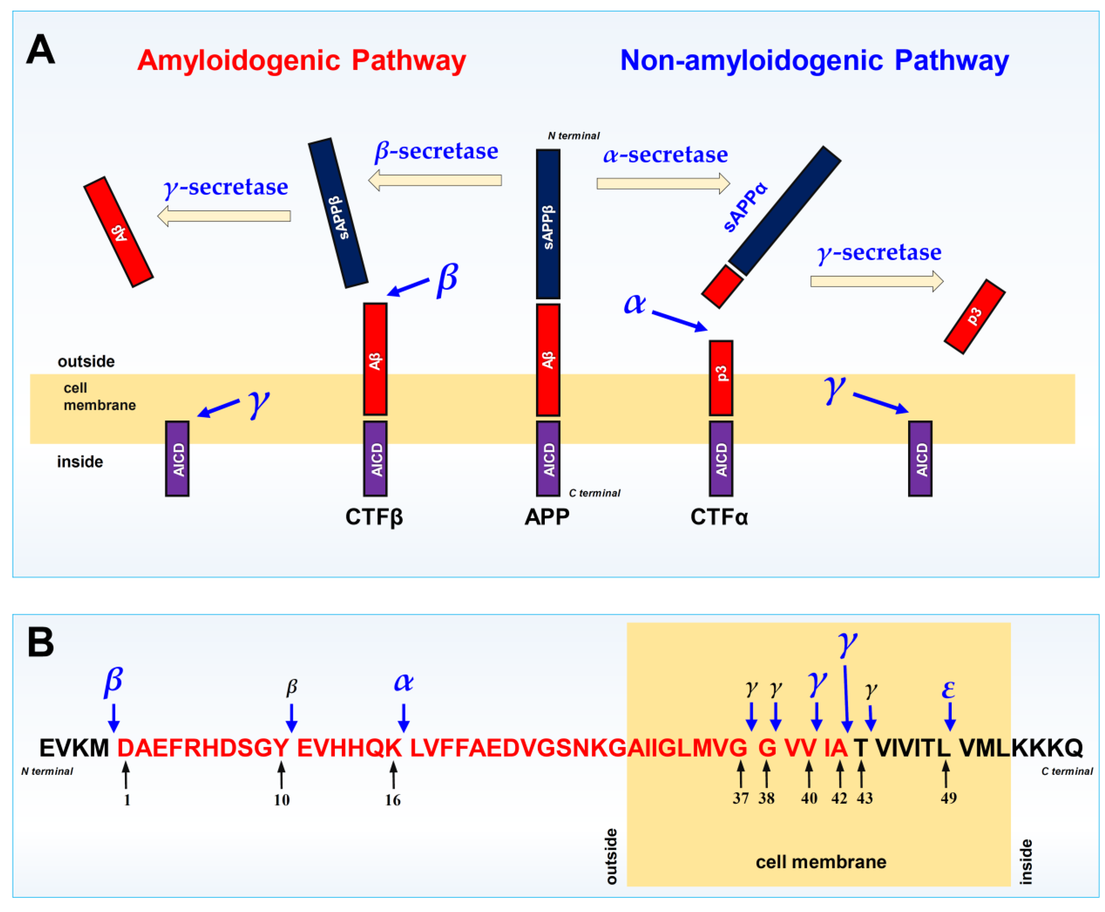
Figure 7.
Role of astrocytic TREM2 in mediating the transcytosis of Aβ-bound ApoE lipoprotein particles from the cerebrospinal fluid (CSF) to the peripheral side. As depicted, the extracellular domain (ECD) of the TREM2 protein plays a crucial role in the binding interaction with the Aβ fragments that are tightly bound to the ApoE lipoprotein particles. The is binding interaction between the two will initiate the internalization (endocytosis) of the Aβ-bound ApoE lipoprotein particles. In addition, TREM2 plays an even more crucial role in mediating the fusion of the ApoE lipoprotein particle with peripheral side of the cell membrane, i.e., carrying out the release of the particles to the peripheral side. When TREM2 has loss-of-function mutations, the exiting process will be stopped, and it will result in the accumulation of the Aβ-bound ApoE lipoprotein particles inside astrocytes. As these ApoE lipoprotein particles are loaded with cholesterol, it is expected that the astrocytes will have abnormally-high levels of cholesterol inside, which will inhibit the ATP synthesis, and will also activate the development of the disease-associated microglia (DAM) phenotype, which is an AD signature phenotype. These changes will also facilitate neuroinflammation. (Note that in the diagram, only ApoE is drawn. It is believed that ApoE and ApoJ may be jointly present in the lipoprotein particles, and the presence of both ApoE and ApoJ may facilitate the process of transcytosis from the CSF side to the peripheral side.).
Figure 7.
Role of astrocytic TREM2 in mediating the transcytosis of Aβ-bound ApoE lipoprotein particles from the cerebrospinal fluid (CSF) to the peripheral side. As depicted, the extracellular domain (ECD) of the TREM2 protein plays a crucial role in the binding interaction with the Aβ fragments that are tightly bound to the ApoE lipoprotein particles. The is binding interaction between the two will initiate the internalization (endocytosis) of the Aβ-bound ApoE lipoprotein particles. In addition, TREM2 plays an even more crucial role in mediating the fusion of the ApoE lipoprotein particle with peripheral side of the cell membrane, i.e., carrying out the release of the particles to the peripheral side. When TREM2 has loss-of-function mutations, the exiting process will be stopped, and it will result in the accumulation of the Aβ-bound ApoE lipoprotein particles inside astrocytes. As these ApoE lipoprotein particles are loaded with cholesterol, it is expected that the astrocytes will have abnormally-high levels of cholesterol inside, which will inhibit the ATP synthesis, and will also activate the development of the disease-associated microglia (DAM) phenotype, which is an AD signature phenotype. These changes will also facilitate neuroinflammation. (Note that in the diagram, only ApoE is drawn. It is believed that ApoE and ApoJ may be jointly present in the lipoprotein particles, and the presence of both ApoE and ApoJ may facilitate the process of transcytosis from the CSF side to the peripheral side.).
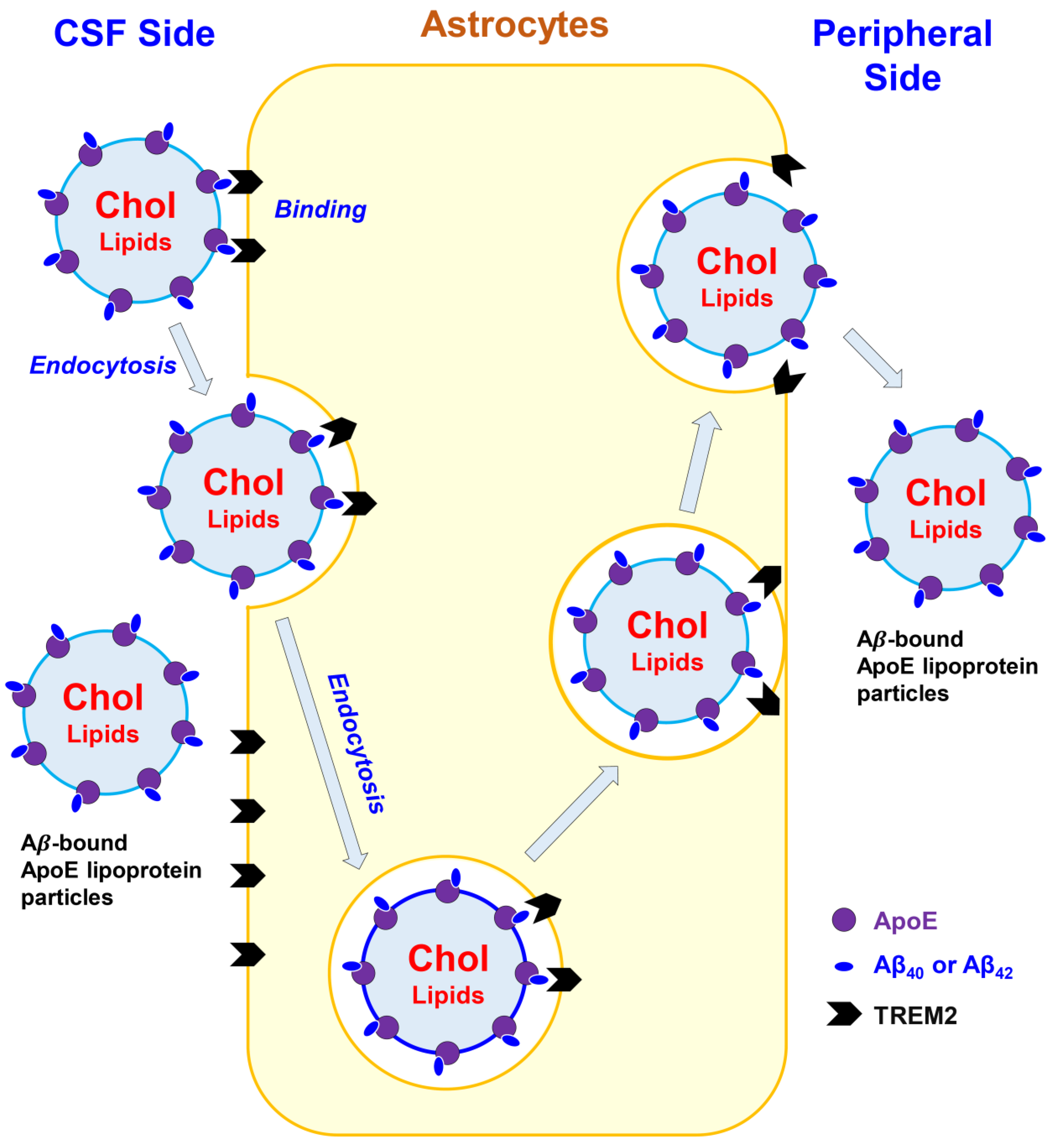
Disclaimer/Publisher’s Note: The statements, opinions and data contained in all publications are solely those of the individual author(s) and contributor(s) and not of MDPI and/or the editor(s). MDPI and/or the editor(s) disclaim responsibility for any injury to people or property resulting from any ideas, methods, instructions or products referred to in the content. |
© 2026 by the authors. Licensee MDPI, Basel, Switzerland. This article is an open access article distributed under the terms and conditions of the Creative Commons Attribution (CC BY) license (http://creativecommons.org/licenses/by/4.0/).
Copyright: This open access article is published under a Creative Commons CC BY 4.0 license, which permit the free download, distribution, and reuse, provided that the author and preprint are cited in any reuse.


