

Submitted:
12 October 2026
Posted:
13 October 2026
You are already at the latest version
Abstract
For decades, classical anatomical dogma has dictated that "without the kidney, the renal fascia cannot form." However, this premise was based on historical anatomical specimens lacking both the kidneys and adrenal glands, likely reflecting broad developmental field defects. In this study, congenital unilateral renal agenesis was examined as a natural "subtraction experiment" to test the mechanical model of retroperitoneal fascial lamination. In three adult cases identified from 5,509 unenhanced abdominal CT scans, despite the lifelong absence of the kidney, a distinct fascial plane corresponding to the parietal lamina (outer layer) of the renal fascia was consistently preserved. Uniform thinning (mean 1.52 mm on the agenesis side vs. 1.85 mm on the contralateral healthy side) was observed, supporting the selective absence of the organ-dependent "inner lamina" and the autonomous formation of the systemically derived "outer lamina." By integrating fetal embryology, materials science, and mechanobiology, we hypothesize that fascial lamination is driven by a strict three-step mechanobiological sequence. First, the primitive jelly-like mesenchyme undergoes a material phase transition into an isotropic poroelastic solid during the mid-trimester (15–20 weeks). Second, coupled with human-specific early trunk stiffening (evolutionary front-loading), this mature matrix unlocks the capacity to respond to multiaxial tension fields. Third, tension-induced Poisson compression may trigger massive fluid expulsion (syneresis), collapsing the isotropic matrix into anisotropic 2D fascial sheets. We outline a two-stage alignment model consisting of this passive Poisson compression followed by active mechanotransduction. Furthermore, we propose a conceptual causal loop wherein fascial sheets structurally primed by fetal tension fields are functionally co-opted (exapted) to disperse and transmit gravitational loads encountered during postnatal upright bipedalism. These findings suggest that retroperitoneal fascial morphogenesis is governed not merely by local organ-dependent mechanisms, but also by systemic biomechanical fields and material phase transitions.
Keywords:
poisson compression
; retroperitoneal fascia
; renal agenesis
; mechanobiology
; fetal development
; parietal lamina
1. Introduction
Although the retroperitoneal fascia exhibits a multi-layered structure and forms critical anatomical boundaries (interfascial planes) for surgical dissection and disease extension, the mechanobiological factors driving its lamination remain incompletely understood (Ishikawa et al., 2006; Sato, 1988, 1995; Tandler, 1923; Wedel et al., 2022). Classical and modern anatomical models have established that the renal fascia consists of a two-layered structure: an "inner lamina" directly enclosing the renal parenchyma and an outer "(parietal) lamina" (Gerota, 1895; Marks et al., 1986; Raptopoulos et al., 1986; Toldt, 1879). However, the systemic mechanics shaping them remain unclear.
Traditionally, fascial formation has been explained by local organ migration and peritoneal fusion. However, while the anatomical fixation of organs is completed by 5 to 12 gestational weeks, distinct fascial layers—as observed in the abdominopelvic region—become clearly recognizable several weeks later, in specimens from around 20 gestational weeks (mid-trimester) onward (Cho et al., 2009; Fritsch, 1993; Fukuoka et al., 2019; Kinugasa et al., 2008; Matsubara et al., 2009). This temporal interval is supported by multiple independent fetal histological studies examining distinct anatomical regions. Matsubara et al. (2009) examined 12 human fetuses and reported that while primitive mesenchymal condensation around the kidney was present at 9–12 gestational weeks (3 fetuses, crown-rump length [CRL] 40–65 mm), lamination of the lateroconal fascia and ureteral sheath was absent; conversely, in all examined specimens at 20–25 weeks (9 fetuses, CRL 152–220 mm), the lateroconal fascia became clearly recognizable as a multilaminar structure, with the posterior renal fascia establishing medial continuity with the great vessel and nerve sheaths. Although specimens from 13–19 weeks were not available in their series, the contrast between the early and late groups suggests that recognizable fascial lamination emerges during this developmental interval. Similarly, in an investigation of the retropancreatic fusion fascia (fascia of Treitz) across 24 fetuses (evaluating 3 fetuses each at 9, 11, 12, 15, 16, 20, 25, and 30 gestational weeks; CRL 90–280 mm), Cho et al. (2009) observed that although retroperitoneal fixation of the pancreas and duodenum occurs around 10 weeks, loose cellular mesenchymal boundaries accompanied by lymphatic follicles persisted through 15–16 weeks, whereas dense collagenous fascial planes became established from 20 weeks onward (20–30 weeks, 9 fetuses). Likewise, in the lumbosacral and pelvic regions, Kinugasa et al. (2008) examined 9 fetuses and demonstrated that poorly defined mesenchymal boundaries at 7–8 weeks (2 fetuses, CRL 23–35 mm) and 10–12 weeks (2 fetuses, CRL 50–67 mm) were transformed into distinct fascial compartments sandwiching the hypogastric nerve at 20–25 weeks (5 fetuses, CRL 152–180 mm). Fritsch (1993) similarly described a mid-trimester transition from a loose mesenchymal period to a fibrous period. Importantly, this fibrous transition may represent not the final step, but the material maturation of the primitive mesenchyme into an isotropic poroelastic solid, establishing the physical prerequisite for subsequent mechanically driven lamination. Collectively, these observations suggest a developmental pattern in which loose mesenchymal boundaries persist through 15–16 weeks and clearly recognizable fascial planes emerge from 20 gestational weeks onward, several weeks after initial organ fixation. This temporal gap suggests that local organ migration or adhesion alone is unlikely to fully account for macroscopic fascial morphogenesis during the mid-trimester. Recent high-resolution 3D micro-CT studies consistently reveal that pelvic fascial architecture is characterized by mechanical compression and interaction rather than simple embryonic fusion. Both Yang et al. (2021) and In et al. (2026) demonstrated that structures like Denonvilliers' fascia consist of multiple layers that are physically compressed within confined anatomical spaces.
Historically, Tobin (1944) and Benjamin and Tobin (1951) established the interpretation that "without the kidney, the renal fascia itself fails to form," based on anomalous anatomical specimens lacking both the kidneys and adrenal glands. However, those specimens likely represented a developmental field defect (Opitz, 1985) rather than isolated renal agenesis. For decades, this prevailing interpretation strongly influenced anatomical thought, leaving the mechanical origins of fascial lamination open to further exploration.
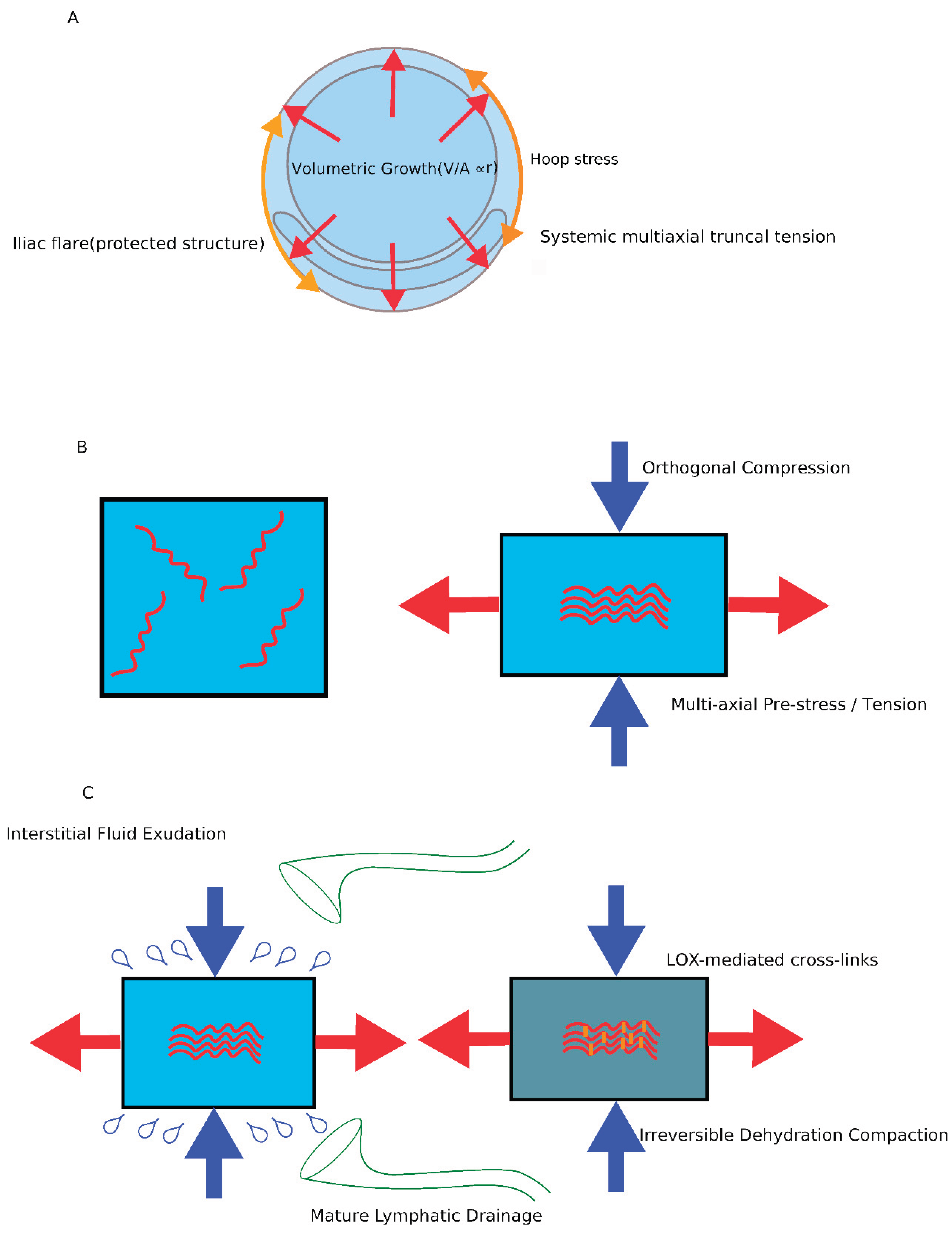
Fetal somatic growth is not merely a process of cellular proliferation. During development, skeletal, muscular, visceral, neural, and mesenchymal tissues do not expand at identical rates. Such differential growth may generate mechanical incompatibilities within the integrated fetal body plan. As development proceeds, these growth-associated stresses are progressively amplified by geometric scaling, whereby volumetric and ponderal expansion (V∝r3) outpaces surface area expansion (S∝r2) toward mid-gestation, as evidenced by standard fetal growth trajectories (Hadlock et al., 1991). Together, these processes may contribute to the emergence of a systemic multiaxial tension field within the water-rich fetal mesenchyme.
Congenital unilateral renal agenesis provides a unique "natural subtraction experiment" to isolate the mechanical variables of fascial development amidst this tension. If fascial lamination relied entirely on organ-driven compaction, the renal fascia would be completely absent on the agenesis side. Conversely, if a systemic tension field were involved, a baseline fascial plane would persist. In this study, we observed the morphological status of the retroperitoneal fascia in adult patients with unilateral renal agenesis.
To precisely interpret these morphological findings and bridge the gap between macroscopic physical tension and microscopic biological structures, we define the following operational terms used in this study:
- Inner lamina: A fascial layer presumed to be formed by local radial pressure (hoop stress) from the expanding kidney (organ-dependent).
- Outer (parietal) lamina / system-derived lamina: A tension-bearing layer hypothesized to form in response to macroscopic, systemically derived mechanical fields rather than local organ expansion.
- Outer fascial laminae complex: The anatomical continuum encompassing the parietal lamina of the posterior renal fascia, the fascia of Toldt (retrocolic fascia), and the fascia of Treitz (retroduodenal fascia) (Sato, 1995; Wedel et al., 2022).
- Evolutionary front-loading: A conceptual evolutionary heterochrony model we propose, describing the hominin-specific heterochronic shift wherein skeletal stiffening of the trunk occurs at an earlier gestational age compared to quadrupedal mammals (Weisbecker et al., 2008).
- Tension-dependent ECM alignment: The critical biological mechanism bridging macro-physics and micro-morphology, whereby mechanical stretch actively guides fibroblasts to reconstruct and align collagen fibers.
2. Materials and Methods
2.1. Study Design and Patient Selection
This retrospective observational study involved a systematic screening of 5,509 consecutive adult unenhanced abdominal CT scans performed at Gakkentoshi Hospital between April 2018 and March 2024. Inclusion criteria strictly selected cases of true congenital unilateral renal agenesis or severe renal dysplasia/regression (less than 3 cm) with no history of surgical intervention or significant trauma in the retroperitoneum, and where the adrenal gland was visibly preserved on CT (to exclude broad developmental field defects).
2.2. Imaging Evaluation
The presence, continuity, and thickness of the fascia were evaluated on axial images using 3D Slicer v5.10 (Fedorov et al., 2012). The display was optimized using specific abdominal soft-tissue window settings (width: 250 HU, level: 150 HU). Slice thickness ranged from 1.0 mm to 5.0 mm (median 3.0 mm). Standard soft-tissue kernels were used for reconstruction. Given the slice thickness of clinical CT and the associated partial volume effects, these measurements represent macroscopic X-ray attenuation profiles reflecting fascial continuity and tissue boundaries rather than true histological micro-measurements. To ensure measurement reproducibility, the fascial thickness on the agenesis side was recorded at a representative axial level—specifically, at the mid-hilar level of the contralateral healthy kidney—where the preserved renal fascia could be most clearly observed and measured orthogonally.
2.3. Ethical Considerations
This study was approved by the Institutional Ethics Committee of Gakkentoshi Hospital (Approval No. GT-R6-07-12-1). Written informed consent was waived due to the retrospective nature of the study, and an opt-out mechanism was provided through the official hospital website.
3. Results
3.1. Clinical Cohort Characteristics
From the 5,509 screened scans, three adults met the inclusion criteria (Table 1). Case 1 (53-year-old, female) and Case 3 (89-year-old, male) exhibited true congenital left renal agenesis. Notably, Case 1 presented with a pathologically characteristic "pancake-shaped" (discoid) adrenal morphology (Kenney et al., 1985), whereas Case 3 showed a linear and flattened adrenal morphology, likely reflecting a combination of the missing renal mold and age-related changes. Case 2 (47-year-old, female) presented with severe left renal dysplasia/regression with normal adrenal morphology. The contralateral side of Case 3 was excluded from comparative measurement due to fascial thickening secondary to pyelonephritis.
Table 1. Clinical characteristics and quantitative radiological findings in 3 adult cases with congenital renal agenesis or severe dysplasia.
- Case 1: 53F / True Left Renal Agenesis / Pancake-shaped Adrenal Morphology / Fascial Thickness (Affected): 1.49 mm / Fascial Thickness (Healthy): 1.88 mm / Δ (mm): -0.39
- Case 2: 47F / Severe Left Renal Dysplasia/Regression / Normal Adrenal Morphology / Fascial Thickness (Affected): 1.46 mm / Fascial Thickness (Healthy): 1.82 mm / Δ (mm): -0.36
- Case 3: 89M / True Left Renal Agenesis / Linear (Atrophic) Adrenal Morphology / Fascial Thickness (Affected): 1.62 mm / Fascial Thickness (Healthy): Excluded* / Δ (mm): N/A
- Mean: Fascial Thickness (Affected): 1.52 mm / Fascial Thickness (Healthy): 1.85 mm / Δ (mm): -0.38 (Note: *The contralateral side of Case 3 was excluded due to fascial thickening secondary to pyelonephritis. Δ (mm): Difference in fascial thickness between affected and healthy sides.)
3.2. Preservation and Continuity of the Parietal Lamina
In all three cases (3/3), objective image analysis revealed a continuous macroscopic fascial plane on the affected side. This plane was consistently observed in the anatomical region corresponding to the parietal lamina of the posterior renal fascia (Figure 1). Despite the complete and lifelong absence of the renal parenchyma, this preserved structure extended from the lateral lateroconal fascia to its anchor points along the medial psoas major or quadratus lumborum muscles, maintaining clear anatomical continuity. This observation aligns closely with the hypothesis that the system-derived outer lamina forms autonomously, independent of the kidney's presence.
3.3. Quantitative Evaluation of Fascial Thickness
Quantitative comparison revealed a consistent reduction in fascial thickness on the renal agenesis side. The mean fascial thickness on the affected side was 1.52 mm, compared to 1.85 mm on the contralateral healthy side (mean difference: -0.38 mm) (Table 1). This uniform thinning is illustrated in Figure 2 and provides morphological data consistent with the dual-origin model, reflecting a combination of the selective loss of the organ-dependent inner lamina and the autonomous preservation of the system-derived outer lamina.
4. Discussion
4.1. Interpretation of the Subtraction Analysis
Why is the parietal lamina (outer layer) preserved despite the complete absence of the kidney? The answer lies in the distinct mechanical origins of the fascial layers: while the inner lamina is strictly organ-dependent, the outer lamina is autonomously formed by a systemic multiaxial tension field.
The uniform thinning (approximately 1.52 mm) observed in our renal agenesis cohort is consistent with the selective aplasia of the organ-dependent inner lamina and the autonomous preservation of the system-derived outer lamina. This radiological finding also aligns directly with clinical surgical observations during laparoscopic colectomy in patients with congenital renal agenesis, where a recognizable retroperitoneal dissection plane (the continuum of Toldt's fusion fascia and the parietal lamina) is preserved despite the absence of the kidney (Tanishima et al., 2013).
Fundamentally, the classical dogma that fascial formation is entirely "organ-dependent" encounters a logical contradiction when explaining Gerota's fascia, which encapsulates both the kidney and the adrenal gland. If local organ expansion were the sole driving force, the integration of these embryologically distinct organs into a single package would be impossible to explain. Crucially, the adrenal gland in this context is not acting as a local compressive "organ-dependent driver." Rather, the connective tissue matrix of the adrenal-gonadal system merely functions as a "histological marker" denoting that the cranial-dorsal developmental field is preserved. Furthermore, unlike the radially expanding solid renal parenchyma, the compliant and highly remodeling nature of the developing fetal adrenal cortex is unlikely to generate the sustained, isotropic hoop stress required to form the inner lamina. Therefore, the outer lamina forms not "because the adrenal gland exerts pressure," but because the systemic multiaxial tension field is maintained within an uncompromised developmental field.
Importantly, one might argue that this preserved fascial plane is merely a non-specific boundary formed by a passive mass effect from retroperitoneal adipose tissue filling the renal fossa or from adjacent intestinal loops. However, the discoid "pancake-shaped" (or flat linear) adrenal morphology (Kenney et al., 1985) observed in our agenesis cases directly refutes this alternative interpretation (Figure 1). If a significant local compressive mass effect or visceral crowding were the primary driving force, the compliant adrenal gland would be irregularly compressed, deformed, or displaced rather than symmetrically flattened along the body wall. This flattened adrenal morphology serves as a direct morphological trace of the complete absence of local outward organ expansion and its exposure to a systemic tension field acting along the posterior abdominal wall.
4.2. Historical Synthesis and Explanatory Asymmetry
This clear distinction between the "absence of an organ" and a "defect in the developmental field" provides a cohesive framework to resolve a century of anatomical debate. When Gerota (1895) first described the renal fascia, his original illustration clearly depicted the fascia as a two-layer structure. However, the spatial and mechanical significance of his drawing was overlooked by subsequent anatomists, reducing the fascia to a simple "single sac."
In congenital unilateral renal agenesis, ipsilateral adrenal agenesis occurs in only about 11% of cases (Ashley & Mostofi, 1960). Nevertheless, Tobin (1944) and Benjamin and Tobin (1951) based their conclusions on specific specimen illustrations from this rare minority—which likely represented true developmental field defects lacking both kidneys and adrenal glands (Opitz, 1985), preventing the formation of the outer lamina network entirely. Derived from these specific observations, Tobin's interpretation likely reflected developmental field defects rather than isolated renal agenesis.
The true robustness of the Poisson Compression Theory (PCT) lies in its explanatory asymmetry when compared to classical peritoneal fusion theories. While the anatomical evidence presented by fusion theorists—such as the continuous Y-shaped reflections derived from the peritoneum (Li et al., 2023) or the tight adhesion of Denonvilliers' fascia to the prostate capsule necessitating sharp dissection (Garcia-Gausi et al., 2022)—can be seamlessly reinterpreted within the PCT framework as the endpoint of extreme orthogonal compression and poroelastic syneresis, the converse is fundamentally impossible. Classical fusion models, which rely solely on local organ-to-organ adhesion, completely fail to explain the anomalous phenomena highlighted by the PCT: the preservation of the parietal lamina in the total absence of a kidney (renal agenesis), the significant temporal gap wherein fascial lamination occurs weeks after organ fixation, and the mechanical lateral exclusion of neurovascular bundles (NVB). Therefore, PCT offers a framework capable of accommodating the morphological observations of classical models while providing plausible explanations for several unresolved biomechanical paradoxes.
4.3. Spatial Paradox in Tissue Lamination
(In vitro Physics) Classical mechanobiological models of tissue lamination, beginning with Stopak and Harris (1982), have broadly assumed an active cellular paradigm in which localized fibroblasts actively exert traction forces to pattern macroscopic ECM orientation. However, this assumption introduces a spatial paradox: how could local cells perceive a global coordinate system without preexisting guidance? This micro-scale paradox was fundamentally resolved by Vader et al. (2009), who demonstrated that uniaxial strain buckles collagen fibers orthogonal to the tension axis, promoting passive compaction that guides initial cell alignment. Ban et al. (2019) mathematically described this phenomenon as an "anomalous Poisson effect" in homogeneous collagen networks.
(In vivo Translation) However, bridging the gap between these homogeneous in vitro models and the highly heterogeneous in vivo environment of a human fetus requires a macro-scale tension source and a tissue matrix capable of undergoing syneresis. During early development (prior to 14 weeks), the fetal mesenchyme exists as an unstructured, highly viscoelastic, hyaluronic acid–rich gel—conceptually described as a "white tablet" (Pirri et al., 2022; Toole, 2001). In this primitive jelly-like state, the tissue lacks an interconnected collagen network (Voronoi structure), making macroscopic poroelastic fluid expulsion (syneresis) physically impossible.
Between approximately 15 and 20 gestational weeks, fetal connective tissue enters the fibrous period (Fritsch, 1993). Driven by mechanical stimulation from fetal movement (Nowlan, 2015; Verbruggen et al., 2018) and biochemical shifts during this interval, the mesenchyme undergoes a fundamental material phase transition into an isotropic poroelastic solid (Chaudhuri et al., 2020; Mow et al., 1980; Pirri et al., 2022). Specifically, this developmental window marks a critical transition wherein the highly hydrated, loose mesenchyme matures and reorganizes into a load-bearing extracellular matrix dominated by type I and type III collagen fiber bundles (Smith et al., 1986; Stecco et al., 2017; Toole, 2001). Only after this isotropic porous scaffold is completed does the tissue acquire the capacity to undergo dramatic internal pressure spikes and fluid expulsion in response to macroscopic tension.
Coupled with the progressive ossification of the axial skeleton that confines the rapid volumetric expansion of the viscera (Nowlan, 2015; Senevirathne et al., 2025; Verbruggen et al., 2018), this biochemical shift transforms the retroperitoneal space into a structurally integrated continuum capable of propagating long-range tensile stress. The resulting multiaxial tension forces orthogonal compression via the Poisson effect onto the newly matured, water-rich retroperitoneal mesenchyme (Figure 3).
The physical footprint of this phenomenon was documented by Kim et al. (2015) and He et al. (2022) in the pelvic retroperitoneum. They demonstrated that Denonvilliers' fascia emerges mechanically in later fetal stages, accompanied by a characteristic lateral displacement of the pelvic plexus and neurovascular bundle (NVB) at its lateral border. Interpreted through our framework, this lateral shift is consistent with a mechanical exclusion effect driven by orthogonal squeezing from anomalous Poisson compression. Crucially, this combined effect of systemic compression and local organ expansion may provide a mechanical explanation for why the NVB in adult anatomy is not a discrete 3D cord, but rather a widely distributed, flattened mesh-like network that envelops the entire prostate, as clearly demonstrated by Costello et al. (2004). The neural mesenchyme may have been constrained by orthogonal compression associated with fascial maturation and local organ expansion, forcing its redistribution into a flattened two-dimensional network rather than a localized bundle.
From the perspective of macroscopic mechanics, the phenomenon traditionally described in developmental biology as biochemical "mesenchymal condensation" or hyaluronic acid degradation (Toole, 2001) is likely functionally two sides of the same coin with physical fluid expulsion (syneresis) driven by anomalous Poisson compression. In this integrated view, the intense physical stress and internal pressure surge generated by growth-associated Poisson compression act as the primary trigger for mechanotransduction, prompting fibroblasts to secrete hyaluronidase and cross-linking enzymes (Kagan & Li, 2003; Stecco et al., 2017). This mechanochemical coupling synchronizes the biochemical breakdown and physical collapse of the gel matrix, seamlessly establishing a "two-stage alignment model" consisting of initial passive physical squeezing followed by active fibroblast-mediated enzymatic stabilization.
4.4. Mechanochemical Coupling in Fascial Lamination
In developmental biology, mesenchymal condensation has traditionally been explained by programmed extracellular matrix remodeling and hyaluronic acid degradation; however, the upstream mechanisms coordinating these events across entire tissues remain incompletely understood. The present model proposes that increasing fetal mechanical loads—specifically, anomalous Poisson compression associated with mid-trimester skeletal stiffening—may directly trigger this matrix remodeling via mechanotransduction pathways. Within this framework, biochemical condensation and biomechanical syneresis are viewed not as competing mechanisms, but as integrally coupled elements of a common developmental process driving fascial lamination. Further research is required to determine whether macroscopic compressive loading directly governs hyaluronic acid metabolism, matrix-remodeling enzymes, and the subsequent spatial organization of fascial sheets.
4.5. Emergence of a Systemic Tension Field and a Conceptual Causal Loop
Beyond Local Organ-Centered Models The conspicuous appearance of retroperitoneal fascial lamination around mid-gestation has long been difficult to reconcile with models based solely on local organ migration or peritoneal fusion. Across the three independent fetal anatomical studies described in the Introduction—despite examining different anatomical regions (the perirenal and lateroconal region, the retropancreatic space, and the pelvic hypogastric sheath)—a remarkably similar developmental sequence is observed. Organ migration and fixation progress during approximately 9–12 weeks, transitional loose mesenchymal stages persist through 15–16 weeks, and clearly recognizable fascial lamination emerges from around 20 weeks onward (Cho et al., 2009; Kinugasa et al., 2008; Matsubara et al., 2009). This sequence also aligns with the developmental staging proposed by Fritsch (1993), who described a mid-trimester transition from a mesenchymal to a fibrous period. This temporal dissociation—wherein fascial planes become distinct several weeks after organ fixation—suggests that fascial lamination may reflect a higher-order mechanical process or phase transition rather than a simple consequence of local positional changes or adhesion (Figure 4). We hypothesize that the fibrous period (15–20 weeks) prepares an isotropic poroelastic scaffold, and that crossing a mechanical threshold during mid-gestation contributes to its structural reorganization into 2D sheets.
Furthermore, the present subtraction analysis demonstrated preservation of a continuous fascial plane corresponding to the parietal lamina despite the lifelong absence of the kidney, which normally represents the major local source of retroperitoneal expansion. While local organ enlargement may contribute to the formation of the organ-dependent inner lamina, the persistence of the outer fascial component indicates that its development is unlikely to depend exclusively on the presence of the kidney itself.
Mechanical Model and Emergence of a Systemic Tension Field We propose that fascial lamination during mid-gestation may be associated with the emergence of a systemic tension field generated by differential growth among developing tissues.
During fetal development, skeletal, muscular, neural, visceral, and mesenchymal tissues do not expand at identical rates. Such spatial and temporal heterogeneity in growth may create mechanical incompatibilities among tissues that remain physically integrated within a single continuous body plan. Developmental biomechanics studies increasingly recognize differential growth as a major driver of tissue stress generation and morphogenesis.
This mechanical imbalance may be further amplified by geometric scaling during fetal growth (Hadlock et al., 1991). As fetal body volume (and weight) increases in proportion to the cube of linear dimensions—expanding nearly sixfold between 12 and 20 gestational weeks while surface area increases only threefold with the square of linear dimensions—the mechanical demands imposed on internal supporting tissues progressively escalate with size. In this framework, the square-cube relationship is viewed not as the primary source of tension itself, but rather as a geometric scaling factor that amplifies growth-induced mechanical disequilibrium.
Concurrently, progressive skeletal stiffening and extracellular matrix maturation during approximately 15–20 weeks of gestation (Smith et al., 1986) may alter the mechanical boundary conditions of the fetal trunk. As tissue rigidity increases, growth-generated stresses become increasingly transmitted across broader anatomical regions rather than remaining locally dissipated. We hypothesize that this transition permits the emergence of a body-wide tension network, which subsequently imposes transverse compressive forces on the hydrated retroperitoneal mesenchyme through the Poisson effect. Evolutionary front-loading is therefore interpreted not as the primary generator of mechanical stress, but as a developmental event that increases boundary rigidity and permits locally generated growth stresses to become integrated into a body-wide tension network (Figure 5).
Integration with the In Vivo Observations of Kim et al. A potentially important anatomical observation supporting this framework was reported by Kim et al. (2015) and He et al. (2022). In their anatomical studies of Denonvilliers' fascia, the authors documented the emergence of a distinct fascial structure during late fetal development accompanied by a characteristic lateral displacement of the neurovascular bundle (NVB) and pelvic plexus. They further described a conspicuous linear organization of the surrounding mesenchymal tissue.
Traditionally, these findings have been interpreted primarily as descriptive anatomical observations. Within the framework of PCT, however, they may also be viewed as signatures of mechanical tissue reorganization induced by orthogonal compression. Specifically, the lateral displacement of the NVB is consistent with a mechanical exclusion effect, whereby developing fascial layers spatially reorganize adjacent structures during progressive tissue compaction.
Moreover, the linear alignment observed by Kim et al. shows notable conceptual correspondence with the strain-induced collagen alignment described by Vader et al. (2009) and with the anomalous Poisson effect observed in collagen networks by Ban et al. (2019). In these experimental systems, tensile loading generates orthogonal matrix compaction and directional collagen reorganization. The fetal observations reported by Kim et al. therefore provide a potentially important in vivo analogue linking experimentally observed Poisson-related phenomena to fascial morphogenesis in the developing human body.
Establishment of a Conceptual Causal Loop D'Arcy Thompson famously stated that "form is a diagram of forces" (Thompson, 1917). Within the present framework, the extensive continuity of the retroperitoneal fascial system may be interpreted as a macroscopic record of systemic tension fields that operated during fetal development.
We further propose that these structures subsequently acquire a secondary functional role after birth. Fascial sheets generated under prenatal tension may later function as part of a body-wide tensional network (tensegrity architecture), participating in the transmission and distribution of longitudinal gravitational loads associated with upright bipedal posture (Ingber, 2003; Schleip et al., 2012). In this view, fetal tension fields contribute to the formation of fascial architecture, whereas the resulting architecture subsequently functions to resist and distribute mechanically analogous loads encountered throughout postnatal life. This relationship suggests a "conceptual causal loop" in which developmental mechanical forces shape fascial morphology, and the resulting morphology later serves as an adaptive structural network for force transmission.
4.6. Integration of Avascular Plane Formation and Pelvic Extension
Tension-induced Poisson compression extends to the pelvic retroperitoneum. We propose that surgical avascular fields can be understood within a common mechanical spectrum formed by the interaction of Poisson compression and shear stress. First, extreme compression promotes syneresis, forming dense fascial sheets, while valleys of compression retain hydration to form a 3D mesh (foamy layer). The macroscopic manifestation of this extreme compression is consistently observed in surgical practice. For instance, Garcia-Gausi et al. (2022) reported in their cadaveric simulation that while loose areolar planes exist in the cranial portion, Denonvilliers' fascia becomes firmly and indistinguishably adhered to the prostate capsule in the narrower caudal space, necessitating sharp dissection. This clinical observation perfectly aligns with the outcome of poroelastic syneresis driven by severe spatial constraint and orthogonal compression.
Second, continuous shear stress induced by organ motion acts as a "mechanical pruning" mechanism against neoangiogenesis at this boundary, maintaining a highly dynamic, water-rich gliding interface facilitated by specialized hyaluronan-secreting cells (fasciacytes) (Stecco et al., 2018). This interaction logically explains regional structural differences, from the robust perirenal space envelope to the loosely defined "Holy plane" in the presacral space (Dogaru et al., 2026; Li et al., 2023). Viewed through the lens of PCT, anatomical densification is consistent with macroscopic manifestations of poroelastic syneresis.
4.7. Comparative Anatomy and Evolutionary Heterochrony
Lamination of the retroperitoneal fascia is prominently developed in humans. While the canine retroperitoneum remains a highly compliant connective tissue mesh (Jeong et al., 2016), our preliminary observations in gorillas confirmed a fascial plane corresponding to the outer lamina, though less distinct and lacking pronounced macroscopic anisotropy compared to humans. To protect laterally expanded iliac bones necessary for bipedalism, humans require early trunk stiffening. By integrating findings on mammalian ossification heterochrony (Weisbecker et al., 2008), rapid mid-trimester fetal ossification and skeletal stress (Nowlan, 2015; Verbruggen et al., 2018), and human pelvic evolution (Senevirathne et al., 2025), we propose a conceptual evolutionary heterochrony model (evolutionary front-loading): an earlier transition of the human trunk into a rigid, closed system compared to quadrupeds. This shift amplifies mechanical stress in the mesenchyme, driving anisotropic lamination.
4.8. Speculative Evolutionary Hypothesis: Prenatal Structural Priming of the Suspension System
Robust fusion to the posterior body wall became evolutionarily indispensable to prevent visceral ptosis under the vertical gravitational vectors unique to human bipedalism (Southam, 1923). Our model provides a mechanical hypothesis for this pre-configuration: human front-loading confines fetal kinetic energy within a stiffened trunk, converting it into multiaxial tension to drive Poisson compression. Through this process, a mechanically competent suspension architecture may be structurally primed in the pseudo-microgravity amniotic environment by approximately the 20th gestational week, synchronizing with the onset of explosive encephalization (Sakai et al., 2013). While this evolutionary framework remains conceptual, it provides a testable biomechanical hypothesis for future cross-species ontogenetic studies.
4.9. Limitations
The primary limitation of this study is its small sample size (n=3), dictated by the extreme rarity of unoperated adult congenital unilateral renal agenesis. Furthermore, observing adult anatomy inevitably includes decades of postnatal mechanical remodeling (e.g., fascial adaptation to bipedal posture and aging). While the preserved fascial planes strongly reflect their embryonic developmental origins, the precise thickness and microstructure may have been modified by continuous mechanotransduction over the patients' lifetimes. Additionally, the retrospective use of clinical CT scans introduces spatial resolution constraints (slice thickness of 1.0–5.0 mm), limiting microstructural evaluation and necessitating macroscopic interpretations. Therefore, measurements should be interpreted as macroscopic optical profiles reflecting fascial persistence rather than true histological dimensions. Furthermore, anomalous Poisson compression and ECM heterogeneity during fetal development cannot be directly measured in vivo, rendering the proposed mechanical sequence theoretical. Finally, while preliminary observations in non-human primates were introduced, a comprehensive cross-species imaging analysis remains a subject for future investigation.
5. Conclusions
By utilizing congenital unilateral renal agenesis as a natural subtraction experiment, this study suggests that retroperitoneal fascial lamination is unlikely to be explained solely by organ-dependent compaction. We observed radiological evidence consistent with a dual-origin model: while the organ-dependent inner lamina is absent without the kidney, the system-derived outer (parietal) lamina is autonomously preserved, provided the developmental field remains intact. We propose that this morphology reflects a three-step mechanobiological sequence: the primitive jelly-like mesenchyme first matures into an isotropic poroelastic solid, unlocking the capacity for tension-induced Poisson compression and syneresis. Driven by growth-associated systemic tension fields, amplified by geometric scaling, and facilitated by early skeletal stiffening (evolutionary front-loading), this compression may physically remodel the mesenchyme into continuous 2D fascial sheets.
Ultimately, this framework presents a conceptual causal loop where multiaxial tension generated in utero sculpts the fascial form, which is later functionally co-opted (exapted) to transmit and disperse the gravitational loads of postnatal upright bipedalism.
Declaration of Generative AI and AI-assisted technologies in the writing process
During the preparation of this work, the author used Microsoft Copilot and Google Gemini to assist with language refinement and structural editing. After using these tools, the author reviewed and edited the content as needed and takes full responsibility for the content of the publication.
Author Contributions
H.T. conceived and designed the study, performed the radiological screening and image analysis, developed the biomechanical framework, and wrote the entire manuscript.
Funding
This research received no specific grant from any funding agency in the public, commercial, or not-for-profit sectors.
Institutional Review Board Statement
This study was conducted in accordance with the Declaration of Helsinki and approved by the Institutional Ethics Committee of Gakkentoshi Hospital (Approval No. GT-R6-07-12-1).
Informed Consent Statement
Due to the retrospective nature of this study, patient consent was waived, and an opt-out mechanism was provided through the hospital website.
Data Availability Statement
The radiological image datasets generated and analyzed during the current study are not publicly available due to patient privacy protection but are available from the corresponding author upon reasonable academic request.
Acknowledgments
The author expresses deep gratitude to colleagues in the Department of Urology at Gakkentoshi Hospital for fostering a collaborative clinical environment. Sincere appreciation is also extended to the radiology staff for their technical assistance. The author is deeply grateful to the Tokyo Zoological Park Society (Ueno Zoological Gardens) and Shizuoka Municipal Nihondaira Zoo for providing gorilla CT data and institutional support.
Conflicts of Interest
The author declares no conflicts of interest.
References
- Ashley, D.J.B.; Mostofi, F.K. Renal agenesis and dysgenesis. J. Urol. 1960, 83(3), 211–230. [Google Scholar] [CrossRef] [PubMed]
- Ban, E.; Wang, H.; Franklin, J.M.; Liphardt, J.T.; Janmey, P.A.; Shenoy, V.B. Strong triaxial coupling and anomalous Poisson effect in collagen networks. Proc. Natl. Acad. Sci. USA 2019, 116(14), 6790–6799. [Google Scholar] [CrossRef] [PubMed]
- Benjamin, J.A.; Tobin, C.E. Abnormalities of the kidneys, ureters, and perinephric fascia: Anatomic and clinical study. J. Urol. 1951, 65(5), 715–731. [Google Scholar] [CrossRef] [PubMed]
- Chaudhuri, O.; Cooper-White, J.; Janmey, P.A.; Mooney, D.J.; Shenoy, V.B. Effects of extracellular matrix viscoelasticity on cellular behaviour. Nature 2020, 584(7822), 535–546. [Google Scholar] [CrossRef] [PubMed]
- Cho, B.H.; Kimura, W.; Song, C.H.; Fujimiya, M.; Murakami, G. An investigation of the embryologic development of the fascia used as the basis for pancreaticoduodenal mobilization. J. Hepato-Biliary-Pancreat. Surg. 2009, 16(6), 824–831. [Google Scholar] [CrossRef] [PubMed]
- Costello, A.J.; Brooks, M.; Cole, O.J. Anatomical studies of the neurovascular bundle and cavernosal nerves. BJU Int. 2004, 94(7), 1071–1076. [Google Scholar] [CrossRef] [PubMed]
- Dogaru, I.A.; Tulin, A.D.; Slavu, I.M.; Gheoca Mutu, D.E.; Enyedi, M.; Stănciulescu, R.; Panțu, C.M.; Filipoiu, Z.F. Morphogenesis and Topography of the Mesorectal Fascia. J. Clin. Med. 2026, 15(4), 1377. [Google Scholar] [CrossRef] [PubMed]
- Fedorov, A.; Beichel, R.; Kalpathy-Cramer, J.; Finet, J.; Fillion-Robin, J.C.; Pujol, S.; Bauer, C.; Jennings, D.; Fennessy, F.M.; Sonka, M.; Buatti, J.; Aylward, S.R.; Miller, J.V.; Pieper, S.; Kikinis, R. 3D Slicer as an image computing platform for the Quantitative Imaging Network. Magn. Reson. Imaging 2012, 30(9), 1323–1341. [Google Scholar] [CrossRef] [PubMed]
- Fritsch, H. Development and organization of the pelvic connective tissue in the human fetus. Ann. Anat. 1993, 175(6), 531–539. [Google Scholar] [CrossRef] [PubMed]
- Fukuoka, K.; Wilting, J.; Rodríguez-Vázquez, J.F.; Murakami, G.; Ishizawa, A.; Matsubara, A. Intermediate morphological changes during renal ascent in human embryos. Anat. Rec. 2019, 302(2), 278–287. [Google Scholar]
- Garcia-Gausi, M.; Garcia-Armengol, J.; Pellino, G.; Mulas, C.; Roig, J.V.; Garcia-Granero, A.; Moro, D.; Valverde, A. Navigating surgical anatomy of the Denonvilliers' fascia and dissection planes of the anterior mesorectum with a cadaveric simulation model. Updat. Surg. 2022, 74(2), 629–636. [Google Scholar] [CrossRef] [PubMed]
- Gerota, D. Beiträge zur Kenntnis des Befestigungsapparates der Niere. Arch. Für Anat. Und Entwicklungsgeschichte 1895, 19, 265–286. [Google Scholar]
- Hadlock, F.P.; Harrist, R.B.; Martinez-Poyer, J. In utero analysis of fetal growth: A sonographic weight standard. Radiology 1991, 181(1), 129–133. [Google Scholar] [CrossRef] [PubMed]
- He, M.; Muro, S.; Akita, K. Positional relationship between the lateral border of Denonvilliers' fascia and pelvic plexus. Anat. Sci. Int. 2022, 97(1), 101–109. [Google Scholar] [CrossRef] [PubMed]
- In, S.Y.; Cho, T.H.; Yang, H.M. 3D micro-CT and immunofluorescence-based characterization of Denonvilliers’ fascia and its neurovascular structures. Sci. Rep. 2026, 16, 18506. [Google Scholar] [CrossRef] [PubMed]
- Ingber, D.E. Tensegrity I. Cell structure and hierarchical systems biology. J. Cell Sci. 2003, 116(7), 1157–1173. [Google Scholar] [CrossRef] [PubMed]
- Ishikawa, K.; Idoguchi, K.; Tanaka, H.; Tohma, Y.; Ukai, I.; Watanabe, H.; Matsuoka, T.; Yokota, J.; Sugimoto, T. Classification of acute pancreatitis based on retroperitoneal extension: Application of the concept of interfascial planes. Eur. J. Radiol. 2006, 60(3), 445–452. [Google Scholar] [CrossRef] [PubMed]
- Jeong, J.; Ko, J.; Lim, H.; Kweon, O.; Kim, W.H. Retroperitoneal laparoscopy in dogs: Access technique, working space, and surgical anatomy. Vet. Surg. 2016, 45(S1), O102–O110. [Google Scholar] [CrossRef] [PubMed]
- Kagan, H.M.; Li, W. Lysyl oxidase: Properties, specificity, and biological roles inside and outside of the cell. J. Cell. Biochem. 2003, 88(4), 660–672. [Google Scholar] [CrossRef] [PubMed]
- Kenney, P.J.; Robbins, G.L.; Ellis, D.A.; Spirt, B.A. Adrenal glands in patients with congenital renal anomalies: CT appearance. Radiology 1985, 155(1), 181–182. [Google Scholar] [CrossRef] [PubMed]
- Kim, J.H.; Kinugasa, Y.; Hwang, S.E.; Murakami, G.; Cho, B.H. Denonvilliers' fascia revisited. Surg. Radiol. Anat. 2015, 37(2), 187–197. [Google Scholar] [CrossRef] [PubMed]
- Kinugasa, Y.; Niikura, H.; Murakami, G.; Suzuki, D.; Saito, S.; Tatsumi, H.; Ishii, M. Development of the human hypogastric nerve sheath with special reference to the topohistology between the nerve sheath and other prevertebral fascial structures. Clin. Anat. 2008, 21(6), 558–567. [Google Scholar] [CrossRef] [PubMed]
- Li, Y.; Zhao, Y.M.; Ma, Y.B.; Chen, Z.J.; Xu, C.; Liu, C.Y.; Yang, Z.W.; Wang, X.S. The characteristics of the urogenital fascia in the retrorectal space based on male cadaveric dissection and its clinical application. BMC Surg. 2023, 23(1), 93. [Google Scholar] [CrossRef] [PubMed]
- Marks, S.C., Jr.; Raptopoulos, V.; Kleinman, P.; Snyder, M. The anatomical basis for retrorenal extensions of pancreatic effusions: The role of the renal fasciae. Surg. Radiol. Anat. 1986, 8(2), 89–97. [Google Scholar] [CrossRef] [PubMed]
- Matsubara, A.; Murakami, G.; Niikura, H.; Kinugasa, Y.; Fujimiya, M.; Usui, T. Development of the human retroperitoneal fasciae. Cells Tissues Organs 2009, 190(5), 286–296. [Google Scholar] [CrossRef] [PubMed]
- Mow, V.C.; Kuei, S.C.; Lai, W.M.; Armstrong, C.G. Biphasic creep and stress relaxation of articular cartilage in compression: Theory and experiments. J. Biomech. Eng. 1980, 102(1), 73–84. [Google Scholar] [CrossRef] [PubMed]
- Nowlan, N.C. Biomechanics of foetal movement. Eur. Cells Mater. 2015, 29, 1–21. [Google Scholar] [CrossRef] [PubMed]
- Opitz, J.M. The developmental field concept. Am. J. Med. Genet. 1985, 21(1), 1–11. [Google Scholar] [CrossRef] [PubMed]
- Pirri, C.; Petrelli, L.; Pérez-Bellmunt, A.; Ortiz-Miguel, S.; Fede, C.; De Caro, R.; Miguel-Pérez, M.; Stecco, C. Fetal fascial reinforcement development: from “a white tablet” to a sculpted precise organization by movement. Biology 2022, 11(5), 735. [Google Scholar] [CrossRef] [PubMed]
- Raptopoulos, V.; Kleinman, P.K.; Marks, S.; Snyder, M.; Silverman, P.M. Renal fascial pathway: Posterior extension of pancreatic effusions within the anterior pararenal space. Radiology 1986, 158(3), 653–658. [Google Scholar] [CrossRef] [PubMed]
- Sakai, T.; Matsui, M.; Mikami, A.; Malkova, L.; Hamada, Y.; Tomonaga, M.; Suzuki, J.; Tanaka, M.; Miyabe-Nishiwaki, T.; Makishima, H.; Nakatsukasa, M.; Matsuzawa, T. Developmental patterns of chimpanzee cerebral tissues provide important clues for understanding the remarkable enlargement of the human brain. Proc. R. Soc. B Biol. Sci. 2013, 280(1753), 20122398. [Google Scholar] [CrossRef] [PubMed]
- Sato, T. I. Kidney B. Renal Fascia. Rinsho Hinyokika (in Japanese). 1988, 42(8), 689–696. [Google Scholar]
- Sato, T. Topographical anatomy of visceral fascia: The basic composition of single-layer structure and differentiation in each region. Nihon Rinsho Geka Igakkai Zasshi (in Japanese). 1995, 56(11), 2253–2272. [Google Scholar]
- Schleip, R.; Findley, T.W.; Chaitow, L.; Huijing, P.A. Fascia: The Tensional Network of the Human Body; Churchill Livingstone / Elsevier: Edinburgh, 2012. [Google Scholar]
- Senevirathne, G.; Fernandopulle, S.C.; Richard, D.; Baumgart, S.L.; Christensen, A.L.; Fabbri, M.; et al. The evolution of hominin bipedalism in two steps. Nature 2025, 645, 952–963. [Google Scholar] [CrossRef] [PubMed]
- Smith, L.T.; Holbrook, K.A.; Madri, J.A. Collagen types I, III, and V in human embryonic and fetal skin. Am. J. Anat. 1986, 175(4), 507–521. [Google Scholar] [CrossRef] [PubMed]
- Southam, A.H. The fixation of the kidney. QJM An. Int. J. Med. 1923, 16(64), 283–308. [Google Scholar] [CrossRef]
- Stecco, C.; Sfriso, M.M.; Porzionato, A.; Rambaldo, A.; Albertin, G.; Macchi, V.; De Caro, R. Microscopic anatomy of the visceral fasciae. J. Anat. 2017, 231(1), 121–128. [Google Scholar] [CrossRef] [PubMed]
- Stecco, C.; Fede, C.; Macchi, V.; Porzionato, A.; Petrelli, L.; Biz, C.; Stern, R.; De Caro, R. The fasciacytes: A new cell devoted to fascial gliding regulation. Clin. Anat. 2018, 31(5), 667–676. [Google Scholar] [CrossRef] [PubMed]
- Stopak, D.; Harris, A.K. Connective tissue morphogenesis by fibroblast traction. I. Tissue culture observations. Dev. Biol. 1982, 90(2), 383–398. [Google Scholar] [PubMed]
- Tandler, J. Lehrbuch der systematischen Anatomie. 2. Band: Die Eingeweide; F.C.W. Vogel: Leipzig, 1923. [Google Scholar]
- Tanishima, H.; Horiuchi, T.; Kimura, M.; Shono, Y. Laparoscopic colectomy for a patient with congenital renal agenesis. J. Surg. Tech. Case Rep. 2013, 5(1), 51–53. [Google Scholar] [CrossRef] [PubMed]
- Thompson, D.W. On Growth and Form; Cambridge University Press: Cambridge, 1917. [Google Scholar]
- Tobin, C.E. The renal fascia and its relation to the transversalis fascia. Anat. Rec. 1944, 89(3), 295–311. [Google Scholar] [CrossRef]
- Toldt, C. Bau und Wachsthumsveränderungen der Gekröse des menschlichen Darmkanales. Denkschr. Der Kais. Akad. Der Wiss. 1879, 41, 1–56. [Google Scholar]
- Toole, B.P. Hyaluronan in morphogenesis. Semin. Cell Dev. Biol. 2001, 12(2), 79–87. [Google Scholar] [CrossRef] [PubMed]
- Vader, D.; Kabla, A.; Weitz, D.; Mahadevan, L. Strain-induced alignment in collagen gels. PLoS ONE 2009, 4(6), e5902. [Google Scholar] [CrossRef] [PubMed]
- Verbruggen, S.W.; Kainz, B.; Shelmerdine, S.C.; Hajnal, J.V.; Rutherford, M.A.; Arthurs, O.J.; Phillips, A.T.M.; Nowlan, N.C. Stresses and strains on the human fetal skeleton during development. J. R. Soc. Interface 2018, 15(138), 20170593. [Google Scholar] [CrossRef] [PubMed]
- Wedel, T.; Heimke, M.; Fletcher, J.; Miskovic, D.; Benz, S.; Stelzner, S.; Heinze, T. The retrocolic fascial system revisited for right hemicolectomy with complete mesocolic excision based on anatomical terminology: Do we need the eponyms Toldt, Gerota, Fredet and Treitz? Colorectal Dis. 2022, 25(4), 764–774. [Google Scholar] [CrossRef] [PubMed]
- Weisbecker, V.; Goswami, A.; Wroe, S.; Sánchez-Villagra, M.R. Ossification heterochrony in the therian postcranial skeleton and the marsupial-placental dichotomy. Evolution 2008, 62(8), 2027–2041. [Google Scholar] [CrossRef] [PubMed]
- Yang, S.Y.; Kim, H.S.; Cho, M.S.; Kim, N.K. Three-dimensional anatomy of the Denonvilliers’ fascia after micro-CT reconstruction. Sci. Rep. 2021, 11(1), 21759. [Google Scholar] [CrossRef] [PubMed]
Figure 1.
Evidence of mechanical preservation of the system-derived parietal lamina in congenital renal agenesis. Unenhanced axial CT image of Case 1 (53-year-old female) with left renal agenesis. Despite the complete and lifelong absence of the kidney and ureter, a distinct and continuous hyperattenuating fascial plane (arrows) is consistently observed in the anatomical position corresponding to the renal fascia. This plane courses smoothly between the peritoneal sac and the posterior abdominal wall, maintaining typical continuity with the lateroconal fascia. On the healthy contralateral side (inset), the fascial complex is thicker, reflecting the presence of both the organ-dependent inner lamina and the system-derived outer lamina. On the affected side, a thin yet clearly identifiable single fascial plane (approximately 1.5 mm) is confirmed, aligning with the selective absence of the inner lamina and preservation of the outer lamina proposed by the tension-driven mechanobiological model.
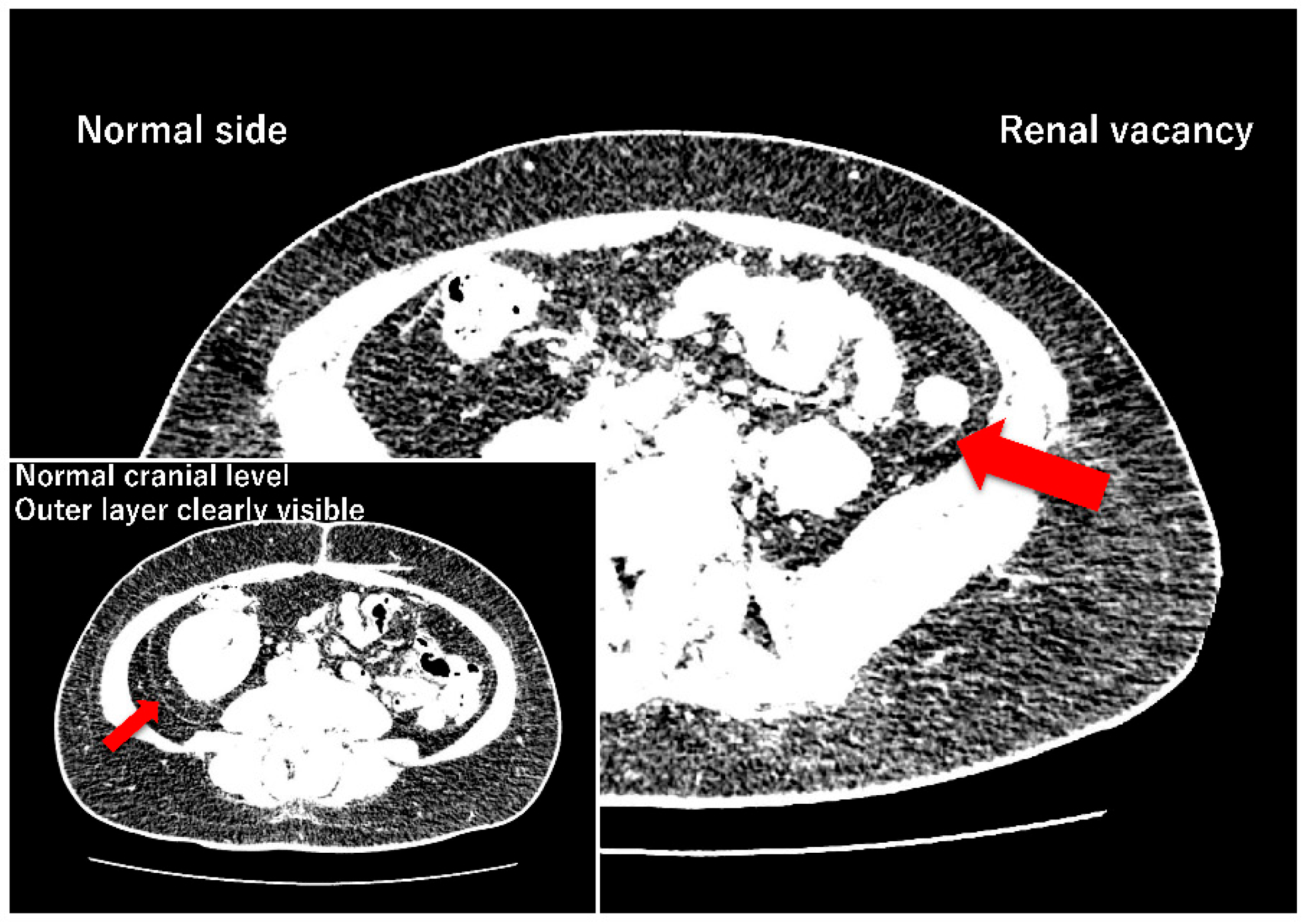
Figure 2.
Proposal of a dual-mechanism model for retroperitoneal fascial lamination. Left: Early renal expansion (9–12 weeks) generates circumferential hoop stress, compacting the adjacent primitive jelly-like mesenchyme to form the anlage of the organ-dependent "inner lamina." Subsequently, alongside ECM maturation into a poroelastic solid during the mid-trimester (15–20 weeks), sustained renal expansion (hoop stress) further compacts this layer, completing it as the inner lamina. In renal agenesis lacking the local hoop stress source, this layer fails to develop. Right: After the mesenchyme undergoes a material phase transition into an isotropic poroelastic solid, system-level multiaxial tension emerging from differential growth and amplified geometric scaling around 20 gestational weeks induces tension-induced Poisson compression (orthogonal compression). This physical sequence remodels the retroperitoneal mesenchyme into the "outer (parietal) lamina" independent of renal expansion. The single preserved fascial plane observed in our renal agenesis cohort is consistent with this system-level mechanical phase transition.
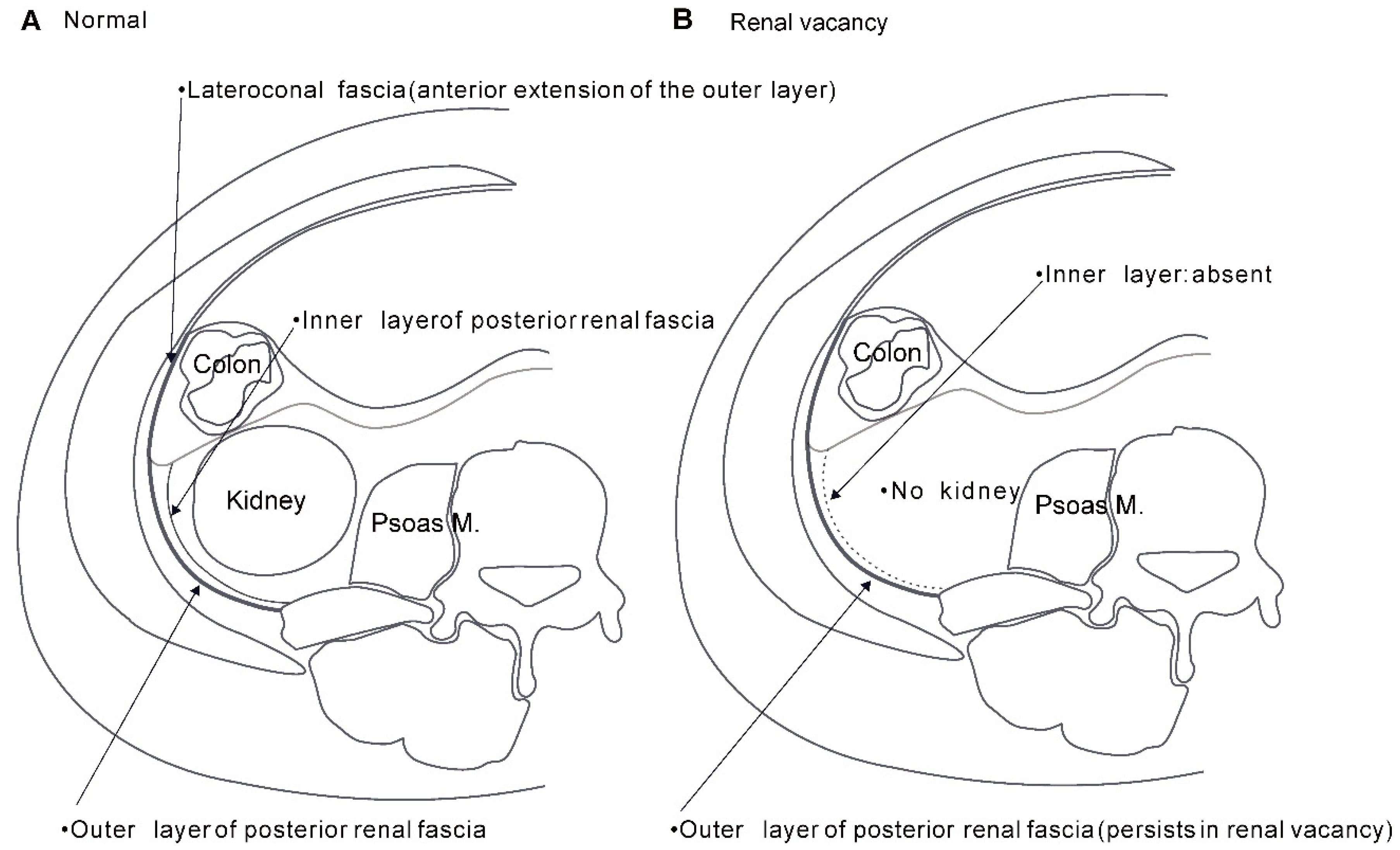
Figure 3.
Theoretical comparison of tissue deformation: Direct compression vs. Poisson effect. (A) Simple direct compression: The classical assumption that local compressive force from an expanding organ crushes mesenchymal tissue without generating a regular layered sheet, resulting in chaotic tissue compaction. (B) Hoop stress-induced Poisson effect: Circumferential tension (hoop stress) from an expanding mass is likely to induce orthogonal (transverse) compression via the Poisson effect, contributing to the regular aggregation of mesenchymal tissue into the characteristic dense layered structure enveloping the renal fascia.
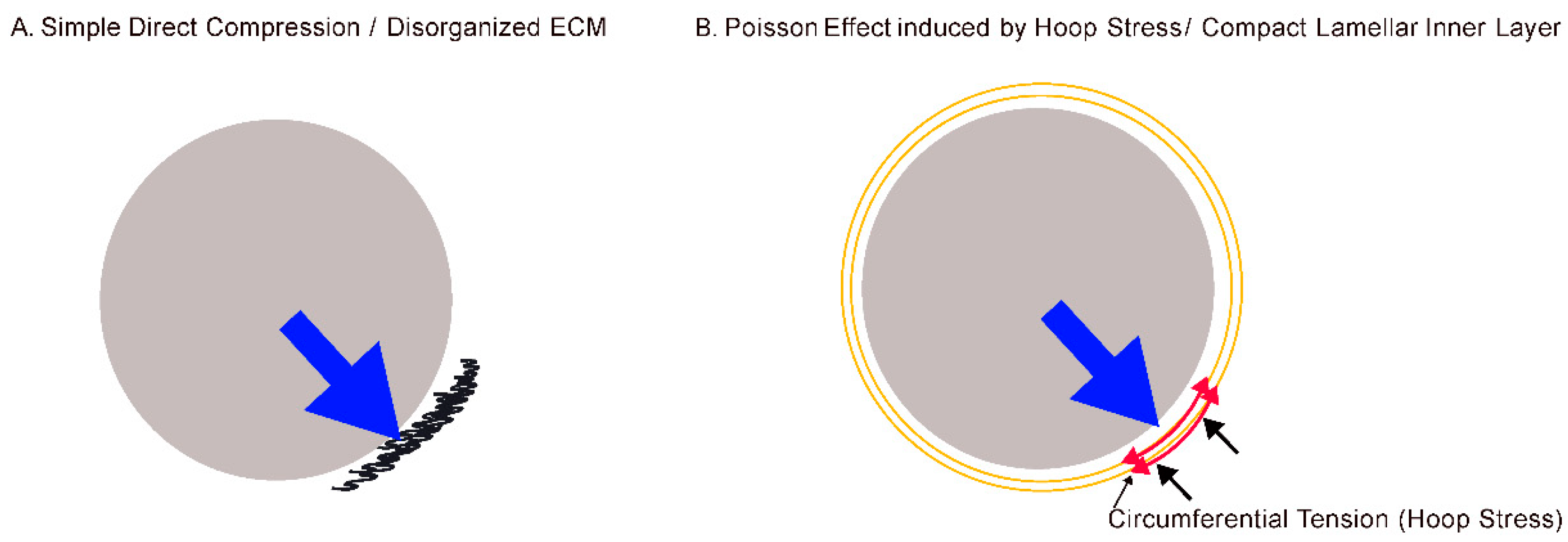
Figure 4.
Spatiotemporal and biomechanical asymmetry in retroperitoneal fascial development. This diagram highlights the temporal divergence in fascial emergence. In Phase 1 (9–12 gestational weeks), medial and lateral mechanical anchors (fixation points) are established. During this initial stage (Panel A), driven by local hoop stress from the expanding kidney, the inner lamina of the renal fascia is presumed to form. In contrast, Phase 2 (approx. 20 gestational weeks; Panel B) represents the systemic mechanical threshold in the mid-trimester, at which point the newly matured isotropic poroelastic scaffold allows the multi-layered "outer lamina" to become macroscopically recognizable across the completed macroscopic tension network between the central and lateral organ anchors. This temporal gap is consistent with the dual-mechanism model, indicating that the inner and outer laminae arise from distinct mechanical origins.
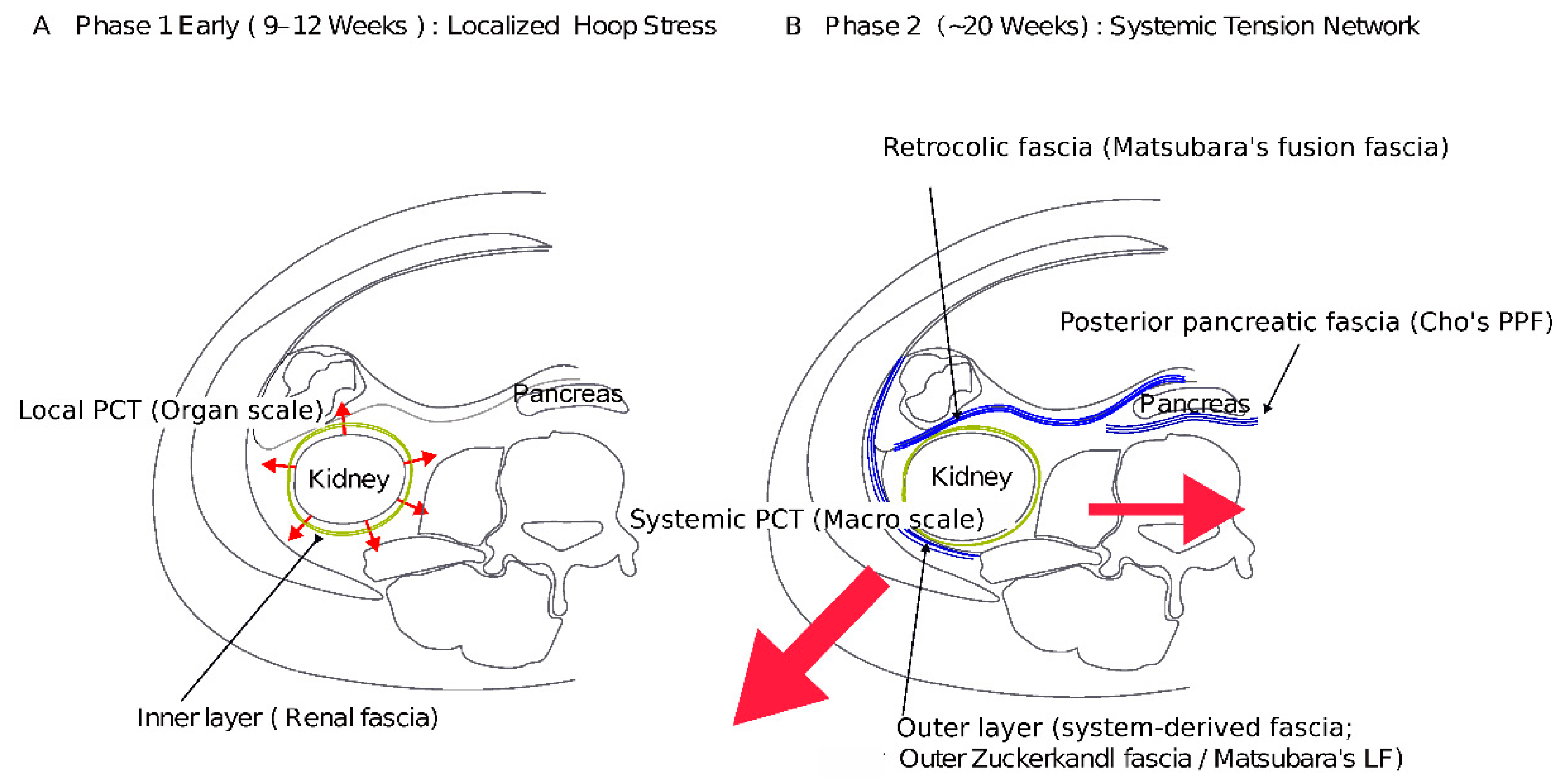
Figure 5.
Emergence of the fetal tension network and theoretical lamination driven by differential growth, geometric scaling, and Poisson compression. (A) Differential growth and stress amplification: Differential growth among skeletal, visceral, and mesenchymal tissues generates mechanical incompatibilities. As development proceeds, geometric scaling (V/S∝r) progressively amplifies these growth-associated stresses. (B) Systemic tension integration: As fetal growth progresses, the mesenchyme matures into a poroelastic solid. Concurrently, increasing boundary rigidity associated with evolutionary front-loading permits locally generated growth stresses to integrate into a systemic multiaxial tension field, producing orthogonal compression through the Poisson effect within the newly matured, water-rich retroperitoneal mesenchyme. (C) Compaction and fixation: This internal mechanical compression expels poroelastic fluid into the maturing lymphatic system. Simultaneously, fibroblast-mediated ECM remodeling and cross-linking stabilize the collapsed collagen framework into a distinct, highly ordered fascial structure. LOX, lysyl oxidase. These sequential mechanical steps delineate a compelling pathway for how systemic growth scaling is converted into permanent, tension-aligned fascial sheets.
Disclaimer/Publisher’s Note: The statements, opinions and data contained in all publications are solely those of the individual author(s) and contributor(s) and not of MDPI and/or the editor(s). MDPI and/or the editor(s) disclaim responsibility for any injury to people or property resulting from any ideas, methods, instructions or products referred to in the content. |
© 2026 by the authors. Licensee MDPI, Basel, Switzerland. This article is an open access article distributed under the terms and conditions of the Creative Commons Attribution (CC BY) license (http://creativecommons.org/licenses/by/4.0/).
Copyright: This open access article is published under a Creative Commons CC BY 4.0 license, which permit the free download, distribution, and reuse, provided that the author and preprint are cited in any reuse.