
Submitted:
26 June 2026
Posted:
01 July 2026
You are already at the latest version
Abstract
The developmental basis of retroperitoneal fascial lamination remains unresolved, as classical peritoneal fusion theories and fat-compaction models cannot fully explain the consistent formation of the anterior and posterior renal fasciae or their behavior in cases of congenital renal agenesis. To clarify the underlying mechanobiology, we conducted a retrospective radiological analysis of unenhanced computed tomography (CT) scans, including rare cases of unilateral renal agenesis, interpreting fascial configurations within a framework incorporating tension-driven lamination, orthogonal Poisson compression, and subtraction-based reasoning. Across all cases, a continuous fascial plane was unequivocally preserved at the predicted anatomical location of the parietal lamina of the posterior renal fascia despite the lifelong absence of the kidney. However, the renal-vacant side consistently exhibited reduced total fascial thickness (mean 1.52 mm vs. 1.85 mm). This asymmetric thinning aligns with the selective absence of the organ-dependent inner lamina and the preservation of a system-derived parietal lamina established during mid-gestation. From a comparative developmental perspective, whereas quadrupedal precocial mammals preferentially allocate fetal movement energy to appendicular stiffening while maintaining a compliant trunk (heterochrony), humans front-load truncal stiffening (an evolutionary shift toward earlier-than-expected ossification timing) to protect a geometrically vulnerable pelvic architecture associated with obligate bipedalism. The convergence of this human-specific "early-completed truncal box" and powerful fetal movement energy concentrates orthogonal Poisson compression (a biological "ironing" effect) in the retroperitoneum, triggering synchronous multilaminar sheet formation. These findings support an evolutionary and mechanobiological model in which the laminated architecture of the retroperitoneal fascia emerges from the interplay of geometric scaling, the human-specific timing of skeletal stiffening, and multiaxial tension fields.
Keywords:
fascia
; retroperitoneal space
; mechanobiology
; heterochrony
; poisson effect
; tensegrity
; evolutionary front-loading
; renal agenesis
; parietal lamina of the posterior renal fascia
1. Introduction
The developmental mechanisms underlying the multilaminar architecture of the retroperitoneal fascia have been debated for over a century. Classical frameworks—including Toldt’s (1879) peritoneal fusion hypothesis and the fixation apparatus models of Zuckerkandl (1883) and Gerota (1895)—provided early morphological descriptions but failed to mechanically explain how and why the highly organized, bilaminar posterior renal fascia emerges. Integrating early cross-sectional imaging with cadaveric liquid latex injection and macroscopic dissection, Raptopolous et al. (1986) demonstrated that the posterior renal fascia consists of two distinct laminae, closely matching Gerota’s original illustrations. Yet, the fundamental question persists: what drives the emergence of these highly organized fascial layers?
In prior research, Matsubara et al. (2009) suggested that early renal parenchymal expansion compresses the adjacent mesenchyme. Although the term "hoop stress" was not explicitly used, this phenomenon is mechanically equivalent to localized Poisson compression and elegantly explains the formation of the inner lamina. Conversely, the development of the parietal (outer) lamina has traditionally been attributed to in situ tissue compaction driven by the expansion of primitive perirenal adipose tissue. However, these localized mechanisms are insufficient to explain the synchronous emergence of highly ordered, multilaminar planar sheets across the entire retroperitoneum at gestational week 20.
First, localized fat compaction lacks the systemic mechanical basis required to generate the comprehensive, sharply demarcated macroscopic compartmentalization originally illustrated by Zuckerkandl (1883) and Gerota (1895). Second, macro-mechanical comparisons with quadrupedal models further highlight these theoretical limitations. As demonstrated by Jeong et al. (2016), the canine retroperitoneum behaves as a continuous, highly compliant connective tissue mesh. Under retroperitoneoscopic conditions, CO2 insufflation at a minimal pressure of 5 mmHg results in rapid, isotropic fluid dissipation across the midline. This fluid-dynamic behavior stands in stark contrast to the human retroperitoneum, which exhibits pronounced anisotropy, retaining and channeling pressure along predefined, two-dimensional fascial planes.
The most decisive counter-evidence arises from the radiological subtraction analysis of congenital renal agenesis. In all examined cases (3/3), a distinct, continuous fascial plane positionally corresponding to the parietal lamina of the posterior renal fascia was unequivocally preserved, despite the lifelong absence of renal parenchyma and its associated adipose dynamics. This autonomy indicates that the parietal lamina is not a passive consequence of localized organ crowding, as presumed by the Matsubara model, but instead reflects a broader mechanobiological process—specifically, a system-wide multiaxial tension field emerging at gestational week 20 that induces an obligatory, systemic Poisson compression.
Accordingly, the present study proposes a unified mechanobiological model integrating the organ-dependent local Poisson effect (inner lamina) and the frame-dependent systemic Poisson effect (outer lamina) into a single overarching physical principle.
Recent advances in fetal biomechanics provide a crucial conceptual foundation for understanding this transition. Nowlan (2015) demonstrated that the fetal musculoskeletal system undergoes a fundamental shift around gestational week 20—from a compliant, pressure-driven continuum to a mechanically active, tension-bearing frame.
We hypothesize that the missing mechanistic link is a genetically timed mechanical convergence. Based on standard fetal growth curves (Hadlock et al., 1991), the internal fetal volume increases nearly sixfold between weeks 12 and 20, whereas the enclosing surface area only triples. This predictable geometric escalation—conceptually expressed as a rapidly increasing volume-to-surface-area ratio (V/A∝r)—imposes an exponentially rising internal load on the fetal trunk. As genetically programmed skeletal elements (e.g., vertebrae, ribs, and uniquely expanding iliac blades) begin to stiffen, the trunk can no longer dissipate this load isotropically. Instead, it transitions into a tension-bearing architecture, generating a systemic multiaxial tension field.
This tension field acts upon the hydrated retroperitoneal mesenchyme, which is anchored medially by the duodenum and pancreas (weeks 9–12) and laterally by the ascending and descending mesocolon (weeks 17–18). Stretching this anchored mesenchyme inevitably induces orthogonal Poisson compression. This compression drives poroelastic fluid exudation and collapses the collagen framework into discrete laminae. Fibroblast traction and subsequent lysyl-oxidase–mediated cross-linking stabilize these mechanically aligned layers, producing a primitive tensegrity network wherein stiffening skeletal elements act as compression-resistant struts and the aligned fascial planes function as tension-bearing cables.
A key unresolved question is whether the outer lamina of the posterior renal fascia depends on the expanding kidney or instead reflects this broader tension field. Congenital renal agenesis provides a natural “subtraction experiment” to resolve this dichotomy.
Finally, comparative developmental anatomy suggests that this mid-gestational mechanical transition may be evolutionarily front-loaded in humans (Verbruggen & Nowlan, 2017; Senevirathne et al., 2025; Weisbecker et al., 2008). Unlike most quadrupedal mammals, which maintain a compliant trunk until late gestation ("back-loading"), humans possess a uniquely laterally flared ilium essential for bipedalism but structurally vulnerable to isotropic pressure. We hypothesize that human ontogeny has shifted the timing of frame-locking forward—an evolutionary front-loading defined as earlier-than-expected structural ossification—to protect this fragile geometry.
To operationalize this structural configuration within our mechanical framework, we collectively define the term “outer fascial laminae” as the anatomical complex comprising the outer (parietal) lamina of the posterior renal fascia, the retrocolic fascia (of Toldt), and the retropancreatic fascia (of Treitz).
Integrating fetal histology, biomechanics, and radiological subtraction analysis, we propose an exploratory mechanobiological model in which the inner lamina of the renal fascia arises from organ-specific hoop stress, whereas the outer fascial laminae (including the parietal lamina) emerge from a systemic tension network established at gestational week 20.
2. Materials and Methods
2.1. Study Design and Conceptual Framework
This study employed a hybrid design integrating (1) a retrospective radiological observational analysis of adult patients with unilateral renal agenesis (a natural subtraction experiment), and (2) a conceptual synthesis of comparative developmental biomechanics in human and non-human mammals.
2.2. Case Selection and Retrospective Radiological Review
A systematic retrospective screening of 5,509 consecutive abdominal unenhanced CT scans performed at a single institution (Gakkentoshi Hospital) between April 2018 and March 2024 was conducted to identify adult patients with unilateral renal vacancy without prior surgical intervention. Inclusion criteria strictly selected for true congenital renal agenesis or severe renal involution (< 3 cm). Fascial thickness was measured at predefined anatomical landmarks on axial images displayed with an optimized window setting (width: 250 HU, level: 150 HU) using 3D Slicer v5.10 (measured by H.T.).
2.3. Literature Review and Embryological Integration
To reconstruct the fetal anatomical and mechanical timeline, we integrated embryological literature focusing on the development of skeletal anchors, qualitative shifts in fetal movement patterns (Verbruggen et al., 2018), and ossification sequence heterochrony in non-human mammals (Weisbecker et al., 2008).
2.4. Ethical Considerations
This retrospective study was approved by the Institutional Review Board of Gakkentoshi Hospital (Approval No. GT-R6-07-12-1). Written informed consent was waived, and an opt-out mechanism was provided via the hospital’s official website.
3. Results
3.1. Radiological Cohort of Renal Agenesis
Among the 5,509 screened scans, three adults met the strict inclusion criteria for unilateral renal vacancy. Cases 1 and 2 demonstrated true congenital renal agenesis (accompanied by the characteristic "lying-down" or pancake adrenal morphology (Hoffman et al., 1992; Potter, 1946)), while Case 3 exhibited a severely involuted dysplastic renal remnant.
3.2. Preservation of the Parietal Lamina of the Posterior Renal Fascia
In all three cases, a continuous macroscopic fascial plane positionally corresponding to the parietal (outer) lamina of the posterior renal fascia was unequivocally preserved at the predicted anatomical location, despite the lifelong absence of renal parenchyma (Figure 1). This plane extended smoothly between the peritoneal sac and the posterior abdominal wall, anchoring reliably to the psoas major or quadratus lumborum, and maintaining continuity with the lateroconal fascia.
3.3. Quantitative Assessment of Fascial Thickness
Quantitative measurements are summarized in Table 1. The mean thickness on the affected (renal-vacant) side was 1.52 mm, compared to 1.85 mm on the contralateral healthy side. This uniform thinning indicates that the organ-dependent inner lamina failed to form, while the system-derived parietal lamina was autonomously maintained.
3.4. Chronological Integration of Fetal Fascial Development and Human-Specific Heterochrony
4. Theoretical Integration and Discussion
4.1. Biological "Ironing" via Orthogonal Poisson Compression
Soft tissues dynamically reorganize their internal architecture in response to sustained mechanical loading (Humphrey, 2003). Conventional models posit that organ pressure or adipose expansion directly squashes the tissue; however, non-directional compression lacks the mechanical capacity to form highly ordered, planar sheets. Conversely, in the highly hydrated, poroelastic retroperitoneal mesenchyme, sustained "multiaxial stretch (tension)" inevitably generates "orthogonal compressive forces" (Fung, 1993). This tension field acts as a biological "ironing" mechanism, driving the exudation of poroelastic fluid (Mow et al., 1980; Swartz & Fleury, 2007) and collapsing the collagen framework into discrete laminae. Subsequently, lysyl-oxidase–mediated cross-linking stabilizes these structures into a permanent tensegrity scaffold (Ingber, 2003). Consistent with this mechanism, histological analyses by Stecco et al. (2017) demonstrate that visceral fasciae exhibit a multilaminar architecture in which collagen bundles are arranged in distinct layers, each with a different fiber orientation, separated by loose connective tissue rich in elastic fibers. This layered, anisotropic organization strongly supports the interpretation that fascial planes arise through tension-dependent remodeling rather than passive adipose expansion. Notably, Stecco et al. identified the renal fascia (Gerota fascia) as one of the thickest and least elastic visceral fasciae, a property consistent with a tension-aligned, load-bearing insertional fascia rather than a product of local adipose compression.
4.2. The Square-Cube Law and the Emergence of Fetal Tensegrity
As somatic growth accelerates, the volume-to-surface-area ratio (V/A∝r) increases, approximately tripling the internal load per unit area between weeks 12 and 20 (Hadlock et al., 1991). To withstand this escalating load, the epidermis keratinizes and the vertebrae and iliac wings stiffen by weeks 18–20. As these anchors rigidify, internal forces can no longer dissipate isotropically, generating a systemic tension field spanning the axial skeleton and the hardened boundary.
A fundamental principle emerging from comparative developmental biology is that the transition from isotropic to anisotropic growth in multicellular organisms is not dictated by cell number or volumetric expansion per se, but by the timing at which the organism acquires a mechanically competent frame capable of sustaining tension. In other words, geometric scaling (V/A∝r) provides the necessary internal load, but anisotropy only emerges once a rigid boundary—such as ossifying vertebrae, ribs, iliac blades, or keratinizing skin—appears early enough to trap and redirect this load. This “structural heterochrony” varies markedly across mammals: precocial quadrupeds delay truncal stiffening to preserve a compliant birth canal and prioritize appendicular ossification, whereas humans front-load truncal frame formation around week 20. Thus, the decisive variable governing laminar morphogenesis is not absolute fetal size but the relative timing between volumetric escalation and the onset of frame-bearing capacity. The human retroperitoneum becomes anisotropically stressed precisely because this timing window is uniquely compressed in our species.
4.3. Resolution of the Historical Controversy: A Natural Subtraction Experiment
The CT analysis of unilateral renal agenesis (n = 3) directly substantiates this dual-mechanism model. In all cases, a continuous fascial plane positionally matching the parietal lamina of the posterior renal fascia was unequivocally preserved despite the lifelong absence of the renal parenchyma. This anatomically proves that while the inner lamina depends on early renal expansion (local hoop stress), the formation of the parietal lamina depends on a system-wide tension field (orthogonal Poisson compression) established at week 20. The historical contradiction of Tobin’s 1944 specimen (which lacked both kidney and adrenal gland) does not negate the existence of a system-derived fascial layer but instead represents a broader mesenchymal field defect. This represents a broader mesenchymal field defect, consistent with Opitz’s developmental field concept (Opitz, 1985).
4.4. Comparison with Non-Human Mammals: Fetal Movement "Quality" and "Heterochrony"
To truly grasp this mechanobiological framework, we must ask why the immense fetal movement energy of large mammals (e.g., horses, elephants) does not form human-like multilaminar fascia. Fetuses of large precocial mammals must bear their own weight and run immediately after birth. Therefore, their mechanical energy from fetal movements is preferentially allocated to the "early stiffening of appendicular long bones and tendons" (Weisbecker et al., 2008). Furthermore, to safely deliver giant fetuses through the birth canal, the truncal frame (thoracic cage and pelvis) is maintained as a compliant, isotropic structure until late gestation. Consequently, their intense fetal movements dissipate isotropically into the flexible trunk and abundant amniotic fluid, failing to convert into the tension field required to induce Poisson compression in the retroperitoneum.
4.5. The Evolutionary Front-Loading Hypothesis
In stark contrast, during the evolution of bipedalism, the human ilium underwent an approximately 90-degree horizontal rotation, acquiring a uniquely laterally flared pelvic geometry (Senevirathne et al., 2025). Because this structure directly receives the gravitational load of visceral ptosis, human ontogeny cannot delay truncal stiffening like large mammals. Instead, it forcefully triggers a "truncal frame-lock" exceptionally early, at gestational week 20. When the rapidly increasing fetal volume and intense, axially-directed fetal kicks (Verbruggen et al., 2018) occur within this human-specific "early-stiffened truncal box," the energy loses its isotropic escape routes. As a result, internal mechanical energy converges into a broad, powerful multiaxial tension field acting upon the retroperitoneal mesenchyme. The highly organized laminar structures observed in adult clinical anatomy—such as the parietal lamina of the posterior renal fascia and Toldt's fusion fascia—are not merely localized mesothelial adhesions. Rather, they represent the biomechanical footprints of intense mechanical stress, resulting from the evolutionary necessity to front-load truncal stiffening in the bipedal human fetus.
4.5.1. Human-Specific Heterochrony and Mechanical Front-Loading Associated with Encephalization
The early stiffening of the truncal frame at gestational week 20 is not merely a localized histological event but should be understood as the convergence of human-specific evolutionary constraints and developmental demands. In large precocial quadrupedal mammals, fetal movement energy is primarily allocated to the early stiffening of appendicular long bones in preparation for immediate postnatal locomotion, thereby maintaining a compliant trunk until late gestation (Weisbecker et al., 2008). In contrast, the highly flared human ilium is structurally vulnerable to isotropic expansion pressure during mid-gestation, rendering an "evolutionary front-loading"—a forward shift in truncal stiffening—inevitable.
Furthermore, this 20-week turning point is precisely synchronized with the developmental trajectory of human-specific encephalization. Comparative embryological studies using MRI have demonstrated that human fetal brain volume is already approximately twice that of chimpanzees by 16 weeks of gestation, and its growth rate accelerates uniquely in humans after 22 weeks (Sakai et al., 2012). This "16–22 week divergence in brain growth" coincides exactly with the timing of the trunk's transition into a closed mechanical system. Because it is impossible for humans to support the escalating reaction forces of fetal movements and an increasingly heavy head using muscle mass alone, the construction of a closed mechanical system—wherein tension systematically circulates—is essential.
Therefore, the retroperitoneal anisotropic planarization and truncal frame stiffening at gestational week 20 are by no means "too early." Rather, they occur at the essential minimum timing required to prepare for the accelerated brain growth that commences at 22 weeks, reflecting a mechanical foundation driven by human evolutionary heterochrony. From this perspective, retroperitoneal lamination at week 20 emerges as a decisive "macro-mechanical turning point" where evolutionary constraints—escalating fetal movement, extreme encephalization, and diminished muscular strength—converge. Thus, the timing of retroperitoneal lamination is not an incidental developmental milestone but a mechanically and evolutionarily constrained necessity imposed by the uniquely human combination of extreme encephalization, reduced muscularity, and a laterally flared pelvis.
4.6. Clinical Relevance and Limitations
Limitations of this study include the lack of formal intra-rater and inter-rater reproducibility assessments for radiological measurements and the small sample size (n = 3). However, this mechanobiological framework provides a clear anatomical context for the avascular dissection planes routinely utilized in modern oncologic procedures, such as complete mesocolic excision (CME) (Kinugasa et al., 2008; Wedel et al., 2022). Furthermore, it elegantly explains the behavior of retroperitoneal fluid tracking in severe acute pancreatitis. High-pressure inflammatory fluid accumulation effectively reopens these tension-aligned planes, mechanically separating the laminae and clinically validating their existence as distinct structural layers (Raptopolous et al., 1986; Molmenti et al., 1996; Ishikawa et al., 2006). This clinical behavior is consistent with histological evidence showing that visceral fasciae—including the renal fascia—possess a multilaminar architecture with alternating collagen orientations and loose connective tissue between layers (Stecco et al., 2017), providing a structural substrate for the reopening and separation of these planes under pathological pressure. Future research, including fetal MRI elastography and comparative modeling in non-human primates, will be essential to further evaluate the biophysical and evolutionary dimensions of this hypothesis.
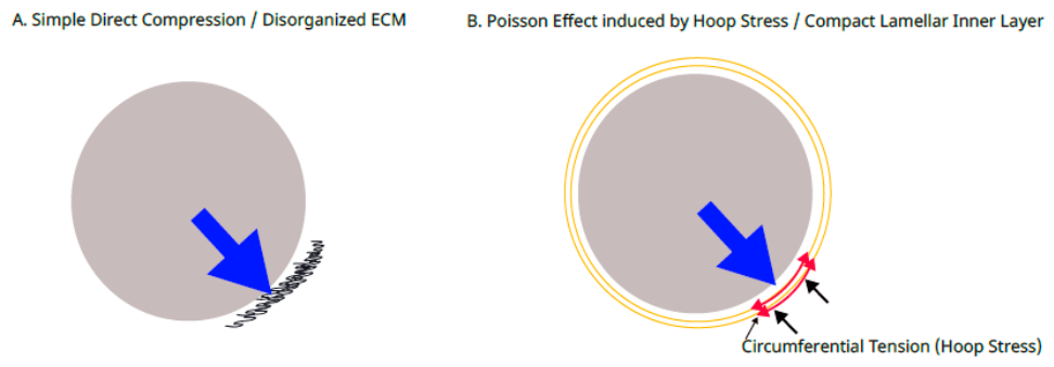
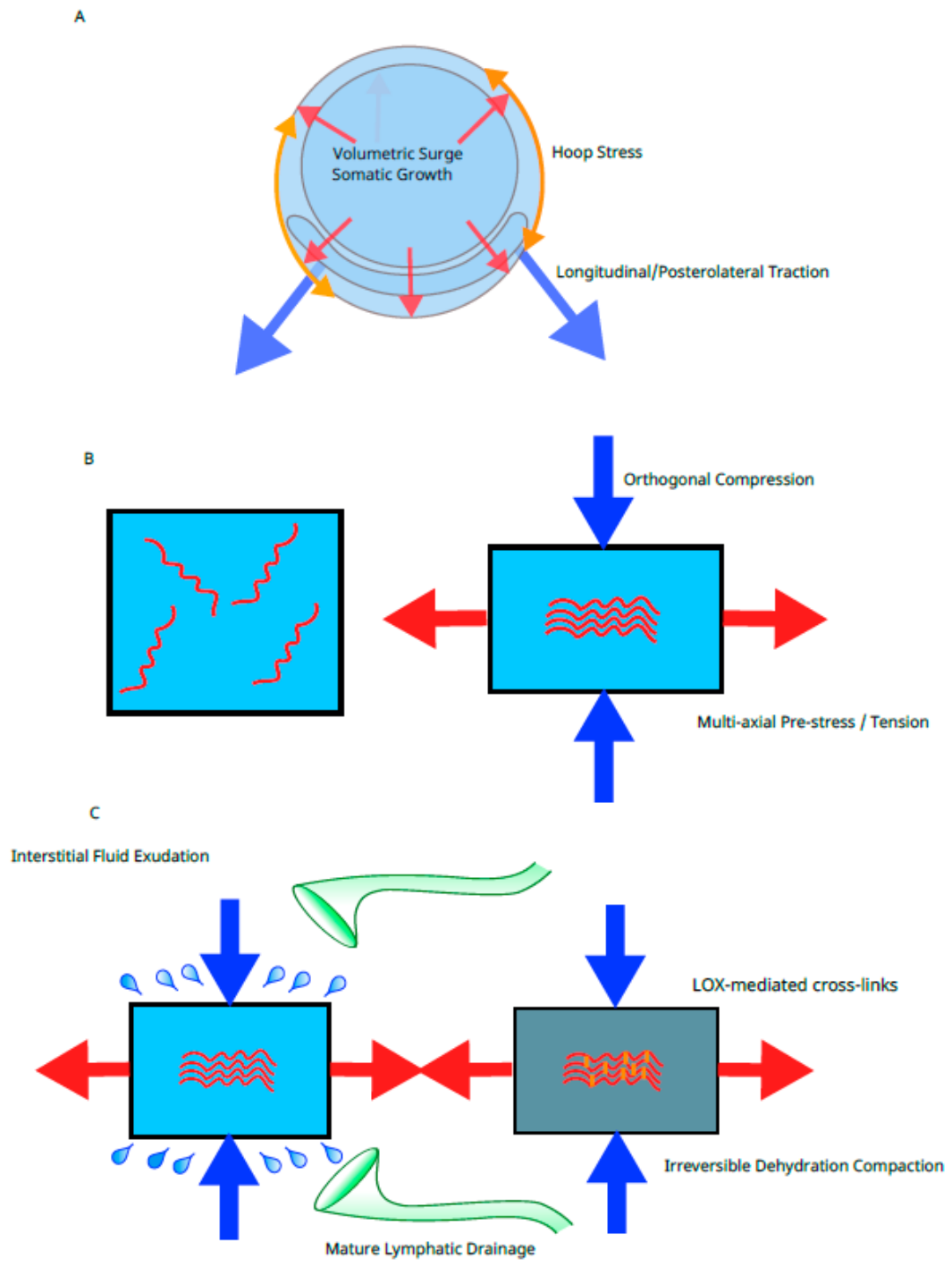
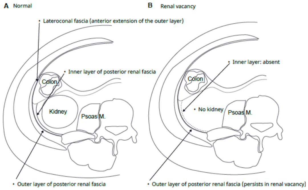
Figure 3.
Theoretical comparison of tissue deformation: Direct Compression vs. Poisson Effect. (A) Simple Direct Compression: A classical assumption where localized compressive forces from expanding organs merely squash the mesenchyme without producing organized lamellar sheets, resulting in disorganized tissue compaction. (B) Hoop Stress-Induced Poisson Effect: Circumferential tension (hoop stress) from the expanding mass induces orthogonal (transverse) compression via the Poisson effect, leading to the orderly condensation of the mesenchyme into the compact lamellar structures characteristic of the investing renal fascia.
Figure 3.
Theoretical comparison of tissue deformation: Direct Compression vs. Poisson Effect. (A) Simple Direct Compression: A classical assumption where localized compressive forces from expanding organs merely squash the mesenchyme without producing organized lamellar sheets, resulting in disorganized tissue compaction. (B) Hoop Stress-Induced Poisson Effect: Circumferential tension (hoop stress) from the expanding mass induces orthogonal (transverse) compression via the Poisson effect, leading to the orderly condensation of the mesenchyme into the compact lamellar structures characteristic of the investing renal fascia.
Figure 4.
Square-cube-driven emergence of a fetal tension network and definitive lamination. (A) Volumetric Surge: Rapid somatic growth (V/A ∝ r) dramatically increases internal mechanical load, generating hoop stress and longitudinal/posterolateral traction. (B) Orthogonal Compression: Because isotropic volumetric expansion is strictly restricted by early frame-locking (evolutionary front-loading), the trapped scaling energy is converted into a powerful multiaxial tension field, forcing the hydrated retroperitoneal mesenchyme to undergo obligatory orthogonal compression via the Poisson effect. (C) Consolidation and Fixation: This internal mechanical compression drives poroelastic fluid exudation into the maturing lymphatic system, while LOX-mediated cross-linking permanently stabilizes the collapsed collagen framework into the definitive, highly ordered fascial architecture. LOX, lysyl oxidase.
Figure 4.
Square-cube-driven emergence of a fetal tension network and definitive lamination. (A) Volumetric Surge: Rapid somatic growth (V/A ∝ r) dramatically increases internal mechanical load, generating hoop stress and longitudinal/posterolateral traction. (B) Orthogonal Compression: Because isotropic volumetric expansion is strictly restricted by early frame-locking (evolutionary front-loading), the trapped scaling energy is converted into a powerful multiaxial tension field, forcing the hydrated retroperitoneal mesenchyme to undergo obligatory orthogonal compression via the Poisson effect. (C) Consolidation and Fixation: This internal mechanical compression drives poroelastic fluid exudation into the maturing lymphatic system, while LOX-mediated cross-linking permanently stabilizes the collapsed collagen framework into the definitive, highly ordered fascial architecture. LOX, lysyl oxidase.
Figure 5.
Dual-mechanism model of retroperitoneal fascial lamination. Left: Early renal expansion (weeks 9–12) generates circumferential hoop stress, compacting adjacent mesenchyme into the organ-dependent inner lamina of the renal fascia. This layer fails to develop in renal agenesis. Right: System-level multiaxial tension emerging near gestational week 20 induces Poisson-driven orthogonal compression, collapsing the retroperitoneal mesenchyme into the outer fascial laminae independently of renal expansion. Notably, Tobin’s 1944 specimen—characterized by concurrent absence of both kidney and adrenal gland—represents a developmental field defect rather than evidence against the existence or formation of the system-derived outer lamina. The preserved solitary fascial plane in the present cohort reflects this system-level mechanism.
Figure 5.
Dual-mechanism model of retroperitoneal fascial lamination. Left: Early renal expansion (weeks 9–12) generates circumferential hoop stress, compacting adjacent mesenchyme into the organ-dependent inner lamina of the renal fascia. This layer fails to develop in renal agenesis. Right: System-level multiaxial tension emerging near gestational week 20 induces Poisson-driven orthogonal compression, collapsing the retroperitoneal mesenchyme into the outer fascial laminae independently of renal expansion. Notably, Tobin’s 1944 specimen—characterized by concurrent absence of both kidney and adrenal gland—represents a developmental field defect rather than evidence against the existence or formation of the system-derived outer lamina. The preserved solitary fascial plane in the present cohort reflects this system-level mechanism.
5. Conclusion
The autonomous preservation of the parietal lamina of the posterior renal fascia in congenital renal agenesis indicates that the retroperitoneal fascia is shaped by a system-level mechanical field rather than localized organ compression. As comparative embryology reveals, while quadrupedal precocial mammals prioritize fetal movement energy for limb development, humans trap this energy internally due to the "early truncal frame-locking" associated with bipedal adaptation. This confined mechanical energy induces orthogonal Poisson compression, forming synchronous multilaminar sheets in the retroperitoneum. The exquisite compartmentalization of the retroperitoneal fascia is a structural footprint of evolution, forged by the mechanical synergy of geometric scaling, human-specific ossification timing (heterochrony), and the biological ironing effect of tension.
Funding
The author received no specific funding for this work.
Data Availability
Radiological data supporting the findings of this study are restricted to protect patient privacy but are available from the corresponding author upon reasonable request.
Declaration of Generative AI and AI-Assisted Technologies in the Manuscript Preparation Process
During the preparation of this work, the author used AI tools to assist with language refinement and structural editing. After using these tools, the author reviewed and edited the content as needed and takes full responsibility for the content of the published article.
Acknowledgments
The author expresses deep gratitude to colleagues at the Department of Urology, Gakkentoshi Hospital, for providing a supportive clinical environment that continually inspires anatomical inquiry. Sincere appreciation is also extended to the radiology staff for their technical assistance.
Conflicts of Interest
The author declares no conflicts of interest.
Ethics Statement
This retrospective study was approved by the Institutional Review Board of Gakkentoshi Hospital (Approval No. GT-R6-07-12-1). Written informed consent was waived, and an opt-out mechanism was provided through the hospital’s official website.
References
- Bagnall KM, Harris PF, Jones PRM. A radiographic study of the human fetal spine. II. The sequence of development of ossification centres in the vertebral column. J Anat. 1977;124(3):791-802.
- Baumann JA. Développement et anatomie de la loge rénale chez l’homme. Acta Anat (Basel). 1945;1:15–65. [CrossRef]
- Baumgart M, Wisniewski M, Grzonkowska M, Badura M, Biernacki M, Siedlecki Z, et al. Quantitative anatomy of the ilium’s primary ossification center in the human fetus. Surg Radiol Anat. 2018;40:1047-1054. [CrossRef]
- Cho BH, Kimura W, Song CH, Fujimiya M, Murakami G. An investigation into the embryological development of the fasciae used as the basis for pancreatoduodenal mobilization. J Hepatobiliary Pancreat Surg. 2009;16:824–831.
- Congdon ED, Edson JN. The cone of renal fascia in the adult white male. Anat Rec. 1941;80:289–313. [CrossRef]
- Fedorov A, Beichel R, Kalpathy-Cramer J, Finet J, Fillion-Robin JC, Pujol S, et al. 3D Slicer as an image computing platform for the quantitative imaging network. Magn Reson Imaging. 2012;30:1323–1341. [CrossRef]
- Fung YC. Biomechanics: Mechanical Properties of Living Tissues. New York: Springer; 1993.
- Gerota, D. Beiträge zur Kenntnis des Befestigungsapparates der Niere. Arch. Anat. Entwicklungsgesch. 1895, 19, 265–286.
- Hadlock FP, Harrist RB, Martinez-Poyer J. In utero analysis of fetal growth: a sonographic weight standard. Radiology. 1991;181:129–133. [CrossRef]
- Hardman MJ, Sisi P, Banbury DN, Byrne C. Patterned acquisition of skin barrier function during development. Development. 1999;126:1541–1552.
- Harris AK, Stopak D, Wild P. Fibroblast traction as a mechanism for collagen morphogenesis. Nature. 1981;290:249–251. [CrossRef]
- Hoffman, C.K.; Filly, R.A.; Callen, P.W. The “lying down” adrenal sign: a sonographic indicator of renal agenesis or ectopia in fetuses and neonates. J. Ultrasound Med. 1992, 11(10), 533–536. [CrossRef]
- Humphrey JD. Continuum biomechanics of soft biological tissues. Proc Math Phys Eng Sci. 2003;459:3-46. [CrossRef]
- Ingber DE. Tensegrity I. Cell structure and hierarchical systems biology. J Cell Sci. 2003;116:1157–1173. [CrossRef]
- Ishikawa, K.; Idoguchi, K.; Tanaka, H.; Matsuoka, T.; Yokota, J.; Sugimoto, T. Classification of acute pancreatitis based on retroperitoneal extension: Application of the concept of interfascial planes. Eur. J. Radiol. 2006, 60, 445–452. [CrossRef]
- Jeong J, Ko J, Lim H, Kweon OK, Kim WH. Retroperitoneoscopy in dogs: Access technique, working space, and surgical anatomy. Vet Surg. 2016;45(Suppl 1):O102–O110.
- Kagan HM, Li W. Lysyl oxidase: properties, specificity, and biological roles inside and outside of the cell. J Cell Biochem. 2003;88:660–672.
- Kinugasa Y, Niikura H, Murakami G, Suzuki D, Saito S, Tatsumi H, Ishii M. Development of the human hypogastric nerve sheath with special reference to the topohistology between the nerve sheath and other prevertebral fascial structures. Clin Anat. 2008;21(6):558–567. [CrossRef]
- Matsubara A, Kinugasa Y, Murakami G, Suzuki D, Fujimiya M, Sugihara K. Development of the lateroconal fascia in human fetuses. Cells Tissues Organs. 2009;190(4):286–296.
- Molmenti, E.P.; Balfe, D.M.; Kanterman, R.Y.; Bennett, H.F. Anatomy of the retroperitoneum: observations of the distribution of pathologic fluid collections. Radiology 1996, 200(1), 95–103. [CrossRef]
- Mow VC, Kuei SC, Lai WM, Armstrong CG. Biphasic creep and stress relaxation of articular cartilage in compression: theory and experiments. J Biomech Eng. 1980;102:73–84. [CrossRef]
- Nowlan NC. Biomechanics of fetal movement. Eur Cell Mater. 2015;29:1–21.
- Opitz JM. The developmental field concept. Am J Med Genet. 1985;21:1–11. [CrossRef]
- Potter, E.L. Bilateral renal agenesis. J. Pediatr. 1946, 29(1), 68–76. [CrossRef]
- Raptopoulos, V.; Kleinman, P.K.; Marks, S.C.; Snyder, M.; Silverman, P.M. Renal fascial pathway: posterior extension of pancreatic effusions within the anterior pararenal space. Radiology 1986, 158(2), 367–374. [CrossRef]
- Sakai, T.; Matsui, M.; Mikami, A.; Malkova, L.; Hamada, Y.; Tomonaga, M.; Suzuki, J.; Tanaka, M.; Miyabe-Nishiwaki, T.; Makishima, H.; Nakatsukasa, M.; Matsuzawa, T. Developmental patterns of chimpanzee cerebral tissues provide important clues for understanding the remarkable enlargement of the human brain. Proceedings of the Royal Society B: Biological Sciences 2013, 280, 20122398. [CrossRef]
- Senevirathne G, Fernandopulle SC, Richard D, Baumgart SL, Christensen AL, Fabbri M, et al. The evolution of hominin bipedalism in two steps. Nature. 2025;645:952–963. [CrossRef]
- Stecco, C.; Sfriso, M.M.; Porzionato, A.; Rambaldo, A.; Albertin, G.; Macchi, V.; De Caro, R. Microscopic anatomy of the visceral fasciae. Journal of Anatomy 2017, 230(6), 848–856.
- Swartz MA, Fleury ME. Interstitial flow and its effects in soft tissues. Annu Rev Biomed Eng. 2007;9:229-256. [CrossRef]
- Thompson DW. On Growth and Form. Cambridge: Cambridge University Press; 1917.
- Tobin, C.E. The renal fascia and its relation to the transversalis fascia. Anat. Rec. 1944, 89(3), 295–311. [CrossRef]
- Toldt C. Bau und Wachsthumsveränderungen der Gekröse des menschlichen Darmkanales. Denkschr Kaiserl Akad Wiss Wien Math-Naturwiss Kl. 1879;41:1–56.
- van der Putte, S.C. The development of the lymphatic system in man. Adv. Anat. Embryol. Cell Biol. 1975, 51(1), 3–60. [CrossRef]
- Verbruggen, S.W.; et al. Stresses and strains on the human fetal skeleton during development.J R Soc Interface. 2018; 15(138): 20170593. [CrossRef]
- Verbruggen, S.W.; Nowlan, N.C. Ontogeny of the human pelvis. Anat. Rec. (Hoboken) 2017, 300(4), 643–652. [CrossRef]
- Verbruggen, S.W.; Loo, J.H.W.; Hayat, T.T.A.; Hajnal, J.V.; Rutherford, M.A.; Phillips, A.T.M.; Nowlan, N.C. Modeling the biomechanics of fetal movements. Biomech. Model. Mechanobiol. 2016, 15, 995–1004.
- Wedel, T.; et al. The retrocolic fascial system revisited for right hemicolectomy with complete mesocolic excision based on anatomical terminology. Colorectal Disease 2022, 24(12), 1466–1477.
- Weisbecker, V., Goswami, A., Wroe, S., & Sánchez-Villagra, M. R. (2008). Ossification heterochrony in the therian postcranial skeleton and the marsupial–placental dichotomy. Evolution, 62(8), 2027–2041.
- Weiss, P. The problem of specificity in growth and development. Yale J. Biol. Med. 1947, 19(3), 235–278.
- Zuckerkandl E. Ueber den Fixationsapparat der Nieren. Med Jahrb. 1883;59-67.
Figure 1.
Preservation of the posterior renal fascial plane in congenital renal agenesis. Axial unenhanced CT image from Case 1 (53-year-old female) with true left renal agenesis. Despite the complete and lifelong absence of the kidney and ureter, a distinct, continuous hyperdense fascial plane (arrows) is unequivocally preserved at the anatomical location corresponding to the posterior renal fascia. This plane extends smoothly between the peritoneal sac and the posterior abdominal wall and maintains typical continuity with the lateroconal fascia. On the contralateral healthy side (inset), the composite fascia is thicker, reflecting the presence of both the organ-dependent inner lamina and the system-derived outer lamina. On the renal-vacant side, a thinner but clearly defined solitary fascial plane (~1.5 mm) remains, consistent with selective absence of the inner lamina and preservation of the system-level outer lamina predicted by the tension-driven mechanobiological model.
Figure 1.
Preservation of the posterior renal fascial plane in congenital renal agenesis. Axial unenhanced CT image from Case 1 (53-year-old female) with true left renal agenesis. Despite the complete and lifelong absence of the kidney and ureter, a distinct, continuous hyperdense fascial plane (arrows) is unequivocally preserved at the anatomical location corresponding to the posterior renal fascia. This plane extends smoothly between the peritoneal sac and the posterior abdominal wall and maintains typical continuity with the lateroconal fascia. On the contralateral healthy side (inset), the composite fascia is thicker, reflecting the presence of both the organ-dependent inner lamina and the system-derived outer lamina. On the renal-vacant side, a thinner but clearly defined solitary fascial plane (~1.5 mm) remains, consistent with selective absence of the inner lamina and preservation of the system-level outer lamina predicted by the tension-driven mechanobiological model.
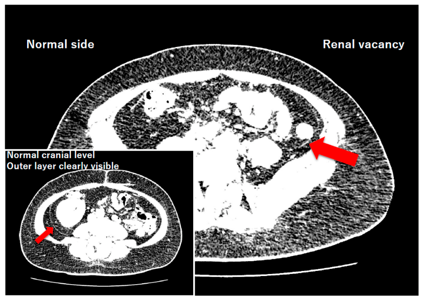
Figure 2.
Spatiotemporal and biomechanical asymmetry in retroperitoneal fascial development. This diagram highlights the chronological discrepancy in fascial emergence. Phase 1 (weeks 9–18) involves the sequential establishment of central and lateral mechanical anchors. During the early part of this phase (weeks 9–12; Panel A), the inner lamina of the renal fascia forms, driven by localized hoop stress from the expanding kidney. In contrast, Phase 2 (around week 20; Panel B) represents the mid-gestational systemic mechanical transition, during which the multilaminated outer layers are synchronously established across the macroscopic tension network completed between these central and lateral visceral anchors.
Figure 2.
Spatiotemporal and biomechanical asymmetry in retroperitoneal fascial development. This diagram highlights the chronological discrepancy in fascial emergence. Phase 1 (weeks 9–18) involves the sequential establishment of central and lateral mechanical anchors. During the early part of this phase (weeks 9–12; Panel A), the inner lamina of the renal fascia forms, driven by localized hoop stress from the expanding kidney. In contrast, Phase 2 (around week 20; Panel B) represents the mid-gestational systemic mechanical transition, during which the multilaminated outer layers are synchronously established across the macroscopic tension network completed between these central and lateral visceral anchors.
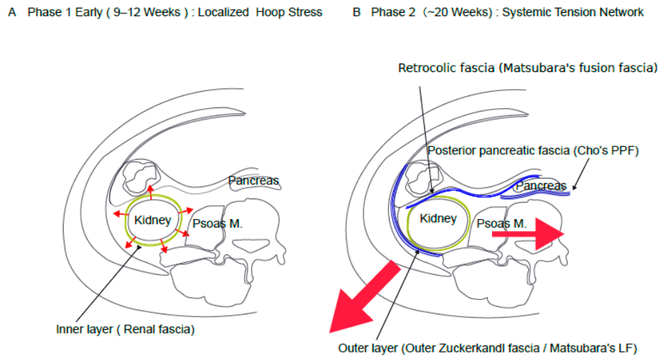

Table 1.
Clinical characteristics and quantitative radiological findings of the renal agenesis cohort (n = 3).
Table 1.
Clinical characteristics and quantitative radiological findings of the renal agenesis cohort (n = 3).
| Case | Age/Sex | Radiological Diagnosis | Adrenal Morphology | Fascial Thickness (Affected) | Fascial Thickness (Normal) | Difference (Δ) |
| 1 | 53F | True left renal agenesis | Pancake | 1.49 mm | 1.88 mm | −0.39 mm |
| 2 | 47F | True left renal agenesis | Pancake | 1.46 mm | 1.82 mm | −0.36 mm |
| 3 | 89M | Severe left renal dysplasia | Normal | 1.62 mm | Excluded* | N/A |
| Mean | — | — | — | 1.52 mm | 1.85 mm | −0.38 mm |
*Contralateral side excluded from measurement due to fascial thickening secondary to pyelonephritis.
Table 2.
Integration of retroperitoneal fascial development and associated human-specific biomechanical events.
Table 2.
Integration of retroperitoneal fascial development and associated human-specific biomechanical events.
| Gestational Age | Anatomical Event | Biomechanical/Evolutionary Context | References |
| Weeks 9–12 | Renal ascent and initial adherence of the proper mesocolon. Emergence of the inner lamina. | Phase 1 (Organ-Dependent): Localized circumferential tension (hoop stress) from the expanding kidney compacts adjacent mesenchyme to form the inner lamina of the renal fascia. | Matsubara et al. 2009 |
| Weeks 14–16 | Initiation of asymmetric pelvic stiffening. Fetal lymphatic maturation. | Increased transverse rigidity and preparation of drainage pathways for poroelastic fluid exudation. | van der Putte 1975; Swartz & Fleury 2007; Sakai et al. 2012 |
| Weeks 17–18 | Adhesion of the mesocolon to the posterior abdominal wall. | Phase 2 (Onset of Human-Specific Front-Loading): Early stiffening of the truncal frame commences. Tension-induced local anisotropy initiates. | Baumann 1945; Verbruggen et al. 2018 |
| ~Week 20 | Synchronous, definitive establishment of the "outer fascial laminae" complex (comprising the posterior renal, retrocolic, and retropancreatic fasciae), accompanied by the anisotropic condensation of mesenchymal tissue surrounding the hypogastric nerve into discrete, tension-aligned fascial planes. | Phase 3 (System-Level Mechanical Transition): Fetal movement energy conversion into multiaxial tension and Poisson compression (ironing effect). Mesenchymal tissues integrate into a tensegrity scaffold along principal tension vectors. | Nowlan 2015; Verbruggen et al. 2016; Weisbecker et al. 2008; Sakai et al. 2012; Kinugasa et al. 2008; Cho et al. 2009; Matsubara et al. 2009 |
*Footnote: In non-human mammals, particularly large precocial species (e.g., horses, elephants), fetal movement energy is preferentially allocated to appendicular/locomotor stiffening (limb-first heterochrony) essential for immediate postnatal standing, while the truncal frame remains compliant until birth. In contrast, in humans, the fetal movement energy during this period collides with the "early-stiffened truncal boundary," uniquely concentrating mechanical stress onto the retroperitoneum.
Disclaimer/Publisher’s Note: The statements, opinions and data contained in all publications are solely those of the individual author(s) and contributor(s) and not of MDPI and/or the editor(s). MDPI and/or the editor(s) disclaim responsibility for any injury to people or property resulting from any ideas, methods, instructions or products referred to in the content. |
© 2026 by the authors. Licensee MDPI, Basel, Switzerland. This article is an open access article distributed under the terms and conditions of the Creative Commons Attribution (CC BY) license (http://creativecommons.org/licenses/by/4.0/).
Copyright: This open access article is published under a Creative Commons CC BY 4.0 license, which permit the free download, distribution, and reuse, provided that the author and preprint are cited in any reuse.


