

Submitted:
23 May 2026
Posted:
26 May 2026
You are already at the latest version
Abstract
The cervical spine is highly susceptible to injury from blunt acceleration/deceleration trauma, most commonly manifesting as whiplash-associated disorder (WAD). Although WAD is often considered a self-limiting soft-tissue injury, certain anatomical variants may influence clinical presentation and management. One such variant is the ponticulus posticus (PP), a bony bridge on the posterior arch of the atlas that can encase the vertebral artery. This narrative review examines the prevalence, anatomy, and potential clinical associations of PP in patients with WAD. While large prospective studies directly linking missed PP to adverse whiplash outcomes are limited, multiple anatomical and imaging studies demonstrate associations between PP and vertebral artery compression or tethering. Current clinical decision rules (NEXUS and Canadian C-Spine Rule) are highly validated for detecting clinically important cervical fractures but were not designed to identify anatomical variants such as PP. Plain-film cervical radiographs can readily visualize PP and may therefore provide additional information in selected trauma patients.
Keywords:
cervical spine
; ponticulus posticus
; whiplash
; vertebral artery
; X-rays
Introduction and Background
Motor vehicle collisions, even at low speeds (approximately 20 km/h), can produce cervical spine injury and related symptoms [1]. Clinicians evaluating acute whiplash patients must balance the need to identify injury with efforts to minimize unnecessary imaging. Two widely used clinical decision rules—the NEXUS criteria and the Canadian C-Spine Rule—have been extensively validated in large trauma populations for detecting clinically important cervical spine injuries (fractures and dislocations) [2,3]. These rules demonstrate high sensitivity and have successfully reduced imaging rates in low-risk patients.
Nevertheless, these guidelines were developed primarily for high-energy trauma and fracture detection. In lower-energy whiplash mechanisms, they may not identify certain anatomical variants that could influence subsequent patient management. One such variant is the ponticulus posticus (PP). Failure to obtain cervical radiographs in neurologically intact patients may therefore overlook PP and other structural findings visible on plain films.
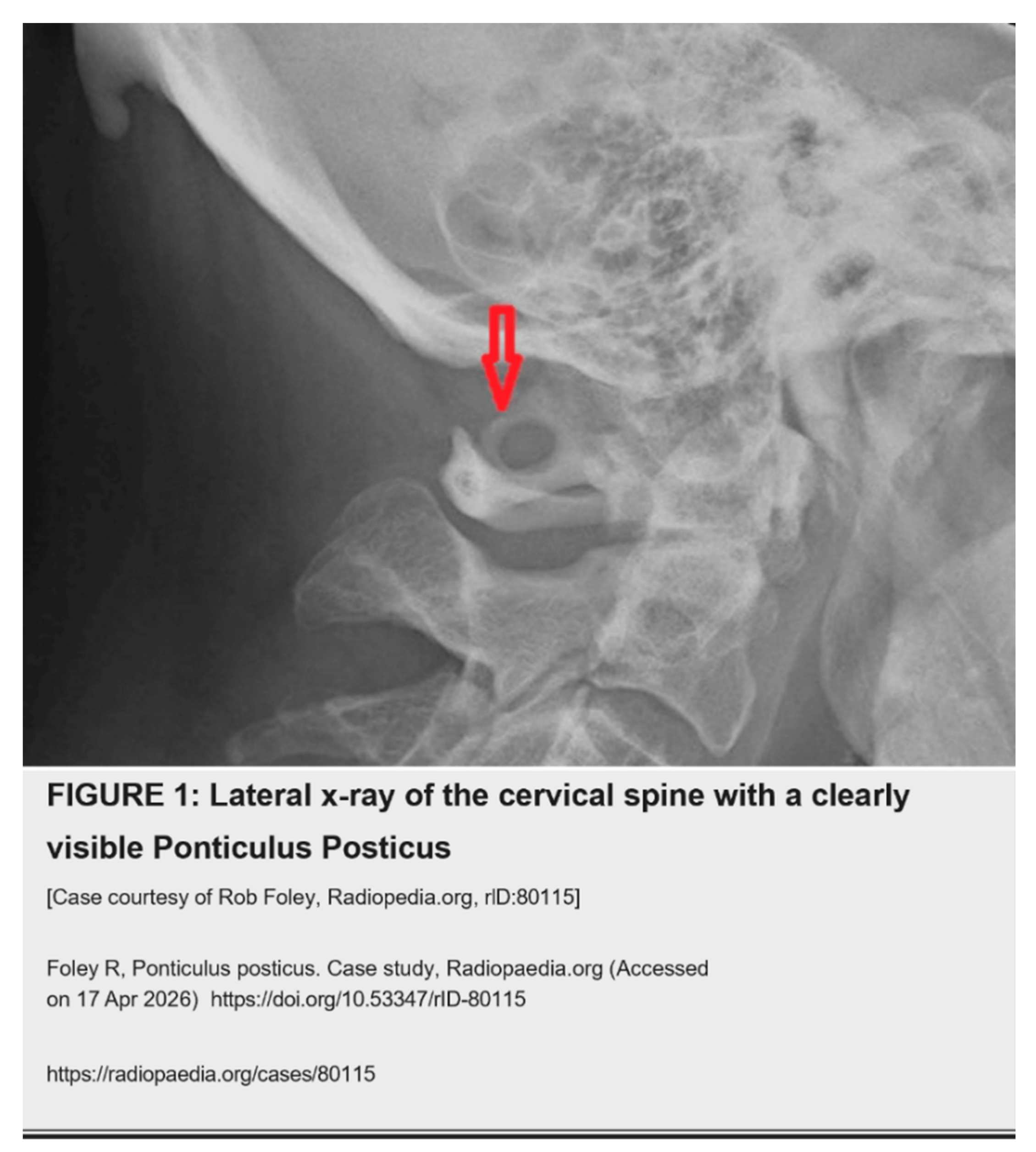
Definition and Anatomy of the Ponticulus Posticus The ponticulus posticus (PP), also known as the arcuate foramen, is a bony bridge that arises from the lateral mass of the atlas (C1) and transforms the groove of the vertebral artery into a canal on the posterior arch [4]. It can be classified as complete, partial, or incomplete [5].
The vertebral artery travels through the transverse foramen of C1 and courses posteriorly along the arch before entering the vertebral canal. The presence of PP places the artery in close proximity to this ossified structure, potentially altering local hemodynamics [6] (Figure 1).
 |
Potential Clinical Associations PP has been associated with a range of symptoms, including headache, vertigo, visual disturbances, and hearing loss—features sometimes grouped under Barré-Liéou syndrome. These associations are thought to result from compression or tethering of the vertebral artery and irritation of the periarterial sympathetic plexus [7,8].
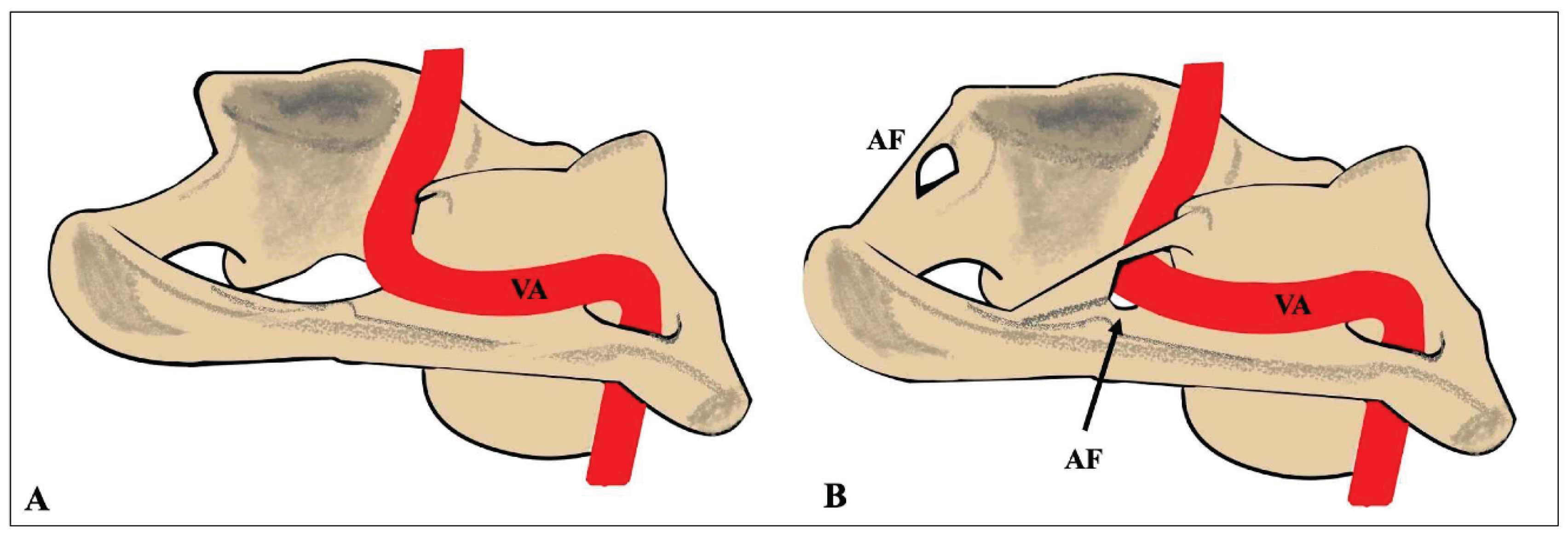
In the context of whiplash, biomechanical studies show that rear-end collisions produce hyperextension, hyperflexion, axial rotation, and shear forces that can elongate or stretch the vertebral artery [9]. The presence of PP may further compromise an already-stressed vessel [10]. Anatomical studies report reduced vertebral artery diameter in patients with complete PP [11] and an increased risk of dynamic compression during neck rotation [12]. While these findings represent associations rather than proven causation, they highlight a potential mechanism by which PP could influence whiplash symptom severity or recovery. (Figure 2 and Figure 3)
Imaging Considerations Standard lateral cervical radiographs readily demonstrate PP. Given that PP is visible on routine views and may carry clinical relevance for manual therapy or ongoing neck pain, some authors have suggested that plain-film imaging may provide useful information even in neurologically intact patients after low-energy trauma [13].
At the same time, the potential harms of over-imaging—radiation exposure, cost, and incidental findings—must be acknowledged. The effective dose of a 3-view cervical series is low (approximately 0.1–0.2 mSv), and the linear no-threshold model of radiation risk continues to be debated in the literature, with some evidence supporting a hormesis effect at very low doses [14,15]. Clinicians must therefore weigh the diagnostic benefit of identifying clinically relevant variants against the small theoretical risk of radiation exposure.
Implications for Manual Therapy Chiropractors and other manual practitioners treat a substantial proportion of whiplash patients [16]. When PP is identified, a more cautious approach to cervical manipulation may be prudent. High-velocity, low-amplitude techniques could theoretically increase stress on a tethered vertebral artery; gentler mobilization, instrument-assisted, or specific upper-cervical protocols may be considered instead. The absolute risk of arterial dissection from cervical manipulation remains extremely low, but identification of PP allows for individualized risk assessment.
Conclusions The ponticulus posticus is a relatively common anatomical variant (prevalence 15–20% in radiographic studies) that can be easily visualized on standard lateral cervical radiographs. While current clinical decision rules are highly effective for ruling out fracture in low-risk trauma patients, they were not designed to detect variants such as PP that may influence vertebral artery dynamics or manual-therapy planning. In selected cases, plain-film imaging may therefore provide additional clinically relevant information.
Limitations This is a narrative review. Large-scale prospective studies directly linking missed PP to worse clinical outcomes in whiplash patients are lacking. All reported associations between PP, vertebral artery compromise, and symptoms remain correlative rather than causal. Further research is needed to clarify the clinical significance of PP in acute and chronic WAD.
Disclosures
Author Contributions
All aspects of the work were performed by Leonard F. Vernon.
Funding
This research received no external funding.
Conflicts of Interest
The authors declare no conflicts of interest regarding the publication of this article.
References
- Severy, D.M.; Mathewson, J.H.; Bechtol, C.O: Controlled automobile rearend collisions, an investigation of related engineering and medical phenomena. Can. Serv. Med. J. 1955, 11:727-759.
- Centeno, C.J.; Freeman, M.; Elkins, W.L: A review of the literature refuting the concept of minor impact soft tissue injury. Pain Res. Manag. 2005, 10:71-74. [CrossRef]
- Baratloo, A.; Ahmadzadeh, K.; Forouzanfar, M.M: NEXUS vs. Canadian C-Spine Rule (CCR) in Predicting Cervical Spine Injuries; a Systematic Review and Meta-analysis. Arch. Acad. Emerg. Med. 2023, 11:66. [CrossRef]
- Paykin G, O’Reilly G, Ackland HM, et.al: The NEXUS criteria are insufficient to exclude cervical spine fractures in older blunt trauma patients. Injury. Injury. 5:1020-1024. [CrossRef]
- Kadom N, Palasis S, Pruthi S, et al.: ACR Appropriateness Criteria® Suspected Spine Trauma-Child . J Am Coll Radiol. 2019, 16:286-299. [CrossRef]
- Pennington CW, Siegel JA: The Linear No-Threshold Model of Low-Dose Radiogenic Cancer: A Failed Fiction. Dose Response. Dose Response. 2019, 7:1559325818824200. [CrossRef]
- Luckey TD: Radiation Hormesis: The Good, the Bad, and the Ugly. . Dose Response . 2006, 4:169-90. [CrossRef]
- Arnone, P.A.; Richards, L.M.; Stittleburg, K.C.; Weiner, M.T.; Dennis: A.K: Risk and Safety in Radiographic Utilization: Evaluating the Evidence From Current Research. Radiol. Res. Pract. 2025, 8843173. [CrossRef]
- Oakley, P.A.; Haas, J.W.; Harrison, D.E: The Rationale and Safety of Routine Imaging in Rehabilitative Spine Care: Delayed Radiographs for Patients Presenting With Spine Disorders is Debatable. Dose Response. 2025, 23:15593258251374411. [CrossRef]
- Failure to Diagnose Spine Injury. (1998). Accessed: April . Accessed: 23rd, 2026: https://www.clinician.com/articles/33884-failure-to-diagnose-spine-injury..
- Lekovic GP, Harrington TR: : Litigation of missed cervical spine injuries in patients presenting with blunt traumatic injury. Neurosurgery. 2007, 3:516-22. [CrossRef]
- Davis, C.G.: Rear-End Impacts: Vehicle And Occupant Response . J. Manip. Physiol. Ther. 1998, 21:629-639.
- Paschopoulos I, Piagkou M, Triantafyllou G: The Potential Morphological Stenosis Pattern of the Arcuate Foramen. Diagnostics (Basel. 2025:1203. [CrossRef]
- Bulut, M.D.; Alpayci, M.; Şenköy: E.;: Decreased Vertebral Artery Hemodynamics in Patients with Loss of Cervical Lordosis. Med. Sci. Monit. 2016, 22:495-500. [CrossRef]
- Zhu, Y.T.; Lyu, L.J.; Zhang, C: Correlative analysis of cervical curvature and atlantoaxial instability . Zhongguo Gu Shang China J. Orthop. Traumatol. 2022, 35:132-135. [CrossRef]
- Buna, M.; Coghlan, W.; deGruchy, M.; Williams, D.; Zmiywsky, O: Ponticles of the atlas: A review and clinical perspective. J. Manip. Physiol. Ther. 1984, 7:261-266.
- Sweat, R. W., & Crowe, H. S: The ponticulus posticus of the atlas vertebra and its significance . Today’s Chiropr. 16:95-97.
- Kuhta, P.; Hart, J.; Greene-Orndorff, L.; McDowell-Reizer, B.; Rush, P: The prevalence of posticus ponticus: Retrospective analysis of radiographs from a chiropractic health center. J. Chiropr. 2010, 9:162-165. [CrossRef]
- Schilling J.; Schilling, A.; Galdames, I.S: Ponticulus posticus on the posterior arch of atlas, prevalence analysis in asymptomatic patients. Int. J. Morphol. 2010, 28:317-322. [CrossRef]
- Xu, X.; Zhu, Y.; Ding, X.; Yin, M.; Mo, W.; Ma, J: Research progress of ponticulus posticus: A narrative literature review. Front. Surg. 2022, 9:834551. [CrossRef]
- Koutsouraki, E.; Avdelidi, E.; Michmizos, D.; Kapsali, S.E.; Costa, V.; Baloyannis, S: Kimmerle’s anomaly as a possible causative factor of chronic tension-type headaches and neurosensory hearing loss: Case report and literature review. Int. J. Neurosci. 2010, 120:236-239. [CrossRef]
- Morinaga Y, Nii K, Sakamoto K, Inoue R, Mitsutake T, Hanada H: Focus on diagnosis, treatment, and problems of Barré-Lièou syndrome: Two case reports. Drug Discov. Ther. 2019, 13:239-243. [CrossRef]
- Pearce, J.M: Barre-Lieou “syndrome: J. Neurol. Neurosurg. Psychiatry. 2004, 75:319.
- Yamamoto, A.; Kunimatsu, Y: Geo: Geographic variation of atlas bridging of Japanese monkey (Macaca fuscata). Primate Res. 2002, 18:215-224. [CrossRef]
- Ivancic, P.C.; Panjabi, M.M.; Ito, S: Cervical spine loads and intervertebral motions during whiplash . Traffic Inj. Prev. 2006, 7:389-999. [CrossRef]
- Hasan, M.; Shukla, S.; Siddiqui, M.S.; Singh, D: Posterolateral tunnels and ponticuli in human atlas vertebrae. J. Anat. 2001, 199:339-343. [CrossRef]
- Cushing, K.E.; Ramesh, V.; Gardner-Medwin, D.; et. al: Tethering of the vertebral artery in the congenital arcuate foramen of the atlas vertebra: A possible cause of vertebral artery dissection in children. Dev. Med. Child Neurol. 2001, 43:491-496. [CrossRef]
- Tubbs, R.S.; Shoja, M.M.; Skokouhl, G.; Farahani, R.M.; Loukas, M: Oakes: Simultaneous lateral and posterior ponticles resulting in the formation of a vertebral artery tunnel of the atlas: Case report and review of the literature. Folia Neuropathol. 2007, 45:43-46.
- Nibu, K.; Cholewicki, J.; Panjabi, M.M: Dynamic elongation of the vertebral artery during an in vitro whiplash simulation. Eur. Spine J. 1997, 6:286-289. [CrossRef]
- Chung, Y.S.; Han, D.H: : Vertebrobasilar dissection: A possible role of whiplash injury in its pathogenesis.
- Neurol. Res. 2002, 24:129-138. [CrossRef]
- Closs, S.; Freire, A.; Costa, S: Ponticulus posticus: Anatomical variation in posterior arch of the atlas vertebra evaluated in lateral cephalometric radiography. Br. J. Med. Med. Res. 2017, 21:1-10.
- Gul, E.; Atik, I: Does ponticulus posticus affect vertebral artery diameter . Surg. Radiol. Anat. SRA. 2024, 46:1517-1524. [CrossRef]
- Ozdemir, B.; Kanat, A.; Durmaz, S.; Ersegun Batcik, O.; Gundogdu, H: Introducing a new possible predisposing risk factor for odontoid type 2 fractures after cervical trauma; Ponticulus posticus anomaly of C1 vertebra. J. Clin. Neurosci. 2022, 96:194-198. [CrossRef]
- Johnson, J.; Bettany-Saltikov, J.; van Schaik: P.; Cordry: J.; Newell, D.; Duangkaew, R: Postural Assessment: An Online Survey of Practicing Chiropractors in the UK. Healthcare. 2025, 13:3212. [CrossRef]
Figure 2.
Ligamentous upper cervical instability can cause the vertebral artery and C 1 nerve root to become encroached upon by the ossification of the ligamentous structures of the Atlas known as a Ponticulus Posticus. [Used by permission of Matthew R. Skalski, DC, DACBR] Skalski M, Posterior ponticle - illustration. Case study, Radiopaedia.org (Accessed on 17 Apr 2026) https://doi.org/10.53347/rID-52537.
Figure 2.
Ligamentous upper cervical instability can cause the vertebral artery and C 1 nerve root to become encroached upon by the ossification of the ligamentous structures of the Atlas known as a Ponticulus Posticus. [Used by permission of Matthew R. Skalski, DC, DACBR] Skalski M, Posterior ponticle - illustration. Case study, Radiopaedia.org (Accessed on 17 Apr 2026) https://doi.org/10.53347/rID-52537.
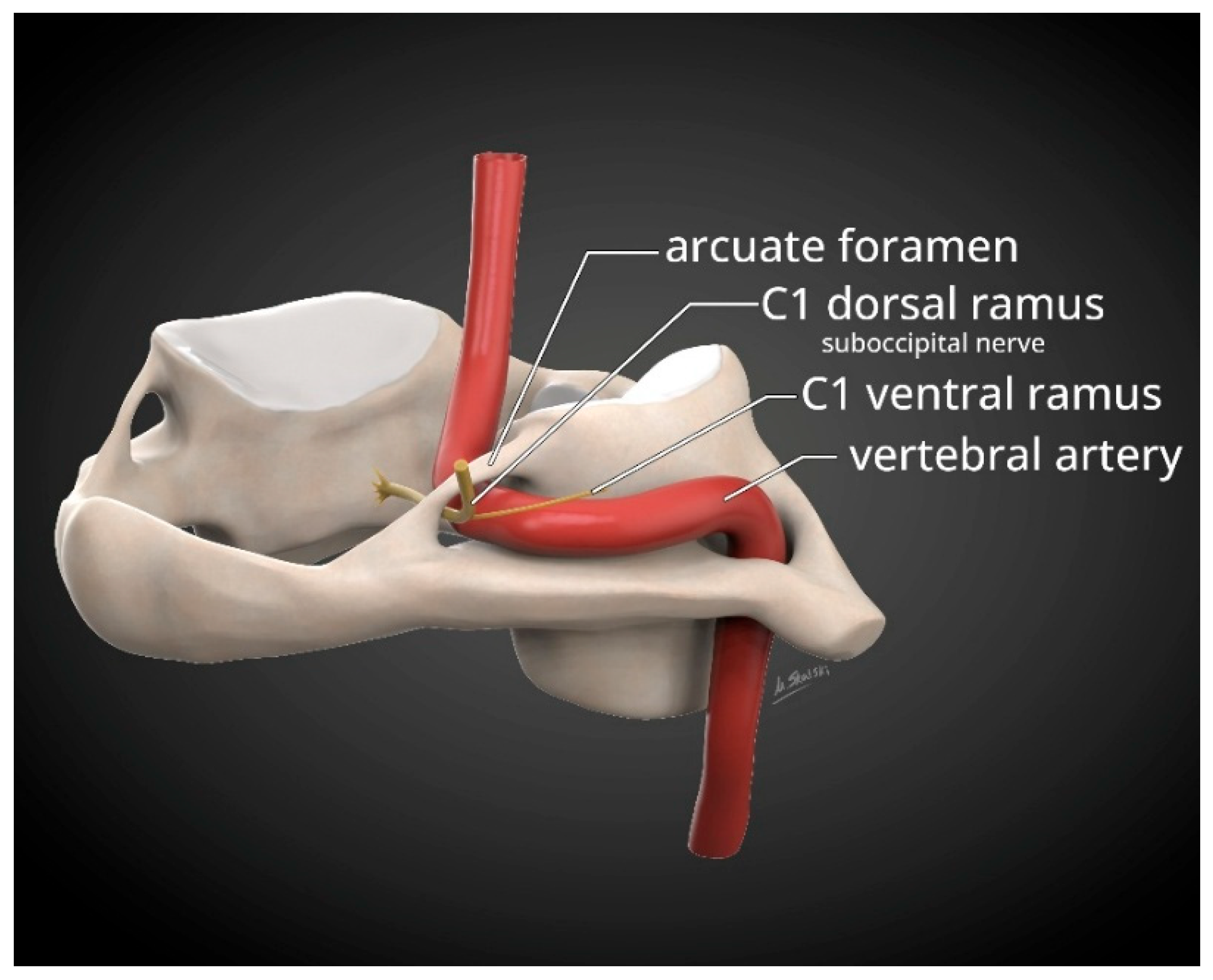
Figure 3.
The vertebral artery is particularly vulnerable to compression during extreme rotation of head and neck movements and that the presence of PP can compromise the caliber of an already-stretched vertebral artery.[Used by permission Mary Piagkou, MD and MDPI Publishing) [13].
Figure 3.
The vertebral artery is particularly vulnerable to compression during extreme rotation of head and neck movements and that the presence of PP can compromise the caliber of an already-stretched vertebral artery.[Used by permission Mary Piagkou, MD and MDPI Publishing) [13].
Disclaimer/Publisher’s Note: The statements, opinions and data contained in all publications are solely those of the individual author(s) and contributor(s) and not of MDPI and/or the editor(s). MDPI and/or the editor(s) disclaim responsibility for any injury to people or property resulting from any ideas, methods, instructions or products referred to in the content. |
© 2026 by the authors. Licensee MDPI, Basel, Switzerland. This article is an open access article distributed under the terms and conditions of the Creative Commons Attribution (CC BY) license (http://creativecommons.org/licenses/by/4.0/).
Copyright: This open access article is published under a Creative Commons CC BY 4.0 license, which permit the free download, distribution, and reuse, provided that the author and preprint are cited in any reuse.