

Submitted:
13 March 2026
Posted:
16 March 2026
You are already at the latest version
Abstract
End-stage lung disease represents a major challenge in modern medicine. Lung transplantation remains the most effective treatment; however, donor shortage and rejection significantly limit its clinical impact. The engineering of bioartificial lung grafts using patient-derived cells may lead to new therapeutic strategies. Advanced culture conditions enable the formation of functional three-dimensional tissues from lineage-committed cells. Currently, bioartificial grafts capable of gas exchange have been created and transplanted in animal models. Ongoing challenges in tissue engineering include the development of ideal scaffolds and the full maturation of engineered structures to ensure graft longevity after in vivo implantation. With collaborative efforts, the goal is to design patient-derived lung grafts and achieve clinically relevant translational milestones such as airway grafts and disease models.
Keywords:
bioartificial lung
; lung transplantation
; tissue engineering
; decellularization
; lung architecture
I. Introduction
Currently, approximately 25 million people worldwide suffer from chronic obstructive pulmonary disease. In the United States, an estimated 12,000 patients die each year because of lung diseases [1]. Lung transplantation remains the only definitive treatment for these conditions. However, there is a significant limitation in the availability of organ donors, and the number of suitable donor lungs is particularly low. The lungs are highly susceptible to damage, infection, and inadequate ventilation, which makes them prone to rapid deterioration after donor death. Consequently, only 10 – 20% of donated lungs are considered suitable for transplantation. In 2022, 3,161 candidates were added to the lung transplant waiting list. Approximately 4,228 candidates were registered on the waiting list throughout the year, a figure that has remained relatively constant since 2011 [2]. Over time, transplant outcomes are negatively affected by chronic rejection and the adverse effects associated with immunosuppressive therapy [3]. The development of a functional lung engineered from scratch could provide a new transplantable organ comparable to a human donor lung. Various approaches have been progressively explored to design functional lung tissue [4]. The use of polymers [5] and natural biomaterials such as collagen [6,7,8], Matrigel [9,10], and Gelfoam [11] highlights the critical role of the extracellular matrix (ECM), not only in maintaining lung architecture [12] but also in directing pulmonary cell differentiation [13].
II. Metodology
A systematic review of the scientific literature was conducted to collect relevant information regarding tissue engineering principles, applied methodologies, recent advancements, and current challenges in the field.
- Data Search and Collection: A comprehensive literature search was performed across several scientific databases, including PubMed, Wiley Online Library, the Royal Society of Chemistry, ScienceDirect, and Google Scholar. The search used key terms such as “Bioartificial lung, lung transplant, tissue engineering, decellularization,” and “lung architecture” to ensure relevant coverage.
- Information Selection and Refinement: The selected literature spanned the period from 2009 to 2025 to ensure an up-to-date and representative overview of the current state of the field.
- Organization of Subtopics: Following the refinement of the collected data, a structured research framework was developed by organizing the content into thematic categories to facilitate a comprehensive analysis of the most relevant aspects of the study.
- Data Analysis and Interpretation: A critical assessment of the selected studies was carried out, identifying emerging trends, knowledge gaps, and potential research opportunities. The insights obtained supported the formulation of evidence-based conclusions, which are presented in the results and discussion section (Rondón et al., 2025).
III. Principles of Lung Engineering
A. Relevant Anatomy and Physiology
The respiratory system enables the exchange of vital gases by obtaining oxygen from inhaled air, delivering it to the bloodstream for transport to all body cells, and simultaneously eliminating carbon dioxide. Within this system, the lungs play a central role [14]. They are located within the thoracic cavity and are responsible for oxygenation of blood by taking in oxygen during inhalation and removing carbon dioxide during exhalation. Internally, the lungs contain an extensive network of bronchi, bronchioles, and alveoli. The bronchi initially inhaled air from the trachea into the lungs. The smaller branches, known as bronchioles, distribute air throughout the pulmonary tissue. At the terminal end of each bronchiole are small sac-like structures called alveoli [15,16]. Within the alveoli, the air–blood interface is formed by an extremely thin layer of alveolar epithelial cells, the type I pneumocytes, which are supported by a shared basement membrane and by capillary endothelial cells. In addition, type II pneumocytes secrete pulmonary surfactant, a substance that reduces surface tension and prevents alveolar collapse [17]. To achieve an efficient transplant, a detailed understanding of the anatomy and physiology of each component is essential. Therefore, a successful bioartificial lung must replicate not only the cellular composition but also the anatomical architecture and physiological properties of the native organ.
B. Requirements for Functional Graft
For a bioartificial lung to accurately replicate the function of a native lung, the primary objective is to restore effective gas exchange. This depends directly on the integrity of the air–blood interface, the tissue’s ventilatory capacity, and continuous blood perfusion through a functional vascular network. Whole-lung extracellular matrix (ECM) scaffolds can be generated by perfusing cadaveric organs with detergents to achieve decellularization, providing an appropriate platform for organ regeneration. It is important to consider that pulmonary epithelial engineering must address both proximal airway cells, which metabolize toxins and promote mucociliary clearance, and distal pneumocytes, which facilitate gas exchange [18]. The pulmonary vasculature must be engineered to support “in vivo” blood perfusion with low resistance while maintaining an intact barrier function.
Initially, the lung structure functions as an empty scaffold. For the decellularized matrix to regain physiological function, viable cells must be reintroduced into their original anatomical locations— for example, epithelial cells within the alveoli and endothelial cells within the vascular network. If these cells are not properly distributed or are present in insufficient numbers, the organ will be unable to ventilate or oxygenate blood in a manner comparable to a native lung [18,19].
IV. Fundamental Technologies
A. Descellularization
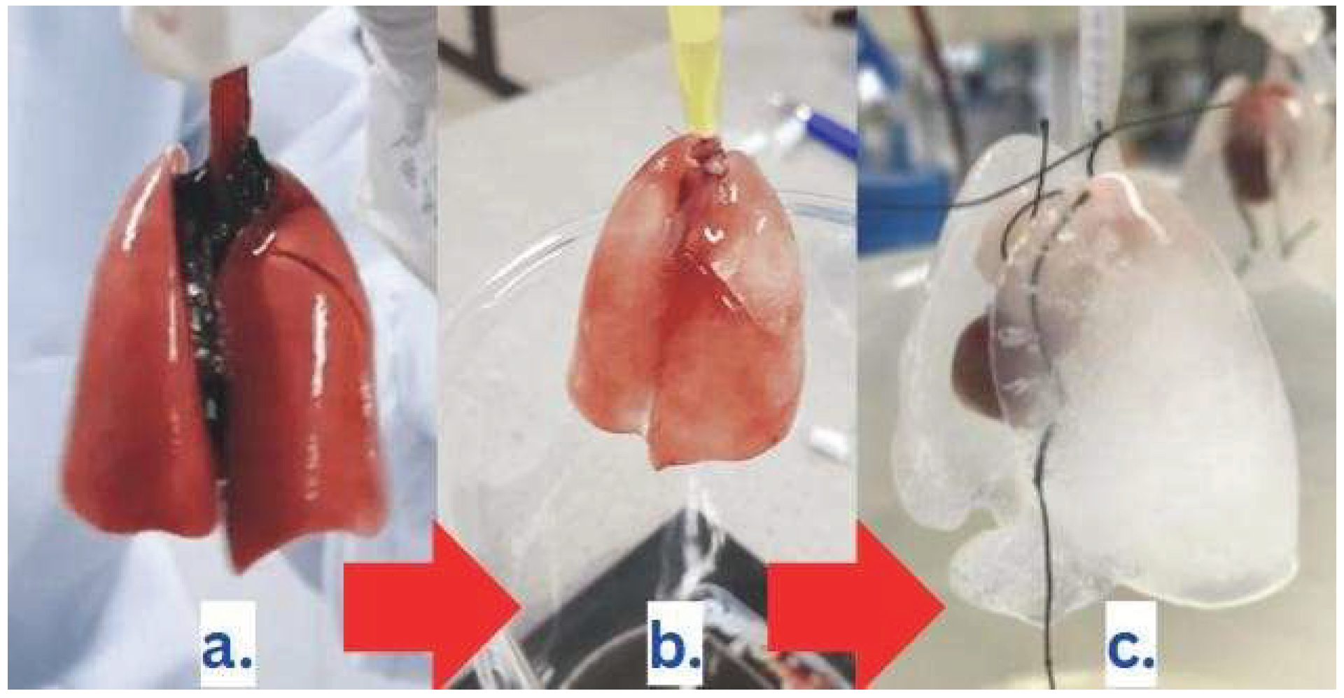
Decellularization is a technique that encompasses a series of processes and methodologies aimed at removing cellular components from a tissue or whole organ while preserving the extracellular matrix (ECM) as a structural scaffold to support subsequent recellularization [20]. Three primary methods are used to achieve this process. The physical method involves freeze–thaw cycles and agitation to induce cellular lysis. The chemical method employs detergents such as sodium dodecyl sulfate, which disrupt cellular membranes and facilitate their removal. Finally, the enzymatic method utilizes enzymes such as trypsin to digest membrane proteins and complete cellular breakdown [21]. Current biomedical research has shifted from the decellularization of isolated tissues toward whole-organ decellularization with the goal of generating transplantable grafts. While this approach has been effective for certain organs, it remains challenging for others. The technique is complex and may cause irreversible damage to the ECM if not properly optimized [20].
Pioneering studies conducted by Dr. Harald Ott and his research group demonstrated, for the first time in 2010, the successful recellularization of decellularized rat lungs, their implantation into living animals, and the achievement of functional in vivo gas exchange. Since then, biomedical research has continued to advance, incorporating larger animal models to further evaluate this approach [21].
Figure 1.
Lung decellularization process: (a) native lung; (b) lung undergoing decellularization; (c) decellularized lung.
Figure 1.
Lung decellularization process: (a) native lung; (b) lung undergoing decellularization; (c) decellularized lung.
B. Three-Dimensional (3D) Bioprinting
Beyond the use of decellularized biological matrices, bioartificial lung engineering has advanced through the application of 3D bioprinting. This technology enables the design of customized pulmonary structures that mimic the anatomical, mechanical, and functional characteristics of native tissue. The lung is a highly complex and dynamic organ, which makes comprehensive “in vitro” modeling particularly challenging. 3D bioprinting is still in its early stages of development toward the long-term goal of constructing dynamic, functional in vitro lung models. Current 3D bioprinting technologies lack the resolution required to accurately print and reproduce the small functional units of the lung, particularly the alveolar tissue [23,24]. However, significant progress has been made in developing bioinks. Bioinks are biomaterials used in 3D bioprinting that contain living cells and mimic the extracellular matrix (ECM) microenvironment. They promote cell adhesion, proliferation, and differentiation following the printing process. These materials exhibit tunable mechanical properties and bioactive compositions, opening new opportunities in lung tissue engineering [25]. By optimizing bioink composition, lung tissue constructs can be fabricated with the necessary mechanical strength, biocompatibility, and biodegradability [23]. A major challenge in these systems is achieving adequate vascularization, since synthetic tissues lack functional blood vessels at the time of fabrication. Sacrificial materials are also used in 3D bioprinting as temporary structural support during the printing process. These materials help overcome the limited mechanical stability of ECM-based hydrogels. Once printing is complete, the sacrificial material is carefully removed without damaging the primary structure, resulting in a complex, stable architecture compatible with living cells [26]. The field of 3D bioprinting has evolved rapidly toward the faithful reconstruction of native human lung structure. Although the development of a fully transplantable bio-printed lung remains a distant goal, current technological advances have significantly improved our understanding of pulmonary diseases and lung architecture
V. Cellular Recellularization
A. Cell Types Used
The functionality of a bioartificial lung depends largely on the type and proper spatial distribution of the cells used to repopulate the scaffold. Achieving effective regeneration requires a heterogeneous cell population that mimics the complexity of native lung tissue, including both the alveolar region and the vascular system [22]. One of the most critical cell groups is the alveolar epithelial cells, which include type I and type II pneumocytes [17]. Type I pneumocytes form the thin diffusion barrier across which gas exchange occurs. In contrast, type II pneumocytes contribute to alveolar epithelial repair and coordinate host defense mechanisms by maintaining a restrictive epithelial barrier [27]. Endothelial cells are also required to form the inner lining of blood vessels and to enable proper blood flow through the engineered lung. Mesenchymal cells play an important role in recellularization by supporting tissue regeneration and reducing the risk of immune rejection [28]. More recently, induced pluripotent stem cells (iPSCs) have gained significant attention. These cells can be derived from the patient and reprogrammed to differentiate into multiple pulmonary cell types, thereby reducing the risk of immunological rejection [29]. The appropriate selection, combination, and precise spatial distribution of these cell types within the scaffold are essential for restoring mature lung functions, including gas exchange, surfactant production, mucociliary clearance, and defense against external agents.
VI. Current Challenges
Despite progress in developing bioartificial lungs, significant challenges still limit their clinical implementation. Early efforts in lung tissue engineering focused on producing a highly elastic matrix capable of reproducing the pulmonary architecture required to support effective gas exchange [32]. This was followed by the need for successful recessing with endothelial and epithelial cells. In two preclinical studies conducted in small-animal models, the engineered lung constructs successfully performed oxygen and carbon dioxide exchange. However, after several hours, significant pulmonary secretions and airway bleeding were observed, indicating that these constructs were not yet suitable for human application [33]. Following these initial studies, similar methodologies were applied to porcine and human lungs, achieving successful decellularization and the generation of scaffolds for recellularization with new cells [34,35]. Additional challenges include the precise delivery and distribution of viable cells within the acellular scaffold to achieve functionality comparable to that of a native lung.
Induced pluripotent stem cells (iPSCs) have emerged as particularly promising cell sources because they enable patient-specific lung bioengineering and reduce the risk of immune rejection. Advances in biomedical engineering and related technologies have also enabled the expression of one of the earliest markers of pulmonary endoderm, Nkx2.1, a transcription factor that regulates lung development and surfactant gene expression [36]. Furthermore, the large size and structural complexity of the human lung require more sophisticated bioprinting technologies and bioreactor systems than those currently used in animal models [36].
VI. Future Applications
At present, tissue engineering has played a relatively limited role in direct patient treatment. Supplementary bladders, small-diameter blood vessels, skin grafts, cartilage, and even complete tracheal constructs have been successfully implanted in patients. However, these procedures remain experimental and are associated with high costs. More complex organs, such as the heart, lung, and liver, have been successfully generated in laboratory settings, but they are not yet sufficiently developed for clinical implantation [39]. In the specific case of the lung, one of the primary future goals is to improve device design and scale these systems to dimensions suitable for clinical use. Initially, these devices are expected to function as acellular systems in adult patients receiving anticoagulant therapy. Subsequently, they may evolve into scaffold-based structures resembling native lungs and lined with endothelial cells. This advancement could enable their use in infants and neonates with reduced anticoagulation requirements [41].
Simultaneously, efforts are focused on optimizing decellularization and cellular repopulation techniques to generate fully transplantable lungs. Current challenges include identifying the ideal cell source and achieving proper cellular maturation both in vitro and in vivo, with the objective of restoring physiological function without compromising lung structure [41]. Large-animal studies using decellularized lungs are also being planned to evaluate their potential clinical applicability. Decellularized tracheal implants have shown promising outcomes, suggesting that other similarly engineered organs may also become viable [41]. In addition, in vitro lung models are being developed to study pulmonary diseases, test therapeutic interventions, and better understand lung responses under different conditions [41].
VI. Conclusion
Lung bioengineering has advanced considerably over the past decades, emerging as a promising alternative to the scarcity of transplantable organs and the limitations associated with conventional respiratory support devices. Approaches based on the decellularization of pulmonary matrices, recellularization with specific cell populations, and the design of biomimetic scaffolds have demonstrated encouraging results in terms of structural and functional viability, particularly in preclinical models. However, the clinical translation of these systems still depends on overcoming critical barriers such as efficient vascularization, immunologically stable integration, and functional tissue maturation both in vitro and in vivo environments. The incorporation of technologies such as 3D bioprinting, dynamic perfusion bioreactor culture, and induced pluripotent stem cells (iPSCs) suggests a future in which bioartificial lungs will not only be viable but also customizable and safe.
In this context, bioartificial lungs are positioned as a multifunctional platform, not only for therapeutic applications in patients with end-stage respiratory failure but also as advanced tools for disease modeling, pharmacological testing, and pathophysiological studies. Their successful consolidation will require multidisciplinary approaches integrating tissue engineering, cellular biotechnology, biomaterials science, and translational medicine.
References
- Office of Disease Prevention and Health Promotion. (n.d.). Respiratory disease. Healthy People 2030. U.S. Department of Health and Human Services. https://odphp.health.gov/healthypeople/objectives-and-data/browse-objectives/respiratorydisease.
- Organ Procurement and Transplantation Network (OPTN); Scientific Registry of Transplant Recipients (SRTR). (2024). OPTN/SRTR 2022 Annual Data Report: Lung. U.S. Department of Health and Human Services, Health Resources and Services Administration. https://srtr.trans-plant.hrsa.gov/ADR/Chapter?name=Lung&year=2022.
- Nichols, J. E.; Niles, J. A.; Cortiella, J. (2009). Design and development of tissue engineered lung: Progress and challenges. Organogenesis 5(2), 5761.
- Nichols, J. E.; Cortiella, J. (2008). Engineering of a complex organ: progress toward development of a tissue-engineered lung. Proceedings of the American Thoracic Society 5(6), 723–730. [CrossRef]
- Olsen, C. E.; Isakson, B. E.; Seedorf, G. J.; Lubman, R. L.; Boitano, S. (2005). Extracellular matrix-driven alveolar epithelial cell differentiation in vitro. Experimental lung research 31(5), 461–482. [CrossRef]
- Mondrinos, M. J.; Koutzaki, S.; Lelkes, P. I.; Finck, C. M. (2007). A tissue-engineered model of fetal distal lung tissue. American journal of physiology-lung cellular and molecular physiology 293(3), L639–L650. [CrossRef] [PubMed]
- Chen, P.; Marsilio, E.; Goldstein, R. H.; Yannas, I. V.; Spector, M. (2005). Formation of lung alveolar-like structures in collagen–glycosaminoglycan scaffolds in vitro. Tissue engineering 11(9-10), 1436–1448. [CrossRef]
- Mondrinos, M. J.; Koutzaki, S.; Jiwanmall, E.; Li, M.; Dechadarevian, J. P.; Lelkes, P. I.; Finck, C. M. (2006). Engineering three-dimensional pulmonary tissue constructs. Tissue engineering 12(4), 717–728. [CrossRef] [PubMed]
- Mondrinos, M. J.; Koutzaki, S. H.; Poblete, H. M.; Crisanti, M. C.; Lelkes, P. I.; Finck, C. M. (2008). In vivo pulmonary tissue engineering: contribution of donor-derived endothelial cells to construct vascularization. Tissue Engineering Part A 14(3), 361–368. [CrossRef] [PubMed]
- Andrade, C. F.; Wong, A. P.; Waddell, T. K.; Keshavjee, S.; Liu, M. (2007). Cell-based tissue engineering for lung regeneration. American journal of physiology-lung cellular and molecular physiology 292(2), L510–L518. [CrossRef]
- Suki, B.; Ito, S.; Stamenovic, D.; Lutchen, K. R.; Ingenito, E. P. (2005). Biomechanics of the lung parenchyma: critical roles of collagen and mechanical forces. Journal of applied physiology 98(5), 1892–1899.
- Coraux, C.; Nawrocki-Raby, B.; Hinnrasky, J.; Kileztky, C.; Gaillard, D.; Dani, C.; Puchelle, E. (2005). Embryonic stem cells generate airway epithelial tissue. American journal of respiratory cell and molecular biology 32(2), 87–92. [CrossRef]
- Ott, H. C.; Clippinger, B.; Conrad, C.; Schuetz, C.; Pomerantseva, I.; Ikonomou, L.; Kotton, D.; Vacanti, J. P. (2010). Regeneration and orthotopic transplantation of a bioartificial lung. Nature medicine 16(8), 927–933. [CrossRef]
- Ritter, P.; Cai, A.; Reischl, B.; Fiedler, M.; Prölß, G.; Frieß, B.; Haug, M. (2022). MyoBio: An automated bioreactor system technology for standardized perfusion-decellularization of whole skeletal muscle. IEEE Transactions on Biomedical Engineering 69(7), 2305–2313. [CrossRef]
- Sánchez, T.; Concha, I. (2018). Estructura y funciones del sistema respiratorio. Neumologia pediatrica 13(3), 101–106. [CrossRef]
- Hall, J. E. (2016). Compendio de fisiología médica. Elsevier Health Sciences Spain-T.
- Rodríguez, R. R.; Hernández, M. L.; García, R. G.; Parrilla, F. J. V. (2012). Papel de los neumocitos II en un modelo experimental de lesión pulmonar aguda. Majorensis: Revista Electrónica de Ciencia y Tecnología (8), 21–31.
- Gilpin, S. E.; Charest, J. M.; Ren, X.; Ott, H. C. (2016). Bioengineering Lungs for Transplantation. Thoracic surgery clinics 26(2), 163–171. [CrossRef]
- Wang, Z.; Wang, Z.; Yu, Q.; Xi, H.; Weng, J.; Du, X.; Chen, D.; Ma, J.; Mei, J.; Chen, C. (2016). Comparative study of two perfusion routes with different flow in decellularization to harvest an optimal pulmonary scaffold for recellularization. Journal of biomedical materials research. Part A 104(10), 2567–2575. [CrossRef] [PubMed]
- Urbano, J. J., da Palma, R. K., de Lima, F. M., Fratini, P., Guimaraes, L. L., Uriarte, J. J., ... & Oliveira, L. V. F. (2017). Effects of two different decellularization routes on the mechanical properties of decellularized lungs. PLoS One 12(6), e0178696.
- Morales-Valencia, M.; Patino-Vargas, M. I.; Correa-Londono, L. A.; Restrepo-Munera, L. M. (2016). Evaluation of the chemical-enzymatic decellularization method for obtaining extracellular trachea matrixes in a porcine model/EvaluaciÃÂ3n del metodo quimico-enzimatico de descelularizaciÃÂ3n para la obtenciÃÂ3n de matrices extracelulares de traquea en el modelo porcino. Iatreia 29(2), 144–157.
- Ott, H.; Clippinger, B.; Conrad, C.; et al. Regeneration and orthotopic transplantation of a bioartificial lung. Nat Med 16, 927–933 (2010). [CrossRef]
- Dabaghi, M.; Carpio, M. B.; Moran-Mirabal, J. M.; Hirota, J. A. (2023). 3D (bio)printing of lungs: past, present, and future. The European respiratory journal 61(1), 2200417. [CrossRef]
- Galliger, Z; Vogt, CD; Panoskaltsis-Mortari, A. 3D bioprinting for lungs and hollow organs. Transl Res 2019, 211, 19–34. [Google Scholar] [CrossRef]
- Petrou, CL; D’Ovidio, TJ; Bölükbas, DA; et al. Clickable decellularized extracellular matrix as a new tool for building hybrid-hydrogels to model chronic fibrotic diseases in vitro. J Mater Chem B 2020, 8, 6814–6826. [Google Scholar] [CrossRef] [PubMed]
- Lee, A; Hudson, AR; Shiwarski, DJ; et al. 3D bioprinting of collagen to rebuild components of the human heart. Science 2019, 365, 482–487. [Google Scholar] [CrossRef] [PubMed]
- Ruaro, B.; Salton, F.; Braga, L.; Wade, B.; Confalonieri, P.; Volpe, M. C.; Baratella, E.; Maiocchi, S.; Confalonieri, M. (2021). The History and Mystery of Alveolar Epithelial Type II Cells: Focus on Their Physiologic and Pathologic Role in Lung. International journal of molecular sciences 22(5), 2566. [CrossRef]
- Wagner, D. E.; Bonvillain, R. W.; Jensen, T.; Girard, E. D.; Bunnell, B. A.; Finck, C. M.; Hoffman, A. M.; Weiss, D. J. (2013). Can stem cells be used to generate new lungs? Ex vivo lung bioengineering with decellularized whole lung scaffolds. Respirology (Carlton, Vic.) 18(6), 895–911. [CrossRef]
- Mejia-Montilla, Jorly & Reyna-Villasmil, Eduardo & Álvarez-Mon, Melchor & Fernández-Ramírez, Andreina. (2018). REVISIÓN CÉLULAS MADRE PLURIPOTENTES INDUCIDAS Y ADIPOGÉ-NESIS. Revista Venezolana de Oncologia. 16.
- Uriarte, J. J.; Uhl, F. E.; Rolandsson Enes, S. E.; Pouliot, R. A.; Weiss, D. J. (2018). Lung bioengineering: advances and challenges in lung decellularization and recellularization. Current opinion in organ transplantation 23(6), 673–678. [CrossRef]
- Welman, T.; Michel, S.; Segaren, N.; Shanmugarajah, K. (2015). Bioengineering for Organ Transplantation: Progress and Challenges. Bioengineered 6(5), 257–261. [CrossRef]
- Nichols, JE; Niles, JA; Cortiella, J. Design and development of tissue engineered lung: Progress and challenges. Organogenesis 2009, 5, 57–61. [Google Scholar] [CrossRef] [PubMed]
- Petersen, TH; Calle, EA; Zhao, L; Lee, EJ; Gui, L; Raredon, MB; Gavrilov, K; Yi, T; Zhuang, ZW; Breuer, C; et al. Tissue-engineered lungs for in vivo implantation. Science 2010, 329, 538–41. [Google Scholar] [CrossRef] [PubMed]
- Gilpin, SE; Guyette, JP; Gonzalez, G; Ren, X; Asara, JM; Mathisen, DJ; Vacanti, JP; Ott, HC. Perfusion decellularization of human and porcine lungs: Bringing the matrix to clinical scale. J Heart Lung Transplant 2014, 33, 298–308. [Google Scholar] [CrossRef] [PubMed]
- O’Neill, JD; Anfang, R; Anandappa, A; Costa, J; Javidfar, J; Wobma, HM; Singh, G; Freytes, DO; Bacchetta, MD; Sonett, JR; et al. Decellularization of human and porcine lung tissues for pulmonary tissue engineering. Ann Thorac Surg 2013, 96, 1046-55; discussion 1055-6. [Google Scholar] [CrossRef] [PubMed]
- Gilpin, SE; Ott, HC. Using nature’s platform to engineer bio-artificial lungs. Ann Am Thorac Soc 2015, 12 (Suppl 1), S45-9. [Google Scholar] [CrossRef] [PubMed]
- Rondón, J.; Sánchez Martínez, V.; Lugo, C.; Gonzalez-Lizardo, A. (2024). Tissue engineering: Advancements, challenges and future perspectives. Ciencia e Ingeniería 46 19–28.
- Rondón, J.; Gutiérrez, Y.; Lugo, C.; GonzalezLizardo, A. (2025). Artificial lung: A biomedical engineering approach Pulmón artificial: Un enfoque de ingeniería biomédica. Revista Ciencia e Ingeniería Vol, 46(2).
- National Institute of Biomedical Imaging and Bioengineering. (2022, abril). Ingeniería de tejidos y medicina regenerativa. https://www.nibib.nih.gov/espanol/temas-cientificos/ingenier%C3%ADa-de-tejidos-y-medicina-regenerativa-0.
- Hoganson, D. M.; Bassett, E. K.; Vacanti, J. P. (2014). Lung tissue engineering. Frontiers in bioscience (Landmark edition) 19(8), 1227–1239. [CrossRef] [PubMed]
- Swaminathan, V.; Bryant, B. R.; Tchantchaleishvili, V.; Rajab, T. K. (2021). Bioengineering lungs—Current status and future prospects. Expert Opinion on Biological Therapy 21(4), 465–471. [CrossRef]
- Organ Procurement and Transplantation Network (OPTN); Scientific Registry of Transplant Recipients (SRTR). (2024). OPTN/SRTR 2022 Annual Data Report: Lung. U.S. Department of Health and Human Services, Health Resources and Services Administration. https://srtr.trans-plant.hrsa.gov/ADR/Chapter?name=Lung&year=2022.
Disclaimer/Publisher’s Note: The statements, opinions and data contained in all publications are solely those of the individual author(s) and contributor(s) and not of MDPI and/or the editor(s). MDPI and/or the editor(s) disclaim responsibility for any injury to people or property resulting from any ideas, methods, instructions or products referred to in the content. |
© 2026 by the authors. Licensee MDPI, Basel, Switzerland. This article is an open access article distributed under the terms and conditions of the Creative Commons Attribution (CC BY) license (http://creativecommons.org/licenses/by/4.0/).
Copyright: This open access article is published under a Creative Commons CC BY 4.0 license, which permit the free download, distribution, and reuse, provided that the author and preprint are cited in any reuse.