

Submitted:
11 March 2026
Posted:
12 March 2026
You are already at the latest version
Abstract
Globally, approximately 18.6 million individuals develop diabetic foot ulcers each year, with an estimated 50–60% of these cases subsequently becoming infected. Diabetes-related foot infections (DFI) are a common and serious complication for patients with diabetes, often resulting in gangrene, lower extremity amputation and eventually death. Multidrug resistance among DFI pathogens aggravates treatment failure, driving up healthcare costs and morbidity. Addressing this multifaceted challenge necessitates the development of novel, synergistic, and innovative therapeutic strategies. Plasma-activated water (PAW) is an emerging solution, produced by treating water with cold plasma; an ionized gas that generates a complex mixture of reactive oxygen and nitrogen species. PAW elicits potent broad-spectrum antimicrobial and antibiofilm activity against a wide range of pathogens implicated in chronic wound infections, including DFI. Moreover, PAW has been shown to accelerate wound healing through modulating immune cell activity and promoting epithelial cell proliferation and migration into wounds. In this review, we summarize: (i) the prevalence and recurrence of DFIs, (ii) methods for PAW generation and its physicochemical properties, (iii) the antimicrobial and antibiofilm efficacy of PAW against clinically relevant DFI pathogens, (iv) its effects on cellular behavior, including immune modulation and promotion of epithelial regeneration, (v) PAW as a stand-alone or synergistic therapy (vi) current limitations in PAW application, including standardization, delivery, and regulatory hurdles. Together, this rationale highlights the notion that PAW holds significant potential as a next-generation therapeutic approach for DFIs and other chronic wounds.
Keywords:
antimicrobial resistance
; biofilm
; chronic wounds
; diabetes-related foot infections
; plasma-activated water
; plasma medicine.
1. Introduction
Chronic wounds represent a significant global health challenge, affecting millions of individuals and imposing substantial economic and social burdens. Specifically in Australia, more than 450,000 Australians suffer from chronic wounds which cost the health and aged care systems $6.6 billion yearly, with each patient spending an average of $4000 in out-of-pocket cost (Wounds Australia, 2024). Diabetes-related foot ulcers (DFUs) are among the most serious form of chronic wounds, as they recur frequently, are prone to persistent microbial infections, and often culminate into lower extremity amputation (LEA) (Armstrong et al. 2017; 2023; Narres et al. 2017; Ezzatvar and García-Hermoso 2023). Each year, an estimated 18.6 million individuals worldwide develop DFUs, with infection occurring in approximately 50–60% of cases (Armstrong et al. 2023). In North America alone, diabetes-related foot infections (DFIs) affects up to 13% of DFU patients (Zhang et al. 2017), highlighting both their global prevalence and disproportionate regional burden. Healing is often delayed by biofilm infections and rising antimicrobial resistance, limiting treatment options. Nevertheless, mortality rates following LEA are comparable to those of several major cancers like breast and lung cancers, underscoring the seriousness of this complication (McDermott et al. 2023). This is because, LEA carries a 5-year mortality risk ( 50–70%) which exceeds the pooled 5-year mortality for other major cancers like lung and breast cancer ( 31%) (Armstrong et al. 2020). Patients with DFU, arterial ulcers, and venous leg ulcers typically suffer from underlying immunodeficiency, peripheral neuropathy, and vascular disease, which compromise the healing process and predispose them to chronic, recurrent infections (Australian Journal of General Practice, 2020). The rise of multidrug-resistant (MDR) pathogens, particularly methicillin-resistant Staphylococcus aureus (MRSA), further undermines treatment success and increases the urgency for innovative, non-antibiotic therapies (Schaper et al. 2024).
Current wound care strategies face significant limitations because they rely on painful and possibly damaging debridement, unsuccessful and improper dressings, systemic antibiotics, and antiseptic solutions (Sood et al. 2014). However, most antiseptics in clinical use are cytotoxic, impair wound healing, and are not recommended for chronic wounds (Norman et al. 2017; Wilkins and Unverdorben 2013). In spite of this, silver-based dressings remain widely used in wound care and management due to the broad-spectrum antimicrobial activity against both Gram positive and Negative bacteria as well as antibiotic-resistant strains (May et al. 2022; Yousefian et al. 2023). The multimodal mechanism of action of silver-based dressings includes disruption of bacterial membranes, inducing oxidative stress, binding to nucleic acids, interfering with protein synthesis, prompting damage to various cellular organelles essential for bacterial gene transcription and cell wall synthesis (Obagi et al. 2019; Yousefian et al. 2023; May et al. 2022; Haidari et al. 2020). On the other hand, silver-based dressings have presented significant benefits in wound healing, with both preclinical and clinical studies reporting reduced microbial burden, improved epithelialization, and lower rates of infection, particularly in burn wounds (Davies et al. 2017; May et al. 2022). They are also highly essential in chronic wounds with impaired vascular supply and where systemic antibiotics may have limited penetration (Leaper 2006). However, there are concerns regarding cytotoxicity, particularly with prolonged use or uncontrolled accruement in wound areas which can inhibit skin cell proliferation and impair wound healing (AshaRani et al. 2009; Haidari et al. 2020). To address this, advancement in silver-based wound dressings led to the development of silver nanoparticles (AgNPs) that supply silver in small, controlled releases into wounds to exert efficacy and improve safety without compromising its antimicrobial and wound healing properties (Haidari et al. 2020; May et al. 2022). Yet, silver-based dressings like AgNPs are significantly expensive and raise environmental concerns due to the energy-intensive and waste-generating processes involved in nanoparticle production (Fahim et al. 2024).
Conversely, antibiotic therapy is increasingly undermined by MDR pathogens, while biofilm formation shields microbes from host immunity and antimicrobials. These limitations underscore the need for alternative or complementary antimicrobial strategies that are dual-action and can eradicate infection while stimulating tissue regeneration without significant adverse effects.
Plasma-activated water (PAW) made from cold plasma technology, presents a promising next-generation therapy capable of overcoming several challenges associated with antibiotics and alternative therapeutics. For instance, PAW generates a mixture of reactive oxygen and nitrogen species (RONS) that exert potent antimicrobial and antibiofilm effects. Moreover, PAW has low cytotoxicity and minimal risk of promoting resistance, highlighting its potential as an effective adjunct therapy.
Cold Plasma Technology
Cold plasma also known as non-thermal or atmospheric-pressure plasma, is a rapidly evolving technology that uses ionized gases for diverse applications in healthcare, materials science, agriculture, and electronics (Tanaka et al. 2021; Okyere et al. 2022; Reema et al. 2022; Rao et al. 2023; Kolbe et al. 2024). In contrast to thermal plasmas, which are generated at very high temperatures, cold plasma is produced by ionizing gases with strong electric fields, resulting in plasma successfully being generated at temperatures below 40°C (Laroussi 2020; Tabares and Junkar 2021). The non-thermal nature makes cold plasma safe for direct application to biological tissues for several minutes without causing heat damage (von Woedtke et al. 2020). Cold plasma devices typically contain electrodes and a power source that generate high-voltage fields to ionize gases such as air, O₂, N₂, He, or Ar (Starikovskiy 2015; Nwabor et al. 2022). The resulting plasma is a complex mixture of radicals, free electrons, ultraviolet radiation, charged ions, and excited molecules (von Woedtke et al. 2020). This unique composition confers broad antimicrobial activity, the ability to regulate inflammation, and stimulation of epithelial tissue growth (Abdo et al., 2022; Dubey et al., 2022; Gan et al., 2021). Several cold plasma devices are now clinically approved in Europe for wound therapy, and accumulating clinical evidence supports their use in wound healing and oncology (Stratmann et al. 2020; Faramarzi et al. 2021; Min et al. 2022; Nguyen et al. 2022; Strohal et al. 2022; Abu Rached et al. 2023; Bolgeo et al. 2023; Sabrin et al. 2024).
PAW is a promising derivative that extends the application of cold plasma. When cold plasma is discharged into water, it produces PAW, an acidic and oxidative liquid enriched with RONS. PAW has demonstrated potent antimicrobial, antibiofilm, anti-cancer, and wound-healing properties (Kim and Kim 2021; von Woedtke et al. 2025). Its liquid form offers practical advantages over direct cold plasma, such as easier storage, transport, and integration into conventional wound cleansing protocols. In preclinical studies, PAW has been shown to eradicate biofilms, including those formed by MDR pathogens like MRSA, while also promoting cellular proliferation, immune modulation, and tissue regeneration (Kaushik et al. 2018; Xu et al. 2020; Kim and Kim 2021; Mai-Prochnow et al. 2021; Abdo et al. 2022; Oliveira et al. 2022; Abdo et al. 2023; Lee et al. 2023; Vyas et al. 2023). Despite these advances, the clinical efficacy of PAW remains untested, and further research is required to optimize generation methods, dosing, and safety protocols (Cheng et al. 2020; Li et al. 2022; Woedtke et al. 2022).
This review summarizes current evidence and future directions for PAW in chronic wound management. We focus particularly on its relevance to DFUs and DFIs, highlighting antimicrobial and antibiofilm efficacy, effects on host cells and wound healing, and limitations that must be addressed for clinical translation. With the first-in-human clinical trial of PAW for DFU about to commence (ACTRN12625000902493), this is a timely opportunity to critically evaluate its potential to evolve from an experimental plasma-derived liquid into a clinically validated therapy for biofilm-associated chronic wounds.
2. Diabetes-Related Foot Ulcer
The prevalence data from the International Diabetes Federation (“IDF Diabetes Atlas 2025,”) found that an estimated 589 million adults aged 20-70, representing 11.1% of this age group, were living with diabetes. This number is projected to rise to 852.5 million by 2050 (“IDF Diabetes Atlas 2025,”). A significant morbidity in a high proportion of patients with diabetes is DFU; an open, chronic, non-healing wound often situated at the plantar surface of the foot of a person with diabetes (Singer et al. 2018). DFU arise from a combination of peripheral neuropathy, angiopathy, immune impairment, and biomechanical and vascular factors, which prevent wound healing and increase susceptibility to microbial infection (Ziegler et al. 2014; Aumiller and Dollahite 2015; Volmer-Thole and Lobmann 2016; Armstrong et al. 2023). These complications not only compromise patient outcomes but also impose a substantial burden of care.
Diabetes-related foot infections (DFIs), frequently associated with DFUs, are typically polymicrobial, comprising diverse Gram-positive (e.g., Staphylococcus aureus/MRSA, Enterococcus faecalis including vancomycin-resistant strains, Streptococcus pyogenes, Streptococcus agalactiae), Gram-negative bacteria (e.g; Pseudomonas aeruginosa, Escherichia coli, Klebsiella pneumoniae, Proteus mirabilis, Acinetobacter baumannii), alongside anaerobic bacteria (e.g; Bacteroides fragilis, Peptostreptococcus spp., Clostridium spp), and fungal species (e.g; Candida albicans, Aspergillus spp.) (Lipsky et al. 2012; Henig et al. 2018; Morton and Coghill 2024). However, a plethora of studies indicate that S. aureus/MRSA remains a prevalent pathogen in chronic wound infections like DFIs (Messad et al. 2013; Dunyach-Remy et al. 2016; Viswanathan et al. 2019; Lipsky et al. 2020; Reina-Bueno et al. 2020; Chen et al. 2023; Morton and Coghill 2024). According to Dunyach-Remy et al., 2016, MRSA commonly colonizes soft tissues and bones, with most infections localized to the feet. These localized infections further impair wound healing, and can rapidly progress into deeper tissues and bone, causing osteomyelitis. If untreated, this cascade often culminates in LEA, severely reducing quality of life and increasing mortality risk (Zubair et al. 2012; Frykberg and Banks 2015). According to Armstrong et al., about 20% of people with DFU will undergo a lower extremity amputation, either minor (ie, part of the foot) or major (ie, above foot) (Armstrong et al. 2023).
The upsurge in MDR amongst DFI pathogens results in treatment failures, increased disease incidence and severity, leading to escalated healthcare costs (López-Montesinos et al. 2020; Wozniak et al. 2022). A major culprit for this problem is biofilms which further exacerbate the severity of the problem. Biofilms are clusters of microbes embedded in a protective extracellular matrix that enables bacteria to: (i) withstand chemical and physical stress from external factors, (ii) communicate and exchange materials via nutrient and water channels, and (iii) become tolerant to antibiotics while sharing antimicrobial resistance genes via horizontal gene transfer (Hussain et al. 1993; Costerton et al. 1995; Munita and Arias 2016; Kragh and Richter 2022). Biofilm formation serves as a microbial tolerance mechanism that hampers antibiotic penetration and efficacy, often leading to sublethal exposure that fosters resistance among the microbes shielded within (Boahen et al. 2022; Jefferson 2004; Sharif and Yadav 2025).
While wound debridement is the gold standard method for physically removing biofilms and necrotic tissues in wound management, it is not adequate as a stand-alone measure as residual biofilm fragments can rapidly re-establish biofilms and perpetuate infection (Schwartz et al. 2014). This is due to the fact that biofilms can extend into deeper tissue layers where debridement cannot reach and restore biofilms often within 24–72 hours (Wolcott et al. 2010). Moreover, debridement is a physical process that may not remove all planktonic cells from the wound bed purulence (Schwartz et al. 2014). The surviving pathogens in the wound area and deeper tissues sustains characteristics of a biofilm and can therefore continue to endure antibiotics and the host defenses (Tian et al. 2023). Here, PAW’s potent antimicrobial action makes it a potential adjunct after debridement, helping to remove remaining pathogens and reduce the risk of re-colonization or infection.
2.1. Diabetes-Related Foot Ulcer Recurrence
By definition, a recurrent foot ulcer is defined as any new ulceration, irrespective of its anatomical location or the time elapsed since the previous event (Cheng et al. 2021). Approximately 40%, 60% and 65% of diabetes patients experience ulcer recurrence within 1, 3 or 5 years after the ulcer healed, respectively (Armstrong et al. 2017). The recurrence of DFU is a significant concern for individuals with diabetes, as a prior foot ulcer greatly increases the risk of recurrence compared to those without a history of DFU, particularly when preventive measures are not consistently followed (Armstrong et al. 2017). Inadequate wound care, including improper cleaning and dressing changes, can delay healing and increase the risk of infections (Nagle et al. 2023). Current management and first line therapy of DFU involves wound cleaning and dressing to prevent infection, offloading to minimize weight on foot, debridement of dead tissue, glycaemic control, and reperfusion of lower extremity ischemic tissue to restore blood flow (Armstrong et al. 2023; Alexiadou and Doupis 2012; Lim et al. 2017; Yang et al. 2022). Patients with DFU are often immunocompromised and have other diabetes-related comorbidities requiring management, including hypertension, neuropathy, renal disease, ischaemic heart disease, peripheral artery disease (PAD), stroke and obesity (Nowakowska et al. 2019; Pearson-Stuttard et al. 2022) that can also impact wound healing dynamics and limit surgical or pharmacological treatment options, response, and prognosis (McDermott et al. 2010; Fowkes et al. 2013; Frykberg et al. 2017; Arora et al. 2019; Verma et al. 2021). Patient factors, such as age, ethnicity, overall health status, adherence to treatment, lifestyle factors (e.g., smoking, nutrition, sedentary lifestyle, socioeconomic status) and geography (rural, remote, urban living) also affect the risk of DFI onset and recurrence (Guo and Dipietro 2010).
2.2. Prevalence of Diabetes-Related Foot Infections
DFI are a common and serious complication of DFU, with 50–70% of DFU becoming infected during the course of the disease (Del Core et al. 2018; Edmonds et al. 2021). The prevalence of DFI varies depending on factors such as the size and depth of the ulcer, presence of neuropathy or PAD, glycaemic control, foot deformities, trauma, previous history of infections and the quality and frequency of wound care (Schaper et al. 2020; Akkus and Sert 2022). These factors contribute to impaired wound healing and weakened immune activity that create an environment conducive to microbial colonization, biofilm growth and infection (Scalise et al. 2015). The most common pathogens isolated from infected DFU include Gram-positive staphylococci (including MRSA), streptococci, enterococci (including Enterococcus faecalis), Actinomyces spp. and aerobic Gram-negative bacilli species (Pseudomonas aeruginosa), Enterobacteriaceae (Escherichia coli, Klebsiella pneumoniae, Enterobacter spp.) and Acinetobacter spp. (Dwedar et al. 2015; Sadeghpour Heravi et al. 2019). As shown in Figure 1, infections caused by these microorganisms in DFU, especially MRSA, are characterized by biofilms which are commonly polymicrobial (Percival et al. 2018; Du et al. 2022; M. S. Khan et al. 2023). This is because the colonizing bacteria has the potential to coordinate with other microorganisms through quorum sensing (QS), to create a symbiotic environment for growth and survival (Pouget et al. 2020; Afonso et al. 2021). This strategy may also include the expression and horizontal gene transfer of resistance genes and the secretion of toxins into the microenvironment to protect their existence whilst allowing the pathogen to exhibit its virulence (Mottola et al. 2016).
The severity of DFI can range from mild to severe, with severe cases progressing to deep tissue infections, abscess formation, osteomyelitis and sepsis (Giurato et al. 2017). Severe infections can also lead to significant morbidity, tissue necrosis, thrombosis of small vessels and localized gangrene that may necessitate hospitalization, or LEA (Lauri et al. 2020; Boulton et al. 2020; Izumi 2018). Patients with DFI have a higher risk of hospitalization and longer hospital stays compared to those without infections, because DFI can become chronic and recurrent, especially if not adequately treated or if underlying risk factors are not addressed (Hicks et al. 2016). As a result, a multidisciplinary approach to treatment is encouraged to reduce incidence and severity of DFI to prevent lower extremity ischemia and amputations (Schaper et al. 2024). Novel therapeutics such as direct cold plasma or PAW together with adjuvant therapies may produce effective therapeutic options to prevent DFI and its recurrence.
3. Plasma-Activated Water Generation and Physicochemical Properties
Cold plasma is generated by applying an electric field to a gas, such as air, N2, O2, He or Ar, or a pure gas spiked with O2 or air, at atmospheric pressure (Woedtke et al. 2022; Tendero et al. 2006). The specific method is dependent on the desired RONS formed, the application requirements, and device design (Hadinoto et al. 2023). As such, cold plasma devices may adjust parameters such as voltage, frequency, operating gas, gas flow rate, and treatment duration to optimize plasma generation and treatment outcomes (Laroque et al. 2022). Cold plasma generates RONS in PAW, including short-lived species like hydroxyl radicals (OH•), superoxide (O 2•), nitric oxide (NO•), singlet oxygen (1O2) and peroxynitrite (OONO-), and long-lived species like hydrogen peroxide (H2O2), ozone (O3) and nitrate/nitrate ions (NO2-/NO3-) which influence the pH, conductivity and oxidation-reduction potential (ORP) of PAW (Chauvin et al. 2017; de Jager et al. 2017; Borkar et al. 2023; Pandey et al. 2023; Rahman et al. 2022). However, spiking noble gases with other molecular gases (e.g., O2, N2, air) can result in increased production of ROS compared with RNS (Lietz and Kushner 2018). Analogous to cold plasma, the choice of method (i.e., direct, or indirect) for generating PAW also depends on factors such as the desired chemical properties of PAW and specific application requirements (Mirpour et al. 2020; Bradu et al. 2020; Jungbauer et al. 2021). In the direct method, the cold plasma plume is discharged straight into water, whereas the indirect method generates reactive species in the gas phase, which subsequently dissolve into the liquid or react at the gas-liquid interphase to create PAW (Milhan et al. 2022).
Cold plasma devices typically utilize one of several methods to generate plasma, including dielectric barrier discharge (DBD), corona discharge, plasma jet, gliding arc, microwave, and piezoelectric direct discharge (PDD) (Dasan et al. 2017; Šimončicová et al. 2019; Korzec et al. 2021; Bae et al. 2022; T. M. Khan et al. 2023). Using DBD to generate PAW uses a high voltage alternating current, passes between two electrodes, separated by a dielectric barrier (Figure 2a). During PAW production, water itself is a dielectric medium and so acts as the dielectric barrier for producing PAW (Wang et al. 2022; Dhakal et al. 2023). Corona discharge involves applying a high-voltage electric field to a fine-tipped point of an electrode, creating an electrically conductive region of air around the electrode tip that becomes ionised and interacts with the water (Figure 2b) (Korachi et al. 2010; Lazra et al. 2020). Plasma jets generate plasma using gas flowing through a nozzle with high-voltage electrodes and typically utilize one of the aforementioned cold plasma generator designs to ionize a gas under high flow rate through a narrow channel to propel the cold plasma discharge into the liquid (Bae et al. 2022; Rupf et al. 2010). The plasma jet can be submerged or directed onto the surface of the water to create PAW (Figure 2c). The gliding arc method involves creating a high-voltage electric arc that slides along a pair of diverging electrodes. The arc ‘glides’ from the narrowest to the widest parts of the electrodes, ionizing the gas to plasma to create PAW (Figure 2d) (Elaragi 2015; Marcinauskas et al. 2024). An operating gas can also be introduced into a microwave cavity resonator that produces microwave radiation to generate cold plasma directly in contact with water or above the water surface (Figure 2e) (Lebedev 2010; Mallick et al. 2020). The PDD is a newer type of atmospheric pressure cold plasma discharge that uses a piezoelectric material to produce high voltage when mechanically stressed with pressure or vibration, creating an electric discharge that ionizes the surrounding gas or directly ionizes the water to form PAW (Figure 2f) (Korzec et al. 2021).
Depending on the conditions used in PAW generation, different physicochemical characteristics can be observed. The RONS composition and concentrations in PAW depend on the cold plasma design, gas feed composition, gas flow rate, power, voltage, exposure time and liquid volume (Rotondo et al. 2025). The RONS reduces the pH and increases the ORP and conductivity of the water (Zhao et al. 2020). The combined effects of acidic pH, high conductivity and elevated ORP make PAW a potent antimicrobial and oxidative agent (Guo et al. 2022). Once generated, PAW can be stored before use, with its stability depending on factors such as storage temperature, exposure to light, degree of cold plasma activation and the intended end-use ((Risa Vaka et al. 2019; Shen et al. 2016; Smet et al. 2019; Tsoukou et al. 2020).
4. Applications of Plasma-Activated Water
PAW can be generally applied for medical surface or equipment disinfection and sanitization (Soni et al. 2021; Park et al. 2017; Bălan et al. 2018; Sakudo et al. 2019), as demonstrated by the potent antimicrobial/antibiofilm activity against a wide range of infectious pathogens (Abdo et al. 2023; Lin et al. 2020; Darmanin et al. 2020; Lee et al. 2022; Rahman et al. 2022; Abdo et al. 2025). The mechanism of action involves disrupting microbial cell membranes, inhibiting enzyme activity, impairing QS, preventing or disrupting biofilm formation, and inducing oxidative stress, which together lead to microbial inactivation or cell death (Zhang et al. 2013; Royintarat et al. 2019; Zhang et al. 2024). PAW also shows promise as a disinfectant for medical equipment (e.g. endoscopes, dental waterlines) and surfaces (Lee et al. 2023; Bălan et al. 2018; Abuzairi et al. 2018; Noopan et al. 2019). It may also be useful for oral hygiene (Milhan et al. 2022; Qiao et al. 2022), and disinfection and irrigation of wounds (Xu et al. 2020; Busco et al. 2020; Wang et al. 2021; Fink et al. 2023).
The prospect to combine antimicrobial properties, immune activation and tissue regeneration potencies of PAW may enhance wound healing outcomes. This makes PAW a potentially valuable preliminary treatment or adjuvant to the current therapeutic arsenal for DFU. There is ongoing in vitro and in vivo research that continue to refine PAW generation, application, and efficacy by exploring the optimal chemical conditions against biofilm-infected wounds, and its long-term safety and effectiveness in wound care. Currently, a world-first Phase I clinical trial (ACTRN12625000902493) using PAW as a dual-action wound cleanser is currently underway, aiming to validate its effectiveness in simultaneously reducing microbial burden and accelerating healing.
4.1. Plasma-Activated Water Antibiofilm Efficacy in Diabetes-Related Foot Infections
PAW is garnering interest particularly in treating chronic and hard-to-heal wounds like DFU due to broad spectrum antimicrobial activity and the ability to enhance various aspects of the wound healing process (Lee et al. 2023; Abdo et al. 2025; Wang et al. 2021; Shen et al. 2016). RONS produced in PAW have a diverse role in disrupting biofilm formation to help prevent and treat wound infection (Figure 3).
The composition of PAW has been reported in various studies to impede biofilms of several microorganisms in vitro (e.g., E. coli, P. aeruginosa, and MRSA) that are relevant to chronic wound infections like DFU (Pereira et al. 2017; Wu et al. 2019; Su et al. 2022). This ‘cocktail’ of reactive species in PAW can disrupt biofilms by penetrating the biofilm matrix and inducing oxidative stress in microbial cells, resulting in damage to cell membranes, proteins, and DNA and eventually cell death (Charoux et al. 2020; Fasnacht and Polacek 2021). Some studies also demonstrated that cold plasma solutions can disrupt early stages of biofilm formation, by downregulating genes involved in bacterial motility and adherence (Joshi et al. 2010; 2015; Han et al. 2016). This broad-spectrum activity of the components of PAW is also beneficial in treating polymicrobial biofilms because they can interrupt QS between microbial cells (Cáp et al. 2012; Ali et al. 2023). Moreover, the interplay between ROS, RNS and water can further result in the generation of acids and oxidants such as nitric acid (HNO3), peroxynitrous acid (ONOOH) and hydrogen peroxide (H2O2), respectively, which are responsible for the pH decrease of water (Lukes et al. 2014; Tibbits et al. 2022). PAW has low pH, and chronic wounds are reported to have a relatively alkaline environment (Sim et al. 2022). As such, an acidic environment rather promotes the healing process and inhibits many skin pathogenic microbes (Qi et al. 2018; Wallace et al. 2019; Derwin et al. 2022; Sim et al. 2022). Despite its acidity, PAW can be made safe for application on the skin (Abdo et al. 2025; Wang et al. 2021). H2O2 can degrade the extracellular polymeric substances that constitute the biofilm matrix (Noh et al. 2020), making the biofilm structure more permeable and easier to eradicate within biofilms (Tibbits et al. 2022). H2O2 also causes oxidative stress by generating additional ROS within the cells, leading to protein denaturation, lipid peroxidation, and DNA strand breaks (Cadet and Wagner 2013; Ransy et al. 2020).
4.2. Cellular Effects of Plasma-Activated Water-Derived Reactive Species
Physiological RONS have a direct and active role in promoting cell survival, proliferation and migration of epithelial cells like keratinocytes to promote wound healing (Abdo and Kopecki 2024). In addition to its antibiofilm properties, PAW-derived RONS play crucial roles in cellular signalling pathways, influencing processes such as cell migration, proliferation, self-killing (apoptosis), self-eating (autophagy) and the release of growth factors and both pro- and anti-inflammatory cytokines/chemokines (Hsieh and Yang 2013; Bekeschus et al. 2016; Redza-Dutordoir and Averill-Bates 2016; Zarkovic 2020; Checa and Aran 2020). RONS can enhance collagen synthesis, and the proliferation and migration of major cell types (i.e., keratinocytes and fibroblasts) involved in wound healing tissue repair/regeneration (Wende et al. 2014; Schmidt et al. 2017; Bhartiya et al. 2021; Khorsandi et al. 2022). For instance, a study by Wende et al. (2014) using direct cold plasma therapy on keratinocytes found that DNA oxidation occurred, which subsequently promoted cell proliferation. The study demonstrated that longer treatment times led to more pronounced effects such as cell cycle arrest and greater extent of apoptosis. Inversely, shorter treatment times caused no detectable damage, as low doses of ROS were well tolerated by the cells, with no apparent effects after 24 hours (Wende et al. 2014). Although these findings were observed with direct cold plasma therapy, PAW exhibits a comparable RONS chemistry to cold plasma and may elicit similar responses.
Similarly, Schmidt et al. (2017) showed that lower cold plasma exposure led to restoration of cells and tissues as compared to longer cold plasma exposure that resulted in apoptosis. This was confirmed by identification of several proteins and growth factors by proteomics, including the upregulation of angio-associated migratory cell protein, a key driver of increased skin cell motility (Schmidt et al. 2017). Bhartiya et al. (2021) confirmed that lower cold plasma exposure is fitting for epithelial skin cell motility and proliferation and stimulates expression and upregulation of important genes involved in cell migration for skin repair.
The skin defends itself against redox-active elements (e.g., UV, ionizing radiation) that can induce DNA damage directly or stimulate intracellular reactive species generation. However, supratherapeutic exposure to cold plasma could modify or destroy the structure and function of lipids, proteins and DNA that can disrupt cell signaling pathways (Xu et al. 2017). As a result, downstream signaling cascades are triggered, leading to altered cytokine release and aggravating inflammatory skin diseases, as well as the development of non-healing wounds (Khorsandi et al. 2022; Xu et al. 2017). For instance, chronic wounds like DFU exhibit impaired phosphatidylinositol 3-kinase/protein kinase B (PI3K/Akt) signaling (an important, redox-sensitive intracellular signaling pathway with diverse regulatory functions in cell survival, angiogenesis, and tumorigenic processes (Kasowanjete et al. 2024), resulting in reduced cell survival and growth factor release that would otherwise stimulate wound healing (Jere et al. 2019). Plasma-activated media (PAM; cell media exposed to cold plasma with similar chemical properties to PAW) was shown to promote PI3K/Akt/mTOR cell signaling in keratinocytes in vitro (Wu et al. 2023), while direct cold plasma exposure promoted Akt activity in vivo (Zhu et al. 2025). Furthermore, PAM led to elevated Akt expression and phosphorylation (activation), with reduced p53 (master tumor suppressor protein that modulates cell cycle and initiates caspase-dependent apoptosis (Aubrey et al. 2018) activity to promote survival of skin-derived mesothelial cells (Holl et al. 2022). As a result, Akt promotes cyclin D expression to tilt cell cycle to G1/S (proliferative) phase (Hwang et al. 2021) and could be a factor that facilitates epithelial and mesothelial cell proliferation to close non-healing wounds.
Other cell signaling pathways, such as the nuclear factor erythroid-2 related factor 2 (Nrf2), mitogen-activated protein kinase (MAPK) and the unfolded protein response (UPR) pathways could also play a role in wound healing stimulated by PAW, as has been the case with direct cold plasma therapy (recently reviewed in (Abdo and Kopecki 2024). Particularly, the redox-sensitive Nrf2 and UPR have been shown to be the most enriched signaling pathways induced in epithelial cells in response to cold plasma exposure, promoting survival by inhibiting apoptosis (Scharf et al. 2019). The Nrf2 pathway activates the antioxidant response element (ARE) encodes a plethora of antioxidant enzymes to protect cells for oxidative stress, while the UPR, in response to endoplasmic reticulum stress, expresses chaperones that repair protein misfolding from oxidative stress (Suzuki and Yamamoto 2015; Liu et al. 2022; Hetz 2012). These pro-survival pathways in epithelial cells could help build an adaptive response to oxidative stress in the chronic wounds and possibly transition them to acute wounds that heal. Unfortunately, a similar analysis of cell signaling pathways involved in PAW therapy for wound healing is yet to be reported and is an open avenue for research.
These studies highlight the potential use of cold plasma treatment for medical applications, particularly in modulating cell growth (Bekeschus et al. 2016), migration (Arndt et al. 2015), inflammation (Bekeschus et al. 2021) and repairing tissue (Brun et al. 2014). This depends on the level of exposure, as excessive RONS exposure can induce various oxidative stress stimuli resulting in cellular dysfunction, trigger apoptotic effects, and promote excessive leukocyte/neutrophil transmigration into inflamed areas by upregulating the expression of cell adhesion molecules (Wende et al. 2014; Khorsandi et al. 2022; Xu et al. 2017; Hasse et al. 2016).
4.3. Wound Healing Studies of Plasma-Activated Water
To date, only a few studies have explored the general application of PAW in wound healing and the treatment of wound infections (Xu et al. 2020; Lee et al. 2023; Vyas et al. 2023; Abdo et al. 2025; Wang et al. 2021), which are summarized in Table 1. In a study by Xu et al. (2020), PAW produced by a DBD device eradicated common wound infection pathogens (P. aeruginosa, E. coli, S. aureus and S. paratyphi-B) in vitro and significantly accelerated wound healing compared to the vehicle control group in a mouse model with full-thickness skin wounds. By day 17, wounds in the PAW-treated group were fully healed, whereas those in the control group reached approximately 80% wound closure (Xu et al. 2020). Furthermore, histological analysis revealed that wounds treated with PAW had fewer inflammatory cells, suggesting reduced infection and inflammation, thus aiding in wound healing (Xu et al. 2020). Additionally, DNA sequencing of wound bacteria showed that PAW significantly reduced bacterial abundance. The authors also tested wound healing potency in a mouse model with PAW generated using a portable surface discharge plasma device compared to vehicle control (water) and medical alcohol-treated groups (Wang et al. 2021). PAW significantly accelerated wound closure as compared to both the alcohol-treated and control groups, reaching wound closure approximately 4 – 5 days faster than the control and comparison groups (Wang et al. 2021). This was linked to PAW promoting the expression of pro-inflammatory (IL-1β, IL-6) and anti-inflammatory (IL-10) cytokines and vascular endothelial growth factor (VEGF), leading to faster blood vessel formation and wound recovery to enhance wound healing (Wang et al. 2021). Finally, both studies showed no significant adverse effects in the internal organs such as the heart, liver, spleen, lung and kidney, or blood biochemical markers of the treated mice (Wang et al. 2021; Xu et al. 2017).
Vyas et al. (2023) researched the potential use of PAW as a pre-treatment for biofilm infected wounds in vitro (Vyas et al. 2023). It was demonstrated that PAW can significantly enhance the effectiveness of topical antiseptics povidone-iodine (PI), polyhexamethylene biguanide (PHMB) and medical-grade manuka honey, against E. coli biofilms (Vyas et al. 2023), which are notoriously difficult to eradicate in chronic wounds. PAW alone also exhibited bactericidal activity on both plastic and keratinocyte monolayers but demonstrated substantially more biofilm-eradicating power when combined with the other antiseptics, reducing the effective biofilm-killing concentration of these agents. Interestingly, scavenging of PAW-derived ROS (O2•-, O3, •OH) during PAW production almost completely abolished its antibiofilm activity against E. coli (Vyas et al. 2023). However, it remains unclear whether this finding diminishes the role of RNS, since their formation depends on the generation of precursory O2•-, •OH and O3 (Mai-Prochnow et al. 2021). Overall, PAW’s antibiofilm efficacy appears to be primarily driven by RONS-induced oxidative stress, which leads to rapid outer membrane permeability, depolarization, and ultimately cell death. Additionally, PAW exposure caused notable morphological changes in biofilm cells, including membrane blebbing and flattening, indicating severe membrane disruption (Vyas et al. 2023).
Another study assessed the effects of PAW produced by a microwave-generated low-pressure plasma system on skin cell migration and wound healing in vitro and in vivo (Lee et al. 2023). PAW treatment enhanced keratinocyte and fibroblast migration without harming cell viability, while increasing metalloproteinase-2 activity involved in tissue remodeling in vitro. PAW also boosted integrin β-subunits expression, and phosphorylation of FAK and paxillin proteins, which promoted cytoskeletal reorganization and cell migration of the skin cells (Lee et al. 2023). These cellular responses to PAW were completely ablated by preloading cells with the ROS-scavenger N-acetylcysteine, indicating that intracellular ROS generated by mitochondria and NADPH oxidase were involved (Lee et al. 2023). Additionally, PAW treatment also accelerated wound healing by enhancing epithelial regeneration and integrin-dependent signaling pathways related to cell adhesion and migration in vivo in rats (Lee et al. 2023).
Another study has investigated both the antimicrobial activity and effects of wound healing of PAW in a wound infection model in vivo (Abdo et al. 2025). Balb/c mice with partial-thickness scald wounds were infected with bioluminescent MRSA and allowed to form a biofilm over 24 hours, before applying twice-daily PAW or vehicle (water) treatment for 7 days with continuous daily infection monitoring by bioluminescence (Abdo et al. 2025). PAW significantly reduced bacterial infection from day 4–7, with biofilm bacteria count at day 8 finding a 1.33 log10 (CFU/g) reduction compared to vehicle treatment (Abdo et al. 2025). Macroscopic wound healing rates did not differ significantly between PAW and control groups, however, histological analysis showed significantly greater re-epithelialization in PAW-treated wounds compared to vehicle (Abdo et al. 2025). These results indicate that PAW is not only antimicrobial and biocompatible but also capable of promoting wound healing. In contrast, most wound antiseptic solutions currently in clinical use impair healing and are therefore not recommended for chronic wounds (Norman et al. 2017; Wilkins and Unverdorben 2013), highlighting PAW’s potential advantages over conventional antiseptics.
Overall, the studies above conclude that PAW is a promising, effective, and safe approach for promoting wound healing, with no reported side effects and a lower risk of driving antimicrobial resistance. However, PAW’s efficacy in treating chronic wounds like DFU/DFI are to be elucidated in clinical trials. Beyond DFU, PAW shows potential for broader application in chronic wounds, including burns, pressure ulcers and venous leg ulcers, where biofilm formation commonly obstructs healing (Bekeschus et al. 2021; Versey et al. 2021; Oliver et al. 2024). Moreover, PAW could reduce postoperative complications by serving as a prophylactic agent against surgical site infections. Preliminary evidence supports its value as an adjunct therapy, particularly for chronic wounds unresponsive to conventional treatments (Abu Rached et al. 2023). Importantly, a first-in-human Phase I clinical trial (ACTRN12625000902493) is about to commence, evaluating PAW as a dual-action wound cleanser for diabetic foot ulcers in comparison with standard-of-care treatments, aiming both to destroy bacteria and to accelerate wound healing. Further clinical trials are needed to confirm safety and efficacy of PAW, determine long-term adverse effects, establish standardized treatment protocols and validate the benefits of PAW across diverse patient populations.
Together, this growing body of evidence underscores the translational potential of PAW, advancing it from an experimental plasma-derived liquid towards a clinically validated therapy for biofilm-associated chronic wounds.
4.4. Integrating Plasma-Activated Water into Wound Management Protocol
The International Working Group on Diabetic Foot (IWGDF) publishes evidence-based guidelines for the diagnosis and treatment of DFI (Senneville et al. 2024; Schaper et al. 2024). In these current guidelines (updates 2023), prophylactic use of systemic or local antibiotics, topical antiseptics (including silver, honey, bacteriophages or negative pressure wound therapy), and topical application of antibiotics at the site of infection is not recommended (Recommendation 11, 23 and 24, respectively). Therefore, there are limited non-surgical strategies clinicians are recommended to perform to complement the surgical debridement of wounds. The overarching rationale of these recommendations is based on low quality of evidence, high risk of bias, and that most topical treatments either do not benefit or impede wound healing (Senneville et al. 2024). As a result, the IWGDF conclude that a key area of concern for the management of wound healing is the potential of topical antimicrobial administration to limit the use of systemic antibiotics (Senneville et al. 2024).
PAW could potentially benefit patients with DFU by promoting wound healing, as we discussed in Section 4.3. Additionally, PAW as a wound healing solution could also act pleiotropically in this use to help prevent infection in non-infected DFU due to its potent antimicrobial properties. This could also help limit reliance on systemic antibiotic use to address the IWGDF concern of systematic antibiotic risks of adverse effects, associated costs and propagation of antimicrobial resistance. Currently, all evidence of PAW therapy for wound healing are in cell and animal models; the efficacy of PAW in treating DFI and in healing DFU has not been reported in clinical trial but are starting (ACTRN12625000902493).
5. Limitations in PAW Applications
To date, animal studies of PAW in wound healing have primarily focused on acute wounds, which are predisposed to heal spontaneously and therefore do not address whether PAW can initiate healing in chronic wounds. Additionally, acute wound models fail to accurately reproduce the immunological and pathological profiles of non-healing wounds. Only a limited number of animal models adequately mimic human non-healing wounds such as DFU. Among these, the db/db mouse strain, carrying a mutation in the leptin receptor gene that causes hyperphagia, obesity, hyperglycemia and pancreatic β cell atrophy in C57BLKS/J mice, closely mimics diabetes (Saeed and Martins-Green 2024). C57BLKS/J db/db mice also exhibit significantly reduced wound revascularization and re-epithelialization, resulting in delayed wound closure of approximately 4 weeks compared to 10 days in wild-type controls, thereby providing at least a partial model of chronic, non-healing wounds (Michaels et al. 2007).
Pig models are considered the most suitable animal models for human wounds due to high similarity in skin morphology, immune response and wound healing processes (Summerfield et al. 2015; Al-Deen Said et al. 2022). Hence, pig models of chronic wounds, including infected wounds, have been explored (Roy et al. 2009; 2014; Nowak et al. 2021). These models produce slow-healing wounds that more closely replicate the characteristics of human non-healing wounds, including those associated with diabetes, and could therefore be valuable for testing the efficacy of PAW in chronic wound treatment. However, large animal models require specialized facilities, are costly and are not widely accessible, underscoring the need for more practical and affordable models to fill this gap.
Despite its promising properties, the practical implementation of PAW faces challenges related to scalability, stability, shelf-life and safety. Variability in the generation and composition of PAW can affect its consistency and reliability. More research is needed to fully understand the mechanisms through which PAW exerts its effects on wound healing (Abdo and Kopecki 2024; Konchekov et al. 2023). Although PAW is generally considered safe for human cells at certain exposure times or frequency, excessive levels of RONS can be cytotoxic and may damage healthy tissues if not carefully controlled (Abdo and Kopecki 2024; Balzer et al. 2015; Girard et al. 2016). Hence, wound treatment with PAW will be acute and intermittent. Additionally, since PAW is a surface disinfectant, it may not be able to penetrate into deeper tissues to clear deep tissues or bone infections. However, it may have bystander effects on surrounding tissues by regulating inflammatory responses which will help create a more favorable healing environment around the wound. Moreover, while the general antimicrobial mechanisms of PAW are known, the specific interactions between RONS and different types of pathogens at the molecular level are not fully understood, potentially hindering optimization and safe translation of PAW into clinical practice. Despite PAW treatment alone being likely limited in cases of advanced infection, the favorable safety profile of PAW also makes it useful as a prophylactic treatment in preventing infection without damaging wounded or surrounding healthy tissue. Accordingly, PAW should not be viewed as a stand-alone antimicrobial solution; rather, it represents a valuable addition to the clinician’s toolbox, complementing rather than replacing established wound care and infection management strategies.
6. Concluding Remarks
PAW is a promising tool for chronic wound management, offering broad-spectrum antimicrobial activity with a reduced risk of resistance development compared to conventional antibiotics. Its dual ability to control infection while modulating inflammation and promoting tissue regeneration positions PAW as a valuable adjunct to existing antimicrobial therapies and wound dressings. The synergistic effects of PAW also highlight its potential for managing and treating other wound types, including burns and surgical wounds. To realize this potential, further research is needed to optimize treatment protocols and conduct rigorous clinical trials that confirm efficacy and safety across diverse clinical settings. Part of clinical trials will also be to identify the best concentrations, preparations, and methods of PAW application, which is another gap in existing studies. In parallel, long-term safety studies will be critical to establish PAW’s suitability for widespread adoption in healthcare.
Author Contributions
Conceptualization: A.B, A.I.A, K.R. Visualization: A.B. Writing - original draft: A.B, A.I.A. Supervision: A.I.A, K.R. Writing – review & editing: A.I.A, K.R, N.M, G.P. All authors read and agreed to the final version of the manuscript.
Funding
The authors gratefully acknowledge the financial support of the University of Adelaide, the Medical Research Future Fund and the Basil Hetzel Institute for Translational Research. The authors would like to also acknowledge Stefania Lebedev for her kind contributions on the plasma device figures presented in the paper.
Conflicts of Interest
KR is co-founder and non-financial board member.of RIBU Plasma Pty Ltd., Australia (ABN 64 659 905 914) and is co-inventor of a patent (Buske, C., & Richter, K. (2024). Process, apparatus and use of an apparatus for producing a plasma-activated liquid. Patent No. WO2024013069. World Intellectual Property Organization). All other authors declare no competing interests.
Abbreviations
| AgNPs | Silver nanoparticles |
| APPJ | Atmospheric pressure plasma jet |
| ARE | Antioxidant response element |
| DBD | Dielectric barrier discharge |
| DFI | Diabetes-related wounds infection |
| DFU | Diabetes-related foot ulcers |
| LEA | Lower extremity amputation |
| MAPK | Mitogen-activated protein kinase |
| MDR | Multidrug resistance |
| MRSA | Methicillin-resistant Staphylococcus aureus |
| Nrf2 | Nuclear factor erythroid-2 related factor 2 |
| PAD | Peripheral artery disease |
| PAW | Plasma-activated water |
| PDD | Piezoelectric electric discharge |
| PI3K/Akt | Phosphatidylinositol 3-kinase/protein kinase B |
| QS | Quorum sensing |
| RNS | Reactive nitrogen species |
| RONS | Reactive oxygen and nitrogen species |
| ROS | Reactive oxygen species |
| UPR | Unfolded protein response |
References
- Abdo, A.; McWhorter, A.; Hasse, D.; Schmitt-John, T.; Richter, K. Efficacy of Plasma-Treated Water against Salmonella Typhimurium: Antibacterial Activity, Inhibition of Invasion, and Biofilm Disruption; Antibiotics: Basel, Switzerland, 2023; Vol. 12. no. 9. [Google Scholar] [CrossRef]
- Abdo, Adrian I.; Nguyen, Hang T.; Kaul, Laurine D.; et al. Plasma-Activated Water Accelerates Wound Healing and Reduces Staphylococcus Aureus Infection in Vivo. Biofilm 2025, 100308. [Google Scholar] [CrossRef]
- Abdo, Adrian I.; Schmitt-John, Thomas; Richter, Katharina. Cold Plasma Therapy as a Physical Antibiofilm Approach. In Antibiofilm Strategies: Current and Future Applications to Prevent, Control and Eradicate Biofilms; Richter, Katharina, Kragh, Kasper Nørskov, Eds.; Springer International Publishing, 2022. [Google Scholar] [CrossRef]
- Abdo, A.I.; Kopecki, Z. Comparing Redox and Intracellular Signalling Responses to Cold Plasma in Wound Healing and Cancer. Current Issues in Molecular Biology 2024, 46(5), 4885–923. [Google Scholar] [CrossRef]
- Abu Rached, N.; Kley, S.; Storck, M.; Meyer, T.; Stücker, M. Cold Plasma Therapy in Chronic Wounds-A Multicenter, Randomized Controlled Clinical Trial (Plasma on Chronic Wounds for Epidermal Regeneration Study): Preliminary Results. Journal of Clinical Medicine 2023, 12(15), 5121. [Google Scholar] [CrossRef] [PubMed]
- Abuzairi, T.; Ramadhanty, S.; Puspohadiningrum, D.F.; Ratnasari, A.; Poespawati, N.R.; Purnamaningsih, R.W. Investigation on Physicochemical Properties of Plasma-Activated Water for the Application of Medical Device Sterilization. AIP Conference Proceedings 2018, 1933(1). [Google Scholar] [CrossRef]
- Afonso, A.C.; Oliveira, D.; Saavedra, M.J.; Borges, A.; Simões, M. Biofilms in Diabetic Foot Ulcers: Impact, Risk Factors and Control Strategies. International Journal of Molecular Sciences 2021, 22(15), 8278. [Google Scholar] [CrossRef]
- Akkus, G.; Sert, M. Diabetic Foot Ulcers: A Devastating Complication of Diabetes Mellitus Continues Non-Stop in Spite of New Medical Treatment Modalities. World Journal of Diabetes 2022, 13(12), 1106–21. [Google Scholar] [CrossRef]
- Al-Deen Said, Sayf; Jatana, Samreen; Ponti, András K.; et al. Development of a Reproducible Porcine Model of Infected Burn Wounds. Journal of Biological Methods 2022, 9(1), e158. [Google Scholar] [CrossRef] [PubMed]
- Alexiadou, K.; Doupis, J. Management of Diabetic Foot Ulcers. Diabetes Therapy : Research, Treatment and Education of Diabetes and Related Disorders 2012, 3(1), 4. [Google Scholar] [CrossRef]
- Ali, A.; Zahra, A.; Kamthan, M.; et al. Microbial Biofilms: Applications, Clinical Consequences, and Alternative Therapies. Microorganisms 2023, 11(8). [Google Scholar] [CrossRef] [PubMed]
- Armstrong, David G.; Swerdlow, Mark A.; Armstrong, Alexandria A.; Conte, Michael S.; Padula, William V.; Bus, Sicco A. Five Year Mortality and Direct Costs of Care for People with Diabetic Foot Complications Are Comparable to Cancer. Journal of Foot and Ankle Research 2020, 13(1), 16. [Google Scholar] [CrossRef]
- Armstrong, D.G.; Boulton, A.J.M.; Bus, S.A. Diabetic Foot Ulcers and Their Recurrence. The New England Journal of Medicine 2017, 376(24), 2367–75. [Google Scholar] [CrossRef]
- Armstrong, D.G.; Tan, T.W.; Boulton, A.J.M.; Bus, S.A. Diabetic Foot Ulcers: A Review. JAMA 2023, 330(1), 62–75. [Google Scholar] [CrossRef]
- Arndt, Stephanie; Landthaler, Michael; Zimmermann, Julia L.; et al. Effects of Cold Atmospheric Plasma (CAP) on ß-Defensins, Inflammatory Cytokines, and Apoptosis-Related Molecules in Keratinocytes in Vitro and in Vivo. PloS One 2015, 10(3), e0120041. [Google Scholar] [CrossRef] [PubMed]
- Arora, E.; Maiya, A.G.; Devasia, T.; Bhat, R.; Kamath, G. Prevalence of peripheral arterial disease among type 2 diabetes mellitus in coastal Karnataka. Diabetes & metabolic syndrome 2019, 13(2), 1251–53. [Google Scholar] [CrossRef]
- AshaRani, P. V.; Mun, Grace Low Kah; Hande, Manoor Prakash; Valiyaveettil, Suresh. Cytotoxicity and Genotoxicity of Silver Nanoparticles in Human Cells. ACS Nano 2009, 3(2), 279–90. [Google Scholar] [CrossRef]
- Aubrey, Brandon J.; Kelly, Gemma L.; Janic, Ana; Herold, Marco J.; Strasser, Andreas. How Does P53 Induce Apoptosis and How Does This Relate to P53-Mediated Tumour Suppression? Cell Death & Differentiation 2018, 25(1), 104–13. [Google Scholar] [CrossRef]
- Aumiller, W.D.; Dollahite, H.A. Pathogenesis and Management of Diabetic Foot Ulcers. JAAPA : Official Journal of the American Academy of Physician Assistants 2015, 28(5), 28–34. [Google Scholar] [CrossRef] [PubMed]
- Australian Journal of General Practice. Diabetic Foot Ulcer. 2020. Available online: https://www1.racgp.org.au/ajgp/2020/may/diabetic-foot-ulcer.
- Bae, G.T.; Jang, H.J.; Jung, E.Y.; et al. Development of an Atmospheric Pressure Plasma Jet Device Using Four-Bore Tubing and Its Applications of In-Liquid Material Decomposition and Solution Plasma Polymerization. Polymers 2022, 14(22), 4917. [Google Scholar] [CrossRef] [PubMed]
- Bălan, G.G.; Roşca, I.; Ursu, E.L.; et al. Plasma-Activated Water: A New and Effective Alternative for Duodenoscope Reprocessing. Infection and Drug Resistance 2018, 11, 727–33. [Google Scholar] [CrossRef]
- Balzer, J.; Heuer, K.; Demir, E.; et al. Non-Thermal Dielectric Barrier Discharge (DBD) Effects on Proliferation and Differentiation of Human Fibroblasts Are Primary Mediated by Hydrogen Peroxide. PloS One 2015, 10(12), 0144968. [Google Scholar] [CrossRef]
- Bekeschus, S.; Schmidt, A.; Bethge, L.; et al. Redox Stimulation of Human THP-1 Monocytes in Response to Cold Physical Plasma. Oxidative Medicine and Cellular Longevity 2016, 5910695. [Google Scholar] [CrossRef] [PubMed]
- Bekeschus, S.; Woedtke, T.; Emmert, S.; Schmidt, A. Medical Gas Plasma-Stimulated Wound Healing: Evidence and Mechanisms. Redox Biology 2021, 46, 102116. [Google Scholar] [CrossRef]
- Bhartiya, P.; Masur, K.; Shome, D.; et al. Influence of Redox Stress on Crosstalk between Fibroblasts and Keratinocytes. Biology 2021, 10(12), 1338. [Google Scholar] [CrossRef] [PubMed]
- Boahen, A.; Than, L.T.L.; Loke, Y.L.; Chew, S.Y. The Antibiofilm Role of Biotics Family in Vaginal Fungal Infections. Frontiers in Microbiology 2022, 13, 787119. [Google Scholar] [CrossRef]
- Bolgeo, T.; Maconi, A.; Gardalini, M.; et al. The Role of Cold Atmospheric Plasma in Wound Healing Processes in Critically Ill Patients. Journal of Personalized Medicine 2023, 13(5), 736. [Google Scholar] [CrossRef] [PubMed]
- Borkar, S.B.; Negi, M.; Kaushik, N.; et al. Plasma-Generated Nitric Oxide Water Mediates Environmentally Transmitted Pathogenic Bacterial Inactivation via Intracellular Nitrosative Stress. International Journal of Molecular Sciences 2023, 24(3). [Google Scholar] [CrossRef]
- Boulton, A.J.M.; Armstrong, D.G.; Hardman, M.J.; et al. Diagnosis and Management of Diabetic Foot Infections. In American Diabetes Association; 2020. [Google Scholar] [CrossRef]
- Bradu, C.; Kutasi, K.; Magureanu, M.; Puač, N.; Živković, S. Reactive Nitrogen Species in Plasma-Activated Water: Generation, Chemistry and Application in Agriculture. Journal of Physics D: Applied Physics 2020, 53(22), 223001. [Google Scholar] [CrossRef]
- Brun, Paola; Pathak, Surajit; Castagliuolo, Ignazio; et al. Helium Generated Cold Plasma Finely Regulates Activation of Human Fibroblast-like Primary Cells. PloS One 2014, 9(8), e104397. [Google Scholar] [CrossRef]
- Busco, G.; Robert, E.; Chettouh-Hammas, N.; Pouvesle, J.M.; Grillon, C. The Emerging Potential of Cold Atmospheric Plasma in Skin Biology. Free Radical Biology & Medicine 2020, 161, 290–304. [Google Scholar] [CrossRef]
- Cadet, J.; Wagner, J.R. DNA Base Damage by Reactive Oxygen Species, Oxidizing Agents, and UV Radiation. Cold Spring Harbor Perspectives in Biology 2013, 5(2), 012559. [Google Scholar] [CrossRef]
- Cáp, M.; Váchová, L.; Palková, Z. Reactive Oxygen Species in the Signaling and Adaptation of Multicellular Microbial Communities. Oxidative Medicine and Cellular Longevity 2012, 976753. [Google Scholar] [CrossRef]
- Charoux, C.M.G.; Patange, A.D.; Hinds, L.M.; Simpson, J.C.; O’Donnell, C.P.; Tiwari, B.K. Antimicrobial Effects of Airborne Acoustic Ultrasound and Plasma Activated Water from Cold and Thermal Plasma Systems on Biofilms. Scientific Reports 2020, 10(1), 17297. [Google Scholar] [CrossRef]
- Chauvin, J.; Judée, F.; Yousfi, M.; Vicendo, P.; Merbahi, N. Analysis of Reactive Oxygen and Nitrogen Species Generated in Three Liquid Media by Low Temperature Helium Plasma Jet. Scientific Reports 2017, 7(1), 4562. [Google Scholar] [CrossRef]
- Checa, J.; Aran, J.M. Reactive Oxygen Species: Drivers of Physiological and Pathological Processes. Journal of Inflammation Research 2020, 13, 1057–73. [Google Scholar] [CrossRef]
- Chen, Y.; Yang, J.; Wang, Y.; et al. Community-Associated Methicillin-Resistant Staphylococcus Aureus Infection of Diabetic Foot Ulcers in an Eastern Diabetic Foot Center in a Tertiary Hospital in China: A Retrospective Study. BMC Infectious Diseases 2023, 23(1), 652. [Google Scholar] [CrossRef] [PubMed]
- Cheng, He; Xu, JiaXing; Li, Xu; Liu, DaWei; Lu, XinPei. On the Dose of Plasma Medicine: Equivalent Total Oxidation Potential (ETOP). Physics of Plasmas 2020, 27(6), 063514. [Google Scholar] [CrossRef]
- Cheng, Y.; Zu, P.; Zhao, J.; et al. Differences in Initial versus Recurrent Diabetic Foot Ulcers at a Specialized Tertiary Diabetic Foot Care Center in China. The Journal of International Medical Research 2021, 49(1), 300060520987398. [Google Scholar] [CrossRef]
- Costerton, J. W.; Lewandowski, Z.; Caldwell, D. E.; Korber, D. R.; Lappin-Scott, H. M. Microbial Biofilms. Annual Review of Microbiology 1995, 49, 711–45. [Google Scholar] [CrossRef]
- Darmanin, M.; Kozak, D.; Oliveira Mallia, J.; Blundell, R.; Gatt, R.; Valdramidis, V.P. Generation of Plasma Functionalized Water: Antimicrobial Assessment and Impact on Seed Germination. Food Control 2020, 113, 107168. [Google Scholar] [CrossRef]
- Dasan, Beyhan Gunaydin; Onal-Ulusoy, Baran; Pawlat, Joanna; Diatczyk, Jaroslaw; Sen, Yasin; Mutlu, Mehmet. A New and Simple Approach for Decontamination of Food Contact Surfaces with Gliding Arc Discharge Atmospheric Non-Thermal Plasma. Food and Bioprocess Technology 2017, 10(4), 650–61. [Google Scholar] [CrossRef]
- Davies, Phil; McCarty, Sara; Hamberg, Kristina. Silver-Containing Foam Dressings with Safetac: A Review of the Scientific and Clinical Data. Journal of Wound Care 2017, 26(Sup6a), S1–32. [Google Scholar] [CrossRef] [PubMed]
- Del Core, M.A.; Ahn, J.; Lewis, R.B.; Raspovic, K.M.; Lalli, T.A.J.; Wukich, D.K. The Evaluation and Treatment of Diabetic Foot Ulcers and Diabetic Foot Infections. Foot & Ankle Orthopaedics 2018, 3(3). [Google Scholar] [CrossRef]
- Derwin, R.; Patton, D.; Avsar, P.; Strapp, H.; Moore, Z. The Impact of Topical Agents and Dressing on pH and Temperature on Wound Healing: A Systematic, Narrative Review. International Wound Journal 2022, 19(6), 1397–408. [Google Scholar] [CrossRef]
- Dhakal, O.B.; Dahal, R.; Acharya, T.R.; et al. Effects of Spark Dielectric Barrier Discharge Plasma on Water Sterilization and Seed Germination. Current Applied Physics 2023, 54, 49–58. [Google Scholar] [CrossRef]
- Du, F.; Ma, J.; Gong, H.; et al. “Microbial Infection and Antibiotic Susceptibility of Diabetic Foot Ulcer in China.” Literature Review. Frontiers in Endocrinology 2022, 13, 881659. [Google Scholar] [CrossRef] [PubMed]
- Dubey, Sunil Kumar; Parab, Shraddha; Alexander, Amit; et al. Cold Atmospheric Plasma Therapy in Wound Healing. Process Biochemistry 2022, 112, 112–23. [Google Scholar] [CrossRef]
- Dunyach-Remy, C.; Essebe, C. Ngba; Sotto, A.; Lavigne, J.P. Staphylococcus Aureus Toxins and Diabetic Foot Ulcers: Role in Pathogenesis and Interest in Diagnosis. Toxins 2016, 8(7), 209. [Google Scholar] [CrossRef]
- Dwedar, R.; Ismail, D.; Abdulbaky, A. Diabetic Foot Infection: Microbiological Causes with Special Reference to Their Antibiotic Resistance Pattern. Egyptian Journal of Medical Microbiology 2015, 24(1), 95–102. [Google Scholar] [CrossRef]
- Edmonds, M.; Manu, C.; Vas, P. The Current Burden of Diabetic Foot Disease. Journal of Clinical Orthopaedics and Trauma 2021, 17, 88–93. [Google Scholar] [CrossRef]
- Elaragi, Gamal. DISCHARGE CHARACTERISTICS OF GLIDING ARC PLASMA REACTOR WITH ARGON/NITROGEN. JOURNAL OF ADVANCES IN PHYSICS 2015, 7, 1316–23. [Google Scholar] [CrossRef]
- Ezzatvar, Y.; García-Hermoso, A. Global Estimates of Diabetes-Related Amputations Incidence in 2010-2020: A Systematic Review and Meta-Analysis. Diabetes Research and Clinical Practice 2023, 195, 110194. [Google Scholar] [CrossRef]
- Fahim, Mohd; Shahzaib, Adnan; Nishat, Nahid; Jahan, Afroz; Bhat, Tahir Ahmad; Inam, Afreen. Green Synthesis of Silver Nanoparticles: A Comprehensive Review of Methods, Influencing Factors, and Applications. JCIS Open 2024, 16, 100125. [Google Scholar] [CrossRef]
- Faramarzi, F.; Zafari, P.; Alimohammadi, M.; Moonesi, M.; Rafiei, A.; Bekeschus, S. Cold Physical Plasma in Cancer Therapy: Mechanisms, Signaling, and Immunity. Oxidative Medicine and Cellular Longevity 2021, 9916796. [Google Scholar] [CrossRef] [PubMed]
- Fasnacht, M.; Polacek, N. Oxidative Stress in Bacteria and the Central Dogma of Molecular Biology. Frontiers in Molecular Biosciences 2021, 8, 671037. [Google Scholar] [CrossRef] [PubMed]
- Fink, S.; Fischer, M.; Spange, S.; et al. Cold Atmospheric Plasma Exerts Antimicrobial Effects in a 3D Skin Model of Cutaneous Candidiasis; Antibiotics: Basel, Switzerland, 2023; Vol. 12. no. 5. [Google Scholar] [CrossRef]
- Fowkes, F.G.; Rudan, D.; Rudan, I.; et al. Comparison of Global Estimates of Prevalence and Risk Factors for Peripheral Artery Disease in 2000 and 2010: A Systematic Review and Analysis; Lancet: London, England, 2013; Vol. 382. no. 9901. [Google Scholar] [CrossRef]
- Frykberg, R.G.; Banks, J. Challenges in the Treatment of Chronic Wounds. Advances in Wound Care 2015, 4(9), 560–82. [Google Scholar] [CrossRef]
- Frykberg, R.G.; Gibbons, G.W.; Walters, J.L.; Wukich, D.K.; Milstein, F.C. A Prospective, Multicentre, Open-Label, Single-Arm Clinical Trial for Treatment of Chronic Complex Diabetic Foot Wounds with Exposed Tendon and/or Bone: Positive Clinical Outcomes of Viable Cryopreserved Human Placental Membrane. International Wound Journal 2017, 14(3), 569–77. [Google Scholar] [CrossRef]
- Gan, Lu; Jiang, Jian; Duan, Jiang Wei; et al. Cold Atmospheric Plasma Applications in Dermatology: A Systematic Review. Journal of Biophotonics 2021, 14(3), e202000415. [Google Scholar] [CrossRef]
- Girard, P.M.; Arbabian, A.; Fleury, M.; et al. Synergistic Effect of H2O2 and NO2 in Cell Death Induced by Cold Atmospheric He Plasma. Scientific Reports 2016, 6, 29098. [Google Scholar] [CrossRef]
- Giurato, L.; Meloni, M.; Izzo, V.; Uccioli, L. Osteomyelitis in Diabetic Foot: A Comprehensive Overview. World Journal of Diabetes 2017, 8(4), 135–42. [Google Scholar] [CrossRef]
- Guo, Jian; Wang, Jiaoyu; Xie, Hui; et al. Inactivation Effects of Plasma-Activated Water on Fusarium Graminearum. Food Control 2022, 134, 108683. [Google Scholar] [CrossRef]
- Guo, S.; Dipietro, L.A. Factors Affecting Wound Healing. Journal of Dental Research 2010, 89(3), 219–29. [Google Scholar] [CrossRef]
- Hadinoto, K.; Niemira, B.A.; Trujillo, F.J. A Review on Plasma-Activated Water and Its Application in the Meat Industry. Comprehensive Reviews in Food Science and Food Safety 2023, 22(6), 4993–5019. [Google Scholar] [CrossRef] [PubMed]
- Haidari, Hanif; Garg, Sanjay; Vasilev, Krasimir; Kopecki, Zlatko; Cowin, Allison J. Silver-Based Wound Dressings: Current Issues and Future Developments for Treating Bacterial Infections. Wound Practice and Research 2020, 28(4). [Google Scholar] [CrossRef]
- Han, L.; Boehm, D.; Patil, S.; Cullen, P.j.; Bourke, P. Assessing Stress Responses to Atmospheric Cold Plasma Exposure Using Escherichia Coli Knock-out Mutants. Journal of Applied Microbiology 2016, 121(2), 352–63. [Google Scholar] [CrossRef]
- Hasse, S.; Tran, T. Duong; Hahn, O.; et al. Induction of Proliferation of Basal Epidermal Keratinocytes by Cold Atmospheric-Pressure Plasma. Clinical and Experimental Dermatology 2016, 41(2), 202–9. [Google Scholar] [CrossRef] [PubMed]
- Henig, Oryan; Pogue, Jason M.; Cha, Raymond; et al. Epidemiology of Diabetic Foot Infection in the Metro-Detroit Area With a Focus on Independent Predictors for Pathogens Resistant to Recommended Empiric Antimicrobial Therapy. Open Forum Infectious Diseases 2018, 5(11), ofy245. [Google Scholar] [CrossRef] [PubMed]
- Hetz, Claudio. The Unfolded Protein Response: Controlling Cell Fate Decisions under ER Stress and Beyond. Nature Reviews Molecular Cell Biology 2012, 13(2), 89–102. [Google Scholar] [CrossRef]
- Hicks, Caitlin W.; Selvarajah, Shalini; Mathioudakis, Nestoras; et al. Burden of Infected Diabetic Foot Ulcers on Hospital Admissions and Costs. Annals of Vascular Surgery 2016, 33, 149–58. [Google Scholar] [CrossRef]
- Holl, Myriam; Rasch, Marie-Lena; Becker, Lucas; et al. Cell Type-Specific Anti-Adhesion Properties of Peritoneal Cell Treatment with Plasma-Activated Media (PAM). Biomedicines 2022, 10(4), 927. [Google Scholar] [CrossRef]
- Hsieh, H.L.; Yang, C.M. Role of Redox Signaling in Neuroinflammation and Neurodegenerative Diseases. BioMed Research International 2013, 484613. [Google Scholar] [CrossRef]
- Hussain, M.; Wilcox, M. H.; White, P. J. The Slime of Coagulase-Negative Staphylococci: Biochemistry and Relation to Adherence. FEMS Microbiology Reviews 1993, 10(3–4), 191–207. [Google Scholar] [CrossRef] [PubMed]
- Hwang, Ji-Hye; Lee, Hyun-Young; Chung, Kyung Bae; et al. Non-Thermal Atmospheric Pressure Plasma Activates Wnt/β-Catenin Signaling in Dermal Papilla Cells. Scientific Reports 2021, 11(1), 16125. [Google Scholar] [CrossRef]
- IDF Diabetes Atlas 2025. Available online: https://diabetesatlas.org/resources/idf-diabetes-atlas-2025/ (accessed on 11 December 2025).
- Izumi, Y. Countermeasures against Infection in Critical Limb Ischemia Treatments. Annals of Vascular Diseases 2018, 11(3), 277–80. [Google Scholar] [CrossRef]
- de Jager, T. L.; Cockrell, A. E.; Du Plessis, S. S. Ultraviolet Light Induced Generation of Reactive Oxygen Species. In Ultraviolet Light in Human Health, Diseases and Environment; Ahmad, Shamim I., Ed.; Springer International Publishing, 2017. [Google Scholar] [CrossRef]
- Jefferson, Kimberly K. What Drives Bacteria to Produce a Biofilm? FEMS Microbiology Letters 2004, 236(2), 163–73. [Google Scholar] [CrossRef]
- Jere, Sandy W.; Houreld, Nicolette N.; Abrahamse, Heidi. Role of the PI3K/AKT (mTOR and GSK3β) Signalling Pathway and Photobiomodulation in Diabetic Wound Healing. Cytokine & Growth Factor Reviews 2019, 50, 52–59. [Google Scholar] [CrossRef]
- Joshi, Suresh G.; Paff, Michelle; Friedman, Gary; Fridman, Greg; Fridman, Alexander; Brooks, Ari D. Control of Methicillin-Resistant Staphylococcus Aureus in Planktonic Form and Biofilms: A Biocidal Efficacy Study of Nonthermal Dielectric-Barrier Discharge Plasma. American Journal of Infection Control 2010, 38(4), 293–301. [Google Scholar] [CrossRef] [PubMed]
- Joshi, Suresh G.; Yost, Adam; Joshi, Siddharth S.; Addya, Sankar; Ehrlich, Garth; Brooks, Ari. Microarray Analysis of Transcriptomic Response of Escherichia Coli to Nonthermal Plasma-Treated PBS Solution. Advances in Bioscience and Biotechnology 2015, 6(2), 2. [Google Scholar] [CrossRef]
- Jungbauer, G.; Moser, D.; Müller, S.; Pfister, W.; Sculean, A.; Eick, S. The Antimicrobial Effect of Cold Atmospheric Plasma against Dental Pathogens-A Systematic Review of In-Vitro Studies; Antibiotics: Basel, Switzerland, 2021; Vol. 10. no. 2. [Google Scholar] [CrossRef]
- Kasowanjete, Patricia; Kumar, Sathish Sundar Dhilip; Houreld, Nicolette N. A Review of Photobiomodulation on PI3K/AKT/mTOR in Wound Healing. Journal of Photochemistry and Photobiology 2024, 19, 100215. [Google Scholar] [CrossRef]
- Kaushik, N.K.; Ghimire, B.; Li, Y.; et al. Biological and Medical Applications of Plasma-Activated Media, Water and Solutions. Biological Chemistry 2018, 400(1), 39–62. [Google Scholar] [CrossRef]
- Khan, M.S.; Azam, M.; Khan, M.N.; et al. Identification of Contributing Factors, Microorganisms and Antimicrobial Resistance Involved in the Complication of Diabetic Foot Ulcer Treatment. Microbial Pathogenesis 2023, 184, 106363. [Google Scholar] [CrossRef]
- Khan, T.M.; Alves, G.A.S.; Iqbal, A. Author Correction: Processing Laser Ablated Plasmonic Nanoparticle Aerosols with Nonthermal Dielectric Barrier Discharge Jets of Argon and Helium and Plasma Induced Effects. Scientific Reports 2023, 13(1), 2566. [Google Scholar] [CrossRef] [PubMed]
- Khorsandi, K.; Hosseinzadeh, R.; Esfahani, H.; Zandsalimi, K.; Shahidi, F.K.; Abrahamse, H. Accelerating Skin Regeneration and Wound Healing by Controlled ROS from Photodynamic Treatment. Inflammation and Regeneration 2022, 42(1), 40. [Google Scholar] [CrossRef]
- Kim, S.; Kim, C.H. Applications of Plasma-Activated Liquid in the Medical Field. Biomedicines 2021, 9(11). [Google Scholar] [CrossRef]
- Kolbe, Bjoern H.; Coad, Bryan; Richter, Katharina. Plasma-Activated Water’s Potential Contribution to ‘One Health. In Microbiology Australia; 10 May 2024. [Google Scholar] [CrossRef]
- Konchekov, E.M.; Gusein-Zade, N.; Burmistrov, D.E.; et al. Advancements in Plasma Agriculture: A Review of Recent Studies. International Journal of Molecular Sciences 2023, 24(20), 15093. [Google Scholar] [CrossRef]
- Korachi, M.; Gurol, C.; Aslan, N. Atmospheric Plasma Discharge Sterilization Effects on Whole Cell Fatty Acid Profiles of Escherichia Coli and Staphylococcus Aureus. Rights and Content 2010, 68(6), 508–12. [Google Scholar] [CrossRef]
- Korzec, Dariusz; Hoppenthaler, Florian; Nettesheim, Stefan. Piezoelectric Direct Discharge: Devices and Applications. Plasma 2021, 4(1), 1. [Google Scholar] [CrossRef]
- Kragh, Kasper Nørskov; Richter, Katharina. Introduction: Biofilms 101. In Antibiofilm Strategies: Current and Future Applications to Prevent, Control and Eradicate Biofilms; Richter, Katharina, Kragh, Kasper Nørskov, Eds.; Springer International Publishing, 2022. [Google Scholar] [CrossRef]
- Laroque, D.A.; Seó, S.T.; Valencia, G.A.; Laurindo, J.B.; Carciofi, B.A.M. Cold Plasma in Food Processing: Design, Mechanisms, and Application. Journal of Food Engineering 2022, 312, 110748. [Google Scholar] [CrossRef]
- Laroussi, M. Cold Plasma in Medicine and Healthcare: The New Frontier in Low Temperature Plasma Applications. Frontiers in Physics 2020, 8(74). [Google Scholar] [CrossRef]
- Lauri, C.; Leone, A.; Cavallini, M.; Signore, A.; Giurato, L.; Uccioli, L. Diabetic Foot Infections: The Diagnostic Challenges. Journal of Clinical Medicine 2020, 9(6), 1779. [Google Scholar] [CrossRef]
- Lazra, Y.; Dubrovin, I.; Multanen, V.; Bormashenko, E.; Bormashenko, Y.; Cahan, R. Effects of Atmospheric Plasma Corona Discharges on Soil Bacteria Viability. Microorganisms 2020, 8(5), 704. [Google Scholar] [CrossRef]
- Leaper, David J. Silver Dressings: Their Role in Wound Management. International Wound Journal 2006, 3(4), 282–94. [Google Scholar] [CrossRef] [PubMed]
- Lebedev, Yu. Microwave Discharges: Generation and Diagnostics. Journal of Physics: Conference Series 2010, 257, 012016. [Google Scholar] [CrossRef]
- Lee, H.R.; Kang, S.U.; Kim, H.J.; et al. Liquid Plasma as a Treatment for Cutaneous Wound Healing through Regulation of Redox Metabolism. Cell Death & Disease 2023, 14(2), 119. [Google Scholar] [CrossRef] [PubMed]
- Lee, H.R.; Lee, Y.S.; You, Y.S.; et al. Antimicrobial Effects of Microwave Plasma-Activated Water with Skin Protective Effect for Novel Disinfectants in Pandemic Era. Scientific Reports 2022, 12(1), 5968. [Google Scholar] [CrossRef]
- Li, Z.; Deng, S.; Chen, J. Surface Modification via Dielectric Barrier Discharge Atmospheric Cold Plasma (DBD-ACP): Improved Functional Properties of Soy Protein Film; Foods (Basel, Switzerland, 2022; Vol. 11. no. 9. [Google Scholar] [CrossRef]
- Lietz, Amanda M.; Kushner, Mark J. Molecular Admixtures and Impurities in Atmospheric Pressure Plasma Jets. Journal of Applied Physics 2018, 124(15), 153303. [Google Scholar] [CrossRef]
- Lim, J.Z.; Ng, N.S.; Thomas, C. Prevention and Treatment of Diabetic Foot Ulcers. Journal of the Royal Society of Medicine 2017, 110(3), 104–9. [Google Scholar] [CrossRef] [PubMed]
- Lin, C.M.; Hsiao, C.P.; Lin, H.S.; et al. The Antibacterial Efficacy and Mechanism of Plasma-Activated Water Against Salmonella Enteritidis (ATCC 13076) on Shell Eggs; Foods: Basel, Switzerland, 2020. [Google Scholar] [CrossRef]
- Lipsky, Benjamin A.; Berendt, Anthony R.; Cornia, Paul B.; et al. 2012 Infectious Diseases Society of America Clinical Practice Guideline for the Diagnosis and Treatment of Diabetic Foot Infections. Clinical Infectious Diseases: An Official Publication of the Infectious Diseases Society of America 2012, 54(12), e132-173. [Google Scholar] [CrossRef]
- Lipsky, Benjamin A.; Senneville, Éric; Abbas, Zulfiqarali G.; et al. Guidelines on the Diagnosis and Treatment of Foot Infection in Persons with Diabetes (IWGDF 2019 Update). Diabetes/Metabolism Research and Reviews 2020, 36, e3280. [Google Scholar] [CrossRef]
- Liu, Shengnan; Pi, Jingbo; Zhang, Qiang. Signal Amplification in the KEAP1-NRF2-ARE Antioxidant Response Pathway. Redox Biology 2022, 54, 102389. [Google Scholar] [CrossRef]
- López-Montesinos, I.; Domínguez-Guasch, A.; Gómez-Zorrilla, S.; et al. Clinical and Economic Burden of Community-Onset Multidrug-Resistant Infections Requiring Hospitalization. The Journal of Infection 2020, 80(3), 271–78. [Google Scholar] [CrossRef]
- Lukes, P.; Dolezalova, E.; Sisrova, I.; Clupek, M. Aqueous-Phase Chemistry and Bactericidal Effects from an Air Discharge Plasma in Contact with Water: Evidence for the Formation of Peroxynitrite through a Pseudo-Second-Order Post-Discharge Reaction of H2O2 and HNO2. Plasma Sources Science and Technology 2014, 23(1), 015019. [Google Scholar] [CrossRef]
- Mai-Prochnow, A.; Zhou, R.; Zhang, T.; et al. Interactions of Plasma-Activated Water with Biofilms: Inactivation, Dispersal Effects and Mechanisms of Action. NPJ Biofilms and Microbiomes 2021, 7(1), 11. [Google Scholar] [CrossRef] [PubMed]
- Mallick, Chinmoy; Bandyopadhyay, Mainak; Kumar, Rajesh; Mallick, Chinmoy; Bandyopadhyay, Mainak; Kumar, Rajesh. Evolution of Microwave Electric Field on Power Coupling to Plasma during Ignition Phase. In Selected Topics in Plasma Physics; IntechOpen, 2020. [Google Scholar] [CrossRef]
- Marcinauskas, Liutauras; Kavaliauskas, Žydrūnas; Jonynaitė, Kamilė; et al. The Influence of Voltage on Gliding Arc Discharge Characteristics, the Composition of Air Plasma, and the Properties of BG-11 Medium. Applied Sciences 2024, 14(5), 5. [Google Scholar] [CrossRef]
- May, Andrew; Kopecki, Zlatko; Carney, Bernard; Cowin, Allison. Antimicrobial Silver Dressings: A Review of Emerging Issues for Modern Wound Care. ANZ Journal of Surgery 2022, 92(3), 379–84. [Google Scholar] [CrossRef]
- McDermott, Katherine; Fang, Michael; Boulton, Andrew J.M.; Selvin, Elizabeth; Hicks, Caitlin W. Etiology, Epidemiology, and Disparities in the Burden of Diabetic Foot Ulcers. Diabetes Care 2023, 46(1), 209–21. [Google Scholar] [CrossRef]
- McDermott, M.M.; Ferrucci, L.; Liu, K.; et al. Leg Symptom Categories and Rates of Mobility Decline in Peripheral Arterial Disease. Journal of the American Geriatrics Society 2010, 58(7), 1256–62. [Google Scholar] [CrossRef]
- Messad, N.; Landraud, L.; Canivet, B.; et al. Distribution of Edin in Staphylococcus Aureus Isolated from Diabetic Foot Ulcers. Clinical Microbiology and Infection : The Official Publication of the European Society of Clinical Microbiology and Infectious Diseases 2013, 19(9), 875–80. [Google Scholar] [CrossRef]
- Michaels, Joseph; Churgin, Samara S.; Blechman, Keith M.; et al. Db/Db Mice Exhibit Severe Wound-Healing Impairments Compared with Other Murine Diabetic Strains in a Silicone-Splinted Excisional Wound Model. Wound Repair and Regeneration: Official Publication of the Wound Healing Society [and] the European Tissue Repair Society 2007, 15(5), 665–70. [Google Scholar] [CrossRef]
- Milhan, N.V.M.; Chiappim, W.; Sampaio, A.D.G.; Vegian, M.R.D.C.; Pessoa, R.S.; Koga-Ito, C.Y. “Applications of Plasma-Activated Water in Dentistry.” A Review. International Journal of Molecular Sciences 2022, 23(8), 4131. [Google Scholar] [CrossRef]
- Min, T.; Xie, X.; Ren, K.; et al. Therapeutic Effects of Cold Atmospheric Plasma on Solid Tumor. Frontiers in Medicine 2022, 9, 884887. [Google Scholar] [CrossRef] [PubMed]
- Mirpour, S.; Fathollah, S.; Mansouri, P.; et al. Cold Atmospheric Plasma as an Effective Method to Treat Diabetic Foot Ulcers: A Randomized Clinical Trial. Scientific Reports 2020, 10(1), 10440. [Google Scholar] [CrossRef] [PubMed]
- Morton, Kate E.; Coghill, Sarah H. Staphylococcus Aureus Is the Predominant Pathogen in Hospitalised Patients with Diabetes-Related Foot Infections: An Australian Perspective. Antibiotics 2024, 13(7), 594. [Google Scholar] [CrossRef] [PubMed]
- Mottola, C.; Matias, C.S.; Mendes, J.J.; et al. Susceptibility Patterns of Staphylococcus Aureus Biofilms in Diabetic Foot Infections. BMC Microbiology 2016, 16(1), 119. [Google Scholar] [CrossRef]
- Munita, Jose M.; Arias, Cesar A. Mechanisms of Antibiotic Resistance 10.1128/microbiolspec.VMBF-0016–2015. Microbiology Spectrum 2016, 4(2). [Google Scholar] [CrossRef]
- Nagle, S.M.; Stevens, K.A.; Wilbraham, S.C. Wound Assessment. 2023. Available online: https://www.ncbi.nlm.nih.gov/books/NBK482198/.
- Narres, M.; Kvitkina, T.; Claessen, H.; et al. Incidence of Lower Extremity Amputations in the Diabetic Compared with the Non-Diabetic Population: A Systematic Review. PloS One 2017, 12(8), 0182081. [Google Scholar] [CrossRef]
- Nguyen, T.X.; Nguyen, D.H.; Ho-Man, T.P.; Bui, V.D.A.; Phan, P.N. Cold Plasmamed Beam as a Supporting Treatment of Soft Tissue Injuries in Severe Covid-19 Patients: A Preliminary Report. Medical Devices (Auckland, N.Z) 2022, 15, 277–83. [Google Scholar] [CrossRef]
- Noh, Jin Hyung; Yoo, Song Hee; Son, Heejong; Fish, Katherine E.; Douterelo, Isabel; Maeng, Sung Kyu. Effects of Phosphate and Hydrogen Peroxide on the Performance of a Biological Activated Carbon Filter for Enhanced Biofiltration. Journal of Hazardous Materials 2020, 388, 121778. [Google Scholar] [CrossRef]
- Noopan, Sarocha; Unchui, Phattranit; Techotinnakorn, Supitcha; Ampornaramveth, Ruchanee Salingcarnboriboon. Plasma Sterilization Effectively Reduces Bacterial Contamination in Dental Unit Waterlines. International Journal of Dentistry 2019, 2019(1), 5720204. [Google Scholar] [CrossRef]
- Norman, Gill; Christie, Janice; Liu, Zhenmi; et al. Antiseptics for Burns. The Cochrane Database of Systematic Reviews 2017, 7(7), CD011821. [Google Scholar] [CrossRef] [PubMed]
- Nowak, Nicole C.; Menichella, Daniela M.; Miller, Richard; Paller, Amy S. Cutaneous Innervation in Impaired Diabetic Wound Healing. Translational Research: The Journal of Laboratory and Clinical Medicine 2021, 236, 87–108. [Google Scholar] [CrossRef]
- Nowakowska, Magdalena; Zghebi, Salwa S.; Ashcroft, Darren M.; et al. The Comorbidity Burden of Type 2 Diabetes Mellitus: Patterns, Clusters and Predictions from a Large English Primary Care Cohort. BMC Medicine 2019, 17(1), 145. [Google Scholar] [CrossRef]
- Nwabor, O.F.; Onyeaka, H.; Miri, T.; Obileke, K.; Anumudu, C.; Hart, A. A Cold Plasma Technology for Ensuring the Microbiological Safety and Quality of Foods. Food Engineering Reviews 2022, 14(4), 535–54. [Google Scholar] [CrossRef]
- Obagi, Zaidal; Damiani, Giovanni; Grada, Ayman; Falanga, Vincent. Principles of Wound Dressings: A Review. Surgical Technology International 2019, 35, 50–57. [Google Scholar]
- Okyere, A.Y.; Rajendran, S.; Annor, G.A. Cold Plasma Technologies: Their Effect on Starch Properties and Industrial Scale-up for Starch Modification. Current Research in Food Science 2022, 5, 451–63. [Google Scholar] [CrossRef] [PubMed]
- Oliveira, M.; Fernández-Gómez, P.; Álvarez-Ordóñez, A.; Prieto, M.; López, M. Plasma-Activated Water: A Cutting-Edge Technology Driving Innovation in the Food Industry. Food Research International (Ottawa, Ont 2022, 156, 111368. [Google Scholar] [CrossRef]
- Oliver, M.A.; Hussein, L.K.; Molina, E.A.; et al. Cold Atmospheric Plasma Is Bactericidal to Wound-Relevant Pathogens and Is Compatible with Burn Wound Healing. Burns : Journal of the International Society for Burn Injuries 2024, 50(5), 1192–212. [Google Scholar] [CrossRef]
- Felgueiras, H.P. Emerging Antimicrobial and Immunomodulatory Fiber-Based Scaffolding Systems for Treating Diabetic Foot Ulcers. Pharmaceutics 2023, 15(1), 258. [Google Scholar] [CrossRef]
- Pandey, S.; Jangra, R.; Ahlawat, K.; et al. Selective Generation of Nitrate and Nitrite in Plasma Activated Water and Its Physicochemical Parameters Analysis. Physics Letters A 2023, 474, 128832. [Google Scholar] [CrossRef]
- Park, J.Y.; Park, S.; Choe, W.; Yong, H.I.; Jo, C.; Kim, K. Plasma-Functionalized Solution: A Potent Antimicrobial Agent for Biomedical Applications from Antibacterial Therapeutics to Biomaterial Surface Engineering. ACS Applied Materials & Interfaces 2017, 9(50), 43470–77. [Google Scholar] [CrossRef] [PubMed]
- Pearson-Stuttard, Jonathan; Holloway, Sara; Polya, Rosie; et al. Variations in Comorbidity Burden in People with Type 2 Diabetes over Disease Duration: A Population-Based Analysis of Real World Evidence. eClinicalMedicine 2022, 52, 101584. [Google Scholar] [CrossRef]
- Percival, S.L.; Malone, M.; Mayer, D.; Salisbury, A.M.; Schultz, G. Role of Anaerobes in Polymicrobial Communities and Biofilms Complicating Diabetic Foot Ulcers. International Wound Journal 2018, 15(5), 776–82. [Google Scholar] [CrossRef]
- Pereira, S.G.; Moura, J.; Carvalho, E.; Empadinhas, N. Microbiota of Chronic Diabetic Wounds: Ecology, Impact, and Potential for Innovative Treatment Strategies. Frontiers in Microbiology 2017, 8, 1791. [Google Scholar] [CrossRef] [PubMed]
- Pouget, C.; Dunyach-Remy, C.; Pantel, A.; Schuldiner, S.; Sotto, A.; Lavigne, J.P. Biofilms in Diabetic Foot Ulcers: Significance and Clinical Relevance. Microorganisms 2020, 8(10), 1580. [Google Scholar] [CrossRef]
- Qi, Z.; Tian, E.; Song, Y.; et al. Inactivation of Shewanella Putrefaciens by Plasma Activated Water. Plasma Chemistry and Plasma Processing 2018, 38, 1035–50. [Google Scholar] [CrossRef]
- Qiao, D.; Li, Y.; Pan, J.; Zhang, J.; Tian, Y.; Wang, K. Effect of Plasma Activated Water in Caries Prevention: The Caries Related Biofilm Inhibition Effects and Mechanisms. Plasma Chemistry and Plasma Processing 2022, 42(4), 801–14. [Google Scholar] [CrossRef]
- Rahman, M.; Hasan, M.S.; Islam, R.; et al. Plasma-Activated Water for Food Safety and Quality: A Review of Recent Developments. International Journal of Environmental Research and Public Health 2022, 19(11), 6630. [Google Scholar] [CrossRef] [PubMed]
- Ransy, C.; Vaz, C.; Lombès, A.; Bouillaud, F. Use of H2O2 to Cause Oxidative Stress, the Catalase Issue. International Journal of Molecular Sciences 2020, 21(23), 9149. [Google Scholar] [CrossRef]
- Rao, W.; Li, Y.; Dhaliwal, H.; et al. The Application of Cold Plasma Technology in Low-Moisture Foods. Food Engineering Reviews 2023, 15(1), 86–112. [Google Scholar] [CrossRef] [PubMed]
- Redza-Dutordoir, M.; Averill-Bates, D.A. Activation of Apoptosis Signalling Pathways by Reactive Oxygen Species. Biochimica et Biophysica Acta 2016, 1863(12), 2977–92. [Google Scholar] [CrossRef]
- Reema; Khanikar, R. R.; Bailung, H.; Sankaranarayanan, K. Review of the Cold Atmospheric Plasma Technology Application in Food, Disinfection, and Textiles: A Way Forward for Achieving Circular Economy. Frontiers in Physics 2022, 10, 942952. [Google Scholar] [CrossRef]
- Reina-Bueno, M.; Palomo-Toucedo, I.C.; Castro-Méndez, A.; Domínguez-Maldonado, G.; Vázquez-Bautista, M.D.C. Methicillin-Resistant Staphylococcus Aureus Diabetic Foot Crossed Infection: A Case Report; Pathogens: Basel, Switzerland, 2020; Vol. 9. no. 7. [Google Scholar] [CrossRef]
- Risa Vaka, M.; Sone, I.; García Álvarez, R.; et al. Towards the Next-Generation Disinfectant: Composition, Storability and Preservation Potential of Plasma Activated Water on Baby Spinach Leaves; Foods: Basel, Switzerland, 2019; Vol. 8. no. 12. [Google Scholar] [CrossRef]
- Rotondo, Palma Rosa; Aceto, Domenico; Ambrico, Marianna; et al. Physicochemical Properties of Plasma-Activated Water and Associated Antimicrobial Activity against Fungi and Bacteria. Scientific Reports 2025, 15(1), 5536. [Google Scholar] [CrossRef] [PubMed]
- Roy, Sashwati; Biswas, Sabyasachi; Khanna, Savita; et al. Characterization of a Preclinical Model of Chronic Ischemic Wound. Physiological Genomics 2009, 37(3), 211–24. [Google Scholar] [CrossRef]
- Roy, Sashwati; Elgharably, Haytham; Sinha, Mithun; et al. Mixed-Species Biofilm Compromises Wound Healing by Disrupting Epidermal Barrier Function. The Journal of Pathology 2014, 233(4), 331–43. [Google Scholar] [CrossRef] [PubMed]
- Royintarat, T.; Seesuriyachan, P.; Boonyawan, D.; Choi, E.H.; Wattanutchariya, W. Mechanism and Optimization of Non-Thermal Plasma-Activated Water for Bacterial Inactivation by Underwater Plasma Jet and Delivery of Reactive Species Underwater by Cylindrical DBD Plasma. 2019. [Google Scholar] [CrossRef]
- Rupf, S.; Lehmann, A.; Hannig, M.; et al. Killing of Adherent Oral Microbes by a Non-Thermal Atmospheric Plasma Jet. Journal of Medical Microbiology 2010, 59 Pt 2, 206–12. [Google Scholar] [CrossRef]
- Sabrin, S.; Hong, S.H.; Kc, S.K.; et al. Electrochemically Enhanced Antimicrobial Action of Plasma-Activated Poly(Vinyl Alcohol) Hydrogel Dressings. Advanced Functional Materials 2024, 23(14), 345. [Google Scholar] [CrossRef]
- Sadeghpour Heravi, F.; Zakrzewski, M.; Vickery, K.; Armstrong, D.G.; Hu, H. Bacterial Diversity of Diabetic Foot Ulcers: Current Status and Future Prospectives. Journal of Clinical Medicine 2019, 8(11). [Google Scholar] [CrossRef]
- Saeed, Shayan; Martins-Green, Manuela. Assessing Animal Models to Study Impaired and Chronic Wounds. International Journal of Molecular Sciences 2024, 25(7), 3837. [Google Scholar] [CrossRef]
- Sakudo, A.; Yagyu, Y.; Onodera, T. Disinfection and Sterilization Using Plasma Technology: Fundamentals and Future Perspectives for Biological Applications. International Journal of Molecular Sciences 2019, 20(20), 5216. [Google Scholar] [CrossRef]
- Scalise, A.; Bianchi, A.; Tartaglione, C.; et al. Microenvironment and Microbiology of Skin Wounds: The Role of Bacterial Biofilms and Related Factors. Seminars in Vascular Surgery, SI: Wound Care in the Vascular Surgery Patient 2015, vol. 28(3), 151–59. [Google Scholar] [CrossRef] [PubMed]
- Schaper, N.C.; Netten, J.J.; Apelqvist, J.; et al. Practical Guidelines on the Prevention and Management of Diabetic Foot Disease (IWGDF 2019 Update. Diabetes/Metabolism Research and Reviews 2020, 36, 3266. [Google Scholar] [CrossRef] [PubMed]
- Schaper, Nicolaas C.; van Netten, Jaap J.; Apelqvist, Jan; et al. Practical Guidelines on the Prevention and Management of Diabetes-Related Foot Disease (IWGDF 2023 Update). Diabetes/Metabolism Research and Reviews 2024, 40(3), e3657. [Google Scholar] [CrossRef]
- Scharf, Christian; Eymann, Christine; Emicke, Philipp; et al. Improved Wound Healing of Airway Epithelial Cells Is Mediated by Cold Atmospheric Plasma: A Time Course-Related Proteome Analysis. Oxidative Medicine and Cellular Longevity 2019, 2019(1), 7071536. [Google Scholar] [CrossRef]
- Schmidt, A.; Bekeschus, S.; Wende, K.; Vollmar, B.; Woedtke, T. A Cold Plasma Jet Accelerates Wound Healing in a Murine Model of Full-Thickness Skin Wounds. Experimental Dermatology 2017, 26(2), 156–62. [Google Scholar] [CrossRef]
- Schwartz, J. A.; Goss, S. G.; Facchin, F.; Avdagic, E.; Lantis, J. C. Surgical Debridement Alone Does Not Adequately Reduce Planktonic Bioburden in Chronic Lower Extremity Wounds. Journal of Wound Care 2014, 23(9), S4, S6, S8 passim. [Google Scholar] [CrossRef] [PubMed]
- Sen, C.K.; Roy, S.; Mathew-Steiner, S.S.; Gordillo, G.M. Biofilm Management in Wound Care. Plastic and Reconstructive Surgery 2021, 148(2), 275–88. [Google Scholar] [CrossRef] [PubMed]
- Senneville, Éric; Albalawi, Zaina; van Asten, Suzanne A.; et al. IWGDF/IDSA Guidelines on the Diagnosis and Treatment of Diabetes-Related Foot Infections (IWGDF/IDSA 2023). Diabetes/Metabolism Research and Reviews 2024, 40(3), e3687. [Google Scholar] [CrossRef]
- Sharif, Shabana; Yadav, Ashok Kumar. Bacterial Biofilm and Its Role in Antibiotic Resistance. The Microbe 2025, 7, 100356. [Google Scholar] [CrossRef]
- Shen, J.; Tian, Y.; Li, Y.; et al. Bactericidal Effects against S. Aureus and Physicochemical Properties of Plasma Activated Water Stored at Different Temperatures. Scientific Reports 2016, 6, 28505. [Google Scholar] [CrossRef]
- Sim, P.; Strudwick, X.L.; Song, Y.; Cowin, A.J.; Garg, S. Influence of Acidic pH on Wound Healing In Vivo: A Novel Perspective for Wound Treatment. International Journal of Molecular Sciences 2022, 23(21), 13655. [Google Scholar] [CrossRef]
- Šimončicová, J.; Kryštofová, S.; Medvecká, V.; Ďurišová, K.; Kaliňáková, B. Technical Applications of Plasma Treatments: Current State and Perspectives. Applied Microbiology and Biotechnology 2019, 103(13), 5117–29. [Google Scholar] [CrossRef]
- Singer, A.J.; Tassiopoulos, A.; Kirsner, R.S. Evaluation and Management of Lower-Extremity Ulcers. The New England Journal of Medicine 2018, 378(3), 302–3. [Google Scholar] [CrossRef]
- Smet, Cindy; Govaert, Marlies; Kyrylenko, Alina; Easdani, Md; Walsh, James L.; Van Impe, Jan F. Inactivation of Single Strains of Listeria Monocytogenes and Salmonella Typhimurium Planktonic Cells Biofilms With Plasma Activated Liquids. Frontiers in Microbiology 2019, 10. [Google Scholar] [CrossRef]
- Soni, A.; Choi, J.; Brightwell, G. Plasma-Activated Water (PAW) as a Disinfection Technology for Bacterial Inactivation with a Focus on Fruit and Vegetables; Foods (Basel, Switzerland, 2021; Vol. 10. no. 1. [Google Scholar] [CrossRef]
- Sood, Aditya; Granick, Mark S.; Tomaselli, Nancy L. Wound Dressings and Comparative Effectiveness Data. Advances in Wound Care 2014, 3(8), 511–29. [Google Scholar] [CrossRef] [PubMed]
- Starikovskiy, A. Physics and Chemistry of Plasma-Assisted Combustion. Philosophical Transactions. Series A, Mathematical, Physical, and Engineering Sciences 2015, 373(2048), 20150074. [Google Scholar] [CrossRef] [PubMed]
- Stratmann, B.; Costea, T.C.; Nolte, C.; et al. Effect of Cold Atmospheric Plasma Therapy vs Standard Therapy Placebo on Wound Healing in Patients With Diabetic Foot Ulcers: A Randomized Clinical Trial. JAMA Network Open 2020, 3(7), 2010411. [Google Scholar] [CrossRef] [PubMed]
- Strohal, R.; Dietrich, S.; Mittlböck, M.; Hämmerle, G. Chronic Wounds Treated with Cold Atmospheric Plasmajet versus Best Practice Wound Dressings: A Multicenter, Randomized, Non-Inferiority Trial. Scientific Reports 2022, 12(1), 3645. [Google Scholar] [CrossRef]
- Su, Y.; Yrastorza, J.T.; Matis, M.; et al. Biofilms: Formation, Research Models, Potential Targets, and Methods for Prevention and Treatment. Advanced Science (Weinheim, Baden-Wurttemberg, Germany) 2022, 9(29), 2203291. [Google Scholar] [CrossRef]
- Summerfield, Artur; Meurens, François; Ricklin, Meret E. The Immunology of the Porcine Skin and Its Value as a Model for Human Skin. Molecular Immunology 2015, 66(1), 14–21. [Google Scholar] [CrossRef]
- Suzuki, Takafumi; Yamamoto, Masayuki. Molecular Basis of the Keap1-Nrf2 System. Free Radical Biology & Medicine 2015, 88 Pt B, 93–100. [Google Scholar] [CrossRef]
- Tabares, F.L.; Junkar, I. Cold Plasma Systems and Their Application in Surface Treatments for Medicine; Molecules: Basel, Switzerland, 2021; Vol. 26. no. 7. [Google Scholar] [CrossRef]
- Tanaka, H.; Bekeschus, S.; Yan, D.; Hori, M.; Keidar, M.; Laroussi, M. Plasma-Treated Solutions (PTS) in Cancer Therapy. Cancers 2021, 13(7), 1737. [Google Scholar] [CrossRef] [PubMed]
- Tendero, Claire; Tixier, Christelle; Tristant, Pascal; Desmaison, Jean; Leprince, Philippe. Atmospheric Pressure Plasmas: A Review. Spectrochimica Acta Part B: Atomic Spectroscopy 2006, 61(1), 2–30. [Google Scholar] [CrossRef]
- Tian, Cuifang; Yuan, Mengqi; Tao, Qian; et al. Discovery of Novel Resistance Mechanisms of Vibrio Parahaemolyticus Biofilm against Aminoglycoside Antibiotics. Antibiotics (Basel, Switzerland) 2023, 12(4), 638. [Google Scholar] [CrossRef]
- Tibbits, G.; Mohamed, A.; Gelston, S.; et al. Efficacy and Toxicity of Hydrogen Peroxide Producing Electrochemical Bandages in a Porcine Explant Biofilm Model. Journal of Applied Microbiology 2022, 133(6), 3755–67. [Google Scholar] [CrossRef]
- Tsoukou, Evanthia; Bourke, Paula; Boehm, Daniela. Temperature Stability and Effectiveness of Plasma-Activated Liquids over an 18 Months Period. Water 2020, 12(11), 11. [Google Scholar] [CrossRef]
- Verma, A.K.; Beg, M.M.A.; Bhatt, D.; et al. Assessment and Management of Diabetic Patients During the COVID-19 Pandemic. Diabetes, Metabolic Syndrome and Obesity : Targets and Therapy 2021, 14, 3131–46. [Google Scholar] [CrossRef]
- Versey, Z.; Cruz Nizer, W.S.; Russell, E.; et al. Biofilm-Innate Immune Interface: Contribution to Chronic Wound Formation. Frontiers in Immunology 2021, 12, 648554. [Google Scholar] [CrossRef]
- Viswanathan, V.; Pendsey, S.; Radhakrishnan, C.; Rege, T.D.; Ahdal, J.; Jain, R. Methicillin-Resistant Staphylococcus Aureus in Diabetic Foot Infection in India: A Growing Menace. The International Journal of Lower Extremity Wounds 2019, 18(3), 236–46. [Google Scholar] [CrossRef] [PubMed]
- Volmer-Thole, M.; Lobmann, R. Neuropathy and Diabetic Foot Syndrome. International Journal of Molecular Sciences 2016, 17(6), 917. [Google Scholar] [CrossRef]
- Vyas, H.K.N.; Xia, B.; Alam, D.; et al. Plasma Activated Water as a Pre-Treatment Strategy in the Context of Biofilm-Infected Chronic Wounds. Biofilm 2023, 6, 100154. [Google Scholar] [CrossRef]
- Wallace, L.A.; Gwynne, L.; Jenkins, T. Challenges and Opportunities of pH in Chronic Wounds. Therapeutic Delivery 2019, 10(11), 719–35. [Google Scholar] [CrossRef]
- Wang, L.H.; Chen, L.; Zhao, S.; Huang, Y.; Zeng, X.A.; Aadil, R.M. Inactivation Efficacy and Mechanisms of Atmospheric Cold Plasma on Alicyclobacillus Acidoterrestris: Insight into the Influence of Growth Temperature on Survival. Frontiers in Nutrition 2022, 9, 1012901. [Google Scholar] [CrossRef]
- Wang, S.; Xu, D.; Qi, M.; et al. Plasma-Activated Water Promotes Wound Healing by Regulating Inflammatory Responses. Biophysica 2021, 1(3), 297–310. [Google Scholar] [CrossRef]
- Wende, K.; Straßenburg, S.; Haertel, B.; et al. Atmospheric Pressure Plasma Jet Treatment Evokes Transient Oxidative Stress in HaCaT Keratinocytes and Influences Cell Physiology. Cell Biology International 2014, 38(4), 412–25. [Google Scholar] [CrossRef]
- Wilkins, Robert G.; Unverdorben, Martin. Wound Cleaning and Wound Healing: A Concise Review. Advances in Skin & Wound Care 2013, 26(4), 160–63. [Google Scholar] [CrossRef]
- von Woedtke, T.; Laroussi, M.; Gherardi, M. Foundations of Plasmas for Medical Applications. Plasma Sources Science and Technology 2022, 31(5), 054002. [Google Scholar] [CrossRef]
- von Woedtke, Thomas; Bekeschus, Sander; Weltmann, Klaus-Dieter; Wende, Kristian. Plasma-Treated Liquids for Medicine: A Narrative Review on State and Perspectives. Plasma Processes and Polymers 2025, 22(1), 2400255. [Google Scholar] [CrossRef]
- von Woedtke, Thomas; Emmert, Steffen; Metelmann, Hans-Robert; Rupf, Stefan; Weltmann, Klaus-Dieter. Perspectives on Cold Atmospheric Plasma (CAP) Applications in Medicine. Physics of Plasmas 2020, 27(7), 070601. [Google Scholar] [CrossRef]
- Wolcott, R. D.; Rumbaugh, K. P.; James, G.; et al. Biofilm Maturity Studies Indicate Sharp Debridement Opens a Time- Dependent Therapeutic Window. Journal of Wound Care 2010, 19(8), 320–28. [Google Scholar] [CrossRef] [PubMed]
- Wounds Australia. Wounds Australia. 2024. Available online: https://woundsaustralia.org/int/woundsaus/uploads/Governance/agm%202024/wa%20annual%20report%202023_24_final_small.pdf.
- Wozniak, T.M.; Dyda, A.; Merlo, G.; Hall, L. Disease Burden, Associated Mortality and Economic Impact of Antimicrobial Resistant Infections in Australia. The Lancet Regional Health. Western Pacific 2022, 27, 100521. [Google Scholar] [CrossRef] [PubMed]
- Wu, Hui; Zhang, Yan; Zhou, Yuanyuan; et al. Low-Dose Non-Thermal Atmospheric Plasma Promotes the Proliferation and Migration of Human Normal Skin Cells. Applied Sciences 2023, 13(5), 2866. [Google Scholar] [CrossRef]
- Wu, Y.K.; Cheng, N.C.; Cheng, C.M. Biofilms in Chronic Wounds: Pathogenesis and Diagnosis. Trends in Biotechnology 2019, 37(5), 505–17. [Google Scholar] [CrossRef]
- Xu, D.; Wang, S.; Li, B.; et al. Effects of Plasma-Activated Water on Skin Wound Healing in Mice. Microorganisms 2020, 8(7), 1091. [Google Scholar] [CrossRef]
- Xu, Hui; Zheng, Yun-Wen; Liu, Qi; et al. “Reactive Oxygen Species in Skin Repair, Regeneration, Aging, and Inflammation.” In Reactive Oxygen Species (ROS) in Living Cells; IntechOpen, 2017. [Google Scholar] [CrossRef]
- Yang, L.; Rong, G.C.; Wu, Q.N. Diabetic Foot Ulcer: Challenges and Future. World Journal of Diabetes 2022, 13(12), 1014–34. [Google Scholar] [CrossRef]
- Yousefian, Faraz; Hesari, Roksana; Jensen, Taylor; et al. Antimicrobial Wound Dressings: A Concise Review for Clinicians. Antibiotics 2023, 12(9), 1434. [Google Scholar] [CrossRef] [PubMed]
- Zarkovic, N. Roles and Functions of ROS and RNS in Cellular Physiology and Pathology. Cells 2020, 9(3), 767. [Google Scholar] [CrossRef]
- Zhang, H.; Wei, J.; Xv, H.; et al. Bactericidal Efficacy of Plasma-Activated Water against Vibrio Parahaemolyticus on Litopenaeus Vannamei. Frontiers in Nutrition 2024, 11, 1365282. [Google Scholar] [CrossRef] [PubMed]
- Zhang, Pengzi; Lu, Jing; Jing, Yali; Tang, Sunyinyan; Zhu, Dalong; Bi, Yan. Global Epidemiology of Diabetic Foot Ulceration: A Systematic Review and Meta-Analysis †. Annals of Medicine 2017, 49(2), 106–16. [Google Scholar] [CrossRef]
- Zhang, Q.; Liang, Y.; Feng, H.; et al. A Study of Oxidative Stress Induced by Non-Thermal Plasma-Activated Water for Bacterial Damage. Applied Physics Letters 2013, 102(20). [Google Scholar] [CrossRef]
- Zhao, Y.M.; Patange, A.; Sun, D.W.; Tiwari, B. Plasma-Activated Water: Physicochemical Properties, Microbial Inactivation Mechanisms, Factors Influencing Antimicrobial Effectiveness, and Applications in the Food Industry. Comprehensive Reviews in Food Science and Food Safety 2020, 19(6), 3951–79. [Google Scholar] [CrossRef]
- Zhu, Yuehan; Lian, Xinrong; Li, Kaici; et al. Low Temperature Plasma Jet Treatment Promotes Skin Wound Healing by Enhancing Cell Proliferation via the PI3K-AKT and AMPK Pathways. International Wound Journal 2025, 22. [Google Scholar] [CrossRef] [PubMed]
- Ziegler, D.; Keller, J.; Maier, C.; Pannek, J.; German Diabetes Association. Diabetic Neuropathy. Experimental and Clinical Endocrinology & Diabetes : Official Journal, German Society of Endocrinology [and] German Diabetes Association 2014, 122(7), 406–15. [Google Scholar] [CrossRef]
- Zubair, M.; Malik, A.; Ahmad, J. Incidence, Risk Factors for Amputation among Patients with Diabetic Foot Ulcer in a North Indian Tertiary Care Hospital. Foot (Edinburgh, Scotland 2012, 22(1), 24–30. [Google Scholar] [CrossRef] [PubMed]
Figure 1.
Biofilm-related diabetic foot infection. Wound healing goes through the stages of haemostasis (1), inflammation (2), proliferation (3) and remodelling (4). For chronic wounds like diabetic foot ulcer wounds do not progress from the inflammatory phase and remain open and inflamed for a prolonged period. The onset of infection (2a) prevails due to factors such as suboptimal or poor hygiene and wound care, elevated pH, immunological factors and patient lifestyle which accommodate bacteria (e.g., methicillin-resistant Staphylococcus aureus) attachment and proliferation at the wound surface. (2b) Multiple other microorganisms (e.g., E. coli, P. aeruginosa) can form nascent polymicrobial biofilms through quorum sensing to prevent high drug concentrations reaching the residing cells and acquiring resistance genes through horizontal gene transfer, thereby progressing the infection. (2c) Eventually, polymicrobial biofilms mature, wound healing fails, infection infiltrates deeper into the blood (bacteraemia/sepsis) and/or bones (osteomyelitis), which can lead to lower- extremity amputation and eventually death. Created in BioRender (https://BioRender.com).
Figure 1.
Biofilm-related diabetic foot infection. Wound healing goes through the stages of haemostasis (1), inflammation (2), proliferation (3) and remodelling (4). For chronic wounds like diabetic foot ulcer wounds do not progress from the inflammatory phase and remain open and inflamed for a prolonged period. The onset of infection (2a) prevails due to factors such as suboptimal or poor hygiene and wound care, elevated pH, immunological factors and patient lifestyle which accommodate bacteria (e.g., methicillin-resistant Staphylococcus aureus) attachment and proliferation at the wound surface. (2b) Multiple other microorganisms (e.g., E. coli, P. aeruginosa) can form nascent polymicrobial biofilms through quorum sensing to prevent high drug concentrations reaching the residing cells and acquiring resistance genes through horizontal gene transfer, thereby progressing the infection. (2c) Eventually, polymicrobial biofilms mature, wound healing fails, infection infiltrates deeper into the blood (bacteraemia/sepsis) and/or bones (osteomyelitis), which can lead to lower- extremity amputation and eventually death. Created in BioRender (https://BioRender.com).
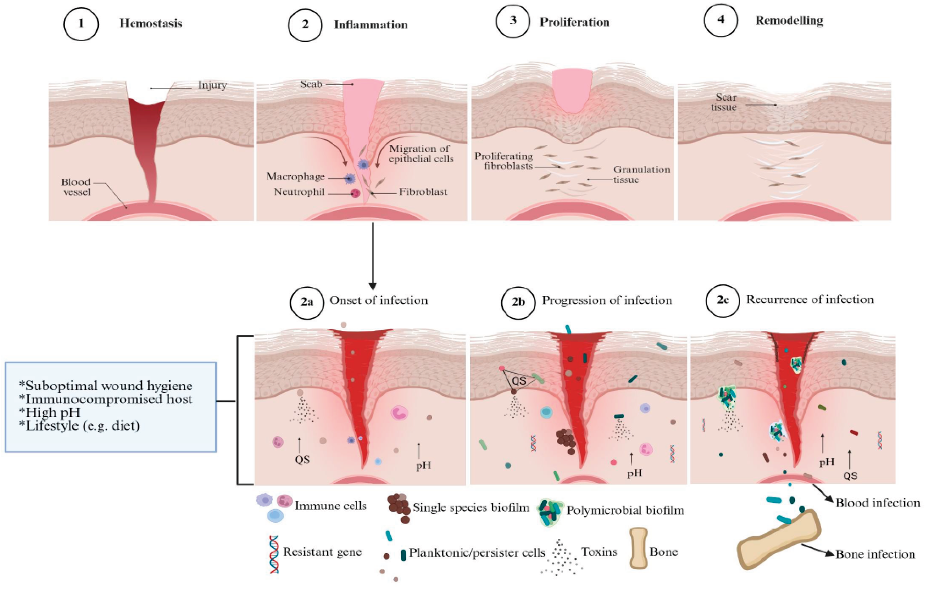
Figure 2.
Representative images of plasma-activated water generating devices and their components with no exception to plasma jet (a), corona discharge (b), gliding arc (c), piezoelectric direct discharge (d), dielectric discharge barrier (e), microwave (f).
Figure 2.
Representative images of plasma-activated water generating devices and their components with no exception to plasma jet (a), corona discharge (b), gliding arc (c), piezoelectric direct discharge (d), dielectric discharge barrier (e), microwave (f).
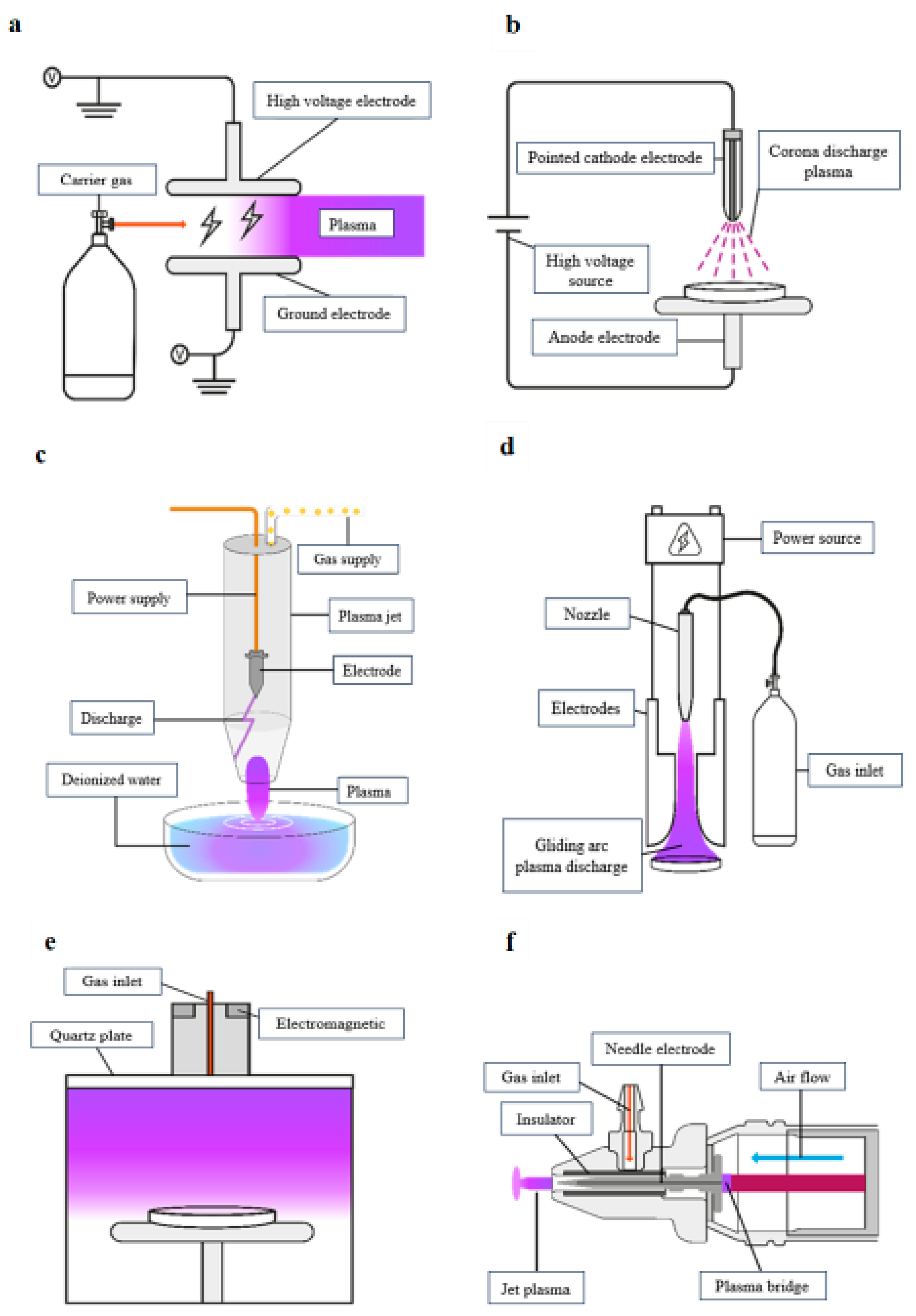
Figure 3.
Application of plasma-activated water (PAW) in wound healing. PAW treatment exposes microbes to reactive oxygen and nitrogen species, that have antibiofilm and antimicrobial effects, interrupting quorum sensing, decreasing pH, encouraging angiogenesis and killing planktonic cells to prevent new colonization of wounds. By chemically disrupting biofilms, PAW may help reduce the reliance on antibiotics. Direct application of PAW to the wound site, such as soaking or dressing wounds with PAW, may diminish and ultimately eliminate microbial biofilms to maintain a biofilm-free environment (Sen et al. 2021). Created in BioRender (https://BioRender.com).
Figure 3.
Application of plasma-activated water (PAW) in wound healing. PAW treatment exposes microbes to reactive oxygen and nitrogen species, that have antibiofilm and antimicrobial effects, interrupting quorum sensing, decreasing pH, encouraging angiogenesis and killing planktonic cells to prevent new colonization of wounds. By chemically disrupting biofilms, PAW may help reduce the reliance on antibiotics. Direct application of PAW to the wound site, such as soaking or dressing wounds with PAW, may diminish and ultimately eliminate microbial biofilms to maintain a biofilm-free environment (Sen et al. 2021). Created in BioRender (https://BioRender.com).
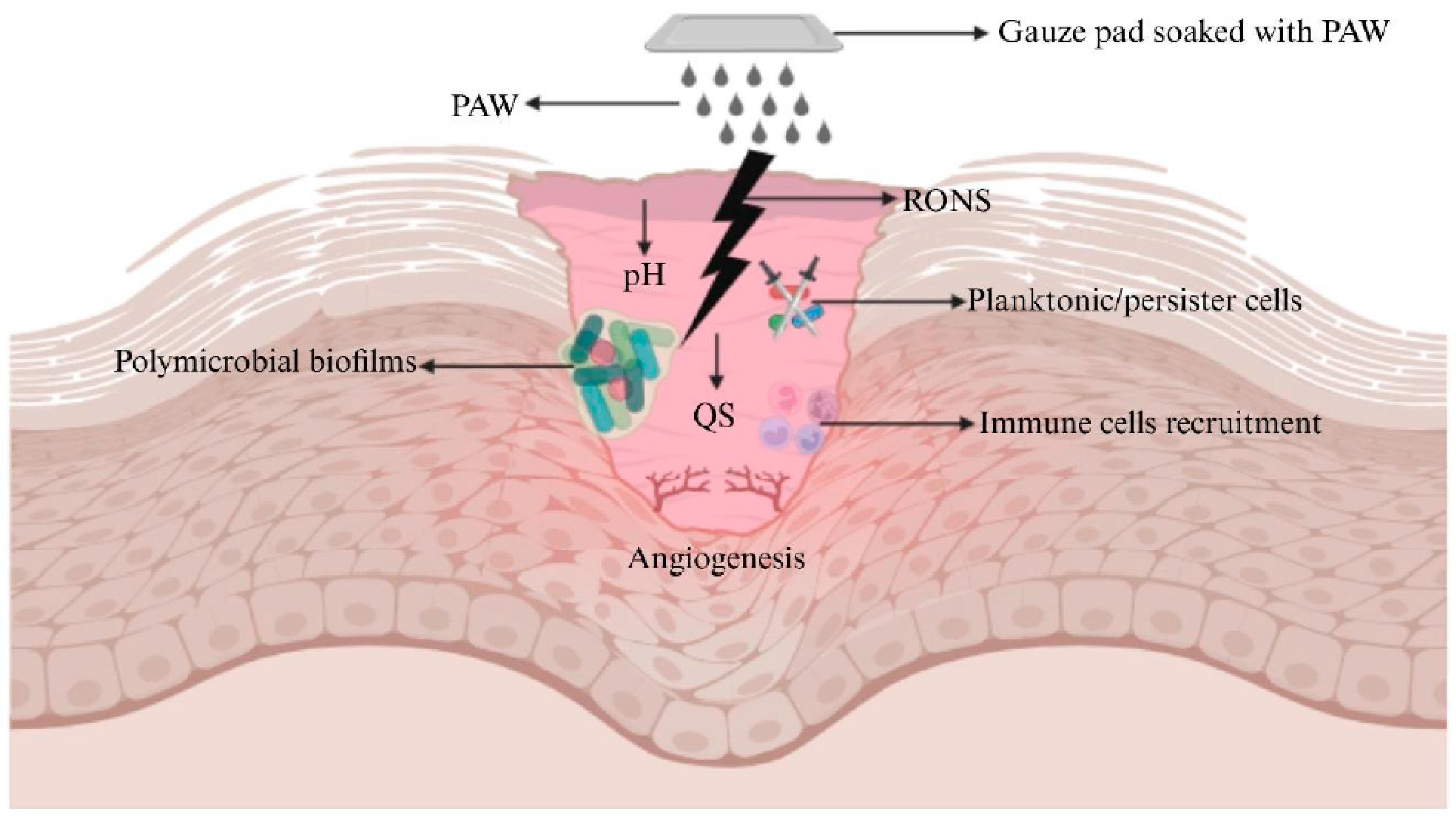

Table 1.
Overview of PAW as an antibiofilm and/or antimicrobial agent in wound healing studies.
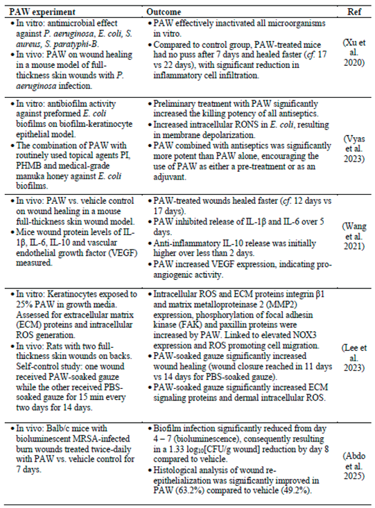
Disclaimer/Publisher’s Note: The statements, opinions and data contained in all publications are solely those of the individual author(s) and contributor(s) and not of MDPI and/or the editor(s). MDPI and/or the editor(s) disclaim responsibility for any injury to people or property resulting from any ideas, methods, instructions or products referred to in the content. |
© 2026 by the authors. Licensee MDPI, Basel, Switzerland. This article is an open access article distributed under the terms and conditions of the Creative Commons Attribution (CC BY) license (http://creativecommons.org/licenses/by/4.0/).
Copyright: This open access article is published under a Creative Commons CC BY 4.0 license, which permit the free download, distribution, and reuse, provided that the author and preprint are cited in any reuse.