Preprint
Brief Report

This version is not peer-reviewed.

A Pragmatic First-Line Screening Assay for PDGFR Rearrangements: A Real-World Clinical Validation

A peer-reviewed version of this preprint was published in:
Hemato 2026, 7(2), 9. https://doi.org/10.3390/hemato7020009

Submitted:

12 February 2026

Posted:

13 February 2026

You are already at the latest version

Abstract

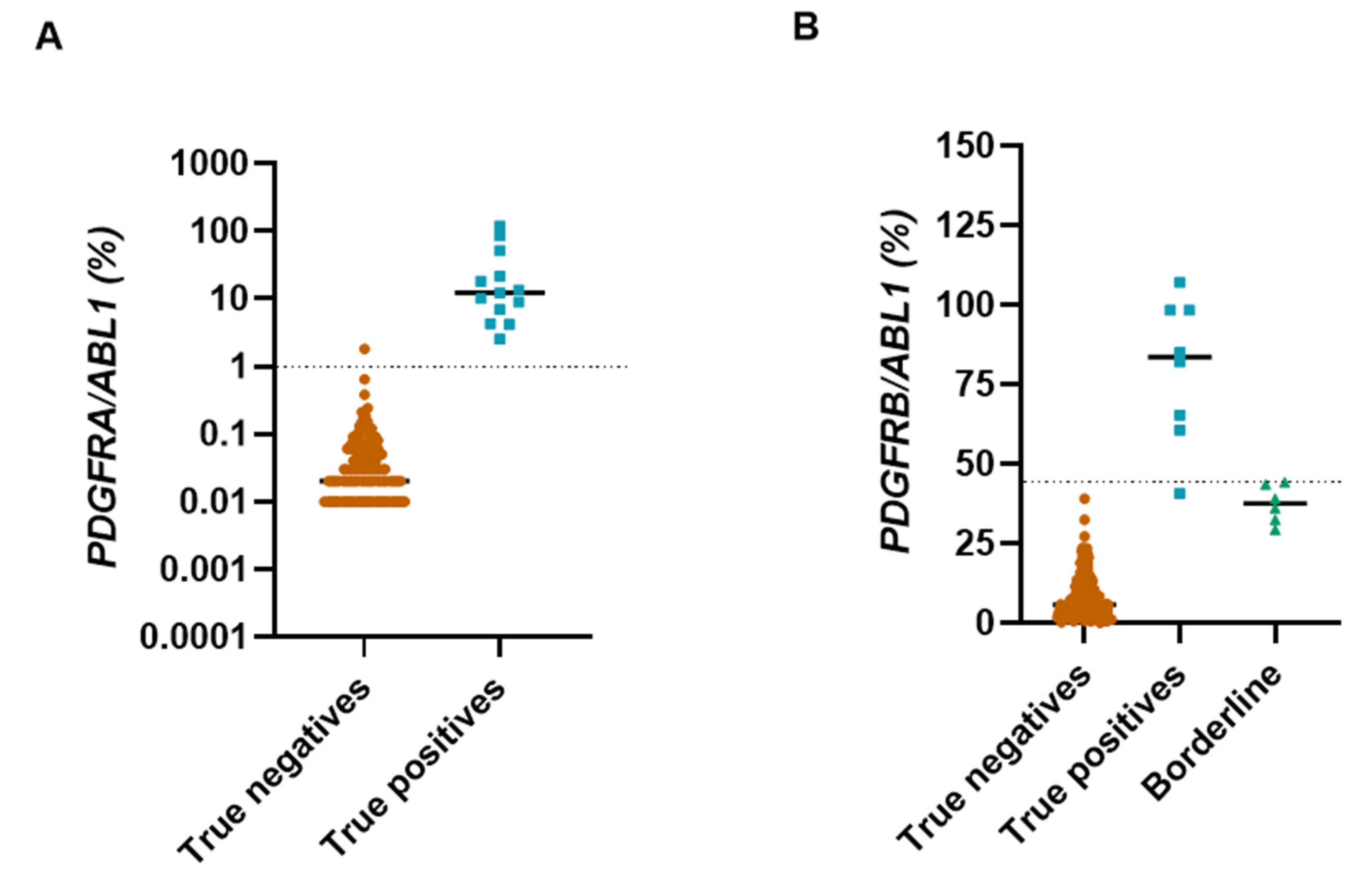
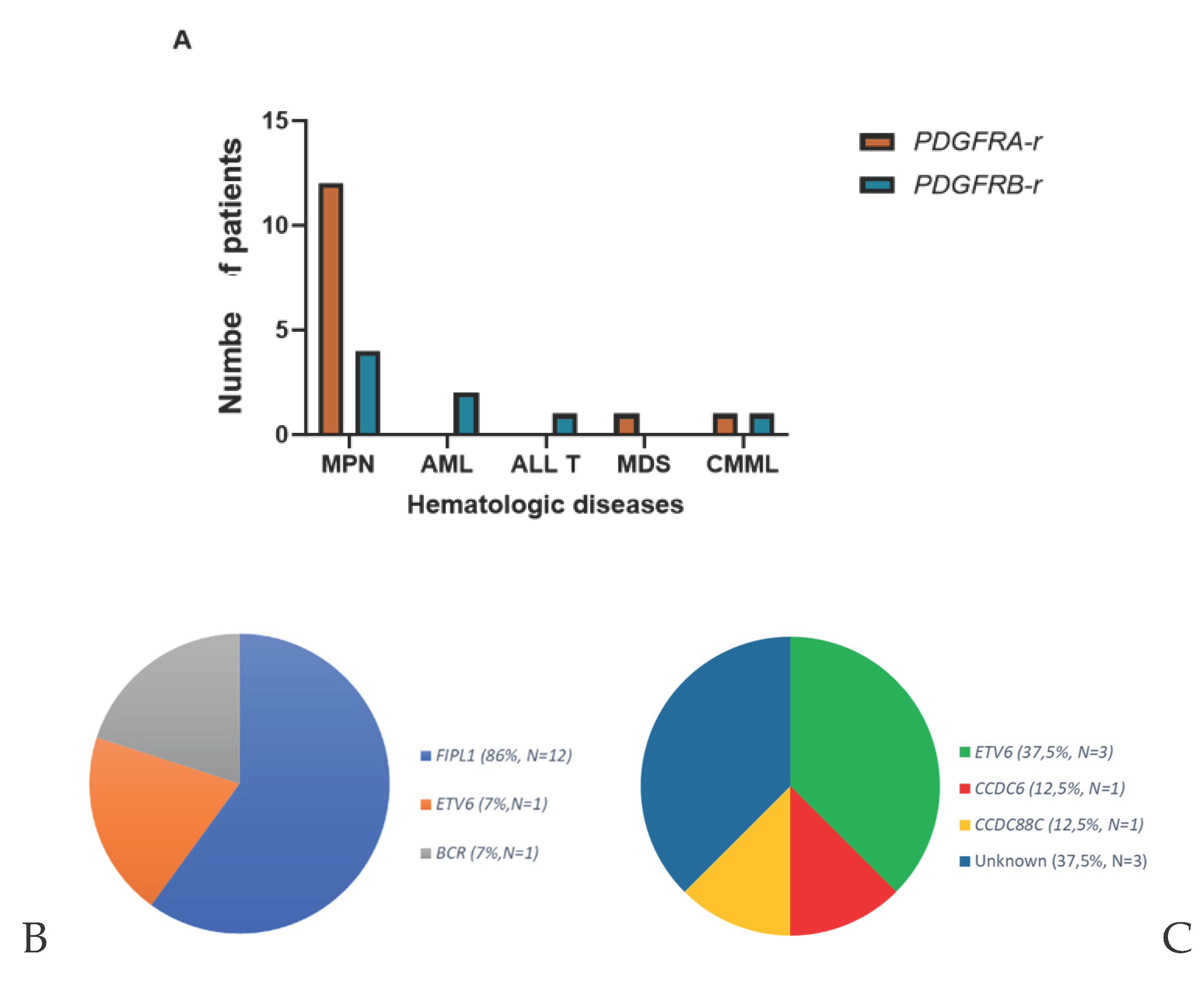
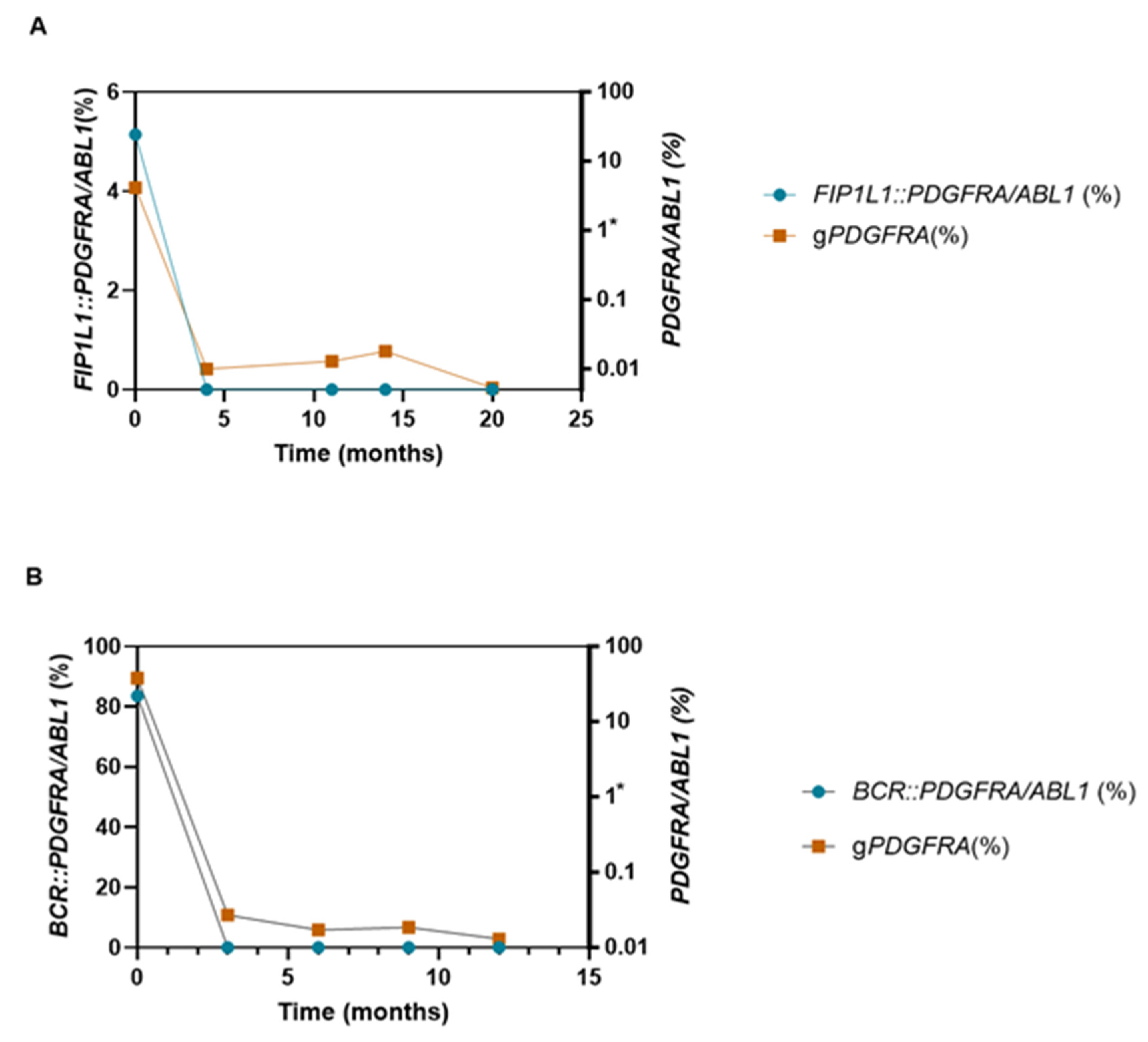
Myeloid/lymphoid neoplasms with tyrosine kinase rearrangements (MLN-TK) are rare clonal eosinophilias driven by PDGFRA, PDGFRB and other kinase fusions, highly sensitive to tyrosine kinase inhibitors. Their detection remains challenging, particularly for cryptic PDGFRA rearrangements. We performed a large multicenter real-world validation of the generic quantitative RT-PCR assay (gPDGFR), which detects 3′ PDGFRA/PDGFRB overexpression independently of fusion partner. A total of 231 consecutive patients with hypereosinophilia from 12 French centers were analyzed, and assay robustness was further assessed in an independent heterogeneous cohort of 102 TKI-treated patients. Twenty-two PDGFR-rearranged cases (14 PDGFRA-r, 8 PDGFRB-r) were identified. The assay demonstrated 100% sensitivity and 100% negative predictive value. For PDGFRA, positive predictive value and specificity reached 100%. In contrast, PDGFRB overexpression showed lower specificity due to borderline false-positive cases, underscoring the need for confirmatory testing. In selected patients, longitudinal gPDGFR kinetics paralleled fusion-specific RT-qPCR, supporting its use for molecular follow-up when dedicated assays are unavailable, although it does not provide quantitative measurable residual disease assessment. Overall, gPDGFR represents a robust, partner-independent first-line screening strategy that can be readily integrated into routine diagnostic workflows to enable timely identification of patients eligible for targeted therapy.

Keywords: 
;  ;  ;  

1. Introduction

Molecular biology has become central to the diagnosis and monitoring of malignant hematologic disorders, a role reinforced by the 2022 WHO Classification of Haematolymphoid Tumours[1]. Within this molecular framework, a new group of diseases has emerged: myeloid/lymphoid neoplasms with tyrosine kinase receptor rearrangements (MLN-TK), often associated with hypereosinophilia (HE). These clonal eosinophilias are driven by rearrangements involving one of five key genes: PDGFRA, PDGFRB, FLT3, JAK2, or FGFR1 and seven other even rarer defined tyrosine kinase fusions. Their identification is crucial, given the remarkable sensitivity to tyrosine kinase inhibitors (TKIs), particularly imatinib for PDGFR rearrangements (PDGFR-r). However, detecting these rearrangements—particularly those involving PDGFRA—is challenging due to cryptic cytogenetic profiles, variable breakpoints, and multiple fusion partners[2]. To address this, Erben et al. developed in 2010[3] a generic quantitative RT-PCR assay (gPDGFR assay) targeting overexpression of the 3′ region of PDGFRA or PDGFRB, independent of fusion partner or breakpoint. Although analytically attractive and technically simple, this strategy has not been widely implemented in routine diagnostics.
The present study provides a large-scale, real-world clinical validation of the gPDGFR assay in patients with unexplained HE, assessing its diagnostic performance, clinical correlates, and utility for longitudinal monitoring.

2. Materials and Methods

2.1. Study Design and Population

We conducted a multicenter observational study including 231 consecutive peripheral blood samples from patients referred for evaluation of HE (Appendix A). Samples originated from 12 hospital centers across France between 2012 and 2024 and were analyzed at the Bordeaux hematology laboratory. HE was defined as eosinophil count >1.5 × 10⁹/L. To further challenge the negative predictive value (NPV) of the assay, we retrospectively analyzed a separate cohort of 102 TKI-treated patients identified through the institutional clinical data warehouse (Entrepôt de Données de Santé, EDS cohort). This cohort was not designed to estimate sensitivity but to assess the risk of false negatives in a heterogeneous TKI-exposed population.

2.2. gPDGFR Assay

This assay was performed as originally described by Erben et al. [3] using successive RT kits (Roche®, SuperScript™ III/IV VILO™). Expression levels were normalized to ABL1. Cut-offs were established locally using 31 healthy controls (mean + 3 SD): PDGFRA/ABL1: 1% and PDGFRB/ABL1: 40%. Historically, a 25% cut-off was initially applied for PDGFRB. Samples between 25% and 40% without confirmed rearrangement were retrospectively classified as false positives.

2.3. Definition of True Positive and True Negative Cases

True positive cases required molecular or cytogenetic evidence of PDGFR rearrangement, including karyotyping, FISH or transcript-specific RT-PCR. Clinical response to TKI was considered supportive but never used as a standalone diagnostic criterion.
True negative cases required: Absence of PDGFR-r by cytogenetic and molecular analyses (qualitative RT-PCR transcription negative[4], RT-multiplex ligation-dependent probe amplification (RT-MLPA) negative [5], combined with clinical, biological, or therapeutic features inconsistent with a clonal PDGFR-driven HE. Diagnostic performance parameters (sensitivity, specificity, positive predictive value (PPV), NPV) were calculated using standard 2×2 contingency tables.

3. Results

3.1. Diagnostic Performance

Among the 231 samples, 194 were classified as negative with no overexpression of PDGFRA/B (mean of 0.02% [0-1.8%] and 5.66% [0.12-39.1%] respectively) and 37 as positive with overexpression of PGDFRA in 14 of them and PDGFRB in the other 23.
Notably, all samples exceeding the 1% PDGFRA expression cut-off (mean of 12.2% [2.53 – 116.6%]) had a confirmed PDGFRA rearrangement (PDGFRA-r), supporting a 100% PPV for this marker. In contrast, only 8 samples with PDGFRB overexpression (mean of 82% [40.7 – 107%]) had a confirmed PDGFRB rearrangement (PDGFRB-r), while 15 samples showed PDGFRB overexpression (mean of 32.81% [25.85 – 67.12%]) without any detectable rearrangement mentioned above (negative cytogenetic analysis, and if not performed, corticosteroid sensitivity or identification of another cause of HE). These borderline cases were classified as false positives, resulting in a PPV of 35% for PDGFRB. The overall sensitivity of the assay was 100%, with a specificity of 100 % for PDGFRA-r and 93% for PDGFRB-r and an exceptionally high negative predictive value of 100%. In the EDS cohort (n=102), none harboured PDGFR-r (gastro-intestinal stromal tumor (37%), chronic myeloid leukemia (31%), solid tumors (10%), and others such as KIT–mutated melanoma, systemic mastocytosis, chronic lymphoid leukemia or systemic sclerosis), confirming assay robustness with no false positives in heterogeneous TKI-treated populations.

3.2. Clinical and Biological Characteristics

We diagnosed 22 patients with PDGFR-r: PDGFRA-r (n=14) or PDGFRB-r (n=8) (Table 1) representing 1.75 new cases per year. This disorder predominantly affects elderly patients, with a median age of 67.5 years [range: 25–93 years]. A strong male predominance was observed for PDGFRA-r cases (sex ratio male / female of 2.5), and all male for PDGFRB-r. HE was higher in PDGFRA-r (mean: 6.42.10⁹/L [2.24–88.13 x 10⁹/L]) vs PDGFRB-r (mean: 2.05 x 10⁹/L [0.6–50.68 x 10⁹/L]). Cytopenias were present in all PDGFRB-r cases (100% anemia (mean: 10.8 g/dL [7.9 – 12.6 g/dL]), 83% thrombocytopenia (mean: 86 x 10⁹/L [34 – 269 x 10⁹/L]), but absent in PDGFRA-r. All PDGFRA-r patients showed elevated serum vitamin B12 (VitB12) and tryptase levels (corresponding data were not available for PDGFRB-r cases). Mean VitB12 concentration reached 2745.9 pg/mL [367–3500 pg/mL], and the mean tryptase level was 37.3 µg/L [22–51.9 µg/L]. Regarding associated hematologic diseases (Appendix B), PDGFRA-r linked mainly to MPN (86%) while PDGFRB-r showed a heterogeneous spectrum: 45% MPN, 22% acute myeloid leukemia (AML), 11% acute lymphoblastic leukemia (ALL), and 22% chronic myelomonocytic leukemia (CMML). This heterogeneity extended to the fusion partners (Appendix B). The PDGFRA-r predominantly involved FIP1L1 (86%), whereas PDGFRB-r were more diverse: ETV6 (n=3), CCDC6 (n=1), CCDC88C (n=1), and in 3 of cases, no fusion partner could be identified despite additional testing. Interestingly, the level of PDGFR/ABL1 overexpression did not correlate with the eosinophil count, the fusion partner, or the associated hematologic malignancy.

3.3. Treatment Response and Molecular Monitoring

All PDGFRA-r patients treated with imatinib (n=14) achieved sustained complete hematologic remission, with no molecular or clinical relapse observed after a median follow-up of 5 years.
Among PDGFRB-r patients, 6 patients received imatinib and one received ruxolitinib. Follow-up data were available for 4 patients with a mean-time of 6 years.
In two patients with adequate longitudinal data, gPDGFR kinetics paralleled transcript-specific RT-qPCR (FIP1L1::PDGFRA, BCR::PDGFRA). While not suitable for quantitative measurable residual disease (MRD) assessment, the assay reliably reflected molecular clearance and stability (Figure 1).

4. Discussion

In this multicenter real-world study, we demonstrate that the generic gPDGFR assay is a robust and clinically relevant screening tool for the detection of PDGFRA rearrangements in patients with unexplained HE. The assay showed excellent diagnostic performance, with 100% sensitivity and negative predictive value in our cohort. Notably, PDGFRA overexpression above the predefined threshold was fully concordant with confirmed PDGFRA rearrangement, resulting in a positive predictive value and specificity of 100% in this setting.
In contrast, PDGFRB overexpression was associated with a lower positive predictive value due to borderline cases without detectable rearrangement. This finding likely reflects higher physiological baseline expression and greater biological heterogeneity of PDGFRB-driven neoplasms. These results indicate that, while gPDGFR is highly reliable for excluding PDGFR rearrangements, PDGFRB-positive or borderline cases require systematic confirmatory cytogenetic or molecular analyses.
Beyond diagnostic performance, our data further delineate the clinical differences between PDGFRA- and PDGFRB-rearranged neoplasms. PDGFRA-r cases were characterized by marked eosinophilia, absence of cytopenias, elevated serum vitamin B12 and tryptase levels, and a predominance of FIP1L1 fusion partners. In contrast, PDGFRB-r cases displayed greater clinical and molecular heterogeneity, frequent cytopenias, and diverse associated hematologic malignancies. Importantly, the level of PDGFR overexpression did not correlate with eosinophil count, fusion partner, or underlying disease, supporting the concept that the assay functions as a qualitative screening tool rather than a quantitative surrogate of disease burden.
Therapeutically, our findings confirm the remarkable sensitivity of PDGFRA-rearranged neoplasms to imatinib, consistent with previously reported remission rates exceeding 90% [6,7]. All treated PDGFRA-r patients achieved sustained hematologic remission. Outcomes were more heterogeneous in PDGFRB-r cases, reflecting both biological diversity and differences in associated hematologic diseases.
Regarding longitudinal monitoring, we observed concordant kinetics between gPDGFR and fusion-specific RT-qPCR in selected patients. Although the assay does not provide the analytical precision required for measurable residual disease assessment, it reliably reflected molecular clearance and sustained response. Thus, gPDGFR may represent a practical alternative for molecular follow-up when fusion-specific assays are unavailable, particularly in cases with rare or unidentified partners.
This study has several strengths. It includes a relatively large consecutive multicenter cohort of patients with HE, reflecting real-world diagnostic practice. The independent evaluation in a heterogeneous TKI-treated population further supports the robustness of the assay and its high negative predictive value.
Limitations include the retrospective design, the limited number of PDGFRB-rearranged cases, and the absence of formal receiver operating characteristic analysis to refine cut-off thresholds. In addition, the monitoring data remain limited to a small number of patients and should be interpreted cautiously.
In conclusion, the gPDGFR assay represents a pragmatic, partner-independent first-line screening strategy for PDGFRA rearrangements and an effective tool for the preliminary exclusion of PDGFR-driven HE. Its integration into routine diagnostic workflows can facilitate timely identification of patients eligible for targeted therapy, while complementary cytogenetic and molecular analyses remain essential, particularly in PDGFRB-positive or borderline cases.

Author Contributions

Conceptualization, FL and AB.; methodology, FL and AB, software, FL and AB.; validation, FL and AB.; formal analysis, FL, LB, MM and CC.; investigation, FL, MMG, DL, CB, GE, WC, LM, JE, FL, JBG, NN, DR, JQ, PCL, EK, EL; data curation, FL.; writing—original draft preparation: FL and AB; writing—review and editing, FL, LB, MMG, DL, CB, GE, WC, LM, JE, FL, JBG, NN, DR, JQ, PCL, EK, EL, AB.; supervision, AB. All authors have read and agreed to the published version of the manuscript.

Funding

This research received no external funding.

Institutional Review Board Statement

This study received a favorable opinion from the Research Ethics Committee of the Bordeaux University Hospital (CER-BDX 2024 – 44).

Acknowledgments

The authors acknowledge the contribution of the Entrepôt de Données de Santé, in particular Mr. Corentin Sinanovic, as well as the CRB-Cancer of Bordeaux University Hospital for their support.

Conflicts of Interest

The authors declare no conflicts of interest.

Appendix A

Figure A1. Diagnostic performance of gPDGFR: Percentage of PDGFR/ABL1 (%). (A): PDGFRA expression. (B): PDGFRB expression. The cut-off point is indicated by dotted lines on both graphs (former cut-off, according to [3], is represented by the blue dotted line).
Preprints 198709 g0a1

Appendix B

Figure A2. Hematologic diseases associated with PDGFR rearrangement and fusion partner. (A): Most of PDGFRA rearrangements are associated with MPN whereas PDGFRB rearrangements are associated with multiple neoplasms, such as MPN, AML, ALLT or CMML. (B): PDGFRA partner is mainly FIP1L1. (C): PDGFRB partner are multiple (ETV6, CCDC6, CCDC88C) and notably unknown in one third of the cases. MPN: myeloproliferative neoplasms, AML: acute myeloblastic leukemia, ALL T: acute lymphoblastic leukemia T, MDS: myelodysplastic neoplasms, CMML: chronic myelomonocytic leukemia.
Preprints 198709 g0a2

References

  1. Khoury, J.D.; Solary, E.; Abla, O.; Akkari, Y.; Alaggio, R.; Apperley, J.F.; Bejar, R.; Berti, E.; Busque, L.; Chan, J.K.C.; Chen, W.; Chen, X.; Chng, W.-J.; Choi, J.K.; Colmenero, I.; Coupland, S.E.; Cross, N.C.P.; De Jong, D.; Elghetany, M.T.; Takahashi, E.; Emile, J.-F.; Ferry, J.; Fogelstrand, L.; Fontenay, M.; Germing, U.; Gujral, S.; Haferlach, T.; Harrison, C.; Hodge, J.C.; Hu, S.; Jansen, J.H.; Kanagal-Shamanna, R.; Kantarjian, H.M.; Kratz, C.P.; Li, X.-Q.; Lim, M.S.; Loeb, K.; Loghavi, S.; Marcogliese, A.; Meshinchi, S.; Michaels, P.; Naresh, K.N.; Natkunam, Y.; Nejati, R.; Ott, G.; Padron, E.; Patel, K.P.; Patkar, N.; Picarsic, J.; Platzbecker, U.; Roberts, I.; Schuh, A.; Sewell, W.; Siebert, R.; Tembhare, P.; Tyner, J.; Verstovsek, S.; Wang, W.; Wood, B.; Xiao, W.; Yeung, C.; Hochhaus, A. The 5th edition of the World Health Organization Classification of Haematolymphoid Tumours: Myeloid and Histiocytic/Dendritic Neoplasms. Leukemia 2022, 36, 1703–1719. [Google Scholar] [CrossRef]
  2. Kaur, M.D. Prabhjot; Khan, P.H.D. Wahab A. Myeloid/Lymphoid Neoplasms with Eosinophilia and Platelet Derived Growth Factor Receptor Alpha (PDGFRA) Rearrangement. In Exon Publications; 2022; pp. 129–146. [Google Scholar] [CrossRef]
  3. Erben, P.; Gosenca, D.; Muller, M.C.; Reinhard, J.; Score, J.; Del Valle, F.; Walz, C.; Mix, J.; Metzgeroth, G.; Ernst, T.; Haferlach, C.; Cross, N.C.P.; Hochhaus, A.; Reiter, A. Screening for diverse PDGFRA or PDGFRB fusion genes is facilitated by generic quantitative reverse transcriptase polymerase chain reaction analysis. Haematologica 2010, 95, 738–744. [Google Scholar] [CrossRef] [PubMed]
  4. Score, J.; Curtis, C.; Waghorn, K.; Stalder, M.; Jotterand, M.; Grand, F.H.; Cross, N.C.P. Identification of a novel imatinib responsive KIF5B-PDGFRA fusion gene following screening for PDGFRA overexpression in patients with hypereosinophilia. Leukemia 2006, 20, 827–832. [Google Scholar] [CrossRef] [PubMed]
  5. Ruminy, P.; Marchand, V.; Buchbinder, N.; Larson, T.; Joly, B.; Penther, D.; Lemasle, E.; Lepretre, S.; Angot, E.; Mareschal, S.; Viailly, P.-J.; Dubois, S.; Clatot, F.; Viennot, M.; Bohers, E.; Rizzo, D.; Cornic, M.; Bertrand, P.; Girod, C.; Camus, V.; Etancelin, P.; Buchonnet, G.; Schneider, P.; Picquenot, J.-M.; Vannier, J.-P.; Bastard, C.; Tilly, H.; Jardin, F. Multiplexed targeted sequencing of recurrent fusion genes in acute leukaemia. Leukemia 2016, 30, 757–760. [Google Scholar] [CrossRef] [PubMed]
  6. Rohmer, J.; Couteau-Chardon, A.; Trichereau, J.; Panel, K.; Gesquiere, C.; Ben Abdelali, R.; Bidet, A.; Bladé, J.-S.; Cayuela, J.-M.; Cony-Makhoul, P.; Cottin, V.; Delabesse, E.; Ebbo, M.; Fain, O.; Flandrin, P.; Galicier, L.; Godon, C.; Grardel, N.; Guffroy, A.; Hamidou, M.; Hunault, M.; Lengline, E.; Lhomme, F.; Lhermitte, L.; Machelart, I.; Mauvieux, L.; Mohr, C.; Mozicconacci, M.-J.; Naguib, D.; Nicolini, F.E.; Rey, J.; Rousselot, P.; Tavitian, S.; Terriou, L.; Lefèvre, G.; Preudhomme, C.; Kahn, J.-E.; Groh, M. CEREO and GBMHM collaborators, Epidemiology, clinical picture and long-term outcomes of FIP1L1-PDGFRA-positive myeloid neoplasm with eosinophilia: Data from 151 patients. Am J Hematol 2020, 95, 1314–1323. [Google Scholar] [CrossRef] [PubMed]
  7. Ogbogu, P.U.; Bochner, B.S.; Butterfield, J.H.; Gleich, G.J.; Huss-Marp, J.; Kahn, J.E.; Leiferman, K.M.; Nutman, T.B.; Pfab, F.; Ring, J.; Rothenberg, M.E.; Roufosse, F.; Sajous, M.-H.; Sheikh, J.; Simon, D.; Simon, H.-U.; Stein, M.L.; Wardlaw, A.; Weller, P.F.; Klion, A.D. Hypereosinophilic syndromes: a multicenter, retrospective analysis of clinical characteristics and response to therapy. J Allergy Clin Immunol 2009, 124, 1319–25.e3. [Google Scholar] [CrossRef] [PubMed]
Figure 1. Two examples of follow-up by gPDGFR and specific quantitative PCR: (A): patient P4 with FIP1L1::PDGFRA, (B): patient P13 with BCR::PDGFRA. The cut-off point is indicated by the asterisk on both graphs.
Preprints 198709 g001
Table 1. Clinical and biological data for patients with PDGFR rearrangements.
UPN Sex Age (years) Hematologic diseases Rearrangement % overexpression Eosinophils (10⁹/L) Tryptase (µg/L) Viamin B12 (pg/mL) Bone marrow Karyotype FISH RT-PCR or RT-MLPA TKI treatment Time to remission
P1 M 49 MPN FIP1L1::PDGFRA 6,85 4,4 unk 1902 HE > 20% Normal n.t FIP1L1::PDGFRA Imatinib 100 mg/d unk
P2 M 85 MPN FIP1L1::PDGFRA 10,1 6,42 51,9 890 unk n.t n.t FIP1L1::PDGFRA Imatinib 100 mg/d unk
P3 M 84 MPN FIP1L1::PDGFRA 4,23 3,25 n.t 367 unk Normal n.t FIP1L1::PDGFRA Imatinib 100 mg/d 3 months
P4 M 42 MPN FIP1L1::PDGFRA 4,16 2,24 22 2160 Mild HE Normal CHIC2 deletion FIP1L1::PDGFRA Imatinib 100 mg/d unk
P5 M 60 MPN FIP1L1::PDGFRA 116,64 10,44 n.t n.t unk Normal n.t FIP1L1::PDGFRA unk unk
P6 M 58 MPN FIP1L1::PDGFRA 51,37 21 Normal 2500 HE Normal CHIC2 deletion FIP1L1::PDGFRA Imatinib 100 mg/d unk
P7 M 54 MPN FIP1L1::PDGFRA 17,93 4,4 unk 1902 unk n.t n.t FIP1L1::PDGFRA Imatinib 100 mg/d unk
P8 M 75 CMML FIP1L1::PDGFRA 13,29 14,5 n.t 8500 unk Normal CHIC2 deletion FIP1L1::PDGFRA Imatinib 100 mg/d unk
P9 M 27 MPN FIP1L1::PDGFRA 2,53 88,13 38 n.t unsuitable n.t n.t FIP1L1::PDGFRA Imatinib 100 mg/d unk
P10 F 77 MPN FIP1L1::PDGFRA 9,81 6,22 unk unk HE=15% Normal CHIC2 deletion FIP1L1::PDGFRA Imatinib 100 mg/d 3 months
P11 F 48 MPN FIP1L1::PDGFRA 12,17 1,43 unk unk HE=13% Normal CHIC2 deletion FIP1L1::PDGFRA « Watch and see » unk
P12 M 76 MPN FIP1L1::PDGFRA 10,78 unk unk unk unk unk unk FIP1L1::PDGFRA unk unk
P13 F 78 MPN BCR::PDGFRA 83,52 3,11 unk unk unk t(4;22) unk BCR ::PDGFRA Imatinib 400 mg/d 6 months
P14 F 91 MDS-IB2 ETV6::PDGFRA 21,4 0,03 unk unk MDS-IB2 t(4;12) ETV6::PDGFRA rearrangement n.t Imatinib 100 mg/d unk
P15 M 57 AML ETV6::PDGFB 98,27 0,6 n.t n.t AML t(5;12) ETV6::PDGFRB rearrangement n.t Imatinib 400 mg/d unk
P16 M 77 MPN ETV6::PDGFRB 60,5 50,68 n.t n.t HE t(5;12) ETV6::PDGFRB rearrangement Negative Ruxolitinib unk
P17 M 58 AML ETV6 ::PDGFRB 98,36 unk n.t n.t unk unk n.t ETV6 ::PDGFRB Imatinib 400 mg/d 1 months
P18 M 77 CMML CCDC88C::PDGFRB 85,09 2,19 n.t n.t CMML t(5;14) PDGFRB rearrangement n.t Imatinib 100 mg/d unk
P19 M 70 CML CCDC6::PDGFRB 40,7 1,9 n.t n.t MPN t(5;10) CCDC6::PDGFRB rearrangement n.t Imatinib 200 mg/d unk
P20 M 93 MPN PDGFRB unknown partner 82 unk n.t n.t unk unk PDGFRB rearrangement n.t unk unk
P21 M 65 MPN PDGFRB
unknown partner
107 16,28 n.t >1476 unk n.t PDGFRB rearrangement n.t Imatinib 100 mg/d unk
P22 M 25 ALL T PDGFRB unknown partner 65,37 1,3 n.t n.t ALL t(5;12) PDGFRB rearrangement Negative Imatinib 500 mg/d unk
M : male, F : female, B12 : Vitamin B12, FISH : Fluorescence In Situ Hybridization), RT-PCR : reverse transcription polymerase Chain reaction, RT-MLPA : reverse transcription multiplex ligation-dependent probe amplification, HE: Hypereosinophilia, MPN : myeloproliferative neoplasm, MDS-IB2 : myelodysplastic neoplasm with increased blasts 2, AML : acute myeloblastic leukemia, CMML : chronic myelomonocytic leukemia, ALL T : acute lymphoblastic leukemia, CML : chronic myeloid leukemia, n.t : not tested, unk : unknown. Time to remission represents time until patients normalized their eosinophilia and PDGFR rearrangement was undetectable.
Disclaimer/Publisher’s Note: The statements, opinions and data contained in all publications are solely those of the individual author(s) and contributor(s) and not of MDPI and/or the editor(s). MDPI and/or the editor(s) disclaim responsibility for any injury to people or property resulting from any ideas, methods, instructions or products referred to in the content.
Copyright: This open access article is published under a Creative Commons CC BY 4.0 license, which permit the free download, distribution, and reuse, provided that the author and preprint are cited in any reuse.
Prerpints.org logo

Preprints.org is a free preprint server supported by MDPI in Basel, Switzerland.

Subscribe

Disclaimer

Terms of Use

Privacy Policy

Privacy Settings

© 2026 MDPI (Basel, Switzerland) unless otherwise stated