Preprint
Case Report

This version is not peer-reviewed.

Aggrecanopathy as an Underrecognized Cause of Idiopathic Short Stature: The Importance of Early Genetic Confirmation for Timely Diagnosis and Management - Case Reports and Literature Review

A peer-reviewed version of this preprint was published in:
Diseases 2026, 14(4), 127. https://doi.org/10.3390/diseases14040127

Submitted:

12 February 2026

Posted:

13 February 2026

You are already at the latest version

Abstract

Background: Short stature is a frequent clinical problem with a broad differential diagnosis. Emerging evidence indicates that pathogenic variants in the ACAN gene represent an underrecognized cause of growth failure and are often misclassified as idiopathic short stature.Case presentation: We report two pediatric patients harboring pathogenic ACAN gene variants, both presenting with short stature and distinctive facial dysmorphism. The first patient, a 15-year-old boy, exhibited short stature, advanced bone age, and a characteristic facial gestalt, including ptosis, hypertelorism, down slanting palpebral fissures, fleshy auricles, features not previously described in association with aggrecanopathy. Genetic analysis revealed a novel heterozygous frameshift variant, c.5677_5684del (p.Glu1893TrpfsTer8), in exon 12 of the ACAN gene. The second patient, a 5.5-year-old girl, presented with short stature, mild facial dysmorphism (down slanting palpebral fissures and retracted mandible), and feeding difficulties. Copy number variation analysis identified a heterozygous deletion encompassing exons 15–19 of the ACAN gene. In both patients, endocrine evaluation was unremarkable, and no chronic systemic disease was identified. The genetic findings were concordant with the clinical phenotype, confirming aggrecanopathy as the underlying cause of growth failure. Conclusion: These cases further delineate the phenotypic spectrum of ACAN-related short stature and underscore the diagnostic value of genetic testing in children with unexplained or idiopathic growth failure. Importantly, we expand the dysmorphological phenotype of aggrecanopathy by describing previously unreported facial features, which may facilitate earlier clinical recognition and diagnosis. Timely identification of pathogenic variants in ACAN gene may have significant implications for patient management and long-term outcomes.

Keywords: 
;  ;  ;  ;  

1. Introduction

Short stature is a common pediatric condition, defined as height below (-) 2 standard deviation scores (SDS) for age and sex, and affects approximately 3% of the general pediatric population [1]. It is among the most frequent reasons for referral to pediatric endocrinology clinics. While nutritional deficiencies, chronic systemic diseases, endocrine disorders, chromosomal aberrations ( e.g. trisomy 21, Turner syndrome) and genetic syndromes (e.g. Noonan syndrome) account for a subset of cases, a substantial proportion of children has been ultimately classified so far as having idiopathic short stature (ISS) following standard clinical evaluation [2,3].
Advances in molecular genetics have fundamentally changed the understanding of growth failure, revealing it to be a highly heterogeneous condition. Pathogenic variants have been identified in genes involved in growth plate biology, skeletal development, and cartilage homeostasis [4]. Among monogenic causes of ISS, SHOX haploinsufficiency is the most common, accounting for approximately 2–3% of cases [4]. More recently, heterozygous pathogenic variants in the gene encoding aggrecan (ACAN) have emerged as an important and previously underrecognized cause of ISS, with reported prevalence ranging from 1.4% to 6% in unselected ISS cohorts [1,5,6,7,8,9,10,11].
The human ACAN gene (OMIM: *155760) is located on chromosome 15q26.1 and consists of 19 exons encoding the full-length aggrecan core protein [12,13]. With the increasing use of next-generation sequencing (NGS) in molecular diagnostics, heterozygous pathogenic variants in ACAN have been recognized as a significant cause of short stature and related skeletal phenotypes [1,2,3,4,5,6,7,8,9,10,11,12,13,14,15]. Pathogenic variants have been reported throughout the gene, affecting distinct functional domains of the protein and resulting in a broad phenotypic spectrum collectively referred to as aggrecanopathies [7,14,15,16,17].
Aggrecan is a large chondroitin sulfate proteoglycan and a major structural component of the extracellular matrix of cartilage, including growth plate, articular, and intervertebral disc cartilage [18,19]. The aggrecan core protein is composed of three globular domains (G1, G2, and G3), separated by an interglobular domain and a centrally located glycosaminoglycan (GAG) attachment region containing keratan sulfate and chondroitin sulfate chains [18,19]. The G1 domain mediates binding to hyaluronan and link protein, enabling the formation of large proteoglycan aggregates essential for cartilage integrity and load-bearing capacity [20]. Although the precise function of the G2 domain remains unclear, the C-terminal G3 domain-containing epidermal growth factor-like repeats, a complement regulatory protein-like domain, and a C-type lectin domain-interacts with extracellular matrix proteins such as tenascins and fibulins, contributing to matrix assembly and stability [7,18,19]. The highly negatively charged GAG-rich region confers cartilage hydration and resistance to compressive forces, which are critical for normal longitudinal bone growth and joint function [21]. Beyond its biomechanical role, aggrecan regulates the availability and distribution of growth factors and signaling molecules within the cartilage extracellular matrix, thereby influencing chondrocyte differentiation, endochondral ossification, and bone morphogenesis [22].
Consistent with these domain-specific functions, pathogenic ACAN variants give rise to a continuum of skeletal phenotypes. Homozygous or compound heterozygous variants result in severe skeletal dysplasias, including autosomal recessive spondyloepimetaphyseal dysplasia, aggrecan type, SEMDAG (OMIM: #612813), and autosomal dominant spondyloepiphyseal dysplasia, Kimberley type, SEDK (OMIM: #608361) [23,24]. In contrast, heterozygous loss-of-function variants typically cause autosomal dominant short stature with variable expressivity, referred to as SSOAOD—short stature and advanced bone age with or without early-onset osteoarthritis and/or osteochondritis dissecans (OMIM: #165800) [7,14,20,22,25,26,27].
The most characteristic feature of autosomal dominant SSOAOD is proportionate short stature, frequently accompanied by advanced bone age, early growth cessation, and reduced adult height [7,14,20,22,25,26,27]. In contrast, disproportionate short stature has been observed in autosomal recessive SEMDAG [28]. Additional manifestations may include early-onset osteoarthritis, osteochondritis dissecans, intervertebral disc disease, and variable skeletal abnormalities. Although advanced bone age is considered a hallmark of heterozygous ACAN-related short stature, delayed or normal bone age has also been reported, underscoring the marked phenotypic heterogeneity of this condition [3]. Facial dysmorphism has been inconsistently described and remains insufficiently characterized [29].
Despite growing recognition of ACAN-related growth disorders, aggrecanopathy remains underdiagnosed, particularly in children who lack overt skeletal dysplasia or endocrine abnormalities. Delayed diagnosis may result in missed opportunities for growth-promoting interventions and suboptimal adult height outcomes.
The aim of this study is to expand the clinical and dysmorphological spectrum of aggrecanopathy and to highlight the therapeutic implications of early versus delayed diagnosis. We present two pediatric patients with pathogenic ACAN variants: a late-diagnosed adolescent boy with untreated aggrecanopathy, normal endocrine evaluation, and established short stature; and an early-diagnosed young girl in whom timely recognition of the genetic etiology-guided by diagnostic pitfalls identified in the first patient- may allow implementation of appropriate management strategies aimed at preventing future growth failure. By contrasting these two clinical trajectories, we underscore the importance of early genetic testing in children with unexplained short stature and demonstrate how prompt diagnosis of aggrecanopathy may directly influence clinical decision-making and long-term growth outcomes.

2. Case Reports

2.1. Patient 1

The male patient was first noted to have short stature at 11 years of age during a routine evaluation by a primary care physician and was subsequently referred for endocrine assessment. At presentation, his height was 135.6 cm (−2.0 SDS), with a body mass index (BMI) of 21.5 kg/m². The mother`s height was 153 cm, whereas the father’s height was 182 cm, yielding a mid-parental height (MPH) of 174 cm.
The patient was born from the second pregnancy and second delivery at 39 weeks of gestation. Pregnancy was complicated by lower-extremity edema. Birth parameters were appropriate for gestational age, with a birth weight of 3660 g, length of 56 cm, and head circumference of 34 cm. Apgar scores were 10 at both 1 and 5 minutes. The neonatal course was unremarkable, and psychomotor development proceeded within normal limits. There was no evidence of intellectual disability.
Physical examination revealed subtle but consistent dysmorphic features, including mild bilateral ptosis of the upper eyelids, hypertelorism, downslanting palpebral fissures, and fleshy auricles. Additional findings included mild limitation of elbow extension and flat valgus feet. Notably, similar facial features were observed in the patient’s mother. Anthropometric assessment demonstrated a slightly increased arm span-to-height ratio (AS/H = 1.1), exceeding age- and sex-specific reference values reported by Gerver et al. [30]
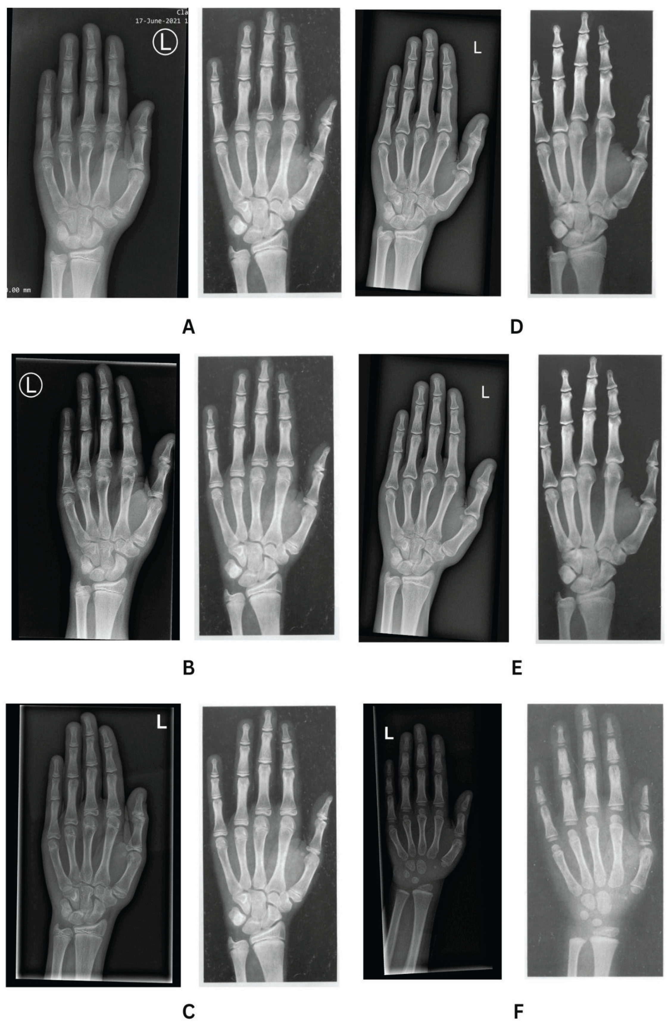
Skeletal maturation was assessed using the Greulich-Pyle comparative method [31]. Radiological evaluation showed advanced bone age, progressing from 14 years at a chronological age of 11 years to 17 years at 15 years of age (Table 1, Figure 1 and Figure 2). During the assessment of the lengths of the phalanges and metacarpal bones, a tendency toward shortening of the metacarpal bones was observed, which may indicate type E brachydactyly (brachymetacarpia) (Figure 2). Longitudinal follow-up between 2021 and 2025 demonstrated progressive pubertal development, with Tanner stage advancement from II to V, an increase in testicular volume from 3–4 mL to 15 mL, and appropriate timing of pubarche and axillarche (Table 1). Endocrine evaluation revealed normal thyroid function throughout follow-up (Table 1). Basal gonadotropin concentrations increased appropriately with pubertal maturation (LH from 1.6 to 2.2 mIU/mL, FSH from 2.0 to 3.6 mIU/mL), and serum testosterone rose from 0.66 to 2.6 ng/mL. Serum insulin-like growth factor 1 (IGF-1) concentrations ranged from 354 to 438 ng/mL and were within the normal range. Overall, the hormonal profile and growth above 3rd centile on growth charts as seen on Figure 1. did not indicate growth hormone deficiency or precocious puberty, and the Patient was not referred to further GH and GnRH stimulation tests and no growth promoting therapy was applied.
Relevant comorbidities included a thyroglossal duct cyst excised at 4 years of age and a benign solitary colonic polyp removed surgically. Histopathological examination demonstrated inflammatory changes, initially raising suspicion for inflammatory bowel disease; however, immunological and serological testing was negative, and no systemic inflammatory disorder was confirmed. The patient had no history of chronic illness, nutritional deficiency, or glucocorticoid exposure.
Due to short stature with advanced bone age and facial dysmorphic features genetic testing was performed. Genetic analysis revealed a novel heterozygous frameshift variant, c.5677_5684del (p.Glu1893TrpfsTer8), in exon 12 of the ACAN gene in the Patient and his mother.

2.2. Patient 2

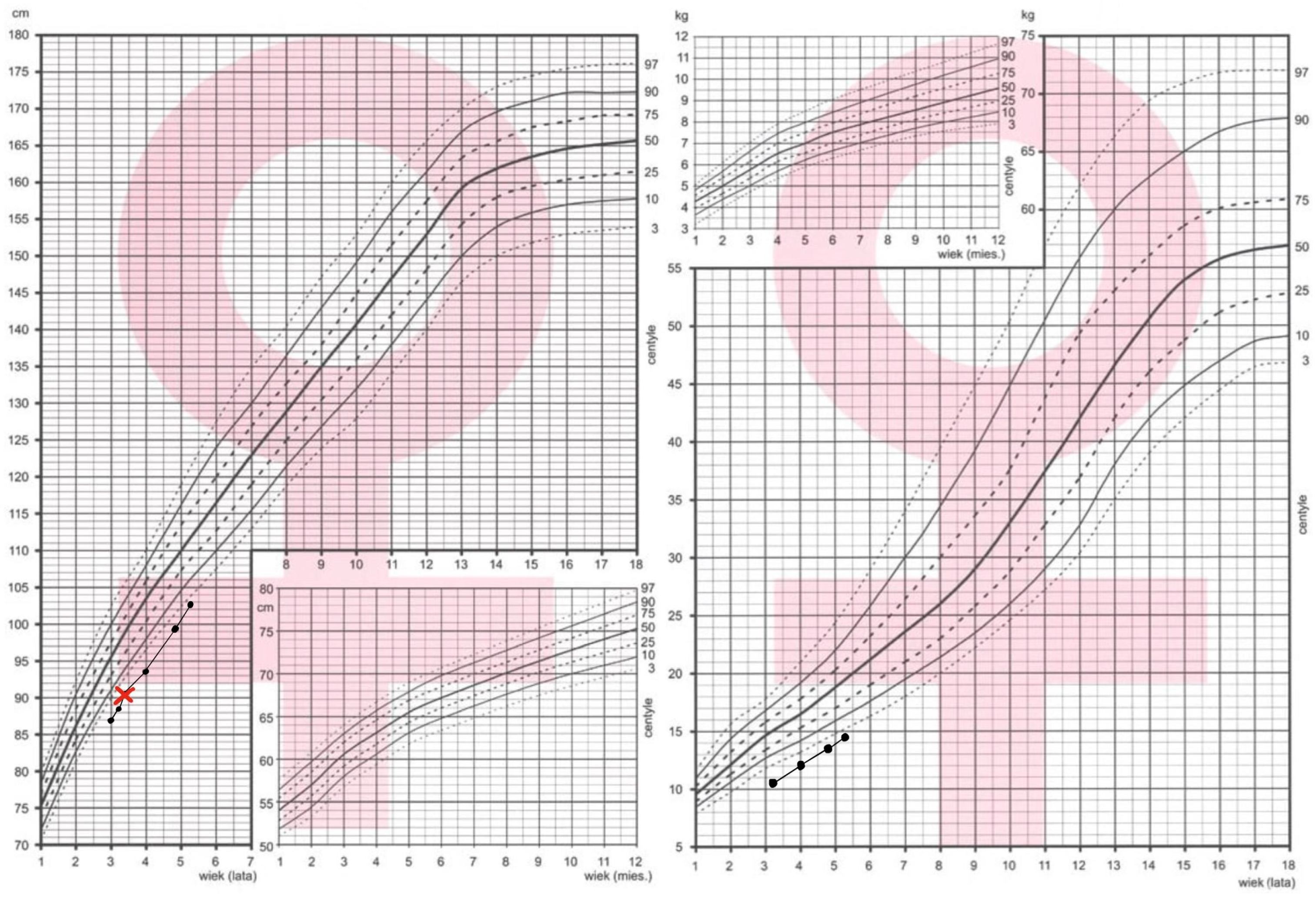
The female patient, currently 5.5 years old, has been under endocrinological follow-up since the age of 2 years because of growth failure and poor weight gain. Since birth, her length has remained below the 3rd percentile (Figure 3). At presentation, her height was 78.7 cm, her height age corresponded to 15 months, and her body weight was approximately 10% below the expected value for her height. Anthropometric assessment demonstrated a slightly increased arm span–to–height ratio (AS/H = 1.1), exceeding age- and sex-specific reference values [30]. Family history was unremarkable. Both parents were healthy. No relatives were reported to have growth failure or skeletal abnormalities. The mother`s height was 155 cm, whereas the father’s height was 178 cm, yielding a mid-parental height (MPH) of 160.5 cm.
The patient was born from the first pregnancy at 39 weeks of gestation by cesarean section. Birth weight was 2830 g (-1.56 SDS), and birth length was 51 cm. Apgar scores were 10 at both 1 and 5 minutes. The perinatal period was uneventful. Psychomotor development was within normal limits, with independent sitting achieved at 8 months and walking at 12 months. From infancy, feeding difficulties were noted, including aversion to solid foods and marked food selectivity, resulting in suboptimal weight gain. Gastroenterological evaluation was performed, and celiac disease was excluded.
Endocrine investigations demonstrated normal thyroid function and a normal insulin-like growth factor 1 (IGF-1) concentration (130.1 ng/mL at 4.5 years of age). Growth hormone deficiency was excluded, with a peak growth hormone level of 11.15 ng/mL during a glucagon stimulation test. Bone age assessment at 3 years and 6 months, based on an X-ray of the left wrist, revealed bone age consistent with chronological age (Figure 2F). Magnetic resonance imaging of the pituitary gland showed no abnormalities.
Due to the child’s short stature and an unfavorable bone age consistent with her chronological age, in the presence of normal endocrine findings and subtle dysmorphic features (including a small, receding mandible and downslanting palpebral fissures), genetic testing was pursued. Conventional cytogenetic analysis revealed a normal female karyotype (46,XX). Copy number variation analysis using next-generation sequencing (NGS) identified a heterozygous deletion encompassing exons 15–19 of the ACAN gene. This variant has not been previously reported in the ClinVar database; however, deletion of five out of nineteen exons is predicted to result in a truncated aggrecan protein with loss of normal function and is therefore considered the most likely molecular cause of the patient’s growth impairment.

3. Genetic Reports

3.1. DNA Sequencing and Bioinformatic Analysis

Genomic DNA was extracted from peripheral blood samples from both patients. Two NGS sequencing strategies were applied depending on the diagnostic indication: whole-exome sequencing (WES) for Patient 1 and targeted multigene panel sequencing for Patient 2.

3.2. Patient 1 – Whole-Exome Sequencing (WES)

Whole-exome sequencing was performed using the Twist Human Core Exome Plus Kit (Twist Bioscience). The enriched DNA libraries were sequenced using the Illumina NovaSeq 6000 instrument (Illumina, Inc., San Diego, CA, USA). All laboratory and sequencing procedures were carried out by CeGaT (Germany). Raw sequencing reads were mapped to the reference human genome using BWA-MEM2 v2.2.1 [32]. Duplicate reads were removed with Picard 2.18.2-SNAPSHOT (Broad Institute), variants were called using GATK HaplotypeCaller (gatk-4.2.6.1), and annotated with VEP version 105 and Samtools software. In silico prediction of variant pathogenicity was carried out using Alamut Visual Plus 1.7.2 (SOPHiA GENETICS), Franklin (Genoox), GeneBe, and additional computational prediction tools. Population frequency data were retrieved from dbSNP [33] and gnomAD.
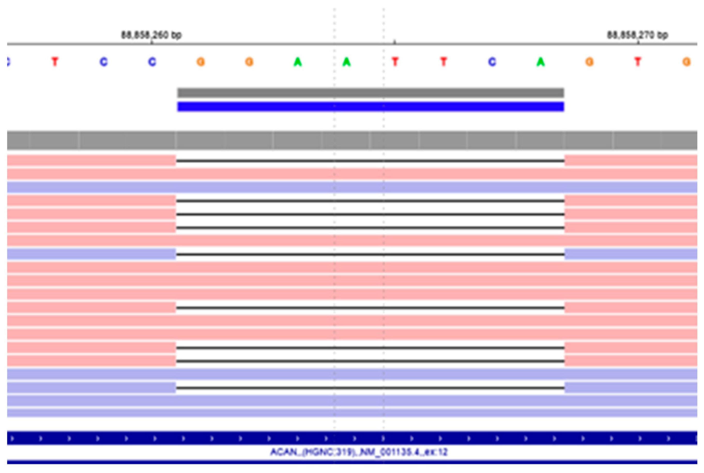
The detected genotype of the Patient 1: NM_001369268.1(ACAN):c.5677_5684del (p.Glu1893TrpfsTer8) in heterozygous (Figure 4).
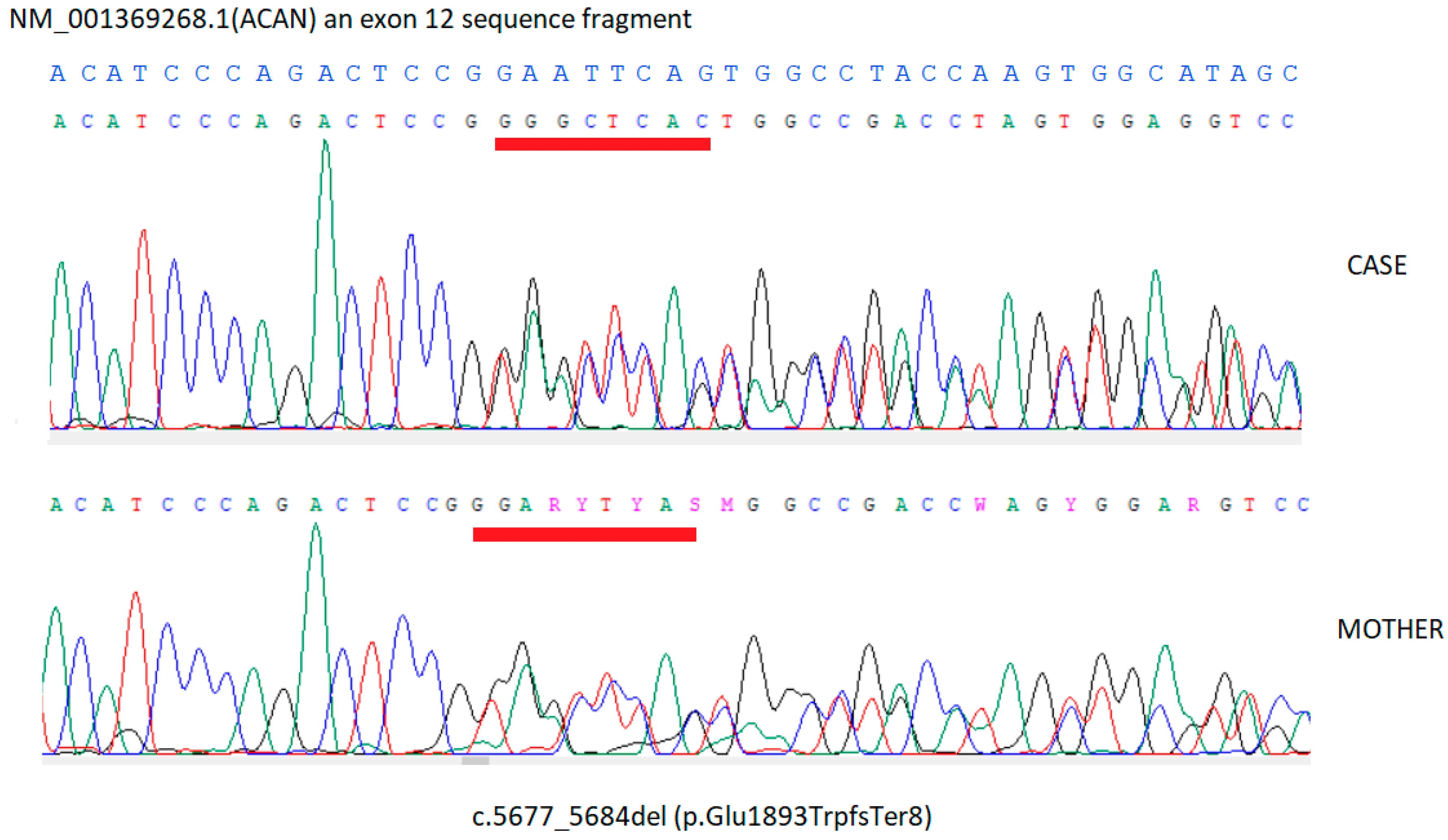
The detected variant was confirmed by Sanger DNA sequencing performed on a second, independent blood sample (Figure 5).

3.3. Patient 2 – Targeted Multigene Panel Sequencing

Targeted Next-Generation Sequencing (NGS) was performed using a custom multigene panel including the following genes: ACAN, ANKRD11, ARCN1, ARID1A, ARID1B, ARID2, ARSB, ARX, ASXL1, ASXL3, ATM, ATR, ATRX, BLM, BMP2, BRAF, BTK, CBL, CCDC8, CDC45, CDC6, CDT1, CHD7, CREBBP, CRIPT, CUL7, DHCR7, DPF2, EP300, FANCA, FANCC, FANCF, FANCG, FGD1, FGFR3, GHR, GHRHR, GHSR, GMNN, GNAS, GSC, HDAC8, HMGA2, HRAS, IGF1, IGF2, KANSL1, KDM6A, KMT2D, KRAS, LZTR1, MAGEL2, MAP2K1, MAP2K2, MCM5, MED12, NBN, NIPBL, NRAS, OBSL1, ORC1, ORC4, ORC6, PHF6, PIGG, PLAG1, POGZ, PTPN11, RAD21, RAF1, RIT1, RPS6KA3, SETBP1, SHOC2, SHOX, SLX4, SMARCA2, SMARCA4, SMARCB1, SMARCE1, SMC1A, SMC3, SOS1, SOS2, SOX11, SOX3, SPRED1, SRCAP, THOC2, TRIM37, UBE2T, USP9X, VPS13B, ZBTB18, ZEB2.Library preparation was conducted using the SureSelectXT Custom kit (Agilent). The sample was sequenced on the Illumina NovaSeq 6000 platform (Illumina, San Diego, CA, USA). Demultiplexing was performed with Illumina’s bcl2fastq2 v2.19.0. Adapter trimming was performed with Skewer v0.2.9 [34]. Reads were aligned to the GRCh37/hg19 human reference genome using BWA-MEM [35]. Duplicate reads were removed using Picard 2.18.2. Variant calling was performed with GATK HaplotypeCaller v4.0.3.0 [36,37] and FreeBayes v1.2.0-2-g29c4002 [38].Variants were annotated using the following resources:VEP97 [39], including SIFT and PolyPhen-2, dbNSFPv4.0 [40] (MutationAssessor, MutationTaster, DANN, FATHMM),ESP6500, gnomAD, dbSNP [33], ClinVar, 1000 Genomes [41].
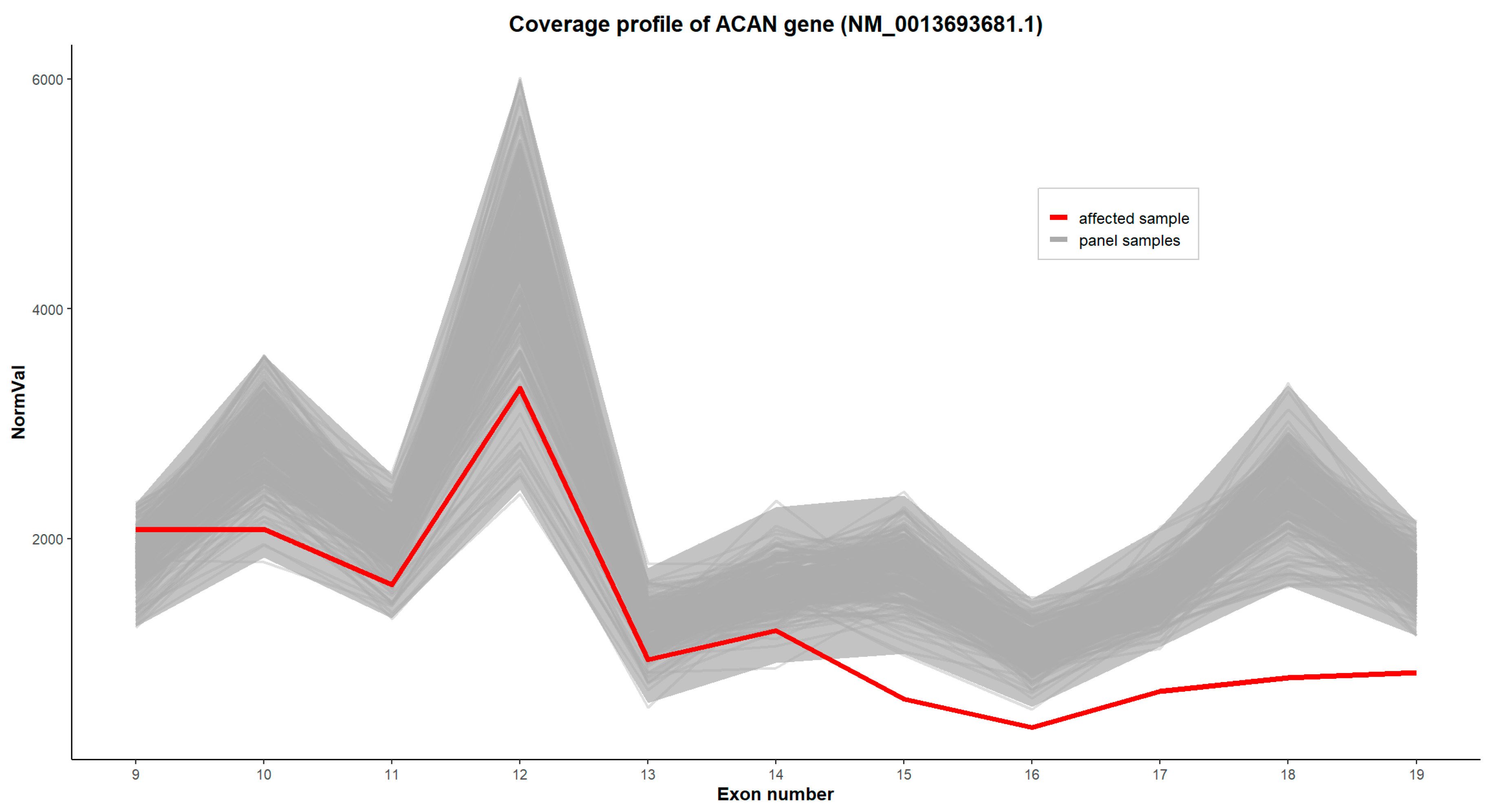
Copy-number variation (CNV) analysis was performed by examining normalized exon-level coverage profiles for all genes included in the panel. For each sample (BAM file), single-base coverage across all exons was calculated and normalized by the total number of mapped reads. Significant deviations in normalized exon coverage were interpreted as potential exon-level deletions or duplications.
A large deletion of 5 exons was identified in ACAN gene. We present the image from in-house CNV analysis, showing a deletion of exons 15–19 of the ACAN gene (chr15: 88,871,381_88,744,481), according to the reference sequence NM_0013693681.1 on Figure 6.

3.4. Variant Interpretation

For both patients, the pathogenicity of identified variants was assessed according to the 2015 American College of Medical Genetics and Genomics and the Association for Molecular Pathology (ACMG-AMP) guidelines and using the bioinformatics tools described above [42].

4. Discussion

4.1. Aggrecanopathies: Established Phenotypic Spectrum

Pathogenic variants in the ACAN gene are now recognized as a significant monogenic cause of idiopathic short stature (ISS), representing a primary disorder of growth plate extracellular matrix rather than an endocrine defect [14,15,43,44,45,46,47,48,49,50,51,52,53,54,55,56,57,58,59,60,61,62,63,64,65,66,67,68,69,70,71,72,73,74,75,76] (Table S1). Since the initial descriptions, ACAN-related disorders-collectively termed aggrecanopathies-have been shown to encompass a broad phenotypic continuum, ranging from severe skeletal dysplasias to isolated short stature with subtle clinical signs (Table S1).
Previously reported phenotypes include recessive spondyloepimetaphyseal dysplasia, aggrecan type; spondyloepiphyseal dysplasia, Kimberley type; autosomal dominant osteochondritis dissecans with early-onset osteoarthritis; and multiple presentations classified clinically as SSOAOD, frequently accompanied by advanced bone age. In addition to growth failure, a number of minor skeletal and dysmorphic features have been reported, including midfacial hypoplasia, flat or broad nasal bridge, frontal bossing, brachydactyly, broad or shortened thumbs, and mild limb disproportion (Table S1). Joint pathology, including early degenerative changes and intervertebral disc disease, has also been described, particularly in adolescence and adulthood. Aggrecanopathy caused by pathogenic ACAN gene variants exhibits a broad phenotypic spectrum; however, analysis of pooled cohort data demonstrates that several clinical features recur with sufficient frequency to define a characteristic pattern (Table S1). When feature occurrence is aggregated across published families and individual cases included in the Table S1, short stature, skeletal disproportionality, brachydactyly, and joint pathology emerge as the most prevalent manifestations, while craniofacial dysmorphism and axial skeletal abnormalities show greater variability.

4.2. Short Stature and Body Disproportion

Short stature is the most consistent feature, reported in approximately 90–100% of affected individuals, and represents the primary reason for clinical referral in nearly all cohorts [20,24,64,66] (Table S1). Disproportionate growth—most commonly reflected by an increased sitting height–to–height ratio or mild rhizomelic or mesomelic limb shortening—was documented in ~50–65% of reported families, particularly in studies with systematic anthropometric assessment [48,69,76]. Importantly, disproportion was often subtle in early childhood and became more apparent with age.

4.3. Brachydactyly and Appendicular Skeletal Features

Brachydactyly is among the most frequent and distinctive skeletal findings, observed in ~60–75% of families overall (Table S1). It was consistently reported across multiple cohorts, including those by Kim et al., Mancioppi et al., Cao et al., and Alexandrou et al., typically affecting distal phalanges, thumbs, and/or great toes [29,44,69,76]. Broadened thumbs or halluces were described in ~30–40% of families, while radiographically evident hand or foot dysplasia-such as shortened metacarpals/metatarsals (brachymetacarpia) or irregular epiphyses-was present in ~40–50%, including patients presented in this work. Nevertheless, Andrade et al. and Denis et al. documented genetically confirmed cases lacking overt limb dysplasia, highlighting incomplete penetrance of these features [48,64].

4.4. Craniofacial Dysmorphism

Craniofacial features were reported in ~45–60% of families, but no single facial trait was universal. The most common findings included midface hypoplasia (~40–50%), flat or low nasal bridge (~35–45%), frontal bossing (~30–40%), and relative macrocephaly (~25–35%). Ptosis and hypertelorism were less frequent, each occurring in approximately 15–25% of reported cases. Kim et al. and Mancioppi et al. described particularly recognizable craniofacial patterns, whereas several other cohorts, including Andrade et al., reported minimal or absent dysmorphism [29,44,64].

4.5. Novel Facial Dysmorphic Features Identified in the Present Study

An important contribution of the present study is the identification of previously unreported facial dysmorphic features. In both patients and in a mother of male patient, we observed a consistent facial pattern including ptosis, hypertelorism, downslanting palpebral fissures, mild mandibular retrognathia, thick, fleshy auricles and cubitus valgus.
Some of these features has not been emphasized in prior aggrecanopathy cohorts and appear to expand the dysmorphological spectrum associated with ACAN variants (Table S1).
The presence of these findings suggests that aggrecan deficiency may also affect craniofacial cartilage development, a hypothesis that aligns with the fundamental role of aggrecan in cartilage matrix integrity. Clinically, this phenotype may overlap with Noonan-like or other syndromic presentations (e.g. Turner syndrome in female patients), potentially contributing to diagnostic delay or misclassification. Recognition of this facial gestalt may therefore aid earlier suspicion of aggrecanopathy in children with short stature and normal endocrine evaluation.

4.6. Bone Age Advancement and Growth Plate Dysfunction

Advanced bone age relative to chronological age was a prominent feature, identified in ~50–70% of evaluated children [3,20,66,76]. This paradoxical association of short stature with accelerated skeletal maturation is a defining pathophysiological characteristic of aggrecanopathy and has significant implications for growth prognosis and treatment planning. In contrast, a subset of patients (~20–30%) exhibited bone age appropriate for age, or even delayed, emphasizing heterogeneity even within this core feature [3]. This feature may depend on age of a patient, prepubertal vs pubertal.

4.7. Axial Skeleton and Vertebral Involvement

Spinal abnormalities—including vertebral end-plate irregularities, mild scoliosis, or platyspondyly—were reported in ~25–35% of families (Table S1). More severe axial involvement resembling spondyloepiphyseal dysplasia Kimberley type was rare and largely confined to the cohort described by Gleghorn et al [24]. Mild scoliosis alone was present in ~10–20% of cases and was often clinically insignificant [10,50,56,63].

4.8. Joint Disease and Osteochondral Pathology

Joint involvement represents one of the most clinically consequential features and shows age-dependent penetrance. Osteochondritis dissecans, early-onset osteoarthritis, or degenerative joint disease was documented in ~30–45% of families overall, increasing to >50% among adolescents and adults [24,48,69]. Knee and elbow involvement predominated, although hip and spinal degeneration were also reported. Importantly, joint pathology was uncommon in early childhood, supporting a progressive disease model.

4.9. Phenotypic Variability

A defining characteristic across all cohorts is pronounced intrafamilial and interfamilial variability. Up to 20–30% of genetically affected individuals were reported to have minimal skeletal or dysmorphic features beyond isolated short stature [48,64]. This variability complicates phenotype-based recognition and underscores the diagnostic value of molecular testing.
In aggregate, the most frequent features of aggrecanopathy in children include short stature (≈90–100%), brachydactyly (≈60–75%), body disproportion (≈50–65%), advanced bone age (≈60–70%), craniofacial dysmorphism (≈45–60%), and joint pathology (≈30–45%). Recognition of this frequency-weighted constellation-rather than reliance on any single hallmark-is essential for timely diagnosis and longitudinal management. The substantial variability observed across and within families further supports early genetic evaluation in children presenting with unexplained short stature, particularly when accompanied by advanced bone age or subtle skeletal anomalies [3,14,15,43].

4.10. Distinct Growth Patterns and the Missed Therapeutic Window

A key clinical observation reinforced by our cases and supported by the literature is the distinctive growth trajectory in many children with aggrecanopathy. Growth during early childhood and the prepubertal period may appear relatively preserved, often with normal or only mildly advanced bone age. However, with the onset of puberty, skeletal maturation may accelerate rapidly, leading to early epiphyseal fusion and abrupt cessation of linear growth [3,14,15].
This pattern was clearly illustrated in the adolescent boy described in our study. At the time of initial evaluation, his auxological parameters and pubertal timing did not fulfill inclusion criteria for the commencement of the diagnostic process prior to growth hormone (GH) therapy or for treatment of precocious puberty national protocols of Narodowy Fundusz Zdrowia (National Fundation of Health). Endocrinological investigations were normal, and therefore no growth-modifying treatment was initiated. In retrospect, once the molecular diagnosis was established, it became evident that the absence of endocrine pathology did not equate to a favorable growth prognosis. By the time advanced bone age was recognized, the therapeutic window for effective intervention had largely closed.

4.11. Therapeutic Implications: Early GH Therapy and Puberty Modulation

These observations underscore a fundamental challenge in aggrecanopathy: standard endocrine-based treatment algorithms may be insufficient for a primary growth plate disorder. Although patients with ACAN variants are usually not growth hormone deficient, GH therapy has been shown in several studies to transiently improve growth velocity and height SDS, particularly when initiated early [3,7,9,29,50,53,74]. Some authors reported poor outcome of GH therapy [49,54,56].
Importantly, emerging evidence suggests that combined GH and gonadotropin-releasing hormone analogue (GnRHa) therapy may be beneficial in selected patients [10,14,27,51]. Suppression of puberty may slow estrogen-mediated growth plate senescence, thereby prolonging the growth period and allowing GH therapy to exert a more meaningful effect on final height. The distinct pubertal growth pattern observed in aggrecanopathy-relatively preserved prepubertal growth followed by rapid bone age advancement during puberty-provides a strong biological rationale for very early intervention, ideally before or at the very beginning of pubertal development.
The response to growth-promoting therapy with recombinant human growth hormone (GH), administered either as monotherapy or in combination with gonadotropin-releasing hormone analogs (GnRHa), is heterogeneous and strongly dependent on age at treatment initiation, pubertal status, and bone age advancement [3]. Across the studies summarized in Table S1, GH monotherapy was consistently associated with an increase in growth velocity, but only modest improvements in height standard deviation score (Ht SDS), with considerable interindividual variability. In cohorts treated with GH alone, most authors reported small first-year gains in height SDS, typically in the range of approximately +0.2 to +0.4 SDS per year [3].
Interestingly, Muthuvel et al. in an open-label, single-arm, prospective study recruiting patients with heterozygous mutation in ACAN, age ≥2 years, prepubertal with bone age (BA) ≥chronological age (CA), and normal insulin-like growth factor I concentration, observed that treatment with rhGH improved linear growth, whereas skeletal maturation did not advance inappropriately [74].
Overall, the available evidence suggests that GH monotherapy provides a limited but clinically relevant benefit, primarily through enhancement of growth velocity, with the greatest effect observed in younger, prepubertal children. The addition of GnRHa may be advantageous in patients with early or rapidly progressive puberty by prolonging the growth period rather than by producing large immediate gains in height SDS. These findings underscore the need for early diagnosis, careful patient selection, and long-term follow-up studies reporting final adult height to more accurately define the role of these therapeutic strategies.
Our younger patient, diagnosed prior to puberty, represents an opportunity to apply this genotype-informed strategy proactively, in contrast to the older boy, in whom delayed diagnosis precluded such intervention.

4.12. Ethical Considerations: Is It Justifiable to Suppress Normally Timed Puberty?

The proposal to suppress normally timed puberty in children without classical endocrine disease raises legitimate ethical concerns. These include the medicalization of genetic risk, potential psychological effects, decrease in bone density, and uncertainty regarding long-term benefit. However, aggrecanopathy represents a distinct clinical context in which puberty, although physiologically timed, may be biologically maladaptive, accelerating irreversible growth plate closure in a structurally compromised cartilage matrix.
From an ethical perspective, the principle of beneficence must be weighed against non-maleficence. In children with a confirmed pathogenic ACAN variant and a predictable natural history of growth failure, withholding intervention may itself result in foreseeable and irreversible harm in the form of severely compromised adult height. Therefore, it is reasonable to argue that aggrecanopathy warrants a dedicated therapeutic program, separate from standard GH deficiency based frameworks, incorporating molecular diagnosis, individualized risk assessment, and shared decision-making with families.

4.13. Insights from Molecular Studies: Lessons from ACAN⁺/⁻ Mouse Models

Recent molecular studies in heterozygous ACAN haploinsufficient mouse models provide critical mechanistic insight into the pathophysiology of aggrecanopathy [43]. These models demonstrate that postnatal growth failure results primarily from reduced extracellular matrix production and impaired chondrocyte hypertrophic differentiation, rather than decreased proliferation [43]. At the molecular level, reduced ACAN expression is associated with suppressed Akt signaling in prehypertrophic and hypertrophic chondrocytes, mediated in part by upregulation of calcium-calmodulin–dependent protein kinase pathways [43].
These findings reinforce the concept that aggrecanopathy is a cartilage-intrinsic disorder and help explain the limited and variable response to systemic endocrine therapies alone. They also open the possibility of future disease-modifying approaches aimed at enhancing proteoglycan synthesis or restoring growth plate signaling pathways, potentially in combination with GH and pubertal modulation [14,15,43].
The research findings reported by Bendre et al. further underscore the pivotal role of impaired IGF-I–Akt signaling in the pathophysiology of aggrecanopathy and provide a clear mechanistic rationale for early therapeutic intervention [43]. The consistent reduction in phosphorylated Akt1 observed in Acan⁺/⁻ growth plates and cultured chondrocytes indicates defective Akt signaling, a pathway that is essential for chondrocyte hypertrophic differentiation and cell size expansion [43]. Given that IGF-I is a principal upstream activator of the PI3K/Akt cascade within the growth plate, reduced Akt phosphorylation establishes a direct mechanistic link between aggrecan deficiency and disrupted IGF-I signaling. Experimental suppression of PI3K/Akt activity leads to shortening of the proliferative and hypertrophic zones and a reduction in hypertrophic chondrocyte size, closely recapitulating the growth plate abnormalities observed in Acan⁺/⁻ models [43]. Importantly, growth hormone therapy increases circulating IGF-I levels and may partially restore Akt signaling, thereby promoting chondrocyte hypertrophy and longitudinal bone growth [43]. This mechanistic framework provides a compelling explanation for the favorable growth responses reported in children with heterozygous ACAN mutations and supports the concept that GH therapy represents a meaningful therapeutic option in aggrecanopathy. Critically, these data emphasize the importance of initiating treatment early, before advanced bone age and growth plate maturation irreversibly limit remaining growth potential [43].

4.14. Arm Span/Height Ratio

Gerver et al. showed that carriers of ACAN haploinsufficiency exhibit an increased arm span–to–height (AS/H) ratio in childhood and adolescence, which remains mildly elevated into adulthood [30]. Our findings are consistent with this observation and can be mechanistically explained by the work of Bendre et al. in Acan⁺/⁻ mice [43]. In this model, growth plate impairment was segment-specific and most pronounced in regions exposed to high growth velocity and mechanical loading, particularly the distal femur [43].
Because the distal femoral growth plate contributes disproportionately to linear growth and bears concentrated axial load, aggrecan deficiency renders it especially vulnerable to functional failure. In contrast, less mechanically stressed regions-such as the upper limbs-are relatively spared [43]. Translating this to humans, preferential impairment of weight-bearing lower-limb growth plates would result in shortened leg length with preserved arm span, producing an elevated AS/H ratio.
Thus, the increased AS/H ratio observed in individuals with heterozygous ACAN mutations reflects selective lower-limb growth attenuation rather than true upper-limb overgrowth, highlighting the load-dependent nature of growth plate dysfunction in aggrecan deficiency [30,43].

5. Conclusion

In summary, our study expands the clinical phenotype of aggrecanopathy by identifying novel dysmorphic features, highlights the distinctive pubertal growth pattern associated with ACAN variants, and emphasizes the critical importance of early molecular diagnosis. These cases illustrate how reliance on conventional endocrine criteria may lead to missed therapeutic opportunities and support the need for genotype-driven management strategies.
Aggrecanopathy should be considered a distinct clinical entity within the spectrum of short stature disorders, warranting dedicated diagnostic pathways, early intervention protocols, and ethically grounded, individualized therapeutic decision-making.

Supplementary Materials

The following supporting information can be downloaded at the website of this paper posted on Preprints.org, Table S1.

Author Contributions

Conceptualization, D.J., A.S., T.T.; methodology, D.J.; software, A.S., T.T.; validation, D.J,A.F,T.K,H.P.,M.J.,K.S.,D.L,S.W..; formal analysis, D.J.,A.F.,T.K., H.P.,M.J.,K.S.,D.L,S.W; investigation, D.J, A.F, T.K, H.P.,M.J.,K.S.,D.L,S.W.; resources, D.J, A.F, T.K, H.P.,M.J.,K.S.,D.L,S.W.; data curation, D.J, A.F, T.K, H.P.,M.J.,K.S.,D.L,S.W.; writing—original draft preparation, A.S, T.T, writing—review and editing, D.J,A.F.; visualization, K.S, T.K., H.P., D.L., S.W..; supervision, D.J., J.S; project administration, D.J.; funding acquisition, D.J. All authors have read and agreed to the published version of the manuscript.”.

Funding

This research received no external funding.

Institutional Review Board Statement

The study was conducted in accordance with the Declaration of Helsinki. Ethical review and approval were waived for this study due to retrospective character of work.

Data Availability Statement

The original contributions presented in this study are included in the article/supplementary material. Further inquiries can be directed to the corresponding author(s).

Conflicts of Interest

The authors declare no conflicts of interest.

References

  1. Hauer, N.N.; Sticht, H.; Boppudi, S.; Büttner, C.; Kraus, C.; Trautmann, U.; Zenker, M.; Zweier, C.; Wiesener, A.; Jamra, R.A.; et al. Genetic screening confirms heterozygous mutations in ACAN as a major cause of idiopathic short stature. Sci. Rep. 2017, 7, 12225. [CrossRef]
  2. Murray, P.G.; Clayton, P.E.; Chernausek, S.D. A genetic approach to evaluation of short stature of undetermined cause. Lancet Diabetes Endocrinol. 2018, 6, 564–574. [CrossRef]
  3. Trigui, M.; Pallares-Ruiz, N.; Geneviève, D.; Amouroux, C.; Edouard, T.; Sigaudy, S.; Willems, M.; Barat-Houari, M. Expanding the molecular spectrum of aggrecanopathies: Exploring 24 patients with ACAN significant variants. Eur. J. Hum. Genet. 2025, 33, 1647–1654. [CrossRef]
  4. Fukami, M.; Seki, A.; Ogata, T. SHOX haploinsufficiency as a cause of syndromic and nonsyndromic short stature. Mol. Syndromol. 2016, 7, 3–11. [CrossRef]
  5. Hu, X.; Gui, B.; Su, J.; Li, H.; Li, N.; Yu, T.; Zhang, Q.; Xu, Y.; Li, G.; Chen, Y.; et al. Novel pathogenic ACAN variants in non-syndromic short stature patients. Clin. Chim. Acta 2017, 469, 126–129. [CrossRef]
  6. Plachy, L.; Strakova, V.; Elblova, L.; Obermannova, B.; Kolouskova, S.; Snajderova, M.; Zemkova, D.; Dusatkova, P.; Sumnik, Z.; Lebl, J.; Pruhova, S. High prevalence of growth plate gene variants in children with familial short stature treated with GH. J. Clin. Endocrinol. MeTable 2019, 104, 4273–4281. [CrossRef]
  7. Gkourogianni, A.; Andrew, M.; Tyzinski, L.; Crocker, M.; Douglas, J.; Dunbar, N.; Fairchild, J.; Funari, M.F.A.; Heath, K.E.; Jorge, A.A.L.; et al. Clinical characterization of patients with autosomal dominant short stature due to aggrecan mutations. J. Clin. Endocrinol. MeTable 2017, 102, 460–469. [CrossRef]
  8. Deng, S.; Hou, L.; Xia, D.; Li, X.; Peng, X.; Xiao, X.; Zhang, J.; Meng, Z.; Zhang, L.; Ouyang, N.; Liang, L. Description of the molecular and phenotypic spectrum in Chinese patients with aggrecan deficiency: Novel ACAN heterozygous variants in eight Chinese children and a review of the literature. Front. Endocrinol. (Lausanne) 2022, 13, 1015954. [CrossRef]
  9. Lin, L.; Li, M.; Luo, J.; Li, P.; Zhou, S.; Yang, Y.; Chen, K.; Weng, Y.; Ge, X.; Mireguli, M.; et al. A high proportion of novel ACAN mutations and their prevalence in a large cohort of Chinese short stature children. J. Clin. Endocrinol. MeTable 2021, 106, E2711–E2719. [CrossRef]
  10. Stavber, L.; Hovnik, T.; Kotnik, P.; Lovrečić, L.; Kovač, J.; Tesovnik, T.; Bertok, S.; Dovč, K.; Debeljak, M.; Battelino, T.; Avbelj Stefanija, M. High frequency of pathogenic ACAN variants including an intragenic deletion in selected individuals with short stature. Eur. J. Endocrinol. 2020, 182, 243–253. [CrossRef]
  11. Sentchordi-Montané, L.; Aza-Carmona, M.; Benito-Sanz, S.; Barreda-Bonis, A.C.; Sánchez-Garre, C.; Prieto-Matos, P.; Ruiz-Ocaña, P.; Lechuga-Sancho, A.; Carcavilla-Urquí, A.; Mulero-Collantes, I.; et al. Heterozygous aggrecan variants are associated with short stature and brachydactyly: Description of 16 probands and a review of the literature. Clin. Endocrinol. (Oxf.) 2018, 88, 820–829. [CrossRef]
  12. Korenberg, J.R.; Chen, X.-N.; Doege, K.; Grover, J.; Roughley, P.J. Assignment of the human aggrecan gene (AGC1) to 15q26 using fluorescence in situ hybridization analysis. Genomics 1993, 16, 546–548. [CrossRef]
  13. Valhmu, W.B.; Palmer, G.D.; Rivers, P.A.; Ebara, S.; Cheng, J.-F.; Fischer, S.; Ratcliffe, A. Structure of the human aggrecan gene: Exon-intron organization and association with the protein domains. Biochem. J. 1995, 309, 535–542. [CrossRef]
  14. Nilsson, O.; Guo, M.H.; Dunbar, N.; Popovic, J.; Flynn, D.; Jacobsen, C.; Lui, J.C.; Hirschhorn, J.N.; Baron, J.; Dauber, A. Short stature, accelerated bone maturation, and early growth cessation due to heterozygous aggrecan mutations. J. Clin. Endocrinol. MeTable 2014, 99, E1510–E1518. [CrossRef]
  15. Nilsson, O. Aggrecanopathies highlight the need for genetic evaluation of ISS children. Eur. J. Endocrinol. 2020, 183, C9–C10. [CrossRef]
  16. Jee, Y.H.; Baron, J.; Nilsson, O. New developments in the genetic diagnosis of short stature. Curr. Opin. Pediatr. 2018, 30, 541–547. [CrossRef]
  17. Tatsi, C.; Gkourogianni, A.; Mohnike, K.; DeArment, D.; Witchel, S.; Andrade, A.C.; Markello, T.C.; Baron, J.; Nilsson, O.; Jee, Y.H. Aggrecan mutations in nonfamilial short stature and short stature without accelerated skeletal maturation. J. Endocr. Soc. 2017, 1, 1006–1011. [CrossRef]
  18. Doege, K.J.; Sasaki, M.; Kimura, T.; Yamada, Y. Complete coding sequence and deduced primary structure of the human cartilage large aggregating proteoglycan, aggrecan: Human-specific repeats, and additional alternatively spliced forms. J. Biol. Chem. 1991, 266, 894–902. [CrossRef]
  19. Doege, K.; Rhodes, C.; Sasaki, M.; Hassell, J.R.; Yamada, Y. Molecular biology of cartilage proteoglycan (aggrecan) and link protein. In Extracellular Matrix Genes; Sandell, L.J.; Boyd, C.D., Eds.; Academic Press: New York, NY, USA, 1990; pp. 137–152. [CrossRef]
  20. Dateki, S.; Nakatomi, A.; Watanabe, S.; Shimizu, H.; Inoue, Y.; Baba, H.; Yoshiura, K.; Moriuchi, H. Identification of a novel heterozygous mutation of the aggrecan gene in a family with idiopathic short stature and multiple intervertebral disc herniation. J. Hum. Genet. 2017, 62, 717–721. [CrossRef]
  21. Day, J.M.; Olin, A.I.; Murdoch, A.D.; Canfield, A.; Sasaki, T.; Timpl, R.; Hardingham, T.E.; Aspberg, A. Alternative splicing in the aggrecan G3 domain influences binding interactions with tenascin-C and other extracellular matrix proteins. J. Biol. Chem. 2004, 279, 12511–12518. [CrossRef]
  22. Quintos, J.B.; Guo, M.H.; Dauber, A. Idiopathic short stature due to novel heterozygous mutation of the aggrecan gene. J. Pediatr. Endocrinol. MeTable 2015, 28, 927–932. [CrossRef]
  23. Tompson, S.W.; Merriman, B.; Funari, V.A.; Fresquet, M.; Lachman, R.S.; Rimoin, D.L.; Nelson, S.F.; Briggs, M.D.; Cohn, D.H.; Krakow, D. A recessive skeletal dysplasia, SEMD aggrecan type, results from a missense mutation affecting the C-type lectin domain of aggrecan. Am. J. Hum. Genet. 2009, 84, 72–79. [CrossRef]
  24. Gleghorn, L.; Ramesar, R.; Beighton, P.; Wallis, G.A. A mutation in the variable repeat region of the aggrecan gene (AGC1) causes a form of spondyloepiphyseal dysplasia associated with severe, premature osteoarthritis. Am. J. Hum. Genet. 2005, 77, 484–490. [CrossRef]
  25. Stougaard, J. Familial occurrence of osteochondritis dissecans. J. Bone Joint Surg. Br. 1964, 46, 542–543. [CrossRef]
  26. Stattin, E.-L.; Wiklund, F.; Lindblom, K.; Onnerfjord, P.; Jonsson, B.-A.; Tegner, Y.; Sasaki, T.; Struglics, A.; Lohmander, S.; Dahl, N.; Heinegard, D.; Aspberg, A. A missense mutation in the aggrecan C-type lectin domain disrupts extracellular matrix interactions and causes dominant familial osteochondritis dissecans. Am. J. Hum. Genet. 2010, 86, 126–137. [CrossRef]
  27. van der Steen, M.; Pfundt, R.; Maas, S.J.W.H.; Bakker-van Waarde, W.M.; Odink, R.J.; Hokken-Koelega, A.C.S. ACAN gene mutations in short children born SGA and response to growth hormone treatment. J. Clin. Endocrinol. MeTable 2017, 102, 1458–1467. [CrossRef]
  28. Fukuhara, Y.; Cho, S.Y.; Miyazaki, O.; Hattori, A.; Seo, J.-H.; Mashima, R.; Kosuga, M.; Fukami, M.; Jin, D.-K.; Okuyama, T.; Nishimura, G. The second report on spondyloepimetaphyseal dysplasia, aggrecan type: A milder phenotype than originally reported. Clin. Dysmorphol. 2019, 28, 26–29. [CrossRef]
  29. Mancioppi, V.; Prodam, F.; Mellone, S.; Ricotti, R.; Giglione, E.; Grasso, N.; Vurchio, D.; Petri, A.; Rabbone, I.; Giordano, M.; Bellone, S. Retrospective diagnosis of a novel ACAN pathogenic variant in a family with short stature: A case report and review of the literature. Front. Genet. 2021, 12, 708864. [CrossRef]
  30. Gerver, W.J.M.; Gkourogianni, A.; Dauber, A.; Nilsson, O.; Wit, J.M. Arm span and its relation to height in a 2- to 17-year-old reference population and heterozygous carriers of ACAN variants. Horm. Res. Paediatr. 2020, 93, 164–172. Erratum - Arm Span and Its Relation to Height in a 2- to 17-Year-Old Reference Population and Heterozygous Carriers of ACAN Variants. Horm Res Paediatr. 2020, 93 (3): 212.https://doi.org/10.1159/000509867. [CrossRef]
  31. Greulich, W.W.; Pyle, S.I. Radiographic Atlas of Skeletal Development of the Hand and Wrist; Stanford University Press: Stanford, CA, USA, 1959. [CrossRef]
  32. Li, H.; Durbin, R. Fast and accurate short read alignment with Burrows–Wheeler transform. Bioinformatics 2010, 26, 589–595. [CrossRef]
  33. Sherry, S.T.; Ward, M.; Sirotkin, K. dbSNP—database for single nucleotide polymorphisms and other classes of minor genetic variation. Genome Res. 1999, 9, 677–679. [CrossRef]
  34. Jiang, H.; Lei, R.; Ding, S.-W.; et al. Skewer: A fast and accurate adapter trimmer for next-generation sequencing paired-end reads. BMC Bioinform. 2014, 15, 182. [CrossRef]
  35. Li, H. Aligning sequence reads, clone sequences and assembly contigs with BWA-MEM. arXiv 2013, arXiv:1303.3997. [CrossRef]
  36. McKenna, A.; Hanna, M.; Banks, E.; et al. The Genome Analysis Toolkit: A MapReduce framework for analyzing next-generation DNA sequencing data. Genome Res. 2010, 20, 1297–1303. [CrossRef]
  37. Poplin, R.; Ruano-Rubio, V.; DePristo, M.A.; et al. Scaling accurate genetic variant discovery to tens of thousands of samples. bioRxiv 2018. [CrossRef]
  38. Garrison, E.; Marth, G. Haplotype-based variant detection from short-read sequencing. arXiv 2012, arXiv:1207.3907. [CrossRef]
  39. McLaren, W.; Gil, L.; Hunt, S.E.; et al. The Ensembl Variant Effect Predictor. Genome Biol. 2016, 17, 122. [CrossRef]
  40. Liu, X.; Wu, C.; Li, C.; Boerwinkle, E. dbNSFP v3.0: A one-stop database of functional predictions and annotations for human non-synonymous and splice-site SNVs. Hum. Mutat. 2016, 37, 235–241. [CrossRef]
  41. 1000 Genomes Project Consortium; Auton, A.; Brooks, L.D.; et al. A global reference for human genetic variation. Nature 2015, 526, 68–74. [CrossRef]
  42. Richards, S.; Aziz, N.; Bale, S.; et al. Standards and guidelines for the interpretation of sequence variants. Genet. Med. 2015, 17, 405–424. [CrossRef]
  43. Bendre, A.; Ottosson, L.; Baroncelli, M.; Dou, Z.; Nilsson, O. Growth failure in aggrecan deficiency is due to decreased extracellular matrix and impaired growth plate chondrocyte hypertrophy. Bone 2025, 200, 117594. [CrossRef]
  44. Kim, T.Y.; Jang, K.M.; Keum, C.W.; Oh, S.H.; Chung, W.Y. Identification of a heterozygous ACAN mutation in a 15-year-old boy with short stature who presented with advanced bone age: A case report and review of the literature. Ann. Pediatr. Endocrinol. MeTable 2020, 25, 272–276. [CrossRef]
  45. Wang, Y.; Ge, J.; Ma, J.; Qiao, L.; Li, T. Short stature with precocious puberty caused by aggrecan gene mutation: A case report. Medicine (Baltimore) 2020, 99, e21635. [CrossRef]
  46. Huang, H.; Jin, J.; Xiang, R.; Wang, X. Case report: A novel heterozygous frameshift mutation of ACAN in a Chinese family with short stature and advanced bone age. Front. Genet. 2023, 14, 1101695. [CrossRef]
  47. Karatas, E.; Demir, M.; Ozcelik, F.; Kara, L.; Akyurek, E.; Berber, U.; Hatipoglu, N.; Ozkul, Y.; Dundar, M. A case of short stature caused by a mutation in the ACAN gene. Mol. Syndromol. 2023, 14, 123–128. [CrossRef]
  48. Denis, A.; Chergui, S.; Basalom, S.; Campeau, P.M.; Janelle, C.; Pauyo, T. Variable expressivity in a family with an aggrecanopathy. Mol. Genet. Genomic Med. 2022, 10, e1773. [CrossRef]
  49. Florio, A.; Papa, R.; Caorsi, R.; Consolaro, A.; Gastaldi, R.; Gattorno, M.; Picco, P. A child with a novel ACAN missense variant mimicking a septic arthritis. Ital. J. Pediatr. 2019, 45, 148. [CrossRef]
  50. Sun, J.; Jiang, L.; Liu, G.; Ma, C.; Zheng, J.; Niu, L. Evaluation of growth hormone therapy in seven Chinese children with familial short stature caused by novel ACAN variants. Front. Pediatr. 2022, 10, 819074. [CrossRef]
  51. Kim, S.J.; Yoon, J.S.; Hwang, I.T. A novel heterozygous ACAN variant in a short patient born small for gestational age with recurrent patellar dislocation: A case report. J. Clin. Res. Pediatr. Endocrinol. 2022, 14, 481–484. [CrossRef]
  52. Yin, L.P.; Zheng, H.X.; Zhu, H. Short stature associated with a novel mutation in the aggrecan gene: A case report and literature review. World J. Clin. Cases 2022, 10, 2811–2817. [CrossRef]
  53. Wu, H.; Wang, C.; Yu, S.; Ye, X.; Jiang, Y.; He, P.; Shan, X. Downregulation of ACAN is associated with the growth hormone pathway and induces short stature. J. Clin. Lab. Anal. 2023, 37, e24830. [CrossRef]
  54. Crippa, M.; Giangiobbe, S.; Villa, R.; Bestetti, I.; De Filippis, T.; Fatti, L.; Taurino, J.; Larizza, L.; Persani, L.; Bellini, F.; et al. A balanced reciprocal translocation t(10;15)(q22.3;q26.1) interrupting ACAN gene in a family with proportionate short stature. J. Endocrinol. Investig. 2018, 41, 929–936. [CrossRef]
  55. Yang, L.; Zhang, C.; Wang, W.; Wang, J.; Xiao, Y.; Lu, W.; Ma, X.; Chen, L.; Ni, J.; Wang, D.; et al. Pathogenic gene screening in 91 Chinese patients with short stature of unknown etiology with a targeted next-generation sequencing panel. BMC Med. Genet. 2018, 19, 212. [CrossRef]
  56. Ahmed, H.A.; Elhossini, R.; Aglan, M.; Amr, K. Aggrecan-related bone disorders; a novel heterozygous ACAN variant associated with spondyloepimetaphyseal dysplasia expanding the phenotypic spectrum and review of literature. J. Genet. Eng. Biotechnol. 2024, 22, 100341. [CrossRef]
  57. Uchida, N.; Shibata, H.; Nishimura, G.; Hasegawa, T. A novel mutation in the ACAN gene in a family with autosomal dominant short stature and intervertebral disc disease. Hum. Genome Var. 2020, 7, 44. [CrossRef]
  58. Lv, S.; Zhao, J.; Liu, L.; Wang, C.; Yue, H.; Zhang, H.; Li, S.; Zhang, Z. Exploring and expanding the phenotype and genotype diversity in seven Chinese families with spondylo-epi-metaphyseal dysplasia. Front. Genet. 2022, 13, 960504. [CrossRef]
  59. Masunaga, Y.; Ohkubo, Y.; Nishimura, G.; Ueno, T.; Fujisawa, Y.; Fukami, M.; Saitsu, H.; Ogata, T. ACAN biallelic variants in a girl with severe idiopathic short stature. J. Hum. Genet. 2022, 67, 481–486. [CrossRef]
  60. Mohammadian Khonsari, N.; Mohammad Poor Nami, S.; Hakak-Zargar, B.; Voth, T. Mutations of uncertain significance in heterozygous variants as a possible cause of severe short stature: A case report. Mol. Cell. Pediatr. 2020, 7, 11. [CrossRef]
  61. Tang, W.; Wu, K.M.; Zhou, Q.; Tang, Y.F.; Fu, J.F.; Dong, G.P.; Zou, C.C. Genotype and phenotype in patients with ACAN gene variants: Three cases and literature review. Mol. Genet. Genomic Med. 2024, 12, e2439. [CrossRef]
  62. Wei, M.; Ying, Y.; Li, Z.; Weng, Y.; Luo, X. Identification of novel ACAN mutations in two Chinese families and genotype-phenotype correlation in patients with 74 pathogenic ACAN variations. Mol. Genet. Genomic Med. 2021, 9, e1823. [CrossRef]
  63. Liang, H.; Miao, H.; Pan, H.; Yang, H.; Gong, F.; Duan, L.; Chen, S.; Wang, L.; Zhu, H. Growth-promoting therapies may be useful in short stature patients with nonspecific skeletal abnormalities caused by ACAN heterozygous mutations: Six Chinese cases and literature review. Endocr. Pract. 2020, 26, 1255–1268. [CrossRef]
  64. Andrade, N.L.M.; Funari, M.F.A.; Malaquias, A.C.; Collett-Solberg, P.F.; Gomes, N.L.R.A.; Scalco, R.; Dantas, N.C.B.; Rezende, R.C.; Tiburcio, A.M.F.P.; Souza, M.A.R.; et al. Diagnostic yield of a multigene sequencing approach in children classified as idiopathic short stature. Endocr. Connect. 2022, 11, e220214. [CrossRef]
  65. Xu, D.; Sun, C.; Zhou, Z.; Wu, B.; Yang, L.; Chang, Z.; Zhang, M.; Xi, L.; Cheng, R.; Ni, J.; Luo, F. Novel aggrecan variant, p.Gln2364Pro, causes severe familial nonsyndromic adult short stature and poor growth hormone response in Chinese children. BMC Med. Genet. 2018, 19, 79. [CrossRef]
  66. Hattori, A.; Katoh-Fukui, Y.; Nakamura, A.; Matsubara, K.; Kamimaki, T.; Tanaka, H.; Dateki, S.; Adachi, M.; Muroya, K.; Yoshida, S.; et al. Next generation sequencing-based mutation screening of 86 patients with idiopathic short stature. Endocr. J. 2017, 64, 947–954. [CrossRef]
  67. Ochoa, M.; Yang, A.; Kollias, C.; Bakir, C.; Carsen, S.; Lazier, J.; Innes, A.M.; Pagé, M.; Dawrant, J.; Robinson, M.E.; et al. From “ACAN” to “I CAN”: Restoring wellness in a boy with severe osteochondritis dissecans through diagnostic precision combined with optimal medical, surgical and rehabilitation management. Bone Rep. 2023, 18, 101663. [CrossRef]
  68. Stattin, E.L.; Lindblom, K.; Struglics, A.; Önnerfjord, P.; Goldblatt, J.; Dixit, A.; Sarkar, A.; Randell, T.; Suri, M.; Raggio, C.; et al. Novel missense ACAN gene variants linked to familial osteochondritis dissecans cluster in the C-terminal globular domain of aggrecan. Sci. Rep. 2022, 12, 5215. [CrossRef]
  69. Alexandrou, E.; Dauber, A.; Tyzinski, L.; Hwa, V.; Andrew, M.; Kim, H.; Elangovan, S.; Gubanich, P.; Taylor-Haas, J.A.; Paterno, M.; Backeljauw, P. Clinical phenotype and musculoskeletal characteristics of patients with aggrecan deficiency. Am. J. Med. Genet. A 2022, 188, 1193–1203. [CrossRef]
  70. Zhao, Q.; Li, Y.; Shao, Q.; Zhang, C.; Kou, S.; Yang, W.; Zhang, M.; Ban, B. Clinical and genetic evaluation of children with short stature of unknown origin. BMC Med. Genomics 2023, 16, 194. [CrossRef]
  71. Freire, B.L.; Homma, T.K.; Funari, M.F.A.; Lerario, A.M.; Vasques, G.A.; Malaquias, A.C.; Arnhold, I.J.P.; Jorge, A.A.L. Multigene sequencing analysis of children born small for gestational age with isolated short stature. J. Clin. Endocrinol. MeTable 2019, 104, 2023–2030. [CrossRef]
  72. Perchard, R.; Murray, P.G.; Payton, A.; Highton, G.L.; Whatmore, A.; Clayton, P.E. Novel mutations and genes that impact on growth in short stature of undefined aetiology: The EPIGROW study. J. Endocr. Soc. 2020, 4, bvaa105. [CrossRef]
  73. Toscano, P.; Di Meglio, L.; Lonardo, F.; Di Meglio, L.; Mazzarelli, L.L.; Sica, C.; Di Meglio, A. Prenatal diagnosis of a novel pathogenic variation in the ACAN gene presenting with isolated shortening of fetal long bones in the second trimester of gestation: A case report. BMC Pregnancy Childbirth 2021, 21, 459. [CrossRef]
  74. Muthuvel, G.; Dauber, A.; Alexandrou, E.; Tyzinski, L.; Andrew, M.; Hwa, V.; Backeljauw, P. Treatment of short stature in aggrecan-deficient patients with recombinant human growth hormone: 1-year response. J. Clin. Endocrinol. MeTable 2022, 107, e2103–e2109. [CrossRef]
  75. Wu, S.; Wang, C.; Cao, Q.; Zhu, Z.; Liu, Q.; Gu, X.; Zheng, B.; Zhou, W.; Jia, Z.; Gu, W.; Li, X. The spectrum of ACAN gene mutations in a selected Chinese cohort of short stature: Genotype–phenotype correlation. Front. Genet. 2022, 13, 891040. [CrossRef]
  76. Cao, Y.; Guan, X.; Li, S.; Wu, N.; Chen, X.; Yang, T.; Yang, B.; Zhao, X. Identification of variants in ACAN and PAPSS2 leading to spondyloepi(meta)physeal dysplasias in four Chinese families. Mol. Genet. Genomic Med. 2022, 10, e1916. [CrossRef]
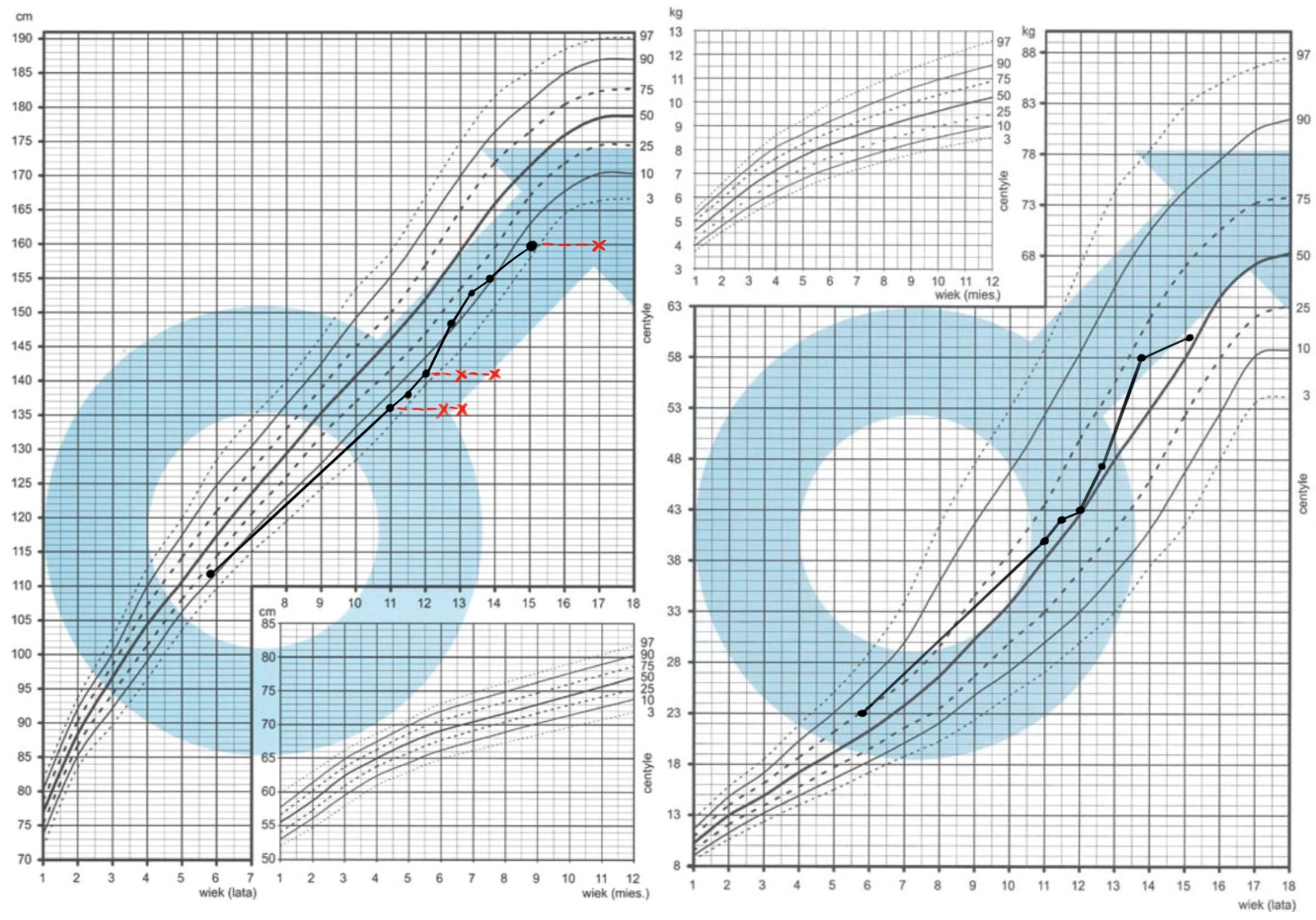
Figure 1. Growth charts of Patient 1. The Polish national charts were obtained from the website: https://www.mp.pl/pediatria/praktyka-kliniczna/procedury/13848,ocena-rozwoju-somatycznego-dzieci-i-mlodziezy,1.
Preprints 198778 g001
Figure 2. Patient 1 (A-E) and Patient 2 ( F). The Patients’ bone ages (on the left side with capital letter L for each pair A-F) compared to female and male standards (on the right side of each pair A-F) showing brachydactyly type E (brachymetacarpia) with shortening of metacarpal bones. A. Male patient’s chronological age: 11 years; bone age: 14 years (left), compared with the male standard for age 14; B. Patient’s chronological age: 11.5 years, bone age: 14 years, compared with the male standard for age 14; C. Patient’s chronological age: 12 years and 8 months, bone age: 14 years, compared with the male standard for age 14; D. Patient’s chronological age: 13 years and 4 months, bone age: 15 years, compared with the male standard for age 15; E. Patient's chronological age: 15 years, bone age: 17 years, compared with the male standard for age 17. F. Female patient’s chronological and bone age: 3 years and 6 months, compared to female standard for this age.
Preprints 198778 g002
Figure 3. Growth chart of Patient 2. The Polish national charts were obtained from the website: https://www.mp.pl/pediatria/praktyka-kliniczna/procedury/13848,ocena-rozwoju-somatycznego-dzieci-i-mlodziezy,1.
Preprints 198778 g003
Figure 4. Integrative Genomics Viewer (IGV) visualization of the heterozygous ACAN frameshift variant NM_001369268.1:c.5677_5684del (p.Glu1893TrpfsTer8). The eight-nucleotide deletion is present in a proportion of aligned reads consistent with heterozygosity and results in a frameshift introducing a premature stop codon eight amino acids downstream.
Preprints 198778 g004
Figure 5. Sanger sequencing chromatograms of the heterozygous NM_001369268.1(ACAN):c.5677_5684del (p.Glu1893TrpfsTer8). variant in Patient 1. and his mother.
Preprints 198778 g005
Figure 6. The normalized coverage profile for ACAN gene (transcript NM_0013693681.1) across the analyzed exons. The x-axis contains exon numbers, and the y-axis shows normalized coverage values scaled to an arbitrary unit. The gray line represents the range of coverage in the reference samples from the panel, while the red line represents the values obtained for the accession under study. The visible decrease in signal corresponds to a deletion spanning chr15: 88,871,381_88,744,481 (Human genome reference: GRCh38).
Preprints 198778 g006
Table 1. Follow-up visits of Patient 1.
Chronological age
(years)
11 12.8 13.4 15
Bone age
(years)
14 14 15 17
Testes volume R,L 3,4 8,8 10,10 15,15
axillarche - - - +
pubarche I I II V
IGF-1 (ng/ml) 354.2 433.8 302.8
LH (mlU/ml) 1.62 2.17
FSH (mlU/ml) 2.2 3.6
Testosterone (ng/ml) 0.6-2.70 2.58
TSH (ulU/ml) 1.3 0.9 0.96 0.8
fT4 (pmol/l) 1.33 14.3 14.3 16.3
Disclaimer/Publisher’s Note: The statements, opinions and data contained in all publications are solely those of the individual author(s) and contributor(s) and not of MDPI and/or the editor(s). MDPI and/or the editor(s) disclaim responsibility for any injury to people or property resulting from any ideas, methods, instructions or products referred to in the content.
Copyright: This open access article is published under a Creative Commons CC BY 4.0 license, which permit the free download, distribution, and reuse, provided that the author and preprint are cited in any reuse.