Submitted:
11 February 2026
Posted:
12 February 2026
You are already at the latest version
Abstract
Background: The Bacillus Calmette–Guérin (BCG) vaccine against tuberculosis is the most widespread vaccine in the world. Discovered by French investigators Albert Calmette and Camille Guérin at the Pasteur Institute, it remains the only effective vaccine against tuberculosis infection. This report describes the recognition and identification of a previously unknown French handwritten laboratory notebook prepared by Drs. Calmette and Camille Guérin recording their experiments performed during the development of the BCG vaccine. Methods: The notebook was examined, translated into English, photographed and the experiments analyzed. Results: The laboratory notebook consists of 69 leaves written in 2 hands, one of which corresponds to that of Albert Calmette. This handwritten notebook contains details of experiments that were performed during the development of the BCG vaccine at the Pasteur Institute by Drs. Calmette and Guérin. These include experimental inoculations of rabbits and guinea pigs describing the pathology including skin lesions, inflammatory reactions, organ pathology and survival. The experiments describe varying inoculative dosages of the bacteria, and different routes of administration including intraperitoneal and subcutaneous injections and administration of bacilli in the ear. In those cases where the animal had died following inoculation of tubercle bacilli, necropsy was performed and the organs examined and the pathology findings described. It describes experimental animal deaths and results of necropsies. Conclusions: this previously unknown notebook is a highly organized and detailed record of investigations using tuberculosis in animal experiments and microbiological culture to produce a safe and effective vaccine, first used in humans in 1921.
Keywords:
tuberculosis
; vaccine development
; Pasteur Institute
; Albert Calmette
; Camile Guérin
; laboratory notebook
; manuscript
; Bacillus Calmette–Guérin
; BCG vaccine
; Mycobacterium bovis
1. Introduction
Vaccines are considered to be one of the greatest triumphs of medicine and public health, being considered by some to be humanity’s greatest invention [1]. In a landmark study published by The Lancet, vaccination for infectious diseases was estimated to have saved 154 million lives in the past 50 years alone, the majority of whom were children [2]. The Bacillus Calmette–Guérin (BCG) vaccine against tuberculosis is the most widespread vaccine in the world [3]. Although the vaccine for smallpox developed by Edward Jenner in 1796 was the first vaccine against an infectious disease, more doses of the BCG vaccine have been administered globally than any other vaccine in history, with greater than 4 billion persons having been vaccinated since 1921 [4].
Tuberculosis, caused by the bacterium Mycobacterium tuberculosis, has ravaged the world for greater than 4000 years and resulted in more than one billion deaths [5]. The discovery by Robert Koch in 1882 that M. tuberculosis was the etiological agent of tuberculosis paved the way for the development of methods of diagnosis, prevention and treatment of this infectious disease [6]. The creation of an effective vaccine against M. tuberculosis by Albert Calmette and Camille Guérin at the Pasteur Institute in Lille and Paris in France was a landmark achievement that demonstrated that tuberculosis was a vaccine-preventable disease, ensuring its continuous use since 1921 [7,8,9].
This article describes the recent recognition and initial description of a previously unknown French manuscript that is a laboratory notebook prepared by Drs. Calmette and Guérin during their experiments to develop the BCG vaccine. This notebook had remained unidentified as to its authorship and content for many decades. Following its recent translation it was found to contain detailed laboratory, microbiological, pathological and animal experiments performed by Drs. Calmette and Guérin that were initially undertaken at the Institut Pasteur de Lille, and later in Paris, that lead to the successful vaccine for tuberculosis. This manuscript attests to the complexity of their research and is a significant historical document.
2. Doctors Calmett, Guérin and the BCG Vaccine
Léon Charles Albert Calmette (1863-1933) was a French bacteriologist, immunologist and physician who was interested in tropical and infectious diseases from his earliest work after studying at the French Navy Medical School and joining the Colonial Medical Corps, first in Hong Kong working with Patrick Manson, and later in Saint-Pierre-et-Miquelon and Africa. Following his studies of malaria, sleeping sickness, pellagra and filariasis, he returned to France in 1890 to study in the Microbiology course of Emile Roux where he met Louis Pasteur. In 1891 Pasteur requested that he travel to French Indochina (Vietnam) to organize and direct a branch of the Institut Pasteur in Saigon. While there he conducted large vaccination campaigns for smallpox and rabies. Calmette returned to France in 1894 where he developed the first snake antivenoms, and in collaboration with Alexandre Yersin developed the first immune serum against the plague bacillus, Yersinia pestis [10,11]. In 1895, Calmette was appointed as the director of the new Pasteur institute in Lille. Two years later, Camille Guérin, a veterinary bacteriologist trained at the Ecole Nationale Vétérinaire, joined him as his assistant, and later in 1900, became Director of Laboratories. When Calmette arrived in Lille tuberculosis was causing approximately 1100 deaths per year, and he believed that an immunological method was the best method for treatment. In 1900 Calmette and Guérin began at program of culturing tubercle bacilli to identify a suitable strain for potential use in a vaccine. Their initial attempts to culture the bacillus on agar and potato medium were hindered because clumping prevented a homogeneous suspension of the medium. They added ox bile to the medium and found that not only was clumping inhibited but also there was diminished virulence of the bacteria in subcultures. In 1908 Calmette and Guérin were working with a virulent strain of the tubercle bacillus that had been provided to them by Edmund Nocard, who had isolated it in 1902 from the udder of a cow with tuberculous mastitis. They found that adding sterile ox bile to the suspension prevented clumping and, surprisingly, diminished the virulence of the strain without altering their antigenicity. This fortuitous finding then permitted Calmette and Guérin to undertake further subcultures, at intervals of three weeks, and develop a vaccine against the attenuated tubercle bacillus. The bacteria from these repeated subcultures were then inoculated into young oxen where there was initially an increase in bacterial virulence, but after approximately 30 subcultures, the virulence was observed to decrease. By 1913 they were prepared to initiate a trial of vaccination in cattle, but the onset of World War I delayed their plans. The German military occupied Lille but permitted them to continue their experiments of subculturing the tuberculosis strains to reduce virulence, with the German veterinary surgeons assisting them in obtaining supplies including ox bile from the abattoir [12]. After the war, Calmette returned to Paris as Assistant Director of the Pasteur Institute and continued research to develop a tuberculosis vaccine. Calmette and Guérin had successfully performed 230 subcultures by 1919, producing a tubercle bacillus that did not cause progressive tuberculosis when inoculated into experimental animals including rabbits and guinea pigs (the topic of this present laboratory notebook), as well as cattle and horses. In 1920, Calmette and Guérin announced that they had developed a successful vaccine against the bovine strain of the tubercule bacillus in which its virulence was suppressed [12]. Guérin suggested that they name it the Bacille Bilie Calmette-Guérin, later omitting “Bilie” and calling it Bacille Calmette-Guérin or BCG [4,10,12]. The initial use of the vaccine in a human occurred in order save the life of a newborn infant whose mother had died of tuberculosis one day after delivery [4,10]. The baby’s grandmother who was caring for the infant also had tuberculosis. At the Charité Hospital in Paris on July 18th, 1921 Drs. Benjamin Weill-Halle and Raymond Turpin successfully administered a dose of the untested BCG vaccine by mouth to the newborn infant on the 3rd, 5th and 7th days of life. At six months of age the baby showed no signs of tuberculosis despite continual exposure at home from its caregiver, a grandmother who had tuberculosis. During the following years, the BCG vaccine was administered to hundreds of infants, and in 1924 it began to be mass produced and distributed. During the following years, the BCG vaccine was administered to hundreds of infants, and in 1924 it began to be mass produced and distributed. Between 1924 and 1928 there were approximately 114,000 infants vaccinated with BCG, and in France by 1931 there were 100,000 infants receiving the vaccine annually [12]. Eventually, administration of the BCG vaccine was mandatory in France starting in 1950 and onwards, leading to a decrease in mortality from tuberculosis. The BCG vaccine was included in the World Health Organization's Expanded Program on Immunization for developing countries [13].
3. Historical Context of the Notebook
This notebook may represent the sole surviving record of experimentation conducted at the Pasteur Institute by Drs. Calmette and Guérin during the laboratory research that led to the development of the BCG vaccine. Laboratory notebooks are unique amongst manuscript documents – they were internal working documents meant to register and memorialize the nature and results of experiments. Because of this, these notebooks often lacking designated authorship or location, were sometimes written in several languages, and typically had handwritten entries from multiple laboratory staff. Microbiology, vaccinology and infectious disease laboratory notebooks such as this one were usually written in chronological sequence and contained experiments, raw data, experimental animal or microbiological culture observations, results and interpretations. Unlike such historical handwritten items as medical lecture notes and correspondence, which typically were neatly penned and often written by professional scribes or secretaries, laboratory notebooks were not meant to be read outside of the lab. Thus, they do not contain elegantly written text, well-organized tables and diagrams and can be challenging to read and interpret. Making their understanding more challenging, they typically employ numerous scientific abbreviations, some of which are peculiar to the laboratory or experiments or which may be outdated. Fortunately, the text in the present notebook by Drs. Calmette and Guérin is actually quite legible, especially by a Francophone with knowledge of medicine and science.
4. History of the Notebook
Unlike many other historical medical manuscripts, this notebook has had a continuous history of identifiable ownership. Written by Drs. Calmette and Guérin in Paris during experimentation to develop the vaccine, after Dr. Calmette’s death in 1933 it was passed to the Calmette family who kept it for many decades. In addition to this manuscript laboratory notebook, Dr. Calmette’s personally collected and professionally bound 2 volumes of his offprints on tuberculosis and the BCG vaccine remained together in the Calmette family, along with another laboratory notebook describing research at a later date. These items eventually passed through a series of book dealers in France and the United States until being passed on to the present owner. This manuscript was analyzed by the co-authors – one having expertise in infectious diseases and vaccinology (DAS) and the other having expertise in French literature pertaining to medicine and tuberculosis (LM), who confirmed it as the laboratory notebook of the development of the BCG vaccine at the Pasteur Institute.
5. Identification of Authorship
Although the authorship of this notebook is not stated, we have no doubts that this manuscript is the personal laboratory logbook used by Drs. Calmette and Guérin during the development of the BCG vaccine in the early 1900s at the Pasteur Institute. This is based upon the following:
- a)
- The manuscript is written in 2 different hands from individuals with technical laboratory and microbiology knowledge. Drs. Calmette and Guérin both worked in the laboratory performing experiments.
- b)
- Comparing the present notebook with published examples of Dr. Calmette’s autographed correspondence, the handwritten notes from one of the writers match the handwriting of Dr. Calmette.
- c)
- There are extensive and detailed descriptions of animal experiments involving tuberculosis preparations via various routes, ex. oral, intravenous and intraperitoneal. These are the procedures used by Drs. Calmette and Guérin to evaluate the effects of their vaccine preparation to cause infection. It is extensively documented that Drs. Calmette & Guérin performed studies on animals to test the safety of their vaccine.
- d)
- The animals used were mostly guinea pigs (cobaye). Throughout this manuscript there are records of experiments performed on guinea pigs in various cages. The guinea pig model has been widely used as a diagnostic tool for detecting Mycobacterium in clinical samples until culture media could demonstrate similar sensitivity. Drs. Calmette & Guérin were known to experimentally infect guinea pigs with various preparations of the tubercle bacillus to test the safety of their vaccine. Guinea pigs serve as an excellent preclinical model due to the similarity to human tuberculosis pathology.
- e)
- Rabbits (lapin) are also described throughout the notebook as experimental animals with tuberculosis infections. The rabbit model of the tuberculosis vaccine effectiveness has been used for a long time. Drs. Calmette & Guérin were known to experimentally infect rabbits with various tubercle bacillus preparations to test the safety of their vaccine.
- f)
- There are efforts to analyze the effects of the tuberculosis preparations on animals who have died. The results of several autopsies are discussed in these notes.
- g)
- There are references in the notebook describing the laboratory culture of tuberculosis. Drs. Calmette & Guérin performed extensive cultures of Mycobacterium bovis in preparing their vaccine.
- h)
- It is clear from the number of experiments, the sophistication of the research design, and especially the large quantity of cages (up to 61) required for housing the experimental animals that this notebook represents research performed at a large French microbiological research institution. During this time period, only the Pasteur Institute would have fulfilled these parameters.
- i)
- There are no other identifiable laboratories from this period in France who were performing the types of extensive microbiological and animal experimentation on tuberculosis and developing a vaccine besides the laboratory of Drs. Calmette and Guérin at the Pasteur Institute in Lille and Paris. Simply put, if this notebook is not from their laboratory, then whose laboratory would it be from?
6. Physical Description of the Notebook
The notebook consists of a disbound group of sheets of typical early 20th century squared paper, each page measuring 212 x 132 mm. The squares measure 6 x 6 mm. There are a total of 69 leaves, most of which are written on both sides. There are 5 blank pages. The left border of the pages is irregular and consistent with having been previously bound, and some pages have remnants of an adhesive. The pages are present variously in variously-sized gatherings and individual leaves, with one large gathering still having 3 rusted metal staples present on the left margin. Other pages contain holes along the left border indicating the previous presence of 3 staples. Thus, the manuscript had been originally stapled in small gatherings, and then bound with glue into a binding.
7. Contents and Topics
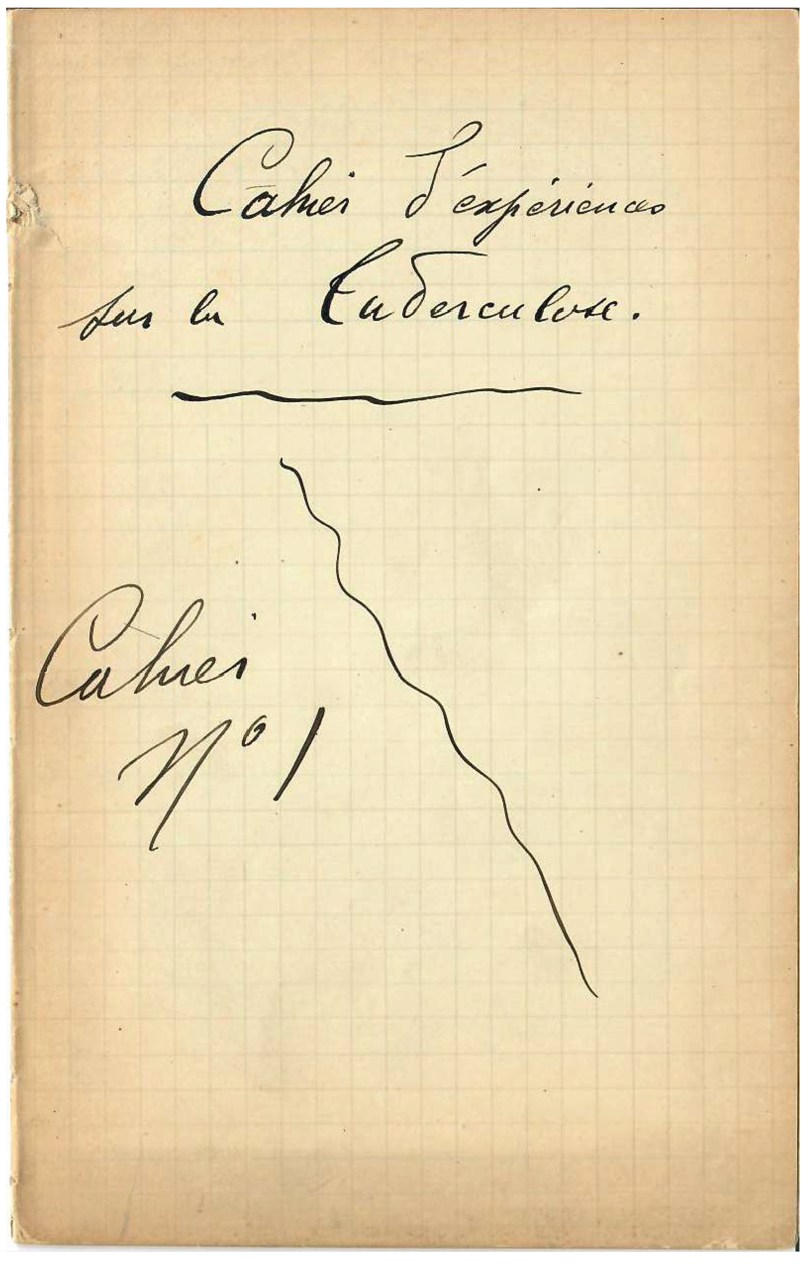
The manuscript is unpaginated and is written in 2 different hands, one of which matches published examples of the handwriting of Dr. Calmette. There are two pages with section headers. One is the initial, or title, page of the notebook and is labelled “Cahier l’experiences sue le tuberculose. Cahier No 1” (Figure 1), and the other states “Cahier No. 3”. The pages are not in order, and there is no location or authorship noted in the manuscript. The entries include dates of the month and day, but not the year.
Based upon the page headings and content, this manuscript can broadly be divided into two groupings - 1) laboratory animal cage-based descriptions of experiments on research animals (guinea pigs and rabbits), and 2) specific research topics.
In this first group the pages have headings referring to a specific cage number followed by a detailed description of the experiments, animals, observations and results, with entries from cage numbers 1, 4, 9, 10, 11, 13, 14, 15, 19, 35, 37, 39, 42, 43, 46, 57, 58, 60, and 61. There are separate pages referring to larger cages, and having with the headings “Grand Cage 2” and “Gde. 42”.
Among the second group, the notebook pages have a topic title, under which experiments and results are described. These topics include Vaccin de passage (passage of vaccine), Vaccin de passage (chez le lapin) (passage of vaccine among rabbits), Culture vaccin (culture of vaccine), Immunite vaccinale (vaccinial immunity), Vaccin pus (vaccine inflammation), Inoculation a l'oreille (inoculation in the ear), Vaccin peau (vaccine in the skin) and Tubes capillaire (capillary tubes).
There are several descriptions of the deaths of experimental animals including necropsy results. The necropsies describe the gross pathology findings with particular attention paid to the presence of inflammatory lesions (“pustulations”) and visceral organ pathology. These were likely performed by Dr. Guérin, who was a veterinarian. There is a mention to Dr. Robert Koch (1843-1910), famed German physician, medical microbiologist and discoverer of the tuberculosis bacterium.
Figure 2.
Experimental cutaneous inoculation of rabbits with vaccine (Number 61), assessment of skin lesions and experimental sacrifice of a rabbit for analysis. It reads: Under what conditions does the rabbit reliably receive the vaccine (continued). 21/11 [November 21] 3 rabbits are vaccinated with vaccine n°61, are placed in darkness at 18°- 20°. 21/11 [November 21] 1 control remains in daylight and at an ordinary temperature of 10°- 14°. On the third day 24/11 [November 24], the pustulation is clear on the 3 animals kept at 20°. Especially on the rabbit that was inoculated by rubbing on the epidermis, one observes in this animal a generalized rash [eruption] of the finest appearance. The control shows a pustulation that is clearly delayed compared to the other rabbits that were kept at 18°- 20°. 26/11 [November 26]. The pustules are very beautiful on all the rabbits, even on the control whose pustules are smaller, however. This vaccine is collected and glycerinated. The vaccine inoculated by rubbing on the skin of one of these 3 rabbits has taken remarkably. This rabbit is sacrificed. The skin bearing pustulation is removed, cut into fragments, and placed in sterilized glycerin in the cool room.
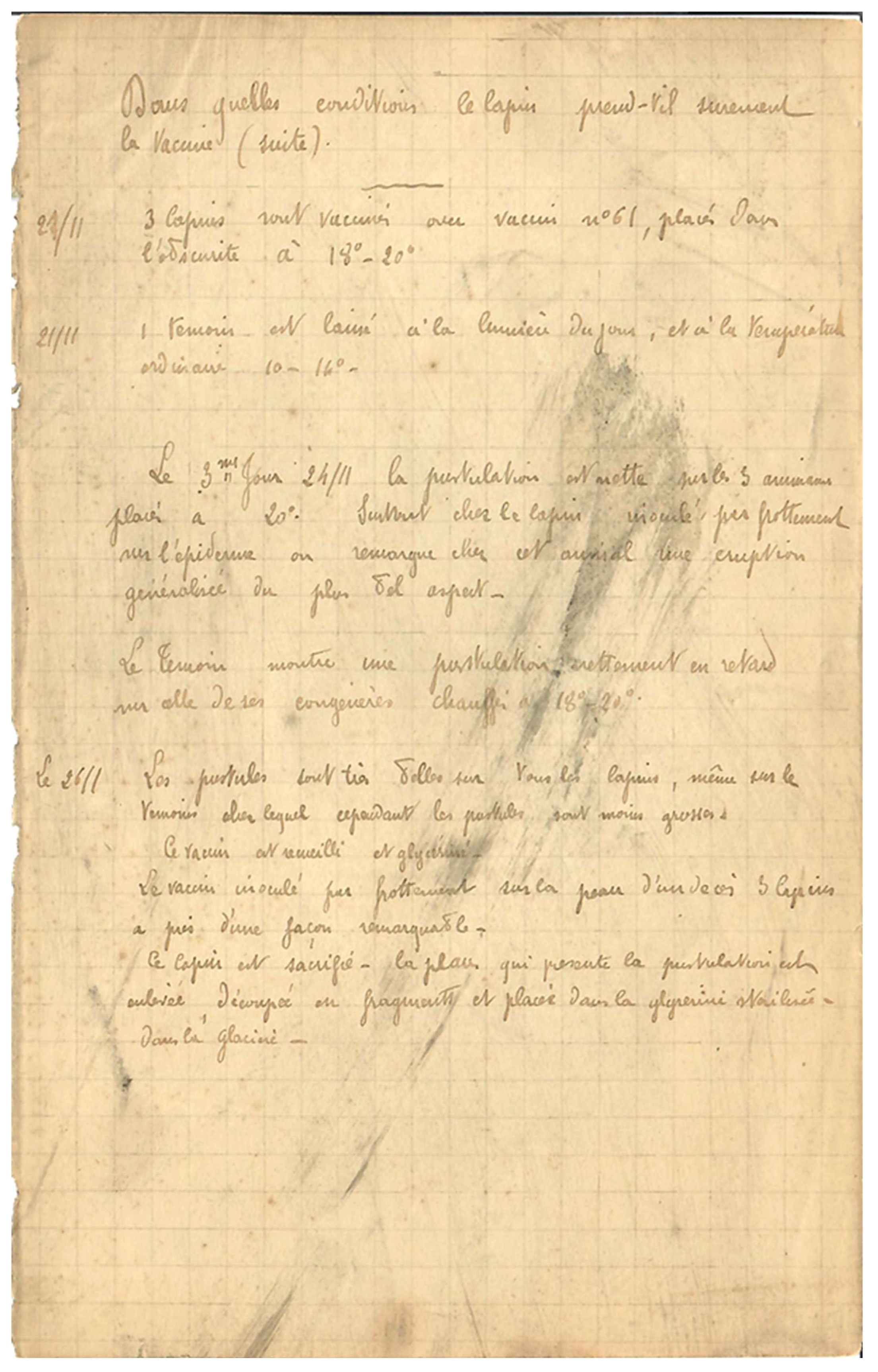
Figure 3.
Tuberculosis vaccine passage with characterization and evolution of pustule formation following inoculation of rabbits – “Pustulation Magnifique”! It reads: Passage of vaccine (3rd passage) 26/11 [November 26] Two rabbits are vaccinated with the rabbit vaccine (2nd passage). Placed in the incubator at 18°- 20°. 30/11. [November 30] Magnificent pustulation. The vaccine is collected from one rabbit and inoculated into another rabbit. Characteristics of the general eruption in the rabbit. As early as the second day, 2 x 24h one observes an extremely intense congestion of the dermis, whose bright red coloration contrasts sharply with the neighboring areas that remained white. One notices that the congested parts are slightly raised above the surrounding parts. On the third day, 3 x 24h the redness is even more intense. However, there is no pustulous process that appears. The fourth day, 4 x 24h On the eve [day] before the surface was uniformly red, one can see discolored points the size of which varies between the head of a pin and a grain of millet. These points become whitish and are surrounded at this level, by the lifting of the epidermis, by a colorless serous fluid that is quite abundant, after having squeezed or pressed these pustules between one’s fingers. The fifth day, 5 x 24h the eruption reaches complete maturity. Small yellowish scabs [craters] start to appear here and there where the pustules are. At this moment, the vaccine is virulent. The following days, the formation regresses, leaving abundant scabs [crusts] that detach by the 11th or 12th day, which leaves visible white scars. The harvesting of the vaccine should preferably be collected on the fourth day, 4 x 24h.
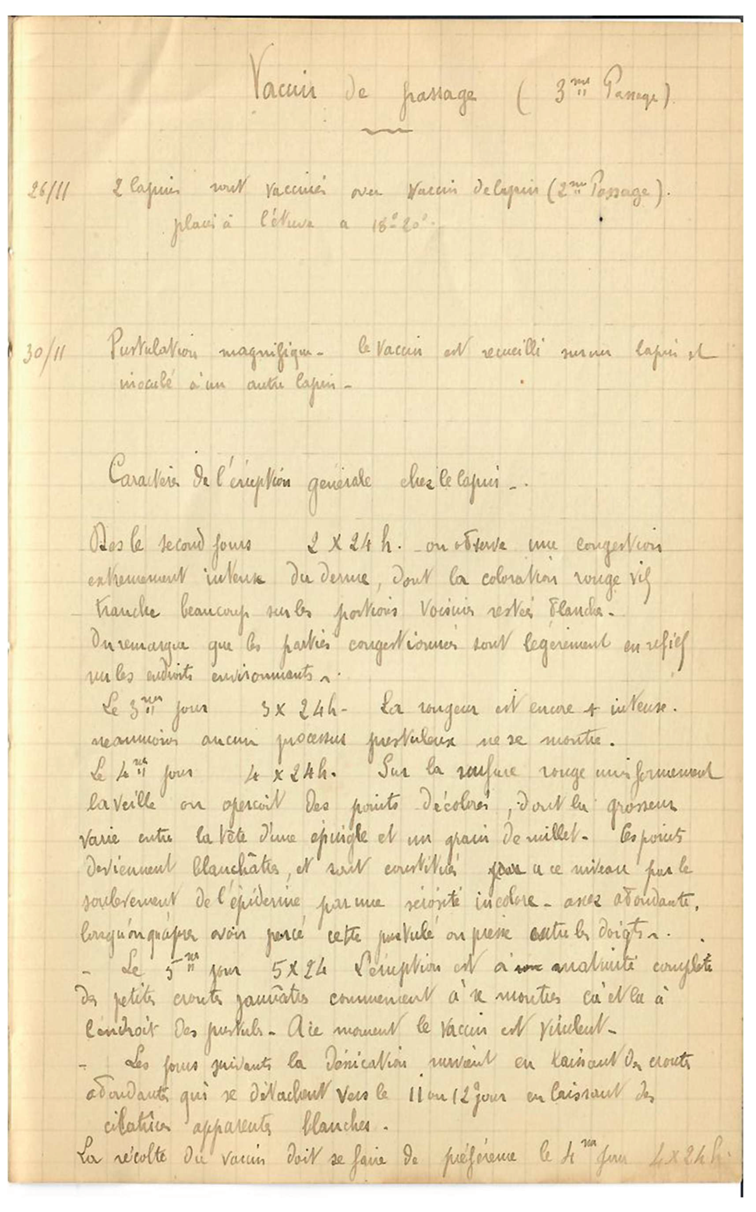
Figure 4.
Inoculation of tubercle bacilli into the ear and cultured. It reads: 13/12 [December 13] 1 rabbit is vaccinated on the external side of its ear by venous cutdown/denudation. Vaccine n°61. 16/12 [December 16] The rabbit receives 2 capillary tubes (model A) under the ear skin. 19/12 [December 19] The 2 tubes are removed and sowed on bouillon – agar – serum. Result: grown. 1 is inoculated on the back of a rabbit. R[esult]: Negative.
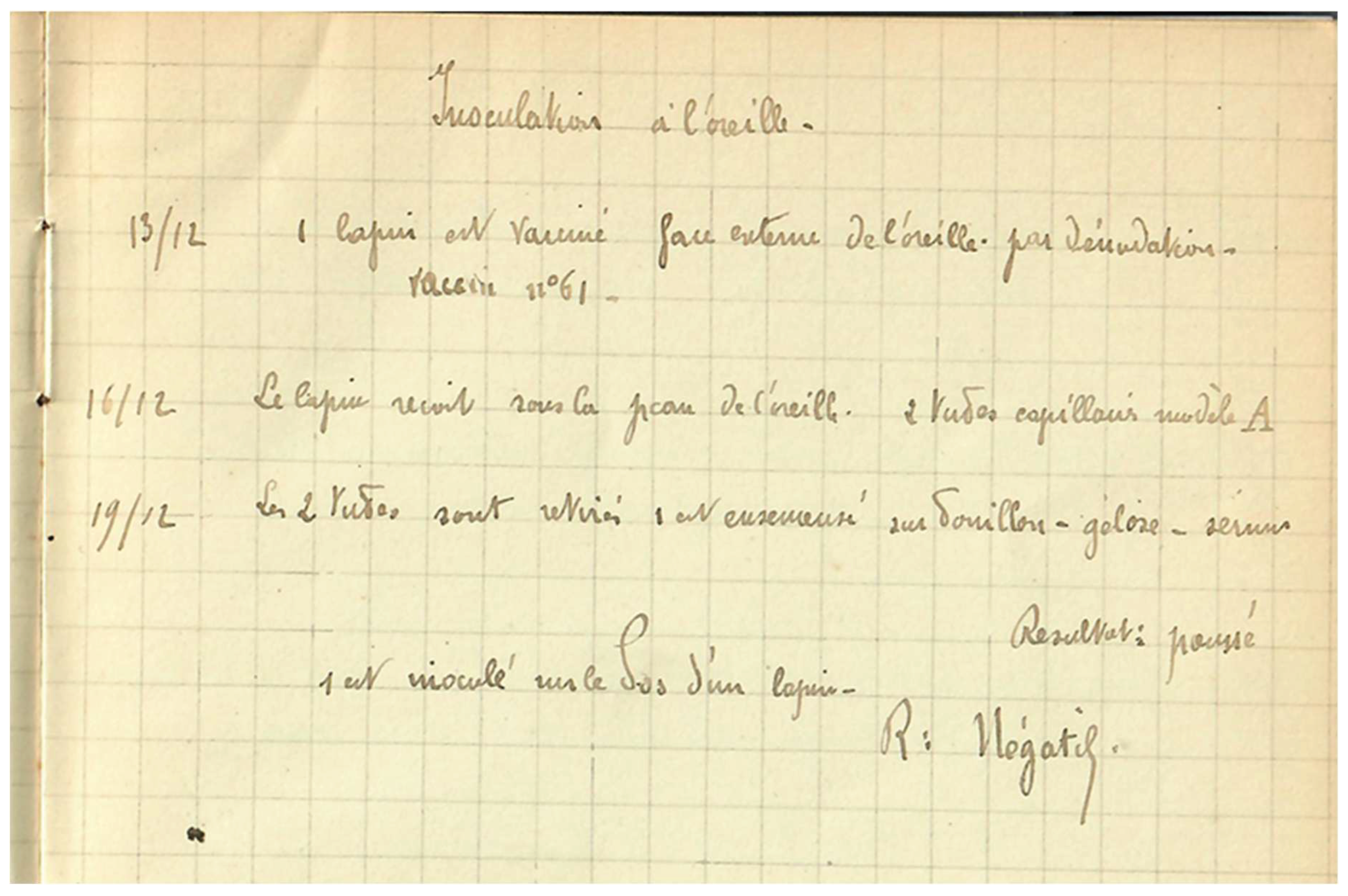
Figure 5.
Death of an experimental rabbit and autopsy findings from a guinea pig following infection with tubercule bacilli. It reads: Large cage 42. The 14/6 [June 14]. Gray rabbit receives oxy + [? had no] eye P = 1980. Dead (failed kidneys). -Glass jar of the room. The 18-6 [June 18] Guinea pig receives 5 c³ liquid F’ under its skin. The same day, injection of virulent tuberculosis in the peritoneum and under the skin. Died on 30-6 [June 30]. Autopsy: the local necrotic sore provoked by the injection of the culture is coming along nicely as a scar. The inguinal lymph nodes are hard, fibrous, without central pus. The opening of the abdominal cavity shows an adhesive peritonitis, excessively intense, the small intestine, the epiploices, the spleen are so closely fused with the parietal peritoneum that one cannot separate them. The liver is healthy. The spleen is normal. The lung is intact. To summarize, this guinea pig seems to have resorbed its bacilli.
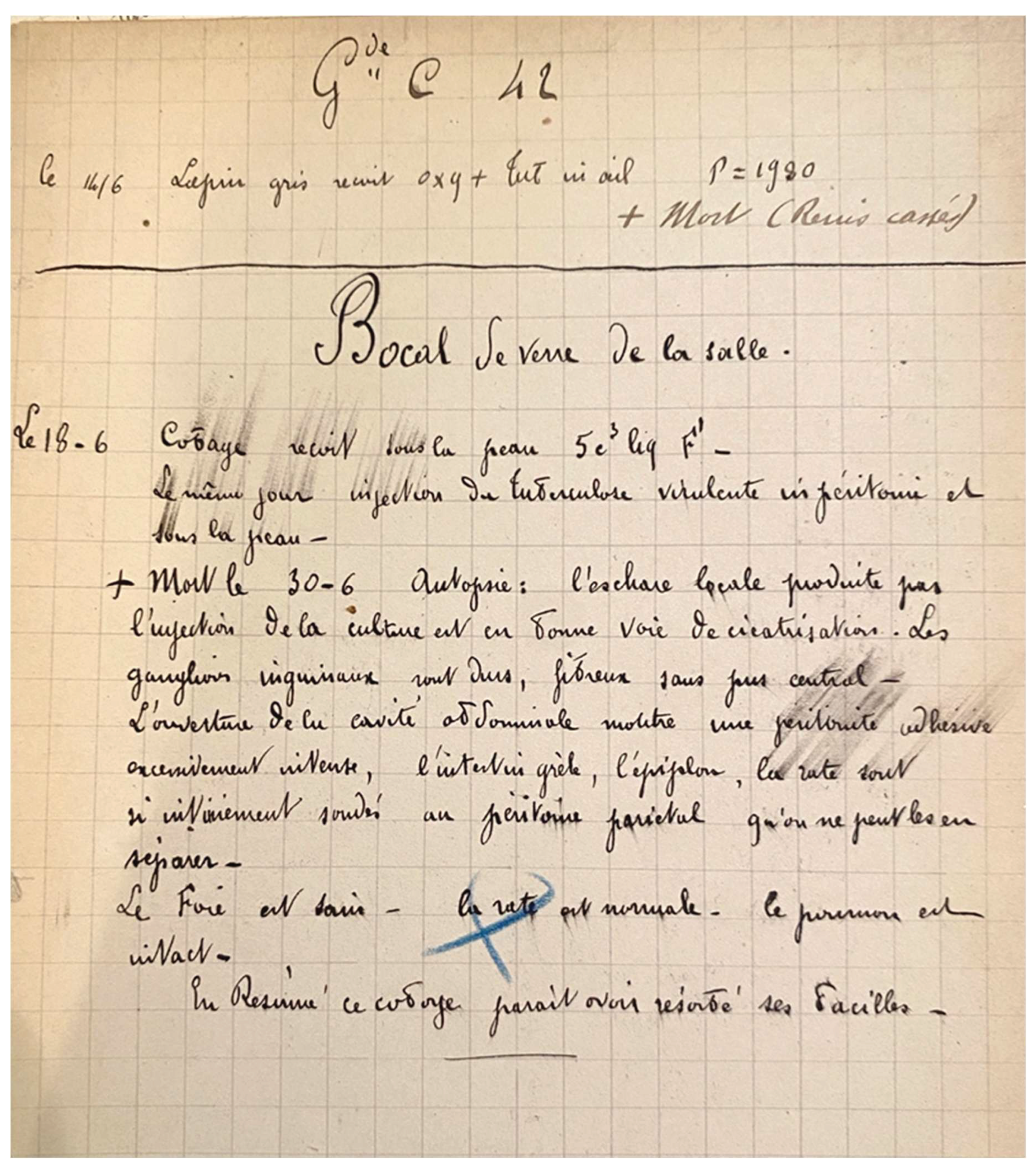
Figure 6.
Experiments in cage 60 including differing routes of inoculation of tubercle bacilli preparations in guinea pigs. It reads: Cage 60 The 24/5 [24 May] Guinea pig W = 400 gr Tricolored female receives 1c3 T.F. under the skin (bacilli from 05/05). Guinea pig W = 320 White Angora “ Guinea pig W = 280 Tricolored female with a white right ear and black left ear receives 1c3 T.F. under the skin in peritoneum (bacilli from 05/05). Guinea pig = 250 White female with a white right ear and a black and yellow left ear receives 1c3 T.F. under the skin in peritoneum (bacilli from 05/05). The 10/6 [10 June] Guinea pig n°183 Tricolored female W = 400gr Guinea pig n°182 White Angora W = 360 Guinea pig n°144 Tricolored female with a black left ear W = 280 Guinea pig n°128 White female with a black and yellow left ear W = 250 [Oblique underline writing on the right]: ‘seen do not treat’. The 17/6 [17 June] Guinea pig n°182 W = 370 receives 1c3 cult. T.F. under the skin. Guinea pig n 144 W = 290 ““ Guinea pig n 183 W = 390 Guinea pig n 128 W = 260 ““ The 20/6 [20 June] Guinea pig n 183 W = 390 Guinea pig n 182 W = 360 Guinea pig n 144 W = 250 Guinea pig n 128 W = 270 The 19/7 [19 July] Guinea pig n°182 W = 380 Guinea pig n 183 W = 390 Guinea pig n 128 W = 270 Guinea pig n 144 new number: 1045 W = 230.
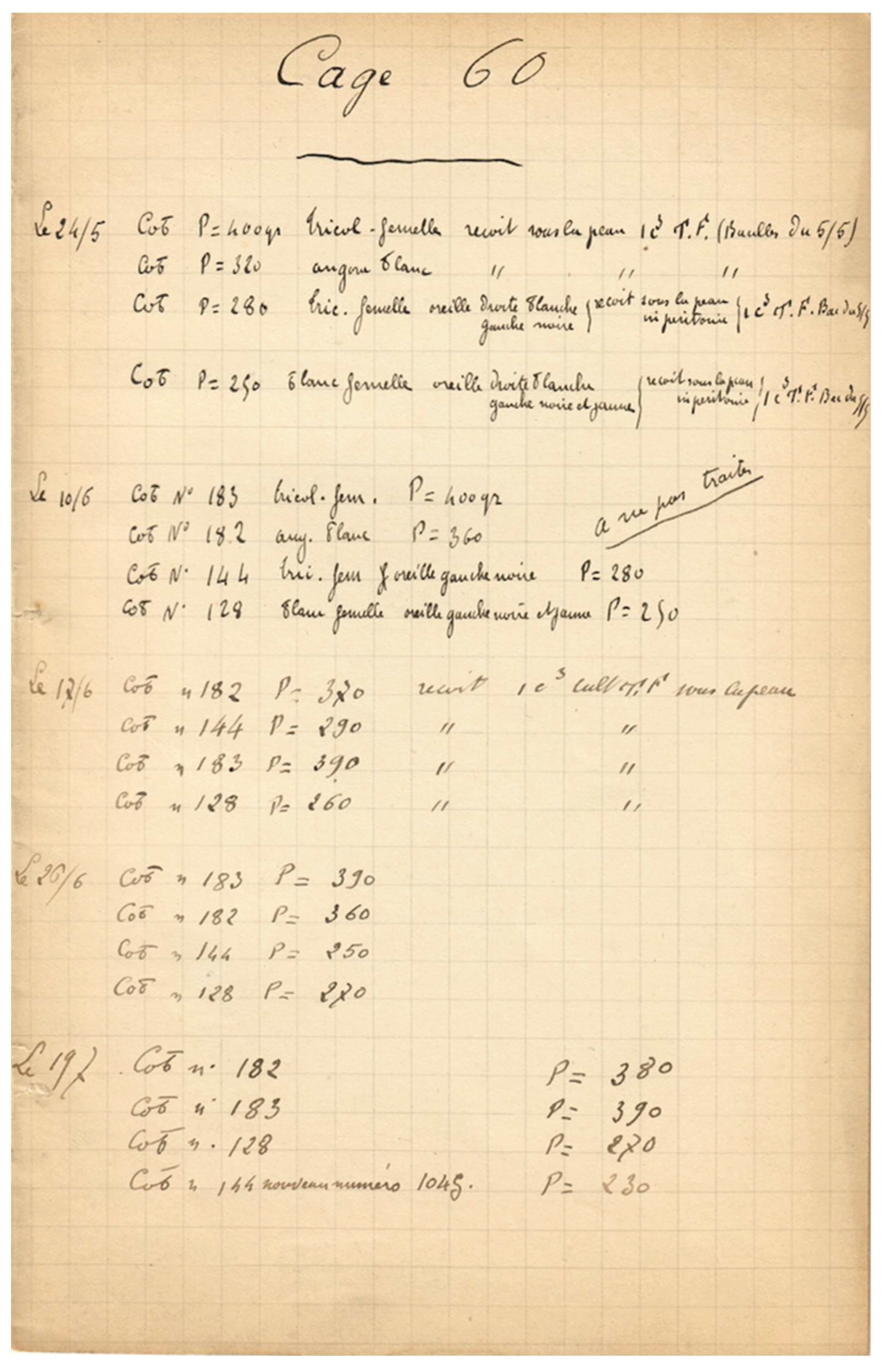
Figure 7.
Cage 61 with experimental inoculations of guinea pigs with virulent tuberculosis both subcutaneously and intraperitoneally, mentioning Dr. Robert Koch. It reads: Cage 61 The 05/11 [May 11] Injections to the guinea pigs of cage 61 with virulent tuberculosis. The 06/01 [June 1]] The guinea pigs of cage 61 that already received tuberculosis R. De Koch are treated with Tub. F., glycerinated set. Guinea pig n°938 W = 280 receives 1c3 under the skin in peritoneum. Guinea pig black and white Angora with brown spots W = 360 receives 1c3 under the skin in peritoneum. Guinea pig n°955 W = 420 receives “ Guinea pig n 973 W = 230 receives “ Guinea pig n 155 W = 300 receives “ All the guinea pigs receive 2c3 of liquid F. centrifuged under the skin. The 06/10 [June 10] All the guinea pigs receive 1c3 of liquid F. in peritoneum. Guinea pig n°155 W = 220 Guinea pig n°938 W = 280 Guinea pig n°955 W = 410 Guinea pig n°973 W = 220 Guinea pig black Angora with reddish brown spots W = 330 06/14 [June 14] All the guinea pigs receive 2c3 of liquid F. in peritoneum. Guinea pig n°155 W = 250 Guinea pig n°973 W = 210 Guinea pig n°938 W = 250 Guinea pig n°955 W = 380 Guinea pig black Angora with reddish brown spots W = 315 + Dead on 06/17, generalized tuberculosis. 06/16 [June 16] All the guinea pigs receive 1c3 of liquid F. in peritoneum.
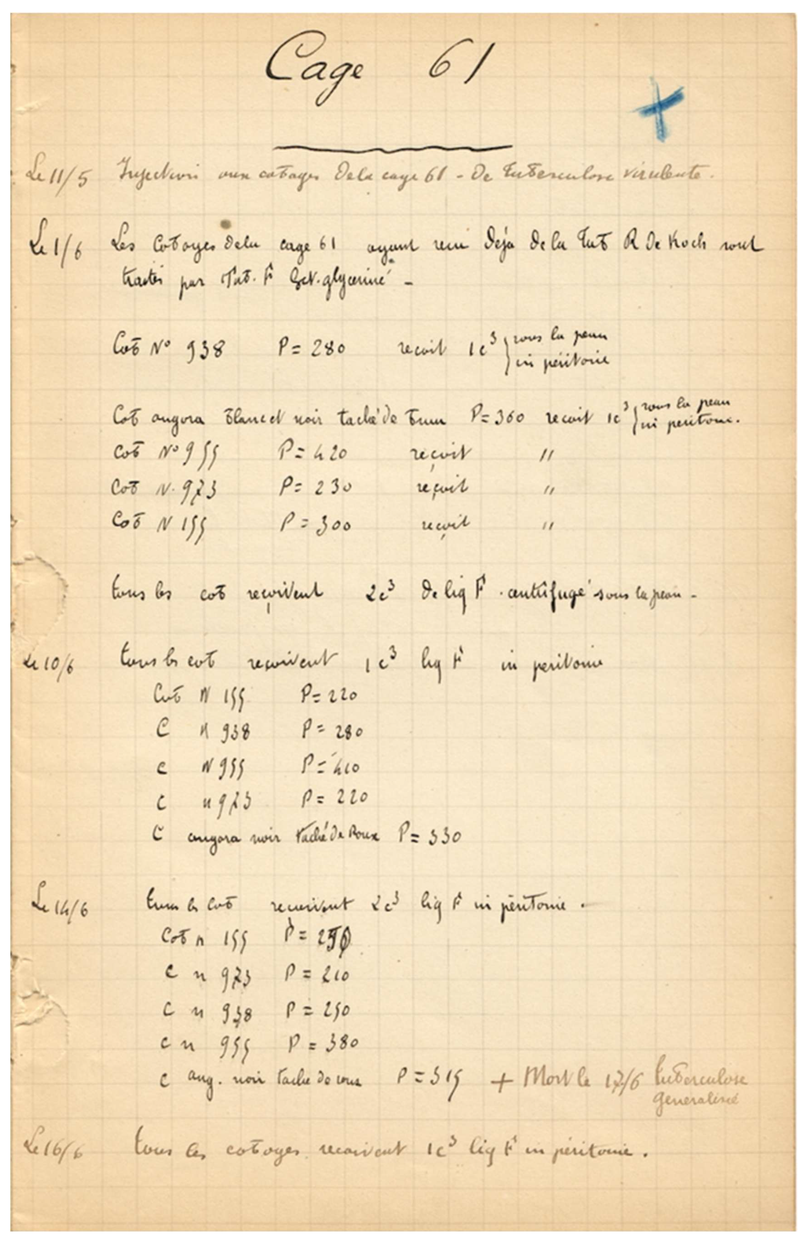
Figure 8.
Experiments in large cage 2. It reads: Large cage 2 The 13/5 [May 13] Guinea pig n 139 W = 490 receives under the skin 1c3 of a seven-day old culture T.F. Filtrated with paper. Guinea pig n 147 W = 560 “ 2c3 “ Guinea pig n 121 W = 460 “ 3c3 “ Guinea pig n 120 W = 430 “ 4c3 “ Guinea pig n 124 W = 330 “ 5c3 “ Guinea pig n 856 W = 280 Control: does not receive anything. The 30/5 [May 30] Guinea pig n°147 W = 540 receives 1c3 [Ext] glycerinated F. under the skin. Guinea pig n 121 W = 510 “ Guinea pig n 139 W = 500 “ Guinea pig n 124 W = 380 “ Guinea pig n 120 W = 220 “ Guinea pig n 856 W = 220 “ [Oblique underline writing on the right:] ‘all these cases are cured on 16/6’. The 16/6 [June 6] Guinea pig n 148 tricolored female with 2 black ears and a white nose W = 440 Preventively treated. 1c3 of F. liquid under the skin. Guinea pig n 390 male, black head W = 440 “ “ Guinea pig n 192 black male W = 390 “ “ Guinea pig n 142 white and yellow female W = 410 “ “ Guinea pig n 166 pale yellow tricolored male with a white left cheek and a black right cheek W = 390 1c3 of F. liquid under the skin. “ “ Guinea pig n 144 tricolored male with a white posterior, 2 black cheeks W = 400 1c3 of F. liquid under the skin. “ “ Rabbit n 1880 receives 5c3 of liquid F. under the skin. The 17/6 [June 17] The 6 guinea pigs receive 1c3 of liquid F. under the skin. [Deleted sentence]: ‘the rabbit receives 2c3 of liquid F. under the skin. The 19/6 [June 19] The 6 guinea pigs receive 1c3 of liquid F. under the skin. The rabbit receives 5c3 of liquid F. under the skin.
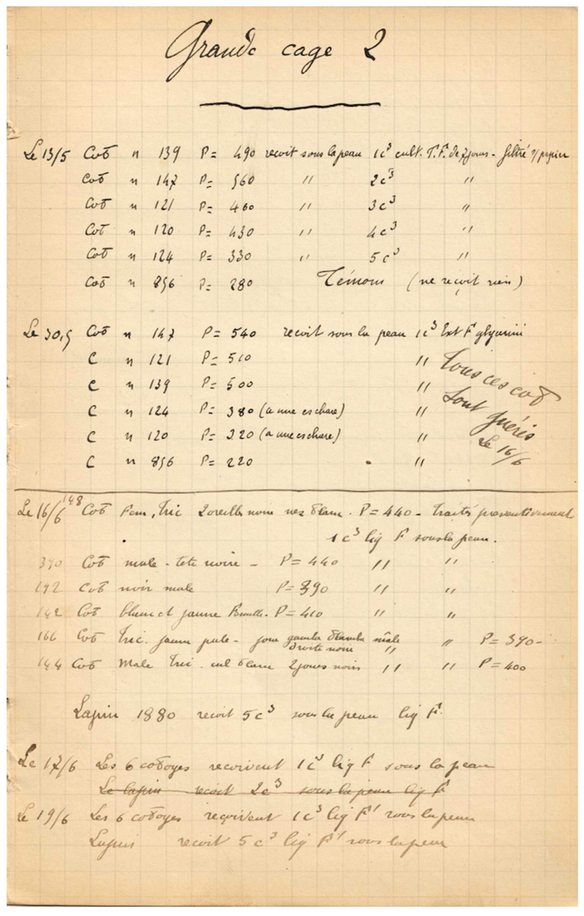
Figure 9.
a. Capillary tubes are used to inoculate rabbits and for inoculation of culture medium, with a drawing of a capillary tube. 7/12 [December 7] 2 rabbits are vaccinated on the back with n° 61 (Serre). 9/12 [December 9] The rabbits receive a few capillary tubes (Model A) under the skin. 12/12 [December 12] The rabbits are sacrificed, the tubes are removed and used to sow 2 tubes bouillon [broth] 2 tubes exudate rabbit 12/12 [December 12] 1 rabbit is vaccinated on the furunculous back with the contents of 3 tubes. Result: the cultures did not grow. Result: negative. [Drawing of Model A capillary tube along right margin, see Figure 9b).
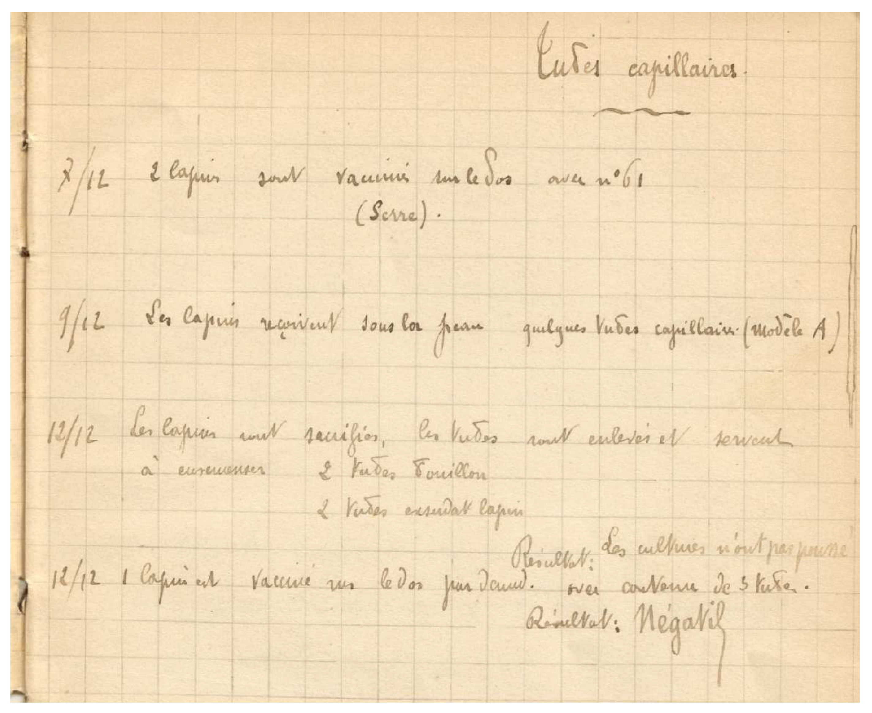
Figure 9.
b. Drawing of Model A capillary tube (magnified).

8. Discussion
The BCG vaccine is administered to between 100 and 200 million children every year, making it still one of the most widely used vaccines in the world [15]. In China alone over 300 million children have been vaccinated using the BCG vaccine since 2002 the BCG vaccine [16]. The current vaccine is still derived from a live attenuated strain of Mycobacterium bovis, as developed by Calmette and Guérin, and remains the only vaccine available for prevention of tuberculosis. Calmette died following a brief illness on October 29, 1933, and unfortunately did not live to see the extent of global acceptance of his vaccine [10].
This laboratory notebook contains the details of experiments that were performed during the development of the BCG vaccine at the Pasteur Institute by Drs. Calmette and Guérin. These include numerous experimental inoculations of laboratory animals – rabbits and guinea pigs – noting observations of the pathology including development of skin lesions, inflammatory reactions (pus formation and eschars), and visceral organ pathology. The experiments describe varying inoculative dosages of the bacteria, as well as using different routes of administration including intraperitoneal and subcutaneous injections and administration of bacilli in the ear. In those cases where the animal had died following inoculation of tubercle bacilli, necropsy was performed and the organs examined and the pathology findings described. To the authors knowledge, this notebook is the sole remaining original record of the laboratory experiments by Drs. Calmette and Guérin of their development of the BCG vaccine.
The handwriting in this notebook matches published examples by Dr. Calmette’s writing. Although it is difficult to identify the author of the second hand used throughout this notebook, it is most probably that of Dr. Guérin. It is beyond the scope of this communication to describe the methodology and results of the numerous experiments contained in these 60-plus manuscript leaves. However, this notebook is clearly a highly organized and detailed record of investigations using tuberculosis in animal experiments and microbiological culture to produce a safe and effective vaccine, which was initially used in humans in 1921.
Understanding and transcribing the contents of antiquated laboratory notebooks is difficult - they were handwritten documents designed for internal use, in which experiments, data and results incorporate numerous abbreviations and terms that were for use of the scientific staff but which might be uninterpretable by those outside of the lab. In addition, the methodological and technical difficulties inherent in understanding obsolete experimental procedures is challenging. Handwriting and variation in script can be daunting to interpret, and differentiating minims and similar strokes can make word transcription a formidable task [17,18]. In the case of this manuscript there are abbreviations and text whose meaning remains unknown. While the authors have attempted to translate the manuscript into English without significantly altering the original meaning, it is often said within the field of translation that there are as many translations as translators. This translation was also challenging for two additional reasons: it was both interlingual and interdisciplinary. From French to English and from medicine to the humanities, the stakes were high. We have attempted to remain as close as possible to the original version so that as little as possible was lost in translation. Thus, although some terms may seem outdated, that it is part of the charm of an early 20th century collaborative laboratory notebook, a tremendous discovery for medical history.
Acknowledgments
The authors wish to express their gratitude to Vincent Bruyère, Winship Distinguished Research Professor and Chairman of French Studies at Emory University, Atlanta, USA who bridges humanities and medicine and whose mentoring is invaluable.
Author Contributions
The authors contributed equally to all aspects of the manuscript.
Funding
This research received no external funding.
Institutional Review Board Statement
Not applicable.
Informed Consent Statement
Not applicable.
Conflicts of Interest
The authors declare no conflict of interest.
References
- Johns Hopkins Bloomberg School of Public Health. Vaccines 101: The basics of vaccines and vaccination. 19 February 2025. Available online: https://publichealth.jhu.edu/2025/the-biology-of-vaccines (accessed on 8 February 2026).
- World Health Organization. Global immunization efforts have saved at least 154 million lives over the past 50 years. 24 April 2024. Available online: https://www.who.int/news/item/24-04-2024-global-immunization-efforts-have-saved-at-least-154-million-lives-over-the-past-50-years (accessed on 7 February 2026).
- Buchholz, K. The most common immunizations around the world. Statista. 24 April 2024. Available online: https://www.statista.com/chart/21644/most-widespread-vaccines-globally/?srsltid=AfmBOoptlo5p9ehjGSuaAoeksyWjFTnEZ1ovwJv342-SrdT7IFTTOCMU (accessed on 8 February 2026).
- Luca, S.; Mihaescu, T. History of BCG vaccine. Maedica (Bucur) 2013, 8, 53–58. [Google Scholar] [PubMed]
- Kim, P.S; Swaminathan, S. Ending TB: the world's oldest pandemic. J Int AIDS Soc. 2021, 24, e25698. [Google Scholar] [CrossRef] [PubMed]
- Barberis, I.; Bragazzi, N.L.; Galluzzo, L.; Martini, M. The history of tuberculosis: from the first historical records to the isolation of Koch's bacillus. J Prev Med Hyg. 2017, 58, E9–E12. [Google Scholar] [PubMed]
- Lobo, N.; Brooks, N. A.; Zlotta, A. R.; Cirillo, J. D.; Boorjian, S.; Black, P. C.; Meeks, J. J.; Bivalacqua, T. J.; Gontero, P.; Steinberg, G. D.; et al. 100 years of Bacillus Calmette-Guérin immunotherapy: from cattle to COVID-19. Nat Rev Urol. 2021, 18, 611–622. [Google Scholar] [CrossRef] [PubMed]
- Lange, C.; Aaby, P.; Behr, M. A.; Donald, P. R.; Kaufmann, S. H. E.; Netea, M. G.; Mandalakas, A. M. 100 years of Mycobacterium bovis bacille Calmette-Guérin. Lancet Infect Dis. 2022, 22, e2–e12. [Google Scholar] [CrossRef] [PubMed]
- La vaccination préventive contre la tuberculose par le "BCG”; Calmette A. Paris, Masson et Cie, 1927.
- Tan, S.Y.; Kwok, E. Albert Calmette (1863-1933): originator of the BCG vaccine. Singapore Med J. 2012, 53, 433–434. [Google Scholar] [PubMed]
- Hawgood, B.J. Albert Calmette (1863-1933) and Camille Guérin (1872-1961): the C and G of BCG vaccine. J Med Biogr. 2007, 15, 139–146. [Google Scholar] [CrossRef] [PubMed]
- Towey, F. Léon Charles Albert Calmette and Jean-Marie Camille Guérin. Lancet Respir Med. 2015, 3, 186–187. [Google Scholar] [CrossRef] [PubMed]
- Bourel, L. French scientists Albert Calmette and Camille Guérin, co-inventors of the Bacillus Calmette-Guérin (BCG) vaccine to protect against tuberculosis at the Pasteur Institute of Lille: A short historical overview. J Clin Pract Res. 2024, 46, 513–518. [Google Scholar] [CrossRef] [PubMed]
- Institut Pasteur. BCG vaccine: the first tuberculosis vaccination took place a century ago. 15 July 2021. Available online: https://www.pasteur.fr/en/home/research-journal/news/bcg-vaccine-first-tuberculosis-vaccination-took-place-century-ago (accessed on 8 February 2026).
- Kaye, A. D.; Giles, T. P.; O'Brien, E.; Zajac, J.; Upshaw, W. C.; Jenks, K.; Arya, P.; Kaye, A. M.; Ahmadzadeh, S.; Chandler, D.; et al. Bacillus Calmette-Guérin (BCG) vaccine in America and overseas: A narrative review. Cureus 2024, 16, e73602. [Google Scholar] [CrossRef] [PubMed]
- Lu, J.; Zhang, X.; Xu, H.; Li, Z. First vaccination after birth: serious adverse events of Bacillus Calmette-Guérin (BCG) in real-world. Hum Vaccin Immunother. 2022, 18, 2080443. [Google Scholar] [CrossRef] [PubMed]
- Lucas, L. Transcribing manuscripts. Minnesota Historical Society. 2004. Available online: https://www2.mnhs.org/library/findaids/CMToolkit/BestPractices/transcribing_manuscripts.pdf (accessed on 9 February 2026).
- Transcription Guide. Bishopsteighton Heritage. Available online: https://www.bishopsteigntonheritage.co.uk/hub-handbook/transcription-guide/ (accessed on 9 February 2026).
Figure 1.
Title page of the notebook. It reads: Cahier L’experiences sur le tubercuose. Cahier No. 1.
Figure 1.
Title page of the notebook. It reads: Cahier L’experiences sur le tubercuose. Cahier No. 1.
Disclaimer/Publisher’s Note: The statements, opinions and data contained in all publications are solely those of the individual author(s) and contributor(s) and not of MDPI and/or the editor(s). MDPI and/or the editor(s) disclaim responsibility for any injury to people or property resulting from any ideas, methods, instructions or products referred to in the content. |
© 2026 by the authors. Licensee MDPI, Basel, Switzerland. This article is an open access article distributed under the terms and conditions of the Creative Commons Attribution (CC BY) license (http://creativecommons.org/licenses/by/4.0/).
Copyright: This open access article is published under a Creative Commons CC BY 4.0 license, which permit the free download, distribution, and reuse, provided that the author and preprint are cited in any reuse.


