Submitted:
30 January 2026
Posted:
02 February 2026
You are already at the latest version
Abstract
Background: Uncontrolled hemorrhage remains a leading cause of preventable trauma deaths. Fish gelatin-chitosan composites offer promising hemostatic alternatives to mammalian-derived materials, yet optimal formulations require systematic characterization. This study investigated fish gelatin-chitosan sponges to determine the most effective compositional ratio for hemostatic applications. Material and Methods: Four formulations were prepared via freeze-drying: SGiK (100% gelatin), SGiK1.1 (50% gelatin, 50% chitosan), SGiK1.3 (25% gelatin, 75% chitosan), and SGiK3.1 (75% gelatin, 25% chitosan). Characterization included FTIR spectroscopy for functional groups, SEM for microstructure analysis, degradation studies in PBS over 4 weeks, and swelling capacity measurements at multiple time points (1, 5, 10, 30 minutes). Mechanical properties were evaluated using tensile testing. Results: Pure gelatin (SGiK) exhibited rapid degradation within 3 days. SGiK3.1 demonstrated optimal characteristics with micro-macroporous architecture, highest swelling capacity (1115.22% at 5 minutes), and balanced degradation rate (18% weight loss over 14 days). SEM revealed interconnected porous structures ideal for blood infiltration. SGiK1.3 showed slowest degradation but lower swelling dynamics compared to SGiK3.1. Conclusion: SGiK3.1 (75% gelatin, 25% chitosan) represents the optimal formulation, combining superior swelling capacity, appropriate degradation kinetics, and ideal micro-macroporous architecture for hemostatic applications. This fish gelatin-based composite offers clinical potential as a biocompatible, effective alternative to mammalian-derived hemostatic materials.
Keywords:
chitosan composite
; fish gelatin
; freeze-drying
; hemostatic sponge
; swelling capacity
1. Introduction
Uncontrolled hemorrhage remains one of the leading causes of potentially preventable death following traumatic injury, accounting for approximately 30-40% of trauma-related mortality in both military and civilian settings.[1,2,3,4] The critical window for hemostatic intervention is remarkably narrow, with most bleeding-related deaths occurring within the first three hours post-injury.[5,6,7] Current hemostatic materials face persistent challenges including limited absorption capacity, inadequate mechanical strength, potential thermal injury from exothermic reactions, and insufficient antibacterial properties.[4,8,9,10] These limitations drive ongoing research toward developing effective, biocompatible hemostatic agents that can rapidly control bleeding while promoting tissue regeneration.
Natural biopolymers have emerged as promising alternatives to synthetic hemostatic materials due to their inherent biocompatibility, biodegradability, and low immunogenicity.[11,12,13,14]. Fish-derived gelatin demonstrates remarkable hemostatic properties through multiple mechanisms including blood absorption, platelet activation, and provision of a scaffold for clot formation.[15,16,17,18] Chitosan, a deacetylated derivative of chitin, promotes hemostasis via electrostatic interactions with negatively charged blood components, enhancing platelet adhesion and aggregation.[19,20,21,22]. Both materials offer excellent fluid absorption capacity and can be fabricated into three-dimensional porous structures suitable for deep wound applications.
Combining fish gelatin with chitosan creates composite materials that exploit synergistic hemostatic mechanisms while addressing individual material limitations.[23,24,25,26]. These composites demonstrate enhanced mechanical properties, improved blood absorption rates, and superior platelet adhesion compared to single-component materials.[9,27,28,29] The porous sponge architecture facilitates rapid blood infiltration and concentration of coagulation factors at wound sites.[23,30,31,32]. Furthermore, the combination offers adjustable degradation rates and potential for incorporating additional bioactive agents to enhance wound healing outcomes.[33,34,35,36]
Despite the promising characteristics of gelatin-chitosan composites, comprehensive evaluation of fish gelatin-based formulations specifically remains limited, particularly regarding their comparative hemostatic efficacy and biocompatibility profiles. This study aims to investigate the potency of fish gelatin and chitosan-based sponges as hemostatic materials by evaluating their physical properties, blood absorption capacity, hemostatic performance in experimental models, and biocompatibility characteristics to establish their potential for clinical translation.
2. Results and Discussion
2.1. Physicochemical Characterization
2.1.1. Functional Group Analysis
FTIR spectroscopy was performed to identify the functional groups and confirm the chemical interactions between gelatin and chitosan in the composite sponges. The FTIR spectra of all four formulations (SGiK, SGiK1.1, SGiK1.3, and SGiK3.1) were recorded in the wavelength range of 600-4000 cm−1.
Figure 1.
FTIR spectra of gelatin-chitosan composite sponges. Black line: SGiK (100% gelatin); Red line: SGiK1.1 (50% gelatin + 50% chitosan); Blue line: SGiK1.3 (25% gelatin + 75% chitosan); Green line: SGiK3.1 (75% gelatin + 25% chitosan).
Figure 1.
FTIR spectra of gelatin-chitosan composite sponges. Black line: SGiK (100% gelatin); Red line: SGiK1.1 (50% gelatin + 50% chitosan); Blue line: SGiK1.3 (25% gelatin + 75% chitosan); Green line: SGiK3.1 (75% gelatin + 25% chitosan).
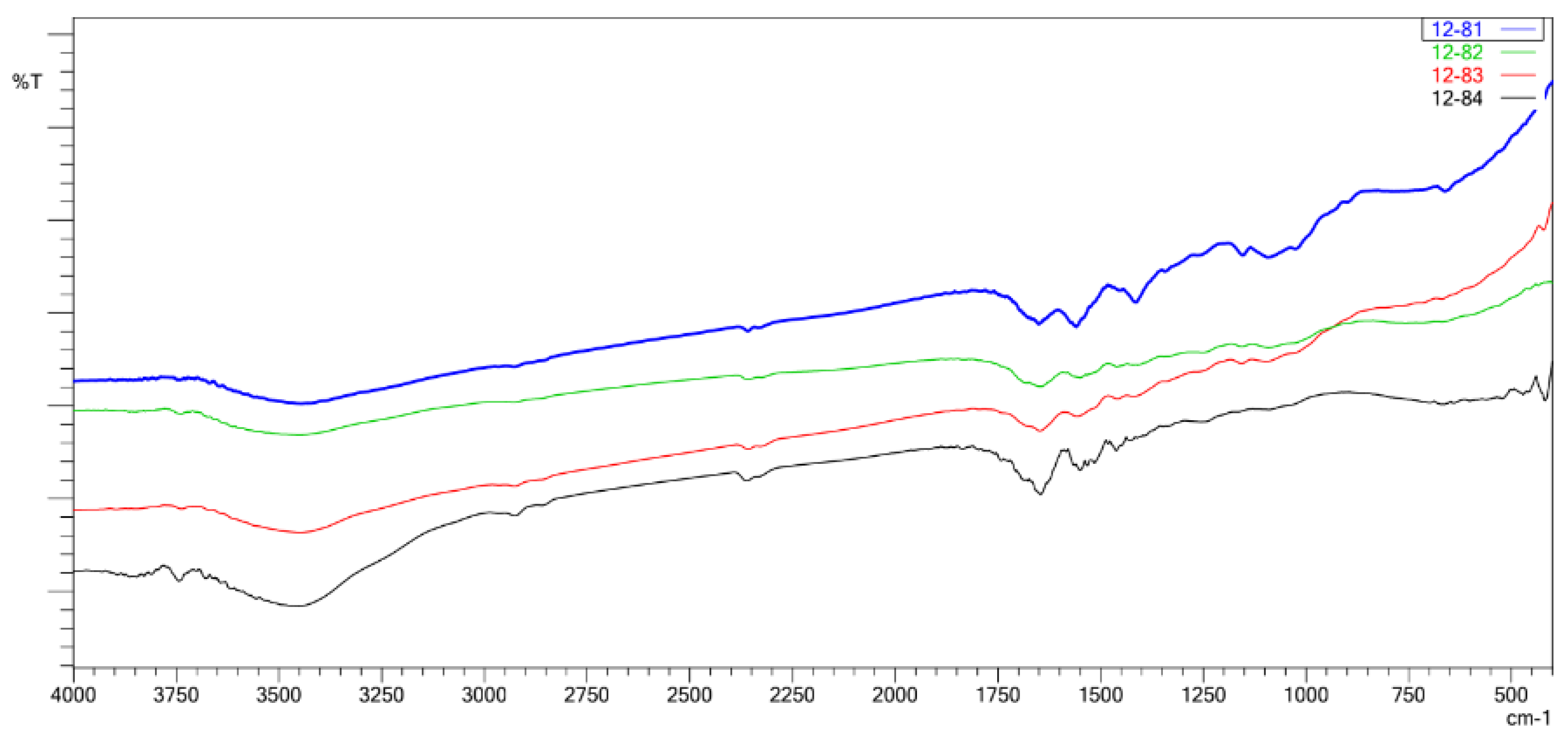
The FTIR analysis revealed characteristic absorption bands corresponding to the functional groups present in gelatin and chitosan. The spectra showed typical amide bands indicating the presence of protein structure from gelatin, while the incorporation of chitosan was evidenced by modifications in the peak positions and intensities, particularly in the regions corresponding to amino and hydroxyl groups. The composite sponges exhibited spectral features that indicated successful blending and potential interactions between the two biopolymers.
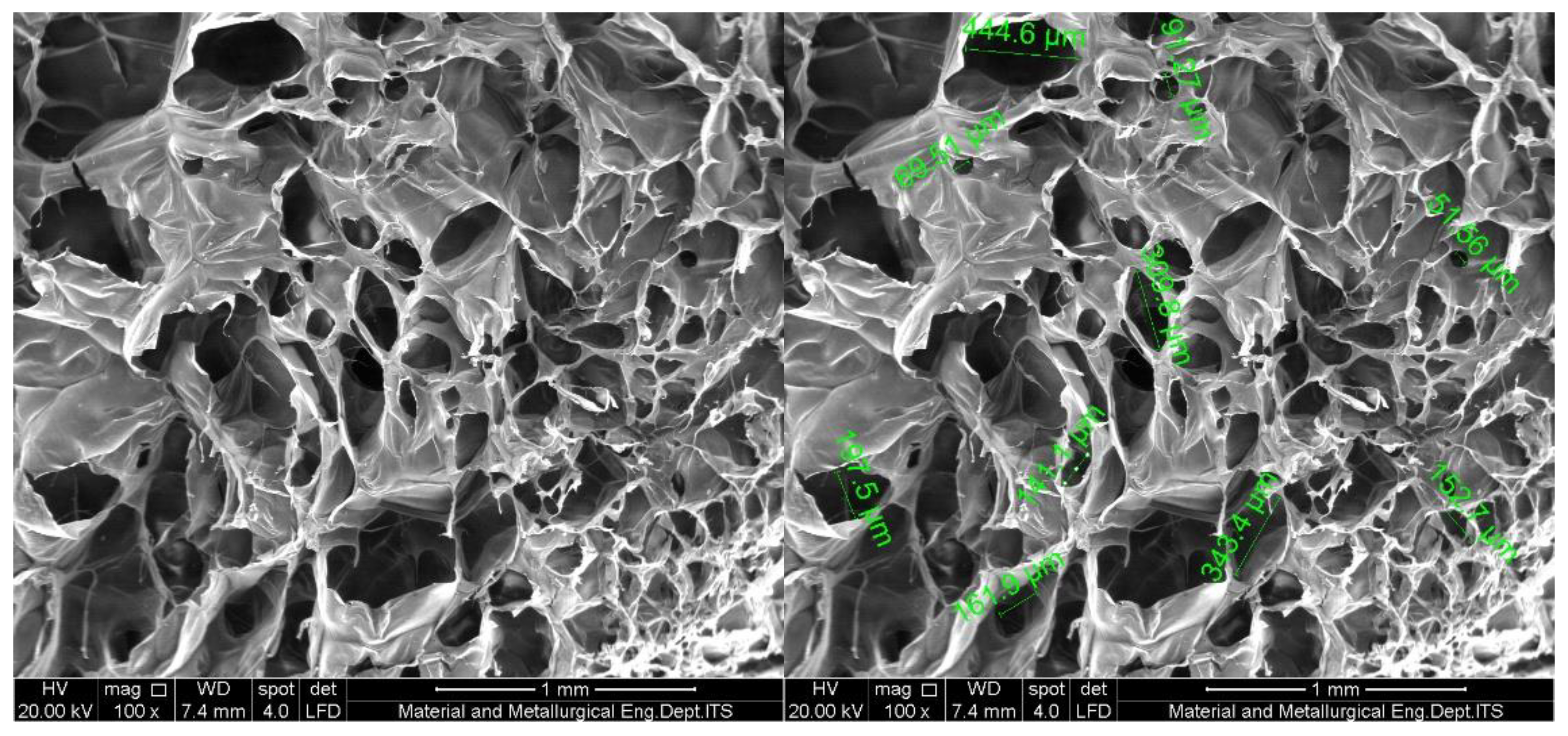
2.1.2. Microstructure Characterization
Scanning electron microscopy (SEM) was employed to examine the morphological characteristics and porous structure of the freeze-dried sponges. The microstructure analysis was performed on all four formulations to evaluate the effect of gelatin-chitosan ratio on the pore architecture, pore size distribution, and overall structural integrity of the sponges.
Figure 2.
SEM micrograph of SGiK sponge (100% gelatin) showing the porous microstructure at 500× magnification.
Figure 2.
SEM micrograph of SGiK sponge (100% gelatin) showing the porous microstructure at 500× magnification.
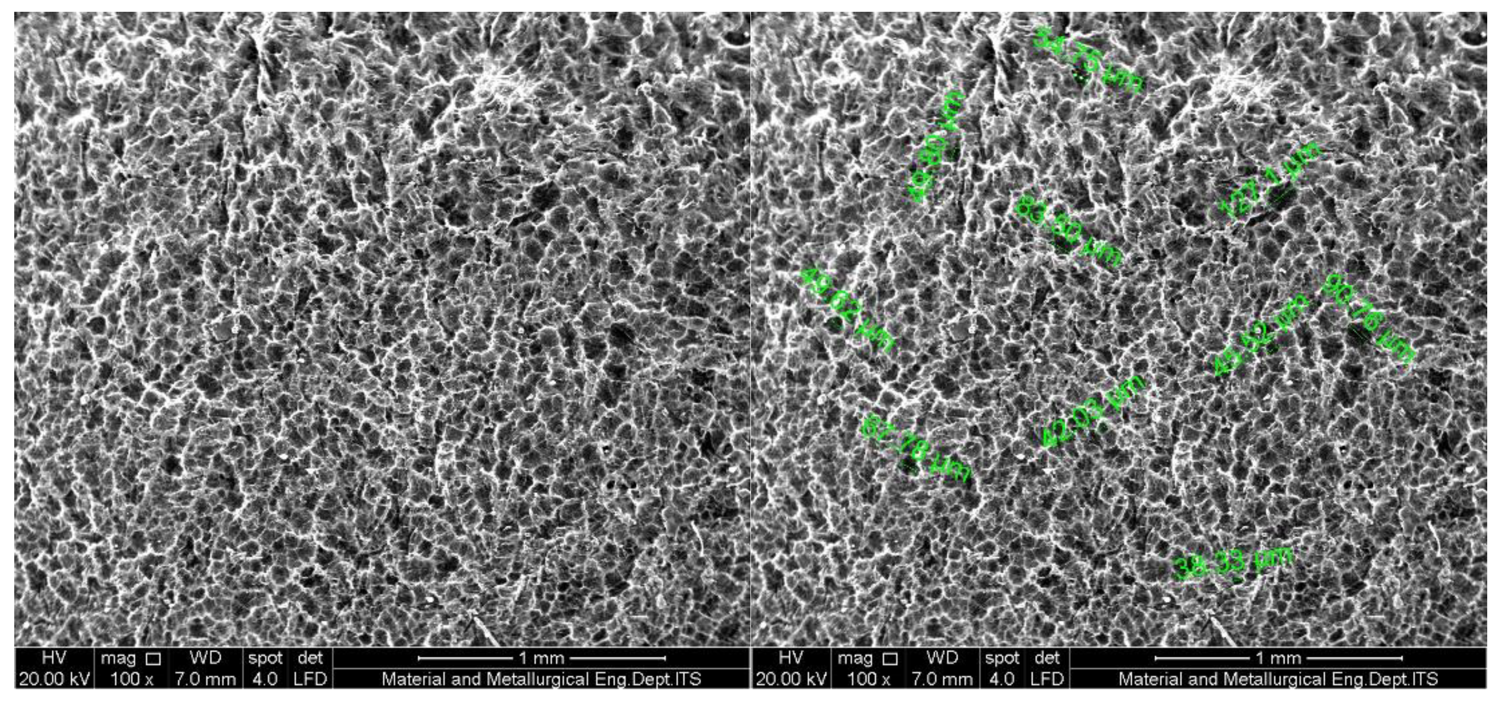
The pure gelatin sponge (SgiK) exhibited a highly porous three-dimensional network structure with interconnected pores. The pore walls appeared relatively thin and delicate, which may explain the rapid degradation behavior observed in subsequent tests.
Figure 3.
SEM micrograph of SGiK1.1 sponge (50% gelatin + 50% chitosan) showing the porous microstructure at 500× magnification.
Figure 3.
SEM micrograph of SGiK1.1 sponge (50% gelatin + 50% chitosan) showing the porous microstructure at 500× magnification.
The 50:50 gelatin-chitosan composite (SgiK1.1) demonstrated a well-defined porous architecture with more robust pore walls compared to pure gelatin. The pores were uniformly distributed throughout the matrix, suggesting good compatibility between the two polymers at this ratio.
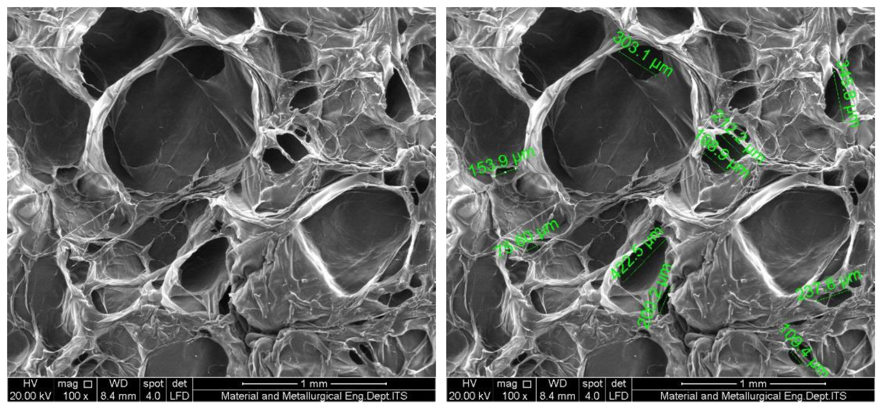
Figure 4.
SEM micrograph of SGiK3.1 sponge (75% gelatin + 25% chitosan) showing the porous microstructure at 500× magnification.
Figure 4.
SEM micrograph of SGiK3.1 sponge (75% gelatin + 25% chitosan) showing the porous microstructure at 500× magnification.
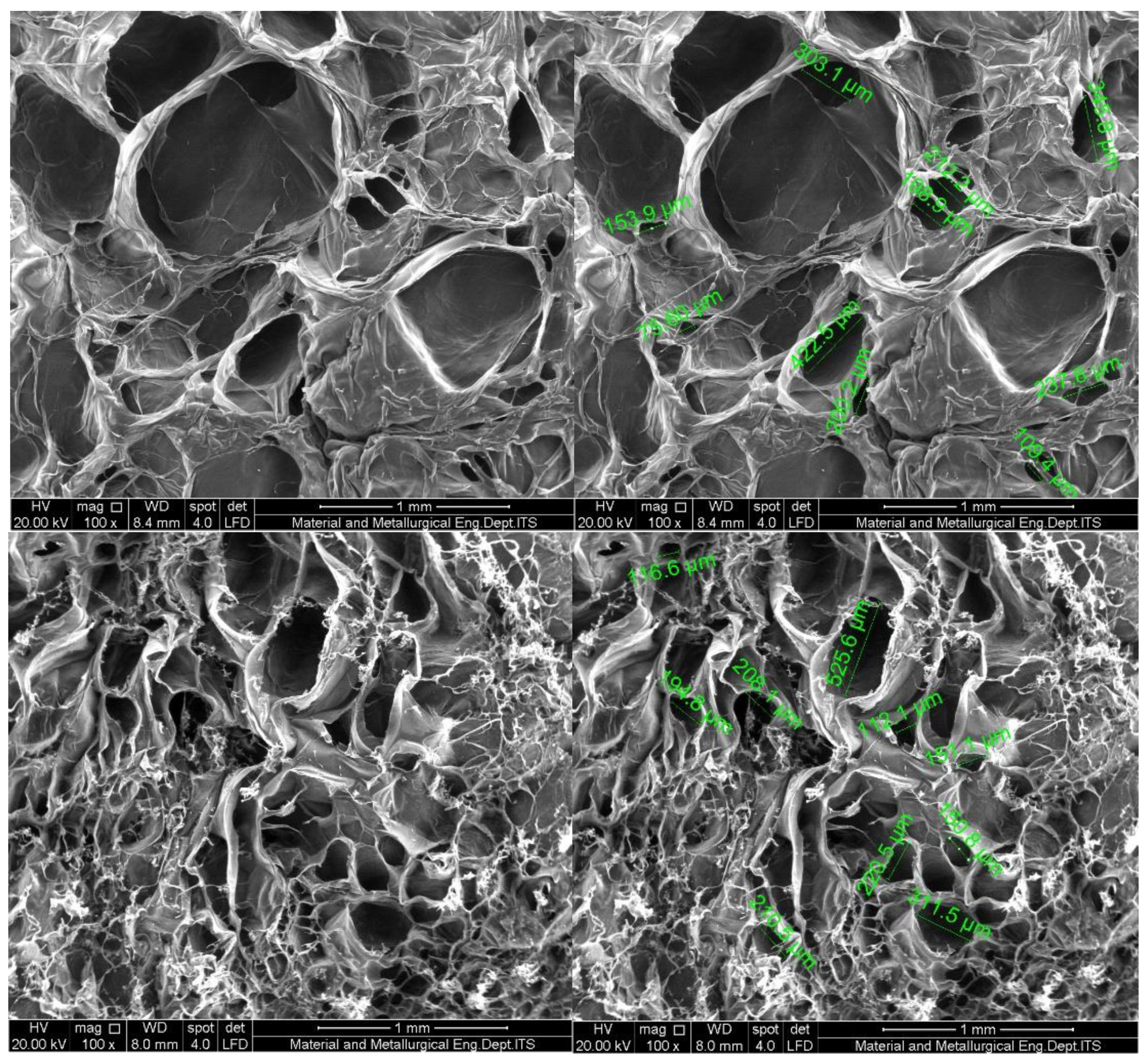
The SgiK3.1 formulation (75% gelatin + 25% chitosan) revealed a porous structure with characteristics intermediate between pure gelatin and the 50:50 composite. The microstructure maintained good interconnectivity while showing slightly thicker pore walls compared to pure gelatin.
Figure 5.
SEM micrograph of SGiK1.3 sponge (25% gelatin + 75% chitosan) showing the porous microstructure at 500× magnification.
Figure 5.
SEM micrograph of SGiK1.3 sponge (25% gelatin + 75% chitosan) showing the porous microstructure at 500× magnification.
The chitosan-rich formulation (SGiK1.3) exhibited a distinct microstructure with more compact and dense pore walls. The increased chitosan content appeared to reinforce the structural framework, potentially contributing to enhanced mechanical stability and slower degradation rates.
2.1.3. Degradation Behavior
The in vitro degradation study was conducted over a period of 4 weeks at 37 °C in PBS solution. The weight of the sponge samples was monitored weekly to assess their biodegradation profile. Table 1 presents the average remaining weight of each formulation throughout the study period.
The pure gelatin sponge (SGiK) showed the most rapid degradation, completely dissolving within 3 days of incubation in PBS. This rapid degradation is attributed to the lack of structural reinforcement from chitosan. In contrast, the chitosan-enriched formulation (SGiK1.3) demonstrated the slowest degradation rate, retaining approximately 82% of its original weight after 14 days. The 50:50 composite (SGiK1.1) exhibited an intermediate degradation profile, showing gradual weight loss throughout the study period. Interestingly, the SGiK3.1 formulation showed faster degradation than SGiK1.1, which may be related to its initial lower weight and potentially different structural characteristics.
2.1.4. Swelling Capacity
The water absorption capacity of the sponges was evaluated by measuring the swelling ratio at different time intervals (1, 5, 10, and 30 minutes) in PBS at 37 °C. Table 2 presents the swelling capacity of all formulations.
The swelling capacity is a critical parameter for hemostatic materials as it reflects the ability to absorb blood and wound exudates. The pure gelatin sponge (SGiK) demonstrated limited swelling capacity and rapidly dissolved in PBS after 1 minute, confirming its poor structural stability in aqueous environments. All composite formulations exhibited significantly higher swelling ratios ranging from approximately 500% to 750% at the 1-minute time point.
The SGiK1.3 formulation (25% gelatin + 75% chitosan) showed the highest initial swelling capacity (740.92 ± 30.87%) at 1 minute, followed by both SGiK1.1 and SGiK3.1 with comparable values around 586%. Interestingly, the SGiK3.1 formulation exhibited a remarkable increase in swelling at 5 minutes (1115.22 ± 136.73%), which may be attributed to the progressive hydration of the gelatin-rich matrix. After 10 minutes, all composite formulations reached equilibrium swelling, with values stabilizing between 570-620%, maintaining these levels through the 30-minute time point.
The incorporation of chitosan clearly enhanced the structural stability and water absorption capacity of the sponges. The high swelling capacity of the composite sponges, particularly SGiK1.3 and SGiK3.1, suggests their potential effectiveness as hemostatic materials, as they can rapidly absorb large volumes of blood while maintaining structural integrity.
Based on the comprehensive physicochemical evaluation, the gelatin-chitosan composite sponges demonstrated distinct characteristics depending on their compositional ratios. The pure gelatin sponge (SGiK) exhibited poor stability with rapid degradation and dissolution, making it unsuitable for hemostatic applications. Among the composite formulations, SGiK1.3 (25% gelatin + 75% chitosan) showed the most favorable properties, including excellent swelling capacity (740.92% at 1 minute), slow degradation rate (18% weight loss over 14 days), and robust microstructure with reinforced pore walls. The SGiK1.1 (50:50) formulation demonstrated balanced properties with good swelling capacity and moderate degradation rate. The SGiK3.1 (75% gelatin + 25% chitosan) formulation exhibited high swelling capacity, particularly at 5 minutes, but showed faster degradation compared to other composites. These findings suggest that SGiK1.3 and SGiK1.1 are the most promising candidates for further evaluation as hemostatic materials, warranting in vitro and in vivo testing to assess their hemostatic efficacy and biocompatibility.
Discussion
The present study demonstrates that fish gelatin-chitosan composite sponges exhibit distinct hemostatic characteristics depending on their compositional ratios, with SGiK3.1 (75% gelatin, 25% chitosan) emerging as the most promising formulation. SEM analysis revealed that SGiK3.1 possessed an optimal porous architecture characterized by both micropores and macropores, creating an interconnected three-dimensional network that facilitates rapid blood infiltration and cellular interaction.[37,38] This dual-scale porosity appears critical for hemostatic efficacy, as micropores concentrate coagulation factors and platelets while macropores allow for adequate blood flow and oxygen exchange.[39,40] The swelling rate data further supported SGiK3.1’s superiority, reaching 1115.22% at 5 minutes compared to other formulations. This exceptional swelling capacity enables rapid blood absorption while maintaining structural integrity, preventing collapse under physiological pressure.[15]
Optimal porosity represents a fundamental determinant of hemostatic sponge performance, with literature consistently identifying specific ranges that promote effective clot formation. Cappella[37] demonstrated that collagen sponges with porosity exceeding 97% and pore sizes averaging 244.69 µm promoted excellent cell adhesion and proliferation. Our SGiK3.1 formulation exhibited comparable characteristics, with SEM analysis revealing pore sizes within this therapeutic range.[24,41] The micro-macroporous structure observed in SGiK3.1 aligns with findings from Du [38] who reported that interconnected pores ranging from 10 to 250 µm enhanced blood permeability and platelet aggregation in chitosan-based hemostats. Asnaghi [39] further emphasized that micrometric and nanometric pores work synergistically to trap red blood cells while facilitating plasma sorption. This architectural configuration in SGiK3.1 likely explains its superior hemostatic performance compared to other formulations tested.
The compositional ratio of gelatin to chitosan profoundly influences both physical properties and hemostatic mechanisms in composite sponges. Lan [24] previously identified 50:50 chitosan-gelatin ratios as optimal for blood-clotting index, yet our 75:25 ratio demonstrated superior swelling dynamics and structural characteristics.[11,25] This discrepancy suggests that optimal ratios may vary depending on gelatin source, with fish gelatin requiring different proportions than mammalian gelatin to achieve peak performance.[15,41] The higher gelatin content in SGiK3.1 provides abundant collagen-derived peptides that activate platelets through glycoprotein receptors, while chitosan’s cationic nature enhances erythrocyte aggregation and coagulation factor concentration.[13] Sharifi [39] noted that gelatin-based sponges with hierarchical porous structures absorbed blood 35 times their weight, comparable to our SGiK3.1 findings.
Swelling kinetics provide critical insights into a hemostatic material’s clinical applicability, particularly during the golden window for hemorrhage control. The remarkable swelling peak of SGiK3.1 at 5 minutes (1115.22%) suggests an optimal balance between rapid fluid absorption and mechanical stability.[22,40] This biphasic swelling pattern differs from the monotonic behavior observed in SGiK1.3, potentially reflecting gelatin’s hydrophilic domains progressively hydrating while chitosan maintains structural scaffolding. [14,15] Research by Chen[16] demonstrated that gelatin sponges achieving high swelling ratios within 3-5 minutes showed optimal hemostatic performance in surgical applications. The stabilization of swelling at 10 minutes across all formulations indicates equilibrium between water absorption and polymer chain relaxation, preventing over-expansion that could compromise wound sealing.[31,33]
Fish gelatin presents distinct advantages over mammalian sources for hemostatic applications, including reduced immunogenicity and absence of religious restrictions, though it typically exhibits inferior mechanical properties. Wang[15] reported that fish collagen sponges activated platelets through specific binding sites on blood cell surfaces, demonstrating comparable hemostatic efficacy to bovine gelatin in arterial bleeding models.[16,18] Our SGiK3.1 formulation addresses fish gelatin’s inherent limitations through strategic chitosan incorporation, which reinforces the polymer network while introducing additional hemostatic mechanisms.[17,41] The composite approach exploits fish gelatin’s excellent biocompatibility and collagen-mediated platelet activation alongside chitosan’s electrostatic interactions with blood components.[13,14] This synergy produces a material that matches or exceeds mammalian gelatin-based hemostats while offering broader applicability.
The integration of gelatin and chitosan creates complementary hemostatic mechanisms that operate simultaneously to achieve rapid hemorrhage control. Gelatin provides a physical scaffold that absorbs water from blood, increasing viscosity and concentrating coagulation factors at the wound site, while its collagen-derived structure activates the intrinsic clotting cascade.[33,37] Chitosan’s polycationic nature neutralizes negative charges on erythrocyte membranes, promoting aggregation independent of normal coagulation pathways.[22,31] The microporous architecture of SGiK3.1 concentrates these effects by creating high surface area contact with blood while macropores ensure adequate cellular infiltration for subsequent wound healing.[25,38]. This dual-mechanism approach explains why composite formulations consistently outperform single-component materials in both experimental models and clinical applications, with SGiK3.1 representing an optimized balance between these complementary properties.
3. Conclusions
This study establishes SGiK3.1 (75% gelatin, 25% chitosan) as the optimal formulation for fish gelatin-based hemostatic sponges, demonstrating superior micro-macroporous architecture and exceptional swelling capacity. These findings support clinical translation of fish gelatin composites as effective, biocompatible alternatives to mammalian-derived hemostatic agents, offering rapid hemorrhage control while addressing religious and safety concerns associated with conventional materials.
4. Materials and Methods
4.1. Materials
Fish gelatin and chitosan were used as the primary materials for sponge fabrication. Four different formulations were prepared with varying ratios of gelatin to chitosan, designated as SGiK (gelatin 100%), SGiK1.1 (gelatin 50% + chitosan 50%), SGiK1.3 (gelatin 25% + chitosan 75%), and SGiK3.1 (gelatin 75% + chitosan 25%). Phosphate-buffered saline (PBS) was used for degradation and swelling tests. All materials were of analytical grade and used as received.
4.2. Methods
4.2.1. Sponge Preparation
Hemostatic sponges were prepared using the freeze-drying method. Four different formulations were prepared based on the weight ratio of gelatin to chitosan. The SGiK formulation consisted of 100% gelatin, while SGiK1.1 contained 50% gelatin and 50% chitosan. The SGiK1.3 formulation was composed of 25% gelatin and 75% chitosan, and SGiK3.1 consisted of 75% gelatin and 25% chitosan. The prepared solutions were subjected to freeze-drying to obtain porous sponge structures. The resulting sponges were stored under appropriate conditions until further characterization.
4.2.2. Physicochemical Characterization
Identification of functional groups in the hemostatic sponges was performed using Fourier Transform Infrared Spectroscopy (FTIR). FTIR spectra were recorded in the wavelength range of 600-4000 cm−1 to identify the characteristic absorption bands and confirm the chemical interactions between gelatin and chitosan in the composite sponges. The microstructure of the sponges (SGiK, SGiK1.1, SGiK1.3, and SGiK3.1) was examined using Scanning Electron Microscopy (SEM, Thermo). Cross-sections of the freeze-dried sponges were prepared using a microtome blade. The samples were then mounted on aluminum stubs, sputter-coated with gold, and examined under the SEM to observe the porous structure, pore size distribution, and morphological characteristics of the sponges.
4.2.3. In Vitro Performance Evaluation
The biodegradation behavior of the sponges was evaluated in vitro using PBS solution. The initial weight of each sponge sample (SGiK, SGiK1.1, SGiK1.3, and SGiK3.1) was recorded as W0. Each sample was placed in a 50 ml conical tube, and 25 ml of PBS was added. The samples were then incubated at 37 °C for 4 weeks. The weight of the samples was measured weekly (Ww1, Ww2, Ww3, and Ww4) to monitor the degradation process. After each measurement, the samples were gently blotted with filter paper to remove excess PBS before weighing. The degradation percentage was calculated using the following formula:
D (%) = [(W0 - Ww)/W0] × 100
where W0 is the initial weight and Ww is the weight at each week of incubation.
The mechanical properties of the sponge samples were evaluated using a Hounsfield H5K-S Universal Testing Machine (UK). The stretching rate was set at 1 mm per minute. Sponge samples were cut into dimensions of 10 × 2 mm and mounted between two clamps. The samples were subjected to tensile/compression testing until failure, and the maximum load was recorded. Three replicates were performed for each formulation to ensure reproducibility.
The water absorption capacity of the sponges was determined by measuring the swelling rate. Each sponge sample was initially weighed to obtain the dry weight (Wk). The samples were then immersed in 5 ml of PBS solution in petri dishes and incubated at 37 °C. At predetermined time intervals (1, 5, 10, and 30 minutes), the samples were carefully removed from the PBS, excess liquid was gently blotted with filter paper, and the samples were immediately weighed to obtain the wet weight (Ww). The swelling rate was calculated using the following equation:
w (%) = [(Ww - Wdry)/Wdry] × 100
where Wdry is the initial dry weight and Ww is the wet weight at each time point. Three replicates were performed for each formulation at each time point.
4.2.4. Statistical Analysis
All experiments were performed in triplicate, and the results were expressed as mean ± standard deviation. Statistical analysis was conducted to compare the physicochemical properties and performance characteristics among different sponge formulations. The data were analyzed to identify the optimal composition that exhibited superior swelling rate, degradation behavior, compressive strength, and microstructure properties for hemostatic applications.
Author Contributions
Conceptualization, Pruput Dwi Mutiari and Lilies Dwi Sulistyani.; methodology, Pruput Dwi Mutiari; software, Vera Julia.; validation, Pruput Dwi Mutiari, Lilies Dwi Sulistyani, Vera Julia; formal analysis, Vera Julia; investigation, Pruput Dwi Mutiari.; resources, Pruput Dwi Mutiari; data curation, Lilies Dwi Sulistyani; writing—original draft preparation, Pruput Dwi Mutiarim.; writing—review and editing, Pruput Dwi Mutiari.; visualization, Vera Julia.; supervision, Lilies Dwi Sulistyani; project administration, Vera Julia.; funding acquisition, Pruput Dwi Mutiari, Vera Julia. All authors have read and agreed to the published version of the manuscript.
Funding
This research received no external funding.
Institutional Review Board Statement
not applicable.
Informed Consent Statement
not applicable.
Data Availability Statement
Acknowledgments
During the preparation of this manuscript/study, the author(s) used fish gelatin from Faculty of marine science and fisheries,Bogor University, and chitosan from BRIN. The authors have reviewed and edited the output and take full responsibility for the content of this publication.”.
Conflicts of Interest
The authors declare no conflicts of interest.
Abbreviations
The following abbreviations are used in this manuscript:
| FTIR | Fourier Transform Infrared Spectroscopy |
| SEM | Scanning Electron Microscopy |
| PBS | Phospate Buffered Saline |
References
- Rossaint, R.; Afshari, A.; Bouillon, B.; Cerny, V.; Cimpoesu, D.; Curry, N.; Duranteau, J.; Filipescu, D.; Grottke, O.; Grønlykke, L.; et al. The European guideline on management of major bleeding and coagulopathy following trauma: sixth edition. Crit. Care 2023, 27, 1–45. [Google Scholar] [CrossRef]
- Eastridge, BJ; Mabry, RL; Seguin, P; Cantrell, J; Tops, T; Uribe, P; et al. Death on the battlefield (2001–2011): Implications for the future of combat casualty care. J Trauma Acute Care Surg. 2012, 73(6), S431–7. [Google Scholar] [CrossRef]
- Hoffman, M.; Cloonan, C.C.; Carr, M.E.; Martinowitz, U. New Concepts in Managing Catastrophic Bleeding: A Combat Perspective. Mil. Med. 2004, 169, 1–3. [Google Scholar] [CrossRef]
- Kozen, B.G.; Kircher, S.J.; Henao, J.; Godinez, F.S.; Johnson, A.S. An Alternative Hemostatic Dressing: Comparison of CELOX, HemCon, and QuikClot. Acad. Emerg. Med. 2008, 15, 74–81. [Google Scholar] [CrossRef] [PubMed]
- Savage, SA; Sumislawski, JJ; Zarzaur, BL; Dutton, WP; Croce, MA; Fabian, TC. The new metric to define large-volume hemorrhage: Results of a prospective study of the critical administration threshold. J Trauma Acute Care Surg. 2015, 78(2), 224–30. [Google Scholar] [CrossRef]
- Gutierrez, G; Reines, HD; Wulf-Gutierrez, ME. No title found. Crit Care 2004, 8(5), 373. [Google Scholar] [CrossRef]
- Matzek, L.J.; Kurian, E.B.; Frank, R.D.; Weister, T.J.; Gajic, O.; Kor, D.J.; Warner, M.A. Plasma, platelet and red blood cell transfusion ratios for life-threatening non-traumatic haemorrhage in medical and post-surgical patients: An observational study. Vox Sang. 2021, 117, 361–370. [Google Scholar] [CrossRef]
- Kunio, N.R.; Riha, G.M.; Watson, K.M.; Differding, J.A.; Schreiber, M.A.; Watters, J.M. Chitosan based advanced hemostatic dressing is associated with decreased blood loss in a swine uncontrolled hemorrhage model. Am. J. Surg. 2013, 205, 505–510. [Google Scholar] [CrossRef]
- Liang, Y.; Zhao, X.; Hu, T.; Han, Y.; Guo, B. Mussel-inspired, antibacterial, conductive, antioxidant, injectable composite hydrogel wound dressing to promote the regeneration of infected skin. J. Colloid Interface Sci. 2019, 556, 514–528. [Google Scholar] [CrossRef] [PubMed]
- Wright, J.K.; Kalns, J.; Wolf, E.A.; Traweek, F.; Schwarz, S.; Loeffler, C.K.; Snyder, W.; Yantis, L.D.; Eggers, J. Thermal Injury Resulting from Application of a Granular Mineral Hemostatic Agent. 2004, 57, 224–230. [Google Scholar] [CrossRef] [PubMed]
- Ghimire, S.; Sarkar, P.; Rigby, K.; Maan, A.; Mukherjee, S.; Crawford, K.E.; Mukhopadhyay, K. Polymeric Materials for Hemostatic Wound Healing. Pharmaceutics 2021, 13, 2127. [Google Scholar] [CrossRef]
- Wang, J; Mao, J; Du, J; Zhao, X. Review of Gelatin Hydrogel Dressings Based on Cross-Linking Technology in the Healing of Hemorrhagic Wounds: Mechanism Exploration, Application Research, and Future Perspectives. Macromol Rapid Commun 2025, 46(17), e00326. [Google Scholar] [CrossRef] [PubMed]
- Su, J.; Liu, C.; Sun, A.; Yan, J.; Sang, F.; Xin, Y.; Zhao, Y.; Wang, S.; Dang, Q. Hemostatic and antimicrobial properties of chitosan-based wound healing dressings: A review. Int. J. Biol. Macromol. 2025, 306, 141570. [Google Scholar] [CrossRef] [PubMed]
- Gheorghiță, D.; Moldovan, H.; Robu, A.; Bița, A.-I.; Grosu, E.; Antoniac, A.; Corneschi, I.; Antoniac, I.; Bodog, A.D.; Băcilă, C.I. Chitosan-Based Biomaterials for Hemostatic Applications: A Review of Recent Advances. Int. J. Mol. Sci. 2023, 24, 10540. [Google Scholar] [CrossRef]
- Wang, L.; Li, W.; Qu, Y.; Wang, K.; Lv, K.; He, X.; Qin, S. Preparation of Super Absorbent and Highly Active Fish Collagen Sponge and its Hemostatic Effect in vivo and in vitro. Front. Bioeng. Biotechnol. 2022, 10, 862532. [Google Scholar] [CrossRef]
- Chen, J.; Gao, K.; Liu, S.; Wang, S.; Elango, J.; Bao, B.; Dong, J.; Liu, N.; Wu, W. Fish Collagen Surgical Compress Repairing Characteristics on Wound Healing Process In Vivo. Mar. Drugs 2019, 17, 33. [Google Scholar] [CrossRef] [PubMed]
- Wang, T.; Yang, L.; Wang, G.; Han, L.; Chen, K.; Liu, P.; Xu, S.; Li, D.; Xie, Z.; Mo, X.; et al. Biocompatibility, hemostatic properties, and wound healing evaluation of tilapia skin collagen sponges. J. Bioact. Compat. Polym. 2020, 36, 44–58. [Google Scholar] [CrossRef]
- Chang, P.; Guo, K.; Li, S.; Wang, H.; Tang, M. In Situ Sodium Chloride Cross-Linked Fish Skin Collagen Scaffolds for Functional Hemostasis Materials. Small 2023, 20, e2208001. [Google Scholar] [CrossRef]
- Biranje, S.S.; Madiwale, P.V.; Patankar, K.C.; Chhabra, R.; Bangde, P.; Dandekar, P.; Adivarekar, R.V. Cytotoxicity and hemostatic activity of chitosan/carrageenan composite wound healing dressing for traumatic hemorrhage. Carbohydr. Polym. 2020, 239, 116106. [Google Scholar] [CrossRef]
- Du, X.; Wu, L.; Yan, H.; Jiang, Z.; Li, S.; Li, W.; Bai, Y.; Wang, H.; Cheng, Z.; Kong, D.; et al. Microchannelled alkylated chitosan sponge to treat noncompressible hemorrhages and facilitate wound healing. Nat. Commun. 2021, 12, 1–16. [Google Scholar] [CrossRef]
- Fan, X.; Li, Y.; Li, N.; Wan, G.; Ali, M.A.; Tang, K. Rapid hemostatic chitosan/cellulose composite sponge by alkali/urea method for massive haemorrhage. Int. J. Biol. Macromol. 2020, 164, 2769–2778. [Google Scholar] [CrossRef]
- Liu, Z.; Xu, Y.; Su, H.; Jing, X.; Wang, D.; Li, S.; Chen, Y.; Guan, H.; Meng, L. Chitosan-based hemostatic sponges as new generation hemostatic materials for uncontrolled bleeding emergency: Modification, composition, and applications. Carbohydr. Polym. 2023, 311. [Google Scholar] [CrossRef] [PubMed]
- Lan, G.; Li, Q.; Lu, F.; Yu, K.; Lu, B.; Bao, R.; Dai, F. Improvement of platelet aggregation and rapid induction of hemostasis in chitosan dressing using silver nanoparticles. Cellulose 2019, 27, 385–400. [Google Scholar] [CrossRef]
- Lan, G.; Lu, B.; Wang, T.; Wang, L.; Chen, J.; Yu, K.; Liu, J.; Dai, F.; Wu, D. Chitosan/gelatin composite sponge is an absorbable surgical hemostatic agent. Colloids Surfaces B: Biointerfaces 2015, 136, 1026–1034. [Google Scholar] [CrossRef]
- Mecwan, M.; Li, J.; Falcone, N.; Ermis, M.; Torres, E.; Morales, R.; Hassani, A.; Haghniaz, R.; Mandal, K.; Sharma, S.; et al. Recent advances in biopolymer-based hemostatic materials. Regen. Biomater. 2022, 9, rbac063. [Google Scholar] [CrossRef]
- Zheng, W.; Chen, C.; Zhang, X.; Wen, X.; Xiao, Y.; Li, L.; Xu, Q.; Fu, F.; Diao, H.; Liu, X. Layer-by-layer coating of carboxymethyl chitosan-gelatin-alginate on cotton gauze for hemostasis and wound healing. Surf. Coatings Technol. 2021, 406. [Google Scholar] [CrossRef]
- Serra, I.; Fradique, R.; Vallejo, M.; Correia, T.; Miguel, S.; Correia, I. Production and characterization of chitosan/gelatin/β-TCP scaffolds for improved bone tissue regeneration. Mater. Sci. Eng. C 2015, 55, 592–604. [Google Scholar] [CrossRef]
- Yuan, H.; Chen, L.; Hong, F.F. A Biodegradable Antibacterial Nanocomposite Based on Oxidized Bacterial Nanocellulose for Rapid Hemostasis and Wound Healing. ACS Appl. Mater. Interfaces 2019, 12, 3382–3392. [Google Scholar] [CrossRef] [PubMed]
- Luo, M.; Shaitan, K.; Qu, X.; Bonartsev, A.P.; Lei, B. Bioactive rare earth-based inorganic-organic hybrid biomaterials for wound healing and repair. Appl. Mater. Today 2022, 26. [Google Scholar] [CrossRef]
- Pan, H.; Fan, D.; Duan, Z.; Zhu, C.; Fu, R.; Li, X. Non-stick hemostasis hydrogels as dressings with bacterial barrier activity for cutaneous wound healing. Mater. Sci. Eng. C 2019, 105, 110118. [Google Scholar] [CrossRef]
- Kang, J.I.; Park, K.M. Advances in gelatin-based hydrogels for wound management. J. Mater. Chem. B 2020, 9, 1503–1520. [Google Scholar] [CrossRef]
- Li, M.; Zhang, Z.; Liang, Y.; He, J.; Guo, B. Multifunctional Tissue-Adhesive Cryogel Wound Dressing for Rapid Nonpressing Surface Hemorrhage and Wound Repair. ACS Appl. Mater. Interfaces 2020, 12, 35856–35872. [Google Scholar] [CrossRef]
- Cao, H.; Wang, J.; Hao, Z.; Zhao, D. Gelatin-based biomaterials and gelatin as an additive for chronic wound repair. Front. Pharmacol. 2024, 15, 1398939. [Google Scholar] [CrossRef]
- Xu, H.; Fang, Z.; Tian, W.; Wang, Y.; Ye, Q.; Zhang, L.; Cai, J. Green Fabrication of Amphiphilic Quaternized β-Chitin Derivatives with Excellent Biocompatibility and Antibacterial Activities for Wound Healing. Adv. Mater. 2018, 30, 1801100. [Google Scholar] [CrossRef]
- Bochani, S.; Kalantari-Hesari, A.; Haghi, F.; Alinezhad, V.; Bagheri, H.; Makvandi, P.; Shahbazi, M.-A.; Salimi, A.; Hirata, I.; Mattoli, V.; et al. Injectable Antibacterial Gelatin-Based Hydrogel Incorporated with Two-Dimensional Nanosheets for Multimodal Healing of Bacteria-Infected Wounds. ACS Appl. Bio Mater. 2022, 5, 4435–4453. [Google Scholar] [CrossRef] [PubMed]
- Leonhardt, E.E.; Kang, N.; Hamad, M.A.; Wooley, K.L.; Elsabahy, M. Absorbable hemostatic hydrogels comprising composites of sacrificial templates and honeycomb-like nanofibrous mats of chitosan. Nat. Commun. 2019, 10, 1–9. [Google Scholar] [CrossRef] [PubMed]
- D’amico, E.; Pierfelice, T.V.; Lepore, S.; Iezzi, G.; D’arcangelo, C.; Piattelli, A.; Covani, U.; Petrini, M. Hemostatic Collagen Sponge with High Porosity Promotes the Proliferation and Adhesion of Fibroblasts and Osteoblasts. Int. J. Mol. Sci. 2023, 24, 7749. [Google Scholar] [CrossRef]
- Jiang, T.; Chen, S.; Xu, J.; Zhang, Y.; Fu, H.; Ling, Q.; Xu, Y.; Chu, X.; Wang, R.; Hu, L.; et al. Superporous sponge prepared by secondary network compaction with enhanced permeability and mechanical properties for non-compressible hemostasis in pigs. Nat. Commun. 2024, 15, 1–16. [Google Scholar] [CrossRef]
- Sharifi, S.; Dizaj, S.M.; Ahmadian, E.; Karimpour, A.; Maleki, A.; Memar, M.Y.; Ghavimi, M.A.; Abdolahinia, E.D.; Goh, K.W. A Biodegradable Flexible Micro/Nano-Structured Porous Hemostatic Dental Sponge. Nanomaterials 2022, 12, 3436. [Google Scholar] [CrossRef] [PubMed]
- Nepal, A.; Tran, H.D.; Nguyen, N.-T.; Ta, H.T. Advances in haemostatic sponges: Characteristics and the underlying mechanisms for rapid haemostasis. Bioact. Mater. 2023, 27, 231–256. [Google Scholar] [CrossRef]
- Herliana, H.; Yusuf, H.Y.; Laviana, A.; Wandawa, G.; Abbas, B. In Vitro Hemostatic Activity of Novel Fish Gelatin–Alginate Sponge (FGAS) Prototype. Polymers 2024, 16, 2047. [Google Scholar] [CrossRef] [PubMed]

Table 1.
Degradation behavior of gelatin-chitosan composite sponges over 4 weeks.
| Formulation | Initial Weight (mg) | Day 3 (mg) | Day 5 (mg) | Day 7 (mg) | Day 14 (mg) | Degradation Rate |
|---|---|---|---|---|---|---|
| SGiK (100% Gel) | 59.5 ± 4.4 | 0 | 0 | 0 | 0 | Complete degradation by day 3 |
| SGiK1.1 (50:50) | 62.8 ± 8.2 | 56.4 ± 6.1 | 51.5 ± 8.8 | 47.0 ± 7.1 | 43.8 ± 7.2 | Gradual (~30% by day 14) |
| SGiK1.3 (25:75) | 75.8 ± 6.3 | 68.2 ± 3.6 | 65.8 ± 3.7 | 63.7 ± 4.4 | 62.0 ± 4.7 | Slow (~18% by day 14) |
| SGiK3.1 (75:25) | 40.3 ± 14.2 | 26.0 ± 6.6 | 24.3 ± 7.0 | 20.2 ± 5.4 | 19.6 ± 5.4 | Moderate-fast (~51% by day 14) |
Table 2.
Swelling capacity (%) of gelatin-chitosan composite sponges at different time intervals.
| Formulation | 1 minute | 5 minutes | 10 minutes | 30 minutes |
|---|---|---|---|---|
| SGiK (100% Gel) | 149.68 ± 42.80 | Dissolved | Dissolved | Dissolved |
| SGiK1.1 (50:50) | 586.34 ± 43.73 | 479.51 ± 81.76 | 614.67 ± 37.95 | 614.67 ± 37.95 |
| SGiK1.3 (25:75) | 740.92 ± 30.87 | 252.50 ± 56.18 | 569.73 ± 82.90 | 569.73 ± 82.90 |
| SGiK3.1 (75:25) | 586.40 ± 40.82 | 1115.22 ± 136.73 | 619.47 ± 35.02 | 619.47 ± 35.02 |
Disclaimer/Publisher’s Note: The statements, opinions and data contained in all publications are solely those of the individual author(s) and contributor(s) and not of MDPI and/or the editor(s). MDPI and/or the editor(s) disclaim responsibility for any injury to people or property resulting from any ideas, methods, instructions or products referred to in the content. |
© 2026 by the authors. Licensee MDPI, Basel, Switzerland. This article is an open access article distributed under the terms and conditions of the Creative Commons Attribution (CC BY) license.
Copyright: This open access article is published under a Creative Commons CC BY 4.0 license, which permit the free download, distribution, and reuse, provided that the author and preprint are cited in any reuse.


