Submitted:
27 January 2026
Posted:
28 January 2026
You are already at the latest version
Abstract
Chronic alcohol exposure disrupts blood-brain barrier (BBB) integrity and promotes neuroinflammation, with P2X7 receptor (P2X7R) signaling playing a critical role. Our prior work in male mice linked P2X7R inhibition to reduced extracellular ATP (eATP) release, modulated extracellular vesicle (EV) cargo, and attenuated neuroinflammation in chronic intermittent ethanol (CIE)-exposed mice. However, sex specific roles of P2X7R signaling and EV-mediated mechanisms in alcohol-induced neuroinflammation remain unclear. Male and female mice were exposed to ethanol vapor for three weeks and treated with Brilliant Blue G (BBG), a P2X7R inhibitor. Compared to their respective CIE-unexposed controls, brain gene expression of Tnf-α, Il-1β, Il-6, Mcp-1, and Fasl sig-nificantly increased in CIE-exposed males, while only Il-1β increased in females. P2X7R inhibition significantly reduced these cytokines. Pericyte immunostaining was decreased by CIE (indicating BBB injury) in male mice only and restored by P2X7R inhibition with no difference between groups in females. Occludin staining (another BBB marker) did not differ between the treatment groups in male and female animals. Circulating cytokines (MIP-1α, TNF-α, IL-1β, and IL-27p28/IL-30) were significantly elevated in CIE-exposed males but not in females, with BBG treatment reducing cytokines in males. Circulating eATP, P2X7R, P-glycoprotein, EVs, and EV-mt-DNA that we identified in our previous study were increased in both sexes and partially decreased by P2X7R blockade. Spatial memory was impaired by CIE exposure in males but not females, and this deficit was reversed by BBG treatment. Our findings reveal sex differences in CIE-induced circu-lating cytokines, neuroinflammation, and memory impairment, with a stronger response in males. However, other markers of cell injury associated with CIE exposure were up-regulated in both sexes; P2X7R inhibition effectively mitigated these effects, highlighting the functional relevance of targeting P2X7R in alcohol-induced injury.
Keywords:
chronic intermittent ethanol
; blood–brain barrier
; ATP
; P2X7R
; neuroinflammation
; object placement test
; sex-specific response
1. Introduction
Chronic alcohol consumption remains a global health concern, contributing significantly to morbidity and mortality through its impact on multiple organs, particularly the central nervous system (CNS). Epidemiological data from the CDC report a 29% rise in alcohol-related deaths during the COVID-19 pandemic [1,2], while the WHO attributes over 3 million annual deaths to alcohol use globally [3]. Among its diverse pathological effects, alcohol-induced neurotoxicity is strongly associated with neuroinflammation and disruption of the blood–brain barrier (BBB), leading to cognitive impairment, mood disorders, and increased vulnerability to neurodegenerative diseases [4,5,6].
One of the key mechanisms underlying alcohol-induced brain injury is the compromise of BBB integrity, characterized by increased permeability and the downregulation of tight junction proteins [4,7]. BBB damage permits the infiltration of peripheral immune cells and the release of pro-inflammatory cytokines, creating a vicious cycle of neuroinflammation and neuronal damage [8,9,10]. The purinergic signaling pathway, particularly the P2X7 receptor (P2X7R), plays a central role in this process [11,12,13]. The P2X7R, an ATP-gated ion channel, gets activated in response to high extracellular ATP (eATP), a well-established danger-associated molecular pattern (DAMP) [14]. Activation of P2X7R promotes calcium influx, inflammasome activation, and release of inflammatory mediators, ultimately exacerbating BBB damage and CNS pathology [15,16,17].
Recent studies have highlighted a critical role for P2X7R in regulating the release of extracellular vesicles (EVs), which serve as potent activators of intercellular communication during stress and injury [18,19,20]. These EVs can carry inflammatory cargo such as eATP, mitochondrial DNA (mtDNA), and cytokines and have been implicated in propagating inflammation across the BBB [21,22]. The interplay between P2X7R signaling and EV dynamics represents a critical underexplored axis in alcohol-induced neuroinflammation. Further, the modulatory role of biological sex within this pathway remains largely uninvestigated.
Sex differences in alcohol-related brain injury are increasingly recognized [23], with evidence suggesting that females metabolize alcohol less efficiently and exhibit heightened sensitivity to oxidative stress [24,25]. At the same time, males often show stronger innate immune responses [26,27]. Hormonal influences [28,29], mitochondrial resilience [30,31], and immune cell profiles [26,27] differ between sexes and likely contribute to differential neuroinflammatory outcomes. Despite its potential significance, the influence of sex on P2X7R activation and EV dynamics in alcohol-induced neuroinflammation remains largely unexplored—a gap this study aims to address.
In this study, we investigated sex-specific differences in P2X7R-driven neuroinflammatory responses and EV release in the context of chronic intermittent ethanol (CIE) exposure. By characterizing inflammatory markers, BBB disruption, and EV cargo profiles in male and female mice, we aim to elucidate novel sex-dependent mechanisms underlying alcohol-induced brain injury. Our findings may point to development of targeted therapeutic strategies that account for sex as a biological variable in neuroinflammatory disorders.
2. Results
2.1. CIE Exposure Resulted in Comparable BEC Levels in Males and Females
Mice were exposed to ethanol vapors for 16 hours per day, 4 days per week, throughout the experimental period to achieve and maintain pathophysiologically relevant blood ethanol concentrations (BECs). We observed that BEC levels of about 150–200 mg/dl was reached after 3-week CIE exposure in both males and females (Figure 1). P2X7R blockade by BBG did not alter the ethanol concentration in blood in both sexes.
2.2. Differences in Cytokine Gene Expression in the Brain of CIE Exposed Male and Female Mice
Previous studies have shown that ethanol exposure activates neuroimmune signaling and increases proinflammatory cytokines [32,33,34,35,36]. Building on these findings, we investigated whether CIE induces similar neuroinflammatory responses in male and female mice. qPCR analysis of total brain tissue revealed a sex-dependent changes in proinflammatory cytokines (Figure 2 A, B). In males, CIE exposure significantly increased the expression of Tnf-α, Il-1β, Mcp-1, Il-6, and Fasl as compared to air control mice. BBG-treatment in these mice reduced the gene expression of these cytokines. Females, however, exhibited a selective increase in Il-1β expression only following CIE exposure. This effect was significantly reduced by BBG treatment.
2.3. CIE Exposure Decreases Pericyte Coverage in Male Mice, Which Was Preserved on P2X7R Inhibition
Pericyte loss and increased permeability due to disrupted tight junctions are early hallmarks of BBB damage. To determine the extent of BBB damage following CIE exposure, pericyte coverage and tight junction expression were determined using intensity of CD13 and occludin immunostainings, respectively. Image analysis of immunohistochemical stains revealed a significant decrease in pericyte coverage in male mice exposed to CIE as compared to air-exposed control group (Figure 3 A, C). Inhibition of P2X7R activity using BBG preserved pericyte coverage in CIE-exposed male mice. In contrast, female mice did not show reduction in pericyte coverage following ethanol exposure (Figure 3 B, D). Moreover, we did not observe any change in occludin expression across the treatment groups in either male or female mice (supplementary figure 1).

2.4. Sex-Specific Modulation of Serum Cytokines by CIE Exposure
Clinical and preclinical studies have demonstrated elevated levels of circulating proinflammatory cytokines, such as TNF-α, IL-1β, and MCP-1, in alcohol-induced inflammation [8,37,38,39]. In the current study, we measured the levels of serum cytokines using multiplex MSD ELISA. CIE-exposed male mice showed increased levels of TNF-α, IL-1β, IL-27p28/IL-30, and MIP-1 as compared to air-exposed control mice, and BBG treatment significantly reduced cytokine levels. While CIE exposure increased KC/GRO and IP-10 levels, P2X7R inhibition did not cause statistically significant reduction. In females, CIE exposure did not elevate any cytokine levels significantly, and BBG had no effect (Figure 4 A, B). These results correlate with the whole brain gene expression pattern in males and females for TNF-α and IL-1β. The effect of alcohol on TNF-α may not have been profound in females, possibly due to the already increased levels at baseline (TNF-α 19.96 ± 2.92 pg/ml in males vs 52.26 ± 13.97 pg/ml in females). However, P2X7R blockade reduced IL-1β levels after CIE exposure in males and not females, given their comparable baseline values.
2.5. No Sex Difference in CIE-Induced Serum P2X7R Levels
Given P2X7R’s central role in driving neuroinflammation through NLRP3 inflammasome activation and proinflammatory cytokine release, the P2X7R is a critical target for understanding and potentially mitigating alcohol-induced neuroinflammatory responses [40,41,42]. Our previous work demonstrated that CIE-exposure in male mice elevated circulating P2X7R levels, reinforcing its contribution to alcohol-induced neuroinflammation. In the present study, we investigated whether similar P2X7R-mediated mechanisms occur and how P2X7R inhibition modulates these responses. We observed a trend toward increased serum P2X7R shedding levels in both male and female mice exposed to CIE compared to their respective air controls. Treatment with the selective P2X7R inhibitor BBG appeared to reduce these levels in CIE-exposed mice (Figure 5A, B); however, the differences among groups were not statistically significant.
2.3. P-Glycoprotein Levels Are Similarly Elevated in Male and Female CIE Exposed Mice
To evaluate the impact of chronic ethanol exposure on BBB function, we measured serum P-gp levels [43]. Consistent with prior findings showing CIE-induced elevation of circulating P-glycoprotein (P-gp) in male mice [22], CIE exposure increased P-gp levels in both male and female mice as compared to respective air-controls (Figure 5C, D). P2X7R inhibition diminished P-gp levels in blood, but it was statistically significant only in females. These results suggest that CIE-exposure caused BBB injury and increase in blood P-gp levels irrespective of sex.
2.6. Increased Release of ATP in Serum (eATP) Is Similar in Male and Female CIE Exposed Mice
The P2X7R plays a pivotal role in regulating eATP levels [44], thereby linking ATP signaling to neuroinflammation [45,46] and CNS pathologies [47]. To explore this, we assessed serum ATP levels and found that CIE exposure increased ATP concentrations in both sexes. There was a trend in reduction of ATP levels in CIE-exposed male and female mice after BBG treatment which did not reach statistical significance (Figure 6 A, B):
2.7. No Sex Bias in CIE-Induced EV Release and P2X7R Inhibition Effect
P2X7R activation promotes EV release in inflammatory and pathological conditions [20,48,49]. In a previous study using male mice, we demonstrated that ethanol exposure increases EV numbers, suggesting alterations in vesicle biogenesis [22]. Here, we examined the effects of CIE exposure and P2X7R inhibition on EV release and assessed sex differences in EV numbers between male and female mice. CIE exposure increased circulatory EVs in both sexes. EV counts were reduced in BBG-treated CIE-exposed male and female mice when compared to their respective CIE-exposed controls (Figure 7 A, B).
Next, we evaluated whether CIE exposure influences EV size and whether P2X7R inhibition modulates these changes in both sexes. CIE exposure increased EV size in male and female mice. BBG-dependent P2X7R inhibition markedly decreased EV size in both BBG-treated CIE-exposed male and female mice as compared to respective CIE-exposed controls (Figure 7 C, D).
2.8. CIE Exposure Induces Similar EV mtDNA Signature in Male and Female Mice and Similar Changes After P2X7R Blockade
Studies from our lab and others have shown that EVs isolated from ethanol-exposed cells contain increased levels of mtDNA [49,50], which act as DAMPs and lead to the activation of autocrine and paracrine signaling [51]. We employed digital PCR to quantify the copy numbers of three mitochondrial genes: mt-Nd2, mt-Atp8, and mt-Cox2. CIE exposure increased the levels of all three mtDNA genes in EVs from CIE-exposed groups compared to air-exposed controls. The baseline copy numbers for mt-Nd2 and mt-Atp8 were much higher in male mice compared to female mice. While baseline levels of mt-Cox2 was similar in male and female mice, CIE increased this expression approximately 5-fold higher in males than females. Notably, P2X7R inhibition with BBG effectively counteracted this increase, substantially reducing mtDNA levels (Figure 8 A, B).
2.9. Sex Bias in Spatial Memory Performance in CIE Exposed Mice
Spatial memory performance after CIE exposure in male and female mice was assessed using the object placement test (OPT). Significant group differences were observed in the discrimination index (DI) and exploration ratio (ER) (Kruskal–Wallis test, p < 0.05). While control males and females showed no difference, CIE exposure significantly impaired ER in males (p < 0.05), but not in females (Figure 9 A, B). Similar effects were observed for the DI (Figure 9 C, D). Notably, P2X7R inhibition with BBG restored the ER and DI in CIE-exposed male mice (p < 0.05 vs. untreated CIE males). CIE did not affect cognition in females and BBG treatment showed no significant effect in females. This indicates sex-specific effect of P2X7R signaling on spatial memory.
3. Discussion
Results of the current study reveal differences in the inflammatory responses to CIE exposure in male and female mice and importance of P2X7R in regulation of these effects. Although male and female mice had similar BEC levels after CIE-exposure, their brain inflammatory profiles diverged considerably. These results reinforce the notion that sex is a critical biological variable in neuroimmune research and suggest that P2X7R-targeted therapies may have different effects in males versus females.
We observed significant upregulation of Il1b, Tnf, Il-6, Ccl2 (MCP-1), and Fasl transcripts in whole brain tissue of male mice after CIE-exposure indicating a strong pro-inflammatory and pro-apoptotic transcriptional response. CIE exposure induces a transcriptional program in the brain consistent with widespread neuroinflammatory activation. Multiple in vivo studies, including ours, have reported ethanol-dependent upregulation of Il1b, Tnf, and Ccl2/Mcp1 in isolated brain microvessels, cortical, hippocampal, and cerebellar regions [9,22,38,52,53,54]. Parallel increases in Il6 expression have been documented in chronic ethanol vapor and liquid-diet models [37,55,56]. Similarly, increase in Fasl transcription after ethanol exposure have been linked to inflammatory cytokine induction and apoptotic mechanisms that underlie ethanol-associated neuronal loss and degeneration [57,58]. Our findings align with those of Niedzwiedz-Massey et al. [65], who reported that chronic plus binge ethanol exposure increases Il1b, Tnf, and Ccl2 expression in the hippocampus and cerebellum of adult mice. Interestingly, we observed an increase only in Il1b levels in females, underscoring the sex- specific nature of ethanol-induced neuroinflammation. Similar sex-specific responses have been reported previously; for example, Anton et al. [34] found that intermittent binge ethanol exposure in aged female mice significantly elevated hippocampal Il1b, Tnf, and Ccl2 expression. In contrast, despite prior reports suggesting heightened ethanol neurotoxicity in females, our results indicate a relatively modest neuroinflammatory response in CIE-exposed females, characterized by an isolated elevation in Il1b. Several reports demonstrate sex-dependent neuroimmune transcriptional differences and highlight the IL-1β pathway as particularly sensitive in females [23,27,59,60]. This suggests that sex-specific outcomes may reflect differences in hormonal status, immune priming, glial reactivity, and regional vulnerability to injury. P2X7R inhibition attenuated CIE-induced Il1b gene upregulation in female mice, mirroring the results in male mice, underscoring P2X7R’s role likely via NLRP3 inflammasome activation in mediating ethanol effects [61,62].
Hormonal and immune regulation likely underlie the different responses exhibited in males and females. For example, estrogens can modulate immune function, suppressing proinflammatory cytokine production and enhancing glucocorticoid anti-inflammatory effects [26]. Numerous studies have demonstrated that alcohol-induced neuroinflammation is characterized by increased expression of proinflammatory cytokines, such as TNF-α, IL-1β, and MCP-1, primarily driven by the activation of innate immune receptors [52,63,64]. In our study, CIE exposure markedly elevated blood cytokines (TNF-α, IL-1β, KC/GRO, IP-10, IL-27p28/IL-30, and MIP-1) in male mice, and BBG treatment significantly reduced these levels, reinforcing P2X7R’s role in peripheral inflammation. These results align with previous studies implicating P2X7R in amplifying inflammatory responses in both the CNS and peripheral tissues during alcohol exposure [22,65,66,67]. Interestingly, a sex-specific pattern emerged where female mice showed no significant serum cytokine changes with CIE exposure or BBG treatment, in contrast to the robust inflammatory response observed in male mice. This observation is consistent with earlier reports indicating attenuated or delayed immune responses in females following ethanol exposure likely due to differences in sex hormones [26,68], immune signaling pathways [26,69], and ethanol metabolism [26]. Females metabolize alcohol faster than males, which may contribute to their blunted peripheral inflammatory profile [68,69].
Sex differences also have been found in pericyte coverage of brain microvessels with significant diminution of CD13 in CIE males but no difference in females. Pericytes play important role supporting functions of brain endothelium, and their loss coincides with compromised BBB, enhanced neuroinflammation, and cognitive decline [70]. P2X7R inhibition normalized CD13 expression in CIE-exposed male animals suggesting important contribution of this receptor activation to pericyte changes in alcohol exposure, similar to pattern of brain cytokine gene expression. Of note, image analysis of occludin immunostaining performed in the same brain samples/areas demonstrated no changes in all treatment groups of both sexes. Occludin, which connects brain endothelial cells, assures BBB tightness and has been shown to be downregulated after alcohol exposure in human brain endothelial cells in vitro [71]. One potential explanation could be that CIE exposure led to phosphorylation/modification of occludin without changing its content [71] or inherent differences between in vivo and in vitro experiments.
Changes in brain cytokines paralleled the systemic inflammatory profile, where serum cytokine levels were significantly increased in ethanol-exposed males but remained relatively unchanged in females. This supports the “two-hit” hypothesis of ethanol-induced neuroimmune damage proposed by Crews and colleagues, in which peripheral cytokines first compromise BBB integrity, facilitating secondary neuroinflammatory insults [72,73,74]. BBG treatment reversed the elevation of peripheral cytokines in males, further strengthening the mechanistic link between P2X7R signaling and systemic inflammation [75]. This receptor, predominantly expressed on immune and endothelial cells, is activated by high eATP and is known to promote cytokine release, immune cell activation, and vascular damage [15].
Quantitative analysis revealed a significant elevation in circulating ATP levels following ethanol exposure in both male and female mice, an effect that was partially, albeit non-significant, attenuated by P2X7R inhibition. The lack of statistical significance may reflect cohort-specific variability, modest effect size, or limited statistical power rather than absence of a biological effect. Consistent with previous reports by Di Virgilio et al., P2X7R-mediated ATP release is recognized as a pivotal event in the initiation and amplification of inflammatory signaling cascades within both the CNS and peripheral compartments [76]. eATP functions as a key DAMP that activates P2X7R, subsequently triggering NLRP3 inflammasome-mediated release of IL-1β and IL-18 [15,61]. The observed elevation in ATP likely both contributed to and resulted from inflammatory processes, establishing a self-perpetuating feed-forward loop of P2X7R activation [22,40].
Consistent with our previous findings in the male cohort [22], CIE exposure significantly increased circulating P-gp levels compared to air controls in both sexes. BBG treatment reduced P-gp level, but statistical significance was observed only in females in this cohort. These data suggest that BBG mitigates CIE-induced brain endothelial injury irrespective of sex, though larger, repeated, and time-course studies are needed to determine if the sex-specific significance reflects true biology or cohort variability. P-gp (ABCB1), a key efflux transporter at the BBB, is transcriptionally regulated by inflammatory cytokines such as TNF-α and endothelin-1 [77,78], and its expression often parallels systemic or CNS inflammation. Elevated eATP levels observed in our study may contribute to this upregulation, since purinergic receptor activation, particularly through P2X7R, can amplify cytokine release and signaling cascades that modulate P-gp expression [79]. Previous reports also indicate that chronic ethanol and other drugs of abuse alter ABC transporter expression and function at the BBB [80]. Taken together, these results bridge our eATP data with subsequent findings on P2X7R activity, suggesting that P-gp elevation may represent an adaptive BBB response secondary to P2X7R-mediated neuroinflammatory signaling rather than a sex-dependent process.
We observed systemic markers consistent with P2X7R-mediated neuroinflammatory mechanisms. Serum P2X7R levels showed a trend toward elevation in both male and female mice following CIE exposure, reflecting the ethanol-induced upregulation of P2X7R observed in microglia in prior studies [81,82]. Notably, treatment with the P2X7R antagonist resulted in a reduction of serum P2X7R levels in CIE-exposed mice. This observation, albeit modest, supports the therapeutic potential of P2X7R inhibition in mitigating alcohol-related neuroimmune activation. We and others showed in preclinical studies that P2X7R antagonists attenuate ethanol-induced neuroinflammatory markers and cognitive deficits [22,83,84]. The trend toward reduction, even without statistical significance, suggests that circulating P2X7R—likely shed from immune, endothelial, or glial cells—could serve as a surrogate biomarker for neuroimmune activation following ethanol exposure. Nonsignificant changes may reflect transient shedding [40], cohort variability, or subtle sex-dependent differences in P2X7R signaling [85,86,87]. Although serum P2X7R levels did not differ significantly between sexes, existing literature suggests that subtle differences in P2X7R signaling could still have functional implications for the neuroimmune outcomes. Several studies have shown that microglial P2 receptor expression, including that of P2X7R, exhibits pronounced sex- and age-dependent variation, with male microglia displaying a more reactive, proinflammatory transcriptional and proteomic profile, whereas female microglia tend to adopt reparative or neuroprotective states [29,87,88]. These intrinsic differences in purinergic responsiveness suggest that equivalent circulating P2X7R levels may yield distinct downstream effects in males and females. Moreover, estrogen signaling can attenuate P2X7R-mediated inflammatory outputs in CNS cells, which may help explain the reduced functional consequences of P2X7R activation in females [89]. P2X7R activation induces membrane blebbing and EV formation through the acid sphingomyelinase, p38 MAPK/ROCK, and inflammasome-linked pathways and has been directly implicated in the export of mitochondrial material in EVs; mechanisms that connect receptor activation to EV abundance and mtDNA cargo [90,91].
In the context of CIE exposure, in vivo studies by our group and others have shown increased eATP, EVs, and EV-mtDNA, whereas pharmacologic or genetic inhibition of P2X7R markedly reduces these response [22,91,92]. Consistent with these reports, we found increased number and size of EVs in both sexes, and BBG reversed these effects with no sex bias. EVs carry bioactive cargo and mediate immune communication [93,94,95,96]. Alcohol induces EV release from liver and immune cells, propagating inflammatory signals to distant tissues, including the brain [21,97,98,99,100,101]. Notably, the CIE-induced shift toward larger EVs indicates cellular activation or injury [102,103]. EVs isolated from CIE-exposed animals likely reflects ethanol-induced mitochondrial damage coupled with active release of mtDNA-containing vesicles [104,105,106]. mt-Nd2, mt-Atp8, and mt-Cox2 encode core subunits of respiratory chain complexes I, V, and IV, respectively, and these loci (and other mitochondrial targets) are commonly used as gene-specific probes to quantify EV-associated mtDNA [107,108]. EV-encapsulated mt-DNA is not an inert cargo: it can be transferred to recipient cells and engage innate nucleic-acid sensors such as TLR9 and the cGAS–STING axis, thereby promoting inflammatory cytokine production [105,106,109]. Alcohol exposure models (including chronic-plus-binge paradigms) have demonstrated increased production of mt-DNA-enriched EVs that potentiate neutrophilia and proinflammatory signaling, supporting the designation of EV-mtDNA as biologically active DAMP cargo in ethanol injury [104,110]. Defects in mitochondrial function and stress-activated kinase pathways have been implicated in mt-DNA release and subsequent packaging into EVs, providing a plausible connection between ethanol-induced mitochondrial dysfunction and altered EV biogenesis [105,109]. P2X7R plays a role in regulating EV release via cytoskeletal and membrane effects [111,112], explaining BBG’s EV-normalizing effect.
Important finding was the increase in mtDNA cargo within EVs (EV-mtDNA) after ethanol exposure in both sexes, which was markedly blunted by P2X7R inhibition. Elevated EV-associated mtDNA has been linked to autoimmune and neurodegenerative diseases [113], yet its role in alcohol-induced neuroinflammation remains underexplored [114]. In our study, EV-mtDNA levels increased along with ATP and P-gp levels and EV numbers in both sexes, suggesting that EV-mtDNA may serve as a sex-independent biomarker of ethanol-induced inflammation. The suppression of EV-mtDNA by BBG highlights P2X7R’s role in dictating EV cargo. P2X7R not only governs EV release but also indirectly enhances their inflammatory potential by promoting conditions that favor mitochondrial stress and mtDNA incorporation into vesicles. This process likely occurs secondary to P2X7R-induced ionic flux, ROS generation, and inflammasome activation, which together destabilize mitochondria and facilitate mtDNA leakage into the cytosol and EVs [20,91,115,116]. Prior reports, including our recent study, suggest that P2X7R-dependent EV-mtDNA release may act as an intermediary between systemic mitochondrial dysfunction and CNS immune activation during CIE-exposure [22,104,116,117], although the direct contribution of this pathway remains to be fully elucidated.
Sex differences in the neuroimmune response to CIE exposure have emerged as critical determinants of cognitive outcomes, with mounting evidence implicating inflammasome activation and neuroinflammatory signaling pathways in mediating these effects [27,118,119]. Our results indicate that CIE exposure impairs spatial memory in males, which was effectively restored by BBG treatment. Females did not exhibit any impairment in special memory after CIE-exposure. These behavioral patterns are consistent with previous molecular evidence demonstrating sex-dependent differences in neuroinflammatory and neurotoxic responses to ethanol exposure [118]. CIE produced a male-specific deficit in object-placement discrimination that co-occurred with a broad proinflammatory/pro-apoptotic whole-brain transcriptional profile (↑Tnfα, ↑Mcp-1/Ccl2, ↑Il-6, ↑Il-1β, ↑Fasl), whereas females showed only a modest Il-1β increase and preserved spatial memory. An isolated Il-1β rise alone does not inevitably produce cognitive impairment; rather, male vulnerability appears to reflect escalation to a multi-node inflammatory and death-pathway program more likely to disrupt hippocampal circuits supporting spatial memory [120,121,122]. Several male-specific mediators plausibly drive cognitive impairment. Dysregulated TNFα and IL-1β are known to suppress hippocampal long-term potentiation and impair memory [123,124], while sustained MCP-1/CCL2 recruits and activates microglia to sustain inflammation and worsen cognition [125,126]. Concurrent FASL upregulation implicates death-receptor signaling that can promote neuronal apoptosis or maladaptive synaptic pruning, compounding cytokine-mediated deficits and helping explain persistent DI impairment in males [29,57,58].
The rescue of male behavior deficits by P2X7R antagonism supports an upstream ATP→P2X7→inflammasome axis as an amplifier of male neuroinflammation. P2X7R activation drives IL-1β release and downstream cytokine cascades; BBG reduces IL-1β/TNF transcription and improves cognition in multiple preclinical paradigms, consistent with our molecular and behavioral normalization in males [116,127].
Our data indicate that P2X7R inhibition is most effective when ethanol exposure triggers inflammatory response (as in males). Key limitations of our study include the whole-brain transcriptomic readout (no cell-type or sub-regional resolution) and lack of protein/time-course or causal manipulations. Overall, CIE-exposure elicits a male-predominant, multi-node inflammatory/apoptotic program that coincides with spatial memory loss, while females show a restricted IL-1β response and behavioral resilience; P2X7R inhibition reverses molecular and behavioral pathology in males, highlighting the P2X7R–inflammasome axis as a sex-dependent therapeutic hub.
Although our findings demonstrate that P2X7R drives sex-specific neuroinflammation with increased proinflammatory transcript and circulatory cytokine levels and cognitive impairment following CIE-exposure, several limitations should be acknowledged. While our study focused on the effects of CIE and P2X7R, we were unable to explore the molecular mediators linking sex hormones with P2X7R signaling. Previous work suggests that estrogen and testosterone distinctly modulate immune signaling and ethanol metabolism, potentially contributing to the observed sex-dependent inflammatory responses [26,29]. Despite comparable BECs between sexes, females are known to metabolize alcohol more rapidly, which could explain their relatively blunted cytokine activation profile [68,69]. Our gene expression analyses were performed on whole-brain tissue, which may mask region- and cell-type–specific responses. Moreover, while this study identified key associations between mitochondrial EVs, mtDNA, and P2X7R signaling, causal relationships remain to be established.
Future studies will be helpful to explore how sex hormones modulate P2X7R and downstream signaling cascades in brain cell types, including microglia, astrocytes, and endothelial cells. High-resolution single-cell or spatial transcriptomics could clarify cell-specific inflammatory signatures, while functional assays using P2X7R-deficient or hormone receptor knockout models could help uncover mechanistic crosstalk. Further, defining the bioactive cargo of EVs, particularly mtDNA, may clarify how peripheral and central inflammation intersect during alcohol exposure [10,104,128]. Lastly, given the promising protective effects of P2X7R inhibition, therapeutic targeting of purinergic signaling or EV-mediated mitochondrial pathways could offer novel sex-specific strategies to prevent or treat alcohol-related neuroinflammation [116].
In summary, our findings support a model in which ethanol-induced ATP release and P2X7R activation drive a systemic inflammatory cascade (cytokines, EVs with mtDNA) that damages the BBB and triggers neuroinflammation. Consistent with the “two-hit” hypothesis, peripheral cytokines (from P2X7R signaling) can prime the brain for injury. In male mice, CIE induced a pronounced increase in systemic ATP and soluble P2X7R, which could further activate P2X7R and the inflammasome, thereby creating a feed-forward loop of inflammation. Concurrently, CIE increased EV number, size, and mt-DNA content in circulation—all reversed by BBG, indicating that P2X7R drives these peripheral signals. Together, elevated cytokines, EVs, and ATP in males likely contribute to signaling processes associated with neuroinflammation. In contrast, females showed similar upstream signals but exhibited a much weaker downstream inflammatory response, which may reflect hormonal modulation of purinergic and inflammasome pathways. This sex-specific immune trajectory may explain why males suffer greater ethanol-induced neural injury and cognitive decline while females remain relatively protected. Our results thus highlight P2X7R as a key mediator of ethanol-induced neuroinflammation and suggest that P2X7R antagonists may offer neuroprotection, particularly in males. Future studies should explore combined or alternative strategies, such as use of hormone modulators, to optimize therapeutic efficacy across sexes in alcohol-related neuroimmune injury.
4. Materials and Methods
4.1. Animals and Experimental Group
C57BL/6 wild-type male and female mice were obtained from Jackson Laboratories. Mice were grouped into eight experimental groups: four for each male and female sexes. Animals were further selected based on average body weight of 30–35 gm to ensure comparable baseline characteristics across groups. Each sex included four groups: air control, BBG-treated CIE-unexposed (BBG), CIE-exposed (CIE), and BBG-treated CIE-exposed (BBG-CIE). Group sizes ranged from 5 to 15 mice to ensure statistical power: air control (n=5), BBG (n=5), CIE (n=10), and BBG-CIE (n=10) per sex.
All mice were housed in groups of five per cage in an uncrowded, quiet animal facility room on a 12-hour light/dark cycle, with free access to lab chow and water. All in vivo procedures were conducted per the National Institutes of Health (NIH) Guide for the Care and Use of Laboratory Animals and the Animal Research: Reporting in Vivo Experiments (ARRIVE) guidelines (www.nc3rs.org.uk/arrive-guidelines; accessed on April 14, 2025). Experimental protocols were approved by the Temple University Institutional Animal Care and Use Committee (IACUC).
4.2. CIE Exposure
CIE exposure protocol was employed to induce alcohol-related neuroinflammation in mice based on previously established methods from our prior published work [22]. Mice assigned to the CIE and BBG-CIE groups were exposed to ethanol vapor for 16 hours daily, followed by 8 hours in ambient air four days per week and three weeks [22]. Mice in the BBG and BBG-CIE groups were administered BBG (45 mg/kg in 100 µL of 0.9% saline; Abcam, ab120389) intraperitoneally prior to ethanol exposure, following protocols established in earlier studies [22,129].
4.3. BEC Determination
BECs were assessed at the end of the exposure protocol to confirm systemic ethanol levels [22]. Blood was collected via submandibular vein puncture into tubes containing 0.5 M EDTA (pH 8) immediately after the mice were removed from the ethanol vapor chamber. The isolated plasma was analyzed using a colorimetric enzymatic assay (ECET-100™ Ethanol Assay Kit; BioAssay Systems, San Francisco, USA), following the manufacturer's instructions.
4.4. qPCR Assay
Total RNA was extracted from whole brain tissue using the Trizol extraction method (Thermo Fisher Scientific, Waltham, MA, USA), following the manufacturer's instructions. The RNA purity and concentration were assessed using a NanoDrop 1000 spectrophotometer (Thermo Fisher Scientific, Waltham, MA, USA). cDNA was synthesized from 300 ng of total RNA isolated from whole brain using the High-Capacity cDNA Reverse Transcription Kit, following the manufacturer's instructions, and stored at -20°C for future analysis.
Real-Time PCR of whole brain cDNA was performed using the QuantStudio™ 3 Real-Time PCR System (Thermo Fisher Scientific, Waltham, USA).
4.5. Immunohistochemistry and Image Analysis
Frozen tissue sections (10 µm) from the right cerebral hemisphere were cut using a cryostat (Leica CM1850). Sections were fixed in methanol/acetone (50:50, v/v), followed by permeabilization with 0.05% Triton X-100 for 20 min at room temperature. After permeabilization, sections were washed with 1× TBS. Tissue sections were incubated with primary antibodies overnight at 4 °C in a humidified chamber. After primary antibody incubation, sections were washed with 1× TBS and incubated with appropriate secondary antibodies for immunofluorescence visualization. Double immunostaining was performed using antibodies against the pericyte marker CD13 (goat polyclonal, 1:50; R&D Systems, #AF2335) and the tight junction protein occludin (rabbit polyclonal, 1:200; Novus Biologicals, #NBP1-87402). Secondary antibodies included Alexa Fluor 488 donkey anti-goat (Invitrogen, #A11055) and Alexa Fluor 594 donkey anti-rabbit (Invitrogen, #A21207). Fluorescent images were acquired under 20X objective lens using upright microscope (i80 Eclipse, Nikon) configured with IRIS 9 camera (Teledyne Photometrics). From each group, 3 mice were utilized for microscopy analysis, and 10 different regions were imaged per mouse brain. All images were analyzed using Nikon’s NIS-Elements software (Version 5.21.03).
To determine pericyte coverage, CD13-positive fluorescence signals associated with occludin-positive fluorescence signals were manually marked as region of interest (ROI) in each image. Using automated measurement tool, green (CD13) and red (occludin) intensity of each ROI was noted. For each mouse, arithmetic mean intensity of CD13-positive ROIs was calculated and plotted as pericyte coverage per mice.
4.6. Multiplex Detection of Serum Proinflammatory Markers
Serum proinflammatory markers were quantified using the V-PLEX Mouse Cytokine 19-Plex Kit (MSD) (Cat No: K15255D; Meso Scale Discovery, Rockville, USA), following the manufacturer's instructions. Data from the V-PLEX Meso Scale assays were analyzed using standard curves within the respective assay programs through MSD Discovery Workbench software (DISCOVERY WORKBENCH version 4.0.13, Meso Scale Discovery).
4.7. EV Isolation and Nanoparticle Tracking Analysis
EVs were isolated from plasma samples using a kit-based protocol (cat. no. 4484450; Invitrogen, USA) [130]. Nanoparticle tracking analysis (NTA) of the isolated EVs was performed using the NanoSight NS300 system, equipped with a 488 nm laser (Malvern Technologies, Malvern, UK). In brief, the EV samples were diluted (1:500) in 1 mL of particle-free Milli-Q water (MilliporeSigma, Burlington, USA) and introduced into the NanoSight chamber with a 1 mL BD slip-tip syringe (Cat. No. 309659, Franklin Lakes, USA). Before analysis, the system was calibrated using 100 nm latex beads (Malvern, UK, Cat. No. NTA4088). The resulting data were processed using NTA 3.3.104 software [22,49].
4.8. Quantification of Serum P2X7R Levels
P2X7R levels in serum were measured using a mouse purinergic P2X7R ELISA kit (Cat. No. E12339m, American Research Products, Waltham, MA, USA) following the manufacturer’s instructions with minor modifications, as described previously [22]. Briefly, serum samples collected at the time of organ harvest were subjected to ELISA, and absorbance was recorded at 450 nm using a SpectraMax® M5 microplate reader (Molecular Devices, San Jose, CA, USA).
4.9. Serum P-gp Measurement
Serum samples were analyzed using commercially available kit (Cat. No. MBS450526; MyBioSource, San Diego, USA) to asses circulatory P-gp levels as described previously [22]. Briefly, serum was incubated in pre-coated wells, followed by the sequential addition of detection antibodies and substrate solution. The final colorimetric reaction was quantified at 450 nm using a SpectraMax® M5 microplate reader, and P-gp concentrations were calculated against a standard curve generated from known concentrations provided with the kit.
4.10. ATP Quantification in Serum
ATP levels in serum samples were measured using the ATP Determination Kit (Cat. no. A22066, Thermo Fisher Scientific; Waltham, USA) with slight modifications to the manufacturer's protocol [131]. Briefly, serially diluted ATP standards (1 nM–1 µM) and 20 µL of serum samples were added to a Corning® black transparent bottom 96-well plate (Cat# 3603, Corning, USA) containing a reaction mix of Tricine buffer, MgSO₄, EDTA, DTT, D-luciferin, and luciferase. Luminescence was recorded immediately using an Infinite® 200 M PRO plate reader (Tecan Austria GmbH), and ATP concentrations were calculated from the standard curve after background subtraction.
4.11. EV-DNA Quantification and Digital PCR Analysis
The DNA attached to the EV surface was eliminated by incubating 100 µL of the EV suspension with 10 U of DNase (LGC Biosearch Technologies, Cat. No. DB0715K, Hoddesdon, UK) for 20 minutes at 37°C. The reaction was stopped by adding 10 µL of 10X DNase stop solution. Afterward, the suspension was diluted with 100 µL of nuclease-free water (NFW), and EV lysis was achieved by adding 20 µL of proteinase K (Cat. no. 4485229, Thermo Fisher Scientific, Waltham, USA) at room temperature. DNA was then isolated from the lysed EV suspension using the DNeasy® Blood & Tissue Kit (Qiagen, Cat. no. 69506, Hilden, Germany) [22,49].
The isolated EV-DNA was diluted to a working concentration of 2 ng/µL with nuclease-free water. Mitochondrial gene-specific Taqman™ probes for ATP8 [mt-Atp8] (Cat. no. 4331182 Mm04225236_g1), NADH dehydrogenase 2 [mt-Nd2] (Cat. no. 4331182 Mm04225288_s1), and cytochrome c oxidase subunit II [mt-Cox2] (Cat. no. 4331182 Mm03294838_g1) were used in this experiment (Thermo Fisher Scientific; Waltham, USA) [110]. PCRs were performed using 2 µL of 5X Absolute Q™ DNA Digital PCR Master Mix (Cat. no. A52490), 2 µL EV-DNA template (2 ng), 0.5 µL FAM-Taqman™ probe, and 5.5 µL NFW. Nine µL of the above reaction mixture was loaded onto the QuantStudioTMMAP16 Digital PCR plate (Cat. no. 10246917). Following the addition of 15 µL QuantStudioTM Absolute QTM Isolation Buffer (Cat. no. A52730) to each sample, the wells were sealed using gaskets that were provided with the dPCR plates. The PCR for mtDNA dPCR was 10 min at 96°C, followed by 40 cycles of 5 s at 96°C and 15 s at 60°C. The QuantStudio™ Absolute Q Digital PCR System and QuantStudio dPCR software were used for DNA amplification, and the number of microchambers with successful mtDNA amplification was counted.
4.12. OPT
We performed the OPT to evaluate hippocampus-dependent spatial memory in mice, with minor modifications from established methods [132]. After the end of the CIE protocol, animals were acclimated to the testing room for 30 minutes and then individually placed into empty testing chambers for 30 minutes to habituate, with chambers cleaned using quatricide between each testing. Next day, after a 30-minute acclimation, mice underwent a 10-minute habituation in the chamber without objects, followed by a 5-minute break in holding cages while two identical objects were positioned parallel to each other about 4–6 cm from the chamber edges. Mice were then returned for a 10-minute familiarization phase, with behavior recorded. After at least a 30-minute, during which one object was moved diagonally to a new location, mice were reintroduced to the chamber for a 10-minute testing phase, and exploration was recorded. Exploration was defined as the animal’s nose being within 2 cm of an object. Animals not meeting minimum exploration criteria were excluded. Exploration times for each object were recorded to calculate the exploration ratio (moved object exploration time / total exploration time) and discrimination index ([moved – unmoved object time] / total exploration time), where a ratio ≥ 0.5 or positive discrimination index indicated intact spatial memory. This protocol was adapted from established methods [133,134].
4.13. Statistical Analysis
Data were analyzed using Prism Version 10.4.2 (534) software (GraphPad Software Inc., La Jolla, CA). A p-value of ≤ 0.05 was considered statistically significant. Results are presented as mean ± SEM. ANOVA with Tukey's post hoc test was used for comparisons between multiple groups. For behavioral outcome we performed OPT test and the differences in groups were assessed using the Kruskal–Wallis test. For microscopic analysis, multiple group comparisons were performed by one-way analysis of variance (Brown-Forsythe and Welch ANOVA test) with Dunnett’s T3 post-hoc test.
Supplementary Materials
The following supporting information can be downloaded at: https://www.mdpi.com/article/doi/s1, Figure S1: CIE exposure did not change tight junction expression in mice.
Author Contributions
Namdev S. Togre: Conceptualization, Methodology, Investigation, Formal analysis, Data curation, Software, Writing – original draft. Priyanka S. Bhoj: Methodology, Investigation, Data curation, review & editing. Naveen Mekala: Methodology, Formal analysis, Data curation. Jayshil Trivedi: Methodology, Formal analysis. Malika Winfield: Methodology, Investigation, Formal analysis. Rebecca Hancock: Methodology. Uma Sriram: Conceptualization, Validation, Writing – review & editing. Slava Rom: Validation, Writing – review & editing. Yuri Persidsky: Conceptualization, Supervision, Resources, Funding acquisition, Validation, Writing – review & editing.
Funding
This study was funded by the NIH grants R01 DA040619 and 1R01AA030841 to Y.P.
Institutional Review Board Statement
All animal protocols were approved by the local Institutional Animal Care and Use Committee (IACUC) (Protocol code 5142-008 and 20-Mar-2023) and performed at an approved facility (Association for Assessment and Accreditation of Laboratory Animal Care International, AAALAC). No human data or tissue was used.
Informed Consent Statement
Not applicable
Data Availability Statement
All data generated or analyzed during this study are included in this published article.
Acknowledgments
We thank F. Del Carpio-Cano for assistance with P-gp ELISA and preparation of brain tissue lysates.
Conflicts of Interest
The authors declare no conflicts of interest
Abbreviations
The following abbreviations are used in this manuscript:
| BBB | Blood–brain barrier |
| EV | Extracellular vesicle |
| CIE | Chronic intermittent ethanol (CIE) |
| BBG | Brilliant Blue G |
| BEC | Blood ethanol concentration |
| eATP | Extracellular ATP |
| P7X7R | Purinergic receptor P2X7 |
| DAMPs | Damage-associated molecular patterns |
| NLRP3 | Nod-like receptor pyrin domain containing 3 |
| EtOH | Ethanol |
| mtDNA | Mitochondrial DNA |
| mt-ATP8 | Mitochondrially encoded ATP synthase membrane subunit 8 |
| mt-ND2 | NADH dehydrogenase 2 |
| mt-COX2 | Cytochrome c oxidase subunit II |
| mt-RNR2 | 16S ribosomal RNA |
| P-gp | P-glycoprotein |
| KC/GRO | Keratinocyte chemoattractant (KC)/human growth-regulated oncogene (GRO) |
| TNF-α | Tumor necrosis factor alpha |
| IFN-γ | Interferon gamma |
| IL-1β | Interleukin 1 beta |
| IL-6 | Interleukin-6 |
| IL-10 | Interleukin-10 |
| IP10 | Interferon-gamma inducible protein 10 |
| MIP-1 | Macrophage Inflammatory Protein-1 alpha |
References
- Esser MB SA, Liu Y, Naimi TS: Deaths from Excessive Alcohol Use — United States, 2016–2021. In.: U.S. Department of Health and Human Services.; 2024.
- Alcohol-Related Disease Impact application .
- Global status report on alcohol and health and treatment of substance use disorders. In.: 24-78.
- Vore, AS; Deak, T. Alcohol, inflammation, and blood-brain barrier function in health and disease across development . Int Rev Neurobiol 2022, 161, 209–249. [Google Scholar]
- Carrino, D; Branca, JJV; Becatti, M; Paternostro, F; Morucci, G; Gulisano, M; Di Cesare Mannelli, L; Pacini, A. Alcohol-Induced Blood-Brain Barrier Impairment: An In Vitro Study . International Journal of Environmental Research and Public Health 2021, 18(5), 2683. [Google Scholar] [CrossRef] [PubMed]
- Crews, FT; Nixon, K. Mechanisms of neurodegeneration and regeneration in alcoholism . Alcohol Alcohol 2009, 44(2), 115–127. [Google Scholar] [CrossRef]
- Takata, F; Nakagawa, S; Matsumoto, J; Dohgu, S. Blood-Brain Barrier Dysfunction Amplifies the Development of Neuroinflammation: Understanding of Cellular Events in Brain Microvascular Endothelial Cells for Prevention and Treatment of BBB Dysfunction . Front Cell Neurosci 2021, 15, 661838. [Google Scholar] [CrossRef] [PubMed]
- Lékó, AH; Ray, LA; Leggio, L. The vicious cycle between (neuro)inflammation and alcohol use disorder: An opportunity to develop new medications? . Alcohol Clin Exp Res (Hoboken) 2023, 47(5), 843–847. [Google Scholar] [CrossRef]
- Crews, FT; Lawrimore, CJ; Walter, TJ; Coleman, LG, Jr. : The role of neuroimmune signaling in alcoholism. Neuropharmacology 2017, 122, 56–73. [Google Scholar] [CrossRef]
- Crews, FT; Sarkar, DK; Qin, L; Zou, J; Boyadjieva, N; Vetreno, RP. Neuroimmune Function and the Consequences of Alcohol Exposure . Alcohol Res 2015, 37(2), 331-341, 344-351. [Google Scholar] [CrossRef] [PubMed]
- Wang, Y; Zhu, Y; Wang, J; Dong, L; Liu, S; Li, S; Wu, Q. Purinergic signaling: A gatekeeper of blood-brain barrier permeation . Front Pharmacol 2023, 14, 1112758. [Google Scholar] [CrossRef]
- Yuemei Wang† YZ, Junmeng Wang, Longcong Dong,, Shuqing Liu SLaQW: Purinergic signaling: A gatekeeper of blood-brain barrier permeation. Frontiers in pharmacology 2023.
- Burnstock, G. Purinergic Signalling and Neurological Diseases: An Update . CNS & Neurological Disorders - Drug Targets (Formerly Current Drug Targets - CNS & Neurological Disorders) 2017, 16(3), 257–265. [Google Scholar]
- Adinolfi, E; Giuliani, AL; De Marchi, E; Pegoraro, A; Orioli, E; Di Virgilio, F. The P2X7 receptor: A main player in inflammation . Biochemical Pharmacology 2018, 151, 234–244. [Google Scholar] [CrossRef]
- Oliveira-Giacomelli, Á; Petiz, LL; Andrejew, R; Turrini, N; Silva, JB; Sack, U; Ulrich, H. Role of P2X7 Receptors in Immune Responses During Neurodegeneration . Frontiers in Cellular Neuroscience 2021, 15. [Google Scholar] [CrossRef]
- Illes P: P2X7 Receptors Amplify CNS Damage in Neurodegenerative Diseases. Int J Mol Sci 2020, 21(17).
- Takenouchi, T; Tsukimoto, M; Iwamaru, Y; Sugama, S; Sekiyama, K; Sato, M; Kojima, S; Hashimoto, M; Kitani, H. Extracellular ATP induces unconventional release of glyceraldehyde-3-phosphate dehydrogenase from microglial cells . Immunol Lett 2015, 167(2), 116–124. [Google Scholar] [CrossRef] [PubMed]
- Lombardi, M; Gabrielli, M; Adinolfi, E; Verderio, C. Role of ATP in Extracellular Vesicle Biogenesis and Dynamics . Frontiers in Pharmacology 2021, 12. [Google Scholar] [CrossRef]
- Drago, F; Lombardi, M; Prada, I; Gabrielli, M; Joshi, P; Cojoc, D; Franck, J; Fournier, I; Vizioli, J; Verderio, C. ATP Modifies the Proteome of Extracellular Vesicles Released by Microglia and Influences Their Action on Astrocytes . Front Pharmacol 2017, 8, 910. [Google Scholar] [CrossRef] [PubMed]
- Golia, MT; Gabrielli, M; Verderio, C. P2X(7) Receptor and Extracellular Vesicle Release . Int J Mol Sci 2023, 24(12). [Google Scholar] [CrossRef]
- Mekala, N; Trivedi, J; Bhoj, P; Togre, N; Rom, S; Sriram, U; Persidsky, Y. Alcohol and e-cigarette damage alveolar-epithelial barrier by activation of P2X7r and provoke brain endothelial injury via extracellular vesicles . Cell Communication and Signaling 2024, 22(1), 39. [Google Scholar] [CrossRef]
- Togre, NS; Mekala, N; Bhoj, PS; Mogadala, N; Winfield, M; Trivedi, J; Grove, D; Kotnala, S; Rom, S; Sriram, U; et al. Neuroinflammatory responses and blood–brain barrier injury in chronic alcohol exposure: role of purinergic P2 × 7 Receptor signaling . Journal of Neuroinflammation 2024, 21(1), 244. [Google Scholar] [CrossRef]
- Hitzemann, R; Bergeson, SE; Berman, AE; Bubier, JA; Chesler, EJ; Finn, DA; Hein, M; Hoffman, P; Holmes, A; Kisby, BR; et al. Sex Differences in the Brain Transcriptome Related to Alcohol Effects and Alcohol Use Disorder . Biol Psychiatry 2022, 91(1), 43–52. [Google Scholar] [CrossRef]
- Colantoni, A; La Paglia, N; De Maria, N; Emanuele, MA; Emanuele, NV; Idilman, R; Harig, J; Van Thiel, DH. Influence of sex hormonal status on alcohol-induced oxidative injury in male and female rat liver . Alcohol Clin Exp Res 2000, 24(9), 1467–1473. [Google Scholar]
- Tsermpini, EE; Plemenitaš Ilješ, A; Dolžan, V. Alcohol-Induced Oxidative Stress and the Role of Antioxidants in Alcohol Use Disorder: A Systematic Review . Antioxidants (Basel) 2022, 11(7). [Google Scholar] [CrossRef] [PubMed]
- Kovacs, EJ; Messingham, KA. Influence of alcohol and gender on immune response . Alcohol Res Health 2002, 26(4), 257–263. [Google Scholar] [PubMed]
- Cruz, B; Borgonetti, V; Bajo, M; Roberto, M. Sex-dependent factors of alcohol and neuroimmune mechanisms . Neurobiology of Stress 2023, 26, 100562. [Google Scholar] [CrossRef]
- Wardhani, K; Yazzie, S; Edeh, O; Grimes, M; Dixson, C; Jacquez, Q; Zychowski, KE. Neuroinflammation is dependent on sex and ovarian hormone presence following acute woodsmoke exposure . Scientific Reports 2024, 14(1), 12995. [Google Scholar] [CrossRef]
- Villa, A; Vegeto, E; Poletti, A; Maggi, A. Estrogens, Neuroinflammation, and Neurodegeneration . Endocr Rev 2016, 37(4), 372–402. [Google Scholar] [CrossRef]
- Khatoon, R; Fick, J; Elesinnla, A; Waddell, J; Kristian, T. Sexual Dimorphism of Ethanol-Induced Mitochondrial Dynamics in Purkinje Cells . International Journal of Molecular Sciences 2024, 25(24), 13714. [Google Scholar] [CrossRef]
- Jung, ME; Metzger, DB. A sex difference in oxidative stress and behavioral suppression induced by ethanol withdrawal in rats . Behav Brain Res 2016, 314, 199–214. [Google Scholar] [CrossRef]
- Pascual, M; Baliño, P; Aragón, CMG; Guerri, C. Cytokines and chemokines as biomarkers of ethanol-induced neuroinflammation and anxiety-related behavior: Role of TLR4 and TLR2 . Neuropharmacology 2015, 89, 352–359. [Google Scholar] [CrossRef] [PubMed]
- Varodayan, FP; Pahng, AR; Davis, TD; Gandhi, P; Bajo, M; Steinman, MQ; Kiosses, WB; Blednov, YA; Burkart, MD; Edwards, S; et al. Chronic ethanol induces a pro-inflammatory switch in interleukin-1β regulation of GABAergic signaling in the medial prefrontal cortex of male mice . Brain, Behavior, and Immunity 2023, 110, 125–139. [Google Scholar] [CrossRef]
- Anton, PE; Rutt, LN; Kaufman, ML; Busquet, N; Kovacs, EJ; McCullough, RL. Binge ethanol exposure in advanced age elevates neuroinflammation and early indicators of neurodegeneration and cognitive impairment in female mice . Brain Behav Immun 2024, 116, 303–316. [Google Scholar] [CrossRef] [PubMed]
- Kane, CJ; Phelan, KD; Douglas, JC; Wagoner, G; Johnson, JW; Xu, J; Phelan, PS; Drew, PD. Effects of ethanol on immune response in the brain: region-specific changes in adolescent versus adult mice . Alcohol Clin Exp Res 2014, 38(2), 384–391. [Google Scholar] [CrossRef]
- Liu M, Guo S, Huang D, Hu D, Wu Y, Zhou W, Song W, Zhu L-Q: Chronic Alcohol Exposure Alters Gene Expression and Neurodegeneration Pathways in the Brain of Adult Mice. Journal of Alzheimer’s Disease 2022, 86(1), 315–331. [CrossRef]
- Qin, L; He, J; Hanes, RN; Pluzarev, O; Hong, J-S; Crews, FT. Increased systemic and brain cytokine production and neuroinflammation by endotoxin following ethanol treatment . Journal of Neuroinflammation 2008, 5(1), 10. [Google Scholar] [CrossRef]
- Lowe, PP; Morel, C; Ambade, A; Iracheta-Vellve, A; Kwiatkowski, E; Satishchandran, A; Furi, I; Cho, Y; Gyongyosi, B; Catalano, D; et al. Chronic alcohol-induced neuroinflammation involves CCR2/5-dependent peripheral macrophage infiltration and microglia alterations . J Neuroinflammation 2020, 17(1), 296. [Google Scholar] [CrossRef]
- Adams, C; Conigrave, JH; Lewohl, J; Haber, P; Morley, KC. Alcohol use disorder and circulating cytokines: A systematic review and meta-analysis . Brain, Behavior, and Immunity 2020, 89, 501–512. [Google Scholar] [CrossRef]
- Giuliani, AL; Berchan, M; Sanz, JM; Passaro, A; Pizzicotti, S; Vultaggio-Poma, V; Sarti, AC; Di Virgilio, F. The P2X7 Receptor Is Shed Into Circulation: Correlation With C-Reactive Protein Levels . Frontiers in Immunology 2019, 10–2019. [Google Scholar] [CrossRef]
- Vultaggio-Poma, V; Sanz, JM; Amico, A; Violi, A; Ghisellini, S; Pizzicotti, S; Passaro, A; Papi, A; Libanore, M; Di Virgilio, F; et al. The shed P2X7 receptor is an index of adverse clinical outcome in COVID-19 patients . Front Immunol 2023, 14, 1182454. [Google Scholar] [CrossRef] [PubMed]
- Hu, Z; Luo, Y; Zhu, J; Jiang, D; Luo, Z; Wu, L; Li, J; Peng, S; Hu, J. Role of the P2 × 7 receptor in neurodegenerative diseases and its pharmacological properties . Cell & Bioscience 2023, 13(1), 225. [Google Scholar] [CrossRef]
- Goebel, J; Chmielewski, J; Hrycyna, CA. The roles of the human ATP-binding cassette transporters P-glycoprotein and ABCG2 in multidrug resistance in cancer and at endogenous sites: future opportunities for structure-based drug design of inhibitors . Cancer Drug Resist 2021, 4(4), 784–804. [Google Scholar] [CrossRef] [PubMed]
- Brandao-Burch, A; Key, ML; Patel, JJ; Arnett, TR; Orriss, IR. The P2X7 Receptor is an Important Regulator of Extracellular ATP Levels . Frontiers in Endocrinology 2012, 3. [Google Scholar] [CrossRef] [PubMed]
- Calzaferri, F; Ruiz-Ruiz, C; de Diego, AMG; de Pascual, R; Méndez-López, I; Cano-Abad, MF; Maneu, V; de los Ríos, C; Gandía, L; García, AG. The purinergic P2X7 receptor as a potential drug target to combat neuroinflammation in neurodegenerative diseases . Medicinal Research Reviews 2020, 40(6), 2427–2465. [Google Scholar] [CrossRef]
- Tao, B; Pei, J; Li, H; Yang, G; Shi, X; Zhang, Z; Wang, H; Zheng, Z; Liu, Y; Zhang, J. Inhibition of P2X7R alleviates neuroinflammation and brain edema after traumatic brain injury by suppressing the NF-κB/NLRP3 inflammasome pathway . Journal of Neurorestoratology 2024, 100106. [CrossRef]
- Bhattacharya, A; Biber, K. The microglial ATP-gated ion channel P2X7 as a CNS drug target . Glia 2016, 64(10), 1772–1787. [Google Scholar] [CrossRef] [PubMed]
- Zhang J, Yu Z, Wang M, Kang X, Wu X, Yang F, Yang L, Sun S, Wu L-a: Enhanced exosome secretion regulated by microglial P2X7R in the medullary dorsal horn contributes to pulpitis-induced pain. Cell & Bioscience 2025, 15(1), 28.
- Mekala, N; Gheewala, N; Rom, S; Sriram, U; Persidsky, Y. Blocking of P2X7r Reduces Mitochondrial Stress Induced by Alcohol and Electronic Cigarette Exposure in Brain Microvascular Endothelial Cells . Antioxidants (Basel) 2022, 11(7). [Google Scholar] [CrossRef]
- Sadikot, RT; Bedi, B; Li, J; Yeligar, SM. Alcohol-induced mitochondrial DNA damage promotes injurious crosstalk between alveolar epithelial cells and alveolar macrophages . Alcohol 2019, 80, 65–72. [Google Scholar] [CrossRef]
- West, AP; Shadel, GS. Mitochondrial DNA in innate immune responses and inflammatory pathology . Nature Reviews Immunology 2017, 17(6), 363–375. [Google Scholar] [CrossRef]
- Lippai, D; Bala, S; Petrasek, J; Csak, T; Levin, I; Kurt-Jones, EA; Szabo, G. Alcohol-induced IL-1β in the brain is mediated by NLRP3/ASC inflammasome activation that amplifies neuroinflammation . J Leukoc Biol 2013, 94(1), 171–182. [Google Scholar] [CrossRef] [PubMed]
- Zhang, K; Wang, H; Xu, M; Frank, JA; Luo, J. Role of MCP-1 and CCR2 in ethanol-induced neuroinflammation and neurodegeneration in the developing brain . Journal of Neuroinflammation 2018, 15(1), 197. [Google Scholar] [CrossRef]
- Holloway, KN; Douglas, JC; Rafferty, TM; Kane, CJM; Drew, PD. Ethanol Induces Neuroinflammation in a Chronic Plus Binge Mouse Model of Alcohol Use Disorder via TLR4 and MyD88-Dependent Signaling . Cells 2023, 12(16), 2109. [Google Scholar] [CrossRef]
- Alfonso-Loeches, S; Pascual-Lucas, M; Blanco, AM; Sanchez-Vera, I; Guerri, C. Pivotal role of TLR4 receptors in alcohol-induced neuroinflammation and brain damage . J Neurosci 2010, 30(24), 8285–8295. [Google Scholar] [CrossRef] [PubMed]
- Gano, A; Doremus-Fitzwater, TL; Deak, T. Sustained alterations in neuroimmune gene expression after daily, but not intermittent, alcohol exposure . Brain Res 2016, 1646, 62–72. [Google Scholar] [CrossRef]
- Hicks, SD; Miller, MW. Effects of ethanol on transforming growth factor Β1-dependent and -independent mechanisms of neural stem cell apoptosis . Experimental Neurology 2011, 229(2), 372–380. [Google Scholar] [CrossRef] [PubMed]
- Mashayekhi-sardoo, H; Razazpour, F; Hakemi, Z; Hedayati-Moghadam, M; Baghcheghi, Y. Ethanol-Induced Depression: Exploring the Underlying Molecular Mechanisms . Cellular and Molecular Neurobiology 2025, 45(1), 49. [Google Scholar] [CrossRef]
- Liss A, Siddiqi M, Podder D, Scroger M, Vessey G, Martin K, Paperny N, Vo K, Astefanous A, Belachew N et al: Ethanol drinking sex-dependently alters cortical IL-1β synaptic signaling and cognitive behavior in mice. bioRxiv 2024:2024.2010.2008.617276.
- Barton, EA; Baker, C. Leasure JL: Investigation of Sex Differences in the Microglial Response to Binge Ethanol and Exercise. Brain Sci 2017, 7(10). [Google Scholar] [CrossRef]
- Karmakar, M; Katsnelson, MA; Dubyak, GR; Pearlman, E. Neutrophil P2X7 receptors mediate NLRP3 inflammasome-dependent IL-1β secretion in response to ATP . Nature Communications 2016, 7(1), 10555. [Google Scholar] [CrossRef]
- Liu, C; She, Y; Huang, J; Liu, Y; Li, W; Zhang, C; Zhang, T; Yu, L. HMGB1-NLRP3-P2X7R pathway participates in PM2.5-induced hippocampal neuron impairment by regulating microglia activation . Ecotoxicology and Environmental Safety 2022, 239, 113664. [Google Scholar] [CrossRef]
- Petrasek, J; Bala, S; Csak, T; Lippai, D; Kodys, K; Menashy, V; Barrieau, M; Min, SY; Kurt-Jones, EA; Szabo, G. IL-1 receptor antagonist ameliorates inflammasome-dependent alcoholic steatohepatitis in mice . J Clin Invest 2012, 122(10), 3476–3489. [Google Scholar] [CrossRef]
- Mezzasoma, L; Schmidt-Weber, CB; Fallarino, F. In Vitro Study of TLR4-NLRP3-Inflammasome Activation in Innate Immune Response . Methods Mol Biol 2023, 2700, 163–176. [Google Scholar]
- Adinolfi, E; Cirillo, M; Woltersdorf, R; Falzoni, S; Chiozzi, P; Pellegatti, P; Callegari, MG; Sandonà, D; Markwardt, F; Schmalzing, G; et al. Trophic activity of a naturally occurring truncated isoform of the P2X7 receptor . Faseb j 2010, 24(9), 3393–3404. [Google Scholar] [CrossRef]
- Asatryan, L; Ostrovskaya, O; Lieu, D; Davies, DL. Ethanol differentially modulates P2X4 and P2X7 receptor activity and function in BV2 microglial cells . Neuropharmacology 2018, 128, 11–21. [Google Scholar] [CrossRef]
- Asatryan, L; Khoja, S; Rodgers, KE; Alkana, RL; Tsukamoto, H; Davies, DL. Chronic ethanol exposure combined with high fat diet up-regulates P2X7 receptors that parallels neuroinflammation and neuronal loss in C57BL/6J mice . J Neuroimmunol 2015, 285, 169–179. [Google Scholar] [CrossRef]
- Mumenthaler, MS; Taylor, JL; O'Hara, R; Yesavage, JA. Gender differences in moderate drinking effects . Alcohol Res Health 1999, 23(1), 55–64. [Google Scholar]
- Goral, J; Karavitis, J; Kovacs, EJ. Exposure-dependent effects of ethanol on the innate immune system . Alcohol 2008, 42(4), 237–247. [Google Scholar] [CrossRef] [PubMed]
- Rom, S; Zuluaga-Ramirez, V; Gajghate, S; Seliga, A; Winfield, M; Heldt, NA; Kolpakov, MA; Bashkirova, YV; Sabri, AK; Persidsky, Y. Hyperglycemia-Driven Neuroinflammation Compromises BBB Leading to Memory Loss in Both Diabetes Mellitus (DM) Type 1 and Type 2 Mouse Models . Mol Neurobiol 2019, 56(3), 1883–1896. [Google Scholar] [CrossRef] [PubMed]
- Haorah, J; Knipe, B; Leibhart, J; Ghorpade, A; Persidsky, Y. Alcohol-induced oxidative stress in brain endothelial cells causes blood-brain barrier dysfunction . J Leukoc Biol 2005, 78(6), 1223–1232. [Google Scholar] [CrossRef]
- Smiley, CE; Wood, SK. Stress- and drug-induced neuroimmune signaling as a therapeutic target for comorbid anxiety and substance use disorders . Pharmacol Ther 2022, 239, 108212. [Google Scholar] [CrossRef]
- Calcia, MA; Bonsall, DR; Bloomfield, PS; Selvaraj, S; Barichello, T; Howes, OD. Stress and neuroinflammation: a systematic review of the effects of stress on microglia and the implications for mental illness . Psychopharmacology 2016, 233(9), 1637–1650. [Google Scholar] [CrossRef] [PubMed]
- He, J; Crews, FT. Increased MCP-1 and microglia in various regions of the human alcoholic brain . Exp Neurol 2008, 210(2), 349–358. [Google Scholar] [CrossRef]
- Savio, LEB; de Andrade Mello, P; da Silva, CG; Coutinho-Silva, R. The P2X7 Receptor in Inflammatory Diseases: Angel or Demon? . Front Pharmacol 2018, 9, 52. [Google Scholar] [CrossRef]
- Di Virgilio, F; Dal Ben, D; Sarti, AC; Giuliani, AL; Falzoni, S. The P2X7 Receptor in Infection and Inflammation . Immunity 2017, 47(1), 15–31. [Google Scholar] [CrossRef]
- Bauer, B; Hartz, AM; Miller, DS. Tumor necrosis factor alpha and endothelin-1 increase P-glycoprotein expression and transport activity at the blood-brain barrier . Mol Pharmacol 2007, 71(3), 667–675. [Google Scholar] [CrossRef]
- Miller, DS; Bauer, B; Hartz, AM. Modulation of P-glycoprotein at the blood-brain barrier: opportunities to improve central nervous system pharmacotherapy . Pharmacol Rev 2008, 60(2), 196–209. [Google Scholar] [CrossRef] [PubMed]
- Vázquez-Cuevas, FG; Martínez-Ramírez, AS; Robles-Martínez, L; Garay, E; García-Carrancá, A; Pérez-Montiel, D; Castañeda-García, C; Arellano, RO. Paracrine stimulation of P2X7 receptor by ATP activates a proliferative pathway in ovarian carcinoma cells . J Cell Biochem 2014, 115(11), 1955–1966. [Google Scholar] [CrossRef]
- Hussein, NA; Muskiewicz, DE; Terrero, D; Malla, S; Hall, FS; Tiwari, AK. The Effects of Drugs of Abuse on ABC Transporters . In Handbook of Substance Misuse and Addictions: From Biology to Public Health.; Patel, VB, Preedy, VR, Eds.; Springer International Publishing: Cham, 2021; pp. 1–26. [Google Scholar]
- Huang S, Dong W, Lin X, Xu K, Li K, Xiong S, Wang Z, Nie X, Bian J-S: Disruption of the Na+/K+-ATPase-purinergic P2X7 receptor complex in microglia promotes stress-induced anxiety. Immunity 2024, 57(3), 495–512.e411. [CrossRef] [PubMed]
- Shah, S; Kondapalli, K; Rasheed, N. Chu X-P: Commentary: P2X7 receptor modulation is a viable therapeutic target for neurogenic pain with concurrent sleep disorders. Frontiers in Neuroscience 2023, 17–2023. [Google Scholar]
- Territo, PR; Zarrinmayeh, H. P2X(7) Receptors in Neurodegeneration: Potential Therapeutic Applications From Basic to Clinical Approaches . Front Cell Neurosci 2021, 15, 617036. [Google Scholar] [CrossRef]
- Freire, D; Reyes, RE; Baghram, A; Davies, DL; Asatryan, L. P2X7 Receptor Antagonist A804598 Inhibits Inflammation in Brain and Liver in C57BL/6J Mice Exposed to Chronic Ethanol and High Fat Diet . Journal of Neuroimmune Pharmacology 2019, 14(2), 263–277. [Google Scholar] [CrossRef]
- Winham, SJ; Bobo, WV; Liu, J; Coombes, B; Backlund, L; Frye, MA; Biernacka, JM; Schalling, M; Lavebratt, C. Sex-specific effects of gain-of-function P2RX7 variation on bipolar disorder . Journal of Affective Disorders 2019, 245, 597–601. [Google Scholar] [CrossRef]
- Perkins, AE; Piazza, MK; Vore, AS; Deak, MM; Varlinskaya, EI; Deak, T. Assessment of neuroinflammation in the aging hippocampus using large-molecule microdialysis: Sex differences and role of purinergic receptors . Brain Behav Immun 2021, 91, 546–555. [Google Scholar] [CrossRef]
- Crain, JM; Watters, JJ. Microglial P2 Purinergic Receptor and Immunomodulatory Gene Transcripts Vary By Region, Sex, and Age in the Healthy Mouse CNS . Transcr Open Access 2015, 3(2). [Google Scholar] [CrossRef] [PubMed]
- Guneykaya, D; Ivanov, A; Hernandez, DP; Haage, V; Wojtas, B; Meyer, N; Maricos, M; Jordan, P; Buonfiglioli, A; Gielniewski, B; et al. Transcriptional and Translational Differences of Microglia from Male and Female Brains . Cell Reports 2018, 24(10), 2773–2783.e2776. [Google Scholar] [CrossRef]
- Bereiter, DA; Rahman, M; Ahmed, F; Thompson, R; Luong, N; Olson, JK. Title: P2x7 Receptor Activation and Estrogen Status Drive Neuroinflammatory Mechanisms in a Rat Model for Dry Eye . Front Pharmacol 2022, 13, 827244. [Google Scholar] [CrossRef]
- Puhm, F; Afonyushkin, T; Resch, U; Obermayer, G; Rohde, M; Penz, T; Schuster, M; Wagner, G; Rendeiro, AF; Melki, I; et al. Mitochondria Are a Subset of Extracellular Vesicles Released by Activated Monocytes and Induce Type I IFN and TNF Responses in Endothelial Cells . Circ Res 2019, 125(1), 43–52. [Google Scholar] [CrossRef] [PubMed]
- Falzoni, S; Vultaggio-Poma, V; Chiozzi, P; Tarantini, M; Adinolfi, E; Boldrini, P; Giuliani, AL; Morciano, G; Tang, Y; Gorecki, DC; et al. The P2X7 Receptor is a Master Regulator of Microparticle and Mitochondria Exchange in Mouse Microglia . Function (Oxf) 2024, 5(4). [Google Scholar] [CrossRef]
- Le Daré, B; Victoni, T; Bodin, A; Vlach, M; Vene, E; Loyer, P; Lagente, V; Gicquel, T. Ethanol upregulates the P2X7 purinergic receptor in human macrophages . Fundamental & Clinical Pharmacology 2019, 33(1), 63–74. [Google Scholar]
- Buzas EI: The roles of extracellular vesicles in the immune system. Nature Reviews Immunology 2023, 23(4), 236–250. [CrossRef]
- Robbins, PD; Morelli, AE. Regulation of immune responses by extracellular vesicles . Nat Rev Immunol 2014, 14(3), 195–208. [Google Scholar] [CrossRef]
- Yáñez-Mó, M; Siljander, PR; Andreu, Z; Zavec, AB; Borràs, FE; Buzas, EI; Buzas, K; Casal, E; Cappello, F; Carvalho, J; et al. Biological properties of extracellular vesicles and their physiological functions . J Extracell Vesicles 2015, 4, 27066. [Google Scholar] [CrossRef] [PubMed]
- Liao, Z; Tong, B; Ke, W; Yang, C; Wu, X; Lei, M. Extracellular vesicles as carriers for mitochondria: Biological functions and clinical applications . Mitochondrion 2024, 78, 101935. [Google Scholar] [CrossRef]
- Saha, B; Momen-Heravi, F; Furi, I; Kodys, K; Catalano, D; Gangopadhyay, A; Haraszti, R; Satishchandran, A; Iracheta-Vellve, A; Adejumo, A; et al. Extracellular vesicles from mice with alcoholic liver disease carry a distinct protein cargo and induce macrophage activation through heat shock protein 90 . Hepatology 2018, 67(5), 1986–2000. [Google Scholar] [CrossRef]
- Saha, B; Momen-Heravi, F; Kodys, K; Szabo, G. MicroRNA Cargo of Extracellular Vesicles from Alcohol-exposed Monocytes Signals Naive Monocytes to Differentiate into M2 Macrophages* . Journal of Biological Chemistry 2016, 291(1), 149–159. [Google Scholar] [CrossRef]
- Ibáñez, F; Montesinos, J; Ureña-Peralta, JR; Guerri, C; Pascual, M. TLR4 participates in the transmission of ethanol-induced neuroinflammation via astrocyte-derived extracellular vesicles . J Neuroinflammation 2019, 16(1), 136. [Google Scholar] [CrossRef]
- Zou, J; Walter, TJ; Barnett, A; Rohlman, A; Crews, FT; Coleman, LG. Ethanol Induces Secretion of Proinflammatory Extracellular Vesicles That Inhibit Adult Hippocampal Neurogenesis Through G9a/GLP-Epigenetic Signaling . Frontiers in Immunology 2022, 13. [Google Scholar] [CrossRef] [PubMed]
- Ibáñez, F; Montesinos, J; Area-Gomez, E; Guerri, C; Pascual, M. Ethanol Induces Extracellular Vesicle Secretion by Altering Lipid Metabolism through the Mitochondria-Associated ER Membranes and Sphingomyelinases . Int J Mol Sci 2021, 22(16). [Google Scholar] [CrossRef] [PubMed]
- Ollen-Bittle, N; Roseborough, AD; Wang, W; Wu, JD; Whitehead, SN. Connecting cellular mechanisms and extracellular vesicle cargo in traumatic brain injury . Neural Regen Res 2024, 19(10), 2119–2131. [Google Scholar] [CrossRef]
- Berumen Sánchez, G; Bunn, KE; Pua, HH; Rafat, M. Extracellular vesicles: mediators of intercellular communication in tissue injury and disease . Cell Communication and Signaling 2021, 19(1), 104. [Google Scholar] [CrossRef]
- Ma, J; Cao, H; Rodrigues, RM; Xu, M; Ren, T; He, Y; Hwang, S; Feng, D; Ren, R; Yang, P; et al. Chronic-plus-binge alcohol intake induces production of proinflammatory mtDNA-enriched extracellular vesicles and steatohepatitis via ASK1/p38MAPKα-dependent mechanisms . JCI Insight 2020, 5(14). [Google Scholar] [CrossRef]
- Rabas, N; Palmer, S; Mitchell, L; Ismail, S; Gohlke, A; Riley, JS; Tait, SWG; Gammage, P; Soares, LL; Macpherson, IR; et al. PINK1 drives production of mtDNA-containing extracellular vesicles to promote invasiveness . J Cell Biol 2021, 220(12). [Google Scholar] [CrossRef]
- Newman, LE; Shadel, GS. Mitochondrial DNA Release in Innate Immune Signaling . Annu Rev Biochem 2023, 92, 299–332. [Google Scholar] [CrossRef] [PubMed]
- Sansone, P; Savini, C; Kurelac, I; Chang, Q; Amato, LB; Strillacci, A; Stepanova, A; Iommarini, L; Mastroleo, C; Daly, L; et al. Packaging and transfer of mitochondrial DNA via exosomes regulate escape from dormancy in hormonal therapy-resistant breast cancer . Proceedings of the National Academy of Sciences 2017, 114(43), E9066–E9075. [Google Scholar] [CrossRef] [PubMed]
- Lazo, S; Noren Hooten, N; Green, J; Eitan, E; Mode, NA; Liu, QR; Zonderman, AB; Ezike, N; Mattson, MP; Ghosh, P; et al. Mitochondrial DNA in extracellular vesicles declines with age . Aging Cell 2021, 20(1), e13283. [Google Scholar] [CrossRef] [PubMed]
- Kim, J; Kim, HS; Chung, JH; Kim, J; et al. Molecular mechanisms of mitochondrial DNA release and activation of the cGAS–STING pathway. Exp Mol Med. 2023;55. Exp Mol Med 2023, 55(3), 510–519. [Google Scholar] [CrossRef]
- Byappanahalli, AM; Noren Hooten, N; Vannoy, M; Mode, NA; Ezike, N; Zonderman, AB; Evans, MK. Mitochondrial DNA and inflammatory proteins are higher in extracellular vesicles from frail individuals . Immunity & Ageing 2023, 20(1), 6. [Google Scholar] [CrossRef]
- Shimoda, M; Khokha, R. Metalloproteinases in extracellular vesicles . Biochimica et Biophysica Acta (BBA) - Molecular Cell Research 2017, 1864(11, Part A), 1989–2000. [Google Scholar] [CrossRef]
- Bianco, F; Pravettoni, E; Colombo, A; Schenk, U; Möller, T; Matteoli, M; Verderio, C. Astrocyte-Derived ATP Induces Vesicle Shedding and IL-1β Release from Microglia1 . The Journal of Immunology 2005, 174(11), 7268–7277. [Google Scholar] [CrossRef]
- Zhou, X; Liu, S; Lu, Y; Wan, M; Cheng, J; Liu, J. MitoEVs: A new player in multiple disease pathology and treatment . Journal of Extracellular Vesicles 2023, 12(4), 12320. [Google Scholar] [CrossRef]
- Cai, Y; Xu, MJ; Koritzinsky, EH; Zhou, Z; Wang, W; Cao, H; Yuen, PS; Ross, RA; Star, RA; Liangpunsakul, S; et al. Mitochondrial DNA-enriched microparticles promote acute-on-chronic alcoholic neutrophilia and hepatotoxicity . JCI Insight 2017, 2(14). [Google Scholar] [CrossRef]
- West, AP; Khoury-Hanold, W; Staron, M; Tal, MC; Pineda, CM; Lang, SM; Bestwick, M; Duguay, BA; Raimundo, N; MacDuff, DA; et al. Mitochondrial DNA stress primes the antiviral innate immune response . Nature 2015, 520(7548), 553–557. [Google Scholar] [CrossRef]
- Togre, NS; Bhoj, PS; Mekala, N; Hancock, R; Trivedi, J; Persidsky, Y. Purinergic and extracellular vesicle signaling in alcohol-induced blood–brain barrier breakdown and neuroimmune activation . Brain, Behavior, and Immunity 2025, 130, 106115. [Google Scholar] [CrossRef]
- Riley, JS; Quarato, G; Cloix, C; Lopez, J; O'Prey, J; Pearson, M; Chapman, J; Sesaki, H; Carlin, LM; Passos, JF; et al. Mitochondrial inner membrane permeabilisation enables mtDNA release during apoptosis . Embo j 2018, 37(17). [Google Scholar] [CrossRef]
- Matthews, DB; Scaletty, S; Trapp, S; Schreiber, A; Rossmann, G; Imhoff, B; Petersilka, Q; Kastner, A; Pauly, J; Nixon, K. Chronic intermittent ethanol exposure during adolescence produces sex- and age-dependent changes in anxiety and cognition without changes in microglia reactivity late in life . Front Behav Neurosci 2023, 17, 1223883. [Google Scholar] [CrossRef]
- Wu, A; Zhang, J. Neuroinflammation, memory, and depression: new approaches to hippocampal neurogenesis . Journal of Neuroinflammation 2023, 20(1), 283. [Google Scholar] [CrossRef] [PubMed]
- Labrousse, VF; Costes, L; Aubert, A; Darnaudéry, M; Ferreira, G; Amédée, T; Layé, S. Impaired interleukin-1beta and c-Fos expression in the hippocampus is associated with a spatial memory deficit in P2X(7) receptor-deficient mice . PLoS One 2009, 4(6), e6006. [Google Scholar] [CrossRef] [PubMed]
- Moore, AH; Wu, M; Shaftel, SS; Graham, KA; O'Banion, MK. Sustained expression of interleukin-1beta in mouse hippocampus impairs spatial memory . Neuroscience 2009, 164(4), 1484–1495. [Google Scholar] [CrossRef]
- Flores, J; Fillion, ML; LeBlanc, AC. Caspase-1 inhibition improves cognition without significantly altering amyloid and inflammation in aged Alzheimer disease mice . Cell Death Dis 2022, 13(10), 864. [Google Scholar] [CrossRef]
- Prieto, GA; Tong, L; Smith, ED; Cotman, CW. TNFα and IL-1β but not IL-18 Suppresses Hippocampal Long-Term Potentiation Directly at the Synapse . Neurochem Res 2019, 44(1), 49–60. [Google Scholar] [CrossRef] [PubMed]
- Mygind, L; Bergh, MS; Tejsi, V; Vaitheeswaran, R; Lambertsen, KL; Finsen, B. Metaxas A: Tumor Necrosis Factor (TNF) Is Required for Spatial Learning and Memory in Male Mice under Physiological, but Not Immune-Challenged Conditions. Cells 2021, 10(3). [Google Scholar] [CrossRef]
- Hinojosa AE, Garcia-Bueno B, Leza JC, Madrigal JL: CCL2/MCP-1 modulation of microglial activation and proliferation. J Neuroinflammation 2011, 8, 77. [CrossRef]
- Nie, W; Yue, Y; Hu, J. The role of monocytes and macrophages in the progression of Alzheimer's disease . Front Immunol 2025, 16, 1590909. [Google Scholar] [CrossRef]
- Neniskyte, U; Gross, CT. Errant gardeners: glial-cell-dependent synaptic pruning and neurodevelopmental disorders . Nature Reviews Neuroscience 2017, 18(11), 658–670. [Google Scholar] [CrossRef] [PubMed]
- Boyapati, RK; Tamborska, A; Dorward, DA; Ho, GT. Advances in the understanding of mitochondrial DNA as a pathogenic factor in inflammatory diseases . F1000Res 2017, 6, 169. [Google Scholar] [CrossRef]
- Santiago-Carvalho, I; Almeida-Santos, Gd; Bomfim, CCB; Souza, PCd; Silva, JCSe; Melo, BMSd; Amaral, EP; Cione, MVP; Lasunskaia, E; Hirata, MH; et al. P2x7 Receptor Signaling Blockade Reduces Lung Inflammation and Necrosis During Severe Experimental Tuberculosis . Frontiers in Cellular and Infection Microbiology 2021, 11. [Google Scholar] [CrossRef]
- Kao, Y-C; Chang, Y-W; Lai, CP; Chang, N-W; Huang, C-H; Chen, C-S; Huang, H-C; Juan, H-F. Ectopic ATP synthase stimulates the secretion of extracellular vesicles in cancer cells . Communications Biology 2023, 6(1), 642. [Google Scholar] [CrossRef]
- López-Lluch, G; Hunt, N; Jones, B; Zhu, M; Jamieson, H; Hilmer, S; Cascajo, MV; Allard, J; Ingram, DK; Navas, P; et al. Calorie restriction induces mitochondrial biogenesis and bioenergetic efficiency . Proceedings of the National Academy of Sciences 2006, 103(6), 1768–1773. [Google Scholar] [CrossRef] [PubMed]
- Wimmer, ME; Hernandez, PJ; Blackwell, J; Abel, T. Aging impairs hippocampus-dependent long-term memory for object location in mice . Neurobiol Aging 2012, 33(9), 2220–2224. [Google Scholar] [CrossRef] [PubMed]
- Denninger, JK; Smith, BM; Kirby, ED. : Novel Object Recognition and Object Location Behavioral Testing in Mice on a Budget. J Vis Exp 2018, 141. [Google Scholar]
- Murai, T; Okuda, S; Tanaka, T; Ohta, H. Characteristics of object location memory in mice: Behavioral and pharmacological studies . Physiology & Behavior 2007, 90(1), 116–124. [Google Scholar] [CrossRef]
Figure 1.
Blood ethanol concentration (BEC) levels in CIE-exposed male and female mice are similar and unaffected by P2X7R blockade. BECs measured at the end of the experiment confirmed that the levels reached were pathophysiologically relevant. BBG treatment did not change the BEC levels in both sexes. There were no significant differences in BECs between male and female groups. One-way ANOVA followed by Tukey’s post hoc test was used. The data presented as mean ± SEM (n = 6–9).
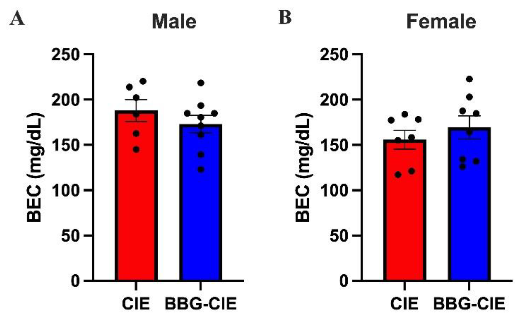
Figure 2.
CIE induced stronger proinflammatory cytokine gene expression in male brains than in female brains. mRNA expression after CIE exposure was analyzed by RT-qPCR and normalized to 18S rRNA. In males (A), upregulated proinflammatory cytokines in CIE mice were significantly reduced by BBG-treatment. In females (B), only Il-1β increased after CIE exposure, and BBG treatment reduced its level. One-way ANOVA followed by Tukey’s post hoc test was used for the statistical analysis; *p ≤ 0.05, **p ≤ 0.005, ***p ≤ 0.0005 compared with CIE-exposed mice as controls. (n = 3–7, mean ± SEM).
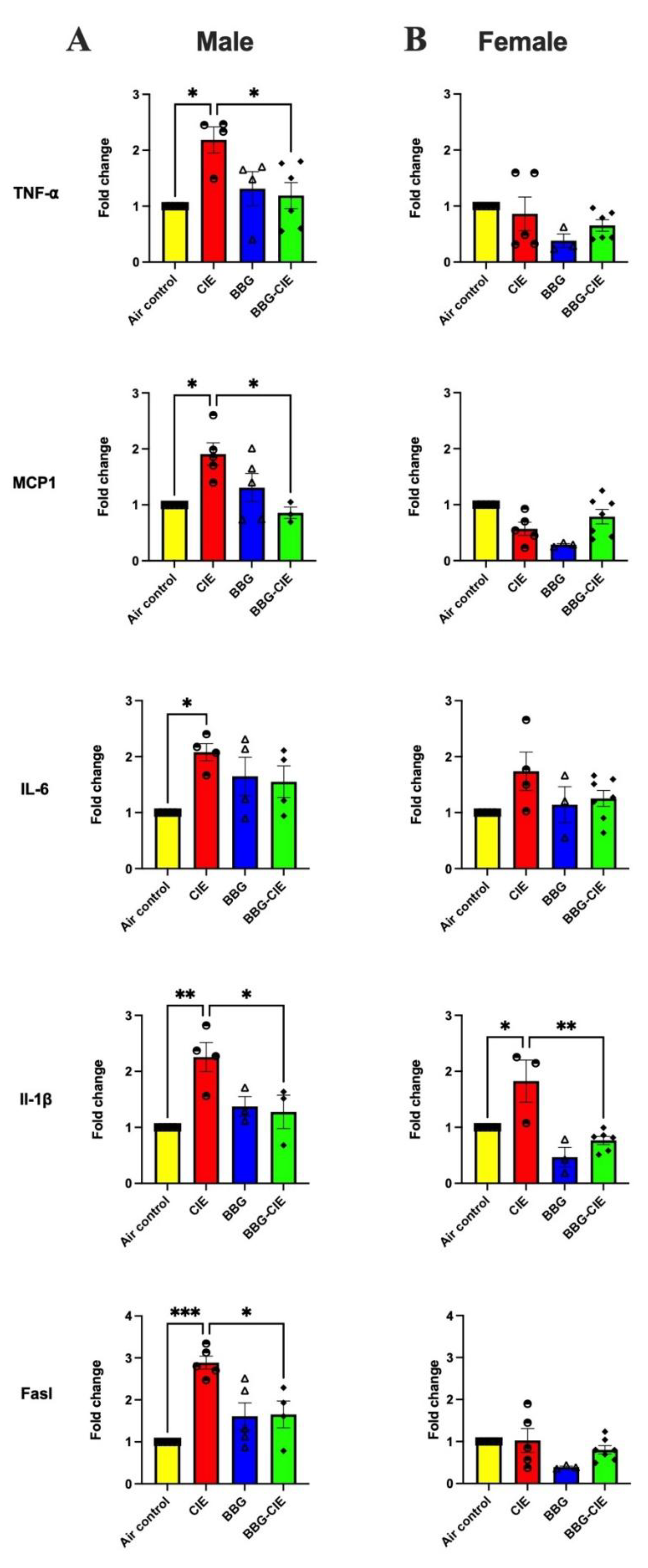
Figure 3.
CIE exposure reduces pericyte coverage in male mice, which was preserved by P2X7R inhibition. Immunofluorescent staining of CD13 (green) in mouse brain of males (A) and females (B) subjected to CIE exposure and/or BBG administration (original magnification: 200X, Scale bar = 500 µm). Intensity analysis of CD13-positive immunofluorescence designated as pericyte coverage in males (C) and females (D). (n = 3/group) (*p < 0.05, **p < 0.005).
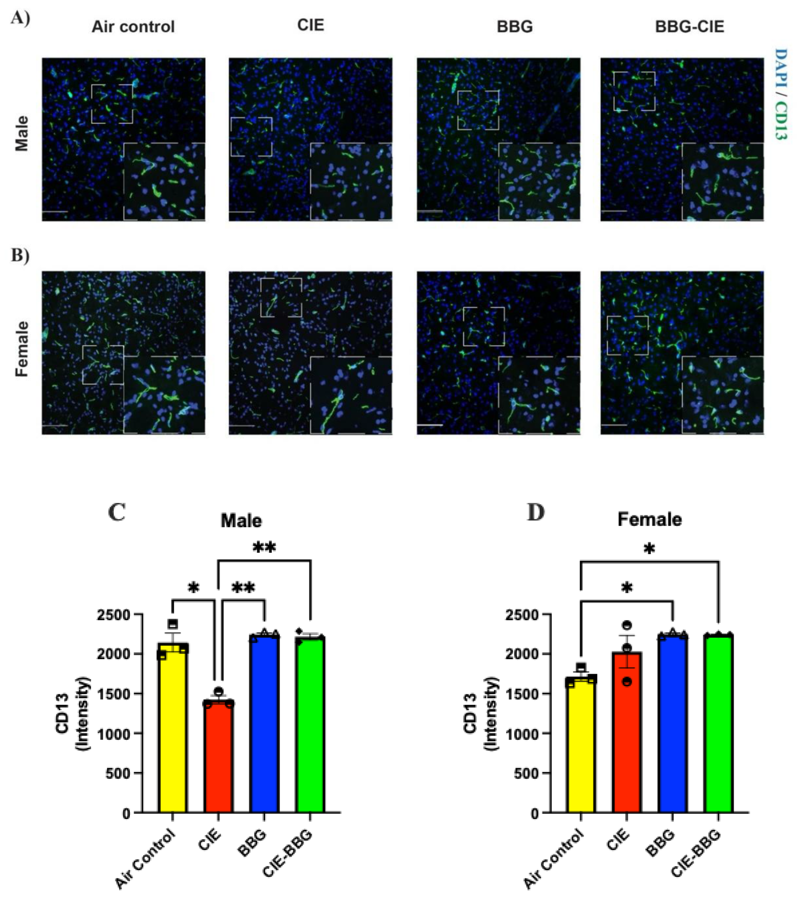
Figure 4.
CIE exposure elevates serum proinflammatory cytokines in male mice without significant effect in females. Cytokine levels following CIE exposure were analyzed by MSD ELISA. In males (A), upregulated cytokine levels in CIE mice were significantly reduced by BBG treatment. In females (B), CIE exposure did not elevate cytokine levels. Statistical analysis was performed using one-way ANOVA followed by Tukey’s post hoc test (*p ≤ 0.05, **p ≤ 0.005). Data presented as mean ± SEM (n = 3–8).
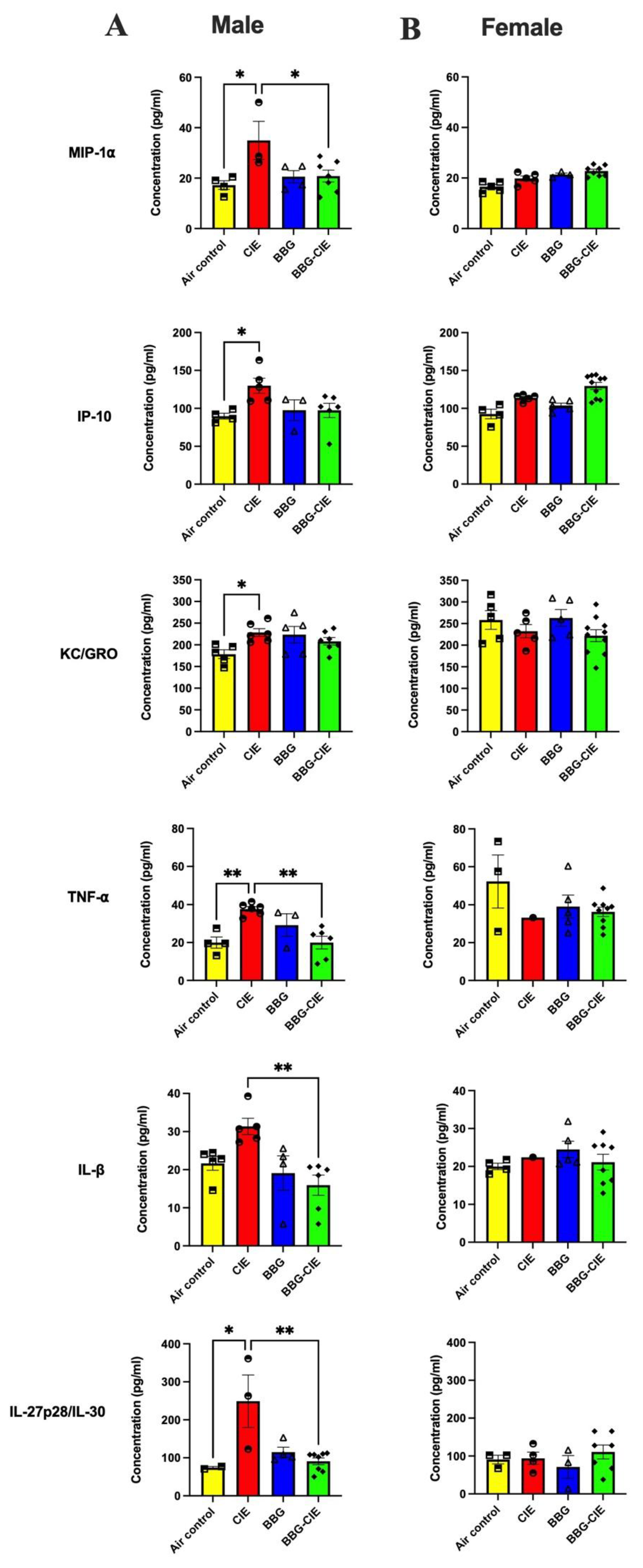
Figure 5.
CIE increases serum levels of P2X7R and P-gp. Serum P2X7R levels were higher in CIE-exposed (A) male and (B) female mice compared to their respective air control groups, and BBG treatment reduced these levels in CIE-exposed mice. CIE exposure elevated P-gp in male (C) and female (D) as compared with air controls. Statistical significance is indicated as ** for male mice (p = 0.0067) and **** for female mice (p < 0.0001). Data represent mean ± SEM from n = 3–5 mice per group. Statistical analysis was performed using one-way ANOVA followed by Tukey’s post-hoc test.
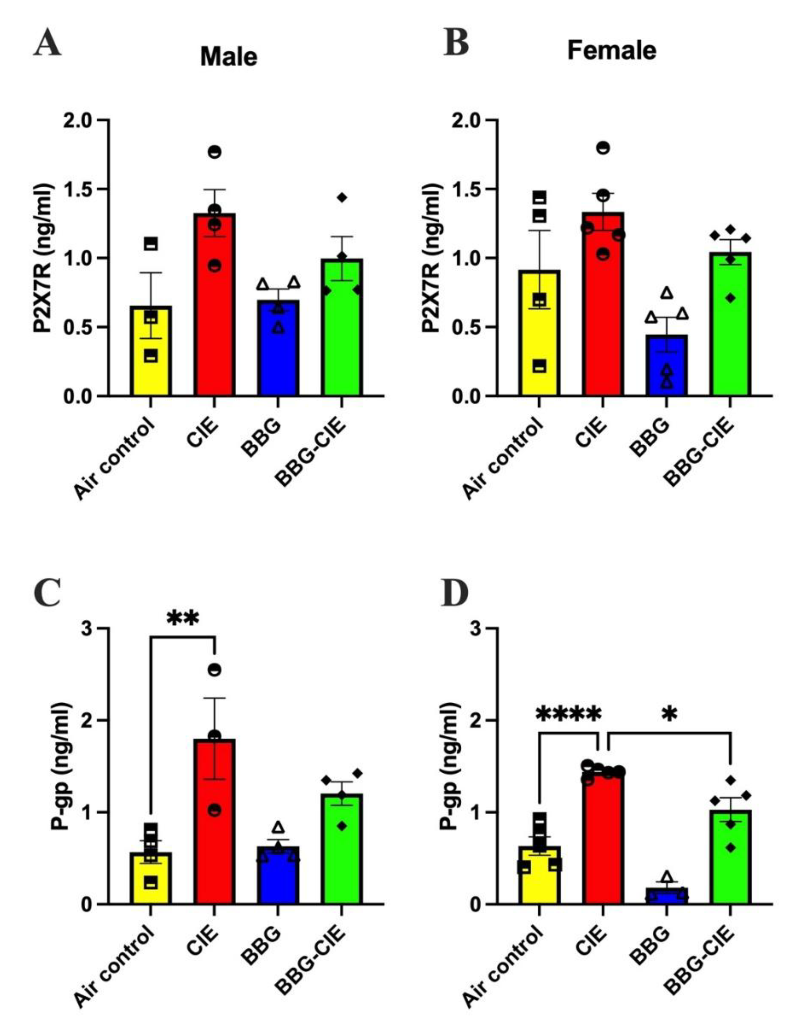
Figure 6.
CIE exposure elevates serum ATP levels similarly in both sexes. Serum ATP (eATP) was measured using luminescence-based ATP assay. ATP levels were significantly higher in CIE-exposed (A) male and (B) female mice compared to their respective air control groups. Statistical analysis was performed using one-way ANOVA followed by Tukey’s post hoc test; *p ≤ 0.05, ***p ≤ 0.0005, compared to the respective air control mice. Data are presented as mean ± SEM (n = 3–8).
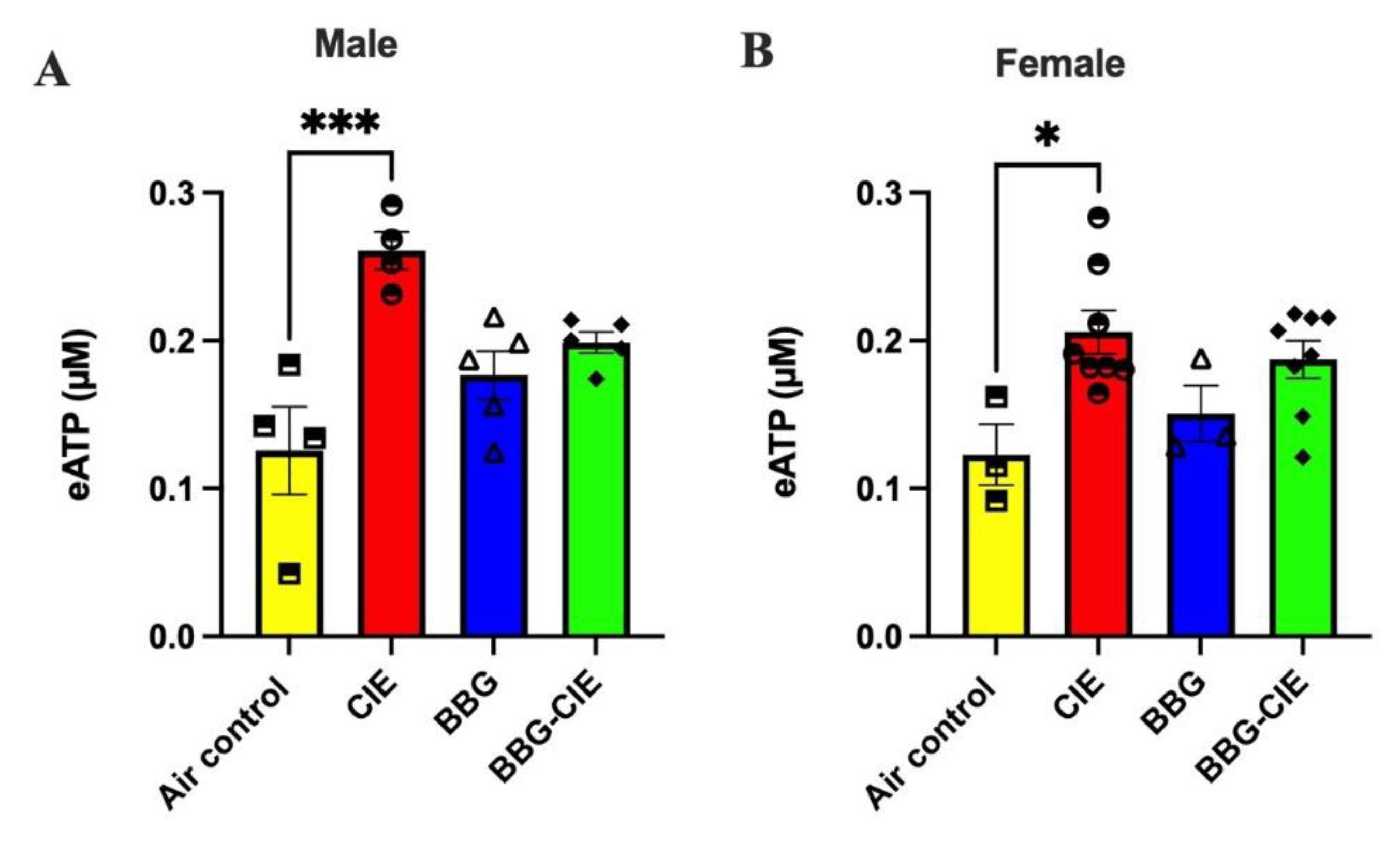
Figure 7.
P2X7R inhibition reduces CIE-induced increase in EV numbers and EV size in male and female mice. EV numbers were significantly increased in CIE male (A) and female mice (B) and were lowered in respective BBG-treated CIE-exposed mice. EV size was significantly increased by CIE and reduced in BBG-treated CIE-exposed (C) male and (D) female mice as compared to their respective CIE-exposed controls. Data are presented as mean ± SEM (n = 4–7). One-way ANOVA followed by Tukey’s post hoc test was used to perform statistical analysis; *p ≤ 0.05, **p ≤ 0.005 compared with the respective CIE-exposed control mice.
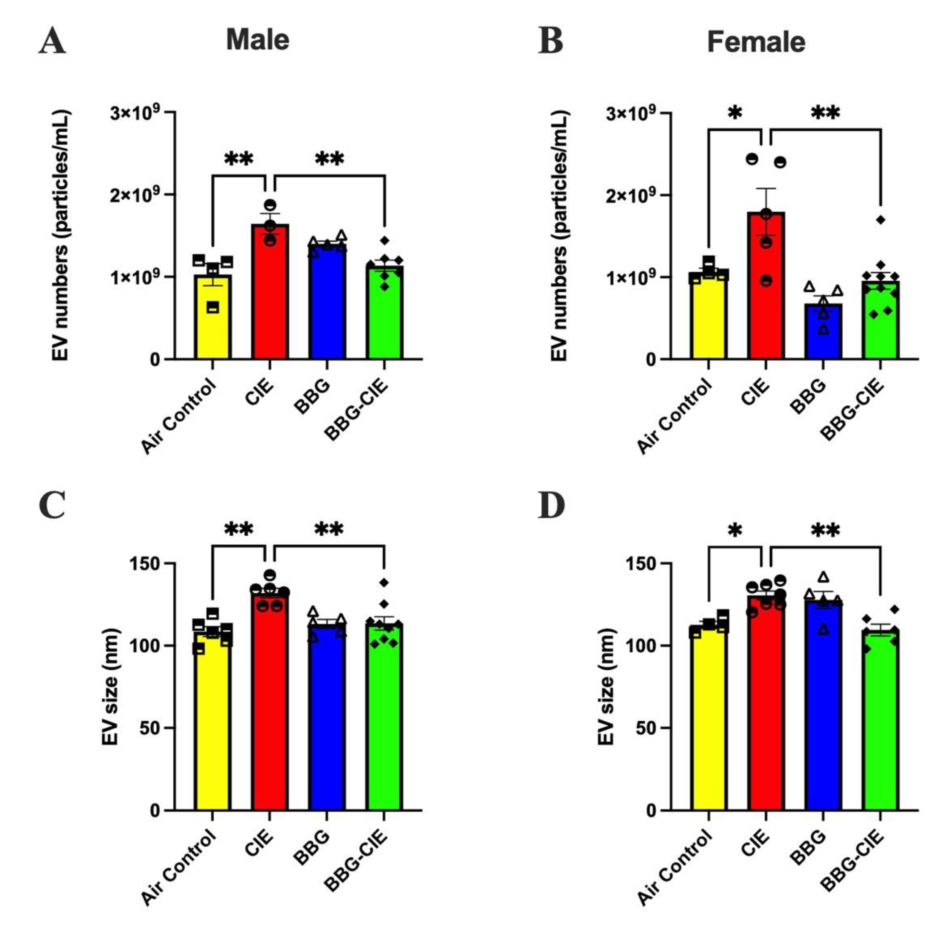
Figure 8.
EV mtDNA content increased by CIE exposure in both sexes was diminished by P2X7R inhibition. Digital PCR was used to quantify mtDNA copy numbers in EVs. Bar graphs show gene copy numbers per microliter of DNA. In both (A) males and (B) females, mtDNA copy numbers were significantly higher in EVs from CIE-exposed mice as compared to air controls. BBG treatment significantly reduced these levels in BBG-treated-CIE-exposed mice. Statistical analysis was performed using one-way ANOVA followed by Tukey’s post hoc test; *p ≤ 0.05, **p ≤ 0.005, ***p ≤ 0.0005 ****p < 0.0005. Data are presented as mean ± SEM (n = 3–7). .
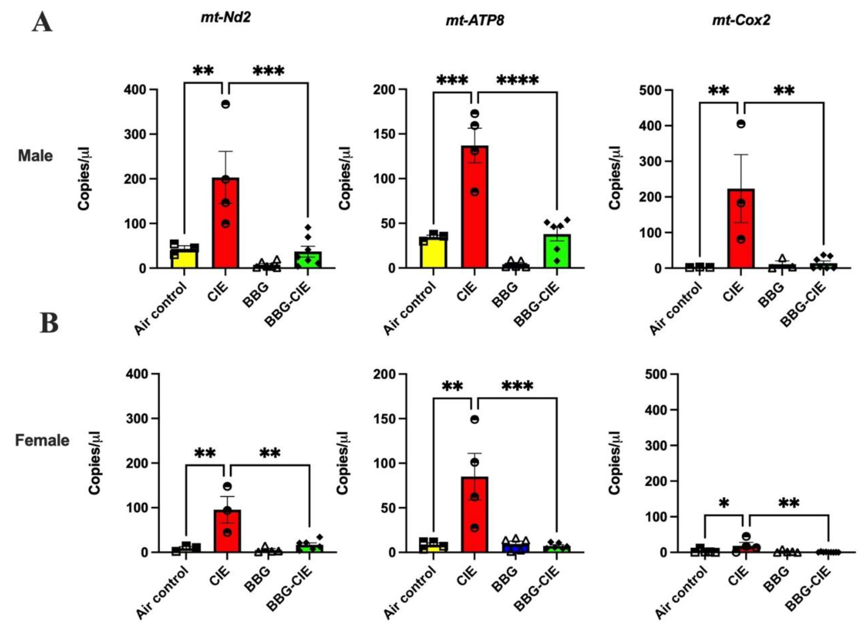
Figure 9.
CIE-induced spatial memory deficits found only in male mice were restored by P2X7R inhibition. Spatial memory performance was evaluated using the object placement test and quantified by the exploration ratio (ER) (A, B) and discrimination index (DI) (C, D). Significant differences between groups were identified using the Kruskal–Wallis test (*p<0.05). Data are shown as mean ± SEM (n = 3–7).
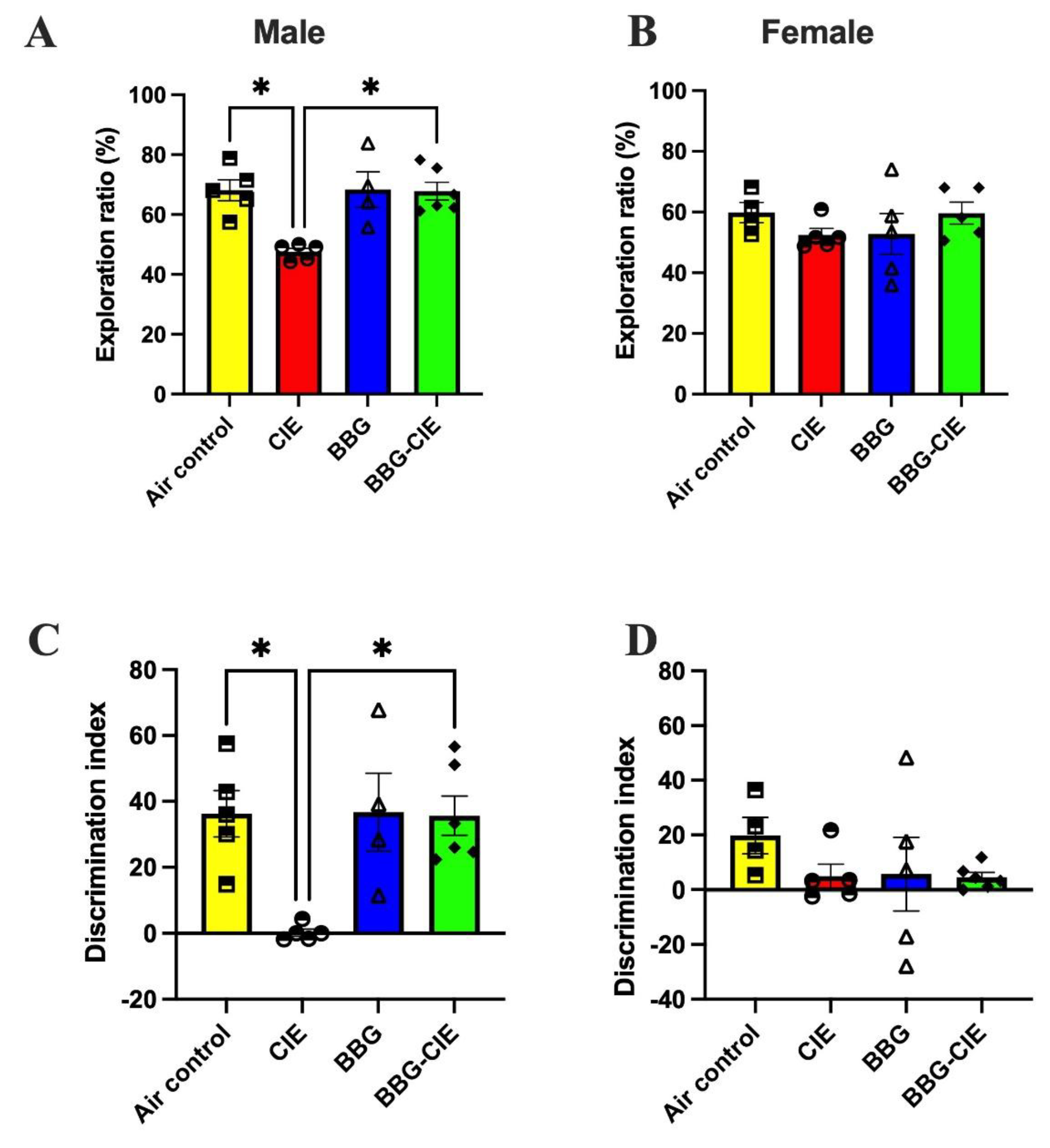
Disclaimer/Publisher’s Note: The statements, opinions and data contained in all publications are solely those of the individual author(s) and contributor(s) and not of MDPI and/or the editor(s). MDPI and/or the editor(s) disclaim responsibility for any injury to people or property resulting from any ideas, methods, instructions or products referred to in the content. |
© 2026 by the authors. Licensee MDPI, Basel, Switzerland. This article is an open access article distributed under the terms and conditions of the Creative Commons Attribution (CC BY) license (http://creativecommons.org/licenses/by/4.0/).
Copyright: This open access article is published under a Creative Commons CC BY 4.0 license, which permit the free download, distribution, and reuse, provided that the author and preprint are cited in any reuse.


