Preprint
Review

This version is not peer-reviewed.

Revisiting the Warburg Effect: Modern Understanding, Existing Misconceptions and Evolving Concepts in Cancer Metabolism

Submitted:

15 January 2026

Posted:

16 January 2026

You are already at the latest version

Abstract

The Warburg effect, classically defined as the preferential use of glycolysis by cancer cells in the presence of oxygen, has been a central concept in cancer biology since a long time. Otto Warburg had originally proposed that defective mitochondrial respiration was the primary cause of aerobic glycolysis in cancer cells. While this hypothesis profoundly influenced early cancer metabolism research, it has now become increasingly clear that this interpretation has gaping. Advances in biochemistry, molecular biology and metabolomics demonstrate that mitochondria in many cancers are functional and play essential roles in biosynthesis, signaling and energy production. Aerobic glycolysis in cancer cells is now recognized as an adaptive metabolic strategy that supports rapid proliferation by providing metabolic intermediates, maintaining redox balance, and enabling cellular signaling rather than maximizing ATP yield. This review discusses the Warburg effect through the lens of modern cancer metabolism. It contrasts classical misconceptions with current evidences, discusses key regulatory pathways like HIF-1α, PI3K/Akt/mTOR, c-Myc and PKM2, and examine the central role of lactate as both a metabolic fuel and a signaling molecule. It further explores metabolic heterogeneity, the reverse Warburg effect, immune–metabolic interactions, and the relevance of oxidative phosphorylation in cancer. Finally, some unresolved questions are highlighted that is critical for future understanding of cancer metabolism.

Keywords: 
;  ;  ;  ;  

1. Introduction

In 1924, Otto Warburg made the seminal observation that cancer cells exhibit unusually high rates of glucose uptake and lactate production, even in the presence of oxygen. This phenomenon was later termed as “Aerobic glycolysis” or “Warburg effect” [1]. Warburg had proposed that this metabolic observation resulted from irreversible damage to mitochondrial respiration, hence forcing cancer cells to rely on glycolysis for ATP generation. After this, the hypothesis dominated the field and strongly influenced how cancer metabolism was conceptualized for decades
However, as biochemical tools improved with passage of time, contradictions to this proposed model started appearing. Studies measuring mitochondrial function, oxygen consumption and tricarboxylic acid (TCA) cycle activity revealed that many cancer cells retain intact and functional mitochondria [2,4]. These findings suggested that aerobic glycolysis in cancer cells arise from a regulated metabolic choice, rather than mitochondrial failure
The modern understanding of cancer metabolism recognizes that high proliferation demand in cancer cells imposes unique biochemical demands. Rapidly dividing cells require not only energy, but also large quantities of amino acids, lipids and nucleotides. Glycolysis, despite being energetically inefficient, provides critical metabolic intermediates for these biosynthetic processes. Hence, the Warburg effect is now understood as a biosynthetic and regulatory adaptation, not a defect
Figure 1. Representation of Warburg effect in cancer cells. (Left side) Normal tissues metabolise glucose depending on available oxygen levels, with normoxic conditions favouring oxidative phosphorylation (maximized ATP production), while low oxygen levels favouring anaerobic glycolysis (minimized ATP production). (Right side) In contrast, cancer tissues metabolize glucose to produce lactate despite the presence of sufficient oxygen.
Preprints 194565 g001

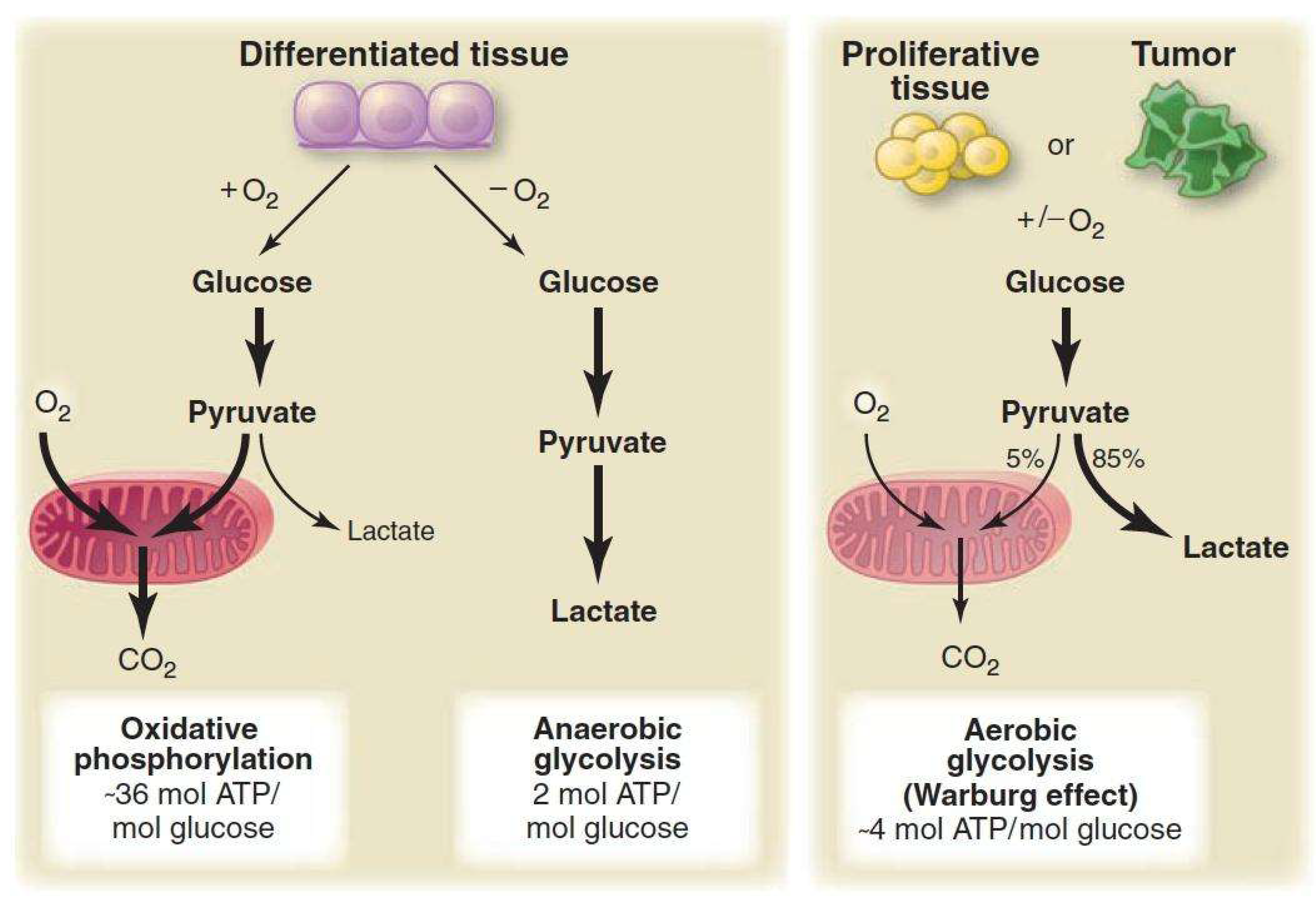
2. Classical Interpretations and Common Misconceptions

Several misconceptions about the Warburg effect exist, like:
a)
Mitochondria are defective in cancer cells- Contrary to Warburg’s original proposal, mitochondria in most cancer cells are intact in terms of structure and function, and actively participate in metabolism [2,4]
b)
Glycolysis is chosen because it is inefficient- Efficiency is not the relevant metric for proliferating cells. The metabolic flexibility and ability to supply biosynthetic precursors rapidly is more crucial [3,20]
c)
Lactate is primarily a waste product- In the cancer microenvironment, lactate is now recognized as an important metabolic fuel and signaling molecule
d)
The Warburg effect is universal- Cancer metabolism is very heterogeneous. Many cancers rely on oxidative phosphorylation, particularly in specific stages and subpopulations [18,21]
These misconceptions arose primarily because early studies focused on ATP yield rather than metabolic adaptations. Modern isotope-tracing studies have clarified that glycolysis has anabolic and regulatory functions beyond energy production, which is important for rapid proliferation of cancer cells [11,13]

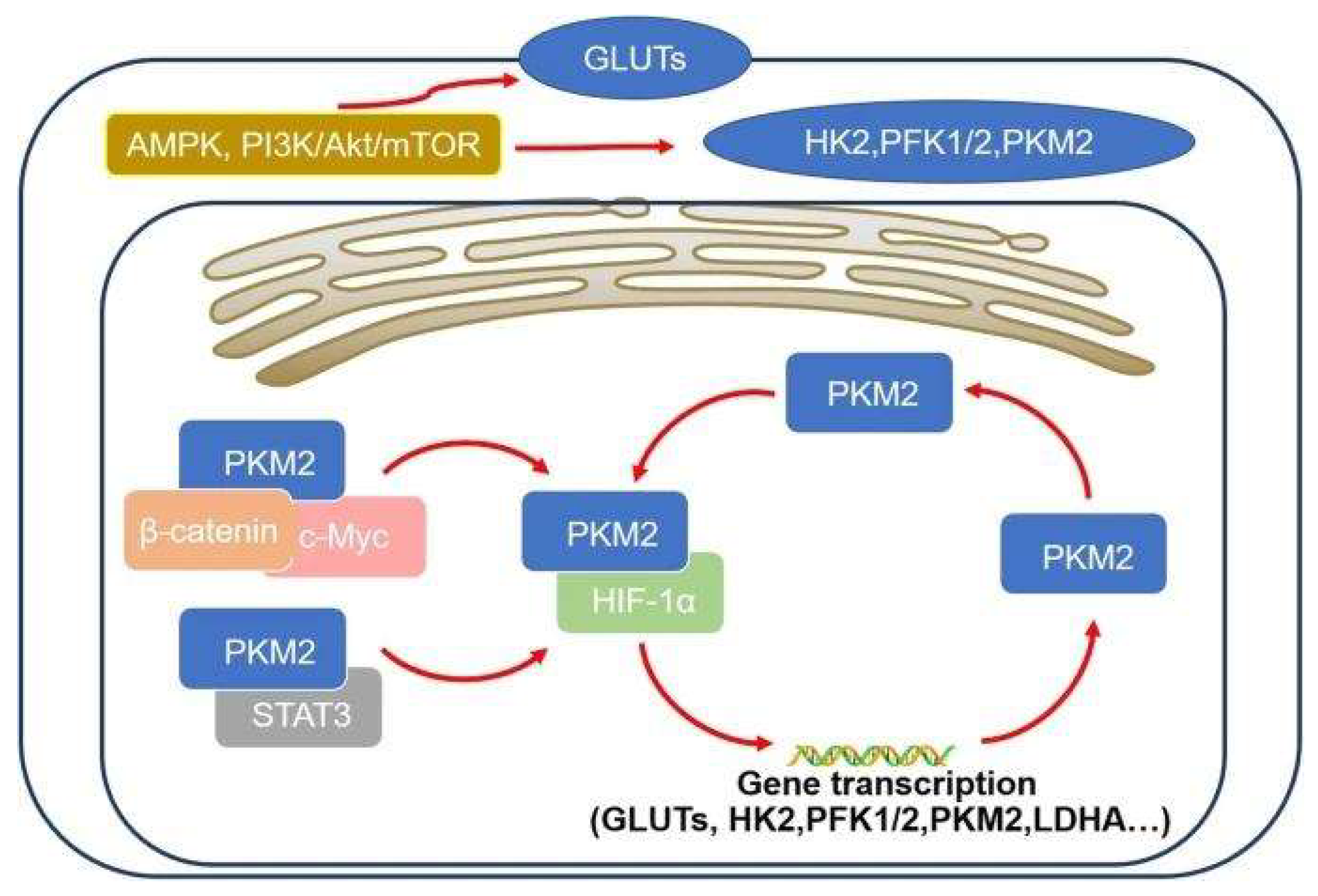
3. Molecular Regulation of Aerobic Glycolysis

While the mechanism of aerobic glycolysis appears simple, it reflects a highly coordinated set of biochemical and signaling changes. These mechanisms operate simultaneously, enabling cancer cells to maintain high glycolytic flux even in oxygen-rich environments

3.1. HIF-1α Signaling

Hypoxia-inducible factor 1α (HIF-1α) is a central regulator of glycolytic mechanism. Under conditions of low oxygen availability, HIF-1α stabilizes and induces the expression of glucose transporters and glycolytic enzymes while suppressing mitochondrial pyruvate oxidation [5]. Importantly, oncogenic signaling pathways can activate HIF-1α even under normoxic conditions, providing a mechanistic basis for aerobic glycolysis in cancer cells

3.2. PI3K/Akt/mTOR Pathway

The PI3K/Akt/mTOR pathway integrates growth factor signaling with nutrient availability. Activation of this pathway enhances glucose uptake, glycolytic flux, protein synthesis, and lipid biosynthesis [6]. In many cancers, constitutive activation of these pathways creates a metabolic environment that favors glycolysis as part of a broader anabolic program

3.3. c-Myc and Metabolic Coordination

The oncogene c-Myc plays a key role in coordinating glycolysis with glutamine metabolism. c-Myc induces transcription of genes involved in glucose metabolism while simultaneously promoting glutaminolysis, ensuring continuous replenishment of TCA cycle intermediates and nitrogen for biosynthesis [7,15]

3.4. PKM2 and Metabolic Control

Pyruvate kinase M2 (PKM2) is preferentially expressed in cancer cells and exists in a regulated low-activity form. This slows the final step of glycolysis, allowing upstream intermediates to be diverted into biosynthetic pathways such as the pentose phosphate pathway and serine biosynthesis [14]. This regulation highlights the intentional nature of glycolytic flux control in cancer
Figure 2. Transcriptional factors like HIF-1α and c-Myc, signaling pathways like PI3K/Akt/mTOR and enzymes like PKM2 play crucial roles in regulation of aerobic glycolysis in cancer cells [22].
Preprints 194565 g002

4. Glycolysis as a Platform for Biosynthesis and Redox Balance

Aerobic glycolysis supports several biosynthetic pathways essential for cell proliferation, such as:
  • Pentose phosphate pathway- Generates ribose-5-phosphate for nucleotide synthesis and NADPH for reductive biosynthesis and antioxidant defense [11]
  • Serine and glycine metabolism- Supplies one-carbon units required for nucleotide and methyl group synthesis [12]
  • Lipid biosynthesis- Glycolytic intermediates contribute to glycerol and fatty acid synthesis
NADPH production is particularly critical, as it maintains redox homeostasis and protects rapidly dividing cells from oxidative stress. Thus, aerobic glycolysis supports both biomass accumulation and redox control, which are key requirements for malignant growth [3,20]

5. Mitochondrial Adaptation and Function in Cancer Cells

While the Warburg effect emphasizes glycolysis, one of the most significant corrections in modern biochemistry is that most cancer cells retain fully functional mitochondria. Early assumptions of mitochondrial “damage” were largely based on limited assays and incomplete techniques. High-resolution respirometry and isotope-based carbon tracing have shown that oxygen consumption remains robust in many tumor types, even when glycolysis is strongly upregulated [2,4]

5.1. Mitochondria Are Hubs of Biosynthesis

Cancer cells divert mitochondrial pathways away from maximal ATP efficiency toward biosynthetic demands. Key functions include:
  • Citrate export for fatty acid and cholesterol synthesis
  • Aspartate production, essential for nucleotide biosynthesis
  • Anaplerotic glutamine metabolism to replenish TCA intermediates
  • NADPH generation for antioxidant defense and lipid synthesis
Even when glycolysis is heavily used, mitochondria maintain a central role in sustaining proliferative metabolism

5.2. Reductive Carboxylation in Hypoxia

Under low oxygen or defective electron transport chain capacity, cancer cells reverse TCA cycle flux through:
  • Reductive carboxylation of α-ketoglutarate to citrate
  • Using NADPH-dependent isocitrate dehydrogenase (IDH)
  • Supporting lipid synthesis under severely hypoxic conditions
This illustrates mitochondrial adaptability beyond classical oxidative metabolism

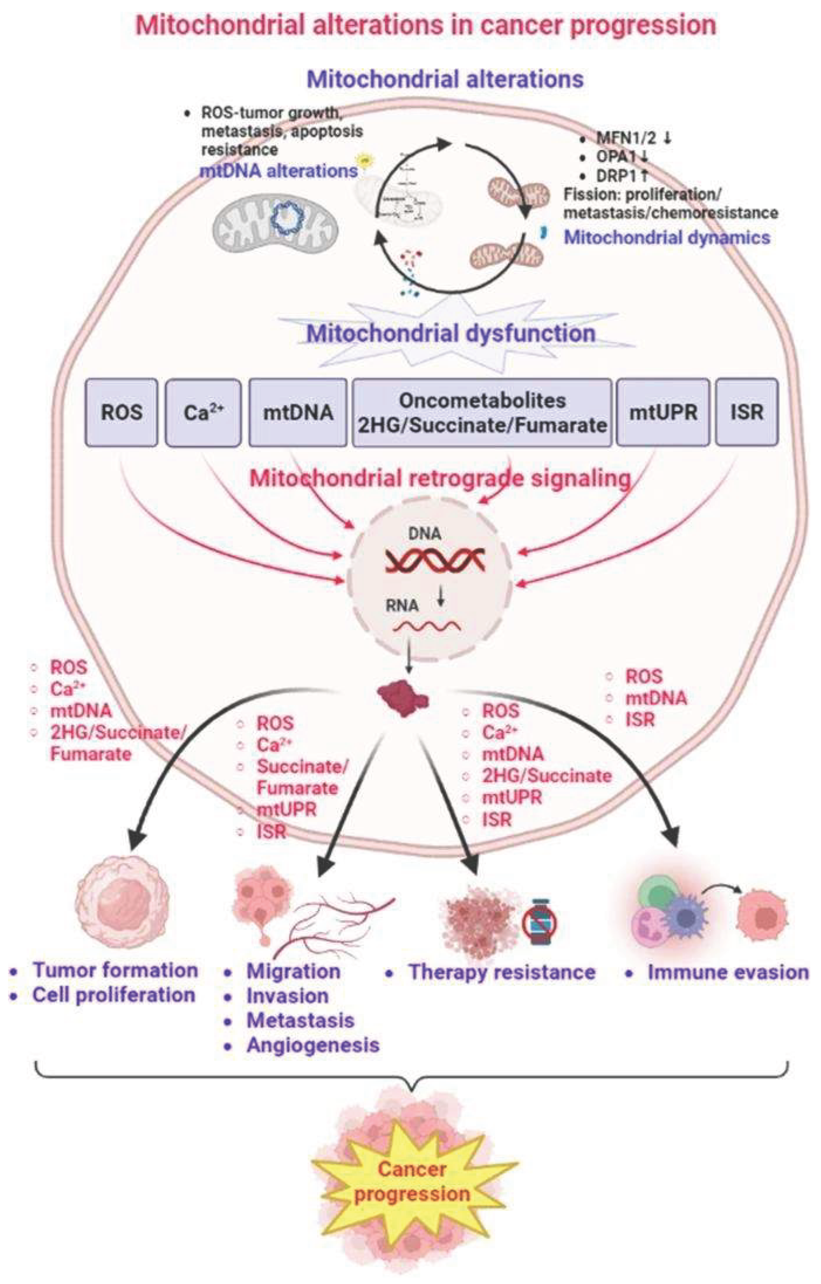
5.3. Mitochondrial Signaling in Cancer Progression

Beyond energy and biosynthesis, mitochondria also regulate:
  • ROS-dependent signaling, affecting proliferation and survival
  • Apoptosis resistance, via Bcl-2 family interactions
  • Mitochondrial biogenesis, coordinated by Myc and PGC-1α pathways
Together, these support survival, motility, and therapy resistance
Figure 3. A representation of mitochondrial alterations in cancer cells, leading to progression of cancer [23].
Preprints 194565 g003

6. The Emerging Role of Lactate as Fuel, Signal and Immune Modulator

Lactate has undergone one of the largest conceptual shifts in cancer metabolism. Once thought to be a metabolic waste-product, lactate is now recognized as a major oxidative substrate, intercellular signal, and immunological regulator

6.1. Lactate as Carbon Source

Contrary to classical assumptions, oxidative tumor cells can use lactate as fuel. This is facilitated through:
  • MCT1, which imports lactate
  • Lactate dehydrogenase B (LDHB), which converts lactate back to pyruvate
  • Oxidation in the tricarboxylic acid (TCA) cycle
This enables oxidative cancer cells to conserve glucose for biosynthetic pathways

6.2. Lactate Shuttle Within Tumors

Studies [9] have described a metabolic symbiosis where:
  • Hypoxic, glycolytic cells produce lactate
  • Lactate is exported via MCT4, induced by HIF-1α
  • Oxygen-rich tumor cells import lactate via MCT1
  • Lactate becomes the major respiratory fuel in these cells
This shuttle reduces competition for glucose while supporting the tumor metabolic efficiency

6.3. Lactate as an Angiogenic and Signaling Molecule

Lactate stabilizes HIF-1α even under normoxia, thereby promoting:
  • VEGF expression
  • Angiogenesis
  • Increased nutrient delivery
It also influences fibroblasts and endothelial cells, transforming the microenvironment into a supportive niche for tumor growth

6.4. Immunosuppressive Functions of Lactate

High lactate levels impair:
  • T-cell proliferation
  • Cytotoxic activity
  • Cytokine (IFN-γ, IL-2) secretion
Lactic acid accumulation directly inhibits human T-cell function [21]. Lactate also promotes regulatory T-cell activity and suppresses dendritic cell maturation
This makes lactate a key mediator of immune evasion, linking metabolism to immunology

7. Tumor Microenvironment and Metabolic Cooperation

Tumors are not homogeneous masses. They are actually ecosystems with diverse metabolic niches shaped by gradients of oxygen, pH, and nutrient availability

7.1. Reverse Warburg Effect

Studies [16] have highlighted phenomenon where:
  • Cancer-associated fibroblasts (CAFs) undergo aerobic glycolysis
  • CAFs produce lactate and high-energy metabolites
  • Cancer cells take up and oxidize this lactate for ATP
  • CAFs undergo metabolic exhaustion while tumors thrive
This contradicts the classical assumption that only tumor cells exhibit glycolysis

7.2. pH Gradients and Acidification

Lactate export acidifies the tumor microenvironment, leading to:
  • ECM degradation
  • Increased invasiveness
  • Selection for acid-resistant clones
  • Impaired T-cell infiltration
Acidosis is therefore a pro-tumorigenic adaptation

8. Oxidative Phosphorylation Dependence and Metabolic Plasticity

Many cancer subpopulations and entire tumor types rely heavily on oxidative phosphorylation

8.1. Oxidative Phosphorylation Dependent Tumor Categories

Several cancers, including lymphomas, leukemias, certain melanomas, and subsets of breast cancer depend primarily on mitochondrial metabolism [18]
These cells prefer:
  • Efficient ATP generation
  • ROS-mediated signaling for proliferation
  • Fatty acid oxidation as fuel
  • A stable redox environment
They are also less sensitive to glycolysis inhibitors

8.2. Cancer Stem-like Cells (CSCs)

CSCs are more reliant on mitochondrial respiration than bulk tumor cells:
  • High mitochondrial mass
  • Elevated oxidative phosphorylation activity
  • Resistance to chemotherapy
  • Ability to survive nutrient stress
CSCs often adopt oxidative phosphorylation to evade metabolic-targeted therapies [19]

8.3. Switching Between Glycolysis and Oxidative Phosphorylation

Cancer cells can dynamically shift metabolic states depending on:
  • Oxygen tension
  • Nutrient availability
  • Therapy exposure
  • Micro environmental interactions
This plasticity explains why single-pathway metabolic inhibitors often fail in clinical settings

10. Future Directions

Future researches need to address several key questions like:
  • What determines metabolic phenotype ?
  • How can metabolic plasticity be predicted ?
  • How does lactate influence immunotherapy outcomes ?
  • How can metabolic therapies be personalized ?

11. Conclusions

Contrary to old proposals, the Warburg effect in cancer cells is a regulated and adaptive metabolic state, not a consequence of mitochondrial injury. Modern research reveals complex interactions among glycolysis, mitochondrial metabolism, lactate signaling, immune suppression, and tumor micro environmental pressures. Understanding this integrated metabolic landscape is essential for developing future diagnostics and metabolically targeted therapies

References

  1. WARBURG, O. On the origin of cancer cells; Science: New York, N.Y.), 1956; Volume 123, 3191, pp. 309–314. [Google Scholar] [CrossRef]
  2. Zu, X. L.; Guppy, M. Cancer metabolism: facts, fantasy, and fiction. Biochemical and biophysical research communications 2004, 313(3), 459–465. [Google Scholar] [CrossRef]
  3. Ward, P. S.; Thompson, C. B. Metabolic reprogramming: a cancer hallmark even warburg did not anticipate. Cancer cell 2012, 21(3), 297–308. [Google Scholar] [CrossRef] [PubMed]
  4. Vyas, S.; Zaganjor, E.; Haigis, M. C. Mitochondria and Cancer. Cell 2016, 166(3), 555–566. [Google Scholar] [CrossRef] [PubMed]
  5. Semenza, G.L. Hypoxia-Inducible Factors in Physiology and Medicine. Cell 2012, 148, 399–408. [Google Scholar] [CrossRef] [PubMed]
  6. Pavlova, N. N.; Thompson, C. B. The Emerging Hallmarks of Cancer Metabolism. Cell metabolism 2016, 23(1), 27–47. [Google Scholar] [CrossRef]
  7. Dang, C. V. MYC on the path to cancer. Cell 2012, 149(1), 22–35. [Google Scholar] [CrossRef]
  8. Halestrap, A. P.; Wilson, M. C. The monocarboxylate transporter family--role and regulation. IUBMB life 2012, 64(2), 109–119. [Google Scholar] [CrossRef]
  9. San-Millán, I.; Brooks, G. A. Reexamining cancer metabolism: lactate production for carcinogenesis could be the purpose and explanation of the Warburg Effect. Carcinogenesis 2017, 38(2), 119–133. [Google Scholar] [CrossRef]
  10. Doherty, J. R.; Cleveland, J. L. Targeting lactate metabolism for cancer therapeutics. The Journal of clinical investigation 2013, 123(9), 3685–3692. [Google Scholar] [CrossRef]
  11. Cairns, R. A.; Harris, I. S.; Mak, T. W. Regulation of cancer cell metabolism. Nature reviews. Cancer 2011, 11(2), 85–95. [Google Scholar] [CrossRef]
  12. Locasale, J. W. Serine, glycine and one-carbon units: cancer metabolism in full circle. Nature reviews. Cancer 2013, 13(8), 572–583. [Google Scholar] [CrossRef]
  13. Liberti, M. V.; Locasale, J. W. The Warburg Effect: How Does it Benefit Cancer Cells? Trends in biochemical sciences 2016, 41(3), 211–218. [Google Scholar] [CrossRef]
  14. Yang, W.; Lu, Z. Regulation and function of pyruvate kinase M2 in cancer. Cancer letters 2013, 339(2), 153–158. [Google Scholar] [CrossRef]
  15. Boroughs, L. K.; DeBerardinis, R. J. Metabolic pathways promoting cancer cell survival and growth. Nature cell biology 2015, 17(4), 351–359. [Google Scholar] [CrossRef] [PubMed]
  16. Sonveaux, P.; Végran, F.; Schroeder, T.; Wergin, M. C.; Verrax, J.; Rabbani, Z. N.; De Saedeleer, C. J.; Kennedy, K. M.; Diepart, C.; Jordan, B. F.; Kelley, M. J.; Gallez, B.; Wahl, M. L.; Feron, O.; Dewhirst, M. W. Targeting lactate-fueled respiration selectively kills hypoxic tumor cells in mice. The Journal of clinical investigation 2008, 118(12), 3930–3942. [Google Scholar] [CrossRef]
  17. Huang, L.; Mellor, A. L. Metabolic control of tumour progression and antitumour immunity. Current opinion in oncology 2014, 26(1), 92–99. [Google Scholar] [CrossRef]
  18. Xu, Y.; Xue, D.; Bankhead, A., 3rd; Neamati, N. Why All the Fuss about Oxidative Phosphorylation (OXPHOS)? Journal of medicinal chemistry 2020, 63(23), 14276–14307. [Google Scholar] [CrossRef] [PubMed]
  19. Weinberg, S. E.; Chandel, N. S. Targeting mitochondria metabolism for cancer therapy. Nature chemical biology 2015, 11(1), 9–15. [Google Scholar] [CrossRef]
  20. Vander Heiden, M. G.; Cantley, L. C.; Thompson, C. B. Understanding the Warburg effect: the metabolic requirements of cell proliferation. Science (New York, N.Y.) 2009, 324(5930), 1029–1033. [Google Scholar] [CrossRef] [PubMed]
  21. Fischer, K.; Hoffmann, P.; Voelkl, S.; Meidenbauer, N.; Ammer, J.; Edinger, M.; Gottfried, E.; Schwarz, S.; Rothe, G.; Hoves, S.; Renner, K.; Timischl, B.; Mackensen, A.; Kunz-Schughart, L.; Andreesen, R.; Krause, S. W.; Kreutz, M. Inhibitory effect of tumor cell-derived lactic acid on human T cells. Blood 2007, 109(9), 3812–3819. [Google Scholar] [CrossRef]
  22. Feng, J.; Li, J.; Wu, L.; et al. Emerging roles and the regulation of aerobic glycolysis in hepatocellular carcinoma. J Exp Clin Cancer Res 2020, 39, 126. [Google Scholar] [CrossRef] [PubMed]
  23. Wang, SF.; Tseng, LM.; Lee, HC. Role of mitochondrial alterations in human cancer progression and cancer immunity. J Biomed Sci 2023, 23 30, 61. [Google Scholar] [CrossRef] [PubMed]
Disclaimer/Publisher’s Note: The statements, opinions and data contained in all publications are solely those of the individual author(s) and contributor(s) and not of MDPI and/or the editor(s). MDPI and/or the editor(s) disclaim responsibility for any injury to people or property resulting from any ideas, methods, instructions or products referred to in the content.
Copyright: This open access article is published under a Creative Commons CC BY 4.0 license, which permit the free download, distribution, and reuse, provided that the author and preprint are cited in any reuse.
Prerpints.org logo

Preprints.org is a free preprint server supported by MDPI in Basel, Switzerland.

Subscribe

Disclaimer

Terms of Use

Privacy Policy

Privacy Settings

© 2026 MDPI (Basel, Switzerland) unless otherwise stated