Preprint
Review

This version is not peer-reviewed.

Amarilloviruses of Aquatic Animals

Submitted:

12 January 2026

Posted:

13 January 2026

You are already at the latest version

Abstract

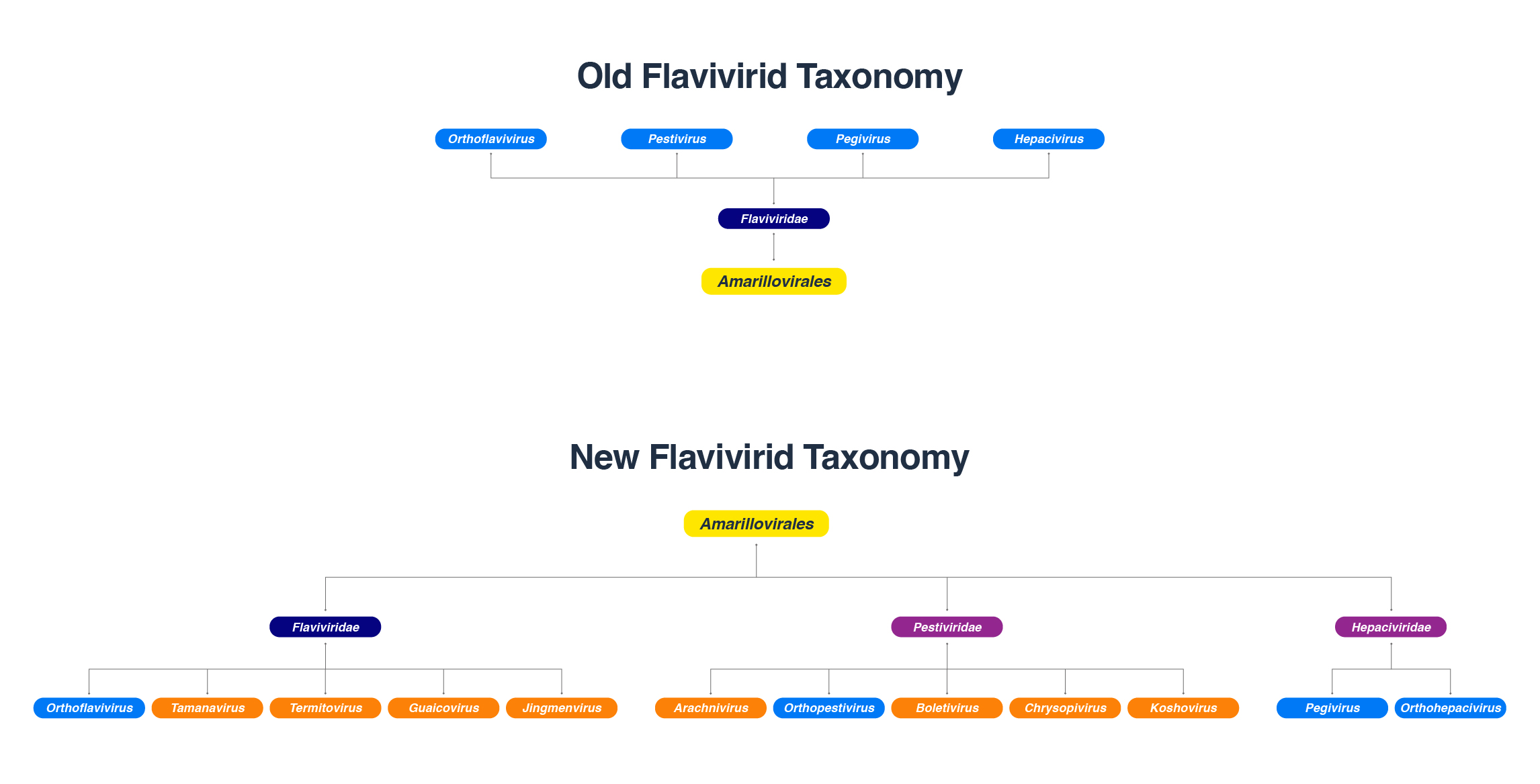
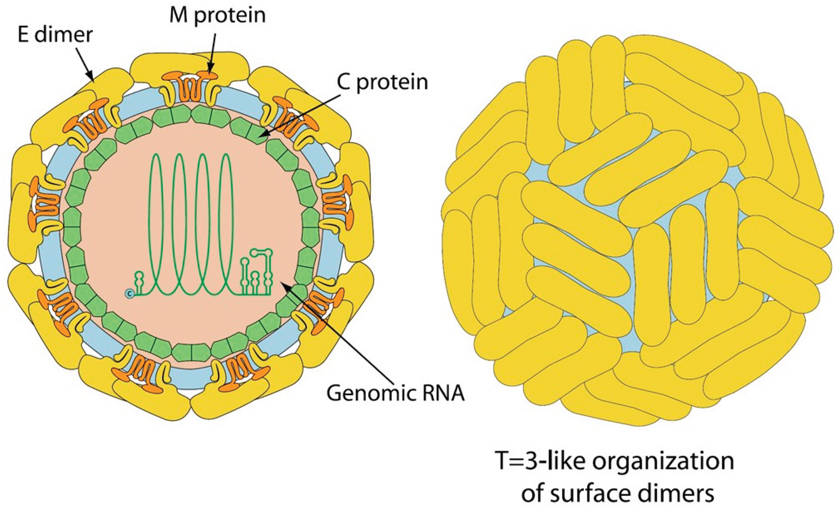
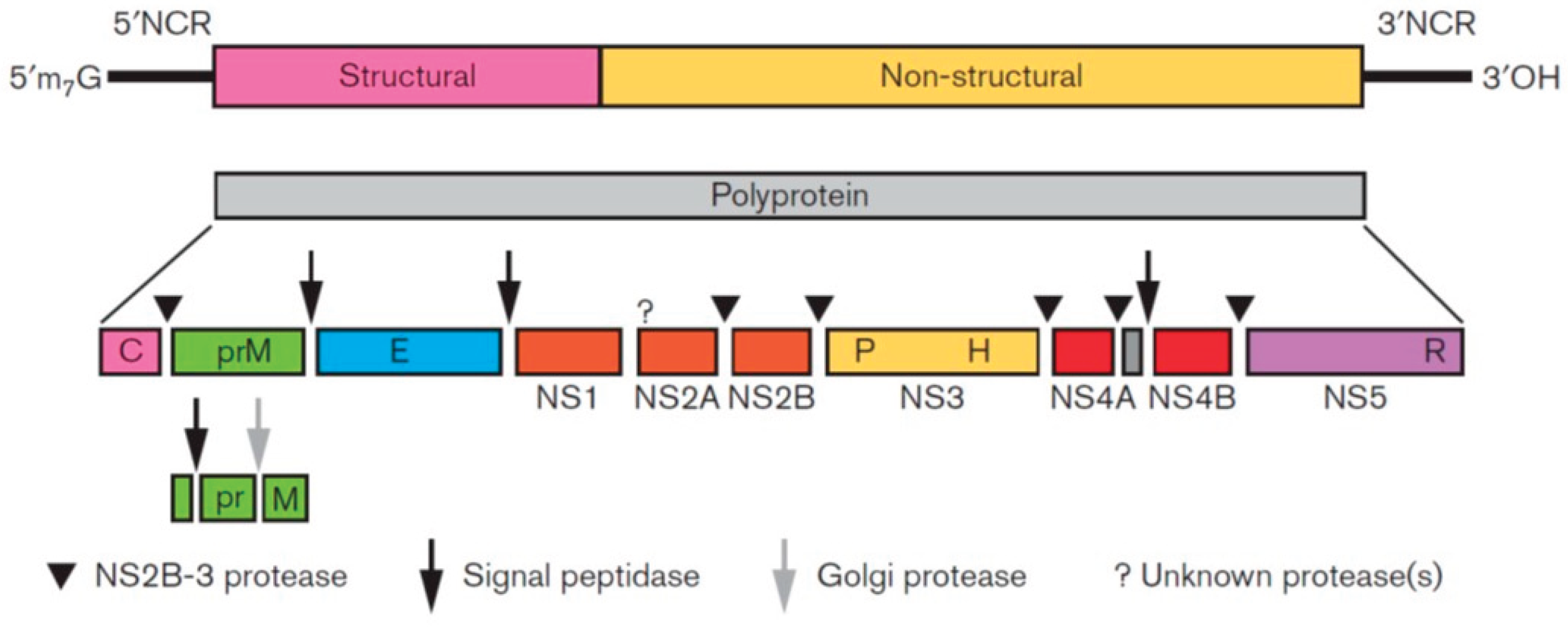
The family Flaviviridae has been expanded to include the highly divergent flavi-like viruses into three new families, Flaviviridae, Pestiviridae, and Hepaciviridae in the order Amarillovirales. Classical flavivirids are small, enveloped viruses with positive-sense ssRNA genomes lacking a 3’ poly(A) tail, and ~ 9.0-13.0 kb in length, with a single ORF encoding structural proteins at the N terminus and nonstructural proteins at the C terminus. Members infect a wide range of mammals, birds, and insects, and many are host-specific and pathogenic. Although the RdRP gene sequences of the flavi-like viruses group phylogenetically with those of classical flavivirids, flavi-like viruses often encode larger polyproteins and possess substantially longer genomes of up to ~ 40 kb, and some have a 3’ poly(A) tail. Their host range extends across the whole animal kingdom and in angiosperm plants. This review describes the reported flavi-like viruses of aquatic animals, providing a meaningful update on all three new families in Amarillovirales that have been discovered in fish, crustaceans, mollusks, and echinoderms, using metagenomics. These amarilloviruses include pathogenic viruses of aquatic animals, such as Cyclopterus lumpus virus (CLuV) detected in moribund lumpfish, and Infectious precocity virus (IPV) found in iron prawn syndrome (IPS)-affected farmed giant freshwater prawns.

Keywords: 
;  ;  ;  ;  ;  ;  ;  ;  ;  ;  

1. Introduction

The highly divergent flavi-like viruses found in aquatic animals are collectively referred to as amarilloviruses in this review, named after the Order Amarillovirales, which contains the new families of flavivirids. In 2025 the International Committee on Taxonomy of Viruses (ICTV) approved the reclassification the family Flaviviridae and its expansion through the incorporation of the large number of additional flavi-like viruses into three new families, Flaviviridae (with 5 genera: Orthoflavivirus, Tamanavirus, Termitovirus, Guaicovirus, and Jingmenvirus), Pestiviridae (with 5 genera: Arachnivirus, Orthopestivirus, Boletivirus, Chrysopivirus and Koshovirus) and Hepaciviridae (with 2 genera: Orthohepacivirus and Pegivirus), and an unnamed family (represented as Clade IV with one floating flavi-like virus, diatom colony-associated ssRNA virus (DCAV)), in the established order Amarillovirales [1,2]. Additionally, there are unnamed genera (designated Lineages) in the three families (5 Lineages in Flaviviridae, 2 in Pestiviridae, and 2 in Hepaciviridae) [2].
Before this reclassification of flavivirids, the only family included in order Amarillovirales was Flaviviridae, which is derived from the Latin flavus, “yellow,” referring to Yellow Fever Virus, the cause of the disease Yellow Fever in humans, characterized by jaundice (yellowing of the skin and the whites of the eyes). The classical flavivirids (i.e., the original family Flaviviridae) are small, enveloped viruses with positive-sense, non-segmented, linear, single-stranded (ss)RNA genomes lacking a 3’ poly(A) tail [3] and approximately 9.0-13.0 kb in length, with a common genome organization. They all have a single 8,000-10,700 base open reading frame (ORF) that is translated and cleaved into structural proteins (core and envelope proteins) located at the N terminus and nonstructural proteins (protease, helicase, and RNA-directed RNA polymerase (RdRP)) at the C terminus. They are homologous in their RdRP gene, superfamily 2 helicase (NS3), and serine protease domain sequences [4]. Viruses of different flavivirid genera (Hepacivirus, Orthoflavivirus, Pegivirus, and Pestivirus in the original family Flaviviridae [5]) differ, however, in their polyprotein translation strategy, which may be 5’-cap-dependent (orthoflaviviruses) or driven through an internal ribosomal entry site (IRES) (other flavivirids), and virion formation [2]. Their host range is primarily mammals and birds [6]. Many are host-specific and pathogenic, such as hepatitis C virus (HCV) in the genus Hepacivirus, a major human pathogen, bovine virus diarrhea virus (BVDV) and classical swine fever virus (CSFV) in genus Pestivirus which cause serious disease in cattle and pigs, respectively, members of genus Pegivirus associated with persistent infections of a wide range of mammalian species but not associated with disease, and genus Orthoflavivirus, which includes 53 species, most of which are arboviruses (arthropod-borne viruses; viruses biologically transmitted by blood-sucking insects and ticks – i.e., vertebrate viruses capable of reproducing in their arthropod vector), and many are important human and veterinary pathogens (e.g., yellow fever virus, YFV; dengue virus, DENV; Zika virus, ZIKV; Japanese encephalitis virus, JEV; West Nile virus, WNV) [7]. In contrast, the majority of flavi-like viruses have been discovered outside the primarily mammalian and vector host range of classified flavivirids, being distributed across the animal kingdom, from poriferans (sponges) [8], cnidarians (jellies) [9], mollusks (squid) [10], arthropods (insects [11]; diplurans and scorpions [12]; crustaceans [9,10,13], nematodes [14], platyhelminths [15,16] to echinoderms (sea cucumbers) [17]), hemichordates (acorn worms) [9], cartilaginous and bony fish [9,18,19,20,21,22], amphibians (frogs) [9,23], reptiles [21], stramenopiles (diatoms and oomycotes) [24,25] and in angiosperm plants [26,27,28].
Evidently, the highly divergent and much wider range of flavi-like viruses has increased considerably. Although these viruses often possess quite different genome organizations, genome lengths, and host ranges, their RdRP gene sequences group phylogenetically with those of classical flavivirids [12], allowing a genomics-based reclassification and expansion of the former family Flaviviridae. Given the fact that the RdRP gene is the most evolutionarily conserved gene within virus groups [29], this reorganization of flavivirids is extremely robust. Thus, the flavi-like viruses have distinct genome organizations from classical flavivirids. They encode larger polyproteins and possess longer genomes (the so-called ‘large-genome flavi-like viruses flaviviruses (LGFs)’ [18,30,31], substantially longer than the classical flavivirids [26,28,32], the longest to date being Maximus pesti-like virus’ of about 40 kb, representing an instance of a flavi-like virus achieving a genome size comparable to that of the order Nidovirales [8], and some have a 3’ poly(A) tail [33]. Moreover, the new Flaviviridae family now includes 3 genera of viruses with non-segmented genomes (Orthoflavivirus, Tamanavirus, and Termitovirus) and 2 genera of viruses with multi-segmented genomes (Guaicovirus with 5 segments and Jingmenvirus with 4 segments [2]). In the old family Flaviviridae, the “segmented flavi-like viruses” with a segmented genome of four to five segments were classified by the ICTV as an unclassified sub-genus in the Orthoflavivirus genus under the putative genus name Jingmenvirus [12,34,35]. Jingmenviruses are found in ticks, nematodes, mosquitoes, humans, cattle, monkeys, bats, rodents, sheep, tortoises, crustaceans, plants, and fungi, and are endemic in a wide range of environments worldwide [34,35]; Jingmen tick virus (JMTV), species Jingmenvirus rhipicephali [2], is one of the novel emerging tick-borne viruses and is pathogenic to humans [35]. Infectious precocity virus (IPV) found in iron prawn syndrome (IPS)-affected farmed giant freshwater prawns Macrobrachium rosenbergii was shown to belong to a proposed new genus Crustaflavivirus [13], which is phylogenetically between Jingmenvirus and Orthoflavivirus [13]. This review describes the reported amarilloviruses of aquatic animals to advance understanding of the wide range of flaviviruses that occur in aquatic animals. These viruses group with the three new families in Order Amarillovirales and have been discovered in various marine hosts, including fish, crustaceans, molluscs, echinoderms, poriferans, and cnidarians using metagenomics [8,9,10,13,17,18,19,20,33,36,37]. Some of them cause disease in species important to the aquaculture industry [13,20,33].
Table 1 lists the highly divergent flavi-like viruses found in aquatic animals. These amarilloviruses in the new family Flaviviridae include Wenzhou shark flavivirus found in both a Pacific spadenose shark (Scoliodon macrorhynchos) [37] and a gazami crab (Portunus trituberculatus) [10], which group with the Orthoflavivirus genus but are not known to be transmitted by arthropods, and the pathogenic fish flavivirus Cyclopterus lumpus virus (CLuV) detected in moribund lumpfish (Cyclopterus lumpus) [20] which groups with the Tamanavirus genus. The fish viruses in the new family Hepaciviridae include Wēnlǐng shark virus (WLSV) (in genus Orthohepacivirus) discovered in the graceful catshark (Proscyllium habereri) [18], Wēnlǐng moray eel hepacivirus (in Lineage IIIt [2]) in Gymnothorax reticularis, Xiàmén guitarfish hepacivirus in Rhinobatos hynnicephalus, Xiàmén sepia Stingray hepacivirus in Urolophus aurantiacus, Western African lungfish hepacivirus in Protopterus annectens, Guangxi houndshark hepacivirus in Mustelus manazo, and Nanhai dogfish shark hepacivirus in Squalus brevirostris, and Nanhai ghost shark hepacivirus 1 and 2 in Chimaera sp [19,37]. The fish flaviviruses that group with the new family Pestiviridae include Xiàmén fanray pesti-like virus (XFPV) in Lineage IIn [2] detected in Platyrhina sp, Nanhai dogfish shark pesti-like virus in Squalus brevirostris, and Wenzhou pesti-like virus 1 and 2 discovered in Sphyrna lewini and Proscyllium habereri, respectively [19,37].

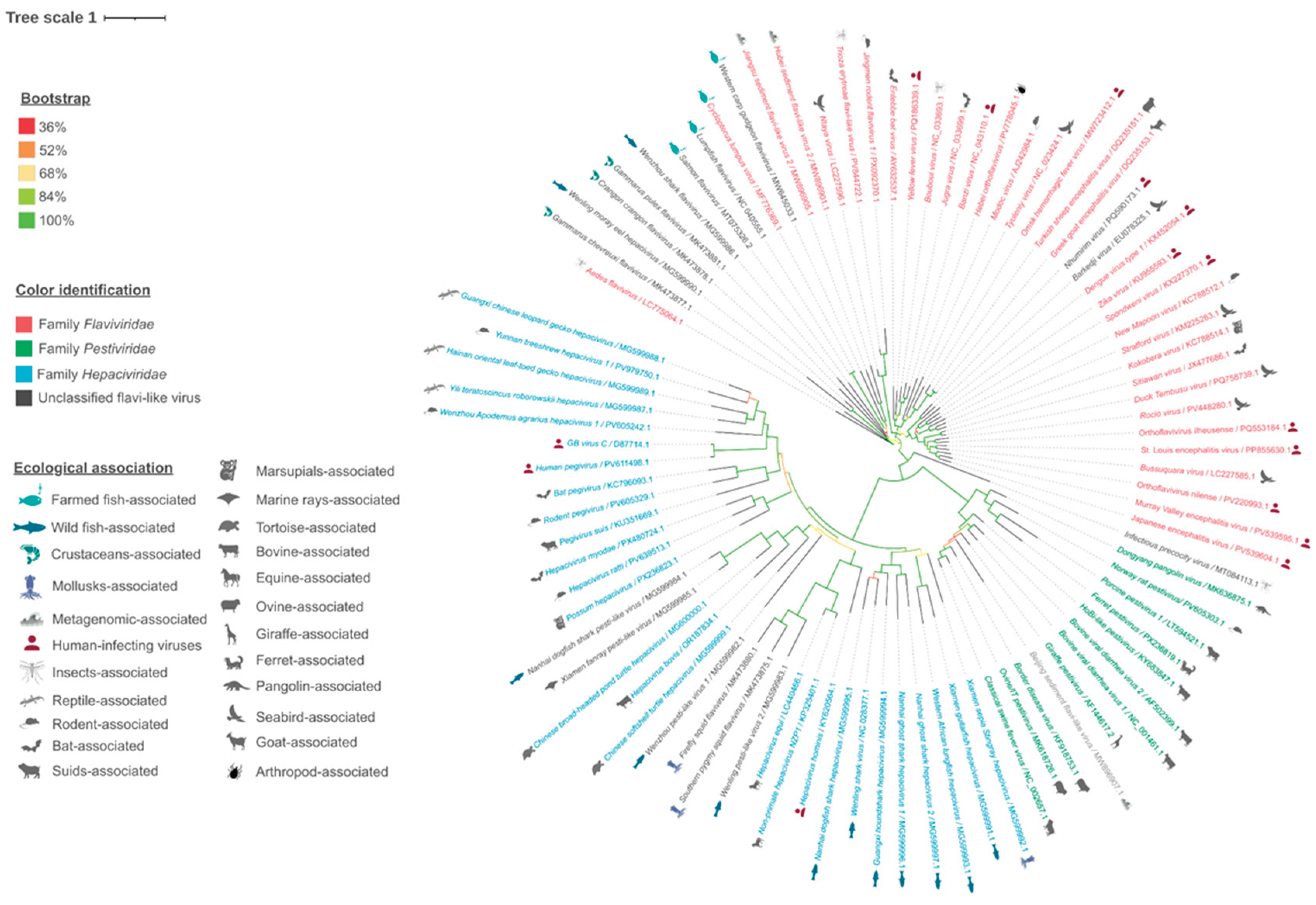
2. Virus Characteristics

The virions of classical flavivirids (i.e., the original family Flaviviridae) are spherical, enveloped, and 37-60 nm in diameter (Figure 1), with a positive-sense ssRNA genome of about 9.5 kb (hepaciviruses) to 12.5 kb (pestiviruses) lacking a 3’ poly(A) tail [3]. Infectious precocity virus (IPV) particles are 40-60 nm in diameter with a genome of 12,630 nt in length [13]. Only the genomes of members of the genus Orthoflavivirus contain a 5’-methylated nucleotide cap (to allow translation). In contrast, others have a genome-linked protein (VPg) and an internal ribosomal entry site (IRES). All flavivirids encode a single large polyprotein, which is cleaved at conserved sites by either the viral serine protease (NS2B/NS3), a host-derived signalase, except for pr/M, which is cleaved after assembly by host-derived furin or a furin-like protease [43]. Thus, the flavivirid polyprotein is effectively processed into at least ten viral proteins: nonstructural (NS) proteins NS1, NS2A, NS2B, NS3, NS4A, NS4B, and NS5, and structural proteins designated C (core) and M (membrane-like), and glycoprotein (E) [13,44,45]. The polyprotein in almost all classical flavivirids is translated from a single open reading frame (ORF) with structural proteins located on the 5’ end (Figure 2) [7].
Some members of the Orthoflavivirus genus, such as the JEV serogroup (e.g., JEV and WNV) (except St. Louis encephalitis virus (SLEV) which lacks the frameshift site [48]) [49,50], Tamanavirus genus (Tamana bat virus (TABV), and the highly divergent flavi-like viruses of aquatic animal (fish and crustacean amarilloviruses in Section 3 and Section 4 below) [10,13,21], have two ORFs (ORF1 and ORF2) and produce a “transframe” polyprotein utilizing program3med-1 ribosomal frameshifting (-1 PRF) (Figure 3) [7,13,48]. The eukaryotic mRNA signal for -1 frameshifting comprises two elements. The first element typically consists of a ‘slippery’ heptanucleotide sequence fitting the consensus motif X_XXY_YYZ, where XXX represents any three identical nucleotides; YYY represents AAA or UUU; Z represents A, C, or U; and underscores separate zero-frame codons [48,51]. In the tandem slippage model, the P-site anticodon repairs from XXY to XXX, whereas the A-site anticodon repairs from YYZ to YYY, thus allowing for perfect repairing except at the wobble position. Certain deviations from the canonical XXX of the slippery site are tolerated in the P-site, including UCC in some members of the JEV serogroup [48]. The second element is a 3’ stable RNA secondary structure, such as a pseudoknot or stem-loop, that is separated from the ‘slippery’ heptanucleotide sequence by a ‘spacer’ region typically of 5-9 nt [48,51]. In the JEV serogroup, frameshifting adds a 52 aa transframe C-terminal extension to the NS1 protein, which is not cleaved at the NS1|NS2A cleavage site, and thus frameshifting results in the production of a C-terminally extended version of NS1, known as NS1’ protein [50]. Members of the genera Orthoflavivirus, Hepacivirus, and Pegivirus have two E proteins (E dimers), whereas the genus Pestivirus has three (E trimers).
The segmented genomes of genera Guaicovirus and Jingmenvirus in the new family Flaviviridae encode up to seven structural proteins and two nonstructural proteins (NSP1 and NSP2), which have a high level of similarity with the nonstructural proteins NS5 (RNA-dependent RNA polymerase [RdRp] and methyltransferase [MTase] domains) and NS2B/NS3 (serine protease and helicase), respectively, of all other flavivirid genera (i.e., all genera with non-segmented genomes) [2,35].
A phylogenetic analysis of the polyprotein nucleotide sequences performed in this study is shown in Figure 4. The phylogenetic tree shows that the reported highly divergent flavi-like viruses of aquatic animals (fish and crustacean amarilloviruses in Section 3 and Section 4 below) are found in all three new families: Flaviviridae, Pestiviridae, and Hepaciviridae, although the grouping is not as clear-cut as with phylogenetic analysis of the RdRp gene [2].
Flavivirids multiply in the cytoplasm and mature into cytoplasmic vesicles derived from the endoplasmic reticulum (ER); assembled virions bud into the lumen of the ER and are secreted through the vesicle transport pathway by the cleavage of prM to M [7]; budding is not seen. Enveloped virions are released through exocytosis [43,52]. In cell cultures examined by electron microscopy, virus replication is commonly accompanied by a characteristic proliferation of intracellular membranes. Infectious precocity virus (IPV) found in farmed giant freshwater prawns, Macrobrachium rosenbergii, produced cytoplasmic inclusions in the eyestalk tissue from infected prawns [13].

3. Fish Amarilloviruses

It is now generally accepted that fish harbor a large and largely uncharacterized virome [19]. It follows that flaviviruses have been found in a variety of fish species. These viruses, which have been discovered among all four original Flaviviridae genera using metagenomics [18,19,37], occur in the three new families, Flaviviridae, Pestiviridae, and Hepaciviridae. Highly divergent amarilloviruses include the pathogenic Cyclopterus lumpus virus (CLuV) detected in moribund lumpfish (Cyclopterus lumpus) [20] in genus Tamanavirus, and Wenzhou shark flavivirus found in both a Pacific spadenose shark (Scoliodon macrorhynchos) [37] and a gazami crab (Portunus trituberculatus) [10], which group with the new family Flaviviridae but are not known to be transmitted by arthropods. The fish viruses that group with the new family Pestiviridae, i.e., fish pesti-like viruses for which full-length polyprotein gene sequences have been published, include Xiàmén fanray pesti-like virus detected in Platyrhina sp, Nanhai dogfish shark pesti-like virus in Squalus brevirostris, Wenzhou pesti-like virus 1 discovered in Sphyrna lewini, and Wēnlǐng pesti-like virus 2 discovered in Proscyllium habereri [19,37]. Fish viruses that group with the new family Hepaciviridae for which full-length polyprotein gene sequences have been published include Wenling shark virus (WLSV) discovered in the graceful catshark (Proscyllium habereri) [18], Wenling moray eel hepacivirus in Gymnothorax reticularis, Xiàmén sepia Stingray hepacivirus in Urolophus aurantiacus, Western African lungfish hepacivirus in Protopterus annectens, Guangxi houndshark hepacivirus in Mustelus manazo, and Nanhai dogfish shark hepacivirus in Squalus brevirostris, and Nanhai ghost shark hepacivirus 1 and 2 in Chimaera sp [19,37], longfin eel flavivirus and shortfin eel flavivirus 1 found in liver and gill samples of longfin eel (Anguilla dieffenbachii) and shortfin eel (Anguilla australis) respectively, in New Zealand and most closely related to Wenling moray eel hepacivirus [42]. The best-characterized fish viruses, most of which group with the new family Flaviviridae, genus Orthoflavivirus, are detailed in the following subsections. In addition, several flavi-like endogenous viral elements (EVEs) (i.e., non-retrovirus EVEs or ‘genomic fossil’) have been identified in various fish species [36,53].

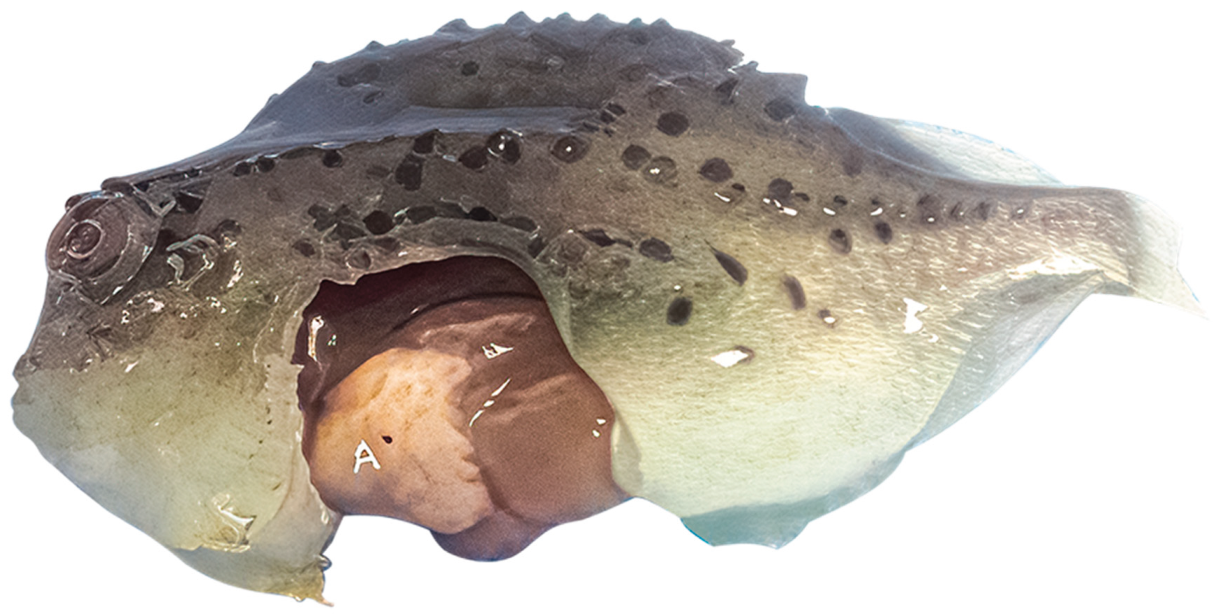
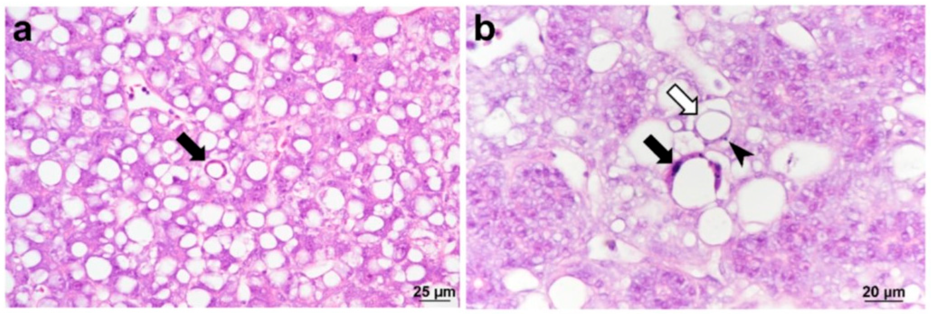
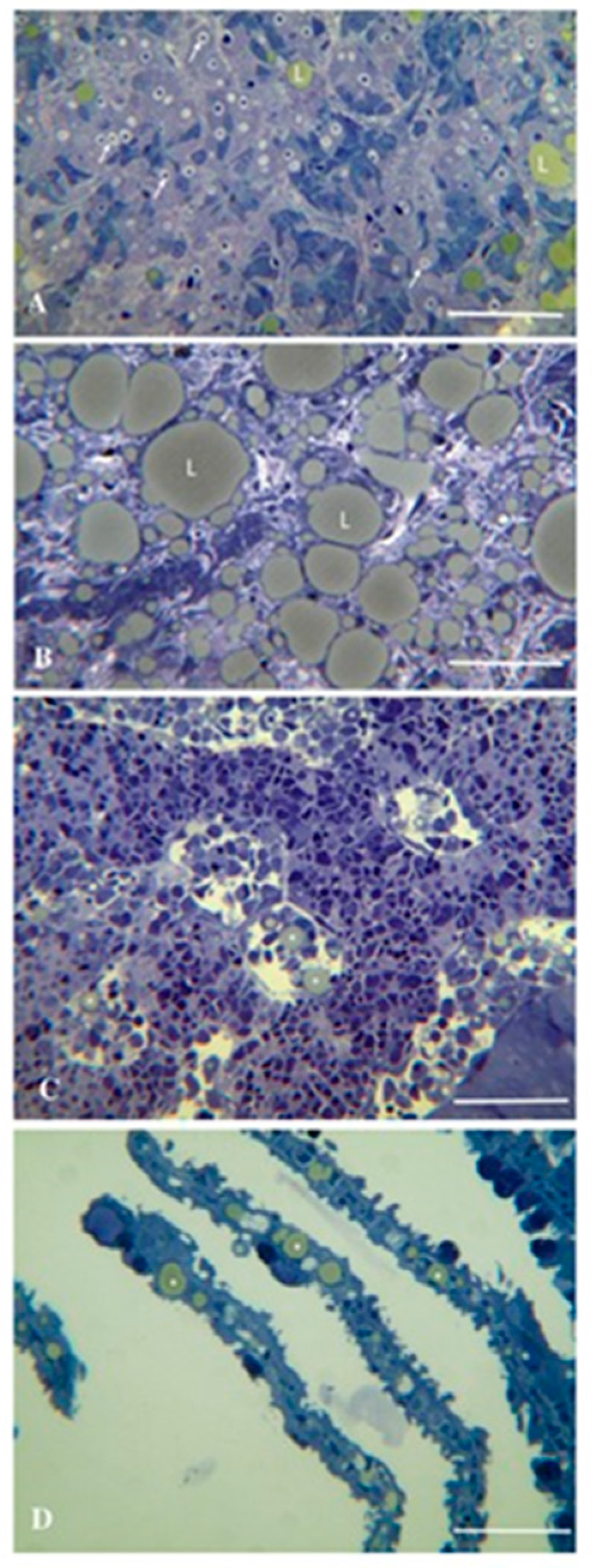
3.1. Cyclopterus Lumpus Virus (CLuV)

Cyclopterus lumpus virus (CLuV) is most similar to Tamana bat virus (TABV) [54], species Tamanavirus parnellis, which is classified in genus Tamanavirus in the new family Flaviviridae [2]. CLuV was the first fish flavivirus to be identified. CLuV was first detected in diseased farmed lumpfish (Cyclopterus lumpus) in 2015 in Western Norway by next-generation sequencing (NGS) [20]. The virus was anecdotally associated with mortalities of >50% and was present in all tissues tested, but pathology was primarily observed in the liver and kidneys. The second detection of CluV was in England using RT-qPCR developed by Skoge et al. [20] and conventional RT-PCR in September 2021 in consignments of lumpfish imported from Norway that experienced mortalities of up to 30% [33]. The fish showed typical clinical signs of CLuV-associated disease (such as increased mortalities, lethargy of pre-nursery fish attaching to the side or bottom of tanks, and loss of appetite) [33]. Gross pathology showed pale livers (Figure 5) and histopathology characterized by mild multifocal hepatocellular degeneration (Figure 6) [33] similar to that in the first CluV detection (Figure 7) [20].
Since 2008, lumpfish (Cyclopterus lumpus) have been used routinely as “cleaner fish” in marine farmed salmonids as a biological control method for sea lice (Lepeophtheirus salmonis) in Norway, UK, Ireland, Iceland, Faroes, and Canada; the process involves the use of wild-caught cleaner fish directly in the salmon farms or as broodstock for hatchery-raised cleaner fish, and are often translocated globally [55,56]. The virus in the second detection had 99.44% nucleotide sequence identity compared to the first [33]. However, in the absence of information on the prevalence of CLuV in the wild lumpfish populations globally, and with no molecular epidemiological data from Norway [57], it is not known if the virus had been introduced to the UK farm site via imported juveniles or transmitted to the farmed fish from a local source of the virus. Therefore, the virus may also be endemic in the UK [33].
CLuV cannot be isolated using common fish cell lines such as CHSE-14, ASK [20], BF, EPC, CHSE-214, or E-11 [33], in contrast to salmon flavivirus (SFV), which was isolated on the SSN-1 cell line [21] (see section 3.5 below). The lack of susceptibility of these cell lines to CluV suggests that its host range is fairly narrow, and there is a need to develop new cell lines from lumpfish to facilitate CLuV cultivation and pathogenesis studies.

3.2. Wenzhou Shark Flavivirus

A large-scale metatranscriptomic study discovered 214 vertebrate-associated viruses, including one flavivirus, the Wenzhou shark flavivirus, in the transcriptome of the Pacific spadenose shark (Scoliodon macrorhynchos) [37]. This virus was also detected in the transcriptome of healthy gazami crab or Japanese blue crab (Portunus trituberculatus), raising the hypotheses of horizontal transmission between the two distantly related hosts in the ocean ecosystem [10] and a correlate with the invertebrate-vertebrate relationship seen in flaviviruses of terrestrial animals [36]. An unconventional flavivirus, most similar to Wenzhou shark flavivirus, Parastichopus californicus flavivirus (PcaFV) was detected in Giant Pacific Sea Cucumber (Apostichopus californicus; formerly Parastichopus californicus) (Holothuroidea; Echinodermata) suffering from wasting disease [17], although the virus was subsequently shown not to cause pathology in its host [58]. It is unknown if Wenzhou shark flavivirus causes disease in sharks or crabs. However, in sharks infected with the virus, the virus was abundant throughout all tissues tested [10], and other aquatic animal flaviviruses such as CLuV [20,33] and IPV [13] are associated with disease in their natural hosts.

3.3. Eastern Red Scorpionfish Flavivirus

A metatranscriptomic study of seemingly healthy fish sold at a fish market in Sydney revealed fragments of a novel aquatic animal flavivirus, Eastern red scorpionfish flavivirus, in the Eastern red scorpionfish (Scorpaena jacksoniensis) gill sample [19]. Eastern red scorpionfish flavivirus polyprotein has 54% amino acid identity to Wenzhou shark flavivirus polyprotein [19].

3.4. Western Carp Gudgeon Flavivirus

Flaviviridae transcripts were detected in the western carp-gudgeon (Hypseleotris klunzingeri) in the Bogan River as part of the metatranscriptomic viral survey of invasive and native fishes across the Murray-Darling Basin in Australia [22]. These transcripts made up 2% of all vertebrate-associated viruses identified in the samples; the viral sequence exhibited 33-36% NS5 amino acid sequence similarity with other aquatic animal flaviviruses, Tamana bat virus (TABV) [23], species Tamanavirus parnellis, which is classified in genus Tamanavirus in the new family Flaviviridae [2,21,22].

3.5. Salmon Flavivirus (SFV)

Salmon flavivirus (SFV) is classified in Lineage Ie in the new family Flaviviridae [2]. SFV was isolated from diseased migrating adult Chinook salmon (Oncorhynchus tshawytscha) from the Eel River, California, USA, in November 2015 [21]. The affected fish observed showed signs of lethargy, congregating at the riverbanks, decreased avoidance of humans, and lenticular opacity due to cataract formation - associated with the presence of eye flukes Diplostomum sp. trematode metacercariae [21]. Examination of affected salmon revealed petechial hemorrhages in the optic lobes, cerebellums, and spinal cords that were also observed microscopically [21]. The necropsied fishes’ hearts, livers, spleens, kidneys, intestines, and skeletal muscles had no significant changes. The cytopathic virus was isolated from brain tissue using the striped-snakehead (SSN-1) fish cell line, and the complete genome sequence was obtained by next-generation sequencing and rapid amplification of cDNA ends [21]. However, experimental infection of fingerling rainbow trout (Oncorhynchus mykiss) and Chinook salmon by immersion and by intracoelomic injection routes resulted only in limited replication in tissues and no pathology, indicating that SFV is nonpathogenic to Chinook salmon and rainbow trout [21]. Interestingly, ongoing sampling in 2019 revealed the continued presence of the virus in California wild adult salmon, as the virus was isolated from the ovarian fluids of returning females [21].

4. Crustacean Amarilloviruses

Flaviviruses have been found in various marine crustacean species. Crustaflavivirus infeprecoquis (Infectious precocity virus, IPV) was identified by metatranscriptomic sequencing of samples from farmed giant freshwater prawns Macrobrachium rosenbergii suffering from iron prawn syndrome (IPS) [13]. Three putative flaviviruses were identified from wild-caught malacostracan crustaceans by querying assembled crustacean transcriptomes for flavivirus polyprotein sequences using the tblastn algorithm [10]: Crangon crangon flavivirus (CcFV), species Orthoflavivirus alphei, genus Orthoflavivirus [2], was identified in the brown shrimp (Crangon crangon) from midgut samples originating from Weser estuary, Germany [30], Gammarus chevreuxi flavivirus (GcFV) was identified in transcriptomes from two publications on Gammarus chevreuxi in both embryonic and adult samples originating from the Plym estuary, Plymouth, United Kingdom [39,40] and in Gammarus pulex flavivirus (GpFV) identified from a male Gammarus pulex wild-caught from the Bourbre River, France [41]. The polyproteins of these viruses were more closely related in amino acid identity to the genus Orthoflavivirus [10]. Changjiang Jingmen-like virus was identified in crayfish [18], and is classified in the genus Jingmenvirus in the new family Flaviviridae [2].

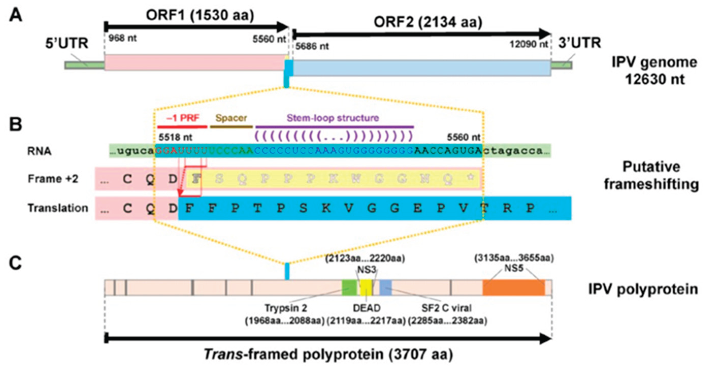
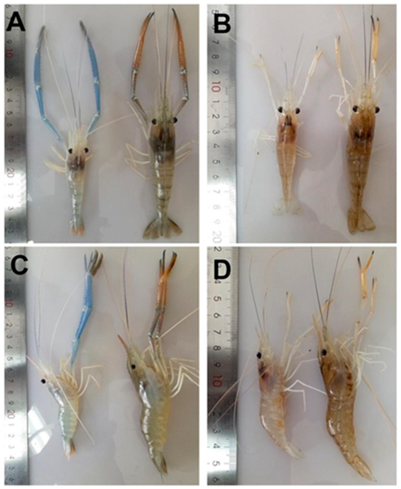
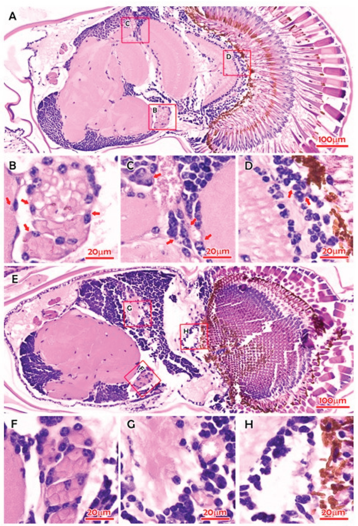
Crustaflavivirus Infeprecoquis (Infectious Precocity Virus, IPV)

Crustaflavivirus infeprecoquis (Infectious precocity virus, IPV) causes iron prawn syndrome (IPS) in farmed giant freshwater prawns Macrobrachium rosenbergii, characterized by clinical signs of sexual precocity and stunted growth, which were first observed on farms in Jiangsu Province, China, during 2018 to 2020. IPV was shown to belong to a new genus named Crustaflavivirus, to denote a virus genus in the family Flaviviridae first identified in crustaceans [13], which corresponds to Lineage Ij in the new family Flaviviridae [2].
IPS has been frequently reported in populations of farmed M. rosenbergii in China since 2010 [13,59,60]. China is the world’s largest M. rosenbergii production country [61]. IPS is part of the growth retardation disease (GRD) in China, characterized by precocity-associated growth retardation [13,62], which results in substantial production losses in the prawn farming industry [63]. Infected pawns grow to only 5–6 cm, resulting in a 50% loss of yield. The affected female prawns show precocious puberty, i.e., they begin holding eggs when they are up to only 5 cm in body size [62]. The affected males engage in mating behaviors and have two elongated front claws (2nd pereiopod) when their body length is about 6 cm, while normal mature prawns are 8 to 10 cm long [62]. The condition could be reproduced experimentally by immersion of M. rosenbergii post larvae in filtered IPV preparation of the IPS-affected prawns. However, clinical signs did not manifest for at least 22–25 weeks [13]. Figure 8 shows the gross signs of IPS-affected M. rosenbergii [13]. Note the elongated blue claws and blue tailfins of the affected prawns; the infected prawns were significantly (P < 0.05) smaller than those in the control group after the 22nd week postinfection. Figure 9 shows the histopathology in IPS-affected M. rosenbergii, including eosinophilic viral inclusions in multiple tissues [13,60]. In-situ hybridization using IPV digoxigenin (DIG)-labeled RNA probe found hybridization signals corresponding with the histopathology in the compound eyes from IPS-affected M. rosenbergii [13].
IPS is a persistent, long-term infection characterized by subtle morphological changes that are difficult to detect [64]. It does not cause significant mortality, and clinical signs only occur in the post larvae, making clinical diagnosis extremely difficult as the retardation in growth needs to be differentiated from the normal development and maturation of prawns in the population. Histopathological examination is more reliable as the virus forms eosinophilic cytoplasmic inclusions in the cells of the lamina ganglionaris and fasciculated zone, as well as neurosecretory cells in the bellonci organ and globuli cells in the hemielipsoid body [13].
Dong et al. [13] developed a nested reverse transcription-PCR diagnostic assay specific for IPS-affected prawns that can be used for quarantine inspection for IPV in the transboundary trade of live M. rosenbergii and enhanced surveillance of IPV in aquaculture in China and globally. The nested RT-PCR assay primers consisted of outer primers IPV_F1 (5′-GCA CAC TCC CAA CAC GTT TC-3′) and IPV_R1 (5′-CGC GCG TAA TCT CTA CAC CT-3′), generating a 1,038-bp product and inner primers IPV_F2 (5′-TCC CTA GGC AGG GGA TAC TG-3′) and IPV_R2 (5′-AGC TAT CCG TGG TGT GGA AC-3′), generating a 395-bp product [13]. Wang et al. [64] developed a semiquantitative approach for diagnosing IPS based on clinical signs. They established a rapid one-step, highly sensitive, and highly specific reverse-transcription quantitative PCR (TaqMan-RT-qPCR) method to detect and quantify IPV. The specific primers were IPV-F (5′ -AGG AGA GGG TTT TGG CTT G-3′ ) and IPV-R (5’-CTG GAT TGG AAG GGA ACT CTG-3′ ), which generate a 139-bp product, and the TaqMan probe was IPV-P: 5’-[6FAM]-CCG CGA CAC TTA CAA CTG CCC TT-[TAMRA]-3′ [64]. The detection limit of the TaqMan-RT-qPCR method was as low as 1.00 × 100 copy/reaction. This assay, about 13 and 1300 times more sensitive than the nested RT-PCR assay, revealed the characteristic neurotropism of IPV with the highest loads in eyestalks and the brain [64]. More recently, Chiang et al. [65] used an immunohistochemistry (IHC) assay to localize IPV in haemocytes in the hepatopancreas, gills, and pleopods and in support cells within the lamina ganglionaris as well as in neurosecretory cells within the bellonci organ, sinus glands, and the X organ of infected prawns with or without clinical signs. Chiang et al. [65] then used RT-qPCR with four different primer pairs (6872F 5’-AAG AAT TCG GAG TCT ATG TTG ACG GC-3’ and 7396R 5’-AAC TCG AGC ACT TTC CTA CCC G-3’, 525 bp product; 6872F 5’-GGA GTC TAT GTT GAC GGC TCT ATC T-3’ and 7127R 5’-GTT GGT GAA CCT ATG ATC CTC TTA-3’, 256 bp product; 7396F 5’-GCT GAG AAA GCG GGT AGG AAA GT-3’ and 7601R 5’-ACC ACG ACA ACA TCA TAG GTA AAG G-3’, 205 bp product; 7828F 5’-GTG TTC TGT CCT AGT GCA GTT GG-3’ and 8048R 5’-CGC GCC ATG AAG CCA TAA TAA C-3’, 220 bp product) and the primer pair of Dong et al. [13] and SYBR Green detection to quantitate viral loads in in prawns identified as IPV-positive and IPV-negative based on the IHC assay. IHC-positive prawns had viral loads of >103.5 copies/μg compared to <103.5 copies/μg in IHC-negative prawns [42]. Moreover, the highest viral loads were in pleopods, which can serve as non-lethal samples for IPS diagnosis [65].
The IPV TaqMan-RT-qPCR assay [64] detected low copies of IPV RNA in red swamp crayfish Procambarus clarkii samples [62], suggesting that P. clarkii if in the same breeding pond with IPS-affected M. rosenbergii may be a reservoir of IPV. Moreover, P. clarkii also has clinical signs similar to IPS, such as sexual precocity and stunted growth [66]. However, whether these clinical signs are caused by infection with IPV needs to be further verified [64]. Zhao et al. [67] developed a TaqMan probe RT-qPCR and a nested RT-PCR assays that were then used to determine the host range of IPV in China. The first step of nested RT-PCR used primers IPV-F1 5′-GCC TCC ACA TCA TTG GCT TCG-3′ and IPV-R1 5′- TCG GGT GTC ATC AAC AAA CTCATA-3’ generating a 754-bp product, and the second step primers were IPV-F2 5′-ACA TCA TTG GCT TCG TAT-3′ and IPV-R2 5′-ACA GAG CAG GAG ATT GGA-3′ generating a 395-bp product [67]. The TaqMan probe RT-qPCR and primer sequences were as follows: primers IPVq-F 5′-GAA GAT GTC ATC GTC CCA GAG TT-3′ and IPVq-R 5′-GGA ATG CCC CCT CCG TAT-3′, and the probe 5′-CCC CAA GGT TTT ATT G-3′ [67]. IPV was detected in crustaceans M. rosenbergii, M. nipponense, Procambarus clarkii, Litopenaeus vannamei, Penaeus monodon, and Oratosquilla oratoria and insect Anisops kuroiwae [67]. IPV was not detected in fishes Misgurnus anguillicaudatus, Carassius auratus, Oreochromis mossambicus, Micropterus salmoides, and Pangasius bocourti; crabs Eriocheir sinensis and Scylla paramamosain; and malacostraca Charybdis feriatus [67]. In M. rosenbergii, IPV mainly infected the brain, abdominal nerve, integument, and gill [67]. Regardless, the high sensitivity of the IPV TaqMan-RT-qPCR assay [64,67] is very useful for screening all potential sources of introduction of IPV into M. rosenbergii aquaculture [60].
It was recently reported that IPV occurs in two distinct phylogenetic clades, the Southeast Asian clade (represented by isolates 01/SEA/202305 and 02/SEA/202401, GenBank Acc. No. PQ786402 and PQ786403, respectively) and the Chinese clade (represented by isolates MR2018, ZJJS2019, ZJHY201110, and JSYZ20170815, GenBank Acc. Nos MT084113.1, ON382579.1, MT648663.1, and MT648664.1, respectively) [68]. The recent IPV isolate, 01/SH/202210 (GenBank Acc. No. PQ786404), from China exhibited features from both groups, suggesting that it may represent an evolutionary intermediate between the two IPV lineages [68].

5. Concluding Remarks

The increased application of viral metagenomics and sequencing activity to commercially important fisheries and aquaculture samples has led to the discovery of novel viruses in aquatic animals thereby broadening the host range of several viral families of terrestrial vertebrate viruses, in this case contributing to the reorganization of the family Flaviviridae and the incorporation of the highly divergent flavi-like viruses into three new families, Flaviviridae, Pestiviridae, and Hepaciviridae in the established order Amarillovirales. This review makes it evident that a wide range of amarilloviruses occur in aquatic animals. Some, such as Cyclopterus lumpus virus (CLuV) detected in moribund lumpfish (Cyclopterus lumpus), and Infectious precocity virus (IPV) found in iron prawn syndrome (IPS)-affected farmed giant freshwater prawns, Macrobrachium rosenbergii, are important causes of disease in the aquaculture industry and should be considered alongside other flavivirids of human and veterinary medical importance.

Author Contributions

Conceptualization, F.K; investigation, data curation, writing—original draft preparation, F.K. and D.V; writing—review and editing, F.K. M.K. D.V. and M.G. All authors have read and agreed to the published version of the manuscript.

Data Availability Statement

The original contributions presented in the study are included in the article, further inquiries can be directed to the corresponding author.

Conflicts of Interest

The authors declare no conflicts of interest.

References

  1. Simmonds, P., Butković, A., Grove, J. et al. 2025a. Taxonomic expansion and reorganization of Flaviviridae. Nat Microbiol 10, 3026-3037. [CrossRef]
  2. Simmonds, P., Butkovic, A., Grove, J., Mayne, R., Mifsud, J.C.O., Beer, M., Bukh, J., Drexler, J.F., Kapoor, A., Lohmann, V., Smith, D.B., Stapleton, J.T., Vasilakis, N., Kuhn, J.H. 2025b. Reorganization of Flaviviridae (order Amarillovirales) and classification of ‘flavi-like’ viruses into three families, 12 genera, and 3 subgenera. 2025.006S.Ac.v2.Amarillovirales_3reorgfam. Available online: https://ictv.global/system/files/proposals/pending/2025/Animal%20%2BssRNA%20%28S%29%20proposals/2025.006S.Ac.v2.Amarillovirales_3reorgfam.docx (accessed on December 23, 2025).
  3. Snyder, J. E., Jose, J., Kuhn, R. J., 2014. The Togaviridae and the Flaviviridae, Reference Module in Biomedical Sciences, Elsevier. [CrossRef]
  4. Choo, Q-L., Richman, K. H., Han, J. H., Berger, K., Lee, C. et al. Genetic organization and diversity of the hepatitis C virus. Proc Natl Acad Sci U S A 1991;88(6):2451-2455. [CrossRef]
  5. Postler, T.S., Beer, M., Blitvich, B.J. et al. 2023. Renaming of the genus Flavivirus to Orthoflavivirus and extension of binomial species names within the family Flaviviridae. Archives of Virology 168, 224. [CrossRef]
  6. Porter, A. F., Pettersson, J. H. O., Chang, W. S., Harvey, E., Rose, K., Shi, M. et al (2020) Novel hepaci- and pegi-like viruses in native Australian wildlife and non-human primates. Virus Evol 6(2). [CrossRef]
  7. Simmonds, P., Becher, P., Bukh, J., Gould, E. A., Meyers, G., Monath, T., Muerhoff, S., Pletnev, A., Rico-Hesse, R., Smith, D. B., Stapleton, J. T., & ICTV Report Consortium., 2017. ICTV Virus Taxonomy Profile: Flaviviridae. The Journal of General Virology, 98(1), 2-3. [CrossRef]
  8. Petrone, M. E., Grove, J., Mélade, J., Mifsud, J. C. O., Parry, R. H. et al. 2024. A ~40-kb flavi-like virus does not encode a known error-correcting mechanism. Proc Natl Acad Sci USA, 121(30):e2403805121. [CrossRef]
  9. Mifsud, J. C. O., Costa, V, A., Petrone, M. E., Marzinelli, E. M., Holmes, E. C. et al. 2023. Transcriptome mining extends the host range of the Flaviviridae to non-bilaterians. Virus evolution 9(1):veac124. [CrossRef]
  10. Parry, R., Asgari, S., 2019. Discovery of novel crustacean and cephalopod flaviviruses: insights into the evolution and circulation of flaviviruses between marine invertebrate and vertebrate hosts. Journal of Virology 93:e00432-19. [CrossRef]
  11. Lay, C. L., Shi, M., Buček, A., Bourguignon, T., Lo, N. et al. 2020. Unmapped RNA Virus Diversity in Termites and their Symbionts. Viruses, 12(10). [CrossRef]
  12. Paraskevopoulou, S., Käfer, S., Zirkel, F. et al. 2021. Viromics of extant insect orders unveil the evolution of the flavi-like superfamily. Virus Evol 7:veab030. [CrossRef]
  13. Dong, X., Wang, G., Hu, T., Li, J., Li, C., Cao, Z., Shi, M., Wang, Y., Zou, P., Song, J., Gao, W., Meng, F., Yang, G., Tang, K. F. J., Li, C., Shi, W., Huang, J., 2021. A Novel Virus of Flaviviridae Associated with Sexual Precocity in Macrobrachium rosenbergii. mSystems 6(3):e0000321. [CrossRef]
  14. Bekal, S., Domier, L. L., Gonfa, B., McCoppin, N. K., Lambert, K. N. et al. 2014. A novel flavivirus in the soybean cyst nematode. J Gen Virol 95(Pt 6):1272-1280. [CrossRef]
  15. Dheilly, N. M., Lucas, P., Blanchard, Y., Rosario, K. A. 2022. World of Viruses Nested within Parasites: Unraveling Viral Diversity within Parasitic Flatworms (Platyhelminthes). Microbiol Spectr 10(3):e0013822. [CrossRef]
  16. Chen, Y-M., Sadiq, S., Tian, J-H., Chen, X., Lin, X-D. et al. 2022. RNA viromes from terrestrial sites across China expand environmental viral diversity. Nat Microbiol 7(8):1312-1323. [CrossRef]
  17. Hewson I., Johnson M. R., Tibbetts I. R., 2020. An unconventional flavivirus and other RNA viruses in the sea cucumber (Holothuroidea; Echinodermata) virome. Viruses 12, 1057. [CrossRef]
  18. Shi, M., Lin, X-D., Vasilakis, N., Tian, J-H., Li, C-X., Chen, L-J., Eastwood, G., Diao, X-N., Chen, M-H., Chen, X., Qin, X-C., Widen, S. G., Wood, T. G., Tesh, R. B., Xu, J., Holmes, E. C., Zhang, Y-Z., 2016. Divergent viruses discovered in arthropods and vertebrates revise the evolutionary history of the Flaviviridae and related viruses. Journal of Virology, 90, 659-669. [CrossRef]
  19. Geoghegan, J. L., Di Giallonardo, F., Cousins, K., Shi, M., Williamson, J. E., Holmes, E. C., 2018. Hidden diversity and evolution of viruses in market fish. Virus evolution, 4(2), vey031. [CrossRef]
  20. Skoge, R.H., Brattespe, J., Økland, A.L. et al. 2018. New virus of the family Flaviviridae detected in lumpfish (Cyclopterus lumpus). Archives of Virology 163:679-685. [CrossRef]
  21. Soto, E., Camus, A., Yun, S., Kurobe, T., Leary, J. H., Rosser, T. G., Dill-Okubo, J. A., Nyaoke, A. C., Adkison, M., Renger, A., Ng, T. F. F., 2020. First Isolation of a Novel Aquatic Flavivirus from Chinook Salmon (Oncorhynchus tshawytscha) and Its In Vivo Replication in a Piscine Animal Model. Journal of Virology 94(15):e00337-20. [CrossRef]
  22. Costa, V. A., Mifsud, J. C. O., Gilligan, D., Williamson, J. E., Holmes, E. C., Geoghegan, J. L., 2021. Metagenomic sequencing reveals a lack of virus exchange between native and invasive freshwater fish across the Murray-Darling Basin, Australia. Virus evolution, 7(1), veab034. [CrossRef]
  23. Parry, R. H., Slonchak, A, Campbell, L. J., Newton, N. D., Debat, H. J., Gifford, R. J., Khromykh, A. A. 2023. A novel tamanavirus (Flaviviridae) of the European common frog (Rana temporaria) from the UK. J Gen Virol. 104(12):001927. [CrossRef]
  24. Urayama, S-I., Takaki, Y., Nunoura, T. 2016. FLDS: A Comprehensive dsRNA Sequencing Method for Intracellular RNA Virus Surveillance. Microbes Environ 31(1):33-40. [CrossRef]
  25. Forgia, M., Daghino, S., Chiapello, M., Ciuffo, M., Turina, M. 2024. New clades of viruses infecting the obligatory biotroph Bremia lactucae representing distinct evolutionary trajectory for viruses infecting oomycetes. Virus evolution, 10(1):veae003. [CrossRef]
  26. Debat, H., Bejerman, N. 2023. Two novel flavi-like viruses shed light on the plant-infecting koshoviruses. Arch Virol 168:184. [CrossRef]
  27. Zhang, S., Yang, C., Qiu, Y., Liao, R., Xuan, Z. et al. 2024. Conserved untranslated regions of multipartite viruses: Natural markers of novel viral genomic components and tags of viral evolution. Virus evolution, 10(1):veae004. [CrossRef]
  28. Schönegger, D., Marais, A., Faure, C. et al. 2022. A new flavi-like virus identified in populations of wild carrots. Arch Virol 167:2407–09. [CrossRef]
  29. Nasir, A., Romero-Severson, E., Claverie, J. M. 2020. Investigating the Concept and Origin of Viruses. Trends Microbiol 28(12):959-967. [CrossRef]
  30. Matsumura, E. E., Nerva, L., Nigg, J. C., Falk, B. W., Nouri, S. 2016. Complete Genome Sequence of the Largest Known Flavi-Like Virus, Diaphorina citri flavi-like virus, a Novel Virus of the Asian Citrus Psyllid, Diaphorina citri. Genome Announc 4(5). [CrossRef]
  31. Kartashov, M. Y., Gladysheva, A. V., Shvalov, A. N., Tupota, N. L., Chernikova, A. A. et al. 2023. Novel Flavi-like virus in ixodid ticks and patients in Russia. Ticks Tick Borne Dis 14(2):102101. [CrossRef]
  32. Atsumi, G., Tomita, R., Kobayashi, K. et al. 2013. Prevalence and genetic diversity of an unusual virus associated with Kobusho disease of gentian in Japan. J Gen Virol 94:2360–65. [CrossRef]
  33. Edwards, M., Bignell, J. P., Papadopoulou, A., Trani, E., Savage, J., Joseph, A. W., Wood, G., Stone, D. M., 2022. First Detection of Cyclopterus Lumpus Virus in England, Following a Mortality Event in Farmed Cleaner Fish. Bulletin of the European Association of Fish Pathologists 43(1): 28-37. [CrossRef]
  34. Qin, X. C., Shi, M., Tian, J. H., Lin, X. D., Gao, D. Y., He, J. R., Wang, J. B., Li, C. X., Kang, Y. J., Yu, B., Zhou, D. J., Xu, J., Plyusnin, A., Holmes, E. C., Zhang, Y. Z., 2014. A tick-borne segmented RNA virus contains genome segments derived from unsegmented viral ancestors. Proceedings of the National Academy of Sciences of the United States of America, 111(18):6744-6749. [CrossRef]
  35. Colmant, A. M. G., Charrel, R. N., Coutard, B., 2022. Jingmenviruses: Ubiquitous, understudied, segmented flavi-like viruses. Frontiers in Microbiology 13:997058. [CrossRef]
  36. Lensink, M. J., Li, Y., Lequime, S., Pierson, T. C., 2022. Aquatic Flaviviruses. Journal of Virology 96(17):e00439-22. [CrossRef]
  37. Shi, M., Lin, X. D., Chen, X., Tian, J. H., Chen, L. J., Li, K., Wang, W., Eden, J. S., Shen, J. J., Liu, L., Holmes, E. C., Zhang, Y. Z., 2018. The evolutionary history of vertebrate RNA viruses. Nature 556(7700), 197-202. [CrossRef]
  38. Martínez-Alarcón, D., Harms, L., Hagen, W., Saborowski, R., 2019. Transcriptome analysis of the midgut gland of the brown shrimp Crangon crangon indicates high polymorphism in digestive enzymes. Marine Genomics 43:1-8. [CrossRef]
  39. Truebano, M., Tills, O., Spicer, J. I., 2016. Embryonic transcriptome of the brackishwater amphipod Gammarus chevreuxi. Marine Genomics 28:5-6. [CrossRef]
  40. Collins, M., Tills, O., Spicer, J. I., Truebano, M., 2017. De novo transcriptome assembly of the amphipod Gammarus chevreuxi exposed to chronic hypoxia. Mar Genom 33:17-19. [CrossRef]
  41. Trapp, J., Geffard, O., Imbert, G., Gaillard, J. C., Davin, A. H., Chaumot, A., Armengaud, J., 2014. Proteogenomics of Gammarus fossarum to document the reproductive system of amphipods. Mol Cell Proteomics 13:3612–3625. [CrossRef]
  42. Waller, S.J., Egan, E., Crow, S. et al., 2024. Host and geography impact virus diversity in New Zealand’s longfin and shortfin eels. Archives of Virology, 169:85. [CrossRef]
  43. Murugesan, A., Manoharan, M., 2020. Dengue Virus. Emerging and Reemerging Viral Pathogens, 281–359. [CrossRef]
  44. Westaway, E. G., Brinton, M. A., Gaidamovich Sya, Horzinek, M. C., Igarashi, A., Kääriäinen, L., Lvov, D. K., Porterfield, J. S., Russell, P. K., Trent, D. W., 1985. Flaviviridae. Intervirology 24(4):183-192. [CrossRef]
  45. Lindenbach, B. D., Murray, C. L., Thiel, H-J., Rice, C. M., 2013. Flaviviridae, p 712–746. In Knipe, D. M., Howley, P. M., Cohen, J. I., Griffin, D. E., Lamb, R. A., Martin, M. A., Racaniello, V. R., Roizman, B. (ed), Fields virology, 6th ed. Lippincott Williams & Wilkins, Philadelphia, PA.
  46. ViralZone Flaviviridae. Available at: https://viralzone.expasy.org/43 (accessed May 27, 2024).
  47. Taniguchi, S., 2019. Detection of Jingmen tick virus in human patient specimens: emergence of a new tick-borne human disease? EBioMedicine 43:18-19. [CrossRef]
  48. Moureau, G., Cook, S., Lemey, P., Nougairede, A., Forrester, N. L., Khasnatinov, M., Charrel, R. N., Firth, A. E., Gould, E. A., de Lamballerie, X., 2015. New Insights into Flavivirus Evolution, Taxonomy and Biogeographic History, Extended by Analysis of Canonical and Alternative Coding Sequences. PLoS ONE 10(2):e0117849. [CrossRef]
  49. Firth, A. E., Blitvich, B. J., Wills, N. M., Miller, C. L., Atkins, J. F., 2010. Evidence for ribosomal frameshifting and a novel overlapping gene in the genomes of insect-specific flaviviruses. Virology, 399(1), 153–166. [CrossRef]
  50. Melian, E. B., Hinzman, E., Nagasaki, T., Firth, A. E., Wills, N. M., Nouwens, A. S., Blitvich, B. J., Leung, J., Funk, A., Atkins, J. F., Hall, R., Khromykh, A. A., 2010. NS1’ of flaviviruses in the Japanese encephalitis virus serogroup is a product of ribosomal frameshifting and plays a role in viral neuroinvasiveness. Journal of Virology 84(3):1641-1647. [CrossRef]
  51. Firth, A. E., Brierley, I., 2012. Non-canonical translation in RNA viruses. Journal of General Virology 93:1385-1409. [CrossRef]
  52. Mukhopadhyay, S., Kuhn, R. J., Rossmann, M. G., 2005. A structural perspective of the flavivirus life cycle. Nature reviews. Microbiology 3(1):13-22. [CrossRef]
  53. Bamford CGG, de Souza WM, Parry R, Gifford RJ. 2022. Comparative analysis of genome-encoded viral sequences reveals the evolutionary history of flavivirids (family Flaviviridae), Virus Evolution, Volume 8, Issue 2, 2022, veac085. [CrossRef]
  54. Parry RH, Slonchak A, Campbell LJ, Newton ND, Debat HJ et al. A novel tamanavirus (Flaviviridae) of the European common frog (Rana temporaria) from the UK. J Gen Virol 2023;104(12).
  55. Imsland, A. K., Reynolds, P., Eliassen, G., Hangstad, T. A., Nytrø, A. V., Foss, A., Vikingstad, E., Elvegård, T. A., 2014. Assessment of Growth and Sea Lice Infection Levels in Atlantic Salmon Stocked in Small-Scale Cages with Lumpfish. Aquaculture 433:137-142. [CrossRef]
  56. Haugland, G. T., Imsland, A. K. D., Reynolds, P., Treasurer, J., 2020. Chapter 10 Application of biological control: use of cleaner fish. In: Kibenge, F. S. B., and Powell, M. D. (Eds.), Aquaculture Health Management: Design and Operational Approaches. Elsevier, Amsterdam. pp. 319-369. [CrossRef]
  57. The Health Situation in Norwegian Aquaculture 2020. Published by the Norwegian Veterinary Institute 2021. Available on line, Sommerset, I., Jensen, B. B., Bronø, G., Haukaas, A., Brun, E. (Eds.). 2021. The Health Situation in Norwegian Aquaculture 2020. Published by the Norwegian Veterinary Institute 2021. Available on line: https://www.vetinst.no/rapporter-og-publikasjoner/rapporter/2022/fish-health-report-2021/_/attachment/inline/3a529735-e485-4bf7-b05b-5677a46b6b09:31cb9146509882b4dc2c60dc915a1be6195ae015/Norwegian%20Fish%20Health%20Report%202021.pdf (accessed December 27 2025).
  58. Crandell JG, Altera AK, DeRito CM, Hebert KP, Lim EG, Markis J, Philipp KH, Rede JE, Schwartz M, Vilanova-Cuevas B, Wang E and Hewson I., 2023. Dynamics of the Apostichopus californicus-associated flavivirus under subtoxic conditions and organic matter amendment. Frontiers in Marine Science 10:1295276. [CrossRef]
  59. Jiang, Q., Qian, L., Gu, S., Guo, X., Zhang, X., Sun, L., 2020. Investigation of growth retardation in Macrobrachium rosenbergii based on genetic/epigenetic variation and molt performance. Comparative Biochemistry and Physiology Part D: Genomics and Proteomics, 35:100683. [CrossRef]
  60. Hooper, C., Debnath, P. P., Stentiford, G. D., Bateman, K. S., Salin, K. R., Bass, D., 2022. Diseases of the giant river prawn Macrobrachium rosenbergii: A review for a growing industry. Reviews in Aquaculture 1-21. [CrossRef]
  61. FAO, Fisheries and Aquaculture, Global production by production source Quantity (1950–2022). Available at: https://www.fao.org/fishery/statistics-query/en/global_production/global_production_quantity (accessed July 23, 2024).
  62. Zhou D, Liu S, Guo G, He X, Xing C, Miao Q, Chen G, Chen X, Yan H, Zeng J, Zheng Z, Deng H, Weng S, He J. 2022. Virome Analysis of Normal and Growth Retardation Disease-Affected Macrobrachium rosenbergii. Microbiology Spectrum 10(6):e0146222. [CrossRef]
  63. Yuan, R., Zhang, Z. H., Chen, H., Fang, P., Chen, J., Liu, X. M., Wu, Y. F., Wang, J. J., 2017. Phenomenon and research progress on prevention and control of “Iron Shell” in giant freshwater prawn Macrobrachium rosenbergii. Fish Sci 36:383-390.
  64. Wang, G., Guo, X., Huang, X., Wang, D., Chen, Y., Qin, J., Yang, G., Tang, K. F. J., Dong, X., Huang, J., 2023. The quantitative characteristics of infection with infectious precocity virus (IPV) revealed with a new TaqMan probe-based real-time RT-PCR method. [CrossRef]
  65. Chiang, Y-R., Lu, Y-Y., Lin, H-Y. 2024. Quantitative analysis of infectious precocity virus load and stunted growth syndrome via an immunohistochemical assay and reverse transcriptase quantitative real-time PCR in Macrobrachium rosenbergii. Aquaculture 583, 740577. [CrossRef]
  66. Tan, H. Y., Shao, G. M., Kang, P. F., Wang, Y. F., 2017. Full-length normalization subtractive hybridization analysis provides new insights into sexual precocity and ovarian development of red swamp crayfish Procambarus clarkii. Aquaculture 468, 417–427. [CrossRef]
  67. Zhao, C., Miu, Q., Liu, S., Zhou, D., He, X., Pang, J., Weng, S., He, J. 2023. Detection methods, epidemiological investigation, and host ranges of infectious precocity virus (IPV). Aquaculture 562, 738818. [CrossRef]
  68. Zhou, C., Wang, G., Zhou, Q.; Meng, F.; Liu, S.; Huang, J.; Dong, X. 2025. Surveillance and Genomic Evolution of Infectious Precocity Virus (IPV) from 2011 to 2024. Viruses 17, 425. [CrossRef]
Figure 1. Schematic of the Flaviviridae virus particle. The left particle is a cross-section with the viral components labeled. Members of the old genera Orthoflavivirus, Hepacivirus, and Pegivirus have two E proteins (E dimers), whereas the genus Pestivirus has three (E trimers). The right particle shows the surface proteins arranged in an icosahedral-like symmetry. (Reproduced from [46]. Source: SwissBioPics. The images are licensed under a Creative Commons Attribution 4.0 International (CC BY 4.0) License https://creativecommons.org/licenses/by/4.0/).
Preprints 193914 g001
Figure 2. Genome organization and polyprotein processing of most classical flavivirids. Boxes below the genome indicate viral proteins generated by proteolytic processing. NCR, non-coding region. (Reproduced from [7], Figure 2, an open-access article distributed under the terms of the https://creativecommons.org/licenses/by/4.0/).
Preprints 193914 g002
Figure 3. Production of a “transframe” polyprotein from two open reading frames (ORFs) utilizing programmed-1 ribosomal frameshifting (-1 PRF), using the IPV genome as an example. (A) The full-length genome of IPV with two predicted ORFs, 59 and 39 UTRs, and potential translational changes caused by the putative frameshifting are indicated. The light yellow block represents the frameshift-off region, taken over by the sky-blue block representing the frameshift-on region. The aqua column indicates the RNA sequence region, which is zoomed out in panel B. (B) The putative frameshifting process. Nucleotide-level details of the aqua column of panel A are presented with the capital RNA sequence with aqua background, in which red letters indicate the potential 21 programmed ribosomal frameshifting (21 PRF), coffee letters indicate the 6-nt spacer, and dark magenta letters indicate the potential stem-loop structure. The red arrow with a dotted line indicates the putative frameshifting. The baby pink background indicates the last fragment of ORF1, in which the hollow letters with a light yellow background indicate the frameshift-off amino acid sequence. The black letters with a sky blue background indicate the frameshift-on amino acid sequence. (C) The predicted conserved domains in the polyprotein. (Reproduced from [13], Figure 3, an open-access article distributed under the terms of the https://creativecommons.org/licenses/by/4.0/).
Preprints 193914 g003
Figure 4. Phylogenetic relationships within the Order Amarillovirales. Maximum-likelihood phylogenetic tree of representative members of the order Amarillovirales inferred from nucleotide sequence alignments using IQ-TREE2 under the TVMe+R6 model. Branch support was assessed using 1,000 ultrafast bootstrap replicates and is indicated by branch color. Taxa are color-coded by family, and icons denote host or ecological associations. Branch lengths represent substitutions per site.
Preprints 193914 g004
Figure 5. Gross pathology on a lumpfish. Liver is shown as pale (A), a potential clinical sign of infection with Cyclopterus lumpus virus (CLuV). (Reproduced from [33], Figure 1, an open-access article distributed under the terms of the https://creativecommons.org/licenses/by/4.0/).
Preprints 193914 g005
Figure 6. Histological sections of liver showing pathology associated with infection with Cyclopterus lumpus virus (CLuV). Haematoxylin and eosin (H&E) stain. (a) Hepatocellular apoptosis (arrow) characterised by cellular shrinking and karyorrhexis (nuclear fragmentation). Note the presence of many hepatocytes exhibiting macrovesicular steatosis and displaced cytoplasmic contents. (b) Higher magnification of hepatocellular apoptosis clearly showing pyknotic nuclear fragments of karyorrhexis (black arrow). Early onset of apoptosis can be observed (white arrow) whereby shrinkage of hepatocyte containing lipid vacuole, is resulting in vacuolation of parenchyma. Note the presence of karyorrhexis (arrowhead) typical of apoptosis. (Reproduced from [33], Figure 2, is an open-access article distributed under the terms of the https://creativecommons.org/licenses/by/4.0/).
Preprints 193914 g006
Figure 7. Liver and head kidney pathology associated with CLuV in lumpfish. A. Early stage of disease development showing degeneration of liver cells (arrows) and beginning accumulation of small lipid droplets (L). Bar = 50 μm. B. Terminal stage of the disease, showing massive changes in the liver and accumulation of large lipid (L) inclusions. Bar = 50 μm. C. Section through the head kidney of CLuV-infected lumpfish. Note the presence of cell-associated lipid droplets (asters) in the blood sinuses. Bar = 500 μm. D. Lipid droplets (asters) present in capillaries in gill lamellas. Bar = 500 μm. (Reproduced from [20], Figure 1, an open-access article distributed under the terms of the https://creativecommons.org/licenses/by/4.0/).
Preprints 193914 g007
Figure 8. Gross signs of M. rosenbergii challenged with IPV preparation. (A and C, left) An infected male M. rosenbergii. (A and C, right) A control male. (B and D, left) An infected female M. rosenbergii. (B and D, right) A control female. (Reproduced from [13], Figure 2. This is an open-access article distributed under the terms of the https://creativecommons.org/licenses/by/4.0/).
Preprints 193914 g008
Figure 9. H&E-stained histological sections of tissues of M. rosenbergii. (A) The overall view of a compound eye of M. rosenbergii (0821007) with IPS. (B) The magnified onion body with eosinophilic inclusions and reduced membrane layers. (C) Cells in the sinus gland area with inclusions. (D) Cells in the cortical glia. (E) The overall view of a compound eye of M. rosenbergii (0821006) without IPS. (F) The magnified onion body. (G) Cells in the sinus gland area. (H) Cells in the cortical glia. Red arrows indicate the eosinophilic inclusion bodies. Bar in panels A and E, 100 μm; bar in panels B, C, D, F, G, and H, 20 μm. (Reproduced from [13], Figure S2. This is an open-access article distributed under the terms of the https://creativecommons.org/licenses/by/4.0/).
Preprints 193914 g009
Table 1. List of highly divergent ‘flavi-like’ viruses (amarilloviruses) found in aquatic animals1.
Family Genus2 Species Virus name GenBank Acc. No Host Reference(s)
Flaviviridae Orthoflavivirus (Sub-genus Euflavivirus) Orthoflavivirus dengue Dengue virus type 2 (DENV-2) U87411 Primates & insects [2]
Orthoflavivirus (Sub-genus Crangovirus) Orthoflavivirus alphei Crangon crangon flavivirus (CcFV) MK473878 Brown shrimp (Crangon crangon) [2,10,38]
Gammarus chevreuxi flavivirus (GcFV) QCH00712.1 Gammaridean amphipod (Gammarus chevreuxi) [10,39,40]
Gammarus pulex flavivirus (GpFV) QCH00716.1 Gammaridean amphipod (Gammarus pulex ) [10,31]
Wenzhou shark flavivirus (WZSFV) AVM87250.1 Pacific spadenose shark (Scoliodon macrorhynchos); gazami crab or Japanese blue crab (Portunus trituberculatus), [10,37]
Eastern red scorpionfish flavivirus
(ERsfFV)*
MH716818 Ray-finned fish eastern red scorpionfish (Scorpaena jacksoniensis) [19]
Tamanavirus Tamanavirus parnellis Tamana bat virus (TABV) AF285080 Parnell’s mustached bat (Pteronotus parnellii) [2]
Tamanavirus Cyclopterus lumpus virus (CLuV) MF776369 Lumpfish (Cyclopterus lumpus) [20,33]
Lumpfish flavivirus (LuFV)* NC_040555 Lumpfish (Cyclopterus lumpus) [20]
Western carp gudgeon flavivirus (WCgFV)* MW645033 Western carp-gudgeon (Hypseleotris klunzingeri) [22]
Lineage Ie Salmon flavivirus (SFV) MT075326.2 Chinook salmon (Oncorhynchus tshawytscha) [2,21]
Lineage Ij Infectious precocity virus (IPV) MT084113 Giant freshwater prawns (Macrobrachium rosenbergii) [2,13]
Gammarus pulex flavivirus (GpFV) MK473881 Gammaridean amphipod (Gammarus pulex ) [10,41]
Jingmenvirus Jingmenvirus rhipicephali Jīngmén tick virus (JMTV) KJ001579 - KJ001582 Ticks & mammals [2]
Jingmenvirus Changjiang Jingmen-like virus APG76081 Crayfish [18]
Pestiviridae Orthopestivirus Orthopestivirus bovis bovine viral diarrhea virus 1 (BVDV1) M96751 Cattle [2]
Wenzhou pesti-like virus 1 (WZPLV-1)* MG599982 Scalloped hammerhead (Sphyrna lewini) [19,37]
Wēnlǐng pesti-like virus 2 (WLPLV-2)* MG599983 Graceful catshark (Proscyllium habereri) [19,37]
Nanhai dogfish pesti-like virus (NDfPLV)* MG599984 Japanese shortnose spurdog (Squalus brevirostris) [19,37]
Xiàmén fanfray pesti-like virus
(XFfPLV)*
MG599985 Fanrays (Platyrhina sp) [19,37]
Hepaciviridae Orthohepacivirus Orthohepacivirus hominis hepatitis C virus (HCV) genotype 1a AF009606 Humans [2]
Wenling shark virus (WLSV) NC_028377 Graceful catshark (Proscyllium habereri) [18]
Xiàmén guitarfish hepacivirus (XgHCV)* MG599991 Ringstreaked guitarfish (Rhinobatos hynnicephalus) [19,37]
Xiàmén sepia Stingray hepacivirus (XsSHCV)* MG599992 sepia stingray
(Urolophus aurantiacus)
[19,37]
Western African lungfish hepacivirus (WAlHCV) MG599993 West African lungfish (Protopterus annectens) [19,37]
Guangxi houndshark hepacivirus (GhHCV) MG599998 Starspotted smooth-hound (Mustelus manazo) [19,37]
Nanhai dogfish shark hepacivirus (NdshHCV)* MG599995 Japanese shortnose spurdog (Squalus brevirostris) [19,37]
Nanhai ghost shark hepacivirus 1 (NgshHV 1) MG599996 Ghost sharks
(Chimaera sp)
[19,37]
Nanhai ghost shark hepacivirus 2 (NgshHV 2) MG599997 Ghost sharks
(Chimaera sp)
[19,37]
Lineage IIIt Wenling moray eel hepacivirus (WmeHCV) MG599990 Moray eel (Gymnothorax reticularis) [2,19]
Longfin eel flavivirus (LeFV)* OR863209 Longfin eel (Anguilla dieffenbachii) [42]
Shortfin eel flavivirus 1 (SeFV1)* OR863218 & OR863219 Shortfin eel (Anguilla australis) [42]
1New flavivirid taxonomy of Order Amarillovirales [2] with aquatic animal virus members and established type species of the genera shown in bold for context; genera may contain additional members and potential species. 2Unnamed genera designated Lineages in clades I and III corresponding to families Flaviviridae and Hepaciviridae, respectively. *not a recognized abbreviation.
Disclaimer/Publisher’s Note: The statements, opinions and data contained in all publications are solely those of the individual author(s) and contributor(s) and not of MDPI and/or the editor(s). MDPI and/or the editor(s) disclaim responsibility for any injury to people or property resulting from any ideas, methods, instructions or products referred to in the content.
Copyright: This open access article is published under a Creative Commons CC BY 4.0 license, which permit the free download, distribution, and reuse, provided that the author and preprint are cited in any reuse.
Prerpints.org logo

Preprints.org is a free preprint server supported by MDPI in Basel, Switzerland.

Subscribe

Disclaimer

Terms of Use

Privacy Policy

Privacy Settings

© 2026 MDPI (Basel, Switzerland) unless otherwise stated